Role of Various Enterotoxins in Aeromonas hydrophila-Induced
12
INFECTION AND IMMUNITY, Apr. 2002, p. 1924–1935 Vol. 70, No. 4 0019-9567/02/$04.000 DOI: 10.1128/IAI.70.4.1924–1935.2002 Copyright © 2002, American Society for Microbiology. All Rights Reserved. Role of Various Enterotoxins in Aeromonas hydrophila-Induced Gastroenteritis: Generation of Enterotoxin Gene-Deficient Mutants and Evaluation of Their Enterotoxic Activity Jian Sha, E. V. Kozlova, and A. K. Chopra* Department of Microbiology and Immunology, The University of Texas Medical Branch, Galveston, Texas 77555-1070 Received 19 September 2001/Returned for modification 14 November 2001/Accepted 16 January 2002 Three enterotoxins from the Aeromonas hydrophila diarrheal isolate SSU have been molecularly character- ized in our laboratory. One of these enterotoxins is cytotoxic in nature, whereas the other two are cytotonic enterotoxins, one of them heat labile and the other heat stable. Earlier, by developing an isogenic mutant, we demonstrated the role of a cytotoxic enterotoxin in causing systemic infection in mice. In the present study, we evaluated the role of these three enterotoxins in evoking diarrhea in a murine model by developing various combinations of enterotoxin gene-deficient mutants by marker-exchange mutagenesis. A total of six isogenic mutants were prepared in a cytotoxic enterotoxin gene (act)-positive or -negative background strain of A. hydrophila. We developed two single knockouts with truncation in either the heat-labile (alt) or the heat-stable (ast) cytotonic enterotoxin gene; three double knockouts with truncations of genes encoding (i) alt and ast, (ii) act and alt, and (iii) act and ast genes; and a triple-knockout mutant with truncation in all three genes, act, alt, and ast. The identity of these isogenic mutants developed by double-crossover homologous recombination was confirmed by Southern blot analysis. Northern and Western blot analyses revealed that the expression of different enterotoxin genes in the mutants was correspondingly abrogated. We tested the biological activity of these mutants in a diet-restricted and antibiotic-treated mouse model with a ligated ileal loop assay. Our data indicated that all of these mutants had significantly reduced capacity to evoke fluid secretion compared to that of wild-type A. hydrophila; the triple-knockout mutant failed to induce any detectable level of fluid secretion. The biological activity of selected A. hydrophila mutants was restored after complementation. Taken together, we have established a role for three enterotoxins in A. hydrophila-induced gastroenteritis in a mouse model with the greatest contribution from the cytotoxic enterotoxin Act, followed by the Alt and Ast cytotonic enterotoxins. Among various Aeromonas species, A. hydrophila is most commonly involved in causing human infections such as septi- cemia and gastroenteritis (16). Isolation of A. hydrophila from water and food sources, as well as the increasing resistance of this organism to antibiotics and chlorination in water, presents a significant threat to public health (2, 4, 10, 11, 16, 26, 27, 30, 34, 48). Although Aeromonas-induced gastroenteritis is most common in young children, the organism is being isolated lately with high frequency from patients with traveler’s diar- rhea (14, 48). The pathogenesis of A. hydrophila infection is complex and multifactorial, with the involvement of a number of virulence factors (1, 5). After initial colonization of the epithelial cells through type IV pili (8, 30, 31, 32), A. hydrophila may cause diarrhea by producing enterotoxins (14, 33). Asao et al. (6) first purified a 49- to 52-kDa -hemolysin to homogeneity from a species of Aeromonas that induced fluid secretion in an animal model. Subsequently, a -hemolysin-related aerolysin from A. bestiarum and A. trota (12, 29) and a cytotoxic enterotoxin (Act, containing 493 amino acid residues) from the A. hydrophila diarrheal isolate SSU were molecularly characterized (17). Act and aerolysin are pore-forming toxins, and we have demon- strated previously that Act has hemolytic, cytotoxic, and en- terotoxic activities (17, 47). Recently, we have shown that Act activates proinflammatory cytokine and eicosanoid cascades in macrophages and a rat intestinal epithelial cell line (IEC-6), leading to tissue damage and a fluid secretory response (22; unpublished data). In addition to Act, we have cloned two cytotonic enterotoxin genes in Escherichia coli from the genomic library of A. hy- drophila SSU (21). Unlike Act, these cytotonic enterotoxins did not cause degeneration of crypts and villi of the small intestine (15, 20, 21). The cell lysates from E. coli clones harboring cytotonic enterotoxin genes caused Chinese hamster ovary (CHO) cells to elongate and to produce cyclic AMP, which are typical enterotoxic responses (21). One of these cytotonic en- terotoxins was heat labile at 56°C and was referred to as Alt, while the other was heat stable at the same temperature and was designated Ast (20, 21). An Ast-related cytotonic entero- toxin gene was first cloned from A. hydrophila (reclassified as A. trota) by Chakraborty et al. (13); however, it was not further characterized. These investigators demonstrated that cell ly- sates from their E. coli clone caused fluid secretion in the rabbit ligated ileal loop and suckling mouse assays (13). We showed previously that the purified native Alt from A. hydrophila SSU exhibited a size of 44 kDa, elongated CHO cells, and evoked fluid secretion in the rabbit ligated ileal loop model (15, 21). More detailed characterization of the recom- binant Alt from E. coli revealed that it consisted of a single polypeptide chain with 368 amino acid residues (20) and that purified Alt elevated cyclic AMP and prostaglandin E 2 levels in * Corresponding author. Mailing address: Department of Microbi- ology and Immunology, University of Texas Medical Branch, Gal- veston, TX 77555-1070. Phone: (409) 747-0578. Fax: (409) 747-6869. E-mail: [email protected]. 1924 on December 28, 2018 by guest http://iai.asm.org/ Downloaded from
Transcript of Role of Various Enterotoxins in Aeromonas hydrophila-Induced

INFECTION AND IMMUNITY, Apr. 2002, p. 1924–1935 Vol. 70, No. 40019-9567/02/$04.00�0 DOI: 10.1128/IAI.70.4.1924–1935.2002Copyright © 2002, American Society for Microbiology. All Rights Reserved.
Role of Various Enterotoxins in Aeromonas hydrophila-InducedGastroenteritis: Generation of Enterotoxin Gene-Deficient
Mutants and Evaluation of Their Enterotoxic ActivityJian Sha, E. V. Kozlova, and A. K. Chopra*
Department of Microbiology and Immunology, The University of TexasMedical Branch, Galveston, Texas 77555-1070
Received 19 September 2001/Returned for modification 14 November 2001/Accepted 16 January 2002
Three enterotoxins from the Aeromonas hydrophila diarrheal isolate SSU have been molecularly character-ized in our laboratory. One of these enterotoxins is cytotoxic in nature, whereas the other two are cytotonicenterotoxins, one of them heat labile and the other heat stable. Earlier, by developing an isogenic mutant, wedemonstrated the role of a cytotoxic enterotoxin in causing systemic infection in mice. In the present study, weevaluated the role of these three enterotoxins in evoking diarrhea in a murine model by developing variouscombinations of enterotoxin gene-deficient mutants by marker-exchange mutagenesis. A total of six isogenicmutants were prepared in a cytotoxic enterotoxin gene (act)-positive or -negative background strain of A.hydrophila. We developed two single knockouts with truncation in either the heat-labile (alt) or the heat-stable(ast) cytotonic enterotoxin gene; three double knockouts with truncations of genes encoding (i) alt and ast, (ii)act and alt, and (iii) act and ast genes; and a triple-knockout mutant with truncation in all three genes, act, alt,and ast. The identity of these isogenic mutants developed by double-crossover homologous recombination wasconfirmed by Southern blot analysis. Northern and Western blot analyses revealed that the expression ofdifferent enterotoxin genes in the mutants was correspondingly abrogated. We tested the biological activity ofthese mutants in a diet-restricted and antibiotic-treated mouse model with a ligated ileal loop assay. Our dataindicated that all of these mutants had significantly reduced capacity to evoke fluid secretion compared to thatof wild-type A. hydrophila; the triple-knockout mutant failed to induce any detectable level of fluid secretion.The biological activity of selected A. hydrophila mutants was restored after complementation. Taken together,we have established a role for three enterotoxins in A. hydrophila-induced gastroenteritis in a mouse model withthe greatest contribution from the cytotoxic enterotoxin Act, followed by the Alt and Ast cytotonic enterotoxins.
Among various Aeromonas species, A. hydrophila is mostcommonly involved in causing human infections such as septi-cemia and gastroenteritis (16). Isolation of A. hydrophila fromwater and food sources, as well as the increasing resistance ofthis organism to antibiotics and chlorination in water, presentsa significant threat to public health (2, 4, 10, 11, 16, 26, 27, 30,34, 48). Although Aeromonas-induced gastroenteritis is mostcommon in young children, the organism is being isolatedlately with high frequency from patients with traveler’s diar-rhea (14, 48).
The pathogenesis of A. hydrophila infection is complex andmultifactorial, with the involvement of a number of virulencefactors (1, 5). After initial colonization of the epithelial cellsthrough type IV pili (8, 30, 31, 32), A. hydrophila may causediarrhea by producing enterotoxins (14, 33). Asao et al. (6) firstpurified a 49- to 52-kDa �-hemolysin to homogeneity from aspecies of Aeromonas that induced fluid secretion in an animalmodel. Subsequently, a �-hemolysin-related aerolysin from A.bestiarum and A. trota (12, 29) and a cytotoxic enterotoxin (Act,containing 493 amino acid residues) from the A. hydrophiladiarrheal isolate SSU were molecularly characterized (17). Actand aerolysin are pore-forming toxins, and we have demon-strated previously that Act has hemolytic, cytotoxic, and en-
terotoxic activities (17, 47). Recently, we have shown that Actactivates proinflammatory cytokine and eicosanoid cascades inmacrophages and a rat intestinal epithelial cell line (IEC-6),leading to tissue damage and a fluid secretory response (22;unpublished data).
In addition to Act, we have cloned two cytotonic enterotoxingenes in Escherichia coli from the genomic library of A. hy-drophila SSU (21). Unlike Act, these cytotonic enterotoxins didnot cause degeneration of crypts and villi of the small intestine(15, 20, 21). The cell lysates from E. coli clones harboringcytotonic enterotoxin genes caused Chinese hamster ovary(CHO) cells to elongate and to produce cyclic AMP, which aretypical enterotoxic responses (21). One of these cytotonic en-terotoxins was heat labile at 56°C and was referred to as Alt,while the other was heat stable at the same temperature andwas designated Ast (20, 21). An Ast-related cytotonic entero-toxin gene was first cloned from A. hydrophila (reclassified asA. trota) by Chakraborty et al. (13); however, it was not furthercharacterized. These investigators demonstrated that cell ly-sates from their E. coli clone caused fluid secretion in therabbit ligated ileal loop and suckling mouse assays (13).
We showed previously that the purified native Alt from A.hydrophila SSU exhibited a size of 44 kDa, elongated CHOcells, and evoked fluid secretion in the rabbit ligated ileal loopmodel (15, 21). More detailed characterization of the recom-binant Alt from E. coli revealed that it consisted of a singlepolypeptide chain with 368 amino acid residues (20) and thatpurified Alt elevated cyclic AMP and prostaglandin E2 levels in
* Corresponding author. Mailing address: Department of Microbi-ology and Immunology, University of Texas Medical Branch, Gal-veston, TX 77555-1070. Phone: (409) 747-0578. Fax: (409) 747-6869.E-mail: [email protected].
1924
on Decem
ber 28, 2018 by guesthttp://iai.asm
.org/D
ownloaded from

CHO and rat intestinal epithelial cells (20, 21). Our recentDNA sequence analysis of the ast gene, which is presented inthis paper, revealed that it was encoded by a 1,911-bp openreading frame (ORF), contained 636 amino acid residues, andhad a predicted molecular mass of 71 kDa with an isoelectricpoint of 6.9 based on computer analysis. Both Alt and Astrepresent novel molecules with no significant homology toknown bacterial enterotoxins (15, 20, 21).
The molecular characterization of these three enterotoxins(Act, Alt, and Ast) (15, 17, 20, 21) will now allow us to definetheir individual contributions in evoking diarrhea and a possi-ble interaction among these enterotoxins during A. hydrophilainfections. In this report, we have generated various entero-toxin gene-deficient mutants of A. hydrophila to precisely eval-uate their role in secretory diarrhea.
MATERIALS AND METHODS
Bacterial strains and plasmids. The sources of A. hydrophila and E. colistrains, as well as the plasmids used in this study, are listed in Table 1. Briefly, thepBlue alt recombinant plasmid contained a 4.0-kb SalI DNA fragment from thechromosome of A. hydrophila SSU that harbored the alt gene in pBluescript(Stratagene, La Jolla, Calif.) and was generated from the original pSL24 recom-binant plasmid (21). The pBlue ast recombinant plasmid contained a 4.8-kbSalI/BamHI A. hydrophila SSU chromosomal DNA fragment in pBluescript andharbored the ast gene (21). This plasmid was generated from the original clonepSBS32, which contained a 6.0-kb SalI DNA fragment harboring the ast gene(21). The suicide vector pJQ200SK contained a P15A origin of replication (ori),a levansucrase gene (sacB) from Bacillus subtilis, and a gentamicin resistance(Gm) gene (28, 41). Suicide vectors pDMS197 and pRE112 had a conditionalR6K ori and a sacB gene and also a tetracycline resistance (Tc) gene and achloramphenicol resistance (Cm) gene, respectively (24). An act isogenic mutantof A. hydrophila SSU (designated SSU�act) was previously generated in ourlaboratory via homologous recombination using suicide vector pJQ200SK (47).
TABLE 1. Strains and plasmids used in this study
Strain or plasmid Relevant characteristic(s) Sourcea or reference
A. hydrophila SSU CDC, Atlanta, GaSSU-R Rifr Laboratory stockSSU�act act isogenic mutant of A. hydrophila SSU-R generated by double crossover; Rifr Kmr; sucrose resistance 47SSU�alt alt isogenic mutant of A. hydrophila SSU-R generated by double crossover; Rifr Kmr; sucrose resistance This studySSU�ast ast isogenic mutant of A. hydrophila SSU-R generated by double crossover; Rifr Kmr; sucrose resistance This studySSU�alt,ast alt and ast gene double-knockout isogenic mutant of A. hydrophila SSU-R generated by double crossover;
Rifr Kmr Smr Spr; sucrose resistanceThis study
SSU�act,ast act and ast gene double-knockout isogenic mutant of A. hydrophila SSU-R generated by double crossover;Rifr Kmr Smr Spr; sucrose resistance
This study
SSU�act,alt act and alt gene double-knockout isogenic mutant of A. hydrophila SSU-R generated by double crossover;Rifr Kmr Tcr; sucrose resistance
This study
SSU�act,alt,ast act, alt, and ast gene triple-knockout isogenic mutant of A. hydrophila SSU-R generated by double crossover;Rifr Kmr Smr Spr Tcr; sucrose resistance
This study
E. coliHB101 recA13 hsdS20 supE44 PromegaDH5� recA gyrA Laboratory stockSM10 Kmr �pir 35S17-1 Smr; trimethoprim resistance; �pir 28HMS174(DE3) Rifr Novagen
PlasmidspRK2013 Helper plasmid, Km ATCC, Manassas, Va.pBR322 Ap Tc AmershampBluescript-SK Ap StratagenepUC-4K Contains a 1.2-kb Km gene cassette AmershampHP45� Contains a 2.0-kb Sm/Sp gene cassette 40pJQ200SK Suicide vector; P15A ori sacB Gm 28, 41pDMS197 Suicide vector; R6K ori sacB Tc 24pRE112 Suicide vector; R6K ori sacB Cm 24pSL24 Contains a 4.0-kb SalI DNA fragment harboring the alt gene in pT7-6 vector; Ap 21pSBS32 Contains a 6.0-kb SalI DNA fragment harboring the ast gene in pBluescript-SK vector; Ap 21pBlue alt pBluescript recombinant plasmid with a 4.0-kb SalI DNA fragment from plasmid pSL24 containing the alt gene This studypBlue ast pBluescript recombinant plasmid with a 4.6-kb SalI/BamHI DNA fragment from plasmid pSBS32 containing
the ast geneThis study
pB ast-Sm/Sp ast gene in plasmid pBlue ast truncated at the SmaI site with a Sm-Sp gene cassette This studypB ast-Km ast gene in plasmid pBlue ast truncated at the SmaI site with a Km gene cassette This studypB alt-Km alt gene in plasmid pBlue alt truncated at the BglII site with a Km gene cassette This studypB alt-Tc alt gene in plasmid pBlue alt truncated at the BglII site with a Tc gene cassette This studypJQ alt-Km Vector pJQ200SK containing a Km gene cassette-truncated alt gene with its flanking sequences for
generating mutant SSU�altThis study
pJQ ast-Km Vector pJQ200SK containing a Km gene cassette-truncated ast gene with its flanking sequences forgenerating mutant SSU�ast
This study
pDMS ast-Sm/Sp Vector pDMS197 containing a Sm-Sp gene cassette-truncated ast gene with its flanking sequences forgenerating SSU�alt,ast and SSU�act,ast mutants
This study
pRE alt-Tc Vector pER112 containing a Tc gene cassette-truncated alt gene with its flanking sequences for generatingSSU�act,alt and SSU�act,alt,ast mutants
This study
pBR alt A. hydrophila alt gene with its putative promoter region, cloned in pBR322 at the EcoRI site This studypBR ast A. hydrophila ast gene with its putative promoter region, cloned in pBR322 at the EcoRI/PstI site This studypBR act A. hydrophila act gene with its putative promoter region, cloned in pBR322 at the EcoRI/PstI site This studypGP1-2 Contains a T7 RNA polymerase gene which is regulated by a thermally inactivated repressor, cI857; Km Laboratory stockpT7-5 Contains a T7 promoter upstream of the multiple cloning site; Ap Laboratory stockpT7-6 Contains a T7 promoter upstream of the multiple cloning site; the multiple cloning site is in the opposite
orientation from the pT7-5 vector; ApLaboratory stock
a CDC, Centers for Disease Control and Prevention; ATCC, American Type Culture Collection.
VOL. 70, 2002 ENTEROTOXIN GENE-DEFICIENT MUTANTS OF A. HYDROPHILA 1925
on Decem
ber 28, 2018 by guesthttp://iai.asm
.org/D
ownloaded from

Enzymes, chemicals, and recombinant DNA techniques. The antibiotics am-picillin, gentamicin (GEN), tetracycline (TET), kanamycin (KAN), chloram-phenicol, spectinomycin (SPT), and streptomycin (STR) were used at concen-trations of 100, 15, 15, 50, 20, 25, and 50 �g/ml, respectively, unless otherwisestated. Rifampin (RIF) was used at a concentration of 40 �g/ml for bacterialgrowth and 300 �g/ml during conjugation experiments. All of the antibiotics usedwere obtained from Sigma (St. Louis, Mo.). Restriction endonucleases and T4DNA ligase were obtained from Promega (Madison, Wis.) and New EnglandBioLabs (Beverly, Mass.). The Advantage cDNA PCR kit was purchased fromClontech (Palo Alto, Calif.). Chromosomal DNA from various A. hydrophilamutants was isolated with a QIAamp DNA Mini kit (Qiagen, Inc., Valencia,Calif.). The plasmid DNA and the DNA fragments from the agarose gel wereprepared and purified with a QIAprep Miniprep Kit (Qiagen). All of the basicmolecular biology techniques used in this study were previously described (7, 47).
Expression of the ast gene with a bacteriophage T7 promoter-polymerasesystem. A dual-plasmid T7 expression system developed by Tabor and Richard-son (46) was used for the expression of the ast gene. The recipient E. coli strainHB101 contained a plasmid, pGP1-2, with a T7 RNA polymerase gene whoseexpression was regulated by a thermally inactivated repressor, cI857, and also aKm gene. The 4.8-kb SalI/BamHI DNA fragment containing the ast gene wascloned in Ap plasmid vectors pT7-5 and pT7-6, which had multiple cloning sitesin opposite orientations and a promoter for the T7 RNA polymerase gene,located upstream of the multiple cloning sites. The recombinant pT7-5 andpT7-6 plasmids were transformed into E. coli HB101(pGP1-2) (39) and exam-ined for the expression of the ast gene. Briefly, 2 ml of the recombinant E. coliclone harboring the ast gene was grown in Luria-Bertani (LB) medium (39, 42)at 30°C with 100 �g of ampicillin/ml and 40 �g of KAN/ml to an optical densityat 600 nm (OD600) of 0.5. The cells were collected by centrifugation and washedthree times with M9 medium (39, 42). The pellet was resuspended in 1 ml of M9medium supplemented with 20 �g of thiamine/ml and 0.01% 18-amino-acidmixture (minus methionine and cysteine) and grown at 30°C for 60 min withshaking (180 rpm). To induce the gene for T7 RNA polymerase, the growthtemperature of the culture was shifted to 42°C for 15 min and RIF (200 �g/ml)was added for an additional 10 min to inhibit E. coli endogenous RNA polymer-ase activity, followed by incubation at 30°C for 20 min. Newly synthesized pro-teins were labeled by adding 30 �Ci of [35S]methionine-cysteine (ICN, Irvine,Calif.) during the last 5 min of incubation of the culture at 30°C. The labeledproteins were precipitated with 10% trichloroacetic acid on ice for 30 min. Aftercentrifugation, the pellet was washed three times with cold 10% trichloroaceticacid and dissolved in 100 �l of the sodium dodecyl sulfate (SDS)-polyacrylamidegel electrophoresis (PAGE) sample buffer (42). Protein samples were subjectedto electrophoresis and autoradiography (39).For preparing cell lysate from anE. coli clone expressing the ast gene with the T7 expression system for biologicalactivity measurement, 100 ml of LB medium was inoculated with the culture andthe culture was grown at 30°C and shaken with appropriate antibiotics until anOD600 of 1.0 was reached. The culture was induced at 42°C for 25 min, and then400 �g of RIF/ml was added. The temperature of the culture was reduced to37°C for an additional 2 h, the cells were harvested and sonicated, and the celllysate was examined for biological activity (15, 47).
DNA sequencing of the ast gene. The entire 4.8-kb SalI/BamHI DNA fragmentcontaining the ast gene was sequenced using a 373XL automated DNA se-quencer (Applied Biosystems, Inc., Foster City, Calif.) in the Protein ChemistryCore Laboratory, University of Texas Medical Branch. The new primers weredesigned based on the confirmed sequence of the ast gene, and both strands ofthe DNA were sequenced.
Development of single-knockout mutants of A. hydrophila SSU with truncationin either the alt or the ast gene. As shown in Fig. 1, the plasmid pBlue altcontaining a 4.0-kb SalI DNA fragment with the alt gene from the chromosomalDNA of A. hydrophila was used to prepare the alt isogenic mutant (SSU�alt). Inthe alt gene, there was a unique BglII restriction site; the plasmid pBlue alt wasthus linearized with BglII enzyme (Fig. 1). A 1.2-kb Km gene cartridge wasisolated from plasmid pUC4K (Amersham Pharmacia Biotech, Piscataway, N.J.)by using restriction enzyme BamHI, which bordered the Km gene cassette. ThisKm gene cassette was ligated to plasmid pBlue alt at the BamHI-compatibleBglII restriction site to truncate the alt gene, which generated a new recombinantplasmid, pB alt-Km (Fig. 1). Subsequently, the SalI DNA fragment, which wasnow 5.2 kb in size due to the insertion of a 1.2-kb Km gene cassette, was removedfrom the plasmid pB alt-Km by SalI digestion and ligated to a suicide vector,pJQ200SK, at the SalI site, forming a new recombinant plasmid, pJQ alt-Km, inE. coli strain S17-1 (Fig. 1 and Table 1). This strategy to prepare an isogenicmutant provided 1.2 and 2.8 kb of the 5� and 3� DNA sequences flanking thetruncated alt gene, respectively, to permit double-crossover homologous recom-bination.
To generate an SSU�ast mutant of A. hydrophila, the ast gene was cleaved atthe unique SmaI restriction site within the 4.8-kb SalI/BamHI DNA fragment inplasmid pBlue ast (Fig. 1 and Table 1). Subsequently, the ast gene was truncatedwith the Km gene cassette, which was removed from the plasmid pUC4K by PstIdigestion. The PstI restriction sites bordered the Km gene cassette. The ends ofthe Km gene cassette were made blunt with a PCR polishing kit (Stratagene),and the cassette was ligated at the blunted SmaI restriction site within the astgene to create the pB ast-Km plasmid (Fig. 1). The truncated ast gene with itsflanking sequences was removed by SalI/BamHI digestion of the plasmid pBast-Km and ligated to the suicide vector pJQ200SK at the compatible restrictionsites, forming a recombinant plasmid, pJQ ast-Km, in E. coli strain S17-1 (Fig. 1).This strategy provided 2.5 and 2.1 kb of the 5� and 3� DNA sequences flankingthe truncated ast gene, respectively, to allow double-crossover homologous re-combination.
The recombinant E. coli S17-1(pJQ alt-Km or pJQ ast-Km) strain (Fig. 1) wasconjugated with Rifr A. hydrophila, as described previously for the developmentof an act isogenic mutant (SSU�act) (47). The transconjugants were plated ontoLB agar plates with RIF, KAN, and 5% sucrose to select double-crossovertransconjugants (47). The cultures were identified as Aeromonas by a positiveoxidase test to differentiate them from E. coli and by an automated identificationsystem (Vitek, Hazelwood, Mo.) (47).
Construction of recombinant plasmids pDMS ast-Sm/Sp and pRE alt-Tc fordeveloping double- and triple-knockout mutants of A. hydrophila SSU. ThepBlue ast plasmid, as described above, was linearized with SmaI restrictionenzyme (Fig. 1). Subsequently, a 2.0-kb Sm-Sp gene cassette was isolated fromplasmid pHP45� by digestion with SmaI enzyme, which bordered the Sm-Spgene cassette, and the 2.0-kb Sm-Sp gene cassette then was inserted at the SmaIrestriction site of the ast gene to create the pB ast-Sm/Sp plasmid (Fig. 1).Finally, a 5-kb KpnI/XbaI DNA fragment from this plasmid, containing the astgene with a 2.0-kb Sm-Sp gene cassette, was cloned at the KpnI/XbaI restrictionsites of a pDMS197 suicide vector containing a Tc gene to create a recombinantplasmid, pDMS ast-Sm/Sp (Fig. 1).
Likewise, the pBlue alt plasmid, as described previously, was linearized withthe BglII restriction enzyme, which cleaved the alt gene, and truncated with a1.3-kb Tc gene cassette obtained by PCR amplification from plasmid pBR322 byusing specific primers (Fig. 1; Table 2). A 5.3-kb KpnI/XbaI DNA fragmentcontaining the alt gene with the Tc gene cassette from plasmid pB alt-Tc wassubcloned in the suicide vector pRE112 containing the Cm gene, to generate arecombinant plasmid, pRE alt-Tc (Fig. 1). The recombinant plasmids pDMSast-Sm/Sp and pRE alt-Tc were transformed into E. coli SM10, as describedpreviously (45, 47). Both E. coli strains, S17-1 and SM10, contained �pir, allowingreplication of the suicide vectors only in these strains (24, 28).
Construction of double-knockout mutants of A. hydrophila SSU. To generatean alt- and ast-negative mutant of A. hydrophila (SSU�alt,ast), E. coli SM10�pir(pDMS ast-Sm/Sp) (Fig. 1) and the Rifr and Kmr SSU�alt mutant, as devel-oped above, were used for conjugation. Cultures that were resistant to KAN,STR-SPT, RIF, and 5% sucrose should have represented genuine double-cross-over mutants. For developing an act- and ast-negative mutant of A. hydrophila(SSU�act,ast), E. coli SM10 �pir(pDMS ast-Sm/Sp) (Fig. 1) and the Rifr andKmr SSU�act mutant, as previously developed (47), were used for conjugation.Cultures resistant to KAN, STR-SPT, RIF, and 5% sucrose were analyzed.For developing an act- and alt-negative mutant of A. hydrophila (SSU�act,alt),E. coli SM10 �pir(pRE alt-Tc) (Fig. 1) and the Rifr and Kmr SSU�act mutant, aspreviously developed (47), were used for conjugation. Cultures resistant to KAN,TET, RIF, and 5% sucrose should have represented double-crossover mutants.
Construction of a triple-knockout mutant of A. hydrophila SSU. To generatean act-, alt-, and ast-negative mutant of A. hydrophila (SSU�act,alt,ast), a double-knockout mutant (SSU�act,ast), as developed above, and E. coli SM10 �pir(pREalt-Tc) (Fig. 1) were used for conjugation. Cultures resistant to KAN, STR-SPT,TET, RIF, and 5% sucrose should have represented genuine triple-knockoutmutants. The identity of various mutants developed was confirmed by PCR,Southern blot analysis, and DNA sequence analysis.
Southern blot analysis. The chromosomal DNA from different enterotoxingene-deficient mutants, as well as the wild-type A. hydrophila, was isolated, andan aliquot (10 �g) of the chromosomal DNA was digested with appropriaterestriction enzymes and subjected to 0.8% agarose gel electrophoresis (42, 47).Next, the digested DNA was transferred to a nylon membrane (Gibco BRL,Gaithersburg, Md.) and baked at 80°C for 2 h. The blots were prehybridized andhybridized by using Quikhyb (Stratagene) at 68°C, as described by the manufac-turer. The blots were hybridized using alt and ast gene probes. The full-lengthtoxin genes for hybridization were obtained by PCR amplification from thechromosomal DNA of A. hydrophila SSU. The sequences of the primers used forPCR amplification of the toxin genes are shown in Table 2. The primers were
1926 SHA ET AL. INFECT. IMMUN.
on Decem
ber 28, 2018 by guesthttp://iai.asm
.org/D
ownloaded from

synthesized commercially by Biosynthesis, Inc. (Lewisville, Tex.), and the pro-gram used for PCR was as follows: 94°C for 2 min (denaturation), followed by 30cycles of 94°C for 1 min and 68°C for 3 min. The final extension was performedat 72°C for 7 min. The PCR-amplified products were isolated from the agarosegel and purified by using a QIAquick Gel Extraction kit (Qiagen). Similarly usedprobes for Southern blot analysis were the Km gene cassette (1.2 kb) obtainedfrom the plasmid pUC4K by PstI restriction enzyme digestion, the Sm-Sp genecassette (2.0 kb) recovered from plasmid pHP45� by SmaI restriction enzymedigestion, and the Tc gene cassette (1.3 kb) obtained by PCR amplification fromthe plasmid pBR322 using specific primers (Table 2). The suicide vector plasmids(pJQ200SK, pDMS197, and pRE112) were probed against linearized plasmidsafter digestion with restriction enzyme XbaI. The probes were labeled with[�-32P]dCTP (ICN) by using a random primer kit (Gibco BRL). The membraneswere washed twice at 68°C in 2 SSC (1 SSC is 0.15 M NaCl plus 0.015 Msodium citrate, pH 7.0) (42, 47) plus 0.1% SDS for 20 min and then twice in 1SSC plus 0.1% SDS for 20 min at 68°C. The blots were exposed to the X-ray filmat 70°C for 2 to 12 h.
Northern blot analysis. Wild-type A. hydrophila SSU and its various entero-toxin gene-deficient isogenic mutants were grown in LB medium at 37°C over-night. The next morning, 200 �l of the overnight cultures was added to 4 ml ofthe fresh LB medium in 50-ml sterilized disposable tubes and allowed to grow for
another 3 h. The cells were collected by centrifugation, and the total RNA wasisolated using the RNA isolation kit from Qiagen. The RNA samples (10 �g)were subjected to electrophoresis on a 1.2% formaldehyde-agarose gel with 1MOPS buffer (0.2 M MOPS [morpholinepropanesulfonic acid] [pH 7.0], 0.005 Msodium acetate, 0.01 M EDTA, pH 8.0) (42, 47). The RNA was transferred to thenylon membrane, and after baking, the filters were prehybridized, hybridized,and washed as described for Southern blot analysis. The 32P-labeled various geneprobes were used for hybridization. The amount of RNA in each lane wasquantitated by scanning 23S or 16S rRNA bands after ethidium bromide stainingof the gel, using the Gel Doc 2000 System (Bio-Rad Laboratories, Hercules,Calif.). All of the reagents used for Northern blot analysis were treated withdiethyl pyrocarbonate (Sigma).
Purification of Ast and Western blot analysis. The coding region of the astgene (1,911 bp) generated by PCR amplification from the pBluescript ast plasmidwith specific primers with 5� NdeI and 3� XhoI restriction sites (Table 2)was ligated to a bacteriophage T7 polymerase-promoter-based pET30a vector(Novagen, Madison, Wis.) at the NdeI/XhoI sites and transformed into E. coliHMS174(DE3). This vector had six histidine residues, which were fused in framewith the protein of interest at the C-terminal end for affinity purification on anickel column. The recombinant E. coli clone (pT30 ast) was grown in LBmedium (200 ml) with shaking (180 rpm) to an OD600 of 0.6 before induction
FIG. 1. Flow diagram showing the construction of various recombinant plasmids for the preparation of enterotoxin gene-deficient mutants ofA. hydrophila SSU. The recombinant plasmid pBlue ast contained a 4.6-kb SalI/BamHI DNA fragment from the chromosome of A. hydrophilawhich harbored the ast gene. The ast gene was truncated at the SmaI restriction site by introducing either a Km gene cassette from plasmid pUC4Kor a Sm-Sp gene cassette from plasmid pHP45� to generate recombinant plasmid pB ast-Km or pB ast-Sm/Sp, respectively. The truncated ast genewith its flanking sequences was cloned into suicide vector pJQ200SK or pDMS197, forming recombinant plasmid pJQ ast-Km or pDMS ast-Sm/Sp,respectively, for the generation of ast gene-deficient mutants of A. hydrophila. The recombinant plasmid pBlue alt contained a 4.0-kb DNAfragment from the chromosome of A. hydrophila which harbored the alt gene. The alt locus was truncated at the BglII restriction site by introducingeither a Km gene cassette from plasmid pUC4K or a Tc gene cassette from plasmid pBR322 to generate recombinant plasmid pB alt-Km or pBalt-Tc, respectively. The truncated alt locus with its flanking sequences was cloned into suicide vector pJQ200SK or pRE112, forming recombinantplasmid pJQ alt-Km or pRE alt-Tc, respectively, for the generation of alt gene-deficient mutants of A. hydrophila. The striped bar represents theast gene, while the dotted bar represents the alt gene. The gray bar represents sequences flanking the ast or alt gene. The open bar indicates theKm, Sm-Sp, or Tc gene cassette. These plasmids are not drawn to scale. MCS, multiple cloning sites. The primers used to amplify the Tc genecassette had a BglII restriction site (Table 2).
VOL. 70, 2002 ENTEROTOXIN GENE-DEFICIENT MUTANTS OF A. HYDROPHILA 1927
on Decem
ber 28, 2018 by guesthttp://iai.asm
.org/D
ownloaded from

TA
BL
E2.
Sequ
ence
sof
the
prim
ers
used
for
ampl
ifica
tion
ofva
riou
sto
xin
gene
san
dth
ean
tibio
ticre
sist
ance
cass
ette
s
Prim
erpo
sitio
nPr
imer
sequ
ence
aL
ocat
ion
Ref
eren
cePu
rpos
e
5�5�
CG
TG
GA
TC
CA
TG
CA
AA
AA
CT
AA
AA
AT
AA
CT
GG
C3�
with
Bam
HI
site
�1
to�
24of
the
act
gene
17A
mpl
ifica
tion
ofth
eac
tge
nefo
rpr
obe
prep
arat
ion
3�5�
TT
AT
TG
AT
TG
GC
TG
CT
GG
CT
GC
AC
GC
T3�
�14
56to
�14
82of
the
act
gene
17
5�5�
AT
AG
AG
GA
AT
TC
CT
CC
AT
GA
TC
GC
CG
GG
CT
GG
TG
GG
CG
CG
GG
CG
3��
72to
�10
3of
the
alt
gene
20A
mpl
ifica
tion
ofth
eal
tge
nefo
rpr
obe
prep
arat
ion
and
iden
tifica
tion
ofth
eal
tis
ogen
icm
utan
tby
PCR
3�5�
CA
TC
CT
AA
GC
TT
TA
AG
CT
TT
CA
AC
GC
GC
CA
TC
GC
CA
AC
GC
TC
TC
3�w
ithE
coR
I(5
�pr
imer
)an
dH
indI
II(3
�pr
imer
)si
tes
�10
82to
�11
14of
the
alt
gene
20
5�5�
AT
GC
AC
GC
AC
GT
AC
CG
CC
AT
G3�
b�
1to
�21
ofth
eas
tge
neT
his
stud
yA
mpl
ifica
tion
ofth
eas
tge
nefo
rpr
obe
prep
arat
ion
and
iden
tifica
tion
ofth
eas
tis
ogen
icm
utan
tby
PCR
3�5�
GG
AC
TT
TT
TC
AC
CG
CA
GC
GG
GT
T3�
b�
1886
to�
1908
ofth
eas
tge
neT
his
stud
y
5�5�
TA
GA
GA
TC
TG
AA
TT
CT
CA
TG
TT
TG
AC
AG
C3�
with
Bgl
IIsi
te
3to
�17
ofpl
asm
idpB
R32
242
Am
plifi
catio
nof
the
Tc
cass
ette
for
the
trun
catio
nof
the
alt
gene
and
prob
epr
epar
atio
n3�
5�G
CT
AG
AT
CT
CC
AA
GG
GT
TG
GT
TT
GC
GC
AT
3�w
ithB
glII
site
�13
55to
�13
74of
plas
mid
pBR
322
42
5�5�
CT
GA
AT
TC
AC
AT
GA
TC
GT
GG
CC
AC
CG
AC
T3�
with
Eco
RI
site
54
3to
52
3of
the
alt
gene
Unp
ublis
hed
data
Am
plifi
catio
nof
the
alt
gene
and
itspu
tativ
epr
omot
erre
gion
for
com
plem
enta
tion
3�5�
CT
GA
AT
TC
TC
AA
CG
CG
CC
AT
CG
CC
AA
CG
AT
CT
3�w
ithE
coR
Isi
te�
1084
to�
1107
ofth
eal
tge
ne20
5�5�
AA
CT
GC
AG
AT
GA
CC
TC
AT
AG
TA
GA
C3�
with
Pst
Isi
te
532
to
514
ofth
eas
tge
neT
his
stud
yA
mpl
ifica
tion
ofth
eas
tge
nean
dits
puta
tive
prom
oter
regi
onfo
rco
mpl
emen
tatio
n3�
5�A
AG
AA
TT
CT
CA
GG
AC
TT
TT
TC
AC
CG
CA
GC
3�w
ithE
coR
Isi
te�
1891
to�
1911
ofth
eas
tge
neT
his
stud
y
5�5�
CG
CT
GA
GG
TC
TG
CC
TC
GT
GA
AG
AA
GG
TG
TT
3��
434
to�
464
ofth
epU
C4K
plas
mid
18A
mpl
ifica
tion
ofth
eK
mge
neca
sset
tefo
rPC
Rid
entifi
catio
nof
the
gene
rate
dm
utan
ts3�
5�A
AA
GC
CA
CG
TT
GT
GT
CT
AA
AA
TC
TC
TG
AT
GT
3��
1613
to�
1643
ofth
epU
C4K
plas
mid
18
5�5�
CT
AT
GG
CG
CC
AT
CA
AC
AG
CT
CG
CC
3��
423
to�
446
ofth
eas
tge
neT
his
stud
ySe
quen
cing
prim
ers
for
furt
her
iden
tifica
tion
ofth
etr
unca
ted
ast
gene
inth
em
utan
ts3�
5�G
CG
AT
AG
CT
GA
GC
GG
CT
TG
CC
CT
G3�
�55
3to
�57
6of
the
ast
gene
Thi
sst
udy
5�5�
CT
CA
AC
AC
CA
TC
AC
CG
AC
GT
G3�
�30
7to
�32
7of
the
alt
gene
20Se
quen
cing
prim
ers
for
furt
her
iden
tifica
tion
ofth
etr
unca
ted
alt
gene
inth
em
utan
ts3�
5�G
CT
CA
GG
GC
GA
AG
CC
GC
GC
TC
3��
454
to�
474
ofth
eal
tge
ne20
5�5�
GT
CT
GC
AG
CA
TG
GC
CG
AC
GC
CA
TC
G3�
with
Pst
Isi
te
546
to
529
ofth
eac
tge
neT
his
stud
yA
mpl
ifica
tion
ofth
eac
tge
nean
dits
puta
tive
prom
oter
regi
onfo
rco
mpl
emen
tatio
n3�
5�T
GG
AA
TT
CT
TA
TT
GA
TT
GG
CT
GC
TG
GC
GT
3�w
ithE
coR
Isi
te�
2345
to�
2365
ofth
eac
tge
neT
his
stud
y
aU
nder
linin
gin
dica
tes
rest
rict
ion
enzy
me
site
sin
the
prim
er.
bSi
mila
rpr
imer
sw
ithN
deI
(5�
prim
er)
and
Xho
I(3
�pr
imer
)re
stri
ctio
nen
zym
esi
tes
wer
eus
edfo
ram
plify
ing
the
codi
ngre
gion
ofth
eas
tge
nean
dcl
onin
gin
toth
epE
T30
ave
ctor
.
1928 SHA ET AL. INFECT. IMMUN.
on Decem
ber 28, 2018 by guesthttp://iai.asm
.org/D
ownloaded from

with 1 mM isopropylthio-�-galactoside (IPTG) for 4 h at 37°C. The expression ofthe ast gene was monitored by SDS–12% PAGE and Coomassie blue staining,with uninduced culture as a control. For purification of Ast, the cells weresonicated after IPTG induction and the pellet was solubilized in 8 M urea indenaturing binding buffer (Invitrogen, Carlsbad, Calif.). The sample was loadedonto a nickel column (Invitrogen), and Ast was eluted with 150 to 200 mMimidazole. The identity of Ast was confirmed by microsequencing of Ast from theImmobilon-P membrane after SDS-PAGE (19) with a 494HT microsequencer(Applied Biosystems, Inc.) at the Protein Chemistry Core Laboratory, Universityof Texas Medical Branch.
The antibodies to Ast were generated in mice after intraperitoneal injectionwith polyacrylamide gel pieces containing Ast mixed with Freund’s incompleteadjuvant. The animals were bled before immunization and every 2 weeks afterimmunization, with booster antigen given every week for 2 months. The antibodytiter was determined by an enzyme-linked immunosorbent assay with Ast as thesource of antigen (15). To examine expression of the ast gene in various entero-toxin gene-deficient mutants of A. hydrophila, the cultures were grown to anOD600 of 0.6. A 2-ml suspension of the culture was centrifuged, and the pelletwas dissolved in 200 �l of the SDS sample buffer (42). An aliquot (5 �l) of eachsample was subjected to SDS–12% PAGE and Western blot analysis (17). Spe-cific polyclonal antibodies to Ast were used as primary antibodies for Westernblot analysis, followed by appropriate secondary antibodies, which were labeledwith horseradish peroxidase (Santa Cruz Biotechnology, Inc., Santa Cruz, Calif.).The blots were developed by using SuperSignal West Pico chemiluminescentsubstrate (Pierce, Rockford, Ill.).
Complementation of SSU�alt, SSU�ast, and SSU�act mutants of A. hy-drophila SSU. By using specific primers (Table 2) with an EcoRI restriction site,a 1.5-kb DNA fragment containing the alt gene and its putative promoter regionwas amplified from the chromosomal DNA of A. hydrophila. It was then ligatedto the vector pBR322 at the EcoRI restriction site to generate recombinantplasmid pBR alt (Table 1), which was first transformed into E. coli HB101, whichcarried a helper plasmid, pRK2013 (with the Km gene). Subsequently, via con-jugation, the recombinant pBR alt plasmid with the helper plasmid pRK2013 wastransformed into an alt gene-deficient mutant of A. hydrophila (SSU�alt) (Table1) that was generated during this study by double-crossover homologous recom-bination (Fig. 1). The transconjugants were screened on LB agar plates contain-ing RIF, KAN, and TET. The presence of the pBR alt recombinant plasmid inthe A. hydrophila SSU�alt mutant was confirmed by plasmid isolation and re-striction enzyme analysis.
By using a strategy similar to that described above and specific primers (Table2), we amplified a 2.5-kb DNA fragment containing the ast gene and its putativepromoter region from A. hydrophila chromosomal DNA. This DNA fragmentwas ligated to the vector pBR322 at the EcoRI/PstI restriction sites to generatethe recombinant plasmid pBR ast (Table 1). Subsequently, the pBR ast plasmidwas transformed into an ast gene -deficient mutant (SSU�ast), as prepared forthe experiment described for Fig. 1.
For SSU�act complementation, a pair of primers was designed (Table 2) toamplify a 2.0-kb DNA fragment containing the act gene and its putative pro-moter region from A. hydrophila chromosomal DNA. The amplified DNA frag-ment was subsequently inserted into vector pBR322 at EcoRI/PstI restrictionsites to generate recombinant plasmid pBR act in E. coli HB101 with helperplasmid pRK2013 (Table 1). The pBR act plasmid was then transformed from E.coli into SSU�act by conjugation as previously described. The wild-type A.hydrophila transformed with pBR322 vector alone served as a control.
CHO cell elongation assay. The assay was performed as previously described(38). Briefly, the 96-well culture plates were seeded with 104 CHO cells/200 �l inF-12 medium, which contained penicillin (100 U/ml), STR (100 �g/ml), GEN (50�g/ml), and fetal bovine serum (1%). The CHO cells were incubated at 37°C with5% CO2 for 1 h. Subsequently, a twofold dilution of cell lysates containing Astwas added to the cells, and the plates were incubated for 24 h at 37°C. The cellswere fixed with 70% methanol, stained with Giemsa stain, and examined forCHO cell elongation.
Hemolytic assay. A twofold dilution of the culture filtrates from wild-type A.hydrophila and its various isogenic mutants was prepared in phosphate-bufferedsaline (PBS) and mixed with rabbit red blood cells (2%; Colorado Serum Co.,Denver, Colo.) in a microtiter plate (47). The plate was incubated at 37°C for 1 hand subsequently at 4°C overnight. The hemolytic activity unit was defined as thereciprocal dilution of Act in the culture filtrate demonstrating 50% lysis of redblood cells. The culture filtrates were prepared by growing cultures in LB me-dium for 18 h at 37°C. After centrifugation, the culture filtrates were filtersterilized before measuring hemolytic activity (17).
Mouse and rat ligated ileal loop assay. A diet-restricted, antibiotic-treatedadult mouse model was used to evaluate the enterotoxic activity of various
enterotoxin gene-deficient mutants of A. hydrophila SSU. Briefly, 20- to 25-gBALB/c mice (Taconic Farms, Inc., Germantown, N.Y.) were restricted in foodintake by 20% for 3 weeks and then given STR (5 g/liter in drinking water) for48 h prior to intraluminal challenge with the organism. The antibiotic wasremoved from water 6 h prior to surgery. The mice were anesthetized withhalothane (Halocarbon Laboratories, River Edge, N.J.). An abdominal incisionwas made, and a single 5-cm segment of the small intestine was constructed with00-size silk suture (18). Approximately 2 105 CFU (per 100 �l) of the testorganism grown in LB medium was inoculated in the ligated small intestinal loopof each mouse (18). After 12 to 16 h of observation, the animals were euthanizedby cervical dislocation, the intestinal loops were excised, and the fluid wascollected in a microcentrifuge tube. The microcentrifuge tubes were centrifugedbriefly, and the amount of luminal fluid was measured (18). When rats were used,the animals (70 to 75 g; Sprague-Dawley, Indianapolis, Ind.) were anesthetizedwith 35 mg of pentobarbital sodium (Nembutal)/kg of body weight intraperito-neally. After an abdominal incision, the rat intestine was ligated into 5-cmsegments, as described above, and injected with 0.5 ml of the cell lysates from E.coli clones containing the ast gene. The animals were sacrificed with an overdoseof pentobarbital sodium (100 mg/kg) after 12 h, and fluid accumulation wasmeasured.
Colonization of the small intestine by A. hydrophila wild type and enterotoxingene-deficient mutants. Mice challenged with various bacteria in the lumen ofthe small intestinal loops were sacrificed at 2 and 12 to 16 h. The small intestinalsegments of the animals were washed with PBS to remove unbound bacteria andhomogenized in PBS. Various dilutions were plated on MacConkey agar plateswith 10 �g of ampicillin (Remel, Lenexa, Kans.)/ml to selectively grow A. hy-drophila. In some experiments, the tissues were homogenized in PBS and thenmixed with the accumulated fluid after 12 to 16 h to determine the increase in thenumber of A. hydrophila organisms.
Statistical analysis. Wherever appropriate, the data were analyzed with themultiple-group comparison Tukey test, and P values of �0.05 were consideredsignificant.
Nucleotide sequence accession number. The sequence of the ast gene has beensubmitted to GenBank with accession no. AF419157.
RESULTS
Expression of the ast gene from a 4.8-kb SalI/BamHI DNAfragment of the A. hydrophila SSU chromosome and DNAsequence analysis of the ast gene. Both Act and Alt enterotox-ins have previously been molecularly characterized in our lab-oratory (15, 16, 17, 20, 21, 22). However, although the geneencoding Ast has been cloned, we only recently sequenced thegene (accession no. AF419157). The sequences of these en-terotoxin genes, as well as those of their flanking DNA, wereneeded to prepare isogenic mutants of A. hydrophila deficientin production of one or more of these enterotoxins. This, inturn, allowed us to delineate their precise role in causing gas-troenteritis.
To obtain an estimate of the potential length of the ast geneORF and the direction of ast gene transcription, we expresseda 4.8-kb SalI/BamHI DNA fragment containing the ast gene inE. coli with a bacteriophage T7 expression system using vectorspT7-5 and pT7-6 (Table 1). This system provided us with twoadvantages: (i) the expression of the genes on the cloned DNAfragment could be exclusively monitored by [35S]methioninelabeling of the proteins, as E. coli protein synthesis was shutdown by RIF, which did not affect T7 RNA polymerase activ-ity, and (ii) pT7-5 and pT7-6 vectors had multiple cloning sitesin opposite orientations, thereby allowing us to determine thedirection of the ast gene transcription.
Our data indicated two radiolabeled polypeptides of 32 and71 kDa when a 4.8-kb SalI/BamHI DNA fragment was ex-pressed from vector pT7-6 in the direction of SalI to BamHI.No radioactive band was detected when we used a pT7-5 vectorto insert this fragment in the opposite orientation (data not
VOL. 70, 2002 ENTEROTOXIN GENE-DEFICIENT MUTANTS OF A. HYDROPHILA 1929
on Decem
ber 28, 2018 by guesthttp://iai.asm
.org/D
ownloaded from

shown). Our DNA sequence analysis of the 4.8-kb SalI/BamHIDNA fragment revealed this fragment to be 4,623 bp withthree ORFs in the direction of SalI to BamHI at nucleotide po-sitions 10 to 894 (encoding a 295-amino-acid-long polypeptide),1007 to 1861 (encoding a 284-amino-acid-long polypeptide), and1964 to 3874 (encoding a 636-amino-acid-long polypeptide) withapproximate molecular sizes of 32, 31, and 71 kDa, respectively.
The cell lysate from this E. coli clone promoted CHO cellelongation (titer, 1:256) and caused fluid secretion (2.8 � 0.2ml/5 cm) in the rat ligated small intestinal loops. The cell lysatefrom an E. coli clone with vector alone or with the 4.6-kbSalI/BamHI DNA fragment inserted in the opposite orienta-tion in vector pT7-5 did not cause any CHO cell elongation orfluid secretion and served as a negative control. Cholera toxin(1 �g) and PBS were used as positive and negative controls,respectively, for the CHO cell and loop assay. The fluid secre-tion caused by cholera toxin was 3.2 � 0.3 ml/5 cm of the loop.A total of 10 rats were used, each with an appropriate negativeand positive control and the test samples. These data suggestedAst to be either 31 to 32 or 71 kDa in size.
We subsequently cloned a 3.6-kb BamHI fragment from thechromosomal DNA of another clinical isolate of A. sobria,which, when expressed with the T7 expression system (usingthe pT7-6 vector) in the correct orientation, exhibited entero-toxic activity similar to that seen with the 4.6-kb SalI/BamHIfragment from A. hydrophila SSU on CHO cells, as well as inrat ligated ileal loops. No biological activity was detected inthose clones in which the 3.6-kb DNA fragment was inserted inthe opposite orientation. The DNA sequence analysis of this3.6-kb fragment revealed only one ORF at nucleotide positions943 to 2853, which encoded a 636-amino-acid-long polypeptidesimilar to Ast from A. hydrophila SSU. A 91% homology wasnoted between Ast from these two isolates of Aeromonas at theamino acid level. These data indicated Ast to be 71 kDa in size.The DNA sequence (943 bp) upstream of the 5� end of the astgene showed only 5% homology between 4.6-kb SalI/BamHIand 3.6-kb BamHI DNA fragments, while the homology was87% in the region spanning 749 bp downstream of the 3� endof the ast gene. These data indicated a significant divergence insequences flanking the ast gene in different species of Aeromo-nas. The DNA sequence of the ast gene, along with its deducedamino acid sequence, is available in GenBank (accession no.AF419157).
The highly purified Ast exhibited a size of 71 kDa afterSDS-PAGE, and the NH2-terminal sequence (5 amino acidresidues sequenced) of the purified Ast matched the DNA-derived amino acid sequence. The availability of the sequenceof the entire 4.6-kb SalI/BamHI fragment allowed us to de-velop an ast isogenic mutant of A. hydrophila to define its rolein causing gastroenteritis.
Characterization of SSU�alt and SSU�ast mutants ofA. hydrophila SSU. The strategies used to develop alt and astisogenic mutants are depicted in Fig. 1. Conjugation of wild-type A. hydrophila SSU with E. coli S17-1 harboring either pJQalt-Km or pJQ ast-Km with a truncated alt or ast gene, respec-tively, should have resulted in transconjugants that were resis-tant to RIF, KAN, and 5% sucrose but sensitive to GEN. Suchmutants should have undergone genuine double-crossover ho-mologous recombination, resulting in the replacement of na-
tive alt and ast genes with truncated alt and ast genes andconcomitant loss of the suicide vector with Gm and sacB genes.
To confirm the identify of these isogenic mutants, the chro-mosomal DNA was isolated and subjected to PCR and South-ern blot analysis. For PCR analysis, specific primers generatedto either the alt and ast genes or the Km gene cassette wereused (Table 2). These primers detected 1.1- and 1.9-kb DNAfragments from the chromosome of wild-type A. hydrophila,which represented the correct sizes of the alt and ast genes,respectively (20). However, the size of the alt and ast genes waslarger by 1.2 kb in the isogenic mutants, due to the insertion ofthe Km gene cassette. Similarly, the primers to the Km genecassette detected a 1.2-kb DNA fragment from the isogenicmutants but not from the chromosomal DNA of wild-type A.hydrophila (data not shown). The PCR data then were con-firmed by performing Southern blot analysis.
As depicted in Fig. 2A, the size of the chromosomal DNAfragment from wild-type A. hydrophila digested with SalI re-striction enzyme was 4.0 kb when an alt-specific gene probe wasused (Fig. 2A-I, lane 2). However, the size of the SalI DNAfragment from the mutant SSU�alt was 5.2 kb due to theinsertion of a Km gene cassette (Fig. 2A-I, lane 1). A similarlysized DNA fragment was detected in the digested chromo-somal DNA of the mutant strain when the Km gene cassettewas used as a probe (Fig. 2A-II, lane 1). This probe did notreact with the digested DNA from the wild-type A. hydrophila(Fig. 2A-II, lane 2). No band was detected in the digestedchromosomal DNA of both the mutant (Fig. 2A-III, lane 1)and wild-type A. hydrophila (Fig. 2A-III, lane 2), when thesuicide vector pJQ200SK was used as a probe. The probereacted with the pJQ200SK plasmid digested with XbaI restric-tion enzyme and served as a positive control (Fig. 2A-III, lane3). These data indicated that the mutant strain SSU�alt hadcompletely lost the suicide vector sequence as a result of dou-ble-crossover homologous recombination.
Similarly, we digested chromosomal DNA from mutantSSU�ast and wild-type A. hydrophila with restriction enzymesSalI/BamHI and subjected them to Southern blot analysis (Fig.2B). As shown in Fig. 2B-I, lane 2, the digested chromosomalDNA from wild-type A. hydrophila exhibited a band of 4.6 kbwhen an ast gene-specific probe was used. The size of thisfragment from the mutant strain SSU�ast was 5.8 kb due to theinsertion of a Km gene cassette (Fig. 2B-I, lane 1). Althoughthe 5.8-kb DNA fragment from the SSU�ast mutant reactedwith the Km cassette gene probe (Fig. 2B-II, lane 1), it failedto react with chromosomal DNA from the wild-type A. hy-drophila as expected (Fig. 2B-II, lane 2). Neither digestedchromosomal DNA from the wild-type strain nor that from themutant strain of A. hydrophila reacted with the suicide vectorprobe, as shown in Fig. 2B-III (lanes 1 and 2). Lane 3 in Fig.2B-III represented a positive control showing the suicide vec-tor probe hybridizing to itself.
Characterization of SSU�alt,ast, SSU�act,ast, and SSU�act,altdouble-knockout mutants and an SSU�act,alt,ast triple-knock-out mutant of A. hydrophila. Since we used the same plasmid,pDMS ast-Sm/Sp, for the generation of mutants SSU�alt,astand SSU�act,ast (Fig. 1), the Southern blot patterns for thesetwo mutants were similar, and therefore, a representative blotfor only one of the mutants is shown here. The chromosomalDNA from these isogenic mutants and wild-type A. hydrophila
1930 SHA ET AL. INFECT. IMMUN.
on Decem
ber 28, 2018 by guesthttp://iai.asm
.org/D
ownloaded from
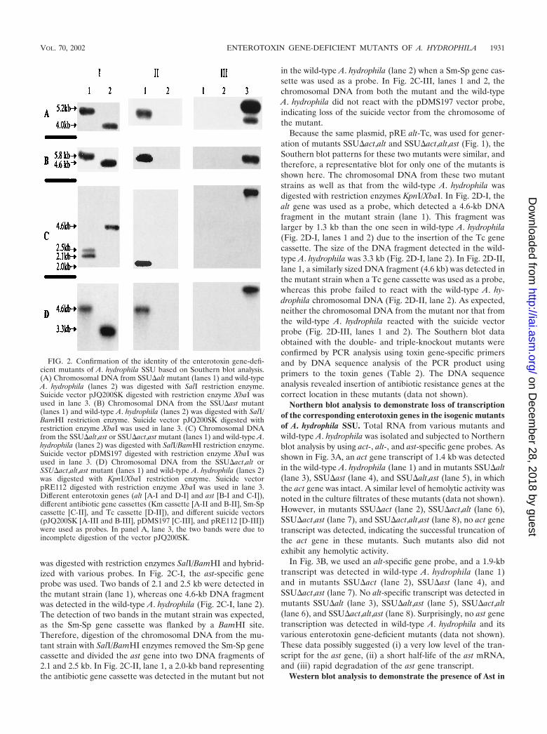
was digested with restriction enzymes SalI/BamHI and hybrid-ized with various probes. In Fig. 2C-I, the ast-specific geneprobe was used. Two bands of 2.1 and 2.5 kb were detected inthe mutant strain (lane 1), whereas one 4.6-kb DNA fragmentwas detected in the wild-type A. hydrophila (Fig. 2C-I, lane 2).The detection of two bands in the mutant strain was expected,as the Sm-Sp gene cassette was flanked by a BamHI site.Therefore, digestion of the chromosomal DNA from the mu-tant strain with SalI/BamHI enzymes removed the Sm-Sp genecassette and divided the ast gene into two DNA fragments of2.1 and 2.5 kb. In Fig. 2C-II, lane 1, a 2.0-kb band representingthe antibiotic gene cassette was detected in the mutant but not
in the wild-type A. hydrophila (lane 2) when a Sm-Sp gene cas-sette was used as a probe. In Fig. 2C-III, lanes 1 and 2, thechromosomal DNA from both the mutant and the wild-typeA. hydrophila did not react with the pDMS197 vector probe,indicating loss of the suicide vector from the chromosome ofthe mutant.
Because the same plasmid, pRE alt-Tc, was used for gener-ation of mutants SSU�act,alt and SSU�act,alt,ast (Fig. 1), theSouthern blot patterns for these two mutants were similar, andtherefore, a representative blot for only one of the mutants isshown here. The chromosomal DNA from these two mutantstrains as well as that from the wild-type A. hydrophila wasdigested with restriction enzymes KpnI/XbaI. In Fig. 2D-I, thealt gene was used as a probe, which detected a 4.6-kb DNAfragment in the mutant strain (lane 1). This fragment waslarger by 1.3 kb than the one seen in wild-type A. hydrophila(Fig. 2D-I, lanes 1 and 2) due to the insertion of the Tc genecassette. The size of the DNA fragment detected in the wild-type A. hydrophila was 3.3 kb (Fig. 2D-I, lane 2). In Fig. 2D-II,lane 1, a similarly sized DNA fragment (4.6 kb) was detected inthe mutant strain when a Tc gene cassette was used as a probe,whereas this probe failed to react with the wild-type A. hy-drophila chromosomal DNA (Fig. 2D-II, lane 2). As expected,neither the chromosomal DNA from the mutant nor that fromthe wild-type A. hydrophila reacted with the suicide vectorprobe (Fig. 2D-III, lanes 1 and 2). The Southern blot dataobtained with the double- and triple-knockout mutants wereconfirmed by PCR analysis using toxin gene-specific primersand by DNA sequence analysis of the PCR product usingprimers to the toxin genes (Table 2). The DNA sequenceanalysis revealed insertion of antibiotic resistance genes at thecorrect location in these mutants (data not shown).
Northern blot analysis to demonstrate loss of transcriptionof the corresponding enterotoxin genes in the isogenic mutantsof A. hydrophila SSU. Total RNA from various mutants andwild-type A. hydrophila was isolated and subjected to Northernblot analysis by using act-, alt-, and ast-specific gene probes. Asshown in Fig. 3A, an act gene transcript of 1.4 kb was detectedin the wild-type A. hydrophila (lane 1) and in mutants SSU�alt(lane 3), SSU�ast (lane 4), and SSU�alt,ast (lane 5), in whichthe act gene was intact. A similar level of hemolytic activity wasnoted in the culture filtrates of these mutants (data not shown).However, in mutants SSU�act (lane 2), SSU�act,alt (lane 6),SSU�act,ast (lane 7), and SSU�act,alt,ast (lane 8), no act genetranscript was detected, indicating the successful truncation ofthe act gene in these mutants. Such mutants also did notexhibit any hemolytic activity.
In Fig. 3B, we used an alt-specific gene probe, and a 1.9-kbtranscript was detected in wild-type A. hydrophila (lane 1)and in mutants SSU�act (lane 2), SSU�ast (lane 4), andSSU�act,ast (lane 7). No alt-specific transcript was detected inmutants SSU�alt (lane 3), SSU�alt,ast (lane 5), SSU�act,alt(lane 6), and SSU�act,alt,ast (lane 8). Surprisingly, no ast genetranscription was detected in wild-type A. hydrophila and itsvarious enterotoxin gene-deficient mutants (data not shown).These data possibly suggested (i) a very low level of the tran-script for the ast gene, (ii) a short half-life of the ast mRNA,and (iii) rapid degradation of the ast gene transcript.
Western blot analysis to demonstrate the presence of Ast in
FIG. 2. Confirmation of the identity of the enterotoxin gene-defi-cient mutants of A. hydrophila SSU based on Southern blot analysis.(A) Chromosomal DNA from SSU�alt mutant (lanes 1) and wild-typeA. hydrophila (lanes 2) was digested with SalI restriction enzyme.Suicide vector pJQ200SK digested with restriction enzyme XbaI wasused in lane 3. (B) Chromosomal DNA from the SSU�ast mutant(lanes 1) and wild-type A. hydrophila (lanes 2) was digested with SalI/BamHI restriction enzyme. Suicide vector pJQ200SK digested withrestriction enzyme XbaI was used in lane 3. (C) Chromosomal DNAfrom the SSU�alt,ast or SSU�act,ast mutant (lanes 1) and wild-type A.hydrophila (lanes 2) was digested with SalI/BamHI restriction enzyme.Suicide vector pDMS197 digested with restriction enzyme XbaI wasused in lane 3. (D) Chromosomal DNA from the SSU�act,alt orSSU�act,alt,ast mutant (lanes 1) and wild-type A. hydrophila (lanes 2)was digested with KpnI/XbaI restriction enzyme. Suicide vectorpRE112 digested with restriction enzyme XbaI was used in lane 3.Different enterotoxin genes (alt [A-I and D-I] and ast [B-I and C-I]),different antibiotic gene cassettes (Km cassette [A-II and B-II], Sm-Spcassette [C-II], and Tc cassette [D-II]), and different suicide vectors(pJQ200SK [A-III and B-III], pDMS197 [C-III], and pRE112 [D-III])were used as probes. In panel A, lane 3, the two bands were due toincomplete digestion of the vector pJQ200SK.
VOL. 70, 2002 ENTEROTOXIN GENE-DEFICIENT MUTANTS OF A. HYDROPHILA 1931
on Decem
ber 28, 2018 by guesthttp://iai.asm
.org/D
ownloaded from

wild-type A. hydrophila SSU and its isogenic mutants. Datapresented in Fig. 4 clearly show the presence of Ast antigen inwild-type A. hydrophila (lane 1) and all of the isogenic mutantsthat had an intact ast gene (lanes 2, 3, and 6) in Western blotsprobed with Ast-specific antibodies. The Ast-specific bandwas missing from the isogenic mutants SSU�ast (lane 4),SSU�alt,ast (lane 5), SSU�act,ast (lane 7), and SSU�act,alt,ast(lane 8). Purified Ast was used as a positive control (lane 9),and cell lysates from E. coli served as a negative control (lane10) to demonstrate the specificity of Ast antibodies.
Evaluation of various isogenic mutants of A. hydrophila in amouse model. A diet-restricted, antibiotic-treated adult mousemodel was used to evaluate different isogenic mutants of A.hydrophila for enterotoxic activity. This model provided opti-mal fluid secretion after A. hydrophila challenge. In animals feda normal diet and challenged with wild-type A. hydrophila (5 105 CFU), a mean fluid secretion of 175 � 55 �l/5 cm of the
loop was recorded over a 12- to 16-h observation period. Thefluid accumulation was 912 � 28 �l/5 cm in animals which werefood restricted but not treated with STR. In food-restrictedand STR-treated mice, the fluid accumulation increased to1,378 � 38 �l/5 cm. A total of 10 mice in each group wereused in the above-mentioned study. We noted that, with theSSU�alt,ast mutant, the fluid secretion occurred rapidly, withmaximum fluid levels noted in 4 h. This mutant induced fluidsecretion as a result of Act production, as the alt and ast geneswere deleted from this strain. However, the fluid secretionevoked by Alt and Ast in an act isogenic mutant of A. hy-drophila was minimal within 4 to 6 h, becoming maximal onlyafter 12 h. Therefore, we opted to observe fluid secretion byvarious enterotoxin gene-deficient mutants of A. hydrophilaafter 12 to 16 h of observation.
As shown in Table 3, single-knockout mutants, such asSSU�act, SSU�alt, and SSU�ast, reduced fluid secretion by64, 48, and 43%, respectively, compared to fluid secretionevoked by the wild-type A. hydrophila. The enterotoxic activityof mutants SSU�alt, SSU�ast, and SSU�act was restored aftercomplementation. The wild-type A. hydrophila with pBR322vector alone was used as a control in the complementationexperiments. The differences in fluid accumulation evoked byvarious mutants not only were significant compared to thewild-type bacterium but also exhibited significance when thesemutants were compared to one another by using Tukey mul-tiple-group comparison analysis (Table 3). The only mutantswhich demonstrated no statistically significant difference influid accumulation were SSU�alt and SSU�ast, which culmi-nated in 445 � 28 and 485 � 22 �l of fluid/5 cm of the loop,respectively. It was noted that, compared to fluid secretion of1,378 � 38 �l/5 cm of the loop observed in a different exper-iment (as stated above) using diet-restricted, antibiotic-treatedanimals, the amount of fluid accumulated was less in the ex-periments presented in Tables 3 and 4, indicating variation inanimals purchased at different times.
In the SSU�alt,ast mutant, the combined reduction in fluidsecretion due to Alt and Ast was 36%, and Act accountedfor 64% of the total fluid accumulated (Table 4). For theSSU�act,ast mutant, which would cause fluid secretion only
FIG. 3. The transcripts for the enterotoxin genes act and alt wereeliminated in the indicated mutants, based on Northern blot analysis.Total RNA from wild-type A. hydrophila and its enterotoxin gene-deficient mutants was isolated and hybridized with the act gene probe(A-I) and the alt gene probe (B-I), as described in Materials andMethods. Lane 1, wild-type A. hydrophila; lane 2, mutant SSU�act;lane 3, mutant SSU�alt; lane 4, mutant SSU�ast; lane 5, mutantSSU�alt,ast; lane 6, mutant SSU�act,alt; lane 7, mutant SSU�act,ast;lane 8, mutant SSU�act,alt,ast. The RNA loaded in each lane wasquantitated by scanning 16S and 23S rRNA bands after ethidiumbromide staining of the gel (A-II and B-II).
FIG. 4. Western blot analysis showing that the ast gene expressionwas eliminated in its corresponding gene-deficient mutant of A. hy-drophila. The cell lysates from different enterotoxin-deficient mutantsof A. hydrophila were subjected to SDS–12% PAGE, and subsequentlythe proteins were transferred to a nitrocellulose membrane for West-ern blot analysis as described in Materials and Methods. Lane 1, wild-type A. hydrophila; lane 2, SSU�act; lane 3, SSU�alt; lane 4, SSU�ast;lane 5, SSU�alt,ast; lane 6, SSU�act,alt; lane 7, SSU�act,ast; lane 8,SSU�act,alt,ast; lane 9, purified Ast (0.1 �g) as a positive control; lane10, cell lysate from E. coli as a negative control.
TABLE 3. Ability of wild-type A. hydrophila SSU, single-knockoutmutants, and their complemented strains to cause
fluid secretion in a mouse model
Organism injected(2 105/100 �l)a
No. ofBALB/c
mice tested
Fluidaccumulation(�l/5-cm loop
� SD)
%Reduction
in fluidsecretion
WT A. hydrophila 25 850 � 50c 0SSU�act 25 300 � 29b,c 64SSU�alt 25 445 � 28b,c,d 48SSU�ast 25 485 � 22b,c,d 43WT A. hydrophila(pBR322) 25 812 � 44 0SSU�alt(pBalt) 25 820 � 24 0SSU�ast(pBast) 25 805 � 20 0SSU�act(pBact) 25 868 � 25 0
a The number of organisms in each loop after 12 to 16 h was increased by2 logs. WT, wild type.
b All groups were significantly different from wild-type A. hydrophila at P �0.001 by multiple-group comparison Tukey test.
c All groups were significantly different from each other at P � 0.001 bymultiple-group comparison Tukey test.
d These groups were not statistically different from each other (P 0.056).
1932 SHA ET AL. INFECT. IMMUN.
on Decem
ber 28, 2018 by guesthttp://iai.asm
.org/D
ownloaded from

through Alt production, 38% of the enterotoxic activity wasnoted, compared to that observed for the wild-type A. hy-drophila. In a double-knockout mutant in which act and altgenes were deleted, fluid secretion totaling 230 � 18 �l/5 cm ofthe loop was measured, indicating the contribution of Ast to be27% (Table 4). No detectable fluid secretion was noted in themutant strain in which all three enterotoxin genes were de-leted. Once again, the differences in fluid accumulation inthese various mutants were statistically significant comparedeither to the wild-type bacterium or among one another withthe Tukey test. All of these mutants colonized the small intes-tine of mice to the same extent [(1.0 � 0.6) 105 to (1.2 � 0.6) 105 CFU/ml] as that noted for the wild-type A. hydrophila(1.1 105 � 0.8 105 CFU/ml) after 2 h, and their numberincreased by approximately 2 logs after 12 to 16 h of incuba-tion.
DISCUSSION
We have performed, for the first time, an extensive case-control human study at the International Center for DiarrhealDiseases Research, Dhaka, Bangladesh, which included chil-dren younger than 5 years of age, to demonstrate the extent ofAeromonas infection in these children. We indeed establishedan association of Aeromonas with diarrhea and showed thatenterotoxins were involved in Aeromonas-associated gastroen-teritis (3). Our recent studies also indicated that bloody diar-rhea was most commonly associated with the production of thecytotoxic enterotoxin Act, while nonbloody diarrhea was cor-related with the elaboration of the cytotonic enterotoxins Altand Ast (3; unpublished data). Diarrheal isolates harboringone, two, or all three of the enterotoxin genes were recoveredfrom patients, although most of the Aeromonas isolates con-tained two of the three enterotoxin genes in various combina-tions. These studies were, therefore, designed to delineateprecisely the roles of individual enterotoxins in evoking diar-rhea by developing mutants with various combinations of de-letions of enterotoxin genes.
Although the genes encoding Act and Alt were previouslysequenced in our laboratory (17, 20), the DNA sequence of theast gene was not available and was needed to generate an astisogenic mutant. Based on the expression of a 4.6-kb SalI/BamHI DNA fragment in E. coli with the T7 expression sys-
tem, biological activity measurement and subsequent DNAsequence analysis revealed that Ast was 71 kDa in size. Thesize of Ast was further confirmed by its molecular character-ization from another clinical isolate of A. sobria and by thedevelopment of an ast isogenic mutant. The SSU�ast mutanttruncated within the ast gene indeed exhibited a reduced en-terotoxic activity, and biological activity associated with Astwas restored after complementation (Table 3).
Our initial strategy was to use purified enterotoxins individ-ually or in various combinations to demonstrate their role inevoking fluid secretion and to determine any synergism amongthese enterotoxins. However, we noted that, when the ast genewas hyperexpressed with the T7 polymerase-promoter-basedpT30a vector system, most of the Ast was membrane bound,requiring harsh treatment for solubilization (e.g., 8 M urea),thereby resulting in the loss of biological activity. We encoun-tered a similar solubility problem when act and alt genes werehyperexpressed in E. coli (20, 25). However, we circumventedthe Act and Alt solubility problem by expressing the corre-sponding genes in A. salmonicida and A. hydrophila strains,lacking act and alt genes, respectively, by using a broad-host-range pMMB66 vector with an IPTG-inducible tac promoter(22; unpublished data). This vector system allowed Act and Altproteins to be secreted out into the medium in a biologicallyactive form (22; unpublished data). The act and alt gene-neg-ative strains of Aeromonas were needed to prevent homolo-gous recombination between the toxin gene on the recombi-nant pMMB66 plasmid and on the chromosome.
We were unable to express the ast gene by using this vector,because until recently we did not have an Aeromonas mutantwith a complete deletion of the ast gene to prevent homolo-gous recombination between the toxin gene on the recombi-nant pMMB66 plasmid and that on the chromosome. Thesepotent enterotoxins are produced in small amounts from thebacterium, and hyperexpression of these toxin genes is essen-tial for generating meaningful data on their relative contribu-tions in evoking fluid secretion. We therefore opted to prepareisogenic mutants deficient in various enterotoxin genes to de-fine their role in causing diarrhea in a murine model.
Inactivation of the coding region of genes by insertion ofantibiotic resistance markers is a general strategy for the con-struction of defined mutants (9, 24, 36, 44). The use of suicidevectors is fundamental to this technique, allowing the creationof deletions or insertions in specific genes on chromosomes(24). However, once the suicide vector is integrated into thechromosome, it is necessary to remove the vector DNA, re-sulting in replacement of the wild-type allele with a mutantallele. A suicide vector carrying a conditional lethal gene, suchas a sacB gene, that discriminates between the integration ofthe vector and double-recombination events has been widelyused (45). Therefore, all of the suicide vectors used in thisstudy (pJQ200SK, pDMS197, and pRE112) (Fig. 1 and Ta-ble 1) harbored the sacB gene. The sacB gene, which en-codes levansucrase, probably polymerizes levan, a productof catabolism of sucrose in the periplasm of gram-negativebacteria, which is toxic to bacteria when they are grown inthe presence of sucrose (23). Vectors pDMS197 andpRE112 had a conditional R6K ori and required � proteinfor replication, while pJQ200SK had a P15A ori from plas-mid pACYC184 (24, 28).
TABLE 4. Ability of wild-type A. hydrophila SSU, double-knockoutmutants (SSU�alt,ast, SSU�act,ast, and SSU�act,alt), and
triple-knockout mutant (SSU�act,alt,ast) tocause fluid secretion in a mouse model
Organism injected(2 105/100 �l)a
No. ofBALB/c
mice tested
Fluidaccumulation(�l/5-cm loop
� SD)
%Reduction
in fluidsecretion
WT A. hydrophila 25 858 � 28b 0SSU�alt,ast 25 552 � 26b 36SSU�act,ast 25 330 � 28b 62SSU�act,alt 25 230 � 18b 73SSU�act,alt,ast 25 NDc 100
a The number of organisms in each loop after 12 to 16 h was increased by2 logs. WT, wild type.
b All groups were significantly different from each other at P � 0.001 bymultiple-group comparison Tukey test.
c ND, not detected. The limit of detection was less than 50 �l.
VOL. 70, 2002 ENTEROTOXIN GENE-DEFICIENT MUTANTS OF A. HYDROPHILA 1933
on Decem
ber 28, 2018 by guesthttp://iai.asm
.org/D
ownloaded from

In this study, six isogenic mutants of A. hydrophila, in whichthree enterotoxin genes were deleted in various combinations,were prepared. The biological activity of the single-knockoutstrains (SSU�act, SSU�alt, and SSU�ast) of A. hydrophilacould be restored by complementation, suggesting no polar ef-fects of marker-exchange mutagenesis on genes downstream ofthose encoding the enterotoxins. Since double- and triple-en-terotoxin-gene-knockout strains of A. hydrophila were preparedusing one of the single-enterotoxin-gene-knockout strains, wedid not expect polar effects. However, complementation ofdouble- and triple-enterotoxin-gene-knockout strains of A. hy-drophila was difficult to perform to completely rule out thepossibility of any polar effects in such mutants. We determinedthe exact insertional sites of the antibiotic resistance cassetteswithin the enterotoxin genes on the chromosomes of variousisogenic mutants and confirmed the absence of frameshift mu-tations within the toxin genes. All of these mutants (single,double, and triple knockout) exhibited similar growth rates andthe ability to colonize and multiply in the small intestine, whichfurther suggested the absence of polar effects in these mutants.However, we should not overlook the possibility of polar ef-fects in the double- and triple-knockout strains of A. hydro-phila, which could have some impact on the fluid accumulationresults. Such polar effects might constitute a limitation of in-sertional mutagenesis.
We noted previously and during present studies that thefrequency of double crossover was very low (0.01%) when themutants SSU�act, SSU�alt, and SSU�ast were prepared usingsuicide vector pJQ200SK (47). We therefore employed re-cently developed suicide vectors pDMS197 and pRE112 (24)for generating double- and triple-knockout mutants of A. hy-drophila, which increased the frequency of obtaining double-crossover mutants by 10-fold. Further, it was noted that, whenthe pJQ200SK suicide vector was used, some false-positivemutants were observed, especially during the preparation ofdouble- and triple-knockout strains of A. hydrophila that weretruncated in two and three enterotoxin genes, respectively. Themajority of the false-positive mutants still had a portion of thesuicide vector plasmid integrated in the chromosomal DNA ofA. hydrophila, based on Southern blot data (data not shown).These mutants, however, exhibited the correct phenotype (i.e.,resistance to sucrose and sensitivity to GEN). Our subsequentanalysis of these mutants indicated that Gmr and sacB geneswere indeed lost from these cultures (data not shown). In someinstances, we could also detect free recombinant pJQ200SKplasmid, suggesting its replication to some extent in A. hydro-phila, which could also interfere with obtaining genuine dou-ble-crossover mutants. The frequency of obtaining false-posi-tive mutants was significantly reduced with pMDS197 andpRE112 vectors.
The correct identity of all of the isogenic mutants was con-firmed by Southern, Northern, and Western blot analysis (Fig.2 to 4). In Northern blot analysis, all of the mutants exhibitedthe correct pattern of inhibition of the corresponding entero-toxin gene transcripts when act and alt gene probes were used(Fig. 3). However, it was noted that the alt mRNA level wassignificantly lower than that of act mRNA (Fig. 3A and B).These data suggested (i) low levels of the alt gene expression,under studied conditions, and (ii) instability or shorter half-lifeof the alt mRNA. It will be intriguing to determine in the
future whether alt gene expression could be increased during invivo conditions. Interestingly, we were unable to detect a tran-script for the ast gene, even in the wild-type A. hydrophila,although the Ast protein could be seen by Western blot anal-ysis in wild-type A. hydrophila and its isogenic mutants with anintact ast gene (Fig. 4). Our data might be suggestive of a shorthalf-life for the ast mRNA or its rapid degradation and needconfirmation. This possibility was based on our observation thatwe could detect the ast gene transcript in significant amountsfrom E. coli when the toxin gene was hyperexpressed using apET30a vector system (data not shown). However, we cannot ruleout the possibility that the ast gene expression requires interactionof A. hydrophila with the host cell, and this is under investigation.
We first developed a mouse model to evaluate the fluidsecretory potential of wild-type A. hydrophila and its variousenterotoxin gene-deficient mutants, based on studies showingthat malnourished animals were more susceptible to infectionthan were adequately nourished animals (37). Further, STRdisrupted the aerobic and anaerobic characteristics of the cecalflora and was particularly effective at decreasing colonizationresistance (43). Deletion of the enterotoxin genes individuallyindicated that Act, Alt, and Ast contributed to the fluid secre-tory response by 64, 48, and 43%, respectively (Table 3). Dataobtained with SSU�act,ast and SSU�act,alt mutants, whichcaused fluid secretion via the production of Alt and of Ast,respectively, conclusively proved their contribution to be 38and 27%, respectively (Table 4). These data coincided with ourobservation that mice immunized with Alt and Ast reducedfluid secretion caused by A. hydrophila challenge by 39 and30%, respectively 20; unpublished data).
Based on the data presented in Tables 3 and 4 for SSU�actand SSU�alt,ast mutants, it was evident that Act was the majorenterotoxin contributing to the fluid secretory response (64%),followed by Alt and Ast. Our data also suggested some inter-action among these various enterotoxins (Tables 3 and 4). Theexact molecular basis of this interaction among the variousenterotoxins is not known yet and needs further study. Theinability of the triple-knockout mutant to exhibit any detect-able enterotoxic response confirmed the role of all three of theenterotoxins (Act, Alt, and Ast) in A. hydrophila-induced di-arrhea. The presence of various combinations of enterotoxinsin different Aeromonas isolates could increase or decrease theexpression of specific enterotoxin genes and thus could dictatethe severity of diarrhea. The availability of purified enterotox-ins would help in further delineating interactions among theseenterotoxins.
In conclusion, we have demonstrated for the first time thecontribution of each of the three enterotoxins of A. hydrophilain causing gastroenteritis in a murine model by developingisogenic mutants. Our data tend to suggest some interactionamong these enterotoxins in vivo that could lead to alteredfluid secretion. Such attenuated strains of A. hydrophila withdeletions of the act, alt, and ast genes could be attractivecandidates for vaccine development.
ACKNOWLEDGMENTS
Jian Sha and E. V. Koslova contributed equally to this work.This work was supported by a grant from the National Institutes of
Health (AI41611). Jian Sha, a postdoctoral fellow, was supported bythe McLaughlin postdoctoral fellowship.
1934 SHA ET AL. INFECT. IMMUN.
on Decem
ber 28, 2018 by guesthttp://iai.asm
.org/D
ownloaded from

The editorial assistance of Mardelle Susman is greatly appreciated.We thank B. Chatuev of our department for providing plasmidspDMS197 and pRE112 and E. coli strain SM10.
REFERENCES
1. Abbott, S. L., S. Seli, M. Catino, Jr., M. A. Hartley, and J. M. Janda. 1998.Misidentification of unusual Aeromonas species as members of the genusVibrio: a continuing problem. J. Clin. Microbiol. 36:1103–1104.
2. Alavandi, S. V., M. S. Subashini, and S. Ananthan. 1999. Occurrence ofhaemolytic and cytotoxic Aeromonas species in domestic water supplies inChennai. Indian J. Med. Res. 110:50–55.
3. Albert, M. J., M. Ansaruzzaman, K. Talukdar, A. K. Chopra, I. Kuhn, M.Rahman, A. S. G. Faruque, M. S. Islam, R. B. Sack, and R. Mollby. 2000.Prevalence of enterotoxin genes in Aeromonas spp. isolated from childrenwith diarrhea, healthy controls, and the environment. J. Clin. Microbiol.38:3785–3790.
4. Altwegg, M., L. G. Martinetti, J. Luthy-Hottenstein, and M. Rohrbach. 1991.Aeromonas-associated gastroenteritis after consumption of contaminatedshrimp. Eur. J. Clin. Microbiol. Infect. Dis. 10:44–45.
5. Annapurna, E., and S. C. Sanyal. 1977. Enterotoxicity of Aeromonas hy-drophila. J. Med. Microbiol. 10:317–323.
6. Asao, T., Y. Kinoshita, S. Kozaki, T. Uemura, and G. Sakaguchi. 1984.Purification and some properties of Aeromonas hydrophila hemolysin. Infect.Immun. 46:122–127.
7. Ausubel, F. M., R. Brent, R. E. Kingston, D. D. Moore, J. Seidman, J. A.Smith, and K. Struhl (ed.). 1989. Current protocols in molecular biology.John Wiley & Sons, Inc., New York, N.Y.
8. Barnett, T. C., S. M. Kirov, M. S. Strom, and K. Sanderson. 1997. Aeromo-nas ssp. possess at least two distinct type IV pilus families. Microb. Pathog.23:241–247.
9. Becker, A., M. Schmidt, W. Jager, and A. Puhler. 1995. New gentamicin-resistance and lacZ promoter-probe cassettes suitable for insertion mutagen-esis and generation of transcriptional fusions. Gene 162:37–39.
10. Brandi, G. M., F. Sisti, F. Giardini, G. F. Schiavano, and A. Albano. 1999.Survival ability of cytotoxic strains of motile Aeromonas spp. in differenttypes of water. Lett. Appl. Microbiol. 29:211–215.
11. Buchanan, R. L., and S. A. Palumbo. 1985. Aeromonas hydrophila and Aero-monas sobria as potential food poisoning species: a review. J. Food Safety7:15–29.
12. Chakraborty, T., B. Huhle, H. Bergbauer, and W. Goebel. 1986. Cloning,expression, and mapping of the Aeromonas hydrophila aerolysin gene deter-minant in Escherichia coli K-12. J. Bacteriol. 167:368–374.
13. Chakraborty, T., M. A. Montenegro, S. C. Sanyal, R. Helmuth, E. Bulling,and K. N. Timmis. 1984. Cloning of enterotoxin gene from Aeromonashydrophila provides conclusive evidence of production of a cytotonic entero-toxin. Infect. Immun. 46:435–441.
14. Chakraborty, T., V. Husslein, B. Huhle, H. Bergbauer, T. Jarchau, H. Hor,and W. Goebel. 1988. Genetics of hemolysins in Aeromonas hydrophila.Zentbl. Bakteriol. Mikrobiol. Hyg. Abt. 1 Suppl. 17:215–222.
15. Chopra, A. K., and C. W. Houston. 1989. Purification and partial character-ization of a cytotonic enterotoxin produced by Aeromonas hydrophila. Can. J.Microbiol. 35:719–727.
16. Chopra, A. K., and C. W. Houston. 1999. Enterotoxins in Aeromonas-asso-ciated gastroenteritis. Microbes Infect. 1:1129–1137.
17. Chopra, A. K., C. W. Houston, J. W. Peterson, and G.-F. Jin. 1993. Cloning,expression, and sequence analysis of a cytolytic enterotoxin gene from Aero-monas hydrophila. Can. J. Microbiol. 39:513–523.
18. Chopra, A. K., J. H. Hung, X.-J. Xu, K. Burden, D. W. Niesel, M. Rosen-baum, and J. W. Peterson. 1999. Role of Salmonella enterotoxin in overallvirulence of the organism. Microb. Pathog. 27:155–171.
19. Chopra, A. K., J. W. Peterson, P. Chary, and R. Prasad. 1994. Molecularcharacterization of an enterotoxin from Salmonella typhimurium. Microb.Pathog. 16:85–98.
20. Chopra, A. K., J. W. Peterson, X.-J. Xu, D. H. Coppenhaver, and C. W.Houston. 1996. Molecular and biochemical characterization of a heat-labilecytotonic enterotoxin from Aeromonas hydrophila. Microb. Pathog. 21:357–377.
21. Chopra, A. K., R. Pham, and C. W. Houston. 1994. Cloning and expressionof putative cytotonic enterotoxin-encoding genes from Aeromonas hydro-phila. Gene 139:87–91.
22. Chopra, A. K., X.-J. Xu, D. Ribardo, M. Gonzalez, K. Kuhl, J. W. Peterson,and C. W. Houston. 2000. The cytotoxic enterotoxin of Aeromonas hydrophilainduces proinflammatory cytokine production and activates arachidonic acidmetabolism in macrophages. Infect. Immun. 68:2808–2818.
23. Donnenberg, M. S., and J. B. Kaper. 1991. Construction of an eae deletionmutant of enteropathogenic Escherichia coli by using a positive-selectionsuicide vector. Infect. Immun. 59:4310–4317.
24. Edwards, R. A., H. K. Linda, and D. M. Schifferli. 1998. Improved allelicexchange vectors and their use to analyze 987P fimbria gene expression.Gene 207:149–157.
25. Ferguson, M. R., X.-J. Xu, C. W. Houston, J. W. Peterson, D. H. Coppen-haver, V. L. Popov, and A. K. Chopra. 1997. Hyperproduction, purification,and mechanism of action of the cytotoxic enterotoxin produced by Aeromo-nas hydrophila. Infect. Immun. 65:4299–4308.
26. Hanninen, M. L., P. Oivanen, and V. Hirvela-Koski. 1997. Aeromonas spe-cies in fish, fish-eggs, shrimp and freshwater. Int. J. Food Microbiol. 34:17–26.
27. Hanninen, M. L., S. Salmi, L. Mattila, R. Taipalinen, and A. Siitonen. 1995.Association of Aeromonas spp. with traveller’s diarrhoea in Finland. J. Med.Microbiol. 41:26–31.
28. Harayama, S., M. Tsuda, and T. Lino. 1980. High frequency mobilization ofthe chromosome of Escherichia coli by a mutant of plasmid RP4 temperaturesensitive for maintenance. Mol. Gen. Genet. 180:47–56.
29. Howard, S. P., and J. T. Buckley. 1986. Molecular cloning and expression inEscherichia coli of the structural gene for the hemolytic toxin aerolysin fromAeromonas hydrophila. Mol. Gen. Genet. 204:289–295.
30. Kirov, S. M., E. K. Ardestani, and L. J. Hayward. 1993. The growth andexpression of virulence factors at refrigeration temperature by Aeromonasstrains isolated from food. Int. J. Food Microbiol. 20:159–168.
31. Kirov, S. M., L. A. O’Donovan, and K. Sanderson. 1999. Functional charac-terization of type IV pili expressed on diarrhea-associated isolates of Aero-monas species. Infect. Immun. 67:5447–5454.
32. Kirov, S. M., T. C. Barnett, C. M. Pepe, M. S. Strom, and M. J. Albert. 2000.Investigation of the role of type IV Aeromonas pilus (Tap) in the pathogen-esis of Aeromonas gastrointestinal infection. Infect. Immun. 68:4040–4048.
33. Ljungh, A., and T. Wadstrom. 1985. Review. Aeromonas and Plesiomonas aspossible causes of diarrhea. Infection 13:169–173.
34. Majeed, K. N., A. F. Egan, and I. C. MacRae. 1990. Production of exotoxinsby Aeromonas ssp. at 5°C. J. Appl. Bacteriol. 69:332–337.
35. Miller, V. L., and J. J. Mekalanos. 1988. A novel suicide vector and its usein construction of insertion mutations: osmoregulation of outer membraneproteins and virulence determinants in Vibrio cholerae requires toxR. J. Bac-teriol. 170:2575–2583.
36. Nishibuchi, M., K. Kumagai, and J. B. Kaper. 1991. Contribution of the tdh1gene of Kanagawa phenomenon-positive Vibrio parahaemolyticus to produc-tion of extracellular thermostable direct hemolysin. Microb. Pathog. 11:453–460.
37. Nzegwu, H. C., and R. J. Levin. 1994. Role of the enteric nervous system inthe maintained hypersecretion induced by enterotoxin STa in the nutrition-ally deprived intestine. Gut 35:1237–1243.
38. Prasad, R., A. K. Chopra, J. W. Peterson, R. Pericas, and C. W. Houston.1990. Biological and immunological characterization of a cloned choleratoxin-like enterotoxin from Salmonella typhimurium. Microb. Pathog. 9:315–329.
39. Prasad, R., A. K. Chopra, P. Chary, and J. W. Peterson. 1992. Expressionand characterization of the cloned Salmonella typhimurium enterotoxin. Mi-crob. Pathog. 13:109–121.
40. Prentki, P., and H. M. Krish. 1984. In vitro insertional mutagenesis with aselectable DNA fragment. Gene 29:303–313.
41. Quandt, J., and M. F. Hynes. 1993. Versatile suicide vectors which allowdirect selection for gene replacement in gram-negative bacteria. Gene 127:15–21.
42. Sambrook, J., E. F. Fritsch, and T. Maniatis. 1989. Molecular cloning: alaboratory manual, 2nd ed. Cold Spring Harbor Laboratory, Cold SpringHarbor, N.Y.
43. Sanderson, K., F. M. Ghazali, and S. M. Kirov. 1996. Colonization ofstreptomycin-treated mice by Aeromonas species. J. Diarrhoeal Dis. Res.14:27–32.
44. Segada, L. M., G. M. Carlone, L. L. Gheesling, and A. J. Lesse. 2000.Characterization of P1-deficient isogenic mutant of Haemophilus influenzaebiogroup aegyptius associated with Brazilian purpuric fever. Microb. Pathog.28:145–155.
45. Steinmetz, M., D. Le Cog, S. Aymerich, G. Gonzy-Treboul, and P. Gay. 1985.The DNA sequence of the gene for the secreted Bacillus subtilis enzymelevansucrase and its genetic control sites. Mol. Gen. Genet. 200:220–228.
46. Tabor, S., and C. C. Richardson. 1985. A bacteriophage T7 RNA polymer-ase/promoter system for controlled exclusive expression of specific genes.Proc. Natl. Acad. Sci. USA 82:1074–1078.
47. Xu, X.-J., M. R. Ferguson, V. L. Popov, C. W. Houston, J. W. Peterson, andA. K. Chopra. 1998. Role of a cytotoxic enterotoxin in Aeromonas-mediatedinfections: development of transposon and isogenic mutants. Infect. Immun.66:3501–3509.
48. Yamada, S., S. Matsushita, S. Dejsirilet, and Y. Kudoh. 1997. Incidence andclinical symptoms of Aeromonas-associated traveller’s diarrhoea in Tokyo.Epidemiol. Infect. 119:121–126.
Editor: J. T. Barbieri
VOL. 70, 2002 ENTEROTOXIN GENE-DEFICIENT MUTANTS OF A. HYDROPHILA 1935
on Decem
ber 28, 2018 by guesthttp://iai.asm
.org/D
ownloaded from


![Fecal contamination and related health effects at a ... contamination and related health effects at a ... Aeromonas hydrophila Aeromonas hydrophila ... Ewers 125.ppt [Read-Only]](https://static.fdocuments.net/doc/165x107/5ada57817f8b9aee348ca8a5/fecal-contamination-and-related-health-effects-at-a-contamination-and-related.jpg)