Role of ultrasound-guided fine-needle aspiration of ... cancer staging 2.pdf · lymph node was...
7
Role of Ultrasound-Guided Fine-Needle Aspiration of Indeterminate and Suspicious Axillary Lymph Nodes in the Initial Staging of Breast Carcinoma Savitri Krishnamurthy, M.D. 1 Nour Sneige, M.D. 1 Deepak G. Bedi, M.D. 2 Beth S. Edieken, M.D. 2 Bruno D. Fornage, M.D. 2 Henry M. Kuerer, M.D., Ph.D. 3 S. Eva Singletary, M.D. 3 Kelly K. Hunt, M.D. 3 1 Department of Pathology, M. D. Anderson Cancer Center, Houston, Texas. 2 Department of Radiology, M. D. Anderson Cancer Center, Houston, Texas. 3 Department of Surgical Oncology, M. D. Ander- son Cancer Center, Houston, Texas. Address for reprints: Savitri Krishnamurthy, M.D., Department of Pathology, The University of Texas M. D. Anderson Cancer Center, P.O. Box 53, 1515 Holcombe Blvd., Houston, TX 77030; Fax: (713) 794-5664; E-mail: [email protected] Received January 8, 2002; revision received March 7, 2002; accepted April 11, 2002. BACKGROUND. Ultrasound (US) is more sensitive than physical examination alone in determining axillary lymph node involvement during preliminary staging of breast carcinoma. Due to occasional overlap of sonographic features of benign and indeterminate lymph nodes, fine-needle aspiration (FNA) of sonographically in- determinate/suspicious lymph nodes can provide a more definitive diagnosis than US alone. This study was undertaken to determine the diagnostic accuracy of US-guided FNA of indeterminate/suspicious/metastatic-appearing axillary lymph nodes during the initial staging of breast carcinoma. METHODS. The cytology of 103 cases of US-guided FNA of nonpalpable indetermi- nate/suspicious/metastatic-appearing lymph nodes was compared with the final histopathologic status of the entire axilla after axillary dissection. The final axillary lymph node status was categorized as either negative when all lymph nodes were negative for metastasis or positive when there was evidence of metastasis in one or more lymph nodes. The sensitivity, specificity, diagnostic accuracy, and false- negative rate of US-guided FNA of nonpalpable axillary lymph nodes in the preliminary staging process were calculated. RESULTS. In 51 of 103 cases (49.5%), the US-guided FNA and histopathology were both positive for metastasis. In 24 of 103 cases (23.3%), both were negative. The apparent false-positive FNA in 16 (15.5%) cases was explained by the complete response of the metastatic lymph nodes to neoadjuvant chemotherapy in the interval between FNA and axillary dissection. In 12 cases (11.6%), US-guided FNA was negative, but metastasis was seen in histologic sections. All cases with three or more lymph nodes with metastatic disease and 93% of those with metastatic deposit measuring more than 0.5 mm were detected by US-guided FNA. The probability of detecting lymph nodes with smaller metastatic deposit measuring less than 0.5 cm was 44%. The overall sensitivity of US-guided FNA was 86.4%, the specificity was 100%, the diagnostic accuracy was 79.0%, the positive predictive value was 100%, and the negative predictive value was 67%. CONCLUSIONS. US-guided FNA of nonpalpable indeterminate and suspicious axil- lary lymph nodes is a simple, minimally invasive, and reliable technique for the initial determination of axillary lymph node status in breast carcinoma. The com- mon causes of discrepancy between the initial and final axillary lymph node status include failure to visualize all lymph nodes during US examination, small-sized metastases, and preoperative neoadjuvant chemotherapy. Cancer 2002;95:982– 8. © 2002 American Cancer Society. DOI 10.1002/cncr.10786 KEYWORDS: ultrasound-guided fine-needle aspiration, axillary lymph nodes, breast carcinoma staging, ultrasound examination. I n the preliminary staging of newly diagnosed cases of breast carci- noma, based on the extent of the primary tumor and the presence or absence of lymph node involvement, various combinations of 982 © 2002 American Cancer Society
Transcript of Role of ultrasound-guided fine-needle aspiration of ... cancer staging 2.pdf · lymph node was...
Role of Ultrasound-Guided Fine-Needle Aspiration ofIndeterminate and Suspicious Axillary Lymph Nodesin the Initial Staging of Breast Carcinoma
Savitri Krishnamurthy, M.D.1
Nour Sneige, M.D.1
Deepak G. Bedi, M.D.2
Beth S. Edieken, M.D.2
Bruno D. Fornage, M.D.2
Henry M. Kuerer, M.D., Ph.D.3
S. Eva Singletary, M.D.3
Kelly K. Hunt, M.D.3
1 Department of Pathology, M. D. Anderson CancerCenter, Houston, Texas.
2 Department of Radiology, M. D. Anderson CancerCenter, Houston, Texas.
3 Department of Surgical Oncology, M. D. Ander-son Cancer Center, Houston, Texas.
Address for reprints: Savitri Krishnamurthy, M.D.,Department of Pathology, The University of TexasM. D. Anderson Cancer Center, P.O. Box 53, 1515Holcombe Blvd., Houston, TX 77030; Fax: (713)794-5664; E-mail: [email protected]
Received January 8, 2002; revision receivedMarch 7, 2002; accepted April 11, 2002.
BACKGROUND. Ultrasound (US) is more sensitive than physical examination alone
in determining axillary lymph node involvement during preliminary staging of
breast carcinoma. Due to occasional overlap of sonographic features of benign and
indeterminate lymph nodes, fine-needle aspiration (FNA) of sonographically in-
determinate/suspicious lymph nodes can provide a more definitive diagnosis than
US alone. This study was undertaken to determine the diagnostic accuracy of
US-guided FNA of indeterminate/suspicious/metastatic-appearing axillary lymph
nodes during the initial staging of breast carcinoma.
METHODS. The cytology of 103 cases of US-guided FNA of nonpalpable indetermi-
nate/suspicious/metastatic-appearing lymph nodes was compared with the final
histopathologic status of the entire axilla after axillary dissection. The final axillary
lymph node status was categorized as either negative when all lymph nodes were
negative for metastasis or positive when there was evidence of metastasis in one or
more lymph nodes. The sensitivity, specificity, diagnostic accuracy, and false-
negative rate of US-guided FNA of nonpalpable axillary lymph nodes in the
preliminary staging process were calculated.
RESULTS. In 51 of 103 cases (49.5%), the US-guided FNA and histopathology were
both positive for metastasis. In 24 of 103 cases (23.3%), both were negative. The
apparent false-positive FNA in 16 (15.5%) cases was explained by the complete
response of the metastatic lymph nodes to neoadjuvant chemotherapy in the
interval between FNA and axillary dissection. In 12 cases (11.6%), US-guided FNA
was negative, but metastasis was seen in histologic sections. All cases with three or
more lymph nodes with metastatic disease and 93% of those with metastatic
deposit measuring more than 0.5 mm were detected by US-guided FNA. The
probability of detecting lymph nodes with smaller metastatic deposit measuring
less than 0.5 cm was 44%. The overall sensitivity of US-guided FNA was 86.4%, the
specificity was 100%, the diagnostic accuracy was 79.0%, the positive predictive
value was 100%, and the negative predictive value was 67%.
CONCLUSIONS. US-guided FNA of nonpalpable indeterminate and suspicious axil-
lary lymph nodes is a simple, minimally invasive, and reliable technique for the
initial determination of axillary lymph node status in breast carcinoma. The com-
mon causes of discrepancy between the initial and final axillary lymph node status
include failure to visualize all lymph nodes during US examination, small-sized
metastases, and preoperative neoadjuvant chemotherapy. Cancer 2002;95:982– 8.
© 2002 American Cancer Society.
DOI 10.1002/cncr.10786
KEYWORDS: ultrasound-guided fine-needle aspiration, axillary lymph nodes, breastcarcinoma staging, ultrasound examination.
In the preliminary staging of newly diagnosed cases of breast carci-noma, based on the extent of the primary tumor and the presence
or absence of lymph node involvement, various combinations of
982
© 2002 American Cancer Society
surgery, radiotherapy, and chemotherapy are used totailor the treatment. The routine staging process in-cludes a thorough history and physical examination,ultrasound (US) of the breast and lymph node basins(axillary, internal mammary, infraclavicular, and su-praclavicular), computed tomography (CT) scan of theabdomen, bone scan, and a chest X-ray. In the ab-sence of distant metastatic disease, assessment of thestatus of the axillary lymph nodes is the most impor-tant component of the initial staging process becauseof their impact on subsequent management. In addi-tion, the presence or absence of axillary metastases isthe strongest prognostic indicator available for breastcarcinoma. Physical examination alone is neither asensitive nor reliable way to ascertain lymph nodestatus, because metastatic lymph nodes are often notpalpable and reactive lymph nodes may be mistakenfor metastases.1–3
Several imaging techniques are used to visualizethe lymph nodes, including US, magnetic resonanceimaging, CT scans, and nuclear medicine. US is themost useful of these techniques for the evaluation oflocal disease in the breast and regional lymphnodes.2– 6 It visualizes not only alterations in the size,shape, and contours of the lymph nodes, but alsochanges in the cortical morphology and texture thatcan reflect the presence of underlying metastasis.However, these early signs of metastatic disease some-times overlap with those of benign reactive changes,resulting in a label of “suspicious” or “indeterminate”for some lymph nodes. Other lymph nodes may beregarded as “metastatic” appearing when sonographyshows compression or displacement of hyperchoicfatty hilum and lymph node enlargement. Fine-needleaspiration (FNA) of sonographically suspicious, inde-terminate, or metastatic-appearing axillary lymphnodes provides a more definitive diagnosis comparedwithsonography alone. Our objective was to evaluatethe accuracy of US-guided FNA of indeterminate andsuspicious axillary lymph nodes in the initial stagingof breast carcinoma.
MATERIALS AND METHODSThe cytology of all cases of US-guided FNA of nonpal-pable indeterminate and suspicious axillary lymphnodes was compared with the final histopathologicstatus of the entire axilla after axillary dissection. US-guided FNA was performed at The University of TexasM. D. Anderson Cancer Center in 1999 during thepreliminary staging of breast carcinoma. The sono-graphic criteria for selecting indeterminate, suspi-cious, or metastatic-appearing nodes were increasedthickening and/or lobulation of the hypoechoic lymphnode cortex compared with other ipsilateral or con-
tralateral lymph nodes, eccentric lobulation of thehypoechoic lymph node cortex with compression ofthe adjacent hilar fat, and complete disappearance ofthe hilar fat, which was replaced by hypoechoic cortex.In all cases, FNA was performed by a radiologist(D.G.B., B.S.E., and B.D.F.) under US guidance, using a20-gauge or 21-gauge needle attached to a 10-mL or20-mL plastic syringe. One percent xylocaine was usedfor local anesthesia. Under real-time visualization, theneedle was directed into the cortical tissue of thelymph node. The tip of the needle was documented ona hard copy image as being within the target and oneor two passes were made. Direct smears from theaspirate were fixed in alcohol and air-dried for Papand Diff-Quik (American Scientific Products, McGrawPark, IL) staining. All of the slides were screened im-mediately for specimen adequacy by the cytopatholo-gist and FNA repeated if necessary; all cases negativefor metastasis in the preliminary screening were thenscreened thoroughly by the cytotechnologist and cy-topathologist. The axillary lymph node status as de-termined using US-guided FNA in the preliminarystaging process was compared with the final statusfollowing histopathologic examination of the axillarylymph nodes that were removed in the axillary dissec-tion. For the purpose of analysis, the final axillarylymph node status was categorized as either negativewhen all of the histopathologically examined lymphnodes were negative for metastasis or positive whenthere was evidence of metastasis in one or morelymph nodes. Because the most abnormal-appearinglymph node was evaluated by FNA, its cytology repre-sented the status of the entire axilla during prelimi-nary staging and was compared with the histopatho-logic status of the whole axilla upon surgicaldissection. The number of dissected axillary lymphnodes having metastasis and the size of each metas-tasis were noted. The discrepant cases were analyzedwith respect to the tumor type, treatment with/with-out preoperative neoadjuvant chemotherapy, and theinterval between US-guided FNA and subsequent sur-gery. Finally, the sensitivity, specificity, diagnostic ac-curacy, and false-negative rate of US-guided FNA ofnonpalpable axillary lymph nodes in the preliminarystaging process were calculated.
RESULTSOne hundred three (103) cases were identified in thecytopathology files of M. D. Anderson Cancer Centeras having undergone US-guided FNA of one or morenonpalpable, sonographically indeterminate or suspi-cious axillary lymph nodes followed by complete ax-illary dissection in 1999. Of the primary tumors, 89were invasive ductal carcinomas, 2 were invasive lob-
Ultrasound-guided FNA of Lymph Nodes/Krishnamurthy et al. 983
ular carcinomas, and 12 were carcinomas of no spec-ified type. Of the 103 cases, 65 received preoperativeneoadjuvant chemotherapy. Table 1 provides detailsof the comparison between preliminary staging andfinal status of the axillary lymph nodes.
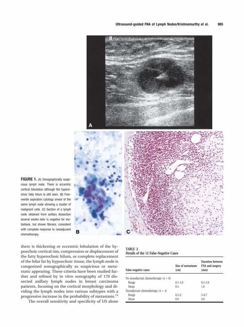
Of the 67 cases in which metastatic adenocarci-noma was exhibited in the cytologic smears, one ormore lymph nodes were identified as histologicallypositive for metastasis after axillary lymph node dis-section in only 51. However, all 16 of 67 cases thatappeared at first to be false-positive for metastasis hadreceived preoperative neoadjuvant chemotherapy.Moreover, the tissue sections of one or more of theirdissected axillary lymph nodes revealed areas of fibro-sis with or without hemosiderin pigment depositionand infiltration by histiocytes, which are features con-sistent with probable complete response of metastaticdisease to neoadjuvant therapy. Figure 1 is an exampleof one such case where the smears showed metastaticcarcinoma but the subsequent axillary dissection wasnegative. Therefore, these 16 cases were not trulyfalse-positive cases.
All cases with three or more lymph nodes withmetastatic disease and 93% of those with metastaticdeposit measuring more than 0.5 mm were detectedby US-guided FNA. The probability of detecting lymphnodes with smaller metastatic deposit measuring lessthan 0.5 cm was 44%.
In the 12 discrepant cases diagnosed as beingnegative by preliminary US-guided FNA, there washistologic evidence of metastasis in one or more of thedissected axillary lymph nodes. The primary tumor inthese 12 discrepant false-negative cases was ductal intype. Table 2 shows the details of the size of themetastases and the interval between FNA and axillarydissection in these 12 cytologically false-negativecases. Four of these patients received preoperative
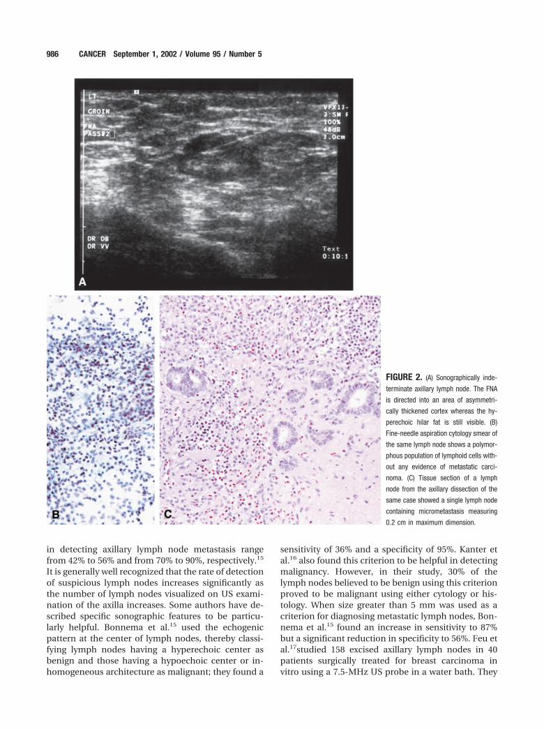
neoadjuvant therapy, underwent axillary dissection 3to 7 months after the primary diagnosis, and the meansize of the metastasis was 0.9 cm (range, 0.4 –2.0 cm).The remaining eight patients did not receive preoper-ative chemotherapy, axillary dissection was performed0.2–3.6 months after the primary diagnosis, and themean size of the metastases was 0.4 cm (range, 0.1–1.6cm). Figure 2 is an example of a discrepant case wherethe cytology smears were negative, but axillary dissec-tion showed a single lymph node containing micro-metastasis measuring 0.2 cm in maximum dimension.
The overall sensitivity of US-guided FNA was86.4%, the specificity was 100%, diagnostic accuracywas 79.0%, positive predictive value was 100%, nega-tive predictive value was 67%, and the false-negativerate was 11.6%. The false-negative rate in the 38 pa-tients who did not receive preoperative chemotherapywas 21.0%.
DISCUSSIONInitial staging of newly diagnosed cases of breast car-cinoma is a routine practice for selecting the appro-priate protocol for treatment. Determination of theaxillary lymph node status in this process plays aparticularly important role in guiding the surgeon andoncologist in planning the management of breast car-cinoma patients. For patients with locally advancedbreast carcinoma with evidence of metastases to theregional lymph nodes or distant spread, neoadjuvantchemotherapy is offered as first-line therapy, whichthen is followed by surgery.7–9 In patients with smaller(T1 and T2) breast carcinomas, when there is no evi-dence of metastasis to the axillary lymph nodes in thepreliminary staging process, segmental mastectomy(lumpectomy) and sentinel lymph node mapping arepreferred instead of complete axillary lymph node dis-section.10 –12
Physical examination alone is very unreliable inassessing axillary lymph node status because meta-static lymph nodes cannot always be distinguishedfrom normal or reactive ones. Specifically, the overallfalse-negative rate of physical examination alone hasbeen reported to be as high as 32–33%.1 US examina-tion can improve the sensitivity of clinical examina-tion in assessing axillary lymph node status in thispreliminary staging process. There are several sono-graphic features that are used to categorize a lymphnode as benign, suspicious, or metastatic. Some of thesonographic features that favor a benign lymph nodeinclude a predominantly hyperechoic lymph node dueto fat replacement, the presence of a thin homoge-neous symmetrical cortical rim around the hypere-choic hilar fat, and symmetric cortical lobulationssimilar to contralateral axillary lymph nodes.13 When
TABLE 1Discrepancy between US-Guided FNA and Axillary Lymph NodeDissectiona
Preliminary axillarylymph node statusfollowing US-guided FNA
Final axillary lymph node status followingaxillary dissection
Positive (%) Negative (%) Total
Positive 51 (49.5) 16 (15.5)b 67 (65)Negative 12 (11.6) 24 (23.3) 36 (34.9)Total 63 40 103
US: ultrasound; FNA: fine-needle aspiration.a Sensitivity, 86.4%; specificity, 100%; diagnostic accuracy, 79%; False-negative rate, 11.6%; Positive
predictive value, 100%; Negative predictive value, 67%.b All 16 patients received neoadjuvant chemotherapy in the interval between FNA and axillary
dissection.
984 CANCER September 1, 2002 / Volume 95 / Number 5
there is thickening or eccentric lobulation of the hy-poechoic cortical rim, compression or displacement ofthe fatty hyperechoic hilum, or complete replacementof the hilar fat by hypoechoic tissue, the lymph node iscategorized sonographically as suspicious or meta-static appearing. These criteria have been studied fur-ther and refined by in vitro sonography of 170 dis-sected axillary lymph nodes in breast carcinomapatients, focusing on the cortical morphology and di-viding the lymph nodes into various subtypes with aprogressive increase in the probability of metastasis.14
The overall sensitivity and specificity of US alone
TABLE 2Details of the 12 False-Negative Cases
False-negative casesSize of metastases(cm)
Duration betweenFNA and surgery(mos)
No neoadjuvant chemotherapy (n � 8)Range 0.1–1.6 0.2–3.6Mean 0.4 1.6
Neoadjuvant chemotherapy (n � 4)Range 0.3–2 3–6.7Mean 0.9 4.9
FIGURE 1. (A) Sonographically suspi-
cious lymph node. There is eccentric
cortical lobulation although the hypere-
choic fatty hilum is still seen. (B) Fine-
needle aspiration cytology smear of the
same lymph node showing a cluster of
malignant cells. (C) Section of a lymph
node obtained from axillary dissection
several weeks later is negative for me-
tastasis, but shows fibrosis, consistent
with complete response to neoadjuvant
chemotherapy.
Ultrasound-guided FNA of Lymph Nodes/Krishnamurthy et al. 985
in detecting axillary lymph node metastasis rangefrom 42% to 56% and from 70% to 90%, respectively.15
It is generally well recognized that the rate of detectionof suspicious lymph nodes increases significantly asthe number of lymph nodes visualized on US exami-nation of the axilla increases. Some authors have de-scribed specific sonographic features to be particu-larly helpful. Bonnema et al.15 used the echogenicpattern at the center of lymph nodes, thereby classi-fying lymph nodes having a hyperechoic center asbenign and those having a hypoechoic center or in-homogeneous architecture as malignant; they found a
sensitivity of 36% and a specificity of 95%. Kanter etal.16 also found this criterion to be helpful in detectingmalignancy. However, in their study, 30% of thelymph nodes believed to be benign using this criterionproved to be malignant using either cytology or his-tology. When size greater than 5 mm was used as acriterion for diagnosing metastatic lymph nodes, Bon-nema et al.15 found an increase in sensitivity to 87%but a significant reduction in specificity to 56%. Feu etal.17studied 158 excised axillary lymph nodes in 40patients surgically treated for breast carcinoma invitro using a 7.5-MHz US probe in a water bath. They
FIGURE 2. (A) Sonographically inde-
terminate axillary lymph node. The FNA
is directed into an area of asymmetri-
cally thickened cortex whereas the hy-
perechoic hilar fat is still visible. (B)
Fine-needle aspiration cytology smear of
the same lymph node shows a polymor-
phous population of lymphoid cells with-
out any evidence of metastatic carci-
noma. (C) Tissue section of a lymph
node from the axillary dissection of the
same case showed a single lymph node
containing micrometastasis measuring
0.2 cm in maximum dimension.
986 CANCER September 1, 2002 / Volume 95 / Number 5
found the absence of hilum to be the most specificsonographic feature for the diagnosis of metastasis.The increase in the long-to-short axis ratio was thefinding that caused the most false-negative interpre-tations, indicating that lymph nodes appearing elon-gated or ovoid can be metastatic. In addition, theyfound that signs of malignancy were more accuratewhen lymph nodes measured 10 mm or more in com-parison to those that measured less than 10 mm. Twoother studies by Kanter et al.16 and Verbanck et al.18
also reported on the utility of US-guided FNA in thepreliminary staging of breast carcinoma. The truefalse-negative rate of US-guided FNA is, however, notapparent from their data. We believe that our criteriabased on cortical morphology are a refinement ofthose described before and are more accurate becausecortical changes predate the secondary effects on thecentral fatty hilum. The true overall sensitivity of USalone was not our objective because we aspirated onlyindeterminate, suspicious, or metastatic-appearinglymph nodes.
Due to overlapping sonographic features of be-nign/reactive and suspicious/metastatic lymph nodes,a large number of lymph nodes that would otherwisebe categorized as indeterminate for metastasis can bemore definitively diagnosed if US is combined withFNA. Using cytomorphology of Diff-Quik and Pap-stained smears of US-guided FNA samples of indeter-minate and suspicious lymph nodes alone withoutusing ancillary cytokeratin immunostaining, we foundthe sensitivity and specificity of this test to be 86.4%and 100%, respectively. This is comparable to the re-sults of Bonnema et al.15 who performed US-guidedFNA of 122 lymph nodes obtained from 81 axilla andfound a sensitivity of 80% and a specificity of 100%.The false-negative rate in their study was 12%, whichwas also similar to the overall false-negative rate forthe whole group in our study. However, the false-negative rate for the group of patients who did notreceive neoadjuvant chemotherapy in our study was21%. Seventy-percent of the false-negative cases in thestudy by Bonnema et al. had only one dissected lymphnode involved with metastatic tumor. Therefore, theyconcluded that a significant cause of discrepancy be-tween US-guided FNA and subsequent axillary dissec-tion was the occurrence of a small number of lymphnodes with metastasis. Our findings agree with thoseof Bonnema et al. in that the majority of our false-negative cases (67%) also had only one dissectedlymph node positive for metastasis. However, all caseswith involvement of three or more lymph nodes withmetastatic tumors were detected by this test.
None of the studies in the literature reporting theutility of US-guided FNA of nonpalpable axillary
lymph nodes comment on the influence of the size ofmetastases in false-negative cases. We found that inthe majority of our false-negative cases (8 of 12 cases,66%), the size of the metastases ranged from 0.1 to 0.5cm. Therefore, US-guided FNA failed to detect onlysmall metastatic deposits in an axillary lymph node.Conversely, we detected 93% of cases with metastasismeasuring more than 0.5 mm.
In conclusion, US-guided FNA of nonpalpable in-determinate and suspicious axillary lymph nodes is asimple, minimally invasive and reliable technique forthe initial determination of axillary lymph node statusin breast carcinoma patients. The positive predictivevalue of 100% and the negative predictive value of 67%in our study indicate that the predictive power of apositive result is excellent. That of a negative result,although much lower, is still acceptable. The resultscompare favorably with those of axillary dissection,thereby lending immense credibility to the procedurein the preliminary staging process. FNA of nonpal-pable axillary lymph nodes can improve markedly thespecificity of both physical examination and US alonein detecting metastatic lymph nodes. When US-guided FNA is positive, the patient need not undergosentinel lymph node mapping, thereby saving timeand expense during surgery. The common causes ofdiscrepancy between the initial and final axillarylymph node staging are the failure to visualize lymphnodes during US examination of the axilla, a smallnumber of lymph nodes positive for metastases,small-sized metastases, and neoadjuvant chemother-apy in the interval between FNA and lymph nodedissection.
The high sensitivity and specificity and relativelylow false-negative rate of US- guided FNA of nonpal-pable axillary lymph nodes indicate that it is a usefulprocedure in the initial staging of breast carcinomaand can be immensely valuable in planning the ap-propriate management of patients.
REFERENCES1. Sacre RA. Clinical evaluation of axillary lymph nodes com-
pared to surgical and pathological findings. Eur J Surg On-col. 1986;12:169 –173.
2. Pamilo M, Soiva M, Lavast EM. Real time ultrasound, axil-lary mammography and clinical examination in the detec-tion of axillary lymph node metastases in breast cancerpatients. J Ultrasound Med. 1989;8:115–120.
3. DeFreitas R Jr., Costa MV, Schneider SV, Nicolau MA, Ma-russi E. Accuracy of ultrasound and clinical examination inthe diagnosis of axillary lymph node metastasis in breastcancer. Eur J Surg Oncol. 1991;17:240 –244.
4. Bruneton JN, Caramella E, Hery M, Aubanel D, Manzino JJ,Picard JL. Axillary node metastases in breast cancer: preop-erative detection with ultrasound. Radiology. 1986;158:325–326.
Ultrasound-guided FNA of Lymph Nodes/Krishnamurthy et al. 987
5. Mustonen P, Farin P, Kosunen O. Ultrasonographic detec-tion of metastatic axillary lymph nodes in breast cancer. AnnChir Gynaecol. 1990;79:15–18.
6. Tate JJT, Lewis V, Archer T, Guyer PG, Royle GT, Taylor I.Ultrasound detection of axillary lymph node metastases inbreast cancer. Eur J Surg Oncol. 1989;15:139 –141.
7. Delena M, Zucali R, Viganotti G, Valagussa P, Bonadonna G.Combined chemotheraphy-radiotherapy approach in lo-cally advanced (T3b–T4) breast cancer. Cancer ChemotherPhamacol. 1978;1:53–59.
8. Lippman ME, Sorace RA, Bagley CS, et al. Treatment oflocally advanced breast cancer using primary induction che-motherapy with hormonal synchronization followed by ra-diation therapy with or without debulking surgery. NatlCancer Inst Monogr. 1986;1:156 –159.
9. Mamounas EP. Overview of national surgical adjuvantbreast project -neoadjuvant chemotherapy studies. SeminOncol. 1998;25:31–35.
10. Giuliano AE, Kirgan DM, Guenther JM, Morton DL. Lym-phatic mapping and sentinel lymphadenectomy for breastcancer. Ann Surg. 1994;220:391– 401.
11. Veronesi V, Paganelli G, Galimberti V, et al. Sentinel nodebiopsy to avoid axillary dissection in breast cancer with clini-cally negative lymph nodes. Lancet. 1997;349:1864–1867.
12. Lam WW, Yang WT, Chan YL, Stewart IE, Metreweli C, KingW. Detection of axillary lymph node metastases in breast
carcinoma by technitium 99m sestamibi breast scintigra-phy, ultrasound and conventional mammography. EurJ Nucl Med. 1996;23:498 –503.
13. Bedi DG, Hunt KK, Delpassand ES, Whitman GJ. Lymphnode mapping (“how-to” workshop), course no. 451, p. 71.Radiology. 2001;21(Suppl. P):213.
14. Krishnamurthy R, Bedi DG, Krishnamurthy S, Edeiken B,Fornage B, Hunt KK. Ultrasound of axillary lymph nodes:classification based on cortical morphology. Radiology.2001;21(Suppl. P):646.
15. Bonnema J, VanGeel AN, Ooijen BV, et al. Ultrasoundguided aspiration biopsy for detection of nonpalpable axil-lary node metastases in breast cancer patients. New Diag-nostic Method. World J Surg. 1997;21:270 –274.
16. Kanter AT de, Van Eijck CHJ, Van Geel AN, et al. Multicentrestudy of ultrasonographically guided axillary node biopsy inpatients with breast cancer. Br J Surg. 1999;86:1459 –1462.
17. Feu J, Tresserra F, Fabregas R, et al. Metastatic breast car-cinoma in axillary lymph nodes: in vitro US detection. Ra-diology. 1997;205:831– 835.
18. Verbanck J, Vandewiele I, DeWinter HD, Tytgat T, Aelst VF,Tanghe W. Value of axillary ultrasonography and sono-graphically guided puncture of axillary nodes. A prospectivestudy in 144 consecutive patients. J Clin Ultrasound. 1997;25:53–56.
988 CANCER September 1, 2002 / Volume 95 / Number 5