Role of TLR-MyD88 signalling in B-cells during Salmonella ... · Role of TLR-MyD88 signalling in...
124
Role of TLR-MyD88 signalling in B-cells during Salmonella infection Vorgelegt von Diplom-Chemikerin Patrícia Neves aus Lissabon Von der Fakultät III – Prozesswissenschaften der Technischen Universität Berlin zur Erlangung des akademischen Grades Doktor der Naturwissenschaften -Dr. rer. nat.- genehmigte Dissertation Promotionsausschuss: Vorsitzender: Prof. Dr. rer. nat. Lothar Kroh Berichter: Prof. Dr. Roland Lauster Berichter: P.D. Dr. Ulrich Steinhof Tag der wissenschaftlichen Aussprache: 10.12.2009 Berlin 2010 D 83
Transcript of Role of TLR-MyD88 signalling in B-cells during Salmonella ... · Role of TLR-MyD88 signalling in...
Role of TLR-MyD88 signalling in B-cells
during Salmonella infection
Vorgelegt von
Diplom-Chemikerin
Patrícia Neves
aus Lissabon
Von der Fakultät III – Prozesswissenschaften der Technischen
Universität Berlin
zur Erlangung des akademischen Grades
Doktor der Naturwissenschaften
-Dr. rer. nat.-
genehmigte Dissertation
Promotionsausschuss:
Vorsitzender: Prof. Dr. rer. nat. Lothar Kroh
Berichter: Prof. Dr. Roland Lauster
Berichter: P.D. Dr. Ulrich Steinhof
Tag der wissenschaftlichen Aussprache: 10.12.2009
Berlin 2010
D 83

I

Contents
II
ABBREVIATIONS ............................................................................................................... VII
1 INTRODUCTION ............................................................................................................ 1
1.1 The innate immune system ...................................................................................................... 3
1.1.1 Innate immune receptors ..................................................................................................... 3
1.1.1.1 Toll-like receptors ............................................................................................................ 4
1.1.1.2 Other receptors: NLRs, CRLs and RLRs ........................................................................ 6
1.1.2 Cells from the innate immune system ................................................................................. 8
1.1.2.1 Granulocytes – neutrophils .............................................................................................. 8
1.1.2.2 Macrophages ................................................................................................................... 9
1.1.2.3 NK cells .......................................................................................................................... 10
1.1.2.4 Dendritic Cells ................................................................................................................ 10
1.2 The adaptive immune system ................................................................................................ 11
1.2.1 Cells from the adaptive immune system ........................................................................... 12
1.2.2 T lymphocytes .................................................................................................................... 12
1.2.2.1 CD4 T cells .................................................................................................................... 12
1.2.2.2 CD8 T cells .................................................................................................................... 14
1.2.2.3 NKT cells ....................................................................................................................... 15
1.2.2.4 Regulatory T cells .......................................................................................................... 15
1.2.2.5 Memory T cells .............................................................................................................. 16
1.2.3 B lymphocytes ................................................................................................................... 17
1.2.3.1 Memory B cells and long lived plasma cells .................................................................. 19
1.2.3.2 Regulatory functions of B cells ...................................................................................... 19
1.2.3.3 TLR-activated B cells ..................................................................................................... 20
1.3 Salmonella enterica infection ................................................................................................. 21
1.3.1 The mouse typhoid model ................................................................................................. 22
1.3.2 The immune response to Salmonella ................................................................................ 23
2 AIMS OF THIS THESIS ................................................................................................26
3 MATERIAL....................................................................................................................27

Contents
III
3.1 Equipment and material .......................................................................................................... 27
3.1.1 Reagents ........................................................................................................................... 28
3.2 Buffers and solutions .............................................................................................................. 29
3.2.1 Buffers / Solutions and media ............................................................................................ 29
3.3 Reagents for cell stimulation ................................................................................................. 30
3.4 Antibodies ................................................................................................................................ 30
3.4.1 Antibodies for flow cytometry ............................................................................................. 30
3.4.2 ................................................................................................................................................... 31
3.4.3 Antibodies for Elisa ............................................................................................................ 32
3.5 Recombinant Cytokines .......................................................................................................... 32
3.5.1 Magnetic Beads ................................................................................................................. 32
3.6 Polymerase Chain Reaction (PCR) ........................................................................................ 32
3.6.1 Reagents ........................................................................................................................... 32
3.6.2 Primers .............................................................................................................................. 33
3.7 Mice ........................................................................................................................................... 34
4 METHODS ....................................................................................................................35
4.1 Bacterial culture ...................................................................................................................... 35
4.2 Generation of bone marrow chimeric mice .......................................................................... 35
4.3 Mice infection ........................................................................................................................... 36
4.4 Serum preparation from mouse blood .................................................................................. 36
4.5 Organ preparation and confirmation of bacterial titres ....................................................... 37
4.6 Flow cytometry and FACS analysis ....................................................................................... 37
4.6.1 Cell surface staining .......................................................................................................... 39
4.6.2 Intracellular staining for cytokines and CD40L .................................................................. 39
4.7 Cell purification ....................................................................................................................... 40
4.7.1 General procedures ........................................................................................................... 40
4.7.2 Purification of B cells ......................................................................................................... 40
4.7.3 Purification of Dendritic cells ............................................................................................. 40
Contents
IV
4.8 Bone marrow derived macrophages ..................................................................................... 41
4.9 In vitro assays .......................................................................................................................... 41
4.9.1 Culture of bone marrow cells ............................................................................................. 41
4.9.2 Stimulation of B cells and dendritic cells ........................................................................... 41
4.9.3 Assay to test the influence of supernatans from HKS activated B cell in the stimulation of
activated macrophages ..................................................................................................................... 42
4.10 ELISA ........................................................................................................................................ 42
4.10.1 Cell-based IL-10 ELISA ..................................................................................................... 42
4.10.2 IFNγ and IL-6 ELISA .......................................................................................................... 43
4.10.3 Serum antibody ELISA ...................................................................................................... 43
4.10.4 Supernatant antibody ELISA ............................................................................................. 44
4.11 Histology .................................................................................................................................. 44
4.11.1 Staining for germinal centre B cells ................................................................................... 44
4.12 Molecular biological methods ................................................................................................ 45
4.12.1 Isolation and purification of RNA from splenocytes ........................................................... 45
4.12.2 Generation of cDNA .......................................................................................................... 45
4.12.3 Real-time polymerase chain reaction (RT-PCR) ............................................................... 45
4.13 Statistical analysis .................................................................................................................. 46
5 RESULTS .....................................................................................................................47
5.1 Effects of heat-killed Salmonella in B cells, DCs and macrophages in vitro .................... 47
5.1.1 HKS induces cytokine production by B cells ..................................................................... 47
5.1.2 Salmonella typhimurium activates B cells through TLR2/4 and MyD88 ............................ 48
5.1.3 HKS induces cytokine production by dendritic cells .......................................................... 49
5.1.4 HKS-stimulated B cells inhibits activation of HKS-stimulated bone marrow derived-
macrophages ..................................................................................................................................... 50
5.2 Role of IL-10 in B cells during S. typhimurium infection .................................................... 52
5.2.1 IL-10 deficient B cells induce a stronger immune response against Salmonella
typhimurium ....................................................................................................................................... 53
5.3 Role of MyD88-signalling in B cells during Salmonella typhimurium infection ............... 55
5.3.1 Course of S. typhimurium infection in MyD88-B cell deficient mice .................................. 55
5.3.2 B cells deficient for MyD88 produce a delayed and attenuated humoral response to S.
typhimurium ....................................................................................................................................... 57

Contents
V
5.3.2.1 Numbers of conventional B-2 cells, follicular B cells and marginal zone B cells in B-
MyD88-/-
and in B-WT during Salmonella infection........................................................................ 57
5.3.2.2 Less germinal centre B cells in MyD88-B cell deficient mice ........................................ 58
5.3.2.3 MyD88 in B cells amplifies humoral immune response ................................................. 59
5.3.2.4 Reduction of bone marrow plasma cells in MyD88-deficient B cell mice ...................... 62
5.3.3 Dual roles of MyD88 in innate immunity ............................................................................ 63
5.3.3.1 The role of MyD88 in B cells in dendritic cell numbers .................................................. 66
5.3.3.2 Effects of MyD88-signalling in B cells on GM-CSF mRNA level in splenocytes from
naïve and infected mice. ................................................................................................................ 67
5.3.4 Dual roles for MyD88 in adaptive T cell response to Salmonella ...................................... 67
5.3.4.1 Role of MyD88 in splenic CD4+ T cell compartment ..................................................... 67
5.3.4.2 MyD88-signalling in B cells influences IL-2 secretion by CD4+ T cells at later time point
of infection ..................................................................................................................................... 68
5.3.4.3 Activation of S. typhimurium-specific CD4+ T cells ........................................................ 70
5.3.4.4 MyD88-signalling in B cells supresses IFN-γ and TNF-α production by CD4+ T cells .. 72
5.3.4.5 CD8+ T cell response during S. typhimurium infection .................................................. 74
5.3.4.6 Summary of absolute cell numbers of Salmonella -specific CD4+ T cells, and cytokines
production by CD4+ T cells and CD8
+ T cells. ............................................................................... 75
5.3.4.7 Effects of MyD88-signalling in B cells on cytokine mRNA levels in splenocytes from
naïve and infected mice. ................................................................................................................ 76
5.3.5 MyD88-signalling in B cells impairs mice survival after a virulent Salmonella infection .... 77
6 DISCUSSION ................................................................................................................79
6.1 Role of B cells during infections ........................................................................................... 80
6.1.1 Effects of MyD88 in B cells ................................................................................................ 80
6.1.2 Link between MyD88-activated B cells and innate immunity ............................................ 83
6.1.3 Effect of MyD88-activated B cells on T cell immunity ........................................................ 84
6.1.4 Summary ........................................................................................................................... 88
7 CONCLUSION ..............................................................................................................89
8 REFERENCES ......................................................................................................... - 92 -
9 SUPPLIERS ........................................................................................................... - 109 -

Contents
VI
10 ACKNOWLEDGMENTS ........................................................................................ - 113 -
11 PUBLICATIONS .................................................................................................... - 114 -

Abbreviations
VII
Abbreviations
Ab Antibody
Ag Antigen
AP Alkaline phosphatase
APC Antigen-presenting cell
BCG Bacille Calmette-Guérin
BCR B cell receptor
BM Bone marrow
BSA Bovine serum albumin
CD# Cluster of differentiation
CFA Complete Freund‟s adjuvant
CIA Collagen-induced arthritis
CNS Central nervous system
CpG Oligodeoxynucleotides carrying unmethylated CpG motifs
CTL Cytotoxic (cytolytic) T lymphocyte
Cy5 Cy-Chrome 5, fluorescent dye
CLR C-type lectin receptor
DC Dendritic cell
DNA Deoxyribonucleic acid
EAE Experimental autoimmune encephalomyelitis
EDTA Ethylenediaminetetraacetic acid
ELISA Enzyme linked immunosorbent assay
ER Endoplasmatic reticulum
FACS Fluorescent-activated cell sorting
FITC Fluorescein isothiocyanate
Foxp3 Forkhead box protein 3
FSC Forward scatter
GC Germinal centre
HKS Heat-killed Salmonella
HLA Histocompatibility leukocyte antigen
HSC Hematopoietic stem cell
IBD Inflammatory bowel disease
IFN Interferon
Ig Immunoglobulin

Abbreviations
VIII
IL Interleukin
iTregs Induced regulatory T cells
i.v. Intravenous
LPS Lipopolysaccharide
LT Lymphotoxin
mAb Monoclonal antibody
MACS Magnetic-activated cell sorting
min. Minute
MHC Major histocompatibility complex
ml Milliliter
mg Milligram
MS Multiple sclerosis
MyD88 Myeloid differentiation primary response gene 88
MZ Marginal zone
NK Natural killer
NKT Natural killer T cells
NO Nitric oxide
NOD Non-obese diabetic
nTregs Natural regulatory T cells
OD Optical density
PBS Phosphate buffered saline
PBMCs Peripheral-blood mononuclear cells
PCR Polymerase chain reaction
PE Phycoerythrin
PerCP Peridinin-chlorophyll-protein complex
PI Propidium iodide
RA Rheumatoid arthritis
RNA Ribonucleic acid
RT Room temperature
SA Streptavidin
SCID Severe Combined Innune Deficiency
SEM Standard errors of mean
SLE Systemic Lupus Erythematosus
SSC Sideward scatter
T1D Type 1 Diabetes Mellitus
TCR T cell receptor

Abbreviations
IX
TGF Transforming growth factor
TH1 T helper cell type 1
TH2 T helper cell type 2
TH17 T helper cell type 17
TLR Toll-like receptor
TNF Tumor necrosis factor
UC Ulcerative colitis
WT Wild-type
Introduction
1
1 Introduction
The immune system provides defences against pathogens (e.g. viruses, bacteria, and
parasites) and tumors. An important process of the immune system is the recognition of the
microorganism by receptors on the cell surface that lead to the activation of the cell and
subsequently the induction of responses. In mammals the immune system consists in two
parts: the innate and the adaptive immunity. In the innate immune response the antigen
recognition is done by evolutionary conserved receptors that recognize conserved microbial
structures common to many pathogens. The binding of specific components of pathogens by
these receptors gives rise to very rapid responses. One of the important responses is the
ingestion of the pathogens after recognition by phagocytic cells, such as neutrophils,
macrophages and dendritic cells. Other responses that are associated to the recognition of
the innate receptors are the induction of soluble components. These components include
cytokines that affect the behaviour of other cells, chemokines that attract cells, growth factors
that control growth, division and maturation of cells, and small soluble proteins that make
part of the complement system. Many of these soluble components have an important role in
the subsequent activation of the adaptive immunity.
In the adaptive immunity the antigen recognition is done by receptors, which are generated
by the random recombination of a variable gene segments creating a highly diverse
repertoire of lymphocyte antigen receptors. This immunity takes longer to develop and
involves antigen specificity of lymphocytes. After antigen-specific recognition, B and T cells
clonally expand and differentiate into effector cells that effectively target the pathogen for
elimination. Another important characteristic of the adaptive immunity is the development of
immunological memory, in which each pathogen is “remembered” by an antigen-specific
antibody and antigen-specific T cells.
These memory cells can be called upon to quickly eliminate the same pathogen on
subsequent infections.
The lymphocytes beside the expression of highly selective antigen-receptors also have
innate receptors, such as Toll-like receptors (TLRs). Particularly, TLR-pathway of
inflammation depends on the signalling protein MyD88 (myeloid differentiation primary
response gene 88). How MyD88-signalling in B cells influences the host response to
intracellular bacteria is the subject of this study. The many known functions and the
involvement of MyD88 in many immune responses by macrophages and dendritic cells as

Introduction
2
well the importance of TLR/MyD88 mediated signalling in B cells during auto-immune
diseases lead us to the analysis of MyD88 during Salmonella typhimurium infection in mice
with MyD88-restricted deficiency on B cells.
Introduction
3
1.1 The innate immune system
The innate immune system constitutes the first line of defence against pathogen invasion. It
is initiated immediately after contact to foreign antigens. The recognition of pathogens relies
on germline-encoded receptors termed pattern recognition receptors (PRR). After pathogen
recognition by these receptors on innate immune cells, the inflammation process starts,
which leads to the local recruitment and activation of different subsets of phagocytes that
limit the multiplication and spread of the microbe very early after infection.
1.1.1 Innate immune receptors
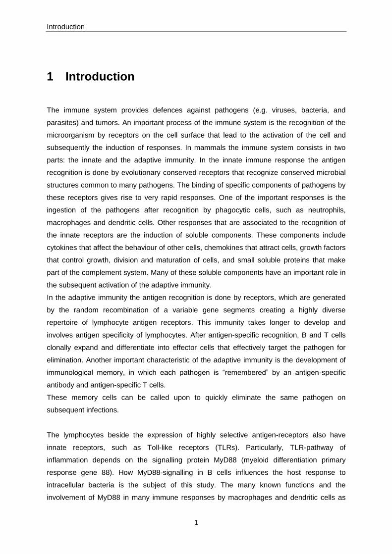
The recognition of microbial threats by PRRs, such as TLRs (Toll-like receptors), NLRs [NOD
(nucleotide-binding oligomerization domain)-like receptors], CLRs (C-type lectin receptors),
and RLRs (RIG-I-like receptors) (Figure 1) is often the first step of the inflammatory cascade.
These receptors are able to recognize a high range of microbial products. The TLRs
recognize a variety of pathogen-associated molecular patterns (PAMPs) derived from
different microbes. The NLRs sense bacteria, RLRs sense viruses and CLRs sense fungi.
These PRRs are localized in different cells of the immune system, such as neutrophils,
macrophages, dendritic cells, endothelial cells, epithelial cells and lymphocytes.
Figure 1: Schematic representation of four types of PRRs and their gene expression in response to invasive
microbes. The TLRs and CLRs activate the transcription factor NF-κB and also MAPKs. Some TLRs and the RLRs activate IRFs, which are required for expression of some antiviral genes. Certain NLRs (e.g. Nalp3) activate caspase 1, which processes the pro-forms of IL-1β and IL-18. Signalling is activated via a receptor-specific subset of adaptors, with MyD88, Mal, TRIF and TRAM mediating the signalling. MyD88 is also used by IL-1R and IL-18R. CLRs signal via the adaptor CARD9. RLRs signal via the adaptor IPS-1; Cardif, CARD adaptor inducing IFNβ; MAVS, mitochondrial antiviral signalling protein; VISA, virus-induced signalling adaptor. The figure is adapted from [1].

Introduction
4
1.1.1.1 Toll-like receptors
Toll-like receptor family are the best characterized class of PRRs in mammalian species. The
first studies identifying the roles of Toll receptors in innate immunity were performed in
Drosophila melanogaster. These studies demonstrated that the gene toll is absolutely
required for activation of antifungal innate immunity [2]. The first ortholog of Drosophila Toll
was found in mammalians in 1996 [3]. Since then ten TLRs (TLR1-TLR10) have been
described for human and twelve TLRs are known in the mouse (TLR10 is not present in
mouse). TLRs are type I transmembrane receptors, which are characterized by an
extracellular Leucine-rich repeat (LLR) domain for ligand binding, a single transmembrane
domain, and an intracellular Toll/IL-1 receptor (TIR) domain involved in signalling [4]. TLRs
can be localized on the cell surface, such as TLR1, TLR2, TLR4, TLR5, TLR6 and TLR11 or
can be localized intracellularly, such as TLR7, TLR8 and TLR9, where their natural ligands
might only be found within acid compartments, such as phagolysomes. The TLR3 could
locate at the cell surface or intracellularly [5].
TLRs detect a broad range of pathogens including viruses [6-10], bacteria [11-14], fungi [15]
and parasites [16]. The recognition of molecular patterns by TLRs includes: lipoproteins from
gram-negative bacteria, Mycoplasma and spirochetes [17-21], lipoteichoic acids and
peptidoglycans from gram-positive bacteria, are recognized by TLR2; TLR9 recognizes
unmethylated CpG motifs from bacterial DNA; flagellin from bacterial flagella is recognized
by TLR5 [11]; double-stranded RNA produced by most viruses during the infection cycle is
recognized by TLR3; and lipopolysaccharide (LPS) of gram-negative bacteria is recognized
by TLR4.
The mechanism of LPS recognition by TLR4 requires several membrane-linked and soluble
molecules, including CD14 and MD-2. CD14, an LRR-containing, GPI-linked molecule binds
LPS binding protein/LPS complexes and is thought to transfer LPS to the TLR4 complex [22,
23]. MD-2 is another protein that interacts with TLR4, and is required for LPS responsiveness
[24, 25].
Signal transduction from TLRs requires adaptor molecules. The protein MyD88 is the major
adaptor molecule in the TLR signalling cascade [26] except for TLR3, and it is also essential
for signalling via interleukin 1 (IL-1) and IL-18 receptors [27]. Other adaptor proteins
contribute to TLR signalling, such as TIRAP (TIR domain-containing adapter protein) also
known as MAL, TRIF (TIR domain-containing adapter-inducing interferon-β), TRAM (TRIF-
related adapter molecule), and SARM (sterile α-and armadillo-motif-containing protein) [28].

Introduction
5
The usage of these adaptor proteins varies between TLRs. For example TLR2 activation on
macrophages triggers via TIRAP and MyD88, a signalling pathway ending in activation of
NF-κB and production of cytokines such as tumour necrosis factor-α (TNFα). TLR4 leads to
the activation of NF-κB via TIRAP or MyD88 but additionally activates the transcription factor
interferon (IFN) regulatory factor-3 (IRF3), leading to the production of type I IFNs in addition
to TNF-α [29, 30].
TLRs can form heterodimers or even associate with non-TLR membrane clusters, to further
diversify their recognition potential [31-33]. The co-activation of TLR2 and TLR4 leads to
higher production of TNF-α, IL-6, and macrophage inflammatory protein 1α (MIP-1α) by
mouse macrophages and human monocytes than either receptor alone elicits [34]. TLR2 and
TLR4 synergize for production of nitric oxide (NO) by macrophages [35]. Activation of TLR4
together with TLR7 increases the production of IL-12p70 by 10 to 100-fold compared to
triggering of either receptor alone [34]. Therefore, TLRs are key receptors in the identification
of pathogens by the innate immune system. Figure 2 summarizes some of the components
in intracellular signalling cascade of the TLRs.

Introduction
6
Figure 2: TLR pathway. TLR1, 2, 4, 5 and 6 are located on the cell surface and TLR3, 7, 8 and 9 are localized to
the endosomal/lysosomal compartment. The activation of the TLR signalling pathway originates from the cytoplasmic Toll/IL-1 receptor (TIR) domain that associates with a TIR domain-containing adaptor, MyD88. Upon stimulation with ligands, MyD88 recruits IL-1 receptor-associated kinase (IRAK) to TLRs through interaction of the death domains of both molecules. IRAK activated by phosphorylation then associates with TRAF6, finally leading to activation of JNK and NF-κB. Tollip and IRAK-M interact with IRAK-1 and negatively regulate the TLR-mediated signalling pathways. MyD88-independent pathways induce activation of IRF3 and expression of interferon-β. TIR-domain containing adaptors such as TIRAP, TRIF and TRAM regulate TLR-mediated signaling pathways by providing specificity for individual TLR signalling cascades. This figure was adapted from www.cellsignal.com
1.1.1.2 Other receptors: NLRs, CRLs and RLRs
NLRs such as NODs (Nucleotide-binding oligomerization domain), NALPs (NACHT-LRR-
PYD-containing protein), NAIP (Neuronal apoptosis inhibitor protein) and IPAF (ICE
protease-activating factor) are cytoplasmic receptors, which recognize microbial products
and/or other danger signals derived from the host [36]. These receptors either sense
organisms that enter the cytoplasm or sense components that may be released or
transported into the cytoplasm by processes such as phagocytosis and degradation of
microbes [34]. Activation of NLRs by bacterial products can stimulate two major signalling
pathways: the nuclear transcription factor (NF-κB) pathway initiated by Nod1 (expressed
ubiquitously) and Nod2 (expressed by monocytes, macrophages, dendritic cells and

Introduction
7
intestinal epithelial cells), or the activation of caspase-1 triggered by IPAF and Nalp3 through
the formation of a multiprotein complex called inflammasome [37]. NF-κB is a key regulator
of the pro-inflammatory response, activating genes that encode cytokines and costimulatory
factors [38]. Caspase-1 leads to the secretion of inflammatory mediators IL-1β and IL-18 and
it can also result in rapid host cell death termed pyroptosis [39-41]. Several studies reported
NAIP5 [42] and IPAF-mediated intracellular recognition of Salmonella flagellin in a TLR5-
independent manner [43-45]. The detection of flagellin by IPAF activates caspase-1 resulting
in the secretion of IL-1β and IL-18 [44, 45].
Other families of different receptors are also important for the innate immune system. This
includes the RIG-I-like receptor (RLR) family and the C-type lectin receptor (CLR) family.
The RLRs are cytosolic domains, such as RIG-1 and MDA-5, that play an important role in
the detection of RNA species derived from viruses in the cytoplasm [46].
CLR family comprises a large family of receptors that binds carbohydrate structures in
calcium-dependent manner but the family also includes proteins that do not bind calcium or
have any carbohydrate specificity. The majority of CLRs mediate endocytosis and/or
phagocytosis, play a role in antigen presentation and keep endogenous glycoprotein levels
constant. Another small subset of CLRs gives rise to altered gene expression in response to
various PAMPs. CLRs are expressed by most cell types including neutrophils, macrophages,
dendritic cells, platelets and B cells [47]. One important group of CLRs is represented by DC-
SIGN. The SIGN (specific ICAM-3-grabbing non-integrin) family of CLRs consists of DC-
SIGN, L-SIGN and LSECtin in humans, and the murine homologues mDC-SIGN and
mSIGNR1 to mSIGR4. For example mSIGNR1 is present on the splenic marginal zone
macrophages and peritoneal macrophages [48]. SIGNR1 is able to capture pathogens from
the blood, such as Escherichia coli and Salmonella typhimurium [49]. It is thought that
pathogen degradation products from MZ macrophages, which do not express MHC class II
molecules, are shed from the cell surface and are taken up by MZ B cells after opsonisation
by complement [48]. Murine Dectin-1 is expressed on dendritic cells, monocytes,
macrophages, neutrophils, and some splenic T cells. In humans, it is also found on mast
cells, eosinophils, and B cells. Dectin-1 is restricted to recognize fungal components [50] and
upon ligand recognition initiates the activation of NK-κB resulting in an increased expression
of pro-inflammatory cytokines [50]. The mannose receptor found in macrophages and DCs
recognizes some sugar molecules found on the surface of many bacteria and some viruses,
including the human immunodeficiency virus (HIV) [47].
In response to a given pathogen, several PRRs can be sequentially or simultaneously
activated on the same cell, and on different cell types. For example, TLR4 and TLR2 are
Introduction
8
sequentially involved in macrophages responses to Salmonella typhimurium. [51]. PRRs are
involved within a system, in which the cross-talk between or even within groups of PRRs is
crucial in balancing immune responses through collaborative induction or negative feedback
mechanisms [52]. For example, to control the over-activation or dysregulation of the TLR-
signalling that can lead to severe diseases, many TLR signal-induced or TLR signal-
associated negative regulators have evolved. For instance, SOCS1 (suppressor of cytokine
signalling 1) is negative regulator of TLR4 through the inhibition of type I IFN signalling
pathway following IFN-β upregulation by TLRs [53]. Also, activation of the same TLR in
different cell types can lead to different functions in the immune response. One example is
that TLR-activated DCs and B cells by LPS provides cytokine environments with opposite
effects on T cell activation [54].
1.1.2 Cells from the innate immune system
Phagocytic cells, such as neutrophils, monocytes, macrophages, DCs, and NK cells are
essential for the innate immune response. Neutrophils are the first cells recruited at the
injured tissue where they contribute to the early defence by eliminating infected cells and by
restricting growth of the microbe [55]. Resident tissue macrophages are also responsible for
the secretion of neutrophil chemoattractants CXCL1 and CXCL2 [56, 57]. If the pathogen
persists, more macrophages are recruited to the site of infection. Macrophages and
neutrophils express receptors for antibody and complement which increases phagocytosis of
antibody- or complement-coated antibodies [58, 59]. NK cells are able to defend the
organism in the early phases of infection against intracellular pathogens or viruses. These
cells are able to kill other cells upon activation by cytokines or upon encountering the target
cells, such as pathogen infected cells and tumours. Dendritic cells are the last ones to be
described since they have an extraordinary capacity to activate naive T cells and elicit the
adaptive immunity. The cascade of cellular events taking place after an infection are
described in more detailed in the following paragraphs.
1.1.2.1 Granulocytes – neutrophils
Granulocytes also called polymorphonuclear leucocytes (PMN) are a group of white blood
cells characterized by the presence of granules in the cytoplasm. The neutrophils belong to
this group and are considered the second major group of phagocytic cells, after
macrophages.
Upon activation neutrophils leave the blood stream and move in the direction of the infection
site as a result of chemotaxis. At sites of infection, neutrophils rapidly phagocytose
pathogens, destroying them with antimicrobial proteins and proteases stored in the

Introduction
9
cytoplasmic granules and with the production of high concentrations of superoxide.
Neutrophils can also generate chemotactic signals to recruit monocytes and dendritic cells,
such as chemerin that is responsible for the recruitment of immature DCs and plasmacytoid
dendritic cells [60]. Neutrophils also produce TNF and other cytokines that differentiate and
activate DCs and macrophages [61-63].
Neutrophils are short lived cells that undergo constitutive apoptosis and are specifically
recognized and phagocytosed by macrophages. Neutrophil apoptosis and clearance is also
very important for the cessation of the acute phase of inflammation. [64].
1.1.2.2 Macrophages
Macrophages are present in almost all tissues. They differentiate from monocytes that leave
the circulation and then migrate into tissue in the steady state or in response to inflammation
[65].
Tissue macrophages have a broad role in the maintenance of tissue homeostasis, through
the clearance of senescent cells and the remodelling and restoration of tissues after
inflammation [66, 67]. These cells show a high degree of heterogeneity according to their
specific function in different organs, such as lungs, brain, in the submucosal layer of the
gastrointestinal tract, spleen and liver. In the liver the resident macrophages are known as
kupffer cells.
Inflammatory macrophages derive from monocytes that leave rapidly the bloodstream upon
injury or infection. This population is subdivided into two other main populations: classically
activated macrophages and alternatively activated macrophages [68].
Classically activated macrophages arise in response to TLR agonists, IFN-γ, which can be
produced during innate immune response by natural killer (NK) T cells or during adaptive
immune response by T helper 1 (TH1) cells or CD8+ T cells, and tumor necrosis factor (TNF),
which is produced by several cells, such as neutrophils, antigen presenting cells, TH1 T cells
[68]. This population have microbicidal activity and secrete high levels of pro-inflammatory
cytokines, such as TNF-α, interleukin 6 (IL-6), IL-1β, and IL-12 [68, 69]. In addition, this
population produces reactive oxygen species and have higher expression of co-stimulatory
molecules [68]. Classically activated macrophages are crucial in host defence against
intracellular pathogens, for example, mice lacking this population are more susceptible to
Salmonella typhimurium infection [70]. However, the activation of classically activated
macrophages must be tightly controlled because cytokines as IL-1, IL-6 and IL-23 give rise to
TH17 cells, which can contribute to autoimmune responses [68].
Alternatively activated macrophages arise in response to IL-4, IL-10 and/or IL-13, cytokines
that are generally produced in TH2 responses, particularly in allergic, cellular and humoral

Introduction
10
responses to parasites and extracelullar pathogens [68, 71]. This population promotes tissue
repair and suppresses inflammation by the secretion of high levels of IL-10 [65, 68].
1.1.2.3 NK cells
Natural killer (NK) cells are large granular lymphocytes that can lyse infected cells with a
variety of pathogens by releasing proteins into the target cell. Their activation is controlled by
NK cell receptors that are encoded in the germline and do not undergo somatic
recombination. They can respond within hours upon infection. The mechanism, by which NK
cells are triggered, depends on a balance between two opposing signals: activating receptors
that recognize target cell surface structures, including viral products, and give a ‟‟kill‟‟ signal,
and inhibitory receptors that recognize MHC class I proteins present in all healthy cells, and
may suppress NK cell responses. When target cells express ligands for both inhibitory and
activating receptors the outcome is determined by the outline of the strength of signals [72].
NK cells can kill target cells through perforin and granzymes, which are proteins, pre-stored
in cytoplasmic granules and can be exocytosed directly in the target cells. This killing
mechanism is very similar to the CD8+ cytotoxic T lymphocytes (CTL).
NK cells upon activation secrete inflammatory cytokines such as, IFN-γ and TNF-α and
chemokines [72, 73], which have a direct role in the activation and function of other cells. For
instance, NK cells facilitate the capacity of DCs to trigger adaptive T cell response [74]. DCs
also secrete IL-12, IFN-α and IFN-β, which activates IFN-γ production by NK cells [74].
Moreover, the IFN-γ produced by NK cells is critical also for the activation of macrophages
[75, 76]. NK cells enhance the response of CD8+ T cells and CD4+ T cells [77] and also IFN-γ
producing NK cells promote a T helper type 1 (TH1) cell polarization.
1.1.2.4 Dendritic Cells
Dendritic cells (DCs) are the most potent antigen presenting cell (APC) of the immune
system, as illustrated by their ability to trigger activation of naïve CD4+ and CD8+ cells
compared to other APCs such as macrophages and B cells [78-80]. Dendritic cells are
present in lymphoid organs, in the epithelia of the skin and gastrointestinal and respiratory
tracts, and in the interstitium of most parenchymal organs.
A large variety of DC subsets has been described. Mouse DCs have been classified into six
subpopulations according to their phenotype and properties [81]. The main populations are
characterized according to the T cell markers CD8 or/and CD4 [81]. There are also the
plasmacytoid DCs (PDC), which also can express CD8 but their principal characteristic is the
expression of B220 [81]. Upon viral activation these DCs are principal producers of type I

Introduction
11
interferons (IFN-α/β) [82]. Additionally, there are also the dermal DCs and the langerhans
cells, both present in the skin [81].
Dendritic cells recognize and ingest pathogens through PRRs that recognize features
common to microbial surfaces, and they are very active in taking antigens by phagocytosis
using receptors such as TLRs. The recognition of pathogens via TLRs activates DC, causing
an increase of expression of class II molecules [83, 84], co-stimulatory molecules (CD80 and
CD86) [85-87] and production of several cytokines such as IL-12 [88]. Remarkably, ligation of
distinct TLRs can trigger differential cytokine production in a single DC type or result in
different cytokines in distinct DC sub-types. Previous studies have shown that CD8- DCs
mainly induce TH2 cell responses, whereas CD8+ DCs elicit strong TH1 cell responses due to
their high capacity to produce IL-12 [89, 90]. However certain characteristics of the microbe
also play an important role in tuning the immune response. TLR4-dependent E. Coli LPS
induces a TH1 response [91], but LPS from the oral bacterium Porphorymonas gingivalis,
which signals through TLR4 independent way [92], induces a TH2 response. Consistent with
this, E. Coli LPS, but not P. gingivalis, LPS induces IL-12 in splenic CD8α+ DCs [91].
Cytokines secreted by other cells can also modulate DC function. For example, TH1-inducing
DCs, when exposed to IL-10 or TGF-β, induce TH2-like responses [93]. On the other hand,
IFN-γ can induce DCs to acquire some TH1-inducing capacity [93].
The DC subsets also comprises different repertoire of TLRs. For example, splenic CD8+ DC
do not express TLR7 mRNA and do not respond to the ligand R-848 in vitro [94]. Other study
claims that CD8+ DCs upregulate co-stimulatoty molecules in response to a different TLR7
ligand in vivo [95]. This subset also expresses lower level of TLR5 mRNA compare to other
splenic DC subsets, but in contrast have the higher expression of TLR3 mRNA, which
appears to be absent in PDCs [94].
1.2 The adaptive immune system
Adaptive immunity also called acquired immunity is characterized by two important
properties: specificity and memory. B and T cells are able to recognize all pathogens
specifically and to provide enhanced protection against reinfection.
Introduction
12
1.2.1 Cells from the adaptive immune system
B and T cells are the main components of adaptive immunity and one of their principal
properties is the specificity, which resides in the antigen receptors. During their development,
every individual pro-B or pro-T cell acquires a unique antigen-specific receptor on its surface.
The broad range of antigen specificities in the antigen receptor repertoire is due to variation
in the amino-acid sequence at the antigen-binding site, which is made up from the variable
(V) regions of the receptor proteins chains. In each chain the V region is linked to an
invariant constant (C) region, which provides effector or signalling functions.
The variable (V) region is encoded by more than one gene segments. The first segment is
termed a V gene segment because it encodes most of V domain. The second segment is
termed diversity (D) gene segment. The third is termed joining (J) gene segment which is
located close to the C region. Each gene segment has multiple different copies, and different
combinations of gene segments can be used in different rearrangement events [96, 97]. This
generates an enormous repertoire of antigen receptors with different specificities.
Upon activation, when the lymphocyte receptor recognizes the specific antigen this leads to
clonal expansion of this specific lymphocyte. The antigen recognition molecules of B cells are
immunoglobulins (Ig), which are membrane-bound receptor for antigen, the B cell receptor
(BCR). Upon antigen recognition, B cells differentiate into cells producing antibody molecules
of the same antigen specificity as the BCR.
T cells recognize antigen through cell-surface receptors called T cell receptor (TCR), which
recognizes a peptide bound to a MHC molecule, which is displayed on the surface of APCs.
T cells are subdivided into two distinct classes based on their TCR. The majority of T cells
express receptors made up of α and β chains. A small group of T cells express receptors
made up of γ and δ chains. Among the α/β T cells are two important subgroups: those that
express the co-receptor molecule CD4 (CD4+ T cells) and those that express CD8 (CD8+ T
cells). These cells differ fundamentally in how they recognize antigen but they mediate
different types of regulatory and effector functions.
1.2.2 T lymphocytes
1.2.2.1 CD4 T cells
The CD4+ T cells are important orchestrators of the immune system. Their functions depend
on the cytokines that they secrete when activated and also on the cell surface molecules that
are induced upon activation. The CD4+ T cells can differentiate into various T helper subsets,
Introduction
13
such as T helper 1 (TH1), T helper 2 (TH2) cells, T follicular helper (TFH) and T helper 17
(TH17) cells, distinguished by the arrays of cytokines they produce.
The TH1 cells produce IL-2, IFN-γ and lymphotoxin (LT). These cytokines are strong inducers
of cellular immune responses. They improve the microbicidal activity of monocytes and
macrophages resulting in an increased efficiency in lysing microorganisms in intracellular
vesicular compartments. In addition these cytokines induce the proliferation and activation of
CD8 T cells. TH1 also provide some B-cell help, but higher TH1-cell numbers can inhibit B cell
activation [98]. There is strong evidence that TH1 T cells are required in bacterial infections.
For instance, mice deficient in IFN-γ receptor are highly susceptible to attenuated Salmonella
typhimurium infection [99] and CD4 T cell production of IFN-γ is required for protection
against Listeria monocytogenes [100].
TH2 cells produce IL-4, IL-5, IL-6, IL-10, TGF-β and IL-13 cytokines [101]. These cytokines
are excellent promoters for antibody production and also enhance eosinophil proliferation
and function. TH2 cytokines are commonly associated with strong antibody and allergic
responses [102].
Follicular helper T cells (TFH) have a distinct phenotype and cytokine profile than other CD4+
T cells. These cells are localized in B-cell areas of lymphoid organs and their main function is
to provide specialized help to germinal centre (GC) B cells, which are involved in the
generation of memory B cells or long-lived plasma cells (explained in detail in section 1.2.3).
TFH cells are distinguished from TH1 and TH2 by chemokine receptors, such as CXC-
chemokine receptor 5 (CXCR5) and transcription factors. Upon activation of TFH cells, the
expression of CXCR5 is up-regulated and binds to CXC-chemokine ligand 13 (CXCL13),
which promotes follicular homing by B and T cells [103]. Several features, not all unique to
TFH cells, help to characterize and define this subset. This includes CD40L, which is crucial
for B cell proliferation, immunoglobulin class-switching, and GC B cell survival [104-106],
ICOS and IL-10, which support GC B-cell growth and differentiation [104, 107], and IL-21,
which also helps B cell activation [108, 109]. TFH can also produce IL-4 upon infection. For
instance, mice infected with a helminth have a strong IL-4 production by TFH cells.
Additionally, TFH-cell derived IL-4 is required for the generation of high IgG1 affinity
antibodies [110].
TH17 T cells are a recently identified CD4+ T cell subset that secretes not only IL-17, but also
IL-22 and IL-21 [111] and exhibit effector functions distinct from TH1 and TH2 cells. TH17 T
cells have been implicated in the pathogenesis of many experimental autoimmune diseases
and human inflammatory conditions [111]. However, the primary function of these cells may

Introduction
14
be the clearance of extracellular pathogens during infections [111]. It seems that TH17 T cells
are required in infections by gram negative bacteria to induce an appropriate host immune
response and for abscess formation [112]. They induce a strong neutrophil response and
also control the migration of various other cell types, such as macrophages and probably
also T cells. For example in mycobacterial infection, TH17 cells trigger the induction of
chemokines that attract TH1 cells, which eventually control the infection [113]. TH17 cells also
play a role in protection against Salmonella. For instance, IL-17 deficient mice have a mild
effect in bacterial clearance during Salmonella infection [114].
Several factors are affecting the differentiation of T helper cells, for example, IL-12
production by DCs and macrophages induces TH1 differentiation, IFN-γ produced by NK cells
and CD8 T cells also induce TH1 differentiation, whereas IL-4 stimulates differentiation of TH2
cells, which may be contributed by mast cells, and basophils [115]. IL-6 production by
activated DCs promotes TH17 differentiation [116].
The characteristic cytokine products of TH1 and TH2 are mutually inhibitory for the
differentiation and effector functions of the reciprocal phenotype. Thus, IFN-γ producing TH1
cells are able to block the growth of TH2 cells [117]. The IL-10 acts on macrophages to inhibit
TH1 activation and TGF-β inhibits the growth of TH1 cells [118, 119]. If a subset is activated
first or preferentially in a response, it can suppress the development of the other subset
[119]. In general, infection are associated with dominance of either a TH2 or a TH1 response
[102] The cross-inhibition may partly explain the strong biases towards TH1 or TH2 responses
during many infections in mice and humans [102].
1.2.2.2 CD8 T cells
CD8+ T cells or cytotoxic T cells (CTL) are important for the host defense against intracellular
pathogens as viruses and bacteria like Listeria and Salmonella [120, 121].
Naive CD8+ T cells differentiate into cytotoxic T cells after recognition of antigen peptide-
MHC class I complexes on the surface of dentritic cells, which express high levels of co-
stimulatory molecules, or they recognize the antigen in weakly co-stimulatory cells requiring
the presence of CD4 T cells bound to the same APC [122]. Both types of activation induce
the autocrine IL-2 production by CD8+ T cells leading to their proliferation [123, 124].
Cytotoxic T cells have several ways of destroying infected cells. One is through the delivery
of cytotoxins (perforins, granzymes and granulysins), contained in specialized lysosomes
called lytic granules that are released and lyse the infected cells upon antigen recognition on
the surface of the infected cell [125]. Another way is through the interaction of Fas (CD95)

Introduction
15
with Fas-ligand on the surface of the cytotoxic cells leading to the apoptosis of the infected
cell [125]. In addition cytotoxic CD8 T cells also secrete cytokines such as IFN-γ, TNF-α and
TNF-β, which contribute to the defense. IFN-γ inhibits viral replication, increases expression
of MHC class I, and activates macrophages. TNF-α and TNF-β can synergize with IFN-γ in
macrophage activation and in killing some target cells through their interaction with TNFR-I.
1.2.2.3 NKT cells
Natural Killer T (NKT) cells are T lymphocytes that express both αβ T cell receptors (TCRs)
and NK cell receptors [126, 127]. NKT cells can respond very fast to their cognate antigens,
which is a characteristic of innate immunity, and they can also promote TH1 and TH2 adaptive
responses [128]. In mice, most NKT cells express an invariant chain Vα14Jα18 TCR with
predominant coexpression of Vβ8.2, Vβ2 and Vβ7 [129]. Human NKT cells express a
Vα24Jα18 rearranged TCR α chain usually coexpressed with Vβ11. NKT cells recognize an
MHC class IB molecule, CD1, which presents endogenous or bacterial lipid antigens.
The most precise description of NKT cells is their expression of an invariant TCRα antigen
receptor and their reactivity to αGalCer, which is an unusual α-glycosylatedsphingolipid
initially derived from marine sponge. NKT cells upon activation release large amounts of TH1
cytokines such as IFN-γ and TNF but also TH2 cytokines like IL-4, IL-10 and IL-13 [130-132].
Deficits in NKT cells are involved with autoimmune diseases, such as diabetes, indicating
that these cells are also important for suppressing autoimmunity and graft rejection. NKT
Cells also facilitate immunity against infections and tumors. For instance, NKT cells
participate in protection of mice from a variety of bacterial, viral, and protozoan parasites
[133].
1.2.2.4 Regulatory T cells
Regulatory T cells (Tregs) are an important subset of T cells that mediate peripheral
tolerance. These cells are classified in two subsets: one is called natural regulatory T cells
(nTregs), which develop in the thymus and the other is called inducible regulatory T cells
(iTregs), which derive from naive T cells in the periphery. The natural regulatory T cells are
CD4+ T cells characterized by high expression of CD25 and the transcriptional factor Foxp3
(forkhead box P3). In humans not all the Foxp3-expressing cells are Tregs. The nTregs
utilize the cytokines IL-10, IL-35 and TGF-β to exert their suppressive effects upon
conventional T cells. Beside the secretion of inhibitory cytokines, nTregs use other
suppression mechanisms, such as cytolysis through granzyme A or B for humans or mice
respectively, perforin, which kills the target cell, metabolic disruption, in which Tregs provoke
death of effector T cells by cytokine deprivation, and modulation of APC function [134-137].

Introduction
16
The iTregs are originally coming from CD4+ CD25- or CD8+ CD25- T cells. The iTregs
population includes distinct subtypes of CD4+ T cell: T regulatory 1 (Tr1), which secrete high
levels of IL-10, TGF-β, no IL-4 and no or low levels of IFN-γ [138]; and T helper 3 (TH3) cells,
which secretes high levels of TGF-β. Although CD8+ T cells are associated with cytotoxic T-
lymphocyte function and IFN-γ production, a subtype of these cells can secrete IL-10 and
TGF-β [139, 140] and have been called CD8+ regulatory T cells.
Regulatory T cells are involved in the control of various immunological processes, including
protective immune responses in autoimmunity, for example nTregs are functionally impaired
or have deficits in their maturation or in their thymic emigration in multiple sclerosis (MS)
[141-144], iTregs and n Tregs are involved in the prevention of type 1 diabetes (T1D) [145,
146], iTregs are also implicated in protection against inflammatory bowel disease (IBD) [147].
The nTregs play also a significant role in the immune response to infections. For instance,
they control an excess of effector immune responses against a pathogen preventing
immunopathology [148], for example, during infection of mice with Candida albicans, a
reduction in nTregs numbers induces better control of the infection but on the other hand
enhances inflammatory gastrointestinal pathology [149]. There are also some pathogens that
favor nTregs induction, promoting their survival inside of the host [148].
1.2.2.5 Memory T cells
Memory T cells can develop directly from effector precursors after the primary response or
are can be generated in parallel with effector populations continuing on to their
predetermined fate after the primary immune response [150, 151].
Memory T cells can generate a rapid recall response to secondary pathogen challenge [152-
154]. CD4 and CD8 memory T cells derived from human blood and mouse spleen can be
distinguished based on the expression of the lymph node homing receptors CD62L and
CCR7.
There are two kinds of memory T cells, the effector memory T cells (TEM), which are
CD62Llow and CCR7- and the central memory T cells (TCM), which are CD62Lhigh and CCR7+.
The TEM cells are localized in the peripheral tissues and display immediate effector function,
whereas central memory T cells home to the T cell areas of secondary lymphoid organs,
have little or no effector function, but readily proliferate and differentiate to effector cells in
response to antigenic stimulation. Both subsets produce large amounts of effector cytokines
such as IFN-γ and TNF-α after antigen stimulation, central memory T cells produce
predominantly IL-2 which could increase their ability to proliferate in response to antigen
[155]. TEM cells have more lytic activity ex vivo, probably because of increase expression of
perforin [156]. Furthermore a small fraction of these cells express the low-affinity Fc receptor

Introduction
17
for IgG IIIa (FcγRIIIa), which allows them to directly mediate antibody-dependent cell
mediated cytotoxicity [157].
Some studies have shown that memory CD8+ T cells after their generation in secondary
lymphoid organs, preferentially locate to BM for homeostatic maintenance [158] and recently
some memory CD4+ T cells have been shown to reside also in the BM [159].
1.2.3 B lymphocytes
There are two main groups of B cells, the B-1 cells, which develop from fetal liver-derived
HSCs and B-2 cells, which derive from bone marrow HSCs.
Most of all studies done with B-1 cells are based on rodent studies, in humans this
population is still not clear [160]. B-1 cells reside mainly in peritoneal and pleural cavities and
are subdivided in B-1a and B-1b B cells. B-1a cells are characterized by the CD5+ CD11b+
B220low IgMhigh IgDlow phenotype, while the B-1b B cells do not express the CD5 [161]. B-1a
cells are the source of natural antibodies, which are predominantly of IgM class and provide
protection during the early stages of infection, whereas induced antibodies derived from B-1b
cells are produced subsequently and are key to the clearance of, and long term protection,
from pathogens [162, 163].
B-2 cell population is heterogeneous, consisting of follicular B cells (FO), in LN and splenic
lymphoid follicles, and a subset residing in the marginal zone (MZ) of the spleen [164]. MZ
and FO B cells are distinguished by differential expression of several cell surface markers:
MZ B cells are IgDlowCD21highCD23low/-, whereas FO B cells are IgDhighCD21interCD23high [165,
166]. Marginal zone B cells are located at the margin between the white pulp and red pulp. In
rodents, MZ B cells primarily appear to mediate T-independent responses to antigens from
blood-borne pathogens. During these pathogen-driven responses, LPS and possibly other
microbial products may drive the migration of activated B cells out of the MZ into the red pulp
where they may differentiate into short-lived plasma cells secreting mainly IgM and IgG3
antibody [164, 167].
The follicular B cells can occupy different niches. Once they mature, they recirculate in the
spleen and lymph nodes (or in the BM). Naive follicular B cells reside in the ‟‟follicular niche‟‟
and may present T-dependent antigens to activated T cells. Differentiation of activated
follicular B cells into antibody-secreting cells is enhanced by TLR activation on follicular B
cells, but only prior to BCR and CD40 activation. It has been previously proposed that for an
effective activation of lymphocyte it is necessary a second signal in addition to the specific
antigen recognition by the BCR or TCR. [168]. Ruprecht and Lanzavecchia [169]
Introduction
18
demonstrated that CpG, provides a strong signal that in synergy with cognate T-cell help and
BCR cross linking helps to sustain optimal proliferation and differentiation of human B cells.
Furthermore, it has been demonstrated by Fillatreau et al. that for experimental autoimmune
encephalomyelitis (EAE), which is an animal model for Multiple Sclerosis (autoimmune
disease that affects the central nervous system (CNS)) CD40 participates in the production
of IL-10 by B cells which stimulates disease resolution [170].
The binding of CD40 by CD40L also induces the expression of co-stimulatory molecules
such as B7-family molecules (CD80/CD86) [171]. In addition, cytokine secretion by T cells,
such as IL-4, IL-5 and IL-6 stimulate B cells to proliferate, to undergo isotype switching and
differentiate into antibody-secreting plasma cells.
B cells, which have recognized antigen trough BCR and received T cell help can also form
germinal centres. Germinal centres are specialized areas in the follicle where B cells
proliferate rapidly and undergo a process of somatic mutation so that a single initial B cell
can give rise to variant progeny [172, 173]. The germinal centre B cells that have high affinity
BCRs for the antigen are selected and differentiate into memory or into plasma cells
secreting antibody of high affinity [173-177]. MZ B cells also participate in T-dependent (TD)
immune responses to protein antigen as well as in responses to lipid antigens. For instance,
Song and colleagues have demonstrated that MZ B cells upon stimulation with TD antigen
can differentiate very rapidly into antibody forming cells (AFC), which have a distinct
clonotypic repertoire and also they can give rise to GCs with somatic hypermutation and
generate immunological memory [178].
The first antibodies to be produced in humoral immune response are always IgM, because
IgM can be expressed without isotype switching. In addition to IgM, there are three antibody
isotypes, such as IgG, IgA and IgE. Among these, IgG are by far the most abundant
immunoglobulin and contain several subclasses. In mouse there are IgG1, IgG2b, IgG2c and
IgG3. In humans there are IgG1, IgG2, IgG3 and IgG4.
It is known that in mice cytokines can induce isotype switching. For example, IFN-γ secretion
by CD4 T cells induce the isotype switching to IgG2c or to IgG3 and the IL-4 switches to
IgG1 or IgE.
The antibodies play different roles in the immune defence. Apart of direct neutralization of
pathogens, antibodies can also activate complement and opsonise microorganisms by
phagocytic cells or killing by NK cells.

Introduction
19
1.2.3.1 Memory B cells and long lived plasma cells
B cell memory develops in GC reactions after primary immune response to thymus-
dependent (TD) antigens [179-182]. Populations of affinity-matured memory B cells persist
for long periods of time in multiple cellular forms [183-185]. These post-germinal center B
cells can be categorized into long-lived plasma cells, which will not respond to antigen recall
and the precursors for the memory response [181, 186, 187].
Memory B cells do not secrete antibodies until expansion and differentiation into plasma cells
after rechallenge with antigen. These memory B cells contain high affinity BCRs, enabling
them to respond in vivo to little amounts of antigen. These cells recirculate in the periphery,
where they can encounter easily the antigen leading to an efficient secondary immune
response [188]. Many of them are localized in the marginal zone of the spleen [189, 190]
Long-lived plasma cells are terminally differentiated, affinity-matured and home to the bone-
marrow [191, 192]. Although there is evidence that some long-lived plasma cells are present
in the spleen [193]. It has been shown in vitro that the survival of these cells is promoted by
IL-6, CXCL12, and ligands for CD44 [194, 195]. The continued production of antibodies by
the long-lived plasma cells is crucial for immunity, because such antibodies provide
immediate protection without the requirement for clonal expansion and differentiation of B
cells [196].
1.2.3.2 Regulatory functions of B cells
B cells positively regulate immune responses through antibody production and CD4+ T cell
activation. However, B cells can also negatively regulate immune responses in mouse
autoimmunity and inflammation models through the production of auto-antibodies. For
instance, it was shown that transfer of serum from patients suffering from idiopathic
thrombocytopenia purpura (ITP), an autoimmune disease associated with low platelet counts
and mucocutaneous bleedings, could transfer disease to healthy patients [197]; in
hyperthyroidism, the increased production of thyroid hormone is sustained through the
stimulation of thyroid-stimulating hormone receptor by auto-antibodies [198].
Despite the role of pathogenic auto-antibodies, recent studies indicate that B cells can have
a regulatory function and suppress autoimmune diseases [170, 199, 200]. The first
observation was provided by Wolf and Janeway who observed that mice lacking B cells fail
to control EAE, while wild-type mice recover after a short episode of paralysis [201].
A characteristic feature of B cell-induced regulation is the involvement of IL-10 in the
regulatory process. B cells are able to produce large amounts of IL-6 and IL-10 that play
instrumental roles in T cell mediated immunity [54]. It has been shown that IL-10 deficient
mice suffer a chronic EAE, a more severe CIA and also develop a spontaneous inflammatory
Introduction
20
bowel disease (IBD) [170, 199, 202]. In fact, it has been revealed that B cells regulate these
autoimmune diseases through the provision of IL-10 [170, 199, 203]. Thus, mice in which
only B cells lack IL-10 develop a chronic EAE while mice with wild type B cells recover after a
short time of paralysis [170]. In addition, B cells isolated from recovered mice secrete IL-10
upon in vitro stimulation and transfer of these cells confers protection to recipient mice [170].
The importance of B cell-derived IL-10 in the regulation of autoimmune diseases has been
extended in numerous studies. IL-10 production by B cells are involved in the suppression of
TH1 and TH17 mediated pathologies such as EAE [204] and T1D [205]. They are also
involved in the suppression of diseases driven by TH1 cells and/or auto-antibodies such as
CIA [199, 206], SLE [207] and act as regulators in TH2 mediated diseases such as UC [202,
203]. The regulatory role of B cells is not limited to autoimmune situations. For instance,
infection with Schistosoma mansoni induces IL-10 production by B cells, which can suppress
anaphylaxis [208]. Similarly, some viruses have built up mechanisms to stimulate IL-10
producing B cells and therefore subvert the immune system [209, 210].
The mechanism how IL-10 production by B cells regulates the immune response is not
completely clear, it has been shown to strongly inhibit macrophages and dendritic cells [211].
IL-10 from B cells also inhibits the capacity of activated CpG-dendritic cells to stimulate T cell
proliferation in vitro. The production of IL-6 and IL-12 by DCs is also repressed by IL-10 from
B cells, which then inhibits differentiation of TH17 and TH1 cells respectively [54]. IL-10
production by B cells elicited by infection with Brugia pahangi attenuates their efficiency as
APCs to CD4+ T cells restricting their expansion [212].
1.2.3.3 TLR-activated B cells
The signalling through B-cell antigen receptor (BCR) is very important in shaping the
development and functions of peripheral follicular B cells; however it has also been described
that, innate TLRs, which recognize specific microbial products, also help to sculpt humoral
immune responses. For instance, MyD88 signalling in B cells can amplify several aspects of
the humoral response, including antibody production, germinal centre formation, and
accumulation of long-lived plasma cells in the bone marrow [213-216]. Beside the fact that B
cells respond to TLR stimulation by proliferating and secreting antibodies, they are also able
to secrete cytokines [54, 217]. Previously it has been demonstrated that LPS or CpG are
potent inducers of IL-10 production by naive B cells [54]. Mice with B cell deficiencies in
MyD88 or both TLR2 and TLR4 do not recover from EAE [54]. In contrary mice with TLR9
deficient B cells are able to recover [54]. In EAE, components from Mycobacterium
tuberculosis are present in the complete freund‟s adjuvant used to induce disease. This
suggests that Mycobacterium tuberculosis provides the TLR agonists that induce the

Introduction
21
regulatory function in B cells. It seems that only particular TLR agonists can trigger a
regulatory function in B cells. This can be explained by the fact that B cells and cells from the
innate immune system, such as DCs or macrophages use TLRs in different ways to sense
their environment. Additionally, MyD88 signalling in B cells controls disease resolution while
MyD88 signalling in other cells is required for disease induction [218]. For example, B cells
activated via TLR can secrete a milieu of cytokines that inhibits T cell activation in an IL-10
dependent manner, whereas activation of DCs in the same way produced a milieu that
contained very little IL-10 and was able to enhance T cell proliferation [54]. These
observations suggest that in EAE model MyD88-signalling in distinct cell types can modulate
immunity in opposite ways.
1.3 Salmonella enterica infection
Salmonella enterica belong to the family of enterobacteriaceae and are facultative
intracellular gram-negative bacteria that infect humans and animals causing a spectrum of
disease ranging from systemic infection to gastroenteritis, depending on the bacterial serovar
and the host species infected. More than 2400 serotypes have been described and all of
them are considered potentially pathogenic. Some serotypes are host specific but the
majority can affect different hosts. The Salmonella serotypes are classified according to the
Kauffman-White classification scheme. The identification is based on the „„O‟‟ antigens, which
consist of the lipopolysaccharide-protein chains exposed on the cell surface, and „„H‟‟
antigens, which consist on the flagellar antigens. Typhoid fever is a systemic disease caused
by Salmonella enterica, serovar typhi, a highly invasive
enteric pathogen that infects only humans. Salmonella
enteric serovar typhimurium (Figure 3) referred usually as
S. typhimurium infects animals and humans. In particular, S.
typhimurium causes gastroenteritis and occasionally
septicemia in humans, and systemic disease in susceptible
mouse strains that is similar to the human typhoid fever.
This model is widely accepted as the best experimental
system for studying human typhoid fever and has proved
extremely valuable in uncovering mechanisms of innate and
adaptive immune resistance to intracellular pathogens.
Figure 3: S. typhimurium. This
figure was adapted from www.britannica.com/EBchecked/topic-art/478531/121515

Introduction
22
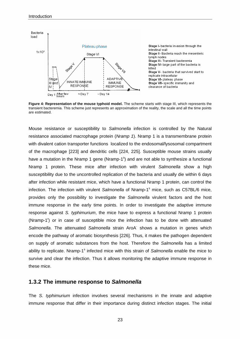
1.3.1 The mouse typhoid model
Salmonella is a food- and water-born pathogen. Therefore, after oral ingestion and
colonization of the small intestine, S. typhimurium penetrates the intestinal epithelium and
enters the Peyer‟s patches through the M cells or through the enterocytes in the microvilli
[219, 220].
After bacteria have penetrated the mucosal barrier, the bacteria moves in the tissues and
many reach the mesenteric lymph nodes. At this stage antibodies, complement and tissue
macrophages exert their protective function, resulting in a reduction of the numbers of
bacteria. Salmonella move with the efferent lymph to the circulation, and are carried by it to
all parts of the body which results in a transient bacteremia [221]. Bacteria are rapidly
removed from the blood by phagocytic cells in the spleen and liver. During this stage a big
part of the Salmonella are killed [219]. The Salmonella that survived this stage starts to
replicate intracellularly in the spleen and liver. This process can take several days, during
which nonspecific host defence mechanisms such as IFN-γ and TNF-α modify the rate of
bacterial growth. Depending on this rate of bacterial multiplication and the initial numbers of
Salmonella in the liver and spleen, the infection can either progress to an overwhelming
disease or be restricted. If the bacteria reach a number of 108 in the organs the host seems
unable to control the infection [219]. As a consequence, secondary bacteremia, in which
bacteria invades hepatocytes, these are lysed and release large numbers of bacteria leading
to an endotoxic shock and soon death [219]. If the growth rate of the bacteria is slower or the
inoculum was smaller, the bacterial multiplication can be suppressed with the recruitment of
more defences. This phase is called plateau phase and is characterized by splenomegaly,
recruitment of bone marrow derived cells and the action of IL-12. The maintenance of
bacterial numbers and formation of granulomas require sustained production of TNF [221],
without which bacterial numbers cannot be suppressed. The plateau level of bacteria can last
from one to several weeks, depending on the mouse strain and the strain of S. tytphimurium
used [222]. The final stage of infection is characterized by the generation of an acquired
immune response able of eliminating S. typhimurium and long-lasting immunity against
reinfection. The Figure 4 illustrates the several phases of the immune response against
Salmonella spp. in the mouse typhoid model.
Introduction
23
Figure 4: Representation of the mouse typhoid model. The scheme starts with stage III, which represents the
transient bacteremia. This scheme just represents an approximation of the reality, the scale and all the time points are estimated.
Mouse resistance or susceptibility to Salmonella infection is controlled by the Natural
resistance associated macrophage protein (Nramp 1). Nramp 1 is a transmembrane protein
with divalent cation transporter functions localized to the endosomal/lysosomal compartment
of the macrophage [223] and dendritic cells [224, 225]. Susceptible mouse strains usually
have a mutation in the Nramp 1 gene (Nramp-1s) and are not able to synthesize a functional
Nramp 1 protein. These mice after infection with virulent Salmonella show a high
susceptibility due to the uncontrolled replication of the bacteria and usually die within 6 days
after infection while resistant mice, which have a functional Nramp 1 protein, can control the
infection. The infection with virulent Salmonella of Nramp-1s mice, such as C57BL/6 mice,
provides only the possibility to investigate the Salmonella virulent factors and the host
immune response in the early time points. In order to investigate the adaptive immune
response against S. typhimurium, the mice have to express a functional Nramp 1 protein
(Nramp-1r) or in case of susceptible mice the infection has to be done with attenuated
Salmonella. The attenuated Salmonella strain AroA- shows a mutation in genes which
encode the pathway of aromatic biosynthesis [226]. Thus, it makes the pathogen dependent
on supply of aromatic substances from the host. Therefore the Salmonella has a limited
ability to replicate. Nramp-1s infected mice with this strain of Salmonella enable the mice to
survive and clear the infection. Thus it allows monitoring the adaptive immune response in
these mice.
1.3.2 The immune response to Salmonella
The S. typhimurium infection involves several mechanisms in the innate and adaptive
immune response that differ in their importance during distinct infection stages. The initial

Introduction
24
stages of infection are characterized by an innate immune response triggered by host
recognition of several microbial structures including PAMPs such as flagellin,
lipopolysaccaride (LPS), and lipoproteins [227, 228]. Interaction of these microbial structures
with specific host receptors ultimately leads to stimulation of innate immune functions of
epithelia and phagocytes. Macrophages phagocytize S. typhimurium, and this process is
facilitated by receptor-mediated uptake after opsonization of Salmonella with antibodies and
complement [229]. Bacterial uptake is not a process done only by phagocytic cells.
Salmonella typhimurium has the ability to invade host cells via the expression of a type-three
secretion system (TTSS) encoded within the Salmonella pathogenicity island I (SPI-1) that
injects virulence factors such as SipA and SopE into target cells and drives cytoskeleton
rearrangements and phagocytosis, this promotes intracellular survival of the pathogen, or
causes apoptosis of macrophages [230].
During the initial stages of infection TNF-α, IFN-γ, IL-12 and IL-18 are important for the host
survival [231, 232]. Monocytes are recruited and they are the major producers of TNF-α and
iNOS [233], the later being crucial for producing reactive nitrogen species important in
Salmonella killing [234, 235]. Macrophages and dendritic cells produce IL-12 and IL-18 these
cytokines are important to induce the expression of IFN-γ produced by NK cells,
macrophages and B cells. The IFN-γ production will also induce the expression of IL-12
through a feedback loop [236, 237]. The recruitment and activation of phagocytes by
chemokines and cytokines respectively characterizes the initial stages of Salmonella
infection. As a consequence, large numbers of bacteria are eliminated and Salmonella
infection is controlled until a certain degree. Then, only the response of antigen specific cells
from the adaptive immunity can allow an effective eradication of bacteria and provide
protection against a secondary infection with S. typhimurium.
The CD4+ T cells have been shown to be crucial for the resolution of S. typhimurium infection
[99, 219, 222, 238, 239]. Salmonella typhimurium induces a strong T-helper 1 (TH1)
response, characterized by the production of large amounts of IFN-γ [240-242] and TNF-α
[238, 243-246]. These cytokines activate bactericidal mechanisms in macrophages and
clearly improve the capacity of these cells to kill S. typhimurium. Additional functions of CD4+
T cells is the regulation of antigen-specific B cell activation and maturation, either by direct
cell-cell contact or by cytokine production. T cell help is essential for B cell differentiation
such as isotype switch and affinity maturation. CD4+ T cells also help in the generation of
Salmonella-specific CD8+ T cells and organization of granuloma formation to control bacterial
spreading [219]. The mechanisms by which CD8+ T cells control S. typhimurium infection are
less well understood. CD8+ T cells are able to lyse infected cells and release bacteria from
their protective environment. The bacteria will be then accessible to activated phagocytes or

Introduction
25
bactericidal molecules such as granulysin [247]. Similar to CD4+ T cells, CD8+ T cells are
also IFN-γ producers and are able of phagocyte activation [222].
Salmonella infection induces a profound antibody response against multiple antigens,
including the outer membrane proteins (OMP), flagellin and LPS [227, 228]. Antibodies
against flagellin and LPS are largely T-independent, while the response against OMP is T
cell dependent [228]. B cell-mediated immune reaction starts with an early antibody
response, mostly of IgM, IgG3 and IgA isotypes, with the later one found principally in the
intestinal lumen. The initial antibody production is usually provided by short-lived plasma
cells. The systemic production of antibodies can drive the activation of complement and also
opsonisation, thereby enhancing bacteria endocytosis. B cells later provide long-lasting
humoral protection following the accumulation of antigen-specific long-lived plasma cells in
the bone marrow. This anti-S. typhimurium humoral response is protective since transfer of
serum from infected mice can protect recipient naïve mice from an otherwise lethal S.
typhimurium challenge [248, 249].

Aims of this thesis
26
2 Aims of this thesis
B cells are known to mediate suppression in autoimmune diseases through inhibition of
inflammatory T cell responses of TH1 and TH17 [170, 199]. This suppression of autoimmune
responses can be mimicked in vitro using supernatants from B cells cultivated with LPS from
gram negative bacteria. LPS activates B cells via TLR4 and MyD88 to secrete IL-10, which
inhibits the DCs to secrete immune stimulatory cytokines, such as IL-6, IL-12 and TNF-α
[198]. IL-10 from B cells also inhibits the capacity of DCs to activate T cells for their
proliferation and for their production of inflammatory cytokines [54]. These data show that B
cell-mediated suppression depends on the provision of microbial products that can activate
signalling via TLR4 and MyD88. Thus, microbial products drive the suppressive function of B
cells. The present thesis investigated whether MyD88-signalling in B cells affects the immune
response to an infection by Salmonella typhimurium.
The specific aims were to:
I. Assess the production of IL-10 by B cells upon Salmonella typhimurium stimulation in
vitro
II. Which receptors are involved in B cell activation by Salmonella typhimurium
III. Effects of S. typhimurium on cytokine production by other APCs, such as dendritic
cells and macrophages
IV. Investigate the role of IL-10 production by B cells in T cell response upon Salmonella
infection in vivo.
V. Determine the role of cell-autonomous MyD88-triggering in B cells for B cell
activation, germinal center formation, plasma cell differentiation, antibody production,
and generation of long-lived plasma cells during Salmonella infection
VI. Characterize the contribution of MyD88-triggering in B cells to innate immune
responses during Salmonella infection
VII. Analyse the influence of MyD88-triggering in B cells on CD4+ T cell and CD8+ T cell
responses.
VIII. Analyse the role of MyD88 expressed in other cells than B cells in innate immune
response and in adaptive immune response against S. typhimurium infection.
IX. Investigate the role of MyD88-triggering in B cells upon infection with virulent
Salmonella
X. Investigate the protective role of MyD88 in B cells in mice vaccinated with attenuated
Salmonella and then infected with virulent Salmonella.

Material
27
3 Material
Addresses and contact information for suppliers are in section 9.
3.1 Equipment and material
Microscope slides, Super Frost plus 25 × 75 × 1.0 mm Menzel Gläser
Cover slips, 24×60 mm Menzel Gläser
Tissue-Tek® Cryomold intermediate Sakura Finetek Europe B.V.
Centrifuge Heraeus, Biofuge fresco WCP Heraeus
Centrifuge Heraeus, Multifuge 3 L-R Heraeus
Microtome, Microm-HM 500 OM Carl Zeiss
Microscope Zeiss 7082 Carl Zeiss
Microscope Leica DM IRE2 Leica
Irradiator Gammacell 40 Exactor MDS Nordion
Real-Time PCR system Mx3000P Stratagene
FACScalibur Becton Dickinson
MACS- Magnetic cell sorting Miltenyi Biotech GmbH
ELISA- Reader Spectra Max 250 Molecular Devices GmbH
Neubauer improved counting chamber Carl Roth
96-well micro titre plates (u-, flat-bottomed) Greiner bio-one
96-well micro titre plates (v-bottomed) Corning , Costar
96-well micro titre plates (high binding EIA/RIA plates) Corning , Costar
Material
28
96-well PCR plate Biozym Scientific GmbH
Safe seal tips, 10 µl, 20 µl, 200 µl and 1000 µl Biozym Scientific GmbH
Safe lock tubes, 0.5 µl Eppendorf
Thermo-Tube, 0.2 µl (DNase; RNase free) Rapidozym
15ml and 50ml conical polypropylene tubes Greiner bio-one
Pre-separation filters Miltenyi Biotec
Liquid sample bags, Whirl-Pak® Carl Roth
3.1.1 Reagents
Bovines Serumalbumin (BSA) PAA Laboratories
Diethanolamin Merck KgaA
Ethanol Carl Roth
EDTA Carl Roth
Tween 20 Carl Roth
Acetone Merck KgaA
DifcoTM MacConkey Agar BD Pharmingen
Baytril 2.5% Bayer
Tissue-Tek® O.C.T. Compound Sakura
Dako Pen Dako
Fluoromount G Southern Biotech
Percoll GE Healthcare
Tryptan blue (0,4%) Sigma-Aldrich Chemie GmbH
Propidiumiodid (PI) Sigma-Aldrich
Extravidin-Alkaline phosphatase Sigma-Aldrich

Material
29
para-nitrophenylphosphate (PNPP) Sigma-Aldrich
BD Cytofix/Cytoperm Plus Kit BD Bioscience
Tri-Reagent Sigma- Aldrich
Chloroform Merck
Isopropanol Roth
Glycogen Roche
Nuclease free water Qiagen
DNA Free Kit Applied Biosystems
Collagenase (from Clostridium histolyticum) Sigma-Aldrich
Deoxyribonuclease 1 (from bovine pancreas) Sigma-Aldrich
3.2 Buffers and solutions
Solutions were made up in H2O prepared with Millipore water purifier, unless stated
otherwise. The solutions were sterilized by autoclaving for 25 min at 121°C, or filter-sterilized
through a 0.2 µm membrane.
3.2.1 Buffers / Solutions and media
Description Composition
PBS-Buffer 130 mM NaCl, 10 mM sodium - phosphate buffer
(100 mM Na2HPO4, 20 mM NaH2PO4), pH 7.4
MACS-Buffer PBS-Buffer with 0.5% (w/v) BSA, pH 8.0
FACS-Buffer (PBA) PBS-Buffer with 0.5% (w/v) Bovine Serum albumin
(BSA) and 0.1% (w/v) NaN3
PBS/3% BSA PBS-buffer with 3% (w/v) BSA
PBS/1% BSA PBS-buffer with 1% (w/v) BSA
PBS/0.05% Tween PBS-buffer with 0.05% Tween20 (Roth)
PBS/0.1% Tween PBS-buffer with 0.1% Tween20 (Roth)

Material
30
Description Composition
Carbonate Buffer 0.05 M Na2CO3, pH 9
ELISA-Buffer 500 ml buffer contain 24.5mg MgCl2.H2O and 48ml
diethanolamine, pH 9.8
Erythrocytes Lysis Buffer 0.83% NH4Cl (w/v), pH 7.3
Serum-free medium RPMI 1640 + GlutaMax-1 (Invitrogen, Gibco)
Cell-culture medium RPMI 1640 + GlutaMax-1 (Invitrogen, Gibco), 50 µM -
mercaptoethanol, 100 U/ml penicillin, 100 µg/ml
streptomycin, 10% Fetal calf serum (FCS), 1mM
sodium pyruvate
D-MEM, 50 µM -mercaptoethanol, 100 U/ml penicillin,
100 µg/ml streptomycin, 10% Fetal calf serum (FCS),
1mM sodium pyruvate
Luria Bertani (LB) Medium 1% (w/v) Bacto-Tryptone, 0.5% (w/v) yeast extract,
85.5 mM NaCl, pH 7.5
3.3 Reagents for cell stimulation
Heat-killed Salmonella (HKS) from Max PIanck Institute provided by Ph.D. Sabine Seibert.
TLR agonist Lipopolysaccharide (LPS) from E. coli O55:B5 purchased from Sigma-Aldrich
were used to stimulate cells in vitro.
3.4 Antibodies
3.4.1 Antibodies for flow cytometry
The monoclonal antibodies (mAb) used for flow cytometry are in the table below. The mAb
were conjugated with the fluorescent dyes FITC, PE, PerCP and Cy5 or APC. Biotinylated
antibodies were detected in a second step using streptavidin (SA) coupled to FITC, PE, APC
or PerCP. The mAb marked with an asterisk (*) were used as isotype controls.
Material
31
Specificity Clone Source
B220 (CD45R) RA3.6B2 DRFZ/ BD Pharmingen
CD1d CD1.1, Ly-38; 1B1 BD Pharmingen
CD3 (CD3ε chain) 145-2c11 BD Pharmingen
CD4 GK1.5/4 DRFZ
CD8α 53-6.7 DRFZ/ BD Pharmingen
CD11b Mac-1 α chain, M1/70 DRFZ/ BD Pharmingen
CD11c N418/ HL3 DRFZ/ BD Pharmingen
CD19 1D3 BD Pharmingen
CD21/CD23 CR2/CR1, CD21a/CD21b
7G6
BD Pharmingen
CD23 (FcεRII) B3B4 BD Pharmingen
CD80 (B7.1) 16-10A1 BD Pharmingen
CD86 (B7.2) GL1 BD Pharmingen
CD138 (Syndecan-1) 281-2 BD Pharmingen
CD154 MR1 Miltenyi Biotec
Fas CD95 (Fas) BD Pharmingen
Gr-1 RB6-8C5 BD Pharmingen
GL7 GL7-Ly 77 BD Pharmingen
IgM Human Goat anti-mouse BD Pharmingen
MHCII M5/114 DRFZ
TCR β chain H57-597 BD Pharmingen
IFNγ XMG1.2 BD Pharmingen
IL-2 JES6-5H4 BD Pharmingen
IL-6 MP5-20F3 BD Pharmingen
IL-10 JES5-16E3 BD Pharmingen
TNFα MP6-XT22 BD Pharmingen
Rat IgG1, κ *
R3-34 BD Pharmingen
Rat IgG2b, κ* A95-1 BD Pharmingen
Streptavidin APC Streptavidin BD Pharmingen
Streptavidin FITC Streptavidin BD Pharmingen
Streptavidin PE Streptavidin BD Pharmingen
Streptavidin PerCP Streptavidin BD Pharmingen
Material
32
3.4.2 Antibodies for Elisa
Specificity Clone Source
IL-10 JES5-2A5 (capture) BD Bioscience
IL-10-Biotin SXC-1 (detection) BD Bioscience
IFNγ R4-6A2 (capture) BD Bioscience
IFNγ-Biotin XMG1.2 (detection) BD Bioscience
IgM- AP Goat anti-mouse BD Bioscience
Ig(H+L)-UNLB Goat anti-mouse BD Bioscience
IgG1-AP Goat anti-mouse Southern Biotech
IgG2b- AP Goat anti-mouse Southern Biotech
IgG2c-Ap Goat anti-mouse Southern Biotech
IgG3-AP Goat anti-mouse Southern Biotech
IgG-AP Goat anti-mouse Southern Biotech
3.5 Recombinant Cytokines
Recombinant cytokines as ELISA standards were from R&D Systems: rmIL-6, rmIL-10 and
rmIFNγ.
3.5.1 Magnetic Beads
Microbeads for magnetic-activated cell sorting (MACS) were purchase from Miltenyi Biotec:
anti-CD43, anti-CD11c, anti-CD90 microbeads.
3.6 Polymerase Chain Reaction (PCR)
3.6.1 Reagents
Description Source
Oligo (dT) primer Promega
Material
33
Description Source
dNTP mix Promega
Reverse transcription 10x buffer Promega
MgCl2 Promega, Sigma
Recombinant RNasin ribonuclease
Inhibitor
Promega
AMV reverse transcriptase Promega
PCR 10x buffer GenExpress
dNTP mix Sigma
Taq polymerase DRFZ
Gel marker VI Boehringer Ingelheim
LightCycler® FastStart DNA
MasterPLUS SYBR Green I
Roche
3.6.2 Primers
The table below shows the sequences of the synthetic oligonucleotides used for Real Time
PCR and conventional PCR. All primers were obtained from MWG and diluted to 100 µM
stock solutions. The working solutions were prepared from stock solutions according to the
requirements.
Primer-name 5’-3’ sequence
-Actin sense TGG AAT CCT GTG GCA TCC ATG AAA C
-Actin antisense TAA AAC GCA GCT CAG TAA CAG TCC
IFN-γ sense GCT CTG AGA CAA TGA ACG CT
IFN-γ antisense AAA GAGA ATA ATC TGG CTC TGC
TNF- sense TCT CAT CAG TTC TAT GGC CC
TNF- antisense GGG AGT AGA CAA GGT ACA AC
GM-CSF-sense GGC CTT GGA AGC ATG TAG AG
GM-CSF-antisense CCG TAG ACC CTG CTC GAA TA

Material
34
3.7 Mice
C57Bl/6, C57B/6 JHT -/- [250], MyD88-/-, IL-10-/- [251] TLR2/4-/- [252], TLR9-/- were kept under
special pathogen-free conditions in the mouse facility of the DRFZ located at the “Bundesamt
für Risikobewertung (BfR)”, Berlin. All mice were used at 6-10 weeks of age. Animals were
treated according to the requirements of the German legislation.

Methods
35
4 Methods
4.1 Bacterial culture
Stocks from attenuated S. typhimurium (SL7207) and virulent S. typhimurium (SL1344) were
provided by Max Plank Institute, Department of Immunology. Bacteria were inoculated in
150ml of LB broth and incubated at 37°C (with shaking) for 3 - 4h then OD600 was measured
and the growth was stopped at OD=0.7. Bacteria were washed twice in sterile cold PBS.
Aliquots of bacteria were in PBS/15% of glycerol frozen at - 80°C. The OD600 was re-
measured in the end and the bacteria were also platted in LB agar and MacConkey agar
plates to confirm the final concentration of live bacteria.
4.2 Generation of bone marrow chimeric mice
The use of knockout mice is an important tool in biological research, which allows us to
analyse precisely the role of specific molecules. In knock-out mice one or more genes are
turned off through a gene knockout, which are genes genetically engineered to become
inactivated and then are introduced in cultures of embryonic stem cells where the
homologous recombination is carried out. Those embryonic stem cells will then be isolated
and introduced into a blastocyt, which will be transplanted into a female mouse. In the end a
mutant mouse is created with a deficiency for the respective gene in all cells. However, if we
want to target a gene in a specific cell type another approach has to be followed.
A way to analyse a restricted deficiency to a particular cell type is the use of bone marrow
chimera system. Here this system was used to restrict a genetic deficiency to B lymphocytes
as already previously described [253]. This is achieved by elimination of the haematopoietic
system of recipient B cell deficient (JHT) mice by lethal irradiation with a 7.2Gy from a γ-
source. The next day the recipient mice are reconstituted with a mixture of haematopoietic
stem cells from B cell-deficient (JHT) mice (80%) and from MyD88-deficient mice (20%). In
the resulting chimera, all the B cells are MyD88-/- while all the other haematopoietic cell types
are wild-type phenotype. Similarly, control mice were obtained by reconstituting irradiated B

Methods
36
cell-deficient (JHT) mice with a mixture of bone marrow cells from C57BL/6 (80%) and from
MyD88-deficient mice (20%). In these mice, all the haematopoietic cell types, including B
cells are mostly of wild type phenotype. The haematopoietic stem cell preparations were
prepared aseptically and depleted of T cells by magnetic separation prior to transfer in order
to eliminate the possibility of graft-versus-host disease. In total, a mixture of 6×10⁶ cells
(4.8×10⁶ cells of 80% donor mice and 1.2×10⁶ cells of 20% donor mice) were transferred in a
volume of 200 µl of sterile PBS into the recipient mice. After six weeks when the
hematopoietic system of the recipient mice is fully reconstituted the chimeric mice are ready
to be used for infection. To prevent any risk of infection by opportunistic bacteria during the
period of immune dysfunction mice are kept on antibiotic Baytril supplied in 1:250 dilution in
the drinking water from one week before irradiation until four weeks after irradiation when the
immune system is completely reconstituted.
A unique feature of this system is the total absence of leakiness i.e. no wild type B cells can
be found in the chimera mice, which would be an unavoidable limitation with conditional
mutagenesis based on the Cre/LoxP technology.
4.3 Mice infection
Mice were infected with the virulent S. typhimurium strain (SL1344) and also with the
attenuated strain Aro A- SL7207. AroA-- Salmonella which shows a mutation in genes which
encode the pathway of aromatic biosynthesis [226]. Such mutation makes the pathogen
unable to supply aromatic substances. Those compounds are not freely available in the host
thus, the deficiency limits the replication ability of Salmonella.
For infection, aliquots were thawed and appropriately diluted in sterile PBS. Mice were
infected by i.v. injection of 1×10⁶ S. typhimurium (SL 7207) and/or by i.v. injection of wild-
type Salmonella (1×10²). Bacteria were injected in a total volume of 200 µl of sterile PBS via
the intravenous route through the lateral tail vein of the mice. Bacterial titres were determined
by plating serial dilutions on LB agar plates before infection.
4.4 Serum preparation from mouse blood
Blood was taken from the tail vein, incubated overnight at 4°C and then centrifuged for 20
min at 13000 rpm. The serum was collected and stored at -20°C.
Methods
37
4.5 Organ preparation and confirmation of bacterial titres
For removal of spleen, liver and bone marrow, mice were killed by cervical dislocation.
Bacterial burden was determined by platting serial dilutions of homogenized spleen and liver
on MacConkey agar plates, and colonies were counted after overnight incubation at 37°C.
For the isolation of lymphocytes in the spleen, single cell suspensions were prepared by
straining the organs through a 24G needle. Erythrocytes were lysed in spleen and bone
marrow preparations by incubation in hypo-osmotic erythrocyte lysis buffer for 3 minutes
(min) at room temperature (RT).
For liver preparation, mice were perfused by injection of 3ml PBS into the hepatic vein.
Lymphocytes and hepatocytes were first separated by several centrifugations followed after
by a centrifugation in 70% and 40% percoll in serum-free medium at 1900rpm for 25 min at
21°C. Afterwards, the interface was collected and the erythrocytes were lysed as explained
above.
For Bone Marrow preparation, femur and tibia were removed and flushed out using RPMI
serum-free medium. Single cell suspensions were prepared using a 25G needle. And the
erythrocytes were lysed as described above.
4.6 Flow cytometry and FACS analysis
Flow cytometry is a very important technique used for enumerating and analysing distinct
characteristics of single cells in a diversified population. To study the properties of different
cell subsets, monoclonal antibodies (mAb) are used to identify specific protein markers of
cells. These are detected with the specific monoclonal antibodies labelled with fluorescent
dyes, or by specific antibodies followed by labelled anti-immunoglobulin antibodies.
During the analysis, cells undergo through a hydrodynamic focusing and each single cell is
analyzed separately. Cells pass through a laser light with a specific wavelength and at this
time several detectors are aimed in the direction of the light beam. One of the detectors is
positioned in line with the light beam called forward scatter (FCS) gives information about the
cell size and another is sideward scatter (SSC) positioned perpendicular to the light beam
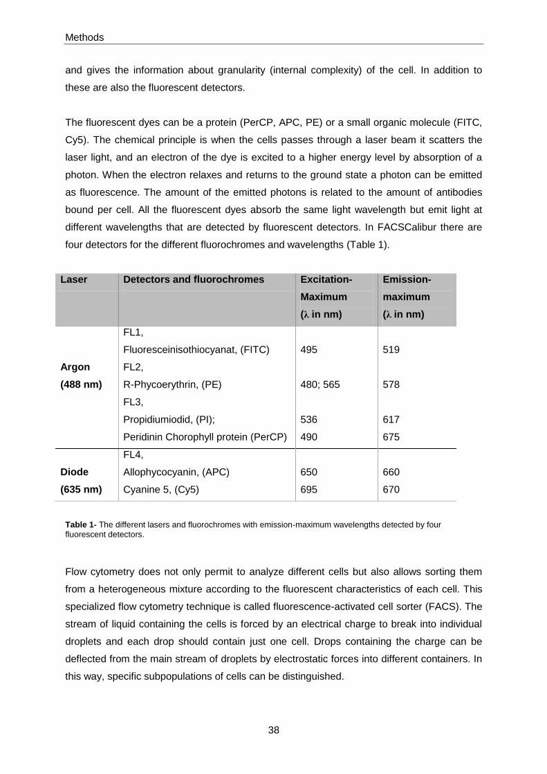
Methods
38
and gives the information about granularity (internal complexity) of the cell. In addition to
these are also the fluorescent detectors.
The fluorescent dyes can be a protein (PerCP, APC, PE) or a small organic molecule (FITC,
Cy5). The chemical principle is when the cells passes through a laser beam it scatters the
laser light, and an electron of the dye is excited to a higher energy level by absorption of a
photon. When the electron relaxes and returns to the ground state a photon can be emitted
as fluorescence. The amount of the emitted photons is related to the amount of antibodies
bound per cell. All the fluorescent dyes absorb the same light wavelength but emit light at
different wavelengths that are detected by fluorescent detectors. In FACSCalibur there are
four detectors for the different fluorochromes and wavelengths (Table 1).
Laser Detectors and fluorochromes Excitation-
Maximum
(λ in nm)
Emission-
maximum
(λ in nm)
FL1,
Fluoresceinisothiocyanat, (FITC)
495
519
Argon
(488 nm)
FL2,
R-Phycoerythrin, (PE)
480; 565
578
FL3,
Propidiumiodid, (PI);
Peridinin Chorophyll protein (PerCP)
536
490
617
675
Diode
(635 nm)
FL4,
Allophycocyanin, (APC)
Cyanine 5, (Cy5)
650
695
660
670
Table 1- The different lasers and fluorochromes with emission-maximum wavelengths detected by four
fluorescent detectors.
Flow cytometry does not only permit to analyze different cells but also allows sorting them
from a heterogeneous mixture according to the fluorescent characteristics of each cell. This
specialized flow cytometry technique is called fluorescence-activated cell sorter (FACS). The
stream of liquid containing the cells is forced by an electrical charge to break into individual
droplets and each drop should contain just one cell. Drops containing the charge can be
deflected from the main stream of droplets by electrostatic forces into different containers. In
this way, specific subpopulations of cells can be distinguished.

Methods
39
The procedure followed for Flow cytometric analyses is described below. For all the
stainings, cells from single cell suspensions were resuspended in PBS/0.2%BSA and
transferred to 96-well V-bottomed plates.
4.6.1 Cell surface staining
In order to prevent unspecific binding of mAb, cells from single cell suspension were
incubated with 30µl/ml anti-FcγReceptor blocking in PBS/0.2%BSA for 20 min at 4°C.
Afterwards samples were stained with the appropriate mAb conjugated to fluorescent dyes or
biotin to cell surface antigens for 15min at 4°C in the dark (Fluorescent dyes are light
sensitive). All the antibodies were used at a concentration determined previously usually at
1-2µg/ml. Cells were washed to remove unbound antibodies. When biotinylated mAbs were
used, another step of incubation with FITC streptavidin, APC streptavidin or PerCP
streptavidin was performed under the same conditions as described above. In order to
exclude dead cells, propidium iodide (PI) was added to the samples immediately before data
acquisition, except for the four color-stained cell samples, in FACSCalibur.
4.6.2 Intracellular staining for cytokines and CD40L
One very important method to measure cytokine production on a single cell level is by
intracellular cytokine staining. This method requires an in vitro stimulation and a blocking
substance that inhibits protein export from the cell. The cytokine thus accumulates within the
endoplasmatic reticulum (ER)/Golgi since this pathway is blocked by substances like
monensin or brefeldin A. In this work we used Heat Killed Salmonella (HKS) to induce
cytokine production by T cells in the presence of antigen specific cells (APC) and stainings
were done using the Cytofix/Cytoperm kit (BD biosciences). Splenocytes were diluted in
complete RPMI medium and 3 × 10⁶ cells were cultured in a volume of 1ml at 37°C, 5% CO2
with or without the addition of HKS (1 × 10⁸/well). After 1h when APCs had time to process
the antigen, GolgiStop was added and incubated for four hours and half. Then surface
staining was done as described before, and cells were fixed and permeabilized using the kit
solution fixation/permeabilization for 20 min at 4°C. Cells were washed twice in perm/wash
buffer and incubated in 100µl Perm/wash buffer containing the anti-cytokine mAb (2µg/ml) for
30min at 4°C. Cells were washed with Perm/Wash buffer and resuspended in 200µl
PBS/BSA for acquisition at FACSCalibur.
Methods
40
4.7 Cell purification
4.7.1 General procedures
Spleen and bone marrow were removed from naive mice and single cell suspensions were
done as described in 4.5 under sterile conditions. Afterwards, cells were purified by magnetic
cell sorting (MACS). Cell specific antigens are labelled with magnetic beads and separated
over a MACS column placed on a MACS separator. Labelled cells are retained in the column
(positive fraction) and unlabelled cells pass through the column (unlabelled fraction). The
positive fraction is then recovered when the column is detached from the magnet. An
autoMACS device was used for the automatic separation of the cells.
4.7.2 Purification of B cells
There are two methods to purify B cells by MACS, one is by depletion of CD43+ cells; this
way allows the obtention of naive B cells, since CD43+ is not expressed on conventional
peripheral resting B cells. The second method is the purification by positive selection of
CD19+ B cells and allows the isolation of mature naive and memory B cells since CD19+ is
expressed in both populations.
Spleens of naive C57BL/6, MyD88-/-, TLR9-/-, TLR2/4-/- and IL-10-/- mice were removed to
make single cell suspensions. Cells were incubated with 33µl of anti-CD19 or 30µl of anti-
CD43 Microbeads per 108 cells in 297µl or 300µl PBS/BSA respectively for 15min at 4°C,
then cells were washed and filtered. Cells were selected by autoMacs using the PosselD2
program for CD19+ sort or the program depleteS for CD43- sort. Purity was checked by
FACS staining for surface CD19+ or B220+ and was routinely above 95%.
4.7.3 Purification of Dendritic cells
Dendritic cells were selected by positive selection of CD11c+ cells. Spleens from naïve
C57BL/6 mice were removed and digested with 1mg/ml Collagenase and 0.5mg/ml DNase in
serum-free medium for 30min at 37°C. After single cell suspension, cells were incubated in
25µl of anti-CD11c Microbeads in 225µl PBS/BSA per 108 cells for 15min at 4°C, and then
cells were washed and filtered. Cells were selected by autoMacs using the PosselD2
program. Purity was checked by staining for surface CD11c+ and MHC class II and was
routinely above 95%.

Methods
41
4.8 Bone marrow derived macrophages
For the generation of BM derived macrophages, BM cells were plated in a cell culture petri
dish and incubated overnight. Therefore, differentiated cells adhere more rapidly and
precursors can be removed and plated in 10 ml of RPMI medium containing 10% FCS/ 1%
Pen-Strep, 2-ME, L-glutamine supplemented with 10% of growth factor M-CSF (produced in
supernatant of X-63 cell line) in a bacteria petri dish. At day 3 and day 6 of culture, cells were
washed in order to remove non-adherent cells and new culture medium was added. On day
7, cells were harvested, counted and cultured under stimulating conditions in 96-well plated
at 1.25x10⁵ cells per well in 50µl of RPMI.
4.9 In vitro assays
Cells were cultivated in growth medium at 37°C and 5% CO2 atmosphere saturated with
water vapour. Cell counts were calculated using a Neubauer improved counting chamber.
Dead cells were stained with 10% trypan blue in PBS solution.
4.9.1 Culture of bone marrow cells
For the analysis of non antigen-specific IgM and IgG antibodies, 8×10⁵ bone marrow cells
from naïve and infected chimera mice were cultivated in 200µl cell culture medium in 96-well
flat-bottom microtitre plates at 37°C and 5% CO2 atmosphere. Cultures were maintained for 8
days without changing the medium. At that time point, supernatants were collected for
antibody detection by ELISA.
4.9.2 Stimulation of B cells and dendritic cells
For the analysis of IL-10, IL-6 and IFN-γ production, 5×10⁵ of purified B cells and dendritic
cells were cultivated in 200µl of RPMI in 96-well flat-bottom microtitre plates at 37°C and 5%
CO2 atmosphere. Cells were stimulated with 10µg/ml and 5µg/ml of LPS and/or a Multiplicity
of infection (MOI) of 100, 50, 25, 12.5 and 6.25 of HKS. After 72 h the supernatants were
collected and cytokines were analysed by cell-based ELISA.
Methods
42
4.9.3 Assay to test the influence of supernatans from HKS activated
B cell in the stimulation of activated macrophages
For the analysis of IFN-γ and IL-6 production by HKS-activated macrophages with activated
B cell supernatants, macrophages were cultivated in 50 µl RPMI in 96-well flat-bottom
microtitre plates at 37°C and 5% CO2 atmosphere. Then, 100 µl of B cell supernatants
(obtained from stimulated B cells at MOI 1:50 of HKS as described on 4.9.2) or medium,
were added to the macrophages. After 2 hours, cells were stimulated with a Multiplicity of
infection (MOI) of 25, 12.5 and 6.25 of HKS or medium at 37°C and 5% CO2 atmosphere, 24
hours later, supernatants were collected and cytokines were analysed by ELISA. These
samples were diluted 1:5 for the ELISA.
4.10 ELISA
Enzyme-Linked Immunosorbent Assay (ELISA) is a method to detect the presence of an
antigen or an antibody in a sample. Usually at least one antibody with specificity for a
particular antigen is used to perform the ELISA. The capture antibody or an antigen is coated
to the surface of high binding 96-EIA/RIA microtitre plate. Free binding sites are blocked by
incubation with blocking buffer. The sample is added and the antigen-specific antibodies are
bounded to the plate. A separate labelled antibody that recognizes a different epitope to the
immobilized first antibody is then used to detect the bound antigen. The detection antibody
can be covalently linked to an enzyme or can itself be detected by a secondary antibody
which is linked to biotin. Biotin is then bounded by streptavidin coupled to an enzyme that
catalyses a colorimetric reaction of its substrate. In this work the detection antibodies are
linked to the enzyme alkaline phosphatase that hydrolyzes the para-nitrophenyl phosphate
(pNPP) into yellow coloured para-nitrophenol and phosphate. Para-nitrophenol absorbs light
between 405nm and 410nm and can be quantified in a photometer.
4.10.1 Cell-based IL-10 ELISA
If cytokines are of difficult detection due to a very low secretion rate by the cells or if they are
quickly consumed a very sensitive method that permits to detect cytokines in these
conditions is the Cell-based ELISA. This procedure is done in sterile conditions until the step
of the cytokine detection. High binding 96 well EIA/RIA plates were coated with 4µg/ml anti-
IL-10 mAb in carbonate buffer overnight at 4°C and blocked afterwards with PBS/3% BSA
Methods
43
buffer for 1h at 37°C. Plates were washed first in PBS/0.1%Tween and in the end in PBS to
remove any antibody not specifically bounded. After 72h of B cells and dendritic cells
stimulation as described in 4.9.2 cells were transferred into the high binding plates and
incubated overnight at 37°C and 5% CO2 atmosphere. During incubation the IL-10 present in
the culture medium and also IL-10 produced by the cells is captured immediately and
consumption is prevented. Recombinant IL-10 mAb was used as standard ranging from
100ng/ml to 0.195ng/ml. For detection 2µg/ml of biotinylated anti-IL-10 mAb in PBS/1%BSA
was added and incubated for 1h at RT. Subsequently ExtrAvidin-Alkaline phosphatase
diluted 1:10000 in PBS/1%BSA were added to the plate and incubated for 30 min at RT. All
incubations steps were followed by repeated washing with PBS/0.1%Tween. 1mg/ml of
substrate pNPP was dissolved in ELISA buffer and added to the plate. Then the
measurement was done at 405nm.
4.10.2 IFNγ and IL-6 ELISA
For IFNγ and IL-6 cytokine detection we used the cell culture supernatants after the 72h
stimulation of B cells and dendritic cells as described before. The high binding 96 well
EIA/RIA plates were coated separately with 2µg/ml anti-IFNγ mAb and/or 4µg/ml anti-IL-6
mAb in carbonate buffer overnight at 4°C and blocked afterwards with PBS/3% BSA buffer
for 1h at 37°C. Afterwards samples and recombinant IFNγ mAb and/or recombinant IL-6 mAb
used as standard ranging from 100ng/ml to 0.195ng/ml were incubated for 2h at 37°C. For
detection 1µg/ml of biotinylated anti-IFNγ mAb and/or 2µg/ml of biotinylated anti-IL-6 in
PBS/1%BSA were used and incubated for 1h at RT. Subsequently ExtrAvidin-Alkaline
phosphatase diluted 1:10000 in PBS/1%BSA were added to the plate and incubated for 30
min at RT. All incubations steps were followed by repeated washing with PBS/0.1%Tween.
The development of the plates was done as described in 4.10.1.
4.10.3 Serum antibody ELISA
This method is used to detect Salmonella-specific and non-specific antibodies in mouse
serum. High binding 96 well EIA/RIA plates were coated with 50µl of heat killed Salmonella
(HKS) (1×10⁸/ml) in PBS overnight at 4°C. After plates were washed in PBS and blocked
with PBS/1%BSA at 4°C overnight or for 3h at 37°C. Then a standard sample and serum
pre-diluted in PBS was added and two fold dilution series were performed and incubated for
2h at RT. As detection antibody an alkaline phosphatase conjugated IgM, IgG, IgG1, IgG2b,
IgG2c and IgG3 diluted 1:1000 in PBS/0.05% Tween were used and incubated for 1h at RT.
All incubations steps were followed by repeated washing with PBS/0.1%Tween followed by a

Methods
44
last wash in PBS. Further steps were as described before. The relative titre was calculated
by comparing measured samples to a standard.
To determine antibody titres (including Salmonella specific and non-specific) in serum, the
plates were coated with 1.5µg/ml anti-Ig(H+L) diluted in carbonate buffer overnight at 4°C.
The procedure was done as described before.
4.10.4 Supernatant antibody ELISA
Supernatants from bone marrow cells obtained as described in 4.9.1 were incubated for 2h
at RT in high binding 96 well EIA/RIA plates previously coated with 1.5µg/ml anti-Ig(H+L)
diluted in carbonate buffer overnight at 4°C. As detection antibody an alkaline phosphatase
conjugated IgM, IgG, IgG1, IgG2b, and IgG2c diluted 1:1000 in PBS/0.05% Tween were
used and incubated for 1h at RT. Further steps were performed as described in 4.10.3.
4.11 Histology
Organs were embedded in tissue-tek (Sakura), frozen in dry ice and kept at -80°C. Using a
cryostat, sections from the organs were cut between 5 microns and 7 microns. Sections were
left air dry and then fixed in cold acetone for 8min left them dry again for 40min and finally
stored at -20°C.
4.11.1 Staining for germinal centre B cells
Sections were defrosted and re-hydrate for 20min in PBS. After, were stained with IgM FITC
or B220 alexa 488 diluted 1:200 in PBS and PNA rhodamine diluted 1:500 in PBS for 90min
at RT in a staining box to maintain a humidified atmosphere and dark environment.
Subsequently, sections were washed three times 10min each in PBS with magnetic stirrer.
Finally sections were mounted with fluoromout G and a cover slip. During the staining,
sections were protected from light. Stained sections were analysed microscopically and
digital pictures were taken.
Methods
45
4.12 Molecular biological methods
4.12.1 Isolation and purification of RNA from splenocytes
RNA was isolated by the Tri-reagent RNA preparation method (Sigma-Aldrich). Briefly,
between 6×10⁶ and 1×10⁷ splenocytes were homogenized in 1ml Tri-Reagent by repeated
pipetting and stored at -70°C. After defrosting samples, 200µl of chloroform was added and
mixed for 15 sec and left for 15min at RT. The mixture was centrifuged at 4°C at 13000rpm
for 15min. The aqueous phase was transferred to a new tube and 500µl isopropanol was
added and left for 10min at 4°C. In order to increase the yield, 1 µl of glycogen was added as
a carrier. Then, RNA was precipitated by centrifugation for 15 min at 13000rpm at 4°C. The
RNA pellet was washed in 75% ethanol, air-dried and dissolved in RNAse free water.
4.12.2 Generation of cDNA
Initially purified RNA samples were treated with DNAse (Applied Biosystems) to eliminate
genomic DNA contamination. For this, 8µl of RNA sample (up to 1µg of RNA), 1µl of
10×DNase I buffer and 1µl of rDNAse were mixed and incubated for 30min at 37°C. The
reaction was stopped by the addition of 1µl DNase inactivation reagent followed by
centrifugation at 13000rpm for 1min at 4°C the DNase was inactivated by incubating the
mixture at 65°C for 10min. After incubation the mixture was centrifuged at 13000rpm for
2min. Later, 9µl of it were used for reverse transcription. For that 1µl oligo(t)N Primer was
added, the mixture was incubated for 10min at 55°C and then placed at RT for 15min. After,
a reaction mix containing 4µl MgCl2 (25mM), 2µl 10× Rev. Trans. Buffer, 2µl dNTP (10mM),
1µl nuclease free water, 0.5µl Rnase inhibiter and 0.5µl AMV-RT (reverse transcriptase)
were added. This mixture was incubated for 3h at 42°C and heated for 99°C for 5min to
inactivate the enzymes and cooled for 5 minutes at 4°C. Finally 30µl of water were added to
cDNA.
4.12.3 Real-time polymerase chain reaction (RT-PCR)
RT-PCR is based in the amplification and simultaneously quantification of DNA. The principle
is that the amplified DNA is quantified as it accumulates in the reaction in real time after each
amplification cycle. This quantification is done with fluorescent dyes that bind to all double-
stranded DNA, as the DNA accumulates in each cycle the fluorescence intensity increases
also. For semi-quantitative real-time PCR total RNA was extracted from organs as described
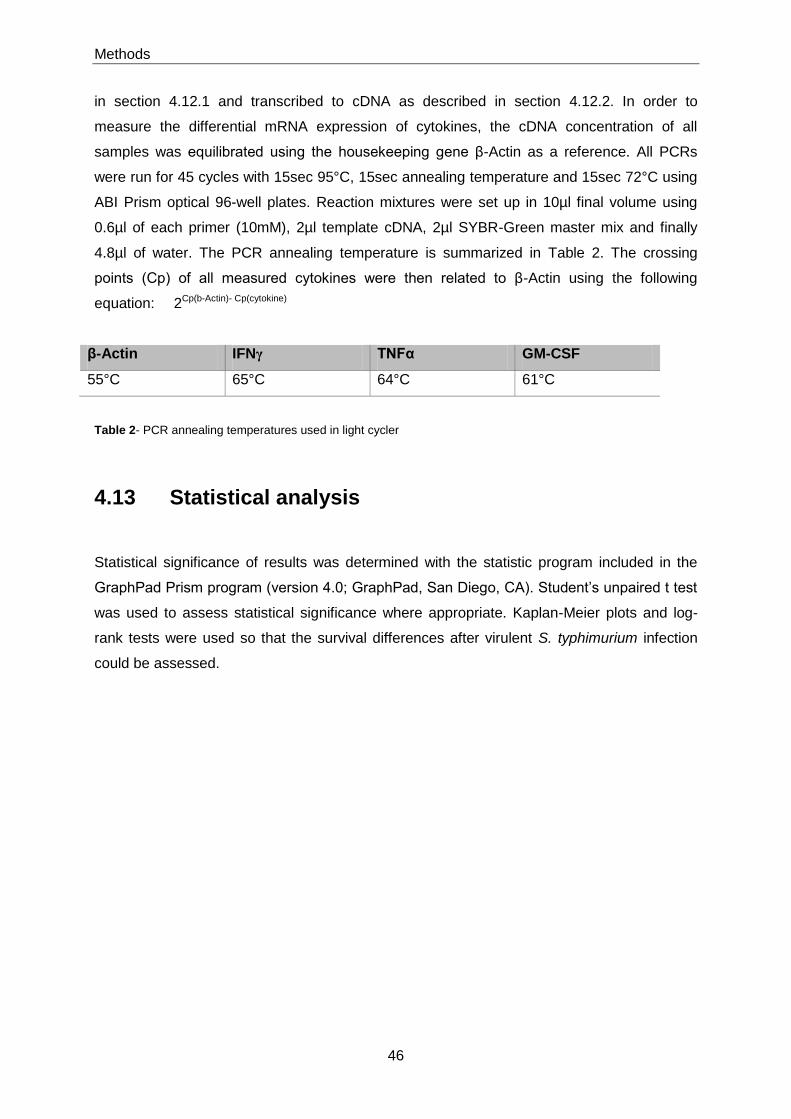
Methods
46
in section 4.12.1 and transcribed to cDNA as described in section 4.12.2. In order to
measure the differential mRNA expression of cytokines, the cDNA concentration of all
samples was equilibrated using the housekeeping gene β-Actin as a reference. All PCRs
were run for 45 cycles with 15sec 95°C, 15sec annealing temperature and 15sec 72°C using
ABI Prism optical 96-well plates. Reaction mixtures were set up in 10µl final volume using
0.6µl of each primer (10mM), 2µl template cDNA, 2µl SYBR-Green master mix and finally
4.8µl of water. The PCR annealing temperature is summarized in Table 2. The crossing
points (Cp) of all measured cytokines were then related to β-Actin using the following
equation: 2Cp(b-Actin)- Cp(cytokine)
β-Actin IFNγ TNFα GM-CSF
55°C 65°C 64°C 61°C
Table 2- PCR annealing temperatures used in light cycler
4.13 Statistical analysis
Statistical significance of results was determined with the statistic program included in the
GraphPad Prism program (version 4.0; GraphPad, San Diego, CA). Student‟s unpaired t test
was used to assess statistical significance where appropriate. Kaplan-Meier plots and log-
rank tests were used so that the survival differences after virulent S. typhimurium infection
could be assessed.

Results
47
5 Results
5.1 Effects of heat-killed Salmonella in B cells, DCs and
macrophages in vitro
5.1.1 HKS induces cytokine production by B cells
Previous studies have shown that IL-10 producing B cells are required for EAE recovery
[170]. The production of IL-10 by B cells in response to microbial products suppressed T cell
activation by inhibiting the response of DCs to TLR agonists. This was confirmed in EAE
studies showing that activation of B cells via TLR2/4 and MyD88 were required for EAE
recovery. This suggests that the TLR agonists controlling the suppressive activity of B cells
are most likely provided by components of Mycobacterium tuberculosis. That gives an idea
that microbes can control the regulatory function of B cells.
In order to investigate the role of B cells during infections, using the S. typhimurium mouse
model, it was decided to test first, whether the B cells are able to produce IL-10 and IL-6 in
vitro, upon nonviable heat-killed Salmonella (HKS) stimulation. Therefore, CD19+ B cells
were isolated from C57BL/6 and stimulated in vitro with HKS or LPS as a control (Figure 5).
Purity of B cells as checked by FACS analysis was routinely higher than 97%.
Stimulation of B cells with HKS induces the same ranges of IL-10 amount as with LPS
(Fig.5A and 5C, respectively). IL-6 and IFN-γ were also tested in order to identify if
production of pro-inflammatory cytokines by B cells will be affected during Salmonella
stimulation. It was observed that HKS triggered naive B cells to produce 0.5-0.75 ng/ml of IL-
6 (Figure 5-B) and IFN-γ production by LPS and HKS activated B cells was not observed
(data not shown). Conclusively, HKS can induce IL-10 and IL-6 by B cells in vitro.

Results
48
5.1.2 Salmonella typhimurium activates B cells through TLR2/4 and
MyD88
The microbial components of S. typhimurium provide various TLR agonists, which activate
the immune cells and induce an appropriate immune response [254].
In order to analyse the relative contributions of TLRs to the bacterial induction of IL-10 in B
cells, splenic B cells from C57BL/6 mice, TLR-9-/- deficient mice, TLR2/4 deficient mice and
MyD88 deficient mice were stimulated in vitro with nonviable S. typhimurium (Figure 6). HKS
triggers IL-10 production by B cells via TLR-2/4 and MyD88 showing that the principal TLR
agonists in B cells of S. typhimurium are LPS, lipoproteins and other PAMPs, excluding the
bacterial DNA, which is a TLR-9 agonist. This observation is not surprising, since Weiss et al.
showed that TLR4, TLR2 and MyD88 are involved in host defence against Salmonella
typhimurium [51].
Figure 5: HKS and LPS induce IL-10 production in B cells. B cells
were isolated from splenocytes of naïve C57/6 mice by positive selection of CD19
+ cells and stimulated in vitro. A) and B), B cells stimulated with
HKS. C), and D), B cells stimulated with LPS. IL-10 and IL-6 production were measured by cell-based ELISA at day 4. Error bar, SEM.
B A
C D
50.00 25.00 12.50 0.000.00
0.25
0.50
0.75
MOI
IL-6
ng
/ml
10 5 00.0
0.5
1.0
1.5
2.0
2.5
g/ml
IL1
0n
g/m
l
10 5 00.00
0.25
0.50
0.75
1.00
1.25
1.50
1.75
g/ml
IL-6
ng
/ml
50.00 25.00 12.50 0.000.0
0.5
1.0
1.5
2.0
2.5
MOI
IL1
0 n
g/m
l

Results
49
5.1.3 HKS induces cytokine production by dendritic cells
In previous studies it was reported that B cells and DCs provide cytokine environments with
different impact on T cell activation following TLR stimulation. Supernatants taken from LPS-
activated B cells repress T cell activation by DC. In contrast, supernatants from TLR
activated DC lead to a stimulation of T cell response [54]. Therefore, DCs and B cells appear
to provide different cytokine milieu upon TLR-triggering.
To further investigate the role of DCs upon HKS stimulation, splenic DCs were isolated with a
purity superior to 97% from C57BL/6 mice and stimulated with HKS or LPS as control (Figure
7). HKS and LPS trigger IL-6 and IFN-γ by DCs but not IL-10. DCs produce 8 - 8.6 ng/ml of
IFN-γ upon HKS stimulation, which is higher compared to the LPS stimulation. IL-6
production by DCs reaches values of 1.5 – 2.5 ng/ml upon HKS stimulation. These
observations show that DCs after HKS stimulation are able to produce high amounts of
inflammatory cytokines.
Figure 6: HKS induces IL-10 production by naïve B cells via TLR2/4 and MyD88.
Splenic B cells from C57BL/6 mice (black bars), TLR-9-/- mice (dark gray bars), TLR-
2/4-/- mice (light gray bars) and MyD88
-/- mice (white bars) were sitmulated with HKS
for 3 days, and IL-10 was determinate at day 4 by cell-based Elisa. Error bar, SEM.
100 50 25 12,5 6,25 00
1
2
3
4C57BL/6
TLR9-/-
MyD88-/-
TLR2/4-/-
MOI
IL1
0 n
g/m
l
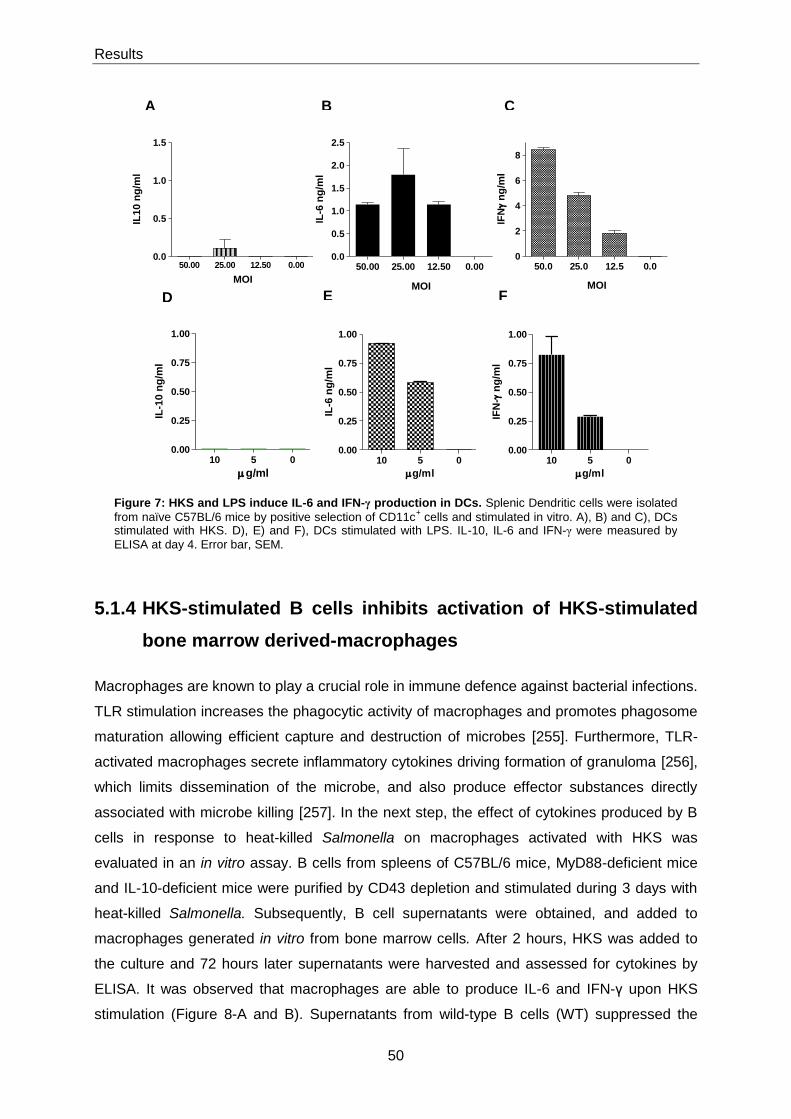
Results
50
5.1.4 HKS-stimulated B cells inhibits activation of HKS-stimulated
bone marrow derived-macrophages
Macrophages are known to play a crucial role in immune defence against bacterial infections.
TLR stimulation increases the phagocytic activity of macrophages and promotes phagosome
maturation allowing efficient capture and destruction of microbes [255]. Furthermore, TLR-
activated macrophages secrete inflammatory cytokines driving formation of granuloma [256],
which limits dissemination of the microbe, and also produce effector substances directly
associated with microbe killing [257]. In the next step, the effect of cytokines produced by B
cells in response to heat-killed Salmonella on macrophages activated with HKS was
evaluated in an in vitro assay. B cells from spleens of C57BL/6 mice, MyD88-deficient mice
and IL-10-deficient mice were purified by CD43 depletion and stimulated during 3 days with
heat-killed Salmonella. Subsequently, B cell supernatants were obtained, and added to
macrophages generated in vitro from bone marrow cells. After 2 hours, HKS was added to
the culture and 72 hours later supernatants were harvested and assessed for cytokines by
ELISA. It was observed that macrophages are able to produce IL-6 and IFN-γ upon HKS
stimulation (Figure 8-A and B). Supernatants from wild-type B cells (WT) suppressed the
Figure 7: HKS and LPS induce IL-6 and IFN-γ production in DCs. Splenic Dendritic cells were isolated
from naïve C57BL/6 mice by positive selection of CD11c+ cells and stimulated in vitro. A), B) and C), DCs
stimulated with HKS. D), E) and F), DCs stimulated with LPS. IL-10, IL-6 and IFN-γ were measured by
ELISA at day 4. Error bar, SEM.
B A C
D E F
50.00 25.00 12.50 0.000.0
0.5
1.0
1.5
MOI
IL10 n
g/m
l
50.00 25.00 12.50 0.000.0
0.5
1.0
1.5
2.0
2.5
MOI
IL-6
ng
/ml
50.0 25.0 12.5 0.00
2
4
6
8
MOI
IFN
ng
/ml
10 5 00.00
0.25
0.50
0.75
1.00
g/ml
IL-1
0n
g/m
l
10 5 00.00
0.25
0.50
0.75
1.00
g/ml
IFN
- n
g/m
l
10 5 00.00
0.25
0.50
0.75
1.00
g/ml
IL-6
ng
/ml

Results
51
secretion of IL-6 by bone marrow derived-macrophages, and this was also observed with
supernatants from MyD88-deficient B cells (MyD88-/-) although to a lesser extent (Figure 8-A
and B). WT-B cell supernatants suppressed almost 2 fold more the amount of IL-6 secreted
by HKS-activated macrophages compared to MyD88-deficient B cell supernatants (Figure 8-
A). In the case of IFN-γ, MyD88-deficient supernatants were found to further increase its
production, whereas WT-supernatants had no effect (Figure 8-B). According to these results
it seems that the suppression is not solely MyD88-dependent. In order to investigate if
suppression of macrophages is due to IL-10 production by B cells, B cells from IL-10-/- mice
were also tested. The amount of IL-6 production by bone marrow derived-macrophages with
IL-10-/- B cell supernatants was similar to the MyD88-deficient B cell supernatants (Figure 8-
A). IFN-γ production by macrophages was higher with IL-10-/- B cell supernatants than the
MyD88-/- B cells supernatants (Figure 8-B).
This suggests that the increased IFN-γ production by macrophages with IL-10-/-- B cell
supernatants can be due to the presence of IFN-γ in IL-10-/- B cell supernatants. Since, IL-10-
/- B cell supernatants had higher amounts of IFN-γ compared to the other strains (Figure 8-C
and D). The high levels of IL-6 and IFN-γ in IL-10-/- B cell supernatants are also not
surprising, considering that the signal transduction pathway by MyD88 it is not blocked
compare to the MyD88 deficient B cell supernatants. Myeloid differentiation factor 88
(MyD88) is critical for TLR-mediated activation of the transcription factor NF-κB and hence
the induction of pro-inflammatory cytokines [258].
Conclusively, HKS-activated B cells are able to suppress HKS-activated bone marrow
derived-macrophages. This suppression can be due to IL-10 most likely via MyD88-
dependent, but also a strong component of suppression is MyD88-independent. Possibly
other anti-inflammatory cytokines are also produced by B cells. As reported before, LPS-
activated B cells are able to produce TGF-β, which is an anti-inflammatory cytokine [259].
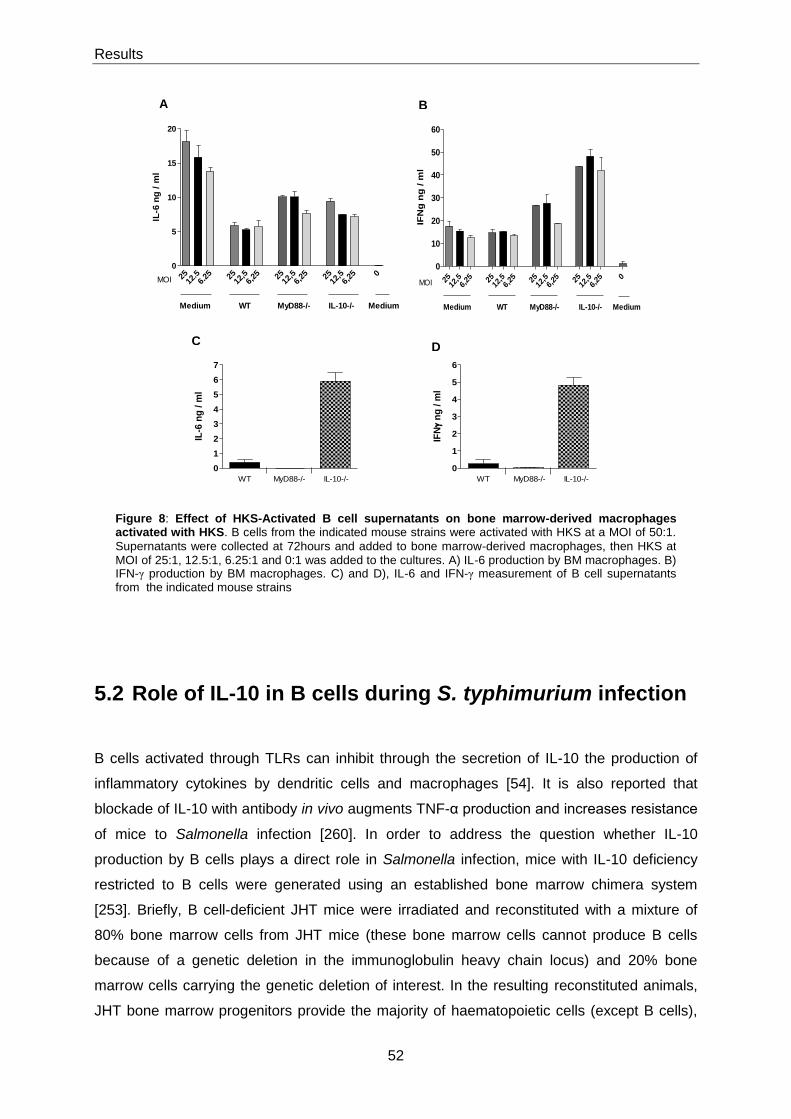
Results
52
5.2 Role of IL-10 in B cells during S. typhimurium infection
B cells activated through TLRs can inhibit through the secretion of IL-10 the production of
inflammatory cytokines by dendritic cells and macrophages [54]. It is also reported that
blockade of IL-10 with antibody in vivo augments TNF-α production and increases resistance
of mice to Salmonella infection [260]. In order to address the question whether IL-10
production by B cells plays a direct role in Salmonella infection, mice with IL-10 deficiency
restricted to B cells were generated using an established bone marrow chimera system
[253]. Briefly, B cell-deficient JHT mice were irradiated and reconstituted with a mixture of
80% bone marrow cells from JHT mice (these bone marrow cells cannot produce B cells
because of a genetic deletion in the immunoglobulin heavy chain locus) and 20% bone
marrow cells carrying the genetic deletion of interest. In the resulting reconstituted animals,
JHT bone marrow progenitors provide the majority of haematopoietic cells (except B cells),
B A
C D
0
1
2
3
4
5
6
7
WT IL-10-/-MyD88-/-
IL-6
ng
/ m
l
0
1
2
3
4
5
6
WT IL-10-/-MyD88-/-
IFN
ng
/ m
l
2512
.56.
25 2512
,56,
25 2512
,56,
25 2512
,56,
25 00
5
10
15
20
WT IL-10-/-MyD88-/- Medium
MOI
IL-6
ng
/ m
l
Medium
2512
,56,25 25
12,5
6,25 25
12,5
6,25 25
12,5
6,25 0
0
10
20
30
40
50
60
WT IL-10-/-MyD88-/- Medium
MOI
IFN
g n
g / m
l
Medium
Figure 8: Effect of HKS-Activated B cell supernatants on bone marrow-derived macrophages activated with HKS. B cells from the indicated mouse strains were activated with HKS at a MOI of 50:1.
Supernatants were collected at 72hours and added to bone marrow-derived macrophages, then HKS at MOI of 25:1, 12.5:1, 6.25:1 and 0:1 was added to the cultures. A) IL-6 production by BM macrophages. B) IFN-γ production by BM macrophages. C) and D), IL-6 and IFN-γ measurement of B cell supernatants from the indicated mouse strains
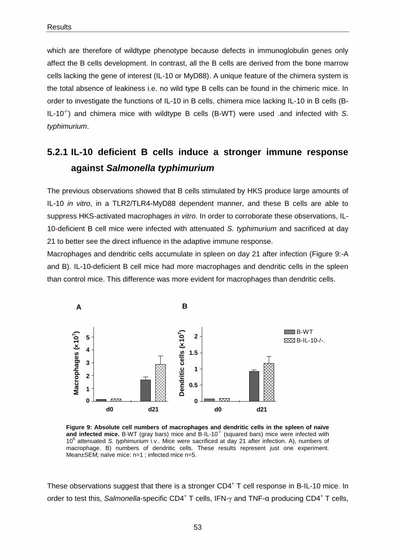
Results
53
which are therefore of wildtype phenotype because defects in immunoglobulin genes only
affect the B cells development. In contrast, all the B cells are derived from the bone marrow
cells lacking the gene of interest (IL-10 or MyD88). A unique feature of the chimera system is
the total absence of leakiness i.e. no wild type B cells can be found in the chimeric mice. In
order to investigate the functions of IL-10 in B cells, chimera mice lacking IL-10 in B cells (B-
IL-10-/-) and chimera mice with wildtype B cells (B-WT) were used .and infected with S.
typhimurium.
5.2.1 IL-10 deficient B cells induce a stronger immune response
against Salmonella typhimurium
The previous observations showed that B cells stimulated by HKS produce large amounts of
IL-10 in vitro, in a TLR2/TLR4-MyD88 dependent manner, and these B cells are able to
suppress HKS-activated macrophages in vitro. In order to corroborate these observations, IL-
10-deficient B cell mice were infected with attenuated S. typhimurium and sacrificed at day
21 to better see the direct influence in the adaptive immune response.
Macrophages and dendritic cells accumulate in spleen on day 21 after infection (Figure 9:-A
and B). IL-10-deficient B cell mice had more macrophages and dendritic cells in the spleen
than control mice. This difference was more evident for macrophages than dendritic cells.
These observations suggest that there is a stronger CD4+ T cell response in B-IL-10 mice. In
order to test this, Salmonella-specific CD4+ T cells, IFN-γ and TNF-α producing CD4+ T cells,
Figure 9: Absolute cell numbers of macrophages and dendritic cells in the spleen of naïve and infected mice. B-WT (gray bars) mice and B-IL-10
-/- (squared bars) mice were infected with
106 attenuated S. typhimurium i.v.. Mice were sacrificed at day 21 after infection. A), numbers of
macrophage. B) numbers of dendritic cells. These results represent just one experiment. Mean±SEM; naïve mice: n=1 ; infected mice n=5.
d0 d21
0.5
2
0
1
1.5
B-WT
B-IL-10-/-.
De
nd
riti
c c
ell
s(
10
7)
A B
d0 d21
1
3
4
5
0
2
Macro
ph
ag
es
(10
7)

Results
54
were determined in naive and infected mice by flow cytometry after a short ex vivo re-
stimulation of splenocytes with HKS. Salmonella-specific CD4+ T cells were identified by co-
expression of CD4 and CD154, which is a marker for antigen-reactive helper CD4 T cells
[261, 262]. According to this technology Salmonella-reactive CD4+ T cells could be detected
on day 21 after infection, showing a clearly stronger accumulation in B-IL-10-/- mice
compared to control mice (Figure 10-A). As expected, IL-10 in B cells reduces the
inflammatory response against Salmonella infection (Figure 10-B and C). However, it was
not possible to detect any differences in the bacterial numbers (data not shown). B-IL-10-/-
mice had 2-fold more IFN-γ and TNF-α producing CD4+ T cells than controls (Figure 10-B).
These observations show for the first time that IL-10 in B cells inhibits an adaptive CD4+ T
cell response in an infection model using Salmonella typhimurium.
Figure 10: Numbers of Salmonella -specific CD4+ T cells (A) and IFN-γ (B) and
TNF-α (C) production by CD4+ T cells in the spleen of naïve and infected mice. B-
WT (gray bars) mice and B-IL-10-/-
(squared bars) mice were infected with 106
attenuated S. typhimurium i.v.. Mice were sacrificed at day 21 after infection. These results represent just one experiment. Mean±SEM; naïve mice: n=1 ; infected mice n=5. (* for p<0.05 and ** for p<0.01).
B
A
C
0.5
0
1
4
7
unst. HKS
d0 d21
HKS
*10B-WT
B-IL-10-/-
CD
40L
+C
D4 T
cells (
10
5)
unst. HKS HKS
d0 d21
0.2
0
0.3
20
40
60
80**
IFN
-+C
D4 T
cells (
10
5)
unst. HKS HKS
d0 d21
0.2
0
0.4
0.6
3.1
**
TN
F-
+C
D4 T
cells (
10
6)
Results
55
5.3 Role of MyD88-signalling in B cells during Salmonella
typhimurium infection
It has been demonstrated that B cells activated by microbial products through MyD88 induce
IL-10 production, and could inhibit T cell-mediated immune responses [54]. In this work it has
been observed that B cells produce IL-10 through TLR/MyD88 activation using heat-killed
Salmonella and that IL-10 production by B cells inhibits CD4+ T cell response during
Salmonella infection in vivo. Therefore, to assess the effect of MyD88 signalling in B cells
during Salmonella infection, mice with MyD88 deficiency restricted to B cells were generated
using the established bone marrow chimera system [253] explained previously. In order to
investigate the functions of MyD88 in B cells, control C57BL/6, MyD88-deficient mice,
chimera mice lacking MyD88 in B cells (B-MyD88-/-) and chimera mice with wildtype B cells
(B-WT) were used and infected with S. typhimurium.
5.3.1 Course of S. typhimurium infection in MyD88-B cell deficient
mice
B-MyD88-/- and B-WT chimera mice were infected via the intravenous route with 1×10⁶ S.
typhimurium SL7207. This route of infection mimics the systemic phase of disease.
In order to have a complete understanding of the different stages of the immune response to
S. typhimurium, previously clarified on section 1.3.1, the infection course was monitored at
several time points (Figure 11) of disease.
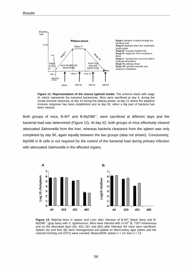
Results
56
Both groups of mice, B-WT and B-MyD88-/-, were sacrificed at different days and the
bacterial load was determined (Figure 12). At day 62, both groups of mice effectively cleared
attenuated Salmonella from the liver, whereas bacteria clearance from the spleen was only
completed by day 90, again equally between the two groups (data not shown). Conclusively,
MyD88 in B cells is not required for the control of the bacterial load during primary infection
with attenuated Salmonella in the affected organs.
Figure 12: Baterial titres in spleen and Liver after infection of B-WT (black bars) and B-
MyD88-/-
(gray bars) with S. typhimurium. Mice were infected with 1×10⁶ SL 7207 intravenous
and on the described days (d4, d10, d21 and d62) after infection the mice were sacrificed. Spleen (A) and liver (B) were homogenized and plated on MacConkey agar plates and the colonies forming unit (CFU) were counted. Mean±SEM; spleen n > 14; liver n > 13.
Figure 11: Representation of the mouse typhoid model. The scheme starts with stage
III, which represents the transient bacteremia. Mice were sacrificed at day 4, during the innate immune response, at day 10 during the plateau phase, at day 21 where the adaptive immune response has been established and at day 62, when a big part of bacteria has been cleared.
B A
0
1
2
3
4
5
6
7
d4 d10 d21 d62
Lo
g 1
0 c
fu/s
ple
en
0
1
2
3
4
5
6
7
d4 d10 d21 d62
Lo
g1
0 c
fu/l
ive
r
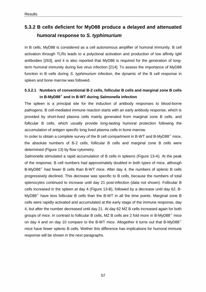
Results
57
5.3.2 B cells deficient for MyD88 produce a delayed and attenuated
humoral response to S. typhimurium
In B cells, MyD88 is considered as a cell autonomous amplifier of humoral immunity. B cell
activation through TLRs leads to a polyclonal activation and production of low affinity IgM
antibodies [263], and it is also reported that MyD88 is required for the generation of long-
term humoral immunity during live virus infection [214]. To assess the importance of MyD88
function in B cells during S. typhimurium infection, the dynamic of the B cell response in
spleen and bone marrow was followed.
5.3.2.1 Numbers of conventional B-2 cells, follicular B cells and marginal zone B cells
in B-MyD88-/- and in B-WT during Salmonella infection
The spleen is a principal site for the induction of antibody responses to blood-borne
pathogens. B cell-mediated immune reaction starts with an early antibody response, which is
provided by short-lived plasma cells mainly generated from marginal zone B cells, and
follicular B cells, which usually provide long-lasting humoral protection following the
accumulation of antigen specific long lived plasma cells in bone marrow.
In order to obtain a complete survey of the B cell compartment in B-WT and B-MyD88-/- mice,
the absolute numbers of B-2 cells, follicular B cells and marginal zone B cells were
determined (Figure 13) by flow cytometry.
Salmonella stimulated a rapid accumulation of B cells in spleens (Figure 13-A). At the peak
of the response, B cell numbers had approximately doubled in both types of mice, although
B-MyD88-/- had fewer B cells than B-WT mice. After day 4, the numbers of splenic B cells
progressively declined. This decrease was specific to B cells, because the numbers of total
splenocytes continued to increase until day 21 post-infection (data not shown). Follicular B
cells increased in the spleen at day 4 (Figure 13-B), followed by a decrease until day 62. B-
MyD88-/- have less follicular B cells than the B-WT in all the time points. Marginal zone B
cells were rapidly activated and accumulated at the early stage of the immune response, day
4, but after the number decreased until day 21. At day 62 MZ B cells increased again for both
groups of mice. In contrast to follicular B cells, MZ B cells are 2 fold more in B-MyD88-/- mice
on day 4 and on day 10 compare to the B-WT mice. Altogether it turns out that B-MyD88-/-
mice have fewer splenic B cells. Wether this difference has implications for humoral immune
response will be shown in the next paragraphs.
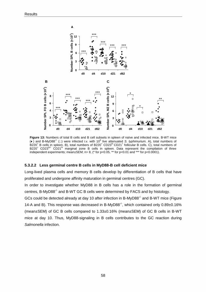
Results
58
5.3.2.2 Less germinal centre B cells in MyD88-B cell deficient mice
Long-lived plasma cells and memory B cells develop by differentiation of B cells that have
proliferated and undergone affinity maturation in germinal centres (GC).
In order to investigate whether MyD88 in B cells has a role in the formation of germinal
centres, B-MyD88-/- and B-WT GC B cells were determined by FACS and by histology.
GCs could be detected already at day 10 after infection in B-MyD88-/- and B-WT mice (Figure
14-A and B). This response was decreased in B-MyD88-/-, which contained only 0.89±0.16%
(mean±SEM) of GC B cells compared to 1.33±0.16% (mean±SEM) of GC B cells in B-WT
mice at day 10. Thus, MyD88-signaling in B cells contributes to the GC reaction during
Salmonella infection.
Figure 13: Numbers of total B cells and B cell subsets in spleen of naïve and infected mice. B-WT mice
() and B-MyD88-/-
() were infected i.v. with 106 live attenuated S. typhimurium. A), total numbers of
B220+ B cells in spleen. B), total numbers of B220
+ CD23
hi CD21
+ follicular B cells. C), total numbers of
B220+ CD23
lo/- CD21
hi marginal zone B cells in spleen. Data represent the compilation of three
independent experiments; mean±SEM; n> 8, (* for p<0.05, ** for p<0.01 and *** for p<0.0001).
d0 d4 d10 d21 d62
9
6
3
0
12
*********
***
***
Nu
mb
er
SP
LB
cells
(10
7)
B
A
C
6
4
3
0
8
d0 d4 d10 d21 d62
***
******
***
***
Nu
mb
er
SP
LF
O B
ce
lls
(10
7)
9
6
3
0
12
d0 d4 d10 d21 d62
*
**
*N
um
be
r S
PL
MZ
Bcells
(10
6)

Results
59
5.3.2.3 MyD88 in B cells amplifies humoral immune response
After an immune challenge, some activated B cells differentiate into short-lived plasma cells
mostly secreting IgM antibodies that supply an early layer of humoral protection. The
mechanisms involved in this response are not yet fully discovered. In B-WT mice, we
observed a striking accumulation of MHC-IIint CD138+ plasma cells at day 4 after infection.
The number of these plasma cells then progressively decreased over time (Figure 15-B). The
kinetic of this response was altered in B-MyD88-/- mice, in which the number of plasma cells
was significantly reduced at day 4. It then continued to increase until day 10 when it reached
control levels. Thus, MyD88 functions in B cells act as an accelerator and an amplifier of the
early plasma cell response. This may be particularly important for protection from infections
primarily controlled by antibodies. Regarding to the previous results, GC B cells were
decreased in MyD88-deficient B cell mice, which suggests that MyD88 potentiates B cell
activation at an early stage, possibly at the initiation of the B cell priming i.e. before the cell
fate decision to become either a short-lived plasma blast or a GC B cell has been made.
Figure 14: Reduced germinal centre B cells in B-MyD88-/-
mice. A), Representative staining for
splenic germinal centre B cells of infected mice on day 10. B), Frequency of B220+ GL7
+ Fas
+ germinal
centre B cells of B-WT () and B-MyD88-/-
() infected mice with 106 attenuated S. typhimurium in
spleen. C), Representative staining of germinal centres from splenic sections prepared 10 days after immunization by immunofluorescence staining with PNA (red) and IgM Alexa 488 (green). Graph B), represents the compilation of three independent experiments; mean±SEM; n> 8.
0
1
2
3
4
d4 d10 d21 d62
% o
f G
C B
ce
lls
(Gate
d o
n B
22
0+ B
cells
)
A
C
B

Results
60
Antibodies are important for protection from Salmonella particularly during the early stages of
infection, while they have little effects at later phases when the bacteria are already
intracellular [264]. The kinetics of the specific antibody response in B-WT and B-MyD88 were
followed. The natural amounts of IgM before immunization as well as levels of Salmonella
reactive IgM antibodies were significantly reduced in B-MyD88-/- mice (Figure 16-A and B).
These mice also showed a delayed Salmonella-specific IgM response, which took 3 weeks to
reach control levels (Figure 16-A). This may be due to the impairment of their early immune
response in spleen (Figure 15-B). The Salmonella-specific IgG response became detectable
around two weeks after infection, and it was also delayed in B-MyD88-/- mice (Figure 16-C).
Beyond these initial differences, the 2 types of mice had similar titres of Salmonella-specific
IgM and IgG antibodies from day 30 to day 62. The analysis of the different IgG isotypes
revealed an enhanced Salmonella-specific IgG1 response in B-MyD88-/- mice (Figure 16-F),
while the IgG2b and IgG2c titres were similar to controls. In contrast, B-MyD88-/- mice
produced less Salmonella-specific IgG3 (Figure 16-G).
Figure 15: Reduction of plasma cells in B-MyD88-/-
mice in early response to Salmonella. A),
Representative staining for frequency of plasma cells in spleen at day 4. B), Total numbers of MHC-IIint
CD138+ plasma cells of naïve and infected B-WT () and B-MyD88
-/- () mice with 10
6 attenuated S.
typhimurium in spleen. mean±SEM; n> 8, (* for p<0.05, ** for p<0.01 and *** for p<0.0001).
3
2
1
0
4
5
d0 d4 d10 d21 d62
***
***
****
*
Nu
mb
er
SP
Lp
las
ma
cells
(10
6)
B A
<
<
<
<
<
<
<
<
<

Results
61
Conclusively, MyD88 signalling in B cells was not strictly required for any of the isotypes
investigated. MyD88 acts as a cell autonomous amplifier for all the facets of the B cell
Figure 16: Relative titers of serum from naïve and infected B-WT (Black Bars) mice and B-MyD88-/-
(gray bars) mice. Mice infected with 10
6 attenuated Salmonella SL 7207and serum levels were determined at
day 7, 14, 21, 30, 40, 50 and 62. A), Natural IgM antibodies. B), C), D), E) and G) Salmonella -specific IgM, IgG, IgG2b, IgG2c, IgG1 and IgG3 respectively. Data represent the compilation of 3 independent experiments. Mean±SEM, (*** for p<0.0001).
0
100
200
300
400
B-WT B-MyD88-/-
Rela
tive t
iters
A
<
<
<
<
<
<
<
<
<
Natural IgM
0
100
200
300
d0 d7 d14 d21 d30 d40 d50 d62
***
rela
tiv
e t
ite
rs
B IgG
C
0
100
200
d0 d7 d14 d21 d30 d40 d50 d62
rela
tiv
e t
ite
rs
IgM
0
100
200
300
400
500
600
700
d0 d7 d14 d21 d30 d40 d50 d62
rela
tive t
iters
E
IgG2c
IgG1
F
0
250
500
750
1000
1250
1500
d0 d7 d14 d21 d30 d40 d50 d62
rela
tiv
e t
ite
rs
G
IgG3
0
250
500
750
1000
1250
1500
d0 d7 d14 d21 d30 d40 d50 d62
rela
tive t
iters
D
0
100
200
300
d0 d7 d14 d21 d30 d40 d50 d62
rela
tiv
e t
ite
rs
IgG2b
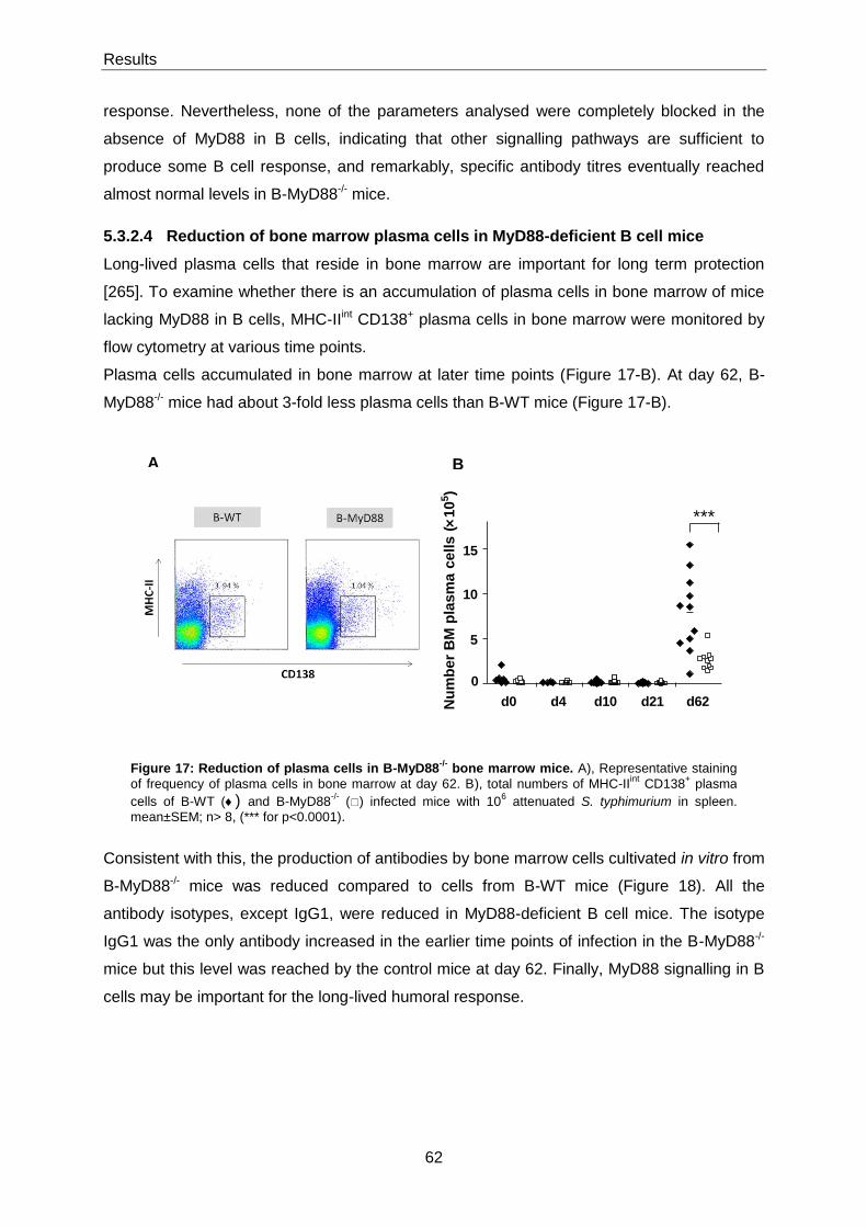
Results
62
response. Nevertheless, none of the parameters analysed were completely blocked in the
absence of MyD88 in B cells, indicating that other signalling pathways are sufficient to
produce some B cell response, and remarkably, specific antibody titres eventually reached
almost normal levels in B-MyD88-/- mice.
5.3.2.4 Reduction of bone marrow plasma cells in MyD88-deficient B cell mice
Long-lived plasma cells that reside in bone marrow are important for long term protection
[265]. To examine whether there is an accumulation of plasma cells in bone marrow of mice
lacking MyD88 in B cells, MHC-IIint CD138+ plasma cells in bone marrow were monitored by
flow cytometry at various time points.
Plasma cells accumulated in bone marrow at later time points (Figure 17-B). At day 62, B-
MyD88-/- mice had about 3-fold less plasma cells than B-WT mice (Figure 17-B).
Consistent with this, the production of antibodies by bone marrow cells cultivated in vitro from
B-MyD88-/- mice was reduced compared to cells from B-WT mice (Figure 18). All the
antibody isotypes, except IgG1, were reduced in MyD88-deficient B cell mice. The isotype
IgG1 was the only antibody increased in the earlier time points of infection in the B-MyD88-/-
mice but this level was reached by the control mice at day 62. Finally, MyD88 signalling in B
cells may be important for the long-lived humoral response.
Figure 17: Reduction of plasma cells in B-MyD88-/-
bone marrow mice. A), Representative staining
of frequency of plasma cells in bone marrow at day 62. B), total numbers of MHC-IIint
CD138+ plasma
cells of B-WT () and B-MyD88-/-
() infected mice with 106 attenuated S. typhimurium in spleen.
mean±SEM; n> 8, (*** for p<0.0001).
10
5
15
0
d0 d4 d10 d21 d62
***
Nu
mb
er
BM
pla
sm
ac
ell
s(
10
5)
B A

Results
63
5.3.3 Dual roles of MyD88 in innate immunity
In previous studies it was shown that MyD88-activated B cells produce cytokines such as IL-
10 that can inhibit DC in vitro [54]. This suggested that MyD88-signalling in B cells could
suppress the innate immune response to Salmonella.
Neutrophils are the first cells recruited at sites of infection. In spleen, Gr-1high MHC-II-
neutrophils increased until day 21 for both chimera mice after infection (Figure 19-A).
Notably, the early phase of this response was significantly amplified in B-MyD88-/- mice,
which had more neutrophils than B-WT mice at day 4 (Figure 19-A). This tendency was still
evident at day 10 but no longer statistically significant. Similar results were obtained using
CD11b and Gr-1 to identify neutrophils (data not shown). This elevated response was not
due to differences in bacterial burdens, because they were similar in both types of mice
(Figure 12). Therefore, MyD88-signalling in B cells inhibits the accumulation of neutrophil in
infected spleens. In contrast, the recruitment of neutrophils was severely impaired at day 4 in
MyD88-deficient mice. However it reached control levels at day 21 after infection (Figure 19-
Figure 18: Relative titers of antibodies in bone marrow supernatants from naïve and infected B-WT
(black bars) mice and B-MyD88-/-
(gray bars) mice. The mice were infected with 106 attenuated
Salmonella SL7207and sacrificed at day 10, day 21 and day 62. Antibody levels were measured in bone marrow supernatants from cultured bone marrow cells. A), B), C), D), and E) Non antigen-specific IgM, IgG, IgG1, IgG2b and IgG2c respectively. Data represent the compilation of 3 independent experiments. Mean±SEM, (* for p<0.05, ** for p<0.01 and *** for p<0.0001).
0
50
100
150
200
d0 d10 d21 d62
**
rela
tive t
iters
0
5
10
15
20
25
30
35
40
45
d0 d10 d21 d62
*
*
rela
tive t
iters
0
10
20
30
40
50
60
70
80
90
d0 d10 d21 d62
* r
ela
tive t
iters
0
10
20
30
40
50
d0 d10 d21 d62
rela
tiv
e t
iters
***
B A
D C E
0
5
10
15
20
25
30
35
40
45
d0 d10 d21 d62
re
lati
ve
tit
ers
IgG IgM
IgG2c IgG2b IgG1
Results
64
B). This reduced response in MyD88-deficient mice was visible in higher bacterial burden in
the spleen (data not shown). Together, these results suggest a dual role for MyD88 in the
neutrophil response: MyD88-signalling in some cells promotes the accumulation of neutrophil
at infected sites, but MyD88-signalling in B cells specifically antagonizes this activity.
The accumulation of macrophages at infected sites is required to stop the expansion of
Salmonella [266, 267]. Interestingly, both groups of chimera mice show an enlarged splenic
macrophage compartment already at day 4 after infection (Figure 19-C). Moreover, B-
MyD88-/- mice contained significantly more splenic macrophages than B-WT mice on day 10
and on day 21, indicating that MyD88-signalling in B cells inhibits the accumulation of
macrophages in infected spleens. In contrast, the macrophage response was strongly
impaired at day 4 in MyD88-deficient mice, although it reached control levels at day 21
(Figure 19-D). Consequently, MyD88-signalling in some cells controls the accumulation of
macrophages at infected sites, but MyD88-signalling in B cells limits this innate immune
response.
NK cells play an important role in immune defence against Salmonella [268, 269]. Among
other functions, NK cells are considered to be as the major source of.macrophage-activating
cytokine IFN-γ. Given the accumulation of macrophages in spleen it was therefore important
to investigate the IFN-γ production by NK cells. Salmonella stimulated a rapid IFN-γ
production by NK cells at day 4 (Figure 19-E). Remarkably, B-MyD88-/- cell deficient mice
had significantly more IFN-γ-producing NK cells than control mice on day 4 (Figure 19-E),
while the total numbers of NK cells were comparable (data not shown). This result, suggests
that MyD88-signalling in B cells strongly inhibits the production of IFN-γ by NK cells. In
MyD88-deficient mice, the NK cell response was almost completely impaired at day 4 and
day 21. This suggests that MyD88-signalling in some cells abrogates NK cell activation, while
MyD88-signalling in B cells reduces NK cell activation possibly by producing inhibitory
signals via B cells.

Results
65
Figure 19: Mice in which B cells lack MyD88 have an increased innate immune response contrary
to the MyD88-deficient B cell mice. B-MyD88-/-
() and B-WT () chimera mice, and C57BL/6 () and
MyD88-/-
() mice were infected with 106 live attenuated S. typhimurium i.v.. A) and B), Total numbers of
neutrophils in spleen. C) and D), Total numbers of splenic CD11bbright
macrophages. E) and F), Total numbers of IFN-γ
+ producing NK cells, splenic cells were stained for DX5 and TCR-β. IFN-γ production
was determined by intracellular cytokine analysis after short-term culture with and without HKS. Mean±SEM, n > 9, (* for p<0.05, ** for p<0.01 and *** for p<0.0001).
A B
C D
E F
d0 d4 d21
0
0.5
1.5
1
***
Neu
tro
ph
ils
(
10
7)
d0 d4 d10 d21
0
0.5
1.5
2
2.5
1
**
Neu
tro
ph
ils (
10
7)
d0 d4 d21
0
1
2
3
***
Ma
cro
ph
ag
es
(
10
7)
0
1
3
4
5
6
2
d0 d4 d10 d21
*
*
Macro
ph
ag
es
(10
7)
d0 d4 d21
unst. unst. unst. HKS HKS HKS
0
1
2
3
4 *
IFN
- p
rod
uc
ing
NK
cell
s (
10
5)
unst. unst. unst. HKS HKS HKS
d0 d4 d21
0
1
2
*** ***
IFN
- p
rod
uc
ing
NK
ce
lls
(
10
5)

Results
66
5.3.3.1 The role of MyD88 in B cells in dendritic cell numbers
Dendritic cells are critical for both innate and adaptive immunity. They are able to secrete
high amounts of IL-12 and TNF-α, which stimulate IFN-γ production by NK cells [270, 271],
and IL-6 [231] and IFN-γ [272].
Salmonella infection stimulated a progressive increase in the number of splenic DCs in both
B-WT and B-MyD88-/- mice (Figure 20-A). Notably, B-MyD88-/- mice had slightly more splenic
DC than B-WT mice on day 10. This difference became statistically significant on day 21
after infection. On the contrary, in MyD88-deficient mice the number of DCs was significantly
lower on day 4, reaching control levels at day 21 (Figure 20-B).
Lack of MyD88 in B cells results in less B cells, but slightly more DCs and macrophages
(Figure 20-C). DCs and macrophages are important APCs during bacterial infections and this
increase might represent a stronger induction of T cell responses through antigen
presentation and cytokine production [51, 273-278].
Figure 20: B-WT () and B-MyD88-/-
() chimera mice, and C57BL/6 () and
MD88-/-
() mice were infected with 106
live attenuated Salmonella i.v..A) and B),
total numbers of CD11c+ dendritic cells determined by flow cytometry. C), ratio of
DCs and macrophages to B cells. The ratio was calculated with the absolute cell numbers. Mean±SEM, n>9, (* for p<0.05 and ** for p<0.01).
d0 d4 d10 d210
0.5
1.5
2
2.5
1*
*
De
nd
riti
c c
ell
s(
10
7)
d0 d4 d210
0.5
1.5
2
1
**
Den
dri
tic c
ells
(10
7)
A B
C
0.0
0.5
1.0
1.5
2.0
2.5
3.0
3.5
4.0
d0 d4 d10 d21
(DC
s+
M)
/ B
ce
lls
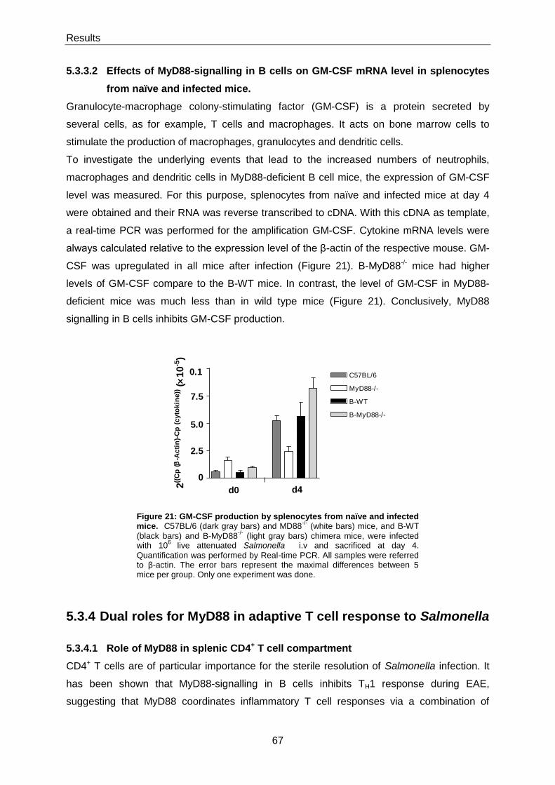
Results
67
5.3.3.2 Effects of MyD88-signalling in B cells on GM-CSF mRNA level in splenocytes
from naïve and infected mice.
Granulocyte-macrophage colony-stimulating factor (GM-CSF) is a protein secreted by
several cells, as for example, T cells and macrophages. It acts on bone marrow cells to
stimulate the production of macrophages, granulocytes and dendritic cells.
To investigate the underlying events that lead to the increased numbers of neutrophils,
macrophages and dendritic cells in MyD88-deficient B cell mice, the expression of GM-CSF
level was measured. For this purpose, splenocytes from naïve and infected mice at day 4
were obtained and their RNA was reverse transcribed to cDNA. With this cDNA as template,
a real-time PCR was performed for the amplification GM-CSF. Cytokine mRNA levels were
always calculated relative to the expression level of the β-actin of the respective mouse. GM-
CSF was upregulated in all mice after infection (Figure 21). B-MyD88-/- mice had higher
levels of GM-CSF compare to the B-WT mice. In contrast, the level of GM-CSF in MyD88-
deficient mice was much less than in wild type mice (Figure 21). Conclusively, MyD88
signalling in B cells inhibits GM-CSF production.
5.3.4 Dual roles for MyD88 in adaptive T cell response to Salmonella
5.3.4.1 Role of MyD88 in splenic CD4+ T cell compartment
CD4+ T cells are of particular importance for the sterile resolution of Salmonella infection. It
has been shown that MyD88-signalling in B cells inhibits TH1 response during EAE,
suggesting that MyD88 coordinates inflammatory T cell responses via a combination of
Figure 21: GM-CSF production by splenocytes from naïve and infected mice. C57BL/6 (dark gray bars) and MD88
-/- (white bars) mice, and B-WT
(black bars) and B-MyD88-/-
(light gray bars) chimera mice, were infected with 10
6 live attenuated Salmonella i.v and sacrificed at day 4.
Quantification was performed by Real-time PCR. All samples were referred to β-actin. The error bars represent the maximal differences between 5 mice per group. Only one experiment was done.
d0 d4
0
2.5
5.0
7.5
0.1 C57BL/6
MyD88-/-
B-WT
B-MyD88-/-
2((
Cp
(
-Ac
tin
)-C
p (
cy
tok
ine
)) (
10
-5)
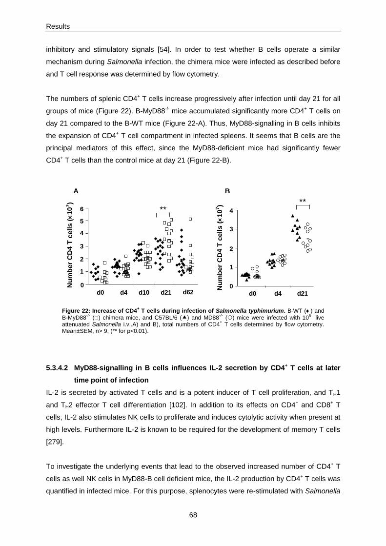
Results
68
inhibitory and stimulatory signals [54]. In order to test whether B cells operate a similar
mechanism during Salmonella infection, the chimera mice were infected as described before
and T cell response was determined by flow cytometry.
The numbers of splenic CD4+ T cells increase progressively after infection until day 21 for all
groups of mice (Figure 22). B-MyD88-/- mice accumulated significantly more CD4+ T cells on
day 21 compared to the B-WT mice (Figure 22-A). Thus, MyD88-signalling in B cells inhibits
the expansion of CD4+ T cell compartment in infected spleens. It seems that B cells are the
principal mediators of this effect, since the MyD88-deficient mice had significantly fewer
CD4+ T cells than the control mice at day 21 (Figure 22-B).
5.3.4.2 MyD88-signalling in B cells influences IL-2 secretion by CD4+ T cells at later
time point of infection
IL-2 is secreted by activated T cells and is a potent inducer of T cell proliferation, and TH1
and TH2 effector T cell differentiation [102]. In addition to its effects on CD4+ and CD8+ T
cells, IL-2 also stimulates NK cells to proliferate and induces cytolytic activity when present at
high levels. Furthermore IL-2 is known to be required for the development of memory T cells
[279].
To investigate the underlying events that lead to the observed increased number of CD4+ T
cells as well NK cells in MyD88-B cell deficient mice, the IL-2 production by CD4+ T cells was
quantified in infected mice. For this purpose, splenocytes were re-stimulated with Salmonella
Figure 22: Increase of CD4+ T cells during infection of Salmonella typhimurium. B-WT () and
B-MyD88-/-
() chimera mice, and C57BL/6 () and MD88-/-
() mice were infected with 106
live attenuated Salmonella i.v..A) and B), total numbers of CD4
+ T cells determined by flow cytometry.
Mean±SEM, n> 9, (** for p<0.01).
0
2
1
3
4
5
6
d0 d4 d10 d21 d62
**
Nu
mb
er
CD
4 T
cells (
10
7)
d0 d4 d210
2
1
4
3
**
Nu
mb
er
CD
4 T
cells (
10
7)
A B
Results
69
for 6 hours in vitro, and then analysed co-expression of CD4 and IL-2 by flow cytometry. In
B-WT and B-MyD88-/- mice, it was detected high numbers of IL-2 producing CD4 T cells at
day 10 and at day 21 after infection, and MyD88-signalling in B cells had no influence on this
response (Figure 23-A). The number of IL-2-producing CD4+ T cells then decreased
significantly at day 62 for both groups. Interestingly, B-MyD88-/- mice had significantly more
IL-2-producing CD4+ T cells than B-WT mice at day 62. This observation suggests that B-
MyD88-/- mice still had a stronger induction of inflammatory response at day 62 compared to
the B-WT mice, since both groups of mice still had bacteria in the spleen at this time point. In
contrast, it was detected less IL-2 secreting CD4+ T cells in MyD88-deficient mice than in
wild-type mice, although this response progressively emerged, demonstrating that other
signalling pathways can partially compensate for the absence of MyD88 (Figure 23-B).

Results
70
5.3.4.3 Activation of S. typhimurium-specific CD4+ T cells
It has been described in other studies that after attenuated Salmonella infection of
susceptible mice there are large numbers of Salmonella -specific CD4+ T cells and CD8+ T
cells [280, 281]. To examine if MyD88-signalling in B cells affects CD4+ antigen specific T
cells in infected mice, the splenocytes were restimulated with Salmonella for 6 hours in vitro,
and then analysed for co-expression of CD4 and CD154. According to this technology,
Salmonella -reactive CD4+ T helper cells could be detected at day 10 after infection, reaching
a peak at day 21 in B-MyD88-/- and B-WT mice, with no influence from MyD88-signalling in B
cells (Figure 24-B). B-WT and B-MyD88-/- mice still had detectable numbers of antigen-
reactive CD4+ T helper cells on day 62. Contrary, the MyD88-deficient mice had only very
Figure 23: Increased of IL-2 production of CD4+ T cells after Salmonella infection. B-WT () and B-
MyD88-/-
() chimera mice, and C57BL/6 () and MD88-/-
() mice were infected with 106
live attenuated Salmonella i.v..A) and B), total numbers of IL-2 producing CD4
+ T cells determined by flow cytometry.
Mean±SEM, n> 9, (* for p<0.05 and ** for p<0.01).
B
unst. unst. unst. HKS HKS HKS
d0 d4 d21
0
2
1
4
3
**
5
**
Nu
mb
er
IL-2
+ C
D4 T
cells (
10
5)
d0 d4 d10 d21 d62
unst. unst. HKS HKS HKSunst. HKSunst. HKSunst.
0
0.5
1
1.5
2
2.5
3
*
Nu
mb
er
IL-2
+C
D4
T c
ells
(
10
6)
A

Results
71
few CD154-expressing CD4+ T cells in all the time points (Figure 24-C), this confirms that
MyD88 plays an essential role for CD4+ T cell activation only in some specific cells.
Figure 24: Activation of Salmonella -specific CD4+ T cells in mice. B-WT () and B-MyD88
-/- ()
chimera mice, and C57BL/6 () and MD88-/-
() mice were infected with 106
live attenuated Salmonella
i.v.. A), Representative staining for CD40L (CD154) of unstimulated and stimulated cells with HKS. B) and C), Frequency numbers of CD40L
+ among CD4
+ T cells. Data shown represent the compilation of at
least two experiments. Mean±SEM, n>9
B
A
C
unst. unst. HKS HKS HKSunst. HKSunst. HKSunst.
d0 d4 d10 d21 d62
0
1
2
3
4
5
6
7
Nu
mb
er
CD
40L
+C
D4 T
cells (
10
6)
0
2
4
6
8
10
unst. unst. unst. HKS HKS HKS
d0 d4 d21
% o
f C
D1
54
+ (
ga
ted
on
CD
4+ T
ce
lls
)
Results
72
5.3.4.4 MyD88-signalling in B cells supresses IFN-γ and TNF-α production by CD4+ T
cells
S. typhimurium induces a strong TH1 response, in which cytokines, such as IFN-γ and TNF-α
play an important role in controlling Salmonella infection [222]. In order to assess the
consequences of MyD88-activation in B cells for CD4+ T cell response, IFN-γ and TNF-α
production were determined in naive and infected mice by flow cytometry after a short ex
vivo re-stimulation of splenocytes with heat-killed Salmonella. MyD88 signalling in B cells
suppressed the accumulation of IFN-γ and TNF-α producing CD4+ T cells on day 21 (Figure
25-B and E, respectively). These data confirm previous studies performed in the EAE model,
where mice lacking MyD88 selectively in B cells make stronger TH1 and TH17 responses than
control mice [54]. IL-17-producing CD4+ T cells were also assessed, but were undetectable
(data not shown), supporting the fact that IL-17 has mild roles during Salmonella infection
[114]. As expected, MyD88-deficient mice had fewer IFN-γ and TNF-α producing CD4+ T
cells than control mice at day 21 (Figure 25-C and F). Surprisingly, in MyD88-deficient mice it
was possible to detect much more IFN-γ producing CD4+ T cells than CD154-producing
CD4+ T cells, suggesting that this cytokine response was stimulated independenty of the
TCR. It is also possible that these two assays have different sensitivities for antigen-reactive
CD4+ T cells. Conclusively, MyD88-signalling in B cells downregulates the inflammatory
response of CD4+ T cells during Salmonella infection.
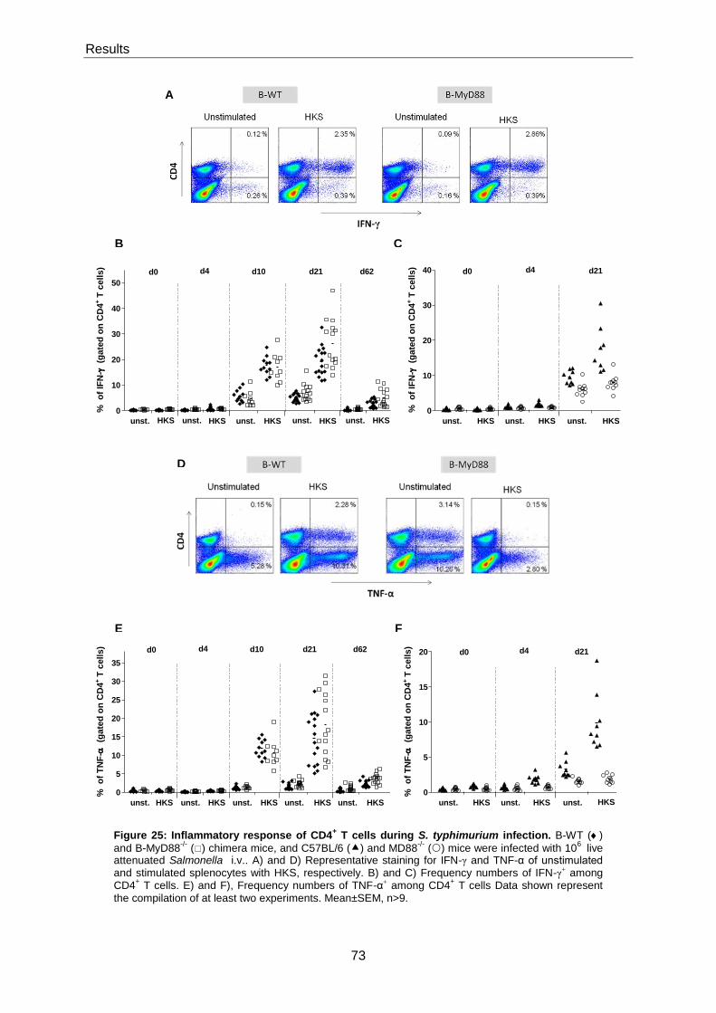
Results
73
Figure 25: Inflammatory response of CD4+ T cells during S. typhimurium infection. B-WT ()
and B-MyD88-/-
() chimera mice, and C57BL/6 () and MD88-/-
() mice were infected with 106
live attenuated Salmonella i.v.. A) and D) Representative staining for IFN-γ and TNF-α of unstimulated and stimulated splenocytes with HKS, respectively. B) and C) Frequency numbers of IFN-γ+ among CD4
+ T cells. E) and F), Frequency numbers of TNF-α+ among CD4
+ T cells Data shown represent
the compilation of at least two experiments. Mean±SEM, n>9.
0
10
20
30
40
unst. unst. unst. HKS HKS HKS
d0 d4 d21
% o
f IF
N-
(g
ate
d o
n C
D4
+ T
cells)
0
10
20
30
40
50
d0 d4 d10 d21 d62
unst. unst. HKS HKS HKSunst. HKSunst. HKSunst.
%
of
IFN
-
(ga
ted
on
CD
4+ T
ce
lls
)
B
A
C
E
D
F
0
5
10
15
20
25
30
35
d0 d4 d10 d21 d62
unst. unst. HKS HKS HKSunst. HKSunst. HKSunst.
%
of
TN
F-
(g
ate
d o
n C
D4
+ T
ce
lls
)
0
5
10
15
20
unst. unst. unst. HKS HKS HKS
d0 d4 d21
% o
f T
NF
- (g
ate
d o
n C
D4
+ T
cells)
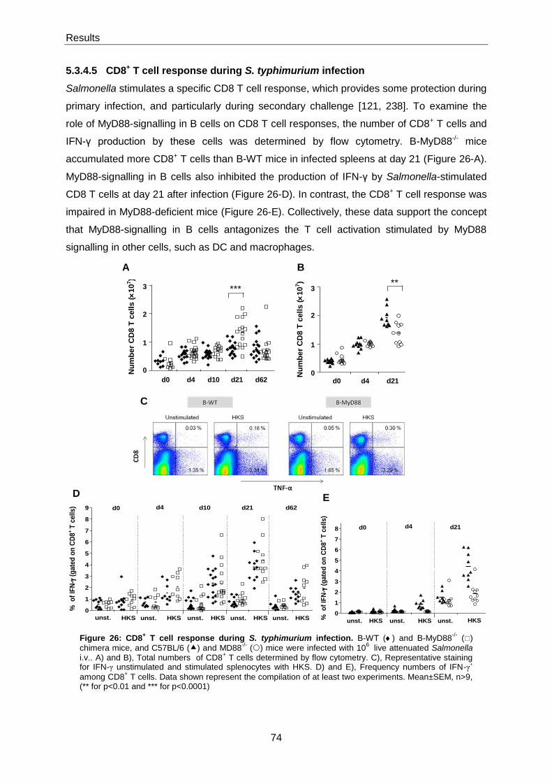
Results
74
5.3.4.5 CD8+ T cell response during S. typhimurium infection
Salmonella stimulates a specific CD8 T cell response, which provides some protection during
primary infection, and particularly during secondary challenge [121, 238]. To examine the
role of MyD88-signalling in B cells on CD8 T cell responses, the number of CD8+ T cells and
IFN-γ production by these cells was determined by flow cytometry. B-MyD88-/- mice
accumulated more CD8+ T cells than B-WT mice in infected spleens at day 21 (Figure 26-A).
MyD88-signalling in B cells also inhibited the production of IFN-γ by Salmonella-stimulated
CD8 T cells at day 21 after infection (Figure 26-D). In contrast, the CD8+ T cell response was
impaired in MyD88-deficient mice (Figure 26-E). Collectively, these data support the concept
that MyD88-signalling in B cells antagonizes the T cell activation stimulated by MyD88
signalling in other cells, such as DC and macrophages.
Figure 26: CD8+ T cell response during S. typhimurium infection. B-WT () and B-MyD88
-/- ()
chimera mice, and C57BL/6 () and MD88-/-
() mice were infected with 106
live attenuated Salmonella i.v.. A) and B), Total numbers of CD8
+ T cells determined by flow cytometry. C), Representative staining
for IFN-γ unstimulated and stimulated splenocytes with HKS. D) and E), Frequency numbers of IFN-γ+
among CD8+ T cells. Data shown represent the compilation of at least two experiments. Mean±SEM, n>9,
(** for p<0.01 and *** for p<0.0001)
0
2
1
3
d0 d4 d21
**
Nu
mb
er
CD
8 T
cells (
10
7)
d0 d4 d10 d21 d62
0
2
1
3 ***
Nu
mb
er
CD
8 T
cell
s (
10
7)
B A
C
D E
0
1
2
3
4
5
6
7
8
9
unst. unst. HKS HKS HKSunst. HKSunst. HKSunst.
d0 d4 d10 d21 d62
% o
f IF
N-
(g
ate
d o
n C
D8
+ T
cell
s)
0
1
2
3
4
5
6
7
8
unst. unst. unst. HKS HKS HKS
d0 d4 d21
% o
f IF
N-
(g
ate
d o
n C
D8
+ T
ce
lls
)

Results
75
5.3.4.6 Summary of absolute cell numbers of Salmonella -specific CD4+ T cells, and
cytokines production by CD4+ T cells and CD8+ T cells.
In addition to the analysis of frequencies of CD40L+ CD4+ T cells, IFN-γ and TNF-α producing
CD4+ T cells, and IFN-γ producing CD8+ T cells, the absolute cell numbers were also
determined in order to obtain a complete analysis of B-WT, B-MyD88-/- mice, C57BL/6 and
MyD88-/- mice. Table 3 and Table 4 summarize the absolute cell numbers in the spleen for
the B-WT and B-MyD88-/- chimera mice and C57BL/6 and MyD88-/- mice respectively.
Salmonella -specific CD4+ T cells increased progressively during infection for all mice (Table
3 and Table 4). However the difference was not statistically significant for the MyD88
deficient B cell mice and B-WT mice (Table 3). On the contrary MyD88 deficient mice had
very few Salmonella-specific CD4+ T cells compare to the WT mice (Table 4). By comparison
to B-WT mice the average number of IFN-γ and TNF-α producing CD4+ T cells was
increased from 5.2×10⁶ to 1.0×10⁷ and 4.1×10⁶ to 7.5×10⁶ in spleens of B-MyD88-/- mice on
day 21, corresponding to a 92% and 83% of increase, respectively (Table 3). For IFN-γ
producing CD8+ T cells, MyD88-deficient B cell mice had an increase of 104% compared to
B-WT mice at day 21 (Table 3).
CD4+
CD40L+ CD4
+ IFN-γ
+ CD4
+ TNF-α
+ CD8
+ IFN-γ
+
Day 4
B-WT 8.5×10
4
± 1.2×10⁴
7.4×10⁴
± 1.3×10⁴
5.2 ×10⁴
± 3.9×10³
8.6×10⁴
± 1.8×10⁴
B-MyD88-/
-
6.1×10⁴
± 8.4×10³
6.3×10⁴
± 6.7×10³
3.8×10⁴
± 3.1×10³
1.5×10⁵
± 3.4×10⁴
Day 10
B-WT 6.1×10
5
± 7.5×10⁴
3.4×10⁶
± 3.4×10⁵
2.7×10⁶
± 2.6×10⁶
1.4×10⁵
± 2.9×10⁴
B-MyD88-/
-
7.5×10⁵
± 1.2×10⁵
4.4×10⁶
± 4.8×10⁵
2.4×10⁶
± 2.7×10⁵
1.7×10⁵
± 2.8×10⁴
Day 21
B-WT 2.5×10
6
± 2.9×10⁵
5.2×10⁶
± 7.7×10
5
4.1×10⁶
± 7.4×10
5
3.1×10⁵
± 4.5×10⁴
B-MyD88-/
-
2.6×10⁶
± 2.9×10⁵
1.0×10⁷
± 1.5×10
6
7.5×10⁶
± 1.3×10⁶
6.2×10⁵
± 1.1×10⁵
P value summary
ns ** * **
Table 3: Numbers of total Salmonella -specific CD4+ T cells, and IFN-γ and TNF-α producing CD4
+ T cells, and
IFN-γ producing CD8+ T cells in spleen for B-WT and B-MyD88
-/- infected mice at day 4, day 10 and day 21 with
attenuated S. typhimurium, were determined by FACS. Mean±SEM; n>14, (ns for p>0.05; * for p<0.05 and ** for p<0.01).
MyD88 deficient mice showed reductions between 65% and 85% compared to C57BL/6 mice
in IFN-γ and TNF-α producing CD4+ T cells at day 21 and IFN-γ production CD8+ T cells
show a reduction of 70% (Table 4).
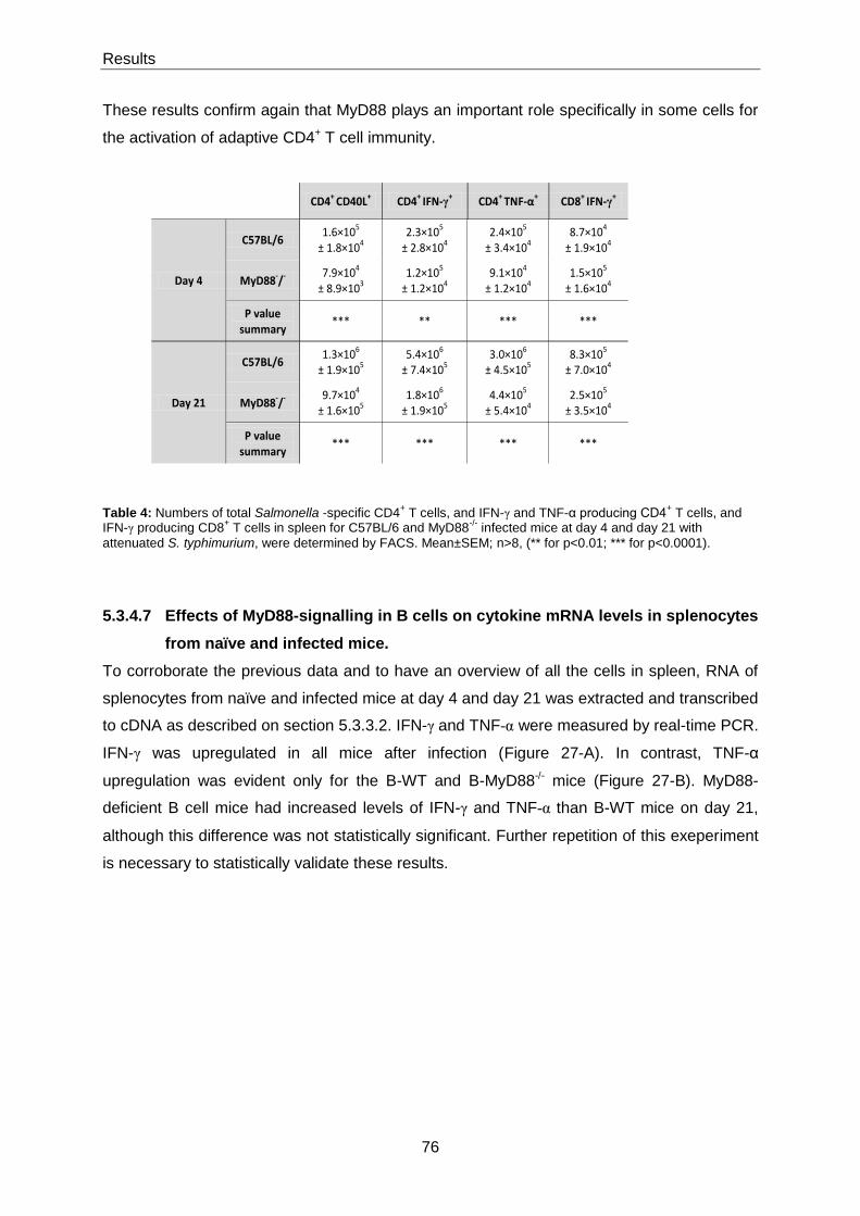
Results
76
These results confirm again that MyD88 plays an important role specifically in some cells for
the activation of adaptive CD4+ T cell immunity.
CD4+
CD40L+ CD4
+ IFN-γ
+ CD4
+ TNF-α
+ CD8
+ IFN-γ
+
Day 4
C57BL/6 1.6×10
5
± 1.8×10⁴
2.3×10⁵
± 2.8×10⁴
2.4×10⁵
± 3.4×10⁴
8.7×10⁴
± 1.9×10⁴
MyD88-/
-
7.9×10⁴
± 8.9×10³
1.2×10⁵
± 1.2×10⁴
9.1×10⁴
± 1.2×10⁴
1.5×10⁵
± 1.6×10⁴
P value summary
*** ** *** ***
Day 21
C57BL/6 1.3×10
6
± 1.9×10⁵
5.4×10⁶
± 7.4×10⁵
3.0×10⁶
± 4.5×10⁵
8.3×10⁵
± 7.0×10⁴
MyD88-/
-
9.7×10⁴
± 1.6×10⁵
1.8×10⁶
± 1.9×10⁵
4.4×10⁵
± 5.4×10⁴
2.5×10⁵
± 3.5×10⁴
P value summary
*** *** *** ***
Table 4: Numbers of total Salmonella -specific CD4+ T cells, and IFN-γ and TNF-α producing CD4
+ T cells, and
IFN-γ producing CD8+ T cells in spleen for C57BL/6 and MyD88
-/- infected mice at day 4 and day 21 with
attenuated S. typhimurium, were determined by FACS. Mean±SEM; n>8, (** for p<0.01; *** for p<0.0001).
5.3.4.7 Effects of MyD88-signalling in B cells on cytokine mRNA levels in splenocytes
from naïve and infected mice.
To corroborate the previous data and to have an overview of all the cells in spleen, RNA of
splenocytes from naïve and infected mice at day 4 and day 21 was extracted and transcribed
to cDNA as described on section 5.3.3.2. IFN-γ and TNF-α were measured by real-time PCR.
IFN-γ was upregulated in all mice after infection (Figure 27-A). In contrast, TNF-α
upregulation was evident only for the B-WT and B-MyD88-/- mice (Figure 27-B). MyD88-
deficient B cell mice had increased levels of IFN-γ and TNF-α than B-WT mice on day 21,
although this difference was not statistically significant. Further repetition of this exeperiment
is necessary to statistically validate these results.
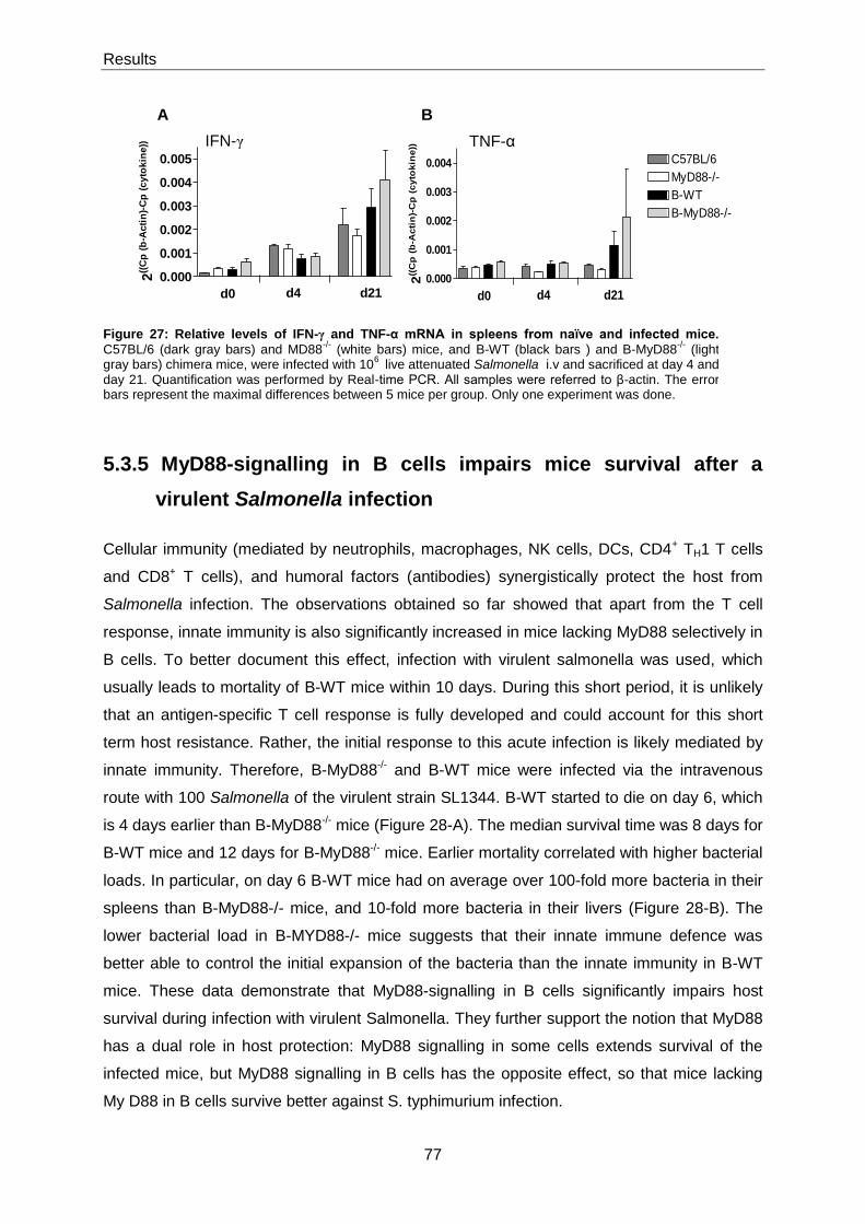
Results
77
5.3.5 MyD88-signalling in B cells impairs mice survival after a
virulent Salmonella infection
Cellular immunity (mediated by neutrophils, macrophages, NK cells, DCs, CD4+ TH1 T cells
and CD8+ T cells), and humoral factors (antibodies) synergistically protect the host from
Salmonella infection. The observations obtained so far showed that apart from the T cell
response, innate immunity is also significantly increased in mice lacking MyD88 selectively in
B cells. To better document this effect, infection with virulent salmonella was used, which
usually leads to mortality of B-WT mice within 10 days. During this short period, it is unlikely
that an antigen-specific T cell response is fully developed and could account for this short
term host resistance. Rather, the initial response to this acute infection is likely mediated by
innate immunity. Therefore, B-MyD88-/- and B-WT mice were infected via the intravenous
route with 100 Salmonella of the virulent strain SL1344. B-WT started to die on day 6, which
is 4 days earlier than B-MyD88-/- mice (Figure 28-A). The median survival time was 8 days for
B-WT mice and 12 days for B-MyD88-/- mice. Earlier mortality correlated with higher bacterial
loads. In particular, on day 6 B-WT mice had on average over 100-fold more bacteria in their
spleens than B-MyD88-/- mice, and 10-fold more bacteria in their livers (Figure 28-B). The
lower bacterial load in B-MYD88-/- mice suggests that their innate immune defence was
better able to control the initial expansion of the bacteria than the innate immunity in B-WT
mice. These data demonstrate that MyD88-signalling in B cells significantly impairs host
survival during infection with virulent Salmonella. They further support the notion that MyD88
has a dual role in host protection: MyD88 signalling in some cells extends survival of the
infected mice, but MyD88 signalling in B cells has the opposite effect, so that mice lacking
My D88 in B cells survive better against S. typhimurium infection.
Figure 27: Relative levels of IFN-γ and TNF-α mRNA in spleens from naïve and infected mice.
C57BL/6 (dark gray bars) and MD88-/-
(white bars) mice, and B-WT (black bars ) and B-MyD88-/-
(light gray bars) chimera mice, were infected with 10
6 live attenuated Salmonella i.v and sacrificed at day 4 and
day 21. Quantification was performed by Real-time PCR. All samples were referred to β-actin. The error bars represent the maximal differences between 5 mice per group. Only one experiment was done.
B A
TNF-α IFN-γ
0.000
0.001
0.002
0.003
0.004
d0 d4 d21
C57BL/6
MyD88-/-
B-WT
B-MyD88-/-
2((
Cp
(b
-Acti
n)-
Cp
(cy
tokin
e))
0.000
0.001
0.002
0.003
0.004
0.005
d0 d4 d21
2((
Cp
(b
-Ac
tin
)-C
p (
cy
tok
ine
))
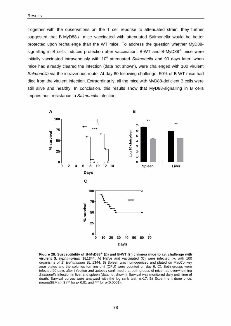
Results
78
Together with the observations on the T cell reponse to attenuated strain, they further
suggested that B-MyD88-/- mice vaccinated with attenuated Salmonella would be better
protected upon rechallenge than the WT mice. To address the question whether MyD88-
signalling in B cells induces protection after vaccination, B-WT and B-MyD88-/- mice were
initially vaccinated intravenously with 106 attenuated Salmonella and 90 days later, when
mice had already cleared the infection (data not shown), were challenged with 100 virulent
Salmonella via the intravenous route. At day 60 following challenge, 50% of B-WT mice had
died from the virulent infection. Extraordinarily, all the mice with MyD88-deficient B cells were
still alive and healthy. In conclusion, this results show that MyD88-signalling in B cells
impairs host resistance to Salmonella infection.
Figure 28: Susceptibility of B-MyD88-/-
() and B-WT () chimera mice to i.v. challenge with virulent S. typhimurium SL1344. A) Naïve and vaccinated (C) were infected i.v. with 100 organisms of S. typhimurium SL 1344. B) Spleen was homogenized and plated on MacConkey agar plates and the colonies forming unit (CFU) were counted on day 6. C), Both groups were infected 90 days after infection and autopsy confirmed that both groups of mice had overwhelming Salmonella infection in liver and spleen (data not shown). Survival was monitored daily until time of death. Survival curves were analyzed with the log rank test, n>17. B) Experiment done once, mean±SEM n> 3 (** for p<0.01 and *** for p<0.0001).
0 10 20 30 40 50 60 700
25
50
75
100
***
Days
% s
urv
ival
B A
C
0 2 4 6 8 10 12 140
25
50
75
100
***
Days
% s
urv
ival
0
1
2
3
4
5
6
7
Spleen Liver
****
Lo
g 1
0 c
fu/s
ple
en
Discussion
79
6 Discussion
The work here presented addressed the role of MyD88-signalling in B cells upon Salmonella
infection. MyD88 is the major adaptor protein involved in the signalling pathway of the Toll-
like receptors upon pathogen recognition. This activation in some innate immune cells can
potentiate inflammatory response, but on the other hand can drive suppression by the
provision of IL-10 by B cells [54]. The major aim of this thesis was the analysis of this
inhibitory circuit and their consequences in the immune response during Salmonella
infection.
In the first part of this study in vitro data with HKS activated B cells, DC and macrophages
revealed production of high amounts of IL-10 by B cells through TLR2/4-MyD88 dependent
manner. On the contrary, DC and macrophages produced big amounts of pro-inflammatory
cytokines upon Salmonella activation. Importantly mice with IL10-deficient B cells showed an
increased inflammatory response by T cells upon Salmonella infection.
In the second part it was demonstrated that MyD88-signalling in B cells inhibits the innate
immunity and adaptive T cell immunity in response to Salmonella typhimurium infection. In
particular, MyD88-signalling in B cells impairs survival of the mice exposed to a primary as
well as to secondary infection by virulent Salmonella. This MyD88-mediated suppression was
specifically elicited by B cells, as total MyD88-deficient mice show impaired protective
immunity against Salmonella.
Thus, MyD88 exerts opposite effects in distinct cells types creating a controlled balance in
the host response against Salmonella infection.

Discussion
80
6.1 Role of B cells during infections
B cells are known to have an important role in immune defence against microbial infections.
Infections with Chlamydia trachomatis, Francisella tularensis, Leishmania major,
Plasmodium chabaudi, Pneumocystis carinii, Mycobacterium tuberculosis and Salmonella
typhimurium have been reported to enhance susceptibility in B cell deficient mice [282-289].
During Salmonella infection, B cells are known to induce a strong antibody response [227]. In
humans, vaccination with purified polyssacharide Vi antigen from serovar Typhi stimulates
antibody production and provides protection [290, 291]. In mice the transfer of serum from
infected mice can transfer protection from an otherwise lethal Salmonella challenge [248,
249]. Furthermore, B cells are required for protection against oral infection with virulent
Salmonella, and vaccination with attenuated Salmonella is less protective in B cell deficient
mice [292, 293]. Urgrinovic and colleagues have described that B cell deficient mice infected
with attenuated Salmonella typhimurium showed an impaired TH1 T cell response [294].
The role of B cells has been studied during Salmonella infection using B cell deficient mice.
However, beside the fact that mice with targeted deletions that „‟knockout‟‟ specific genes are
useful tools in immunology, they also can lead to inaccurate conclusions regarding the role of
a particular component of immunity, if not interpreted carefully. In order to have a better
approach for studying the role of molecules of interest in B cells during infection, chimeric
mice were generated in which a genetic deficiency of interest, e.g MyD88 or IL-10, was
restricted solely to B cells. We observed that B-MyD88-/- mice were more resistant to primary
infection with virulent Salmonella than mice with wild-type B cells. This observation contrasts
other studies where a „‟knockout mouse‟‟ was used. For instance, B cell deficient mice have
been reported to succumb to virulent Salmonella infection within the same time range as wild
type mice [292]. MyD88-deficient mice died much earlier after oral infection with virulent
Salmonella than the wild-type mice [295].
6.1.1 Effects of MyD88 in B cells
B cell development is dependent upon and closely regulated by the expression of the antigen
receptor genes [296]. We have observed that naive MyD88-deficient B cell mice have fewer
B cells than control mice. This observation is maintained after infection with Salmonella
typhimurium. Is MyD88 important in the homeostasis and development of the B cell
Discussion
81
compartment? Some studies have shown that TLR-4 and TLR-9 are expressed on the
surface of B cells during early stages of development [297] suggesting that these receptors
have a role in the development of B cells. In vitro studies with pre-B cell line showed that
LPS can provide stimulus for B cell differentiation [298]. Moreover, Hayashi and colleagues
have shown that TLR-4 signalling favours B lymphocyte maturation, whereas TLR-2 retards
this process [299]. However other studies have shown that MyD88 and TLR-9 deficient mice
have normal B cell populations [27, 300, 301]. This is in agreement with our observation, as
MyD88 knockout mice have normal B cell numbers (data not shown). This suggests that
maybe in knockout mice the cell numbers are under homeostatic control. The exact reasons
for our observations in the MyD88-deficient B cell mice are still unclear, but further analysis
of homeostatic survival and proliferation of B cells should shed light on the possible role of
MyD88 in B cell populations. There are possible endogenous ligands for the regulation of
these processes since TLR can recognize self-antigens, such as heat-shock proteins [302,
303].
In addition to the role served by BCR signals in shaping the development and function of B
cells. Innate immune receptors, such as TLRs have also important roles. TLRs recognize
specific microbial products that also help to sculp humoral immune response. B cells express
most TLR and can respond to a variety of TLR ligands. Their response to these stimuli can
be to proliferate, to differentiate into antibody secreting plasma cells, to become more
efficient antigen presenting cells (APC) or to secrete cytokines [304, 305].
The role of TLRs for antibody responses is still object of controversy. Pasare and Medzhitov
reported that T-dependent antigen-specific antibody responses require activation of TLRs in
B cells [215], but Gavin and colleagues have later reported that B cells can produce
antibodies independently of TLR activation [306]. Furthermore, Ruprecht and Lanzavechia
demonstrated that naive human B cells require TLR signals for productive T cell-dependent
activation [169]. Several studies have evaluated humoral immune responses in mice with
MyD88 deficiency restricted to B cells. In particular, it was found that mice with MyD88-
deficient B cells mount a weaker antibody response to antigen mixed with LPS than mice
carrying wild type B cells [307]. MyD88-deficient B cells also form less germinal centres and
produce less specific antibodies than wild-type B cells in chimera mice harbouring both types
of B cells following infection with murine gamma herpes virus 68 [213].
Our results show that MyD88-deficient B cell mice infected with Salmonella typhimurium
have less germinal centres but they are able to mount a normal specific antibody response of
all antibody isotypes with exception of IgG1 and IgG3. MyD88-deficient B cell mice had a
Discussion
82
slightly increase of IgG1 production and a delayed IgG3 production compared to the control
mice. These observations are in agreement with other studies, which demonstrated that B
cells from MyD88-deficient mice resulted in a biased-TH2 response with increased IgG1
production and decreased levels of IgG3 [27, 308-310]. However, Barr and colleagues have
shown that MyD88-deficient B cell mice infected with Salmonella show loss of IgG2c and
lgM, which we do not [311]. Nevertheless they demonstrated that MyD88 B cell-deficient
mice have less germinal centres and were detected on day 14 after infection [311].
Interesting are also the findings of Cunningham, which show that in WT mice infected with
attenuated Salmonella. Plasma cells appear in spleen by day 3 and germinal centres one
month later when the infection has been resolved [228].
The natural IgM pool is largely derived by the B1 population of B cells [312, 313]. Barr et al.
also demonstrated that natural antibodies, such as IgM depends on B cell intrinsic MyD88-
transduced signals [311]. We also observed a decrease of natural IgM in the MyD88 deficient
mice, and this observation is not surprising considering that B1 population in peritoneal cavity
was reduced in MyD88 deficient B cell mice (data not shown). Despite the fact that we
observed a decrease in natural IgM, the mice were able to restore the levels of antigen
specific antibodies at later time points of infection. Our work indicates that in mice infected
i.v. with Salmonella typhimurium, the antibodies mainly derive from follicular and marginal
zone B cells. We had observed that MyD88-deficient B cell mice had fewer follicular B cells
but more MZ B cells after infection compared to the control mice. Both groups showed a
decrease in the MZ B cell population after a late time point of infection. A decrease in
marginal zone B cells has been associated to antigen transport after exposure to opsonised
antigen [314, 315]. Furthermore, LPS induces a reduction of MZ B cells, associated with their
migration into follicles [314, 316, 317]. Oliver et al. also have demonstrated that stimulation
with LPS of MZ B cells results in their rapid proliferation, IgM and IgG3 antibody secretion
and up-regulation of the co-stimulatory molecule CD86 [166, 167]. This observation suggests
that since MZ B cells from MyD88-deficient B cell mice cannot be efficiently stimulated, this
leads to a decrease in the levels of IgM and IgG3 in the beginning of the immune response
against to Salmonella, but this levels are restored at a later time point after infection. MZ B
cells differentiate into antibody-forming cells during the first week after infection whereas,
follicular B cells respond slower. Both populations are known to undergo GC reactions that
give rise to memory B cells and long-lived plasma cells [166]. We observed that B-MyD88-/-
have fewer long-lived plasma cells in the bone marrow and as well as, reduced levels of all
antibody isotypes except IgG1. This observation is in agreement with Guay and colleagues,
who showed that MyD88-deficient mice infected with polyoma virus can form germinal

Discussion
83
centres and mount early specific antibody responses, but they fail to generate bone marrow
plasma cells and to maintain long-term humoral immunity [214].
Overall these data suggest that MyD88-signalling in B cells acts as a cell autonomous
amplifier for all the facets of B cell response. MyD88-dependent signalling in B cells is not
totally required for primary B cell immune response, but it seems necessary for effectively
generating long-term antibody responses to Salmonella infection.
6.1.2 Link between MyD88-activated B cells and innate immunity
The innate immune system provides the first layer of protection and MyD88 is a crucial
component of this response. MyD88 stimulates resident tissue macrophages to produce
neutrophil chemoattractants [56, 57]. The recruitment of neutrophils to infected tissues can
be or is impaired in MyD88-deficient mice [57, 318-320]. We observed that in the absence of
MyD88, the accumulation of neutrophils and macrophages within infected spleens was
severely delayed. However they reached control levels by day 21 after infection, indicating
that other signalling pathways can substitute for MyD88. In contrast, the lack of IFN-γ-
producing NK cells was not recovered even at later time points in MyD88-deficient mice.
Given that the early provision of IFN-γ by NK cells is essential for macrophage activation and
for protection of Salmonella [99], it is likely that this defect contributes to the impaired survival
of MyD88-deficient mice after infection by virulent Salmonella [51]. The activation of NK cells
has been described in mice infected with Listeria monocytogenes [321]. Myeloid cells and
NK cells are recruited by CD11c+ DC into clusters localized around the central arterioles in
the T cell zones of the white pulp areas in spleen, where the bacteria is trapped. In these
clusters, IL-12 and IL-18 production by DCs stimulates NK cells to produce IFN-γ, which is
necessary also for the activation and maturation of colocalized monocytes into TNF- and
iNOS-producing DCs [321]. MyD88 plays also an important role in this response, since NK
cells from MyD88-deficient mice do not form clusters and do not produce IFN-γ upon Listeria
infection [321]. It is possible that a similar process occurs during Salmonella infection.
MyD88 would drive the NK cell response by stimulating their recruitment around central
arterioles, and by inducing signals via IL-12 and IL-18 that are necessary for IFN-γ
expression.
Remarkably, multiple aspects of the innate immune response were increased in the absence
of MyD88-signalling selectively in B cells: the numbers of neutrophils and IFN-γ-producing
NK cells were higher at day 4, and the numbers of macrophages were higher at day 10 and
at day 21. The innate immune response was therefore stronger during the whole course of

Discussion
84
infection in B-MyD88-/- mice, although different players were involved at different time points.
This increased innate response most likely explains why B-MyD88-/- were more resistant to a
primary infection by virulent Salmonella than mice with wild-type B cells. These observations
show that absence of MyD88-signalling restricted to B cells augmented the immune
response against Salmonella, whereas the absence of MyD88-signalling in other cells has
opposite effects. How MyD88-signalling B cells influences the innate immune cells?
Neutrophils are known to be crucial in survival of infected mice with Salmonella [322]. The
recruitment of these cells at the site of infection is orchestrated by several chemokines, such
as CXCL1, CXCL2, CCL2 and CCL3 [56, 57, 323, 324]. Recently it has been described that
IFN-γ coordinates CCL3-mediated neutrophil recruitment in vivo [323]. This suggests that
IFN-γ production by NK cells can affect the recruitment of neutrophils. Furthermore, IFN-γ is
secreted by NK cells in response to the IL-12 produced by dendritic cells and macrophages
[269, 325]. Macrophages provide an important line of defense against Salmonella [221].
Supernatants from Salmonella TLR-activated B cells can suppress production by
macrophages of proinflammatory cytokines that are usually produced by classically activated
macrophages. This suppression can be due to presence of IL-10 in the supernatants since B
cells stimulated with Salmonella typhimurium produces large amounts of IL-10 in vitro, in a
TLR2/TLR4-MyD88 dependent manner. Furthermore, it has been reported that IL-10 can
polarize monocytes into alternatively activated macrophages, which play a role in resolution
of inflammation, accompanied by reduced pro-inflammatory cytokine secretion [326].
Therefore it is possible that B cells influence the polarization of activated macrophages. For
example, it has been reported that IL-10 inhibits TNF-α and IL-12 production by
macrophages and their stimulatory effect on IFN-γ production by NK cells [270, 327]. In
general, these observations suggest that MyD88-activated B cells can regulate the response
of macrophages and dendritic cells, which are able to control activation of NK cells and
neutrophils.
6.1.3 Effect of MyD88-activated B cells on T cell immunity
In antigen-presenting cells (APCs), TLRs can induce signals that, through expression of
cytokines and co-receptors, are capable of activating T cell immunity [3, 328]. Mice lacking
MyD88 show impaired TH1 responses to antigens emulsified in complete Freund‟s adjuvant,
whereas TH2 responses were normal [328]. This effect was also observed for Toxoplasma
gondii and Leishmania major [329, 330]. More specifically, MyD88-deficient dendritic cells
migrate normally into lymph nodes and up-regulate co-stimulatory receptors as efficiently as
wild type DCs [331], but they do not secrete cytokines that are essential for T cell immunity
[332].

Discussion
85
According to our results, the absence of MyD88 leads to delayed accumulation of dendritic
cells within infected spleens. Furthermore, Salmonella-specific inflammatory TH1 response
was strongly impaired by means of IFN-γ and TNF-α production by CD4+ T cells in MyD88
deficient mice compared to wild-type mice. This observation is in contrast to previous
findings, which demonstrated that MyD88-deficient mice can mount TH1 responses against
some microbes, such as Listeria monocytogenes [333], Mycobacterium tuberculosis [334], or
influenza virus [335], implying that MyD88-independent receptors are able to recognize these
pathogens and initiate T cell priming. Possibly, MyD88-deficient mice remain less resistant to
these microbes, because of defective innate immune responses.
Interesting, it was the observation done in the B-MyD88-/- mice, these mice had stronger
Salmonella -specific inflammatory TH1 response compared to B-WT mice. This corroborates
the previous findings, which show that absence of MyD88 in B cells results in heightened TH1
and TH17 responses during EAE, leading to a chronic form of disease [54]. During
Salmonella infection, deficiency of MyD88 signalling in B cells does not increase the number
of antigen-reactive CD4+ T cells, as determined by staining intracellular CD154 or IL-2, but
rather enhance the polarization of the reactive CD4+ T cells towards the TH1 pathway
characterized by the secretion of IFN-γ and TNF-α. These results are in agreement with the
observations made by Lampropoulou et al. and Fillatreau et al., demonstrating that the B
cell-mediated regulation of EAE does not influence the production of IL-2 by antigen-reactive
T cells, but specifically suppress the inflammatory TH1 and TH17 responses [54, 170]. This B
cell-mediated regulation of TH1 immunity could be mimicked in vitro, and IL-10 was identified
as a key suppressive mediator [54]. During in vitro culture, B cells stimulated through MyD88
using TLR ligands, such as LPS, secrete large amounts of IL-10 and supernatants from
these B cells inhibit in an IL-10-dependent manner the differentiation of naive CD4+ T cells
into TH1 cells [54]. This inhibition operates indirectly via suppression of the production of IL-
12, IL-6, IL-23 and TNF-α by DCs [54]. Therefore, TLR activated B cells can control TH1
immunity by inhibiting via IL-10 the production of inflammatory mediators such as IL-12 by
DC [170, 336]. There is evidence for this regulatory circuit in vivo, since mice with B cell-
restricted deficiency in IL-10 make stronger TH1 responses than mice with wild-type B cells,
and DCs isolated from B cell-deficient mice produce higher amounts of IL-12 and induce
stronger TH1 responses than DCs from wild-type mice upon adoptive transfer [170, 336].
Maybe IL-10 is involved in the suppressive function of B cells during Salmonella infection
because heat-killed Salmonella induces IL-10 secretion by B cells via a MyD88-dependent
pathway in vitro and mice with IL-10-deficiency restricted to B cells have also a stronger TH1
response with higher amounts of IFN-γ and TNF-α during Salmonella infection. Other
studies, dissecting the role of IL-10 and other cytokines during bacterial infections showed

Discussion
86
that IL-10 suppresses IFN-γ and TNF-α production by T cells [270, 327]. Sashinami et al.
reported that neutralization of endogenous IL-10 reduced bacterial growth and this can be
due to the increase of IFN-γ and TNF-α production [337]. B cells can interfere with the
activation of TH1 cells by suppressing and/or modulating the function of DCs indirectly by
their production of IL-10. If this mechanism is important, where and when can B cells instruct
DC function in vivo? There is evidence that B cells interact directly with DC at a very early
stage of the immune response. Following activation, B cells rapidly up-regulate the
chemokine receptor CCR7, which confers them responsiveness for the T cell zone specific
chemokines CCL19 and CCL21 [338]. The newly equipped B cells then migrate into the T
cell area, where they accumulate at the periphery of the periarteriolar lymphoid sheath and
near the terminal arteriolar branches. It has been previously shown that primed B cells start
to proliferate there, within clusters of DC and T cells, within few days after immunization [339,
340]. In this microenvironment, B cells can directly interact with DC via both antigen-
independent and antigen-dependent mechanisms [341, 342]. Even though this interaction
has been regarded principally as a place where DC can provide B cells with signals
facilitating antibody responses [343, 344], it is possible that B cells equally modulate the
functions of DC, possibly through cytokines such as IL-10. The suppression of DC function
through the production of IL-12, could explain why B-MyD88-/- mice show enhanced NK cell
response already at day 4 after infection with Salmonella.
Beside DCs, macrophages are also an important APC in the combat of bacterial infections by
phagocytosing and destroying bacteria and presenting bacteria-derived antigens to T cells.
We have previously shown that B cells can modulate macrophage function in the production
of cytokines, such as IL-6 and IFN-γ. Maybe B cells influence the T cell response trough
macrophages via IL-10. For instance, it has been shown that B1 cells are able to control the
effector functions of macrophages via IL-10 secretion [345] and Fiorentino and colleagues
demonstrated that IL-10 inhibits the ability of LPS-activated macrophages to produce
inflammatory cytokines, such as IL-1, TNF-α and IL-6 leading to the inhibition of T cell
activation [346]. If this is true, where and when macrophages interact with B cells? In the
spleen, the marginal zone has two main populations of macrophages: the marginal-zone
metallophilic macrophages and marginal-zone macrophages. The latter population, is
characterized by expression of DC-SIGN and the type I scavenger receptor MARCO
(macrophages receptor with collagenous structure). MZ macrophages are located together
with the marginal zone B cells. It has been suggested that MZ B cells can be activated by
marginal zone macrophages and that MZ macrophages are able to transfer processed
antigens to MZ B cells [347]. Moreover, Koppel and colleagues have shown that SINGR1-
expressing macrophages interact with MZ B cells and that are involved in the early
Discussion
87
production of antibodies [348]. Maybe, in this microenvironment B cells can simultaneously
secrete IL-10 and modulate macrophages function.
However we have to consider that maybe it is not only IL-10 secretion by MyD88-activated B
cells regulating innate and adaptive immune responses. For instance, B cells can suppress
inflammation in bowel disease by innate immune cells via IL-10-independent mechanism
[349]. Moreover, Tian and colleagues have shown that LPS-activated B cells can inhibit T
cell immunity via TGF-β secretion in NOD mice [259]. Scott et al. have described that LPS-
activated B cells can directly suppress T cells independently of IL-10, via antigen
presentation involving MHC-II and CD86 [350]. Overall, these studies suggest that MyD88-
signalling in B cells can induce several molecular mechanisms to suppress immunity. During
Salmonella infection, we observed that the absence of MyD88 in B cells induces an increase
in immune response involving different cell populations: neutrophils and NK cells,
macrophages and DC, and finally CD4 and CD8 T cells. These effects had a dramatic
consequence on the protection of vaccinated mice.
We have observed that all vaccinated B-MyD88-/- mice survived an infection with virulent
Salmonella while half of the B-WT succumbed to the infection. Several studies have
described the role of B cells in the formation and maintenance of T cell memory and in
protection against infection [284, 351-353]. For example, it has been demonstrated that
antigen presentation and antibody production are necessary to maintain memory CD4+ T
cells [284, 354]. Other studies show the contrary, that for instance, B cell-deficient mice
infected with Listeria monocytogenes have defective CD4+ T cell responses, although B cells
do not express listerial antigens [355]. Moreover, Whitmire and colleagues have shown that
B cells are required for CD4+ T cell memory generation in infection with lymphocytic
choriomeningitis virus (LCMV) independently of antibody production [353]. These studies
imply that maybe B-cell functions other than antibody production are important for memory
CD4+ T cell responses.
With regard to the role of B cells in CD8+ T cell memory, several studies have demonstrated
that CD8 memory can be maintained in the absence of B cells [356-358]. Moreover, CD8
memory can be maintained in the absence of specific antigens [359-361] and in the absence
of cross-reactive antigens and MHC class I [362]. However, other studies have shown the
opposite. For example, Shen and colleagues have demonstrated that B cells influence the
pool of CD8 T cell memory, since in absence of B cells there is an increased death of
activated CD8+ T cells in contraction phase, leading to a decrease of Listeria
monocytogenes-specific CD8+ T cell memory [352].

Discussion
88
In addition studies have revealed a role for MyD88 in the generation and maintenance of T
cell memory. Rahman et al. have demonstrated that MyD88-deficent mice can provide CD8+
T cell response in the early stages of LCMV infection, but afterwards showed a reduced
accumulation of CD8+ T cells [363]. Moreover, Quigley and colleagues have shown that
activation of TLR2-MyD88 signalling pathway is critical for CD8 T cell clonal expansion and
memory formation [364]. Pasare and Medzhitov demonstrate that TLRs contribute to cell
memory because MyD88-deficient mice show weak secondary responses [331].
Collectively, our studies demonstrate that MyD88-signalling in B cells decreases the
inflammatory T cell response against S. typhimmurium and this is reflected in the protection
of vaccinated mice against subsequent challenge.
6.1.4 Summary
Here we identified that MyD88-signalling in B cells functions as a regulator of inflammation
during infection.
MyD88-deficient B cell mice develop a stronger innate and TH1 immune response to
Salmonella than mice with wild-type B cells. In contrast, mice lacking MyD88 in all cells show
an impaired immune response. Remarkably, B-MyD88-/- mice were more resistant to a
primary virulent Salmonella infection than control mice. Moreover, these mice were more
resistant to secondary infection with the virulent Salmonella after vaccination with attenuated
Salmonella that the control mice.
The mechanism involved in immunosuppression of the immune response via MyD88-
signalling in B cells during Salmonella infection is still not completely clear. One possibility
could be a set of distinct processes acting at different time points or a central mechanism
acting at the initiation of the immune response. All our observations indicate that it is a
central property of B cells. TLR/MyD88 activated B cells produce IL-10 upon infection
resulting in the inhibition of the immune response. If so, why upon infection B cells would
produce an anti-inflammtory cytokine via MyD88 inhibiting the immune response, while in
other cells, such as macrophages and dendritc cells, MyD88 functions as an activator of the
immune response? Can this mechanism result in an effective immune response and have
benefits to the organism? These questions are addressed in the next chapter.
Conclusion
89
7 Conclusion
B cells are known to have a suppressive function through the provision of IL-10, which plays
an important role in resolution of EAE. Beside the fact that suppressive functions of activated
B cells are beneficial to the host in autoimmune diseases, they also can be favourable in the
combat against pathogens by improving the dynamic of the response and the robustness of
pathogen sensing.
In the case of EAE model, TLR-agonists were identified as inducers of IL-10 production in
naive B cells. In vivo experiments showed that mice in which only B cells lack MyD88 or
TLR2/TLR4 develop a chronic form of EAE [54]. These data suggest that microbial products
are required for the recovery from EAE, by stimulating MyD88, TLR2 and TLR4 on B cells.
However, MyD88 signalling in different cell types, such as DCs stimulates pathogenic T cell
responses and induces EAE [54]. This indicates that TLR agonists have a different impact on
different cells of the immune system, which drive the induction and the resolution of this
autoimmune pathology.
The present study demonstrated that MyD88-mediated B cell suppression operates also
during bacterial infection with Salmonella. In vitro experiments showed that heat-killed
Salmonella induce IL-10 production by B cells through MyD88 and TLR2/TLR4. Mice in
which only B cells lack MyD88 or IL-10 mount an intense T cell response of TH1 type,
suggesting that TLR-activated B cells have a suppressive effect also in a infection model. On
the other hand the same pathogens induce DCs and macrophages to produce pro-
inflammatory cytokines, which presumably stimulates T cell response helping in the
clearance of the pathogen.
Collectively, these two models demonstrate the identification of an immunosuppressive role
of MyD88-activated B cells, as a common mechanism of immunoregulation.
In fact this form of regulation fits to a known network motif often found in transcription
networks, and termed as a type 1-incoherent feedforward loop (I1-FFL) in system biology
[365]. The I1-FFL is made of 3 elements X, Y, and Z, in which X is the input signal and
stimulates directly Z, but also Y, which regulates the stimulatory function by inhibiting Z. This
network motif is defined as “incoherent” because it includes opposite types of connections,
stimulatory from X to Z and from X to Y, and inhibitory from Y to Z. In our case, microbial
products (X), stimulate DC or macrophages (Z), and induce B cells (Y) to secrete IL-10 that

Conclusion
90
inhibits DC and/or macrophages. During infections, macrophages also play an important role
in the orchestration of immunity. Therefore, the regulatory functions of MyD88-activated B
cells mediated by IL-10 can operate through an inhibition of the inflammatory effects of DC or
macrophages exposed to TLR agonists [54, 170, 211, 345, 366, 367]. Consequently, we
consider Z as a DC or a macrophage, since it is not clarified yet which cellular components
are involved in this regulatory mechanism.
This network motif can provide the immune system with advantageous properties. Since in
the I1-FFL, the microbial input (X) will directly activate Z and concomitantly trigger Y. The
activation of Z will induce an increased immune response by production of pro-inflammatory
cytokines, promotion of TH1 responses and also an immunological memory. In a I1-FFL this
activation can exceed and be transiently superior to the final state. This can occur, since the
input signal X rapid stimulates a powerful induction of Z and Y which after a certain time
progressively reduces the intensity of response until a defined steady state. Thus, toxic side
effects inherent to inflammation and the risk of immunopathology can be avoided [368]. For
instance, it was previously described that the disruption of the inhibitory loop can lead to a
severe immunopathology: mice lacking MyD88 or IL-10 restricted in B cells develop a chronic
EAE, while mice with wild-type B cells rapidly recover after a short-time of paralysis [54, 170].
Another feature of an effective immune response is the induction of a fast response. This
means that Z can respond rapidly to the input signal X. The intensity of the final steady state,
and the time until it is reached is controlled by the Y inhibitory loop. The result is an
accelerated induction and intense response that is transiently superior to the steady state,
until the inhibitory arm regulates the response. In contrast, a simple regulation from X to Z
has slower kinetics of induction due to the limitations imposed by the steady state [369].
Furthermore, I1-FFL provides also the immune system with increased robustness in the
microbial sensing. Microbes acquiring mutations that reduce the affinity of their agonists for
their receptor would become less immunogenic and could easily escape the host defence
mechanism. The I1-FFL may limit this risk. In fact, the reduced positive signal produced by a
mutated agonist is compensated by a proportional reduction of the inhibitory signals, so that
the equilibrium remains, and an effective immune response can still develop. This confers
robustness to the process of microbial recognition by innate receptors because it allows to
protect the system from fluctuations imposed on it by the environment.
Altogether, the I1-FFL described here, shows that IL-10 production by B cells regulates the
response presumably of DC and/or macrophages to TLR stimulation with Salmonella
typhimurium. However it would be interesting to test this regulatory function of MyD88-
activated B cells in infections by other pathogens, including different bacteria and viruses,

Conclusion
91
which induce distinct immune responses and cause immunopathologies other than the ones
induced by the specific microorganism studied in this work.
Furthermore, this network motif helps to clarify the fact that microbes can also have a
protective effect in the development of autoimmune diseases in the so-called ‟‟hygiene
hypothesis‟‟ [366]. It has been described that infection of NOD mice with attenuated
Salmonella typhimurium can reduce the incidence of type 1 diabetes (T1D) [370].
Conclusively, together with previously reported findings, the current work points out that a
main function of MyD88-activated B cells is to regulate the magnitude of innate and adaptive
immune responses, thereby protecting the host from excessive immunopathology.
References
- 92 -
8 References
1. Palsson-McDermott, E.M. and L.A. O'Neill, Building an immune system from nine domains. Biochem Soc Trans, 2007. 35(Pt 6): p. 1437-44.
2. Lemaitre, B., et al., The dorsoventral regulatory gene cassette spatzle/Toll/cactus controls the potent antifungal response in Drosophila adults. Cell, 1996. 86(6): p. 973-83.
3. Medzhitov, R., P. Preston-Hurlburt, and C.A. Janeway, Jr., A human homologue of the Drosophila Toll protein signals activation of adaptive immunity. Nature, 1997. 388(6640): p. 394-7.
4. Xu, Y., et al., Structural basis for signal transduction by the Toll/interleukin-1 receptor domains. Nature, 2000. 408(6808): p. 111-5.
5. Ulevitch, R.J., Therapeutics targeting the innate immune system. Nat Rev Immunol, 2004. 4(7): p. 512-20.
6. Diebold, S.S., et al., Innate antiviral responses by means of TLR7-mediated recognition of single-stranded RNA. Science, 2004. 303(5663): p. 1529-31.
7. Heil, F., et al., Species-specific recognition of single-stranded RNA via toll-like receptor 7 and 8. Science, 2004. 303(5663): p. 1526-9.
8. Hoebe, K., et al., Identification of Lps2 as a key transducer of MyD88-independent TIR signalling. Nature, 2003. 424(6950): p. 743-8.
9. Hornung, V., et al., Replication-dependent potent IFN-alpha induction in human plasmacytoid dendritic cells by a single-stranded RNA virus. J Immunol, 2004. 173(10): p. 5935-43.
10. Lund, J., et al., Toll-like receptor 9-mediated recognition of Herpes simplex virus-2 by plasmacytoid dendritic cells. J Exp Med, 2003. 198(3): p. 513-20.
11. Hayashi, F., et al., The innate immune response to bacterial flagellin is mediated by Toll-like receptor 5. Nature, 2001. 410(6832): p. 1099-103.
12. Poltorak, A., et al., Defective LPS signaling in C3H/HeJ and C57BL/10ScCr mice: mutations in Tlr4 gene. Science, 1998. 282(5396): p. 2085-8.
13. Underhill, D.M., et al., The Toll-like receptor 2 is recruited to macrophage phagosomes and discriminates between pathogens. Nature, 1999. 401(6755): p. 811-5.
14. Zhang, D., et al., A toll-like receptor that prevents infection by uropathogenic bacteria. Science, 2004. 303(5663): p. 1522-6.
15. Meier, A., et al., Toll-like receptor (TLR) 2 and TLR4 are essential for Aspergillus-induced activation of murine macrophages. Cell Microbiol, 2003. 5(8): p. 561-70.
16. Campos, M.A., et al., Activation of Toll-like receptor-2 by glycosylphosphatidylinositol anchors from a protozoan parasite. J Immunol, 2001. 167(1): p. 416-23.
17. Aliprantis, A.O., et al., Cell activation and apoptosis by bacterial lipoproteins through toll-like receptor-2. Science, 1999. 285(5428): p. 736-9.
18. Aliprantis, A.O., et al., The apoptotic signaling pathway activated by Toll-like receptor-2. EMBO J, 2000. 19(13): p. 3325-36.
19. Brightbill, H.D., et al., Host defense mechanisms triggered by microbial lipoproteins through toll-like receptors. Science, 1999. 285(5428): p. 732-6.
20. Hirschfeld, M., et al., Cutting edge: inflammatory signaling by Borrelia burgdorferi lipoproteins is mediated by toll-like receptor 2. J Immunol, 1999. 163(5): p. 2382-6.
21. Lien, E., et al., Toll-like receptor 2 functions as a pattern recognition receptor for diverse bacterial products. J Biol Chem, 1999. 274(47): p. 33419-25.
22. Ulevitch, R.J., Recognition of bacterial endotoxins by receptor-dependent mechanisms. Adv Immunol, 1993. 53: p. 267-89.

References
- 93 -
23. Wright, S.D., et al., CD14, a receptor for complexes of lipopolysaccharide (LPS) and LPS binding protein. Science, 1990. 249(4975): p. 1431-3.
24. Nagai, Y., et al., Essential role of MD-2 in LPS responsiveness and TLR4 distribution. Nat Immunol, 2002. 3(7): p. 667-72.
25. Shimazu, R., et al., MD-2, a molecule that confers lipopolysaccharide responsiveness on Toll-like receptor 4. J Exp Med, 1999. 189(11): p. 1777-82.
26. O'Neill, L.A. and A.G. Bowie, The family of five: TIR-domain-containing adaptors in Toll-like receptor signalling. Nat Rev Immunol, 2007. 7(5): p. 353-64.
27. Adachi, O., et al., Targeted disruption of the MyD88 gene results in loss of IL-1- and IL-18-mediated function. Immunity, 1998. 9(1): p. 143-50.
28. Kenny, E.F. and L.A. O'Neill, Signalling adaptors used by Toll-like receptors: an update. Cytokine, 2008. 43(3): p. 342-9.
29. Doyle, S., et al., IRF3 mediates a TLR3/TLR4-specific antiviral gene program. Immunity, 2002. 17(3): p. 251-63.
30. Kawai, T., et al., Lipopolysaccharide stimulates the MyD88-independent pathway and results in activation of IFN-regulatory factor 3 and the expression of a subset of lipopolysaccharide-inducible genes. J Immunol, 2001. 167(10): p. 5887-94.
31. Gantner, B.N., et al., Collaborative induction of inflammatory responses by dectin-1 and Toll-like receptor 2. J Exp Med, 2003. 197(9): p. 1107-17.
32. Hoebe, K., et al., CD36 is a sensor of diacylglycerides. Nature, 2005. 433(7025): p. 523-7.
33. Trinchieri, G. and A. Sher, Cooperation of Toll-like receptor signals in innate immune defence. Nat Rev Immunol, 2007. 7(3): p. 179-90.
34. Underhill, D.M., Collaboration between the innate immune receptors dectin-1, TLRs, and Nods. Immunol Rev, 2007. 219: p. 75-87.
35. Paul-Clark, M.J., et al., Differential effects of Gram-positive versus Gram-negative bacteria on NOSII and TNFalpha in macrophages: role of TLRs in synergy between the two. Br J Pharmacol, 2006. 148(8): p. 1067-75.
36. Carneiro, L.A., et al., Nod-like proteins in inflammation and disease. J Pathol, 2008. 214(2): p. 136-48.
37. Mariathasan, S. and D.M. Monack, Inflammasome adaptors and sensors: intracellular regulators of infection and inflammation. Nat Rev Immunol, 2007. 7(1): p. 31-40.
38. Bonizzi, G. and M. Karin, The two NF-kappaB activation pathways and their role in innate and adaptive immunity. Trends Immunol, 2004. 25(6): p. 280-8.
39. Chen, Y., et al., A bacterial invasin induces macrophage apoptosis by binding directly to ICE. EMBO J, 1996. 15(15): p. 3853-60.
40. Fink, S.L. and B.T. Cookson, Pyroptosis and host cell death responses during Salmonella infection. Cell Microbiol, 2007. 9(11): p. 2562-70.
41. Miura, M., et al., Induction of apoptosis in fibroblasts by IL-1 beta-converting enzyme, a mammalian homolog of the C. elegans cell death gene ced-3. Cell, 1993. 75(4): p. 653-60.
42. Ren, T., et al., Flagellin-deficient Legionella mutants evade caspase-1- and Naip5-mediated macrophage immunity. PLoS Pathog, 2006. 2(3): p. e18.
43. Franchi, L., et al., Cytosolic flagellin requires Ipaf for activation of caspase-1 and interleukin 1beta in salmonella-infected macrophages. Nat Immunol, 2006. 7(6): p. 576-82.
44. Franchi, L., et al., Nucleotide-binding oligomerization domain-like receptors: intracellular pattern recognition molecules for pathogen detection and host defense. J Immunol, 2006. 177(6): p. 3507-13.
45. Miao, E.A., et al., Cytoplasmic flagellin activates caspase-1 and secretion of interleukin 1beta via Ipaf. Nat Immunol, 2006. 7(6): p. 569-75.
46. Kawai, T. and S. Akira, The roles of TLRs, RLRs and NLRs in pathogen recognition. Int Immunol, 2009. 21(4): p. 317-37.
47. Geijtenbeek, T.B. and S.I. Gringhuis, Signalling through C-type lectin receptors: shaping immune responses. Nat Rev Immunol, 2009. 9(7): p. 465-79.
References
- 94 -
48. Koppel, E.A., et al., Distinct functions of DC-SIGN and its homologues L-SIGN (DC-SIGNR) and mSIGNR1 in pathogen recognition and immune regulation. Cell Microbiol, 2005. 7(2): p. 157-65.
49. Takahara, K., et al., Functional comparison of the mouse DC-SIGN, SIGNR1, SIGNR3 and Langerin, C-type lectins. Int Immunol, 2004. 16(6): p. 819-29.
50. Brown, G.D., Dectin-1: a signalling non-TLR pattern-recognition receptor. Nat Rev Immunol, 2006. 6(1): p. 33-43.
51. Weiss, D.S., et al., Toll-like receptors are temporally involved in host defense. J Immunol, 2004. 172(7): p. 4463-9.
52. Lee, M.S. and Y.J. Kim, Signaling pathways downstream of pattern-recognition receptors and their cross talk. Annu Rev Biochem, 2007. 76: p. 447-80.
53. Gingras, S., et al., Re-examination of the role of suppressor of cytokine signaling 1 (SOCS1) in the regulation of toll-like receptor signaling. J Biol Chem, 2004. 279(52): p. 54702-7.
54. Lampropoulou, V., et al., TLR-activated B cells suppress T cell-mediated autoimmunity. J Immunol, 2008. 180(7): p. 4763-73.
55. Conlan, J.W. and R.J. North, Neutrophil-mediated dissolution of infected host cells as a defense strategy against a facultative intracellular bacterium. J Exp Med, 1991. 174(3): p. 741-4.
56. De Filippo, K., et al., Neutrophil chemokines KC and macrophage-inflammatory protein-2 are newly synthesized by tissue macrophages using distinct TLR signaling pathways. J Immunol, 2008. 180(6): p. 4308-15.
57. Miller, L.S., et al., MyD88 mediates neutrophil recruitment initiated by IL-1R but not TLR2 activation in immunity against Staphylococcus aureus. Immunity, 2006. 24(1): p. 79-91.
58. McKenzie, S.E. and A.D. Schreiber, Fc gamma receptors in phagocytes. Curr Opin Hematol, 1998. 5(1): p. 16-21.
59. Zhang, W., Y.M. Lucisano, and P.J. Lachmann, Complement in IgA immune-complex-induced neutrophil activation. Biochem Soc Trans, 1997. 25(2): p. 462-6.
60. Wittamer, V., et al., Neutrophil-mediated maturation of chemerin: a link between innate and adaptive immunity. J Immunol, 2005. 175(1): p. 487-93.
61. Bennouna, S., et al., Cross-talk in the innate immune system: neutrophils instruct recruitment and activation of dendritic cells during microbial infection. J Immunol, 2003. 171(11): p. 6052-8.
62. Tsuda, Y., et al., Three different neutrophil subsets exhibited in mice with different susceptibilities to infection by methicillin-resistant Staphylococcus aureus. Immunity, 2004. 21(2): p. 215-26.
63. van Gisbergen, K.P., et al., Neutrophils mediate immune modulation of dendritic cells through glycosylation-dependent interactions between Mac-1 and DC-SIGN. J Exp Med, 2005. 201(8): p. 1281-92.
64. Kobayashi, S.D., et al., Neutrophils in the innate immune response. Arch Immunol Ther Exp (Warsz), 2005. 53(6): p. 505-17.
65. Gordon, S. and P.R. Taylor, Monocyte and macrophage heterogeneity. Nat Rev Immunol, 2005. 5(12): p. 953-64.
66. Gordon, S., Biology of the macrophage. J Cell Sci Suppl, 1986. 4: p. 267-86. 67. Gordon, S., The role of the macrophage in immune regulation. Res Immunol, 1998.
149(7-8): p. 685-8. 68. Mosser, D.M. and J.P. Edwards, Exploring the full spectrum of macrophage
activation. Nat Rev Immunol, 2008. 8(12): p. 958-69. 69. Nauciel, C. and F. Espinasse-Maes, Role of gamma interferon and tumor necrosis
factor alpha in resistance to Salmonella typhimurium infection. Infect Immun, 1992. 60(2): p. 450-4.
70. Bishop, J.L., et al., The inositol phosphatase SHIP controls Salmonella enterica serovar Typhimurium infection in vivo. Infect Immun, 2008. 76(7): p. 2913-22.

References
- 95 -
71. Gordon, S., Alternative activation of macrophages. Nat Rev Immunol, 2003. 3(1): p. 23-35.
72. Lanier, L.L., NK cell recognition. Annu Rev Immunol, 2005. 23: p. 225-74. 73. Lodoen, M.B. and L.L. Lanier, Natural killer cells as an initial defense against
pathogens. Curr Opin Immunol, 2006. 18(4): p. 391-8. 74. Degli-Esposti, M.A. and M.J. Smyth, Close encounters of different kinds: dendritic
cells and NK cells take centre stage. Nat Rev Immunol, 2005. 5(2): p. 112-24. 75. Gregory, S.H., X. Jiang, and E.J. Wing, Lymphokine-activated killer cells lyse Listeria-
infected hepatocytes and produce elevated quantities of interferon-gamma. J Infect Dis, 1996. 174(5): p. 1073-9.
76. Orange, J.S., et al., Requirement for natural killer cell-produced interferon gamma in defense against murine cytomegalovirus infection and enhancement of this defense pathway by interleukin 12 administration. J Exp Med, 1995. 182(4): p. 1045-56.
77. Andoniou, C.E., J.D. Coudert, and M.A. Degli-Esposti, Killers and beyond: NK-cell-mediated control of immune responses. Eur J Immunol, 2008. 38(11): p. 2938-42.
78. Jung, S., et al., In vivo depletion of CD11c(+) dendritic cells abrogates priming of CD8(+) T cells by exogenous cell-associated antigens. Immunity, 2002. 17(2): p. 211-20.
79. Steinman, R.M. and M.D. Witmer, Lymphoid dendritic cells are potent stimulators of the primary mixed leukocyte reaction in mice. Proc Natl Acad Sci U S A, 1978. 75(10): p. 5132-6.
80. Trinchieri, G., Pillars of immunology: The birth of a cell type. J Immunol, 2007. 178(1): p. 3-4.
81. Ardavin, C., Origin, precursors and differentiation of mouse dendritic cells. Nat Rev Immunol, 2003. 3(7): p. 582-90.
82. Asselin-Paturel, C., et al., Mouse type I IFN-producing cells are immature APCs with plasmacytoid morphology. Nat Immunol, 2001. 2(12): p. 1144-50.
83. Cella, M., et al., Inflammatory stimuli induce accumulation of MHC class II complexes on dendritic cells. Nature, 1997. 388(6644): p. 782-7.
84. Pierre, P., et al., Developmental regulation of MHC class II transport in mouse dendritic cells. Nature, 1997. 388(6644): p. 787-92.
85. Caux, C., et al., B70/B7-2 is identical to CD86 and is the major functional ligand for CD28 expressed on human dendritic cells. J Exp Med, 1994. 180(5): p. 1841-7.
86. Inaba, K., et al., Expression of B7 costimulator molecules on mouse dendritic cells. Adv Exp Med Biol, 1995. 378: p. 65-70.
87. Inaba, K., et al., The tissue distribution of the B7-2 costimulator in mice: abundant expression on dendritic cells in situ and during maturation in vitro. J Exp Med, 1994. 180(5): p. 1849-60.
88. Cella, M., F. Sallusto, and A. Lanzavecchia, Origin, maturation and antigen presenting function of dendritic cells. Curr Opin Immunol, 1997. 9(1): p. 10-6.
89. Maldonado-Lopez, R., et al., Role of CD8alpha+ and CD8alpha- dendritic cells in the induction of primary immune responses in vivo. J Leukoc Biol, 1999. 66(2): p. 242-6.
90. Pulendran, B., et al., Distinct dendritic cell subsets differentially regulate the class of immune response in vivo. Proc Natl Acad Sci U S A, 1999. 96(3): p. 1036-41.
91. Pulendran, B., K. Palucka, and J. Banchereau, Sensing pathogens and tuning immune responses. Science, 2001. 293(5528): p. 253-6.
92. Hirschfeld, M., et al., Signaling by toll-like receptor 2 and 4 agonists results in differential gene expression in murine macrophages. Infect Immun, 2001. 69(3): p. 1477-82.
93. Kalinski, P., et al., T-cell priming by type-1 and type-2 polarized dendritic cells: the concept of a third signal. Immunol Today, 1999. 20(12): p. 561-7.
94. Edwards, A.D., et al., Toll-like receptor expression in murine DC subsets: lack of TLR7 expression by CD8 alpha+ DC correlates with unresponsiveness to imidazoquinolines. Eur J Immunol, 2003. 33(4): p. 827-33.

References
- 96 -
95. Rissoan, M.C., et al., Reciprocal control of T helper cell and dendritic cell differentiation. Science, 1999. 283(5405): p. 1183-6.
96. Jung, D., et al., Mechanism and control of V(D)J recombination at the immunoglobulin heavy chain locus. Annu Rev Immunol, 2006. 24: p. 541-70.
97. von Boehmer, H., et al., The T-cell receptor for antigen in T-cell development and repertoire selection. Immunol Rev, 1988. 101: p. 21-37.
98. Mosmann, T.R., T lymphocyte subsets, cytokines, and effector functions. Ann N Y Acad Sci, 1992. 664: p. 89-92.
99. Hess, J., et al., Salmonella typhimurium aroA- infection in gene-targeted immunodeficient mice: major role of CD4+ TCR-alpha beta cells and IFN-gamma in bacterial clearance independent of intracellular location. J Immunol, 1996. 156(9): p. 3321-6.
100. Pamer, E.G., Immune responses to Listeria monocytogenes. Nat Rev Immunol, 2004. 4(10): p. 812-23.
101. Cherwinski, H.M., et al., Two types of mouse helper T cell clone. III. Further differences in lymphokine synthesis between Th1 and Th2 clones revealed by RNA hybridization, functionally monospecific bioassays, and monoclonal antibodies. J Exp Med, 1987. 166(5): p. 1229-44.
102. Mosmann, T.R. and S. Sad, The expanding universe of T-cell subsets: Th1, Th2 and more. Immunol Today, 1996. 17(3): p. 138-46.
103. Schaerli, P., P. Loetscher, and B. Moser, Cutting edge: induction of follicular homing precedes effector Th cell development. J Immunol, 2001. 167(11): p. 6082-6.
104. Breitfeld, D., et al., Follicular B helper T cells express CXC chemokine receptor 5, localize to B cell follicles, and support immunoglobulin production. J Exp Med, 2000. 192(11): p. 1545-52.
105. Casamayor, A., et al., DNA sequence analysis of a 13 kbp fragment of the left arm of yeast chromosome XV containing seven new open reading frames. Yeast, 1995. 11(13): p. 1281-8.
106. Ebert, L.M., et al., B cells alter the phenotype and function of follicular-homing CXCR5+ T cells. Eur J Immunol, 2004. 34(12): p. 3562-71.
107. Schaerli, P., et al., CXC chemokine receptor 5 expression defines follicular homing T cells with B cell helper function. J Exp Med, 2000. 192(11): p. 1553-62.
108. Chtanova, T., et al., T follicular helper cells express a distinctive transcriptional profile, reflecting their role as non-Th1/Th2 effector cells that provide help for B cells. J Immunol, 2004. 173(1): p. 68-78.
109. Parrish-Novak, J., et al., Interleukin 21 and its receptor are involved in NK cell expansion and regulation of lymphocyte function. Nature, 2000. 408(6808): p. 57-63.
110. Reinhardt, R.L., H.E. Liang, and R.M. Locksley, Cytokine-secreting follicular T cells shape the antibody repertoire. Nat Immunol, 2009. 10(4): p. 385-93.
111. Oukka, M., Th17 cells in immunity and autoimmunity. Ann Rheum Dis, 2008. 67 Suppl 3: p. iii26-9.
112. Chung, Y., et al., Expression and regulation of IL-22 in the IL-17-producing CD4+ T lymphocytes. Cell Res, 2006. 16(11): p. 902-7.
113. Khader, S.A., et al., IL-23 and IL-17 in the establishment of protective pulmonary CD4+ T cell responses after vaccination and during Mycobacterium tuberculosis challenge. Nat Immunol, 2007. 8(4): p. 369-77.
114. Schulz, S.M., et al., IL-17A is produced by Th17, gammadelta T cells and other CD4- lymphocytes during infection with Salmonella enterica serovar Enteritidis and has a mild effect in bacterial clearance. Int Immunol, 2008. 20(9): p. 1129-38.
115. Yoshimoto, T., et al., Basophils contribute to T(H)2-IgE responses in vivo via IL-4 production and presentation of peptide-MHC class II complexes to CD4+ T cells. Nat Immunol, 2009. 10(7): p. 706-12.
116. O'Shea, J.J., et al., Signal transduction and Th17 cell differentiation. Microbes Infect, 2009. 11(5): p. 599-611.
References
- 97 -
117. Mosmann, T.R. and R.L. Coffman, TH1 and TH2 cells: different patterns of lymphokine secretion lead to different functional properties. Annu Rev Immunol, 1989. 7: p. 145-73.
118. Farrar, J.D., H. Asnagli, and K.M. Murphy, T helper subset development: roles of instruction, selection, and transcription. J Clin Invest, 2002. 109(4): p. 431-5.
119. Fiorentino, D.F., M.W. Bond, and T.R. Mosmann, Two types of mouse T helper cell. IV. Th2 clones secrete a factor that inhibits cytokine production by Th1 clones. J Exp Med, 1989. 170(6): p. 2081-95.
120. Kaufmann, S.H., C.H. Ladel, and I.E. Flesch, T cells and cytokines in intracellular bacterial infections: experiences with Mycobacterium bovis BCG. Ciba Found Symp, 1995. 195: p. 123-32; discussion 132-6.
121. Lo, W.F., et al., T cell responses to Gram-negative intracellular bacterial pathogens: a role for CD8+ T cells in immunity to Salmonella infection and the involvement of MHC class Ib molecules. J Immunol, 1999. 162(9): p. 5398-406.
122. den Haan, J.M. and M.J. Bevan, A novel helper role for CD4 T cells. Proc Natl Acad Sci U S A, 2000. 97(24): p. 12950-2.
123. Cheng, L.E., et al., Enhanced signaling through the IL-2 receptor in CD8+ T cells regulated by antigen recognition results in preferential proliferation and expansion of responding CD8+ T cells rather than promotion of cell death. Proc Natl Acad Sci U S A, 2002. 99(5): p. 3001-6.
124. Shi, M., et al., CD4(+) T cells stimulate memory CD8(+) T cell expansion via acquired pMHC I complexes and costimulatory molecules, and IL-2 secretion. J Leukoc Biol, 2006. 80(6): p. 1354-63.
125. Harty, J.T. and M.J. Bevan, Responses of CD8(+) T cells to intracellular bacteria. Curr Opin Immunol, 1999. 11(1): p. 89-93.
126. Beckman, E.M., et al., Recognition of a lipid antigen by CD1-restricted alpha beta+ T cells. Nature, 1994. 372(6507): p. 691-4.
127. Vincent, M.S., J.E. Gumperz, and M.B. Brenner, Understanding the function of CD1-restricted T cells. Nat Immunol, 2003. 4(6): p. 517-23.
128. Van Kaer, L., Regulation of immune responses by CD1d-restricted natural killer T cells. Immunol Res, 2004. 30(2): p. 139-53.
129. Balato, A., D. Unutmaz, and A.A. Gaspari, Natural killer T cells: an unconventional T-cell subset with diverse effector and regulatory functions. J Invest Dermatol, 2009. 129(7): p. 1628-42.
130. Chen, H. and W.E. Paul, Cultured NK1.1+ CD4+ T cells produce large amounts of IL-4 and IFN-gamma upon activation by anti-CD3 or CD1. J Immunol, 1997. 159(5): p. 2240-9.
131. Mendiratta, S.K., et al., CD1d1 mutant mice are deficient in natural T cells that promptly produce IL-4. Immunity, 1997. 6(4): p. 469-77.
132. Smyth, M.J. and D.I. Godfrey, NKT cells and tumor immunity--a double-edged sword. Nat Immunol, 2000. 1(6): p. 459-60.
133. Wong, P. and E.G. Pamer, CD8 T cell responses to infectious pathogens. Annu Rev Immunol, 2003. 21: p. 29-70.
134. Cao, X., et al., Granzyme B and perforin are important for regulatory T cell-mediated suppression of tumor clearance. Immunity, 2007. 27(4): p. 635-46.
135. Sojka, D.K., Y.H. Huang, and D.J. Fowell, Mechanisms of regulatory T-cell suppression - a diverse arsenal for a moving target. Immunology, 2008. 124(1): p. 13-22.
136. Tang, Q. and J.A. Bluestone, The Foxp3+ regulatory T cell: a jack of all trades, master of regulation. Nat Immunol, 2008. 9(3): p. 239-44.
137. Vignali, D.A., L.W. Collison, and C.J. Workman, How regulatory T cells work. Nat Rev Immunol, 2008. 8(7): p. 523-32.
138. Roncarolo, M.G., et al., Interleukin-10-secreting type 1 regulatory T cells in rodents and humans. Immunol Rev, 2006. 212: p. 28-50.
References
- 98 -
139. Garba, M.L., et al., HIV antigens can induce TGF-beta(1)-producing immunoregulatory CD8+ T cells. J Immunol, 2002. 168(5): p. 2247-54.
140. Haynes, L.M., et al., CD8(+) T cells from Theiler's virus-resistant BALB/cByJ mice downregulate pathogenic virus-specific CD4(+) T cells. J Neuroimmunol, 2000. 106(1-2): p. 43-52.
141. Haas, J., et al., Reduced suppressive effect of CD4+CD25high regulatory T cells on the T cell immune response against myelin oligodendrocyte glycoprotein in patients with multiple sclerosis. Eur J Immunol, 2005. 35(11): p. 3343-52.
142. Hug, A., et al., Thymic export function and T cell homeostasis in patients with relapsing remitting multiple sclerosis. J Immunol, 2003. 171(1): p. 432-7.
143. Kumar, M., et al., CD4+CD25+FoxP3+ T lymphocytes fail to suppress myelin basic protein-induced proliferation in patients with multiple sclerosis. J Neuroimmunol, 2006. 180(1-2): p. 178-84.
144. Viglietta, V., et al., Loss of functional suppression by CD4+CD25+ regulatory T cells in patients with multiple sclerosis. J Exp Med, 2004. 199(7): p. 971-9.
145. Alonso, N., et al., Regulatory T cells in diabetes and gastritis. Autoimmun Rev, 2009. 8(8): p. 659-62.
146. Paust, S. and H. Cantor, Regulatory T cells and autoimmune disease. Immunol Rev, 2005. 204: p. 195-207.
147. Workman, C.J., et al., The development and function of regulatory T cells. Cell Mol Life Sci, 2009.
148. Belkaid, Y. and B.T. Rouse, Natural regulatory T cells in infectious disease. Nat Immunol, 2005. 6(4): p. 353-60.
149. Montagnoli, C., et al., B7/CD28-dependent CD4+CD25+ regulatory T cells are essential components of the memory-protective immunity to Candida albicans. J Immunol, 2002. 169(11): p. 6298-308.
150. Bushar, N.D. and D.L. Farber, Recalling the year in memory T cells. Ann N Y Acad Sci, 2008. 1143: p. 212-25.
151. van Leeuwen, E.M., J. Sprent, and C.D. Surh, Generation and maintenance of memory CD4(+) T Cells. Curr Opin Immunol, 2009. 21(2): p. 167-72.
152. Badovinac, V.P., B.B. Porter, and J.T. Harty, Programmed contraction of CD8(+) T cells after infection. Nat Immunol, 2002. 3(7): p. 619-26.
153. Flynn, K.J., et al., Virus-specific CD8+ T cells in primary and secondary influenza pneumonia. Immunity, 1998. 8(6): p. 683-91.
154. Kaech, S.M. and R. Ahmed, Memory CD8+ T cell differentiation: initial antigen encounter triggers a developmental program in naive cells. Nat Immunol, 2001. 2(5): p. 415-22.
155. MacLeod, M.K., et al., CD4 memory T cells divide poorly in response to antigen because of their cytokine profile. Proc Natl Acad Sci U S A, 2008. 105(38): p. 14521-6.
156. Champagne, P., et al., Skewed maturation of memory HIV-specific CD8 T lymphocytes. Nature, 2001. 410(6824): p. 106-11.
157. Clemenceau, B., et al., Effector memory alphabeta T lymphocytes can express FcgammaRIIIa and mediate antibody-dependent cellular cytotoxicity. J Immunol, 2008. 180(8): p. 5327-34.
158. Becker, T.C., et al., Bone marrow is a preferred site for homeostatic proliferation of memory CD8 T cells. J Immunol, 2005. 174(3): p. 1269-73.
159. Tokoyoda, K., et al., Professional memory CD4+ T lymphocytes preferentially reside and rest in the bone marrow. Immunity, 2009. 30(5): p. 721-30.
160. Genestier, L., et al., TLR agonists selectively promote terminal plasma cell differentiation of B cell subsets specialized in thymus-independent responses. J Immunol, 2007. 178(12): p. 7779-86.
161. Martin, F. and J.F. Kearney, B1 cells: similarities and differences with other B cell subsets. Curr Opin Immunol, 2001. 13(2): p. 195-201.

References
- 99 -
162. Alugupalli, K.R., et al., B1b lymphocytes confer T cell-independent long-lasting immunity. Immunity, 2004. 21(3): p. 379-90.
163. Haas, K.M., et al., B-1a and B-1b cells exhibit distinct developmental requirements and have unique functional roles in innate and adaptive immunity to S. pneumoniae. Immunity, 2005. 23(1): p. 7-18.
164. Martin, F. and J.F. Kearney, Marginal-zone B cells. Nat Rev Immunol, 2002. 2(5): p. 323-35.
165. Gray, D., et al., Migrant mu+ delta+ and static mu+ delta- B lymphocyte subsets. Eur J Immunol, 1982. 12(7): p. 564-9.
166. Oliver, A.M., F. Martin, and J.F. Kearney, IgMhighCD21high lymphocytes enriched in the splenic marginal zone generate effector cells more rapidly than the bulk of follicular B cells. J Immunol, 1999. 162(12): p. 7198-207.
167. Oliver, A.M., et al., Marginal zone B cells exhibit unique activation, proliferative and immunoglobulin secretory responses. Eur J Immunol, 1997. 27(9): p. 2366-74.
168. Bretscher, P. and M. Cohn, A theory of self-nonself discrimination. Science, 1970. 169(950): p. 1042-9.
169. Ruprecht, C.R. and A. Lanzavecchia, Toll-like receptor stimulation as a third signal required for activation of human naive B cells. Eur J Immunol, 2006. 36(4): p. 810-6.
170. Fillatreau, S., et al., B cells regulate autoimmunity by provision of IL-10. Nat Immunol, 2002. 3(10): p. 944-50.
171. Sharpe, A.H. and G.J. Freeman, The B7-CD28 superfamily. Nat Rev Immunol, 2002. 2(2): p. 116-26.
172. Jacob, J., et al., Intraclonal generation of antibody mutants in germinal centres. Nature, 1991. 354(6352): p. 389-92.
173. Ziegner, M. and C. Berek, Analysis of germinal centres in the immune response to oxazolone. Adv Exp Med Biol, 1994. 355: p. 201-5.
174. Berek, C., A. Berger, and M. Apel, Maturation of the immune response in germinal centers. Cell, 1991. 67(6): p. 1121-9.
175. Liu, A.H., P.K. Jena, and L.J. Wysocki, Tracing the development of single memory-lineage B cells in a highly defined immune response. J Exp Med, 1996. 183(5): p. 2053-63.
176. McHeyzer-Williams, M.G., et al., Antigen-driven B cell differentiation in vivo. J Exp Med, 1993. 178(1): p. 295-307.
177. Smith, K.G., et al., The extent of affinity maturation differs between the memory and antibody-forming cell compartments in the primary immune response. EMBO J, 1997. 16(11): p. 2996-3006.
178. Song, H. and J. Cerny, Functional heterogeneity of marginal zone B cells revealed by their ability to generate both early antibody-forming cells and germinal centers with hypermutation and memory in response to a T-dependent antigen. J Exp Med, 2003. 198(12): p. 1923-35.
179. Kelsoe, G., The germinal center: a crucible for lymphocyte selection. Semin Immunol, 1996. 8(3): p. 179-84.
180. MacLennan, I.C. and D. Gray, Antigen-driven selection of virgin and memory B cells. Immunol Rev, 1986. 91: p. 61-85.
181. McHeyzer-Williams, M.G. and R. Ahmed, B cell memory and the long-lived plasma cell. Curr Opin Immunol, 1999. 11(2): p. 172-9.
182. Rajewsky, K., Clonal selection and learning in the antibody system. Nature, 1996. 381(6585): p. 751-8.
183. Liu, Y.J., S. Oldfield, and I.C. MacLennan, Memory B cells in T cell-dependent antibody responses colonize the splenic marginal zones. Eur J Immunol, 1988. 18(3): p. 355-62.
184. McHeyzer-Williams, L.J., M. Cool, and M.G. McHeyzer-Williams, Antigen-specific B cell memory: expression and replenishment of a novel b220(-) memory b cell compartment. J Exp Med, 2000. 191(7): p. 1149-66.
References
- 100 -
185. O'Connor, B.P., M. Cascalho, and R.J. Noelle, Short-lived and long-lived bone marrow plasma cells are derived from a novel precursor population. J Exp Med, 2002. 195(6): p. 737-45.
186. McHeyzer-Williams, M., et al., Helper T-cell-regulated B-cell immunity. Microbes Infect, 2003. 5(3): p. 205-12.
187. McHeyzer-Williams, M.G., B cells as effectors. Curr Opin Immunol, 2003. 15(3): p. 354-61.
188. Tangye, S.G. and D.M. Tarlinton, Memory B cells: effectors of long-lived immune responses. Eur J Immunol, 2009. 39(8): p. 2065-75.
189. Dunn-Walters, D.K., P.G. Isaacson, and J. Spencer, Analysis of mutations in immunoglobulin heavy chain variable region genes of microdissected marginal zone (MGZ) B cells suggests that the MGZ of human spleen is a reservoir of memory B cells. J Exp Med, 1995. 182(2): p. 559-66.
190. Stephens, R., F.M. Ndungu, and J. Langhorne, Germinal centre and marginal zone B cells expand quickly in a second Plasmodium chabaudi malaria infection producing mature plasma cells. Parasite Immunol, 2009. 31(1): p. 20-31.
191. Shapiro-Shelef, M. and K. Calame, Regulation of plasma-cell development. Nat Rev Immunol, 2005. 5(3): p. 230-42.
192. Slifka, M.K., M. Matloubian, and R. Ahmed, Bone marrow is a major site of long-term antibody production after acute viral infection. J Virol, 1995. 69(3): p. 1895-902.
193. Sze, D.M., et al., Intrinsic constraint on plasmablast growth and extrinsic limits of plasma cell survival. J Exp Med, 2000. 192(6): p. 813-21.
194. Cassese, G., et al., Plasma cell survival is mediated by synergistic effects of cytokines and adhesion-dependent signals. J Immunol, 2003. 171(4): p. 1684-90.
195. Hauser, A.E., et al., Chemotactic responsiveness toward ligands for CXCR3 and CXCR4 is regulated on plasma blasts during the time course of a memory immune response. J Immunol, 2002. 169(3): p. 1277-82.
196. Zinkernagel, R.M., What is missing in immunology to understand immunity? Nat Immunol, 2000. 1(3): p. 181-5.
197. Harrington, W.J., et al., Demonstration of a thrombocytopenic factor in the blood of patients with thrombocytopenic purpura. J Lab Clin Med, 1951. 38(1): p. 1-10.
198. Hoehlig, K., et al., Immune regulation by B cells and antibodies a view towards the clinic. Adv Immunol, 2008. 98: p. 1-38.
199. Mauri, C., et al., Prevention of arthritis by interleukin 10-producing B cells. J Exp Med, 2003. 197(4): p. 489-501.
200. Mizoguchi, A. and A.K. Bhan, A case for regulatory B cells. J Immunol, 2006. 176(2): p. 705-10.
201. Wolf, S.D., et al., Experimental autoimmune encephalomyelitis induction in genetically B cell-deficient mice. J Exp Med, 1996. 184(6): p. 2271-8.
202. Mizoguchi, A., et al., Chronic intestinal inflammatory condition generates IL-10-producing regulatory B cell subset characterized by CD1d upregulation. Immunity, 2002. 16(2): p. 219-30.
203. Mizoguchi, A., et al., Suppressive role of B cells in chronic colitis of T cell receptor alpha mutant mice. J Exp Med, 1997. 186(10): p. 1749-56.
204. Park, H., et al., A distinct lineage of CD4 T cells regulates tissue inflammation by producing interleukin 17. Nat Immunol, 2005. 6(11): p. 1133-41.
205. Hussain, S. and T.L. Delovitch, Intravenous transfusion of BCR-activated B cells protects NOD mice from type 1 diabetes in an IL-10-dependent manner. J Immunol, 2007. 179(11): p. 7225-32.
206. Evans, J.G., et al., Novel suppressive function of transitional 2 B cells in experimental arthritis. J Immunol, 2007. 178(12): p. 7868-78.
207. Lenert, P., et al., TLR-9 activation of marginal zone B cells in lupus mice regulates immunity through increased IL-10 production. J Clin Immunol, 2005. 25(1): p. 29-40.
208. Mangan, N.E., et al., Helminth infection protects mice from anaphylaxis via IL-10-producing B cells. J Immunol, 2004. 173(10): p. 6346-56.
References
- 101 -
209. Jude, B.A., et al., Subversion of the innate immune system by a retrovirus. Nat Immunol, 2003. 4(6): p. 573-8.
210. Velupillai, P., R.L. Garcea, and T.L. Benjamin, Polyoma virus-like particles elicit polarized cytokine responses in APCs from tumor-susceptible and -resistant mice. J Immunol, 2006. 176(2): p. 1148-53.
211. Moore, K.W., et al., Interleukin-10 and the interleukin-10 receptor. Annu Rev Immunol, 2001. 19: p. 683-765.
212. Gillan, V., R.A. Lawrence, and E. Devaney, B cells play a regulatory role in mice infected with the L3 of Brugia pahangi. Int Immunol, 2005. 17(4): p. 373-82.
213. Gargano, L.M., J.M. Moser, and S.H. Speck, Role for MyD88 signaling in murine gammaherpesvirus 68 latency. J Virol, 2008. 82(8): p. 3853-63.
214. Guay, H.M., et al., MyD88 is required for the formation of long-term humoral immunity to virus infection. J Immunol, 2007. 178(8): p. 5124-31.
215. Pasare, C. and R. Medzhitov, Control of B-cell responses by Toll-like receptors. Nature, 2005. 438(7066): p. 364-8.
216. Zhou, S., et al., Role of MyD88 in route-dependent susceptibility to vesicular stomatitis virus infection. J Immunol, 2007. 178(8): p. 5173-81.
217. Andersson, J., O. Sjoberg, and G. Moller, Induction of immunoglobulin and antibody synthesis in vitro by lipopolysaccharides. Eur J Immunol, 1972. 2(4): p. 349-53.
218. Prinz, M., et al., Innate immunity mediated by TLR9 modulates pathogenicity in an animal model of multiple sclerosis. J Clin Invest, 2006. 116(2): p. 456-64.
219. Mittrucker, H.W. and S.H. Kaufmann, Immune response to infection with Salmonella typhimurium in mice. J Leukoc Biol, 2000. 67(4): p. 457-63.
220. Takeuchi, A., Electron microscope studies of experimental Salmonella infection. I. Penetration into the intestinal epithelium by Salmonella typhimurium. Am J Pathol, 1967. 50(1): p. 109-36.
221. Kaufmann, S.H.E., Host response to intracellular pathogens. 1997, New York Austin: Chapman & Hall ; R.G. Landes. 345 p.
222. Mittrucker, H.W., A. Kohler, and S.H. Kaufmann, Characterization of the murine T-lymphocyte response to Salmonella enterica serovar Typhimurium infection. Infect Immun, 2002. 70(1): p. 199-203.
223. Gruenheid, S., et al., Natural resistance to infection with intracellular pathogens: the Nramp1 protein is recruited to the membrane of the phagosome. J Exp Med, 1997. 185(4): p. 717-30.
224. Stober, C.B., et al., Slc11a1, formerly Nramp1, is expressed in dendritic cells and influences major histocompatibility complex class II expression and antigen-presenting cell function. Infect Immun, 2007. 75(10): p. 5059-67.
225. Valdez, Y., et al., Nramp1 expression by dendritic cells modulates inflammatory responses during Salmonella Typhimurium infection. Cell Microbiol, 2008. 10(8): p. 1646-61.
226. Hoiseth, S.K. and B.A. Stocker, Aromatic-dependent Salmonella typhimurium are non-virulent and effective as live vaccines. Nature, 1981. 291(5812): p. 238-9.
227. Brown, A. and C.E. Hormaeche, The antibody response to salmonellae in mice and humans studied by immunoblots and ELISA. Microb Pathog, 1989. 6(6): p. 445-54.
228. Cunningham, A.F., et al., Salmonella induces a switched antibody response without germinal centers that impedes the extracellular spread of infection. J Immunol, 2007. 178(10): p. 6200-7.
229. Mosser, D.M., Receptors on phagocytic cells involved in microbial recognition. Immunol Ser, 1994. 60: p. 99-114.
230. Galan, J.E., Salmonella interactions with host cells: type III secretion at work. Annu Rev Cell Dev Biol, 2001. 17: p. 53-86.
231. Eckmann, L. and M.F. Kagnoff, Cytokines in host defense against Salmonella. Microbes Infect, 2001. 3(14-15): p. 1191-200.
References
- 102 -
232. Jung, H.C., et al., A distinct array of proinflammatory cytokines is expressed in human colon epithelial cells in response to bacterial invasion. J Clin Invest, 1995. 95(1): p. 55-65.
233. Wick, M.J., Monocyte and dendritic cell recruitment and activation during oral Salmonella infection. Immunol Lett, 2007. 112(2): p. 68-74.
234. Mastroeni, P., et al., Antimicrobial actions of the NADPH phagocyte oxidase and inducible nitric oxide synthase in experimental salmonellosis. II. Effects on microbial proliferation and host survival in vivo. J Exp Med, 2000. 192(2): p. 237-48.
235. Vazquez-Torres, A., et al., Antimicrobial actions of the NADPH phagocyte oxidase and inducible nitric oxide synthase in experimental salmonellosis. I. Effects on microbial killing by activated peritoneal macrophages in vitro. J Exp Med, 2000. 192(2): p. 227-36.
236. Schaible, U.E., H.L. Collins, and S.H. Kaufmann, Confrontation between intracellular bacteria and the immune system. Adv Immunol, 1999. 71: p. 267-377.
237. Trinchieri, G., Interleukin-12: a proinflammatory cytokine with immunoregulatory functions that bridge innate resistance and antigen-specific adaptive immunity. Annu Rev Immunol, 1995. 13: p. 251-76.
238. Mastroeni, P., B. Villarreal-Ramos, and C.E. Hormaeche, Role of T cells, TNF alpha and IFN gamma in recall of immunity to oral challenge with virulent salmonellae in mice vaccinated with live attenuated aro- Salmonella vaccines. Microb Pathog, 1992. 13(6): p. 477-91.
239. Nauciel, C., Role of CD4+ T cells and T-independent mechanisms in acquired resistance to Salmonella typhimurium infection. J Immunol, 1990. 145(4): p. 1265-9.
240. Pie, S., et al., Th1 response in Salmonella typhimurium-infected mice with a high or low rate of bacterial clearance. Infect Immun, 1997. 65(11): p. 4509-14.
241. Ramarathinam, L., et al., Interferon gamma (IFN-gamma) production by gut-associated lymphoid tissue and spleen following oral Salmonella typhimurium challenge. Microb Pathog, 1991. 11(5): p. 347-56.
242. Thatte, J., S. Rath, and V. Bal, Immunization with live versus killed Salmonella typhimurium leads to the generation of an IFN-gamma-dominant versus an IL-4-dominant immune response. Int Immunol, 1993. 5(11): p. 1431-6.
243. Gulig, P.A., et al., Systemic infection of mice by wild-type but not Spv- Salmonella typhimurium is enhanced by neutralization of gamma interferon and tumor necrosis factor alpha. Infect Immun, 1997. 65(12): p. 5191-7.
244. Mastroeni, P., et al., Serum TNF alpha in mouse typhoid and enhancement of a Salmonella infection by anti-TNF alpha antibodies. Microb Pathog, 1991. 11(1): p. 33-8.
245. Mastroeni, P., B. Villarreal-Ramos, and C.E. Hormaeche, Effect of late administration of anti-TNF alpha antibodies on a Salmonella infection in the mouse model. Microb Pathog, 1993. 14(6): p. 473-80.
246. Tite, J.P., G. Dougan, and S.N. Chatfield, The involvement of tumor necrosis factor in immunity to Salmonella infection. J Immunol, 1991. 147(9): p. 3161-4.
247. Stenger, S., et al., An antimicrobial activity of cytolytic T cells mediated by granulysin. Science, 1998. 282(5386): p. 121-5.
248. Eisenstein, T.K., L.M. Killar, and B.M. Sultzer, Immunity to infection with Salmonella typhimurium: mouse-strain differences in vaccine- and serum-mediated protection. J Infect Dis, 1984. 150(3): p. 425-35.
249. McSorley, S.J. and M.K. Jenkins, Antibody is required for protection against virulent but not attenuated Salmonella enterica serovar typhimurium. Infect Immun, 2000. 68(6): p. 3344-8.
250. Gu, H., Y.R. Zou, and K. Rajewsky, Independent control of immunoglobulin switch recombination at individual switch regions evidenced through Cre-loxP-mediated gene targeting. Cell, 1993. 73(6): p. 1155-64.
251. Kuhn, R., et al., Interleukin-10-deficient mice develop chronic enterocolitis. Cell, 1993. 75(2): p. 263-74.

References
- 103 -
252. Takeuchi, O., et al., Differential roles of TLR2 and TLR4 in recognition of gram-negative and gram-positive bacterial cell wall components. Immunity, 1999. 11(4): p. 443-51.
253. Fillatreau, S. and D. Gray, T cell accumulation in B cell follicles is regulated by dendritic cells and is independent of B cell activation. J Exp Med, 2003. 197(2): p. 195-206.
254. Lembo, A., et al., Differential contribution of Toll-like receptors 4 and 2 to the cytokine response to Salmonella enterica serovar Typhimurium and Staphylococcus aureus in mice. Infect Immun, 2003. 71(10): p. 6058-62.
255. Blander, J.M. and R. Medzhitov, Regulation of phagosome maturation by signals from toll-like receptors. Science, 2004. 304(5673): p. 1014-8.
256. Mastroeni, P., J.N. Skepper, and C.E. Hormaeche, Effect of anti-tumor necrosis factor alpha antibodies on histopathology of primary Salmonella infections. Infect Immun, 1995. 63(9): p. 3674-82.
257. Ulevitch, R.J. and P.S. Tobias, Receptor-dependent mechanisms of cell stimulation by bacterial endotoxin. Annu Rev Immunol, 1995. 13: p. 437-57.
258. Takeda, K., T. Kaisho, and S. Akira, Toll-like receptors. Annu Rev Immunol, 2003. 21: p. 335-76.
259. Tian, J., et al., Lipopolysaccharide-activated B cells down-regulate Th1 immunity and prevent autoimmune diabetes in nonobese diabetic mice. J Immunol, 2001. 167(2): p. 1081-9.
260. Arai, T., et al., Effects of in vivo administration of anti-IL-10 monoclonal antibody on the host defence mechanism against murine Salmonella infection. Immunology, 1995. 85(3): p. 381-8.
261. Frentsch, M., et al., Direct access to CD4+ T cells specific for defined antigens according to CD154 expression. Nat Med, 2005. 11(10): p. 1118-24.
262. Kirchhoff, D., et al., Identification and isolation of murine antigen-reactive T cells according to CD154 expression. Eur J Immunol, 2007. 37(9): p. 2370-7.
263. Iwasaki, A. and R. Medzhitov, Toll-like receptor control of the adaptive immune responses. Nat Immunol, 2004. 5(10): p. 987-95.
264. Saxen, H., Mechanism of the protective action of anti-Salmonella IgM in experimental mouse salmonellosis. J Gen Microbiol, 1984. 130(9): p. 2277-83.
265. Manz, R.A., et al., Maintenance of serum antibody levels. Annu Rev Immunol, 2005. 23: p. 367-86.
266. Hormaeche, C.E., Dead salmonellae or their endotoxin accelerate the early course of a Salmonella infection in mice. Microb Pathog, 1990. 9(3): p. 213-8.
267. O'Brien, A.D., E.S. Metcalf, and D.L. Rosenstreich, Defect in macrophage effector function confers Salmonella typhimurium susceptibility on C3H/HeJ mice. Cell Immunol, 1982. 67(2): p. 325-33.
268. Ashkar, A.A., et al., Interleukin-15 and NK1.1+ cells provide innate protection against acute Salmonella enterica serovar Typhimurium infection in the gut and in systemic tissues. Infect Immun, 2009. 77(1): p. 214-22.
269. Schafer, R. and T.K. Eisenstein, Natural killer cells mediate protection induced by a Salmonella aroA mutant. Infect Immun, 1992. 60(3): p. 791-7.
270. Tripp, C.S., S.F. Wolf, and E.R. Unanue, Interleukin 12 and tumor necrosis factor alpha are costimulators of interferon gamma production by natural killer cells in severe combined immunodeficiency mice with listeriosis, and interleukin 10 is a physiologic antagonist. Proc Natl Acad Sci U S A, 1993. 90(8): p. 3725-9.
271. Wherry, J.C., R.D. Schreiber, and E.R. Unanue, Regulation of gamma interferon production by natural killer cells in scid mice: roles of tumor necrosis factor and bacterial stimuli. Infect Immun, 1991. 59(5): p. 1709-15.
272. Hochrein, H., et al., Differential production of IL-12, IFN-alpha, and IFN-gamma by mouse dendritic cell subsets. J Immunol, 2001. 166(9): p. 5448-55.
References
- 104 -
273. Belz, G.T., et al., CD8alpha+ dendritic cells selectively present MHC class I-restricted noncytolytic viral and intracellular bacterial antigens in vivo. J Immunol, 2005. 175(1): p. 196-200.
274. Henderson, R.A., S.C. Watkins, and J.L. Flynn, Activation of human dendritic cells following infection with Mycobacterium tuberculosis. J Immunol, 1997. 159(2): p. 635-43.
275. Maldonado-Lopez, R., et al., Cytokines regulate the capacity of CD8alpha(+) and CD8alpha(-) dendritic cells to prime Th1/Th2 cells in vivo. J Immunol, 2001. 167(8): p. 4345-50.
276. Maroof, A. and P.M. Kaye, Temporal regulation of interleukin-12p70 (IL-12p70) and IL-12-related cytokines in splenic dendritic cell subsets during Leishmania donovani infection. Infect Immun, 2008. 76(1): p. 239-49.
277. Yrlid, U., et al., Salmonella infection of bone marrow-derived macrophages and dendritic cells: influence on antigen presentation and initiating an immune response. FEMS Immunol Med Microbiol, 2000. 27(4): p. 313-20.
278. Yrlid, U. and M.J. Wick, Antigen presentation capacity and cytokine production by murine splenic dendritic cell subsets upon Salmonella encounter. J Immunol, 2002. 169(1): p. 108-16.
279. Hoyer, K.K., et al., Interleukin-2 in the development and control of inflammatory disease. Immunol Rev, 2008. 226: p. 19-28.
280. Ravindran, R. and S.J. McSorley, Tracking the dynamics of T-cell activation in response to Salmonella infection. Immunology, 2005. 114(4): p. 450-8.
281. Srinivasan, A., J. Foley, and S.J. McSorley, Massive number of antigen-specific CD4 T cells during vaccination with live attenuated Salmonella causes interclonal competition. J Immunol, 2004. 172(11): p. 6884-93.
282. Culkin, S.J., T. Rhinehart-Jones, and K.L. Elkins, A novel role for B cells in early protective immunity to an intracellular pathogen, Francisella tularensis strain LVS. J Immunol, 1997. 158(7): p. 3277-84.
283. Langhorne, J., et al., A role for B cells in the development of T cell helper function in a malaria infection in mice. Proc Natl Acad Sci U S A, 1998. 95(4): p. 1730-4.
284. Lund, F.E., et al., B cells are required for generation of protective effector and memory CD4 cells in response to Pneumocystis lung infection. J Immunol, 2006. 176(10): p. 6147-54.
285. Maglione, P.J., J. Xu, and J. Chan, B cells moderate inflammatory progression and enhance bacterial containment upon pulmonary challenge with Mycobacterium tuberculosis. J Immunol, 2007. 178(11): p. 7222-34.
286. Su, H., et al., Chlamydia trachomatis genital tract infection of antibody-deficient gene knockout mice. Infect Immun, 1997. 65(6): p. 1993-9.
287. Vordermeier, H.M., et al., Increase of tuberculous infection in the organs of B cell-deficient mice. Clin Exp Immunol, 1996. 106(2): p. 312-6.
288. Woelbing, F., et al., Uptake of Leishmania major by dendritic cells is mediated by Fcgamma receptors and facilitates acquisition of protective immunity. J Exp Med, 2006. 203(1): p. 177-88.
289. Yang, X. and R.C. Brunham, Gene knockout B cell-deficient mice demonstrate that B cells play an important role in the initiation of T cell responses to Chlamydia trachomatis (mouse pneumonitis) lung infection. J Immunol, 1998. 161(3): p. 1439-46.
290. Acharya, I.L., et al., Prevention of typhoid fever in Nepal with the Vi capsular polysaccharide of Salmonella typhi. A preliminary report. N Engl J Med, 1987. 317(18): p. 1101-4.
291. Klugman, K.P., et al., Immunogenicity, efficacy and serological correlate of protection of Salmonella typhi Vi capsular polysaccharide vaccine three years after immunization. Vaccine, 1996. 14(5): p. 435-8.
292. Mittrucker, H.W., et al., Cutting edge: role of B lymphocytes in protective immunity against Salmonella typhimurium infection. J Immunol, 2000. 164(4): p. 1648-52.

References
- 105 -
293. Mastroeni, P., et al., Igh-6(-/-) (B-cell-deficient) mice fail to mount solid acquired resistance to oral challenge with virulent Salmonella enterica serovar typhimurium and show impaired Th1 T-cell responses to Salmonella antigens. Infect Immun, 2000. 68(1): p. 46-53.
294. Ugrinovic, S., et al., Characterization and development of T-Cell immune responses in B-cell-deficient (Igh-6(-/-)) mice with Salmonella enterica serovar Typhimurium infection. Infect Immun, 2003. 71(12): p. 6808-19.
295. Ko, H.J., et al., Innate immunity mediated by MyD88 signal is not essential for induction of lipopolysaccharide-specific B cell responses but is indispensable for protection against Salmonella enterica serovar Typhimurium infection. J Immunol, 2009. 182(4): p. 2305-12.
296. Hardy, R.R. and K. Hayakawa, B cell development pathways. Annu Rev Immunol, 2001. 19: p. 595-621.
297. Applequist, S.E., R.P. Wallin, and H.G. Ljunggren, Variable expression of Toll-like receptor in murine innate and adaptive immune cell lines. Int Immunol, 2002. 14(9): p. 1065-74.
298. Paige, C.J., P.W. Kincade, and P. Ralph, Murine B cell leukemia line with inducible surface immunoglobulin expression. J Immunol, 1978. 121(2): p. 641-7.
299. Hayashi, E.A., S. Akira, and A. Nobrega, Role of TLR in B cell development: signaling through TLR4 promotes B cell maturation and is inhibited by TLR2. J Immunol, 2005. 174(11): p. 6639-47.
300. Hemmi, H., et al., A Toll-like receptor recognizes bacterial DNA. Nature, 2000. 408(6813): p. 740-5.
301. Kawai, T., et al., Unresponsiveness of MyD88-deficient mice to endotoxin. Immunity, 1999. 11(1): p. 115-22.
302. Cohen-Sfady, M., et al., Heat shock protein 60 activates B cells via the TLR4-MyD88 pathway. J Immunol, 2005. 175(6): p. 3594-602.
303. Liu, B., et al., Cell surface expression of an endoplasmic reticulum resident heat shock protein gp96 triggers MyD88-dependent systemic autoimmune diseases. Proc Natl Acad Sci U S A, 2003. 100(26): p. 15824-9.
304. Gerondakis, S., R.J. Grumont, and A. Banerjee, Regulating B-cell activation and survival in response to TLR signals. Immunol Cell Biol, 2007. 85(6): p. 471-5.
305. Gray, D., M. Gray, and T. Barr, Innate responses of B cells. Eur J Immunol, 2007. 37(12): p. 3304-10.
306. Gavin, A.L., et al., Adjuvant-enhanced antibody responses in the absence of toll-like receptor signaling. Science, 2006. 314(5807): p. 1936-8.
307. Meyer-Bahlburg, A., S. Khim, and D.J. Rawlings, B cell intrinsic TLR signals amplify but are not required for humoral immunity. J Exp Med, 2007. 204(13): p. 3095-101.
308. Lin, L., A.J. Gerth, and S.L. Peng, CpG DNA redirects class-switching towards "Th1-like" Ig isotype production via TLR9 and MyD88. Eur J Immunol, 2004. 34(5): p. 1483-7.
309. Silver, K., et al., TLR4, TLR9 and MyD88 are not required for the positive selection of autoreactive B cells into the primary repertoire. Eur J Immunol, 2006. 36(6): p. 1404-12.
310. Woods, A., et al., MyD88 negatively controls hypergammaglobulinemia with autoantibody production during bacterial infection. Infect Immun, 2008. 76(4): p. 1657-67.
311. Barr, T.A., et al., B cell intrinsic MyD88 signals drive IFN-gamma production from T cells and control switching to IgG2c. J Immunol, 2009. 183(2): p. 1005-12.
312. Casali, P. and E.W. Schettino, Structure and function of natural antibodies. Curr Top Microbiol Immunol, 1996. 210: p. 167-79.
313. Forster, I. and K. Rajewsky, Expansion and functional activity of Ly-1+ B cells upon transfer of peritoneal cells into allotype-congenic, newborn mice. Eur J Immunol, 1987. 17(4): p. 521-8.

References
- 106 -
314. Cinamon, G., et al., Follicular shuttling of marginal zone B cells facilitates antigen transport. Nat Immunol, 2008. 9(1): p. 54-62.
315. Gray, D., et al., Relation of intra-splenic migration of marginal zone B cells to antigen localization on follicular dendritic cells. Immunology, 1984. 52(4): p. 659-69.
316. Fischer, M.B., et al., The presence of MOMA-2+ macrophages in the outer B cell zone and protection of the splenic micro-architecture from LPS-induced destruction depend on secreted IgM. Eur J Immunol, 2007. 37(10): p. 2825-33.
317. Lu, T.T. and J.G. Cyster, Integrin-mediated long-term B cell retention in the splenic marginal zone. Science, 2002. 297(5580): p. 409-12.
318. Lebeis, S.L., et al., TLR signaling mediated by MyD88 is required for a protective innate immune response by neutrophils to Citrobacter rodentium. J Immunol, 2007. 179(1): p. 566-77.
319. Leendertse, M., et al., TLR2-dependent MyD88 signaling contributes to early host defense in murine Enterococcus faecium peritonitis. J Immunol, 2008. 180(7): p. 4865-74.
320. Power, M.R., et al., The development of early host response to Pseudomonas aeruginosa lung infection is critically dependent on myeloid differentiation factor 88 in mice. J Biol Chem, 2004. 279(47): p. 49315-22.
321. Kang, S.J., et al., Regulation of hierarchical clustering and activation of innate immune cells by dendritic cells. Immunity, 2008. 29(5): p. 819-33.
322. Vassiloyanakopoulos, A.P., S. Okamoto, and J. Fierer, The crucial role of polymorphonuclear leukocytes in resistance to Salmonella dublin infections in genetically susceptible and resistant mice. Proc Natl Acad Sci U S A, 1998. 95(13): p. 7676-81.
323. Bonville, C.A., et al., Interferon-gamma coordinates CCL3-mediated neutrophil recruitment in vivo. BMC Immunol, 2009. 10: p. 14.
324. Reichel, C.A., et al., Ccl2 and Ccl3 Mediate Neutrophil Recruitment via Induction of Protein Synthesis and Generation of Lipid Mediators. Arterioscler Thromb Vasc Biol, 2009.
325. Mastroeni, P., et al., Interleukin-12 is required for control of the growth of attenuated aromatic-compound-dependent salmonellae in BALB/c mice: role of gamma interferon and macrophage activation. Infect Immun, 1998. 66(10): p. 4767-76.
326. Martinez, F.O., et al., Macrophage activation and polarization. Front Biosci, 2008. 13: p. 453-61.
327. D'Andrea, A., et al., Interleukin 10 (IL-10) inhibits human lymphocyte interferon gamma-production by suppressing natural killer cell stimulatory factor/IL-12 synthesis in accessory cells. J Exp Med, 1993. 178(3): p. 1041-8.
328. Schnare, M., et al., Toll-like receptors control activation of adaptive immune responses. Nat Immunol, 2001. 2(10): p. 947-50.
329. Muraille, E., et al., Genetically resistant mice lacking MyD88-adapter protein display a high susceptibility to Leishmania major infection associated with a polarized Th2 response. J Immunol, 2003. 170(8): p. 4237-41.
330. Scanga, C.A., et al., Cutting edge: MyD88 is required for resistance to Toxoplasma gondii infection and regulates parasite-induced IL-12 production by dendritic cells. J Immunol, 2002. 168(12): p. 5997-6001.
331. Pasare, C. and R. Medzhitov, Toll-dependent control mechanisms of CD4 T cell activation. Immunity, 2004. 21(5): p. 733-41.
332. Kaisho, T., et al., Endotoxin-induced maturation of MyD88-deficient dendritic cells. J Immunol, 2001. 166(9): p. 5688-94.
333. Kursar, M., et al., Protective T cell response against intracellular pathogens in the absence of Toll-like receptor signaling via myeloid differentiation factor 88. Int Immunol, 2004. 16(3): p. 415-21.
334. Fremond, C.M., et al., Fatal Mycobacterium tuberculosis infection despite adaptive immune response in the absence of MyD88. J Clin Invest, 2004. 114(12): p. 1790-9.

References
- 107 -
335. Heer, A.K., et al., TLR signaling fine-tunes anti-influenza B cell responses without regulating effector T cell responses. J Immunol, 2007. 178(4): p. 2182-91.
336. Moulin, V., et al., B lymphocytes regulate dendritic cell (DC) function in vivo: increased interleukin 12 production by DCs from B cell-deficient mice results in T helper cell type 1 deviation. J Exp Med, 2000. 192(4): p. 475-82.
337. Sashinami, H., T. Yamamoto, and A. Nakane, The cytokine balance in the maintenance of a persistent infection with Salmonella enterica serovar Typhimurium in mice. Cytokine, 2006. 33(4): p. 212-8.
338. Reif, K., et al., Balanced responsiveness to chemoattractants from adjacent zones determines B-cell position. Nature, 2002. 416(6876): p. 94-9.
339. Jacob, J., R. Kassir, and G. Kelsoe, In situ studies of the primary immune response to (4-hydroxy-3-nitrophenyl)acetyl. I. The architecture and dynamics of responding cell populations. J Exp Med, 1991. 173(5): p. 1165-75.
340. Liu, Y.J., et al., Sites of specific B cell activation in primary and secondary responses to T cell-dependent and T cell-independent antigens. Eur J Immunol, 1991. 21(12): p. 2951-62.
341. Huang, N.N., et al., B cells productively engage soluble antigen-pulsed dendritic cells: visualization of live-cell dynamics of B cell-dendritic cell interactions. J Immunol, 2005. 175(11): p. 7125-34.
342. Kushnir, N., L. Liu, and G.G. MacPherson, Dendritic cells and resting B cells form clusters in vitro and in vivo: T cell independence, partial LFA-1 dependence, and regulation by cross-linking surface molecules. J Immunol, 1998. 160(4): p. 1774-81.
343. Wykes, M. and G. MacPherson, Dendritic cell-B-cell interaction: dendritic cells provide B cells with CD40-independent proliferation signals and CD40-dependent survival signals. Immunology, 2000. 100(1): p. 1-3.
344. Wykes, M., et al., Dendritic cells interact directly with naive B lymphocytes to transfer antigen and initiate class switching in a primary T-dependent response. J Immunol, 1998. 161(3): p. 1313-9.
345. Popi, A.F., J.D. Lopes, and M. Mariano, Interleukin-10 secreted by B-1 cells modulates the phagocytic activity of murine macrophages in vitro. Immunology, 2004. 113(3): p. 348-54.
346. Fiorentino, D.F., et al., IL-10 inhibits cytokine production by activated macrophages. J Immunol, 1991. 147(11): p. 3815-22.
347. van Rooijen, N., Antigen processing and presentation in vivo: the microenvironment as a crucial factor. Immunol Today, 1990. 11(12): p. 436-9.
348. Koppel, E.A., et al., Interaction of SIGNR1 expressed by marginal zone macrophages with marginal zone B cells is essential to early IgM responses against Streptococcus pneumoniae. Mol Immunol, 2008. 45(10): p. 2881-7.
349. Gerth, A.J., et al., An innate cell-mediated, murine ulcerative colitis-like syndrome in the absence of nuclear factor of activated T cells. Gastroenterology, 2004. 126(4): p. 1115-21.
350. Skupsky, J., et al., Tolerance induction by gene transfer to lymphocytes. Curr Gene Ther, 2007. 7(5): p. 369-80.
351. Seder, R.A., P.A. Darrah, and M. Roederer, T-cell quality in memory and protection: implications for vaccine design. Nat Rev Immunol, 2008. 8(4): p. 247-58.
352. Shen, H., et al., A specific role for B cells in the generation of CD8 T cell memory by recombinant Listeria monocytogenes. J Immunol, 2003. 170(3): p. 1443-51.
353. Whitmire, J.K., et al., Requirement of B cells for generating CD4+ T cell memory. J Immunol, 2009. 182(4): p. 1868-76.
354. Kosco, M.H., A.K. Szakal, and J.G. Tew, In vivo obtained antigen presented by germinal center B cells to T cells in vitro. J Immunol, 1988. 140(2): p. 354-60.
355. Matsuzaki, G., et al., The role of B cells in the establishment of T cell response in mice infected with an intracellular bacteria, Listeria monocytogenes. Cell Immunol, 1999. 194(2): p. 178-85.
References
- 108 -
356. Asano, M.S. and R. Ahmed, CD8 T cell memory in B cell-deficient mice. J Exp Med, 1996. 183(5): p. 2165-74.
357. Brundler, M.A., et al., Immunity to viruses in B cell-deficient mice: influence of antibodies on virus persistence and on T cell memory. Eur J Immunol, 1996. 26(9): p. 2257-62.
358. Di Rosa, F. and P. Matzinger, Long-lasting CD8 T cell memory in the absence of CD4 T cells or B cells. J Exp Med, 1996. 183(5): p. 2153-63.
359. Bruno, L., J. Kirberg, and H. von Boehmer, On the cellular basis of immunological T cell memory. Immunity, 1995. 2(1): p. 37-43.
360. Lau, L.L., et al., Cytotoxic T-cell memory without antigen. Nature, 1994. 369(6482): p. 648-52.
361. Mullbacher, A., The long-term maintenance of cytotoxic T cell memory does not require persistence of antigen. J Exp Med, 1994. 179(1): p. 317-21.
362. Murali-Krishna, K., et al., Persistence of memory CD8 T cells in MHC class I-deficient mice. Science, 1999. 286(5443): p. 1377-81.
363. Rahman, A.H., et al., MyD88 plays a critical T cell-intrinsic role in supporting CD8 T cell expansion during acute lymphocytic choriomeningitis virus infection. J Immunol, 2008. 181(6): p. 3804-10.
364. Quigley, M., et al., A critical role for direct TLR2-MyD88 signaling in CD8 T-cell clonal expansion and memory formation following vaccinia viral infection. Blood, 2009. 113(10): p. 2256-64.
365. Alon, U., Network motifs: theory and experimental approaches. Nat Rev Genet, 2007. 8(6): p. 450-61.
366. Fillatreau, S., D. Gray, and S.M. Anderton, Not always the bad guys: B cells as regulators of autoimmune pathology. Nat Rev Immunol, 2008. 8(5): p. 391-7.
367. Herrero, C., et al., Reprogramming of IL-10 activity and signaling by IFN-gamma. J Immunol, 2003. 171(10): p. 5034-41.
368. Mangan, S. and U. Alon, Structure and function of the feed-forward loop network motif. Proc Natl Acad Sci U S A, 2003. 100(21): p. 11980-5.
369. Beutler, B., Microbe sensing, positive feedback loops, and the pathogenesis of inflammatory diseases. Immunol Rev, 2009. 227(1): p. 248-63.
370. Zaccone, P., et al., Salmonella typhimurium infection halts development of type 1 diabetes in NOD mice. Eur J Immunol, 2004. 34(11): p. 3246-56.

Suppliers
- 109 -
9 Suppliers
Supplier Location URL
Adobe Systems GmbH Ohmstrasse 1
D-85716
www.adobe.de
Applied Biosystems Divison Headquarters 850
Lincoln Centre Drive
Foster City, CA 94404
USA
www.appliedbiosystems.com
Amersham Biosciences
Europe GmbH
Munzinger Strasse 9
D-79111 Freiburg
www.amershambiosciences.com
Bayer AG D-51368 Leverkusen
www.bayer.com
Becton Dickinson GmbH
(BD Biosciences):
comprising products from
Pharmingen and Clontech
Tullastrasse 8-12
D-69126 Heidelberg
www.bdbiosciences.com
Biozym Scientific GmbH Postfach 163
D - 31833 Hess. Oldendorf
www.biozym.com
Boehringer Ingelheim
Pharma GmbH & Co. KG
Binger Straße 173
D-55216 Ingelheim am
Rhein
www.boehringer-ingelheim.de
Carl Roth GmbH & Co.KG Schoemperlenstrasse 1-5
D-76185 Karlsruhe
www.carl-roth.de
Carl Zeiss AG Democenter Berlin
Rankestrasse 26
D-10789 Berlin
www.zeiss.de

Suppliers
- 110 -
Supplier Location URL
Corning Costar Am Kümmerling 21
D- 55294 Bodenheim
www.corning.com
Dako GmbH Neue Flora
Stresemannstraβe 161
D-22769 Hamburg
www.dako.com
Eppendorf Barkhausenweg 1
D-22339 Hamburg
www.eppendorf.com
Graph Pad Software, Inc. 11452 El Camino Real
215 San Diego, CA 92130
USA
www.graphpad.com
GenExpress Gesellschaft
für protein design GmbH
Eresburgstrasse 22-23
D-12103 Berlin
www.genexpress.de
GE Healthcare Oskar-Schlemmer-Str. 11
D-80807 München
www.gehealthcare.com
Gerhard Menzel,
Glasbearbeitungswerk
GmbH & Co. KG
Saarbrückener Str. 248 –
D-38116 Braunschweig
www.menzel.de
Greiner Bio-One GmbH Rosenkamper Straße 6
D-42719 Solingen-Wald
www.gbo.com/bioscience
Heraeus Instruments Kendro Laboratory Products
GmbH
Robert-Bosch-Strasse 1
D-63505 Langenselbold
www.heraeus-instruments.de
Invitrogen GmbH:
comprising products from
Invitrogen, Novex and
Technologiepark Karlsruhe
Emmy-Noether-Strasse 10
D-76131 Karlsruhe
www.invitrogen.com
Suppliers
- 111 -
Supplier Location URL
Gibco
QIAGEN GmbH
QIAGEN Strasse 1
D- 0724 Hilden
www.qiagen.com
Leica Microsystems AG Leica Mikrosysteme
Vertrieb GmbH
Lilienthalstrasse 39-45
D-64625 Bensheim
www.leica-microsystems.com
Merck Frankfurter Strasse 250
D-64293 Darmstadt
www.merck.de
Microsoft Deutschland
GmbH
Katharina-Heinroth-Ufer 1
D-10787 Berlin
www.microsoft.de
Millipore GmbH Am Kronberger Hang 5
D-65824 Schwalbach
www.millipore.de
Miltenyi Biotech GmbH Miltenyi Biotec GmbH,
Headquarters Friedrich-
Ebert-Straße 68
D-51429 Bergisch Gladbach
www.milteyibiotec.com
Molecular Devices GmbH Gutenbergstrasse 10
D-85737
Ismaning/München
www.moleculardevices.com
MWG-Biotech AG Anzingerstr. 7a
D-85560 Ebersberg
www.mwg-biotech.com
PAA Laboratories GmbH PAA-Strasse 1
4061 Pasching
Austria
www.paa.com
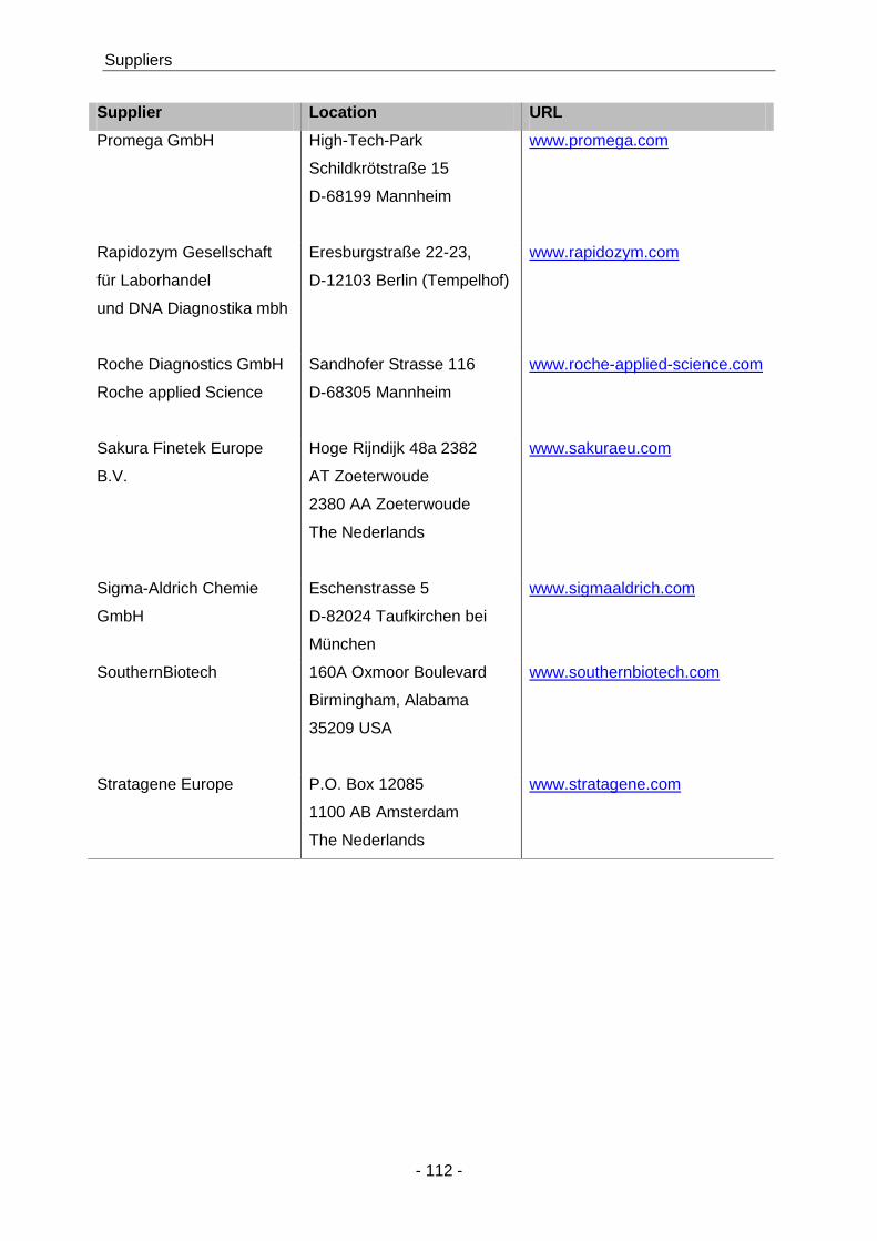
Suppliers
- 112 -
Supplier Location URL
Promega GmbH High-Tech-Park
Schildkrötstraße 15
D-68199 Mannheim
www.promega.com
Rapidozym Gesellschaft
für Laborhandel
und DNA Diagnostika mbh
Eresburgstraße 22-23,
D-12103 Berlin (Tempelhof)
www.rapidozym.com
Roche Diagnostics GmbH
Roche applied Science
Sandhofer Strasse 116
D-68305 Mannheim
www.roche-applied-science.com
Sakura Finetek Europe
B.V.
Hoge Rijndijk 48a 2382
AT Zoeterwoude
2380 AA Zoeterwoude
The Nederlands
www.sakuraeu.com
Sigma-Aldrich Chemie
GmbH
Eschenstrasse 5
D-82024 Taufkirchen bei
München
www.sigmaaldrich.com
SouthernBiotech 160A Oxmoor Boulevard
Birmingham, Alabama
35209 USA
www.southernbiotech.com
Stratagene Europe P.O. Box 12085
1100 AB Amsterdam
The Nederlands
www.stratagene.com

Aknowledgments
- 113 -
10 Acknowledgments
First of all I would like to thank my supervisor Simon Fillatreau for the opportunity to do my
Ph.D. work in his lab. I am very grateful for learning from his profound knowledge of
immunology and for his excellent scientific guidance throughout my time as a Ph.D. student.
I would like to thank Prof. Dr. Roland Lauster for his interest and willingness to be my “doctor
father” and evaluate my thesis.
Furthermore, a special thank you to P.D. Dr. Ulrich Steinhoff for his scientific and technical
support, which made possible to perform most of the experiments described on this thesis.
A special thank you also to Sabine Seibert, who introduced me to the Salmonella world.
Thanks to my lab mates Toralf Roch, Ping Shen, Ulrik Stervbo who contributed to this work
through productive scientific discussions and constructive critics. I would like to thank
especially Vicky and Eli, for their precious help in a lot of experiments, for their
encouragement and principally for their friendship during this time.
Finally, I wish to thank my wonderful family for their endless support and belief on me.
Especially my love and friend Matthias, for his support and patience during my Ph.D. work.
Publications
- 114 -
11 Publications
Lampropoulou V.*, K. Hoehlig *, T. Roch*, P. Neves, E. Calderón Gomez, C. H. Sweenie, Y.
Hao, A. A. Freitas, U.Steinhoff, S. M. Anderton, and S. Fillatreau, (* equally contributing
authors), Toll-like receptor-activated B cells suppress T cell-mediated autoimmunity. J
Immunol, 2008. 180(7): p.4763-73.
Hoehlig K., V. Lampropoulou, T. Roch, P. Neves, E. Calderón Gomez, S. M. Anderton,
U.Steinhoff, and S. Fillatreau, Immune regulation by B cells and antibodies: a review towards
the clinic. Adv Immunol, 2008. 98: in Press.
Lampropoulou V., E. Calderón Gomez, T. Roch, P. Neves, P. Shen, U. Stervbo, P. Boudinot,
S. M. Anderton, and S. Fillatreau, Suppressive functions of activated B cells in autoimmune
diseases reveal the dual roles of Toll-like receptors in immunity. Immunol reviews, 2009:
manuscript accepted.
Neves P.*, V. Lampropoulou*, E. Calderón-Gomez, Christoph Loddenkemper, Sergei A.
Nedospasov, U.Steinhoff, and S. Fillatreau, (* equally contributing authors), MyD88-signaling
in B cells suppresses immunity to Salmonella typhimurium. Immunity, 2009. Manuscript
submitted.