Thermodynamics AP Physics Chapter 15. Thermodynamics 13.3 Zeroth Law of Thermodynamics.

Subscriber access provided by Columbia Univ Libraries
Journal of the American Chemical Society is published by the American ChemicalSociety. 1155 Sixteenth Street N.W., Washington, DC 20036
Article
Role of the Active-Site Solvent in theThermodynamics of Factor Xa Ligand Binding
Robert Abel, Tom Young, Ramy Farid, Bruce J. Berne, and Richard A. FriesnerJ. Am. Chem. Soc., 2008, 130 (9), 2817-2831 • DOI: 10.1021/ja0771033
Downloaded from http://pubs.acs.org on November 18, 2008
More About This Article
Additional resources and features associated with this article are available within the HTML version:
• Supporting Information• Access to high resolution figures• Links to articles and content related to this article• Copyright permission to reproduce figures and/or text from this article

Role of the Active-Site Solvent in the Thermodynamics ofFactor Xa Ligand Binding
Robert Abel,† Tom Young,† Ramy Farid,‡ Bruce J. Berne,† andRichard A. Friesner*,†
Department of Chemistry, Columbia UniVersity, 3000 Broadway, New York, New York 10027,and Schro¨dinger, Inc., 120 West 45th Street, New York, New York 10036
Received September 13, 2007; E-mail: [email protected]
Abstract: Understanding the underlying physics of the binding of small-molecule ligands to protein activesites is a key objective of computational chemistry and biology. It is widely believed that displacement ofwater molecules from the active site by the ligand is a principal (if not the dominant) source of binding freeenergy. Although continuum theories of hydration are routinely used to describe the contributions of thesolvent to the binding affinity of the complex, it is still an unsettled question as to whether or not thesecontinuum solvation theories describe the underlying molecular physics with sufficient accuracy to reliablyrank the binding affinities of a set of ligands for a given protein. Here we develop a novel, computationallyefficient descriptor of the contribution of the solvent to the binding free energy of a small molecule and itsassociated receptor that captures the effects of the ligand displacing the solvent from the protein activesite with atomic detail. This descriptor quantitatively predicts (R2 ) 0.81) the binding free energy differencesbetween congeneric ligand pairs for the test system factor Xa, elucidates physical properties of the active-site solvent that appear to be missing in most continuum theories of hydration, and identifies several featuresof the hydration of the factor Xa active site relevant to the structure-activity relationship of its inhibitors.
Introduction
Understanding the underlying physics of the binding of small-molecule ligands to protein active sites is a key objective ofcomputational chemistry and biology. While a wide range oftechniques exist for calculating binding free energies, rangingfrom methods that should be accurate in principle (e.g., freeenergy perturbation theory) to relatively simple approximationsbased on empirically derived scoring functions, no completelysatisfactory and robust approach has yet been developed.Furthermore, physical insight into the sources of binding affinityis, arguably, as important as computing accurate numbers; assuch, insight would be extremely valuable in the design ofpharmaceutical candidate molecules.
It is widely believed that displacement of water moleculesfrom the active site by the ligand is a principal (if not thedominant) source of binding free energy. Water moleculessolvating protein active sites are often entropically unfavorabledue to the orientational and positional constraints imposed bythe protein surface, or they are energetically unfavorable dueto the water molecule’s inability to form a full complement ofhydrogen bonds when solvating the protein surface. This leadsto free energy liberation when a ligand that is suitably com-plementary to the active site displaces these waters into bulksolution, thus providing a relatively more favorable environment.Free energy perturbation methods are capable of computingthese free energy gains explicitly (within the accuracy of the
force field used in the simulations) but are computationally veryexpensive. Empirical scoring functions require negligible com-putational effort for a single ligand, but it has proven verydifficult to achieve high accuracy and robustness in this way.
“Standard” empirical scoring functions are dominated bylipophilic atom-atom contact terms that reward the closeapproach of lipophilic atoms of the ligand and protein. Suchfunctions are implicitly attempting to model the free energy gainupon displacement of waters by a given ligand atom, which ispresumed to depend upon the hydrophobicity of the proteinenvironment at the location of the ligand atom. Reasonableresults can be obtained in a fraction of cases with such anapproximation. However, as we have recently pointed out, thesimple atom-atom pair term fails to take into account thespecific positioning of the hydrophobic groups of the activesite.1,2 In particular, regions that exhibit “hydrophobic enclo-sure”, i.e., are surrounded by hydrophobic protein atoms, providea much less favorable environment for water molecules than isreflected in additive pair scoring. This argument applies not onlyto purely hydrophobic cavities but also to regions in which theligand must make a small number of hydrogen bonds butotherwise is hydrophobically enclosed by protein groups. A newempirical scoring function, implemented in the Glide dockingprogram as Glide XP,1 incorporates these geometrical factors
† Columbia University.‡ Schrodinger, Inc.
(1) Friesner, R. A.; Murphy, R. B.; Repasky, M. P.; Frye, L. L.; Greenwood,J. R.; Halgren, T. A.; Sanschagrin, P. C.; Mainz, D. T.J. Med. Chem.2006, 49, 6177-6196.
(2) Young, T.; Abel, R.; Kim, B.; Berne, B. J.; Friesner, R. A.Proc. Natl.Acad. Sci. U.S.A.2007, 104, 808-813.
Published on Web 02/12/2008
10.1021/ja0771033 CCC: $40.75 © 2008 American Chemical Society J. AM. CHEM. SOC. 2008 , 130, 2817-2831 9 2817

and has been shown to substantially improve the ability of thescoring function to separate active and inactive compounds.
While the Glide XP model represented a significant improve-ment as compared to previous empirical approaches, it shouldbe possible to achieve a higher level of detail, and numericalprecision, by mapping out the thermodynamics of water mole-cules in the active site, using explicit solvent simulations andappropriate approximations for the thermodynamic functions.In ref 2, we presented an initial effort in this direction, demon-strating that regions of the active site identified by Glide XP ashydrophobically enclosed dramatically affected the structure andthermodynamic properties of solvating water molecules. In onecase, the active site of cyclooxygenase-2 (COX-2), the activesite cavity dewetted; in a second, the active site of streptavidin,the solvating water molecules formed an ice-like five-memberedring, incurring a large entropic penalty in order to avoid loss ofhydrogen bonds. The thermodynamics of the solvating waterin these cases was analyzed via inhomogeneous solvation theory,proposed originally by Lazaridis,3 which provides an approxi-mate description of the hydration thermodynamics using datafrom relatively short (∼10 ns) molecular dynamics simulations.
In the present paper, we continue the line of researchdescribed in ref 2 by applying the inhomogeneous solvationtheory approach to study ligand binding in factor Xa (fXa), animportant drug target in the thrombosis pathway, severalinhibitors of which are currently in Phase III clinical trials.4
We use a clustering technique to build a map of water occupancyin the fXa active site, and we assign chemical potentials to thewater sites using the inhomogeneous solvation theory discussedabove.2 We then construct a semiempirical extension of themodel which enables computation of free energy differences(∆∆G values) for selected pairs of fXa ligands, and we comparethe success of this approach with the more standard technique,MM-GBSA.5,6 The free energy differences calculated from oursemiempirical model are shown to correlate exceptionally wellwith experimental data (R2 ) 0.81, reduced to 0.80 after leave-one-out (LOO) validation) via the use of only three adjustableparameters and to substantially out-perform the analogous MM-GBSA calculations (R2 ) 0.29). We investigated 31 pairs ofligands using data from only a single 10 ns MD simulation,illustrating the high computational efficiency of our methodol-ogy. Furthermore, the solvent chemical potential map producedhere appears to elucidate features of the known fXa structure-activity relationship (SAR) and would very likely provide auseful starting point for efforts to design novel compounds. Aneffort to calculate absolute binding free energies for highlydiverse ligands displays less accuracy and some over-fitting (aswould be expected, since the displacement of water moleculesis not the only factor determining binding affinity) but still showsa significant correlation with experimental data for this chal-lenging data set.
Results and Discussion
1. Mapping of the Thermodynamic Properties of theActive-Site Solvent.When a ligand binds to a protein, the watersolvating the active site is expelled into the bulk fluid. Thisexpulsion of the active-site solvent makes enthalpic and entropic
contributions to the binding free energy of the complex. Theless energetically or entropically favorable the expelled water,the more favorable its contributions to the binding free energy.The active sites of proteins provide very diverse environmentsfor solvating water. Water solvating narrow hydrophobicenclosures such as the COX-2 binding cavity is energeticallyunfavorable because it cannot form a full complement ofhydrogen bonds.2 Similarly, water molecules solvating enclosedprotein hydrogen-bonding sites are entropically unfavorablesince the number of configurations they can adapt whilesimultaneously forming hydrogen bonds with the protein andtheir water neighbors is severely reduced.2 The expulsion ofwater from such enclosed regions has been shown to lead toenhancements in protein binding affinity.1 From these observa-tions, we wanted to determine if a computationally derived mapof the thermodynamic properties of the active-site solvent couldbe used to rank the binding affinities of congeneric compounds.We hypothesized that the contributions to the binding freeenergy of adding acomplementarychemical groups i.e.,chemical groups that make hydrogen bonds where appropriateand hydrophobic contacts otherwises to a given ligand scaffoldcould largely be understood by an analysis of the solvent alone.
Testing this hypothesis requires a method to compute the localthermodynamic properties of the fXa active-site solvent. Weutilized data from 10 ns of explicitly solvated moleculardynamics simulations of fXa to sample the active-site solventdistribution for this receptor. We then clustered the active-sitesolvent distribution into high-occupancy 1 Å spheres, whichwe denoted as the “hydration sites” of the active-site cavity.Using inhomogeneous solvation theory, we then computed theaverage system interaction energy and excess entropy terms forthe water in each hydration site. Comparing the systeminteraction energy of the hydration sites with the bulk referencevalue allowed us to estimate the enthalpic cost of transferringthe water in the hydration site from the active site to the bulkfluid. The excess entropy calculated here can be used similarly.We also computed several other descriptors of the hydrationsite’s local environment. More details of these procedures andmeasurements are given in the Methods section. The data foreach hydration site are presented in Table 1. Figure 1 showsthe calculated energies and excess entropies for each of thehydration sites in the fXa binding cavity. Relative to otherhydration sites, the hydration sites circled in gray had poorsystem interaction energies, the hydration sites circled in greenhad unfavorable excess entropies, and the hydration sites circledin purple had both relatively poor system interaction energiesand entropies. We show the resulting three-dimensional active-site hydration map with this same color coding in Figure 2.
The hydration site map depicted in Figure 2 elucidated severalfeatures of the experimentally known SAR of the fXa ligands.Factor Xa inhibitors generally bind in an L-shaped conformation,where one group of the ligand occupies the anionic S1 pocketlined by residues Asp189, Ser195, and Tyr228 and another groupof the ligand occupies the aromatic S4 pocket lined by residuesTyr99, Phe174, and Trp215. Typically, a fairly rigid linker groupwill bridge these two interaction sites. The solvent analysisidentified three enthalpically unfavorable hydration sites, sites13, 18, and 21, solvating the fXa S4 pocket. This finding agreed
(3) Lazaridis, T.J. Phys. Chem. B1998, 102, 3531-3541.(4) Turpie, A. G.Arterioscler. Thromb. Vasc. Biol.2007, 27, 1238-1247.(5) Huang, N.; Kalyanaraman, C.; Bernacki, K.; Jacobson, M. P.Phys. Chem.
Chem. Phys.2006, 8, 5166-5177.(6) Lyne, P. D.; Lamb, M. L.; Saeh, J. C.J. Med. Chem.2006, 49, 4805-
4808.
A R T I C L E S Abel et al.
2818 J. AM. CHEM. SOC. 9 VOL. 130, NO. 9, 2008

with the experimental result that the S4 pocket has anexceptionally high affinity for hydrophobic groups.7,8 We alsoidentified a single, very high excess chemical potential hydration
site, site 12, solvating Tyr228 in the S1 pocket. Several studieshave found that introducing a ligand chlorine atom at thislocation, and hence displacing the water from this site, makesa large favorable contribution to the binding affinity.9-12
Additionally, we identified an energetically depleted hydrationsite, site 17, solvating the disulfide bridge between Cys191 andCys220. We expect that displacement of water from this sitewould make favorable contributions to the binding free energy.This agrees with several reported chemical series targeting thissite.13-15
We compared this hydration map with the locations of active-site crystallographic waters from the fXa apo-structure, crystalstructure 1HCG.16 Of the 11 crystallographic waters that resolvewithin the fXa active site, 9 are within 1.5 Å of a hydrationsite, and all of the crystallographic waters are within 2.5 Å ofa hydration site. One difficulty in the comparison is that weidentified in the active site many more hydration sites thancrystallographically resolved waters. However, this discrepancyis expected since the 1HCG crystal structure was only solvedto a resolution of 2.2 Å, and it has been noted that the numberof crystallographic water molecules identified in X-ray crystal-lography of proteins is quite sensitive to resolution (an average
(7) Young, R. J.; et al.Bioorg. Med. Chem. Lett.2006, 16, 5953-5957.(8) Matter, H.; Defossa, E.; Heinelt, U.; Blohm, P. M.; Schneider, D.; Muller,
A.; Herok, S.; Schreuder, H.; Liesum, A.; Brachvogel, V.; Lonze, P.;Walser, A.; Al-Obeidi, F.; Wildgoose, P.J. Med. Chem.2002, 45, 2749-2769.
(9) Adler, M.; Kochanny, M. J.; Ye, B.; Rumennik, G.; Light, D. R.; Biancalana,S.; Whitlow, M. Biochemistry2002, 41, 15514-15523.
(10) Matter, H.; Will, D. W.; Nazare, M.; Schreuder, H.; Laux, V.; Wehner, V.J. Med. Chem.2005, 48, 3290-3312.
(11) Nazare, M.; Will, D. W.; Matter, H.; Schreuder, H.; Ritter, K.; Urmann,M.; Essrich, M.; Bauer, A.; Wagner, M.; Czech, J.; Lorenz, M.; Laux, V.;Wehner, V.J. Med. Chem.2005, 48, 4511-4525.
(12) Maignan, S.; Guilloteau, J. P.; Choi-Sledeski, Y. M.; Becker, M. R.; Ewing,W. R.; Pauls, H. W.; Spada, A. P.; Mikol, V.J. Med. Chem.2003, 46,685-690.
(13) Maignan, S.; Guilloteau, J. P.; Pouzieux, S.; Choi-Sledeski, Y. M.; Becker,M. R.; Klein, S. I.; Ewing, W. R.; Pauls, H. W.; Spada, A. P.; Mikol, V.J. Med. Chem.2000, 43, 3226-3232.
(14) Mueller, M. M.; Sperl, S.; Sturzebecher, J.; Bode, W.; Moroder, L.J. Biol.Chem.2002, 383, 1185-1191.
(15) Quan, M. L.; et al.J. Med. Chem.2005, 48, 1729-1744.(16) Padmanabhan, K.; Padmanabhan, K. P.; Tulinsky, A.; Park, C. H.; Bode,
W.; Huber, R.; Blankenship, D. T.; Cardin, A. D.; Kisiel, W.J. Mol. Biol.1993, 232, 947-966.
Table 1. Calculated Thermodynamic and Local Water StructureData for Each of the 43 Hydration Sites We Identified byClustering the Factor Xa Active-Site Solvent Density Distributiona
hyd site occupancy−TSe
(kcal/mol)E
(kcal/mol) #nbrs #HBnbrs % HB exposure
neat 1385 n/ab -19.67 5.09 3.53 0.69 1.001 9347 4.00 -20.34 1.54 1.30 0.84 0.302 9062 3.91 -22.59 3.13 1.99 0.64 0.613 8425 2.61 -20.85 3.45 2.27 0.66 0.684 8383 2.93 -19.55 3.12 2.79 0.89 0.615 8157 3.24 -23.18 2.52 1.88 0.75 0.506 8123 3.20 -21.86 3.62 2.24 0.62 0.717 8116 3.37 -21.82 3.22 2.12 0.66 0.638 8081 2.74 -22.73 3.05 2.39 0.78 0.609 7257 2.13 -19.38 4.30 2.76 0.64 0.84
10 7172 2.52 -21.04 3.75 2.85 0.76 0.7411 6886 2.05 -20.71 3.41 2.24 0.66 0.6712 6815 2.28 -16.93 1.62 1.49 0.92 0.3213 6238 1.72 -17.88 2.72 2.05 0.75 0.5314 6081 1.95 -19.89 2.58 2.11 0.82 0.5115 5441 1.83 -22.62 4.66 3.63 0.78 0.9216 5078 1.51 -20.01 3.30 2.56 0.78 0.6517 4919 1.33 -17.04 2.45 1.78 0.73 0.4818 4887 1.35 -17.74 3.38 2.46 0.73 0.6619 4466 1.20 -19.48 4.11 2.77 0.67 0.8120 4386 1.37 -22.14 3.69 2.79 0.76 0.7221 4356 1.23 -18.50 3.75 2.67 0.71 0.7422 4241 1.22 -20.27 3.72 2.63 0.71 0.7323 4189 1.13 -19.58 3.87 2.84 0.73 0.7624 4170 1.17 -19.64 3.69 2.51 0.68 0.7225 4137 1.12 -20.85 4.61 2.59 0.56 0.9126 4067 1.07 -20.19 4.23 3.09 0.73 0.8327 4046 1.03 -20.72 4.37 3.48 0.80 0.8628 3921 1.10 -16.74 2.66 2.00 0.75 0.5229 3833 1.03 -21.44 4.27 2.57 0.60 0.8430 3793 1.04 -21.97 4.05 2.68 0.66 0.8031 3786 0.99 -20.00 4.70 3.39 0.72 0.9232 3686 0.99 -22.61 4.48 2.69 0.60 0.8833 3618 1.00 -20.46 4.34 2.56 0.59 0.8534 3570 0.95 -19.75 4.36 2.92 0.67 0.8635 3312 0.90 -24.24 4.41 2.74 0.62 0.8736 3296 0.84 -19.66 4.06 2.66 0.66 0.8037 3152 0.79 -18.87 4.57 3.15 0.69 0.9038 3094 0.73 -19.09 4.70 3.25 0.69 0.9239 3089 0.92 -21.61 3.55 2.55 0.72 0.7040 3007 0.79 -19.96 4.20 2.79 0.67 0.8241 3003 0.78 -20.41 3.71 2.70 0.73 0.7342 2862 0.73 -19.26 4.72 3.28 0.69 0.9343 2791 0.75 -20.93 3.98 2.84 0.71 0.78
a Occupancy is the number of water-oxygen atoms found occupying agiven hydration site during the 10 ns of molecular dynamics simulation.-TSe is the excess entropic contribution to the free energy calculated froma truncated expansion of the excess entropy in terms of correlations in thesingle particle translational and rotational density.E is average energy ofinteraction of the water molecules in a given hydration site with the rest ofthe system. The #nbrs value is the average number of neighboring watersfound within a 3.5 Å oxygen atom-to-oxygen atom distance from a wateroccupying the specified hydration site. The #HBnbrs value is the averagenumber of neighboring water oxygens found within a 3.5 Å distance fromthe water oxygen occupying the specified hydrations site that make a lessthan 30° oxygen-oxygen-hydrogen hydrogen-bonding angle with thiswater., The %HB value is the #HBnbrs/#nbrs fraction. Exposure is the #nbrsvalue divided by the bulk #nbrs value found in the bulk fluid.b The truncatedexpansion of the excess entropy used included only the first-order terms.The first-order excess entropic term for all neat fluids is strictly zero;however, the second-order and larger terms will be quite large.
Figure 1. System interaction energies (E) and the excess entropiccontribution to the free energy (-TSe) of water molecules in the principalhydration sites of the factor Xa active site. The system interaction energyis the average energy of interaction of the water molecules in a givenhydration site with the rest of the system, and the excess entropiccontribution to the free energy is calculated from a truncated expansion ofthe excess entropy in terms of correlation functions. Those hydration sitesthat were expected to make large energetic contributions when evacuatedby the ligand are circled in gray, those expected to make large entropiccontributions are circled in green, and those expected to make both entropicand enthalpic contributions are circled in purple.
Thermodynamics of Factor Xa Ligand Binding A R T I C L E S
J. AM. CHEM. SOC. 9 VOL. 130, NO. 9, 2008 2819

of 1.0 crystal waters per protein residue is expected at aresolution of 2 Å, but an average of 1.6-1.7 crystal waters perresidue is expected at a resolution of 1 Å).17 The number andlocation of crystallographic waters identified in X-ray crystal-lography of proteins have also been found to be sensitive totemperature, pH, solvent conditions, and the crystal packingconfiguration.18,19 Given these sources of noise, we found ouragreement was satisfactory and in line with other similarcomparisons of the solvent distributions obtained from moleculardynamics simulations and with those obtained from X-raycrystallography.20
To better quantify the visual correlation of the known SARof fXa binding compounds and thermodynamic properties ofthe hydration sites, we constructed a simple five-parameterscoring function based upon the hydration map of the fXa activesite that attempts to rank the relative binding affinities ofcongeneric fXa ligands. This scoring function was based on thefollowing physical principles: (1) if a heavy atom of a ligandoverlapped with a hydration site, it displaced the water fromthat site; and (2) the less energetically or entropically favorablethe expelled water, the more favorable its contributions to thebinding free energy. A hydration site would contribute to thebinding free energy if its excess entropy or system interactionenergy were beyond the fitted entropy and energy cutoffparameters,Sco andEco, respectively. A flat reward was givenfor any hydration site that had excess entropies or systeminteraction energies that were beyond these values. The ampli-tudes of the reward values,Srwd andErwd, were fit accordingly.A fit cutoff distance (Rco) was used to determine whether aheavy atom of the ligand displaced water from a hydration site.If the ligand heavy atom had the same position as the hydrationsite, the full values ofSrwd and Erwd would be awarded. The
reward was then linearly reduced to zero over the distanceRco.This scoring function was implemented as
where∆Gbind is the predicted binding free energy of the ligand,Ehs is the system interaction energy of a hydration site,Shs
e isthe excess entropy of a hydration site, andΘ is the Heavisidestep function. We will refer to this implementation as the“displaced-solvent functional”. Implementing this displaced-solvent functional was particularly simple since it is merely asum over the ligand heavy atoms and a restricted sum over theentropically structured and energetically depleted hydration sites,with a linear function of the hydration-site-ligand-atom approachdistance as its argument. Note that some hydration sitescontributed in both the entropic and energetic sums. We alsoconstructed a three-parameter scoring function based on thesame principles as the five-parameter scoring function, wherethe value ofRco was set to 2.8 Å and the values ofSrwd andErwd were forced to be equal, and an “ab initio” parameter freeform of the scoring function, where contributions from all ofthe hydration sites were included, theSrwd andErwd values weretaken to beSrwd ) Shs
e and Erwd ) Ebulk - Ehs, and anapproximate valueRco ) 2.24 Å was deduced from physicalarguments (see Methods). One minor technical point was that,in the ab intio form of the scoring function, the maximumcontribution from any given hydration was capped to neverexceed∆Ghs ) (Ebulk - Ehs) - TSe, i.e., the total computedtransfer free energy of a hydration site into the bulk fluid. Forthe three- and five-parameter functionals, we determined theoptimal values of parametersRco, Eco, Erwd, Sco, and Srwd by
(17) Carugo, O.; Bordo, D.Acta Crystallogr. D: Biol. Crystallogr.1999, 55,479-483.
(18) Mattos, C.Trends Biochem. Sci.2002, 27, 203-208.(19) Nakasako, M.J. Mol. Biol. 1999, 289, 547-564.(20) Makarov, V. A.; Andrews, B. K.; Smith, P. E.; Pettitt, B. M.Biophys. J.
2000, 79, 2966-2974.
Figure 2. Those hydration sites expected to contribute favorably to binding when evacuated by the ligand are here shown within the factor Xa active sitein wireframe. Those expected to contribute energetically are shown in gray, those expected to contribute entropically are shown in green, and those expectedto contribute energetically and entropically are shown in purple. The S1 and S4 pockets are labeled in yellow, as are several hydration sites discussed in thetext.
∆Gbind ) ∑lig,hs
Erwd(1 -| rblig - rbhs|
Rco) Θ(Ehs - Eco)
× Θ(Rco - | rblig - rbhs|)- T ∑lig,hs
Srwd(1 -| rblig - rbhs|
Rco)
× Θ(Shse - Sco) Θ(Rco - | rblig - rbhs|) (1)
A R T I C L E S Abel et al.
2820 J. AM. CHEM. SOC. 9 VOL. 130, NO. 9, 2008

fitting to the binding thermodynamics of a set of 31 congenericligand pairs and a set of 28 ligands found in crystal structures(see Methods).
It is important to note that this functional was not intendedto compute the absolute binding affinity of a given ligand andreceptor. Computing absolute binding affinities would requireterms that describe the loss of entropy of binding the ligand,the strength of the interaction energy between the ligand andthe protein, and the reorganization free energy of the protein inaddition to the contributions of solvent expulsion described here.However, for congeneric ligands that differ by only smallchemical modifications, these additional contributions are likelyquite small (given that those modifications are complementaryto the protein surface). The ability of the proposed form of thescoring function to describe free energy differences betweensuch congeneric ligand pairs tests if the thermodynamicconsequences to the binding free energy of these smallmodifications can be largely understood from only the propertiesof the excluded solvent.
2. Development and Testing of the Displaced-SolventFunctional on the Set of the Congeneric Inhibitor Pairs.Weprepared a data set of 31 congeneric inhibitor pairs of fXa (seeMethods) (Table 2). These 31 congeneric inhibitor pairs werepairs of fXa ligands that differed by at most three chemicalgroups. We expected that excluded solvent density effects woulddominate this data set since the other termss the proteinreorganization free energy, ligand conformational entropy, etc.swould be largely a consequence of the ligand scaffold sharedby both members of the pair. We optimized the parameters of
the displaced-solvent functionals to reproduce the experimentallymeasured differences in binding affinity between each of thesecongeneric ligand pairs. We also estimated the error of theresulting functionals with LOO cross-validation. The resultingvalues of the parameters can be found in the SupportingInformation Table 1, and plots of the predicted differences inbinding free energy versus the experimental values are shownin Figures 3 and 4, and Supporting Information Figure 1. Theagreement of the predictions of the functionals with theexperimental data was quite striking: the Pearson correlation
Table 2. Inhibition Data for the Congeneric Ligand Pairs Binding to Factor Xaa
initial ligand final ligand∆∆Gexp
(kcal/mol)∆∆G3p
(kcal/mol)∆∆G5p
(kcal/mol)∆∆Gab initio
(kcal/mol)∆∆GMM-GBSA
(kcal/mol) ref
1MQ5:XLC 1MQ6:XLD -2.94 -2.85 -2.54 -2.97 -4.22 Adler021NFU:RRP 1NFY:RTR -1.56 -2.56 -2.98 -3.23 -0.47 Maignan031NFX:RDR 1NFW:RRR -0.59 1.35 0.94 0.21 2.01 Maignan03Matter:25 Matter:28 -0.62 -0.61 -0.62 -2.17 -0.48 Matter05Matter:25 2BMG:I1H -1.05 -1.31 -1.31 -3.52 -3.8 Matter05Matter:28 2BMG:I1H -0.43 -0.70 -0.69 -1.35 -3.32 Matter05Mueller:3 Mueller:2 -0.90 -2.05 -2.34 -4.53 -8.35 Mueller02Haginoya:56 Haginoya:57 -0.59 -1.15 -1.12 -0.23 1.41 Haginoya04Haginoya:60 Haginoya:56 -0.19 0.00 0.00 0.08 0.81 Haginoya04Haginoya:56 1V3X:D76 -0.54 -1.15 -1.12 -0.31 -5.04 Haginoya04Haginoya:60 Haginoya:57 -0.79 -1.15 -1.12 -0.15 2.22 Haginoya041V3X:D76 Haginoya:57 -0.05 0.00 0.00 0.08 6.45 Haginoya04Haginoya:60 1V3X:D76 -0.74 -1.15 -1.12 -0.23 -4.23 Haginoya042BQ7:IID 2BQW:IIE -2.01 -1.73 -1.95 -5.42 -8.81 Nazare052BQ7:IID 2BOH:IIA -2.01 -1.80 -2.09 -1.98 -6.7 Nazare052BQW:IIE 2BOH:IIA 0.00 -0.07 -0.13 3.44 2.11 Nazare05Quan:11a Quan:43 -0.09 0.04 0.39 0.27 0.3 Quan05Quan:43 1Z6E:IK8 -0.68 -0.04 -0.45 -0.49 -3.47 Quan05Quan:11a 1Z6E:IK8 -0.77 0.00 -0.06 -0.22 -3.17 Quan051G2L:T87 1G2M:R11 -0.21 0.79 0.20 -0.32 10.1 Nar01Guertin:5c 1KSN:FXV -0.13 -0.87 -0.78 -0.68 -4.42 Guertin022FZZ:4QC 2G00:5QC -1.06 -1.70 -1.68 0.06 0.31 Pinto06Matter:107 1LQD:CMI -3.93 -2.52 -2.48 -2.84 -15.71 Matter02Matter:108 Matter:46 -3.09 -2.52 -2.48 -2.78 -11.49 Matter021F0R:815 1F0S:PR2 -0.12 0.09 0.14 -1.06 -3.53 Maignan00Young:33 2J4I:GSJ -0.82 0.00 0.00 0.36 -7.53 Young06Young:32 2J4I:GSJ -4.93 -4.87 -4.83 -5.31 -15.25 Young06Young:38 2J4I:GSJ -6.26 -4.87 -4.83 -5.67 -7.27 Young06Young:32 Young:33 -4.11 -4.87 -4.83 -5.67 -7.72 Young06Young:38 Young:33 -5.44 -4.87 -4.83 -6.03 0.26 Young06Young:38 Young:32 -1.33 0.00 0.00 -0.36 7.98 Young06
a Our predicted activity differences from the trained three-parameter and five-parameter displaced-solvent functionals and MM-GBSA method. When aligand was taken from a solved crystal structure, the ligand was designated “(PDB id):(ligand residue name)”, and when the ligand was built from congenericseries data, the ligand was designated “(first author of the reporting publication):(molecule number in the reporting publication)”.
Figure 3. Computed relative activities using the five-parameter form ofeq 1 versus experimental relative activities of the 31 congeneric inhibitorpairs with factor Xa. Note the stability of this fit under leave-one-out cross-validation.
Thermodynamics of Factor Xa Ligand Binding A R T I C L E S
J. AM. CHEM. SOC. 9 VOL. 130, NO. 9, 2008 2821
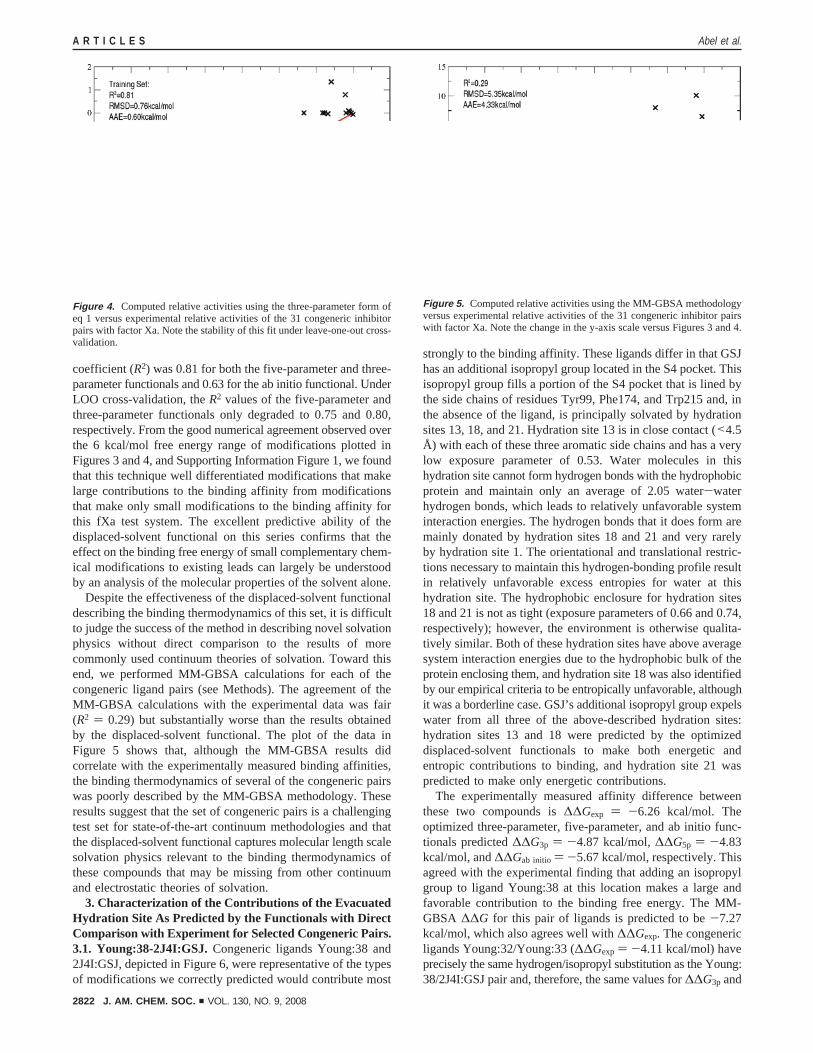
coefficient (R2) was 0.81 for both the five-parameter and three-parameter functionals and 0.63 for the ab initio functional. UnderLOO cross-validation, theR2 values of the five-parameter andthree-parameter functionals only degraded to 0.75 and 0.80,respectively. From the good numerical agreement observed overthe 6 kcal/mol free energy range of modifications plotted inFigures 3 and 4, and Supporting Information Figure 1, we foundthat this technique well differentiated modifications that makelarge contributions to the binding affinity from modificationsthat make only small modifications to the binding affinity forthis fXa test system. The excellent predictive ability of thedisplaced-solvent functional on this series confirms that theeffect on the binding free energy of small complementary chem-ical modifications to existing leads can largely be understoodby an analysis of the molecular properties of the solvent alone.
Despite the effectiveness of the displaced-solvent functionaldescribing the binding thermodynamics of this set, it is difficultto judge the success of the method in describing novel solvationphysics without direct comparison to the results of morecommonly used continuum theories of solvation. Toward thisend, we performed MM-GBSA calculations for each of thecongeneric ligand pairs (see Methods). The agreement of theMM-GBSA calculations with the experimental data was fair(R2 ) 0.29) but substantially worse than the results obtainedby the displaced-solvent functional. The plot of the data inFigure 5 shows that, although the MM-GBSA results didcorrelate with the experimentally measured binding affinities,the binding thermodynamics of several of the congeneric pairswas poorly described by the MM-GBSA methodology. Theseresults suggest that the set of congeneric pairs is a challengingtest set for state-of-the-art continuum methodologies and thatthe displaced-solvent functional captures molecular length scalesolvation physics relevant to the binding thermodynamics ofthese compounds that may be missing from other continuumand electrostatic theories of solvation.
3. Characterization of the Contributions of the EvacuatedHydration Site As Predicted by the Functionals with DirectComparison with Experiment for Selected Congeneric Pairs.3.1. Young:38-2J4I:GSJ.Congeneric ligands Young:38 and2J4I:GSJ, depicted in Figure 6, were representative of the typesof modifications we correctly predicted would contribute most
strongly to the binding affinity. These ligands differ in that GSJhas an additional isopropyl group located in the S4 pocket. Thisisopropyl group fills a portion of the S4 pocket that is lined bythe side chains of residues Tyr99, Phe174, and Trp215 and, inthe absence of the ligand, is principally solvated by hydrationsites 13, 18, and 21. Hydration site 13 is in close contact (<4.5Å) with each of these three aromatic side chains and has a verylow exposure parameter of 0.53. Water molecules in thishydration site cannot form hydrogen bonds with the hydrophobicprotein and maintain only an average of 2.05 water-waterhydrogen bonds, which leads to relatively unfavorable systeminteraction energies. The hydrogen bonds that it does form aremainly donated by hydration sites 18 and 21 and very rarelyby hydration site 1. The orientational and translational restric-tions necessary to maintain this hydrogen-bonding profile resultin relatively unfavorable excess entropies for water at thishydration site. The hydrophobic enclosure for hydration sites18 and 21 is not as tight (exposure parameters of 0.66 and 0.74,respectively); however, the environment is otherwise qualita-tively similar. Both of these hydration sites have above averagesystem interaction energies due to the hydrophobic bulk of theprotein enclosing them, and hydration site 18 was also identifiedby our empirical criteria to be entropically unfavorable, althoughit was a borderline case. GSJ’s additional isopropyl group expelswater from all three of the above-described hydration sites:hydration sites 13 and 18 were predicted by the optimizeddisplaced-solvent functionals to make both energetic andentropic contributions to binding, and hydration site 21 waspredicted to make only energetic contributions.
The experimentally measured affinity difference betweenthese two compounds is∆∆Gexp ) -6.26 kcal/mol. Theoptimized three-parameter, five-parameter, and ab initio func-tionals predicted∆∆G3p ) -4.87 kcal/mol,∆∆G5p ) -4.83kcal/mol, and∆∆Gab initio ) -5.67 kcal/mol, respectively. Thisagreed with the experimental finding that adding an isopropylgroup to ligand Young:38 at this location makes a large andfavorable contribution to the binding free energy. The MM-GBSA ∆∆G for this pair of ligands is predicted to be-7.27kcal/mol, which also agrees well with∆∆Gexp. The congenericligands Young:32/Young:33 (∆∆Gexp ) -4.11 kcal/mol) haveprecisely the same hydrogen/isopropyl substitution as the Young:38/2J4I:GSJ pair and, therefore, the same values for∆∆G3p and
Figure 4. Computed relative activities using the three-parameter form ofeq 1 versus experimental relative activities of the 31 congeneric inhibitorpairs with factor Xa. Note the stability of this fit under leave-one-out cross-validation.
Figure 5. Computed relative activities using the MM-GBSA methodologyversus experimental relative activities of the 31 congeneric inhibitor pairswith factor Xa. Note the change in the y-axis scale versus Figures 3 and 4.
A R T I C L E S Abel et al.
2822 J. AM. CHEM. SOC. 9 VOL. 130, NO. 9, 2008

∆∆G5p of -4.87 and-4.83 kcal/mol, respectively, which matchvery well with ∆∆Gexp. However, for this pair of ligands, theMM-GBSA predicted∆∆G is -7.72 kcal/mol, which is morenegative than the experimental value by 3.61 kcal/mol. Visualinspection of the MM-GBSA structure does not reveal the originof this discrepancy.
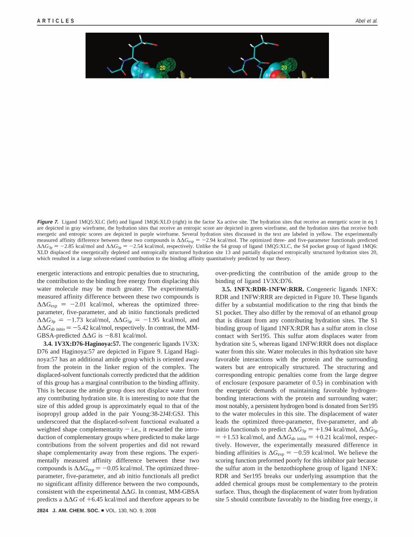
3.2. 1MQ5:XLC-1MQ6:XLD. The congeneric ligands 1MQ5:XLC and 1MQ6:XLD are depicted in Figure 7. This pair has amore subtle modification of the group binding the S4 pocketthan the Young:38-2J4I:GSJ congeneric pair described above.For this pair, the S4 binding group found in ligand 1MQ6:XLDoverlapped with hydration sites 13 and 20, whereas the S4binding group of ligand 1MQ5:XLC did not. As noted above,expulsion of water from hydration site 13 is expected to makeboth favorable energetic and entropic contributions to binding.Water in hydration site 20 has favorable energetic interactionsdue to several well-formed hydrogen bonds: water moleculesoccupying this the site predominately donate a hydrogen bondto the backbone carbonyl group of Glu97, nearly always receivea hydrogen bond from hydration site 4, and have good hydrogen-bonding interactions with hydration site 35. Hydration site 20,though, also incurred unfavorable contributions to its excessentropy due to the structuring required to maintain thesefavorable interactions. When displaced by the S4 binding groupof ligand 1MQ6:XLD, an electropositive carbon (the carbon isbound to an oxygen) comes into close contact with the backbonecarbonyl group of Glu97. This electropositive carbon likelyrecaptures much of the interaction energy between the proteincarbonyl group and the water in hydration site 20 without theassociated entropic cost. From these water thermodynamicsconsiderations, the optimized three-parameter, five-parameter,
and ab initio displaced-solvent functionals predict affinitydifferences of∆∆G3p ) -2.85 kcal/mol,∆∆G5p ) -2.54 kcal/mol, and ∆∆Gab initio ) -2.97 kcal/mol, respectively. Theexperimental difference in binding affinity between the twoligands is∆∆Gexp ) -2.94 kcal/mol. The MM-GBSA-predicted∆∆G for this pair of ligands is-4.22 kcal/mol.
3.3. 2BQ7:IID-2BQW:IIE. The congeneric ligands 2BQ7:IID and 2BQW:IIE are depicted in Figure 8. This congenericpair isolates the contribution of inserting a ligand chlorine atominto the region of the S1 pocket lined by the side chains ofresidues Ala190, Val213, and Tyr228. The chlorine atom on2BQW:IIE displaces water from hydration site 12, which istightly enclosed by the side chains of residues Ala190, Val213,and Tyr228. The exposure parameter of this hydration site isonly 0.32. This extremely tight enclosure by hydrophobic groupscaused the system interaction energy of water in this hydrationsite to be several kilocalories per mole less favorable than inthe neat fluid. Water molecules in this site maintained hydrogenbonds with its few water neighbors 92% of the simulation time,which made unfavorable contributions to its excess entropy. Thelocation of this hydration site coincided with the location of astructurally conserved water molecule that several studies haveshown is favorable to displace.10,11 Several studies have sug-gested that the free energy contribution of expelling thisstructurally conserved water should be close to the theoreticalmaximum of 2.0 kcal/mol derived by Dunitz from the thermo-dynamics of inorganic hydrates.10,12,21The Dunitz upper bound,however, is inappropriate here since it includes only entropiccontributions. Since water in this region suffers from both poor
(21) Dunitz, J. D.Science1994, 264, 670.
Figure 6. Ligand Young:38 (left) and ligand 2J4I:GSJ (right) in the factor Xa active site. The hydration sites that receive an energetic score in eq 1 aredepicted in gray wireframe, the hydration sites that receive an entropic score are depicted in green wireframe, and the hydration sites that receive bothenergetic and entropic scores are depicted in purple wireframe. Several hydration sites discussed in the text are labeled in yellow. The experimentallymeasured affinity difference between these two compounds is∆∆Gexp ) -6.26 kcal/mol. The optimized three- and five-parameter functionals predicted∆∆G3p ) -4.87 kcal/mol and∆∆G5p ) -4.83 kcal/mol, respectively. The isopropyl group of ligand 2J4I:GSJ displaces three energetically depleted hydrationsites, two of which are predicted to also be entropically structured, which resulted in a large predicted contribution to the binding affinity of the complex.
Thermodynamics of Factor Xa Ligand Binding A R T I C L E S
J. AM. CHEM. SOC. 9 VOL. 130, NO. 9, 2008 2823
energetic interactions and entropic penalties due to structuring,the contribution to the binding free energy from displacing thiswater molecule may be much greater. The experimentallymeasured affinity difference between these two compounds is∆∆Gexp ) -2.01 kcal/mol, whereas the optimized three-parameter, five-parameter, and ab initio functionals predicted∆∆G3p ) -1.73 kcal/mol,∆∆G5p ) -1.95 kcal/mol, and∆∆Gab initio ) -5.42 kcal/mol, respectively. In contrast, the MM-GBSA-predicted∆∆G is -8.81 kcal/mol.
3.4. 1V3X:D76-Haginoya:57.The congeneric ligands 1V3X:D76 and Haginoya:57 are depicted in Figure 9. Ligand Hagi-noya:57 has an additional amide group which is oriented awayfrom the protein in the linker region of the complex. Thedisplaced-solvent functionals correctly predicted that the additionof this group has a marginal contribution to the binding affinity.This is because the amide group does not displace water fromany contributing hydration site. It is interesting to note that thesize of this added group is approximately equal to that of theisopropyl group added in the pair Young:38-2J4I:GSJ. Thisunderscored that the displaced-solvent functional evaluated aweighted shape complementaritys i.e., it rewarded the intro-duction of complementary groups where predicted to make largecontributions from the solvent properties and did not rewardshape complementarity away from these regions. The experi-mentally measured affinity difference between these twocompounds is∆∆Gexp ) -0.05 kcal/mol. The optimized three-parameter, five-parameter, and ab initio functionals all predictno significant affinity difference between the two compounds,consistent with the experimental∆∆G. In contrast, MM-GBSApredicts a∆∆G of +6.45 kcal/mol and therefore appears to be
over-predicting the contribution of the amide group to thebinding of ligand 1V3X:D76.
3.5. 1NFX:RDR-1NFW:RRR. Congeneric ligands 1NFX:RDR and 1NFW:RRR are depicted in Figure 10. These ligandsdiffer by a substantial modification to the ring that binds theS1 pocket. They also differ by the removal of an ethanol groupthat is distant from any contributing hydration sites. The S1binding group of ligand 1NFX:RDR has a sulfur atom in closecontact with Ser195. This sulfur atom displaces water fromhydration site 5, whereas ligand 1NFW:RRR does not displacewater from this site. Water molecules in this hydration site havefavorable interactions with the protein and the surroundingwaters but are entropically structured. The structuring andcorresponding entropic penalties come from the large degreeof enclosure (exposure parameter of 0.5) in combination withthe energetic demands of maintaining favorable hydrogen-bonding interactions with the protein and surrounding water;most notably, a persistent hydrogen bond is donated from Ser195to the water molecules in this site. The displacement of waterleads the optimized three-parameter, five-parameter, and abinitio functionals to predict∆∆G3p ) +1.94 kcal/mol,∆∆G5p
) +1.53 kcal/mol, and∆∆Gab initio ) +0.21 kcal/mol, respec-tively. However, the experimentally measured difference inbinding affinities is∆Gexp ) -0.59 kcal/mol. We believe thescoring function preformed poorly for this inhibitor pair becausethe sulfur atom in the benzothiophene group of ligand 1NFX:RDR and Ser195 breaks our underlying assumption that theadded chemical groups must be complementary to the proteinsurface. Thus, though the displacement of water from hydrationsite 5 should contribute favorably to the binding free energy, it
Figure 7. Ligand 1MQ5:XLC (left) and ligand 1MQ6:XLD (right) in the factor Xa active site. The hydration sites that receive an energetic score in eq 1are depicted in gray wireframe, the hydration sites that receive an entropic score are depicted in green wireframe, and the hydration sites that receive bothenergetic and entropic scores are depicted in purple wireframe. Several hydration sites discussed in the text are labeled in yellow. The experimentallymeasured affinity difference between these two compounds is∆∆Gexp ) -2.94 kcal/mol. The optimized three- and five-parameter functionals predicted∆∆G3p ) -2.85 kcal/mol and∆∆G5p ) -2.54 kcal/mol, respectively. Unlike the S4 group of ligand 1MQ5:XLC, the S4 pocket group of ligand 1MQ6:XLD displaced the energetically depleted and entropically structured hydration site 13 and partially displaced entropically structured hydration sites 20,which resulted in a large solvent-related contribution to the binding affinity quantitatively predicted by our theory.
A R T I C L E S Abel et al.
2824 J. AM. CHEM. SOC. 9 VOL. 130, NO. 9, 2008

is more than offset by the loss of hydrogen-bonding energybetween the water and Ser195. This resulted in the displaced-solvent functional predicting 1NFX:RDR would be the tighterbinding ligand, in disagreement with the experimental data.Interestingly, MM-GBSA also over-predicts the stability ofligand 1NFX:RDR relative to ligand 1NFW:RRR; the MM-GBSA ∆∆G is +2.01 kcal/mol. The minimized MM-GBSAcomplex associated with ligand 1NFX:RDR incorrectly producesa strong hydrogen bond between the Ser195 side chain and thesulfur atom in the benzothiophene group of the ligand, whichis the result of erroneous conformational change in the side chainof Ser195.
4. Development and Testing of the Displaced-SolventFunctional on the Set of 28 Factor Xa Crystal StructureLigands. In addition to the set of 31 congeneric pairs, weprepared a data set of 28 inhibitors taken from solved fXa crystalstructures (see Methods) (Table 3). These fXa ligands belongedto many different congeneric series and typically did not sharea common chemical scaffold with each other. In the previoussection, we hypothesized that the contributions to the free energyof binding from changes in conformational entropy, protein-ligand interaction energy, and protein reorganization free energywould be similar for ligand pairs that shared a common chemicalscaffold. If this was the case, we posited that the differences inthe binding free energies of congeneric pairs could be understoodmainly by an analysis of the displaced solvent alone. The successof the displaced-solvent functionals outlined in the previoussection supports the validity of this hypothesis. However, forligand pairs that do not share a common scaffold, we wouldexpect that differences in these contributions would not be small
and that predictions based solely on an analysis of the solventwould be less successful. Despite this concern, since thefunctional performed well over the set of congeneric pairs, wewere interested in determining how much of the bindingaffinities of these ligands could be understood from only thecontributions described by the displaced-solvent functional, asmeasured by the root-mean-square deviation (rmsd), absoluteaverage error, andR2 values. To study this question, weoptimized the three- and five-parameter displaced-solvent func-tionals to reproduce the experimentally measured differencesin binding affinities between 378 unique ligand pairs (allcombinations) of this 28 ligand set, and we performed LOOcross-validation to better estimate the error of the functionals.The optimal values of the parameters can be found in SupportingInformation Table 2, and the agreement of the fit functionalsand the ab initio functional with the experimental data can befound in Figures 11 and 12, and Supporting Information Figure2. Although the three- and five-parameter functionals could betuned to correlate reasonably well with the experimental data(R2 ) 0.50 and 0.48, respectively), the performance under LOOcross-validation suggested substantial over-fitting of the five-parameter functional (LOOR2 ) 0.11). Notably though, thecross-validatedR2 of 0.30 (p-value of 0.24% as determined bya Monte Carlo permutation test) for the three-parameter fitindicated that terms of the type described by the displaced-solvent functional are likely important to understanding theabsolute binding thermodynamics of fXa ligand, but it alsoclearly indicated that more traditional terms will also be neededto quantitatively predict absolute binding free energies withdesired accuracies.
Figure 8. Ligand 2BQ7:IID (left) and ligand 2BQW:IIE (right) in the factor Xa active site. The hydration sites that receive an energetic score in eq 1 aredepicted in gray wireframe, the hydration sites that receive an entropic score are depicted in green wireframe, and the hydration sites that receive bothenergetic and entropic scores are depicted in purple wireframe. Several hydration sites discussed in the text are labeled in yellow. The experimentallymeasured affinity difference between these two compounds is∆∆Gexp ) -2.01 kcal/mol. The optimized three- and five-parameter functionals predicted∆∆G3p ) -1.73 kcal/mol and∆∆G5p ) -1.95 kcal/mol, respectively. Unlike the S1 group of ligand 2BQ7:IID, the S1 pocket group of ligand 2BQW:IIEdisplaces the energetically depleted and entropically structured hydration site 12 found within the S1 subgroove. The contribution to the binding affinitypredicted by the three-parameter and five-parameter displaced-solvent functionals agreed with experiment.
Thermodynamics of Factor Xa Ligand Binding A R T I C L E S
J. AM. CHEM. SOC. 9 VOL. 130, NO. 9, 2008 2825

In both the three- and five-parameter fits of the displaced-solvent functional to the set of 28 crystal structure ligands, twoparticular ligands, 1MQ6:XLD and 1FJS:Z34, were consistentlythe worst outliers in the set. Both of these ligands have excellentoverlap with contributing hydration sites but were out scoredby ligands that placed larger aromatic groups at similar positionsin the binding pocket, such as ligands 1Z6E:IK8, 2FZZ:4QC,and 2G00:5QC. This error was expected because our pairwisereward of atoms in close contact with the hydration sitesapproximated to what degree the contributing hydration siteswere displaced by the surface of the ligand. Thus, when anaromatic group displaced a hydration site, a disproportionatelylarge number of ligand atoms contributed in the displaced-solvent functional, since the tighter covalent bonding in thesegroups placed many ligand atoms closer in space to thehydration site than could be seen otherwise. We should notethat this systematic error was likely much less problematic inthe set of congeneric pairs because the pathological bulkyaromatic groups typically appeared in both congeners, leadingto an exact cancellation of this error. When ligands 1MQ6:XLDand 1FJS:Z34 were excluded from the fit, the LOO cross-validation of the three- and five-parameter functionals yieldR2
values of 0.40 and 0.55, respectively. This dramatic improve-ment of the stability and quality of the fit underscores how poorthe linear pairwise approximation of the excluded volume ofthe ligand was for inhibitors 1MQ6:XLD and 1FJS:Z34. It isalso possible that the known favorable electrostatic interactionbetween 1FJS:Z34 and the fXa S4 pocket, which was notdescribed by the displaced-solvent functional, contributed to1FJS:Z34 being an outlier in this data set.22
5. Cross Testing of the Trained Displaced-Solvent DensityFunctionals. It was interesting to check the transferability ofthe parameters trained on the set of 31 congeneric inhibitor pairsto the set of 28 crystal structure ligands (Supporting InforfmationTable 3). The optimized three- and five-parameter functionalstrained on the set of 31 congeneric inhibitor pairs each hadR2
values of 0.17 when predicting the relative binding affinitiesof the 28 crystal structure ligands to fXa. The functionalsperformed poorly because the values of the parameters weobtained from training to the set of congeneric pairs typicallypredicted the difference in binding affinity between crystalstructure pairs to be much too large (often greater than 10 kcal/mol). The reason for this may be subtle: typically only thetightest binding compound of a series will be crystallized, andeven then it is typically crystallized only if it binds with asubmicromolar affinity. Thus, if a ligand displaces a suboptimalportion of the active-site solvent density, then it, by construction,becomes a crystallized ligand only if it is possible to tune theother contributions to the free energy (ligand entropy, liganddesolvation free energy, protein ligand interaction energy, etc.)to offset this suboptimal active-site-solvent evacuation, resultingin the needed submicromolar affinity. So the magnitude of thecontributions predicted by the displaced-solvent functionals maybe qualitatively correct, but the other terms not described bythe functional systematically offset them.
We found an interesting contrast to this result when we usedthe three- and five-parameter functionals trained on the set of28 crystal structure ligands to predict the binding affinity
(22) Adler, M.; Davey, D. D.; Phillips, G. B.; Kim, S. H.; Jancarik, J.; Rumennik,G.; Light, D. R.; Whitlow, M.Biochemistry2000, 39, 12534-12542.
Figure 9. Ligand 1V3X:D76 (left) and ligand Haginoya:57 (right) in the factor Xa active site. The hydration sites that receive an energetic score in eq 1are depicted in gray wireframe, the hydration sites that receive an entropic score are depicted in green wireframe, and the hydration sites that receive bothenergetic and entropic scores are depicted in purple wireframe. Several hydration sites discussed in the text are labeled in yellow. The experimentallymeasured affinity difference between these two compounds is∆∆Gexp ) -0.05 kcal/mol. The optimized three- and five-parameter functionals predicted∆∆G3p ) 0.0 kcal/mol and∆∆G5p ) 0.0 kcal/mol, respectively. The addition of the amide group to ligand D76 contributes negligibly to the bindingaffinity of the complex, which the method predicted from the location of the amide group away from any structured or energetically depleted hydrationsites.
A R T I C L E S Abel et al.
2826 J. AM. CHEM. SOC. 9 VOL. 130, NO. 9, 2008

differences of the set of 31 congeneric inhibitor pairs. We foundthe three- and five-parameter functionals trained on the set ofcrystal structure ligands predicted the binding affinity differencesof the set of 31 congeneric inhibitor pairs withR2 values of0.53 and 0.59, respectively. This result suggested that thefunctional form of the displaced-solvent functional may havefundamental features that lend themselves to ranking the bindingaffinities of compounds that differ by deletions of atomss i.e.,as long as the chosen parameters are physically reasonable, theperformance of the functional over congeneric sets of this kindmay be quite good.
Table 3. Inhibition Data for the 28 Ligands Extracted from SolvedCrystal Structures Binding to Factor Xa and Our Predicted ActivityDifferences from the Trained Three-Parameter and Five-ParameterDisplaced-Solvent Functionals
liganda
∆Gexp
kcal/mol)∆G3p
(kcal/mol)∆G5p
(kcal/mol)∆Gab initio
(kcal/mol)
2BOK:784 -9.39 -6.12 -7.24 0.002J2U:GSQ -9.61 -7.26 -8.60 3.342BQ7:IID -9.62 -7.78 -8.77 3.001G2L:T87 -9.88 -6.86 -8.93 -0.032J34:GS5 -10.00 -6.73 -7.80 1.571G2M:R11 -10.09 -6.54 -8.42 0.291KYE:RUP -10.37 -7.47 -8.88 -0.031F0R:815 -10.45 -6.26 -7.53 -6.911F0S:PR2 -10.57 -6.39 -7.77 -5.852BMG:I1H -10.57 -8.49 -9.34 5.491NFU:RRP -10.57 -6.98 -8.50 -2.212J38:GS6 -10.67 -6.94 -8.06 2.151LQD:CMI -10.98 -8.22 -9.30 4.312CJI:GSK -11.22 -7.48 -8.52 1.762BQW:IIE -11.63 -8.07 -9.16 8.421NFX:RDR -11.63 -7.58 -8.97 0.692BOH:IIA -11.63 -8.61 -9.72 4.981NFY:RTR -12.12 -7.47 -8.89 1.011NFW:RRR -12.22 -7.21 -8.46 0.481MQ5:XLC -12.28 -8.53 -9.58 3.772J4I:GSJ -12.28 -7.98 -9.33 2.011EZQ:RPR -12.34 -8.41 -9.91 -1.991KSN:FXV -12.82 -8.10 -9.39 -2.591Z6E:IK8 -13.26 -9.90 -11.55 5.222FZZ:4QC -13.29 -9.93 -11.33 4.941FJS:Z34 -13.59 -7.04 -8.75 -0.052G00:5QC -14.36 -9.98 -11.44 4.881MQ6:XLD -15.22 -8.66 -9.75 6.74
a Each ligand was designated “(PDB id):(ligand residue name)”.
Figure 10. Ligand 1NFX:RDR (left) and ligand 1NFU:RRR (right) in the factor Xa active site. The hydration sites that receive an energetic score in eq 1are depicted in gray wireframe, the hydration sites that receive an entropic score are depicted in green wireframe, and the hydration sites that receive bothenergetic and entropic scores are depicted in purple wireframe. Several hydration sites discussed in the text are labeled in yellow. The experimentallymeasured affinity difference between these two compounds is∆∆Gexp ) -0.59 kcal/mol. The optimized three- and five-parameter functionals predicted∆∆G3p ) +1.94 kcal/mol and∆∆G5p ) +1.53 kcal/mol, respectively. The poor agreement of the theory with experiment here is due to the poor interactionenergy of the S1 pocket sulfur atom of 1NFX:RDR with Ser195 compared with hydration 5, which is not displaced when ligand 1NFU:RRR docks with thereceptor.
Figure 11. Computed activities using the five-parameter form of eq 1 versusexperimental activities for the set of 28 inhibitors with factor Xa. The poorstability of the fit under cross-validation suggested substantial over-fitting.
Thermodynamics of Factor Xa Ligand Binding A R T I C L E S
J. AM. CHEM. SOC. 9 VOL. 130, NO. 9, 2008 2827

Conclusions
Our results suggest that the expulsion of active-site waterstrongly impacts protein-ligand binding affinities in twoways: (1) hydrophobic ligand groups that displace water fromenergetically unfavorable (hydrophobically enclosed) environ-ments contribute enthalpically since the water molecules willmake more favorable interactions in the bulk fluid; and (2)ligand groups that displace entropically structured solventcontribute even when the solvent interacts favorably with theprotein since well-designed ligands will recapture the protein-water interaction energy. The congeneric inhibitor pair Young:38-2J4I:GSJ is a particularly clear example where the expulsionof active-site water that solvates an energetically unfavorableenvironment led to large favorable contributions to the bindingfree energy. In contrast, the congeneric pair 1MQ5:XLC-1MQ6:XLD offered an interesting example of the expulsion of waterfrom a hydration site with a favorable interaction energy andunfavorable excess entropy. The expulsion of water from thishydration site was found to be favorable by our empiricalcriteria, presumably because the ligand group that displaces thiswater does a reasonably good job recapturing the interactionenergy of the solvent with the protein with less entropic cost.The congeneric inhibitor pair 2BQ7:IID-2BQW:IIE illustratedthat these two solvent categories, energetically unfavorable andentropically unfavorable, are by no means mutually exclusiveand that the evacuation of solvent from the protein active sitewill often make both entropic and enthalpic contributions tothe binding free energy. Instrumental to our analysis is theassumption of complementaritys that is, that the differencebetween the water-protein energetic interactions and theligand-protein interactions was expected to be small. Thisassumption is valid when the ligands form hydrogen bonds withthe protein where appropriate and hydrophobic contacts other-wise; however, the congeneric ligand pair 1NFX:RDR/1NFW:RRR illustrated that ligands that violate this hypothesis will oftenbe mistreated by the method. This has relevance to modern drugdesign since it suggests that it is misleading to look at particularcrystal waters as favorable or unfavorable to displace, as is oftendone in structure-based drug design. Instead, it may be moreproductive to consider how thermodynamically favorable dis-
placing a crystal water will be when it is displaced by acomplementary chemical group of a ligand.
The empirical functionals we developed were quite successfulat quantifying the contributions to the free energy of bindingdue to the ligand evacuating energetically unfavorable andentropically structured solvent for the set of congeneric pairs.They were able to differentiate those modifications to an existingligand scaffold that made small contributions to the bindingaffinity of the complex from those modifications that made largecontributions over a 6 kcal/mol range. In their present form,the three- and five-parameter functionals may be useful to leadoptimization, since the functionals appeared to well describethe thermodynamics of adding small chemical groups to a givenligand scaffold that are complementary to the protein surface.The performance of the functionals on the set of 28 crystalstructure ligands suggests that terms of this type may make largecontributions to binding; however, these functionals should notbe used as a stand-alone tool for computational screening ofchemically diverse compounds. The reason for this was clear:the displaced-solvent functionals presented here neglect severalterms which will vary considerably over sets of chemicallydiverse ligands. These terms include the protein-ligand elec-trostatic and van der Waals interaction energies, ligand solvationfree energy, ligand configurational entropy, and protein-reorganization free energy. Thus, a functional designed forcomputational screening would have to include additional termsdescribing these types of contributions to the free energy inaddition to those contributions captured by the displaced-solventfunctional.
Methods
1. Structure Preparation and Simulation. We chose to use PDBcrystal structure 1FJS as our initial model of the fXa protein.22 Thisstructure was imported into the Maestro23 program, all crystallographicwaters were deleted, and hydrogens were added to the structureassuming a pH 7 environment. Chain L of the crystal structure wasalso deleted, since it contained no atoms within 20 Å of the fXa activesite. We then used the protein preparation utility found in Maestro torun a restrained minimization of the protein in the presence of the 1FJScrystal structure ligand.24 This removed bad steric contacts and improvedthe quality of the protein-protein and protein-ligand hydrogen-bondingwithout large rearrangements of the protein heavy atoms. Using theOPLSAA-200125 potential, we imported this model of the protein intoa modified version of GROMACS26,27 prepared by Shirts et al. Wethen solvated the system in a cubic TIP4P28 water box, where eachboundary was greater than 10 Å away from the protein, and added onechlorine ion to neutralize the system.
We minimized the energy of the system to relieve bad steric contactsbetween the protein and the water and equilibrated the system for 100ps with the velocity version of the Verlet integrator29 and Berendsen30
temperature and pressure controls at 298 K and 1 bar, where a frameof the system was saved every 1 ps. The Lennard-Jones interactionswere truncated at 9 Å, the electrostatic interactions were describedexactly for pairs within 10 Å and by Particle Mesh Ewald31,32 for pairs
(23) Banks, J. L.; et al.J. Comput. Chem.2005, 26, 1752-1780.(24) Friesner, R. A.; Banks, J. L.; Murphy, R. B.; Halgren, T. A.; Klicic, J. J.;
Mainz, D. T.; Repasky, M. P.; Knoll, E. H.; Shelley, M.; Perry, J. K.;Shaw, D. E.; Francis, P.; Shenkin, P. S.J. Med. Chem.2004, 47, 1739-1749.
(25) Kaminski, G. A.; Friesner, R. A.; Tirado-Rives, J.; Jorgensen, W. L.J.Phys. Chem. B2001, 105, 6474-6487.
(26) Lindahl, E.; Hess, B.; van der Spoel, D.J. Mol. Mod.2001, 7, 306-317.(27) Shirts, M. R.; Pande, V. S.J. Chem. Phys.2005, 122, 134508-134508.(28) Jorgensen, W. L.; Chandrasekhar, J.; Madura, J. D.; Impey, R. W.; Klein,
M. J. Chem. Phys.1983, 79, 926-935.
Figure 12. Computed activities using the three-parameter form of eq 1versus experimental activities for the set of 28 inhibitors with factor Xa.The moderate stability of the fit under cross validation suggested theproblems associated with over fitting were reduced when the three parameterform of eq 1 was used.
A R T I C L E S Abel et al.
2828 J. AM. CHEM. SOC. 9 VOL. 130, NO. 9, 2008

outside of this radius, and all protein heavy atoms were harmonicallyrestrained with spring constants of 1000 kJ mol-1 nm-2. We used thefinal 10 ps of equilibration data to seed 10 different 1 ns moleculardynamics trajectories with the velocity version of the Verlet integrator,29
Andersen33 temperature controls, and Parrinello-Rahman34,35pressurecontrols at 298 K and 1 bar. For these simulations, the Lennard-Jones,electrostatic forces, and harmonic restraints on the heavy atoms of theprotein were the same as in the equilibration simulations. Frames ofthis simulation were saved every 1 ps.
The MM-GBSA5,6 calculations of the protein-ligand bindingaffinities were carried out using Prime 1.6 and were set-up using thegraphical user interface available in Maestro 8.0. The free energy ofbinding was estimated using the following equation:∆Gbinding ) Ecomplex-(minimized)- Eligand(from minimized complex)- Ereceptor(from mini-mized complex). The OPLS-AA-2001 potential25 was used to modelthe protein and the ligand, and the Surface Generalized Born49 modelwas used to describe the polar and nonpolar contributions of the solvent.The protein and ligand coordinates used for these calculations weretaken directly from the reported crystal structure for the particularligand. The energy of each protein/ligand complex was determined byminimizing the ligand and residues within 8 Å of the ligand. Theenergies of the ligand and receptor are from the minimized complex,so the estimated binding energy is the protein-ligand interaction energywithout accounting for ligand or receptor strain. For each ligand pairthat involved one cocrystallized ligand and one modified ligand(constructed as described in Method section 3), the receptor that wasused for the MM-GBSA was the prepared (as described Methods section3) PDB structure associated with the co-cocrystallized ligand. For ligandpairs in which both ligands are cocrystallized, two MM-GBSA runswere conducted using both protein structures associated with each ofthe ligands, and the results were reported for the receptor structure thatyielded the smallest change in the ligand conformation following theMM-GBSA calculation.
2. Active-Site Hydration Analysis. In order to analyze thethermodynamic and structural properties of the water moleculeshydrating the fXa active site, we needed to develop some sensibledefinition for when a solvating water should be considered within thefXa active site and when it should not.2 We used a set of 35 fXa crystalstructures with bound inhibitors to define the volume of the active site(PDB structures 1EZQ,13 1F0R,13 1F0S,13 1FAX,36 1FJS,22 1G2L,37
1G2M,37 1IOE,38 1IQE,38 1IQF,38 1IQG,38 1IQH,38 1IQI,38 1IQJ,38
1IQK,38 1IQL,38 1IQM,38 1IQN,38 1KSN,39 1KYE,14 1MQ5,9 1MQ6,9
1NFU,12 1NFW,12 1NFX,12 1NFY,12 1V3X,41 1XKA,41 1XKB,41
2BOK,42 2CJI,43 2J2U,44 2J34,44 2J38,44 and 2J4I7). We computed amultiple structure alignment between the 35 fXa crystal structurescontaining inhibitors and our prepared fXa model structure. Thisalignment rotated the crystal structures onto our prepared fXa structure.This procedure also rotated the inhibitors found in these crystalstructures into the active site of our prepared model fXa structure. Theresults of these alignments were hand-inspected for severe steric clashes,and none were found. Using this set of aligned structures, we definedthe active site as the volume containing all points in space that arewithin 3 Å of any ligand heavy atom. The position of the active-sitevolume was constant throughout the simulation because the proteinheavy atoms were harmonically restrained. The coordinates of all watersobserved within this region of space during the 10 ns of simulationdata were saved every 1 ps. We considered this water distribution tobe the equilibrium distribution of water within the fXa active site, andwe characterized its thermodynamic properties with inhomogeneoussolvation theory along with several other measures of local waterstructure.
The application of inhomogeneous solvation theory to the hetero-geneous surface of a protein active site where the solvating waters canexchange with the bulk fluid is highly nontrivial. Although the difficultyposed by waters exchanging with the bulk fluid is alleviated by ourdefinition of the active site, the inhomogeneous topography of theprotein surface made the orientational distributions of the watermolecules highly dependent on their position within the active site.Following procedures we previously developed,2 we partitioned theactive-site volume into small subvolumes which we denote “hydrationsites” and treated the angular distributions as independent of positionin these subvolumes. We identified the subvolumes by applying aclustering algorithm to partition the solvent density distribution into aset of high-water-occupancy, 1 Å radius spheres. This algorithm cycledthrough the positions of the oxygen atom of every water molecule foundin the active-site solvent density distribution and found the positionthat has the greatest number of water neighbors within a 1 Å radius.We denoted this position as a hydration site and removed it and all ofthe oxygen positions within 1 Å of it from the solvent densitydistribution. This process was then repeated, cycling through theremaining positions. This loop was terminated when the clusteringalgorithm identified a hydration site with a water-oxygen occupancyless than twice the expected value of a 1 Å radius sphere in the bulkfluid. These hydration sites are well-defined subvolumes of the activesite and have good convergence properties for the inhomogeneoussolvation theory machinery since they have sparse water density towardthe edges of the clusters.
We performed an inhomogeneous solvation theory analysis of thethermodynamic properties of each hydration site to elucidate how theproperties of the solvating water may affect the thermodynamics offXa inhibitor association. Consistent with our prior work, we definedthe system interaction energy (Ehs) of each hydration site to be theaverage energy of interaction of the water molecules in a givenhydration site with the rest of the system.2 We also computed the partialexcess entropy (Se) of each hydration site by numerically integratingan expansion of the entropy in terms of orientational and spatialcorrelation functions.3,45,46In this work we included only contributionsfrom the first-order term for each hydration site:
(29) Swope, W. C.; Anderson, H. C.; Berens, P. H.; Wilson, K. R.J. Chem.Phys.1982, 76, 637-649.
(30) Berendsen, H. J. C.; Postma, J. P. M.; DiNola, A.; Haak, J. R.J. Chem.Phys.1984, 81, 3684-3690.
(31) Darden, T.; York, D.; Pedersen, L.J. Chem. Phys.1993, 98, 10089-10092.(32) Essmann, U.; Perera, L.; Berkowitz, M. L.; Darden, T.; Lee, H.; Pedersen,
L. G. J. Chem. Phys.1995, 103, 8577-8592.(33) Andersen, H. C.J. Chem. Phys.1980, 72, 2384-2393.(34) Parrinello, M.; Rahman, A.J. Appl. Phys.1981, 52, 7182-7190.(35) Nose, S.; Klein, M. L.Mol. Phys.1983, 50, 1055-1076.(36) Brandstetter, H.; Kuhne, A.; Bode, W.; Huber, R.; von der Saal, W.;
Wirthensohn, K.; Engh, R. A.J. Biol. Chem.1996, 271, 29988-29992.(37) Nar, H.; Bauer, M.; Schmid, A.; Stassen, J. M.; Wienen, W.; Priepke, H.
W.; Kauffmann, I. K.; Ries, U. J.; Hauel, N. H.Structure2001, 9, 29-38.(38) Matsusue, T.; Shiromizu, I.; Okamoto, A.; Nakayama, K.; Nishida, H.;
Mukaihira, T.; Miyazaki, Y.; Saitou, F.; Morishita, H.; Ohnishi, S.;Mochizuki, H. To be published.
(39) Guertin, K. R.; et al.Bioorg. Med. Chem. Lett. 2002, 12, 1671-1674.(40) Haginoya, N.; Kobayashi, S.; Komoriya, S.; Yoshino, T.; Suzuki, M.;
Shimada, T.; Watanabe, K.; Hirokawa, Y.; Furugori, T.; Nagahara, T.J.Med. Chem.2004, 47, 5167-5182.
(41) Kamata, K.; Kawamoto, H.; Honma, T.; Iwama, T.; Kim, S. H.Proc. Natl.Acad. Sci. U.S.A.1998, 95, 6630-6635.
(42) Scharer, K.; Morgenthaler, M.; Paulini, R.; Obst-Sander, U.; Banner, D.W.; Schlatter, D.; Benz, J.; Stihle, M.; Diederich, F.Angew. Chem., Int.Ed. 2005, 44, 4400-4404.
(43) Watson, N. S.; et al.Bioorg. Med. Chem. Lett.2006, 16, 3784-3788.(44) Senger, S.; Convery, M. A.; Chan, C.; Watson, N. S.Bioorg. Med. Chem.
Lett. 2006, 16, 5731-5735.(45) Baranyai, A.; Evans, D. J.Phys. ReV. A 1989, 40, 3817-3822.(46) Morita, T.; Hiroike, K.Prog. Theor. Phys. 1961, 25, 537-578.(47) Pinto, D. J.; et al.Bioorg. Med. Chem. Lett.2006, 16, 5584-5589.(48) Pinto, D. J.; et al.Bioorg. Med. Chem. Lett.2006, 16, 4141-4147.(49) Ghosh, A.; Rapp, C. S.; Friesner, R. A.J. Phys. Chem. B1998, 102, 10983-
10990.
Se ) -kbFw
Ω ∫gsw(r ,ω) ln(gsw(r ,ω)) dr dω
≈ -kbFw ∫gsw(r ) ln(gsw(r )) dr
-kbNW
V
Ω ∫gsw(ω) ln(gsw(ω)) dω (2)
Thermodynamics of Factor Xa Ligand Binding A R T I C L E S
J. AM. CHEM. SOC. 9 VOL. 130, NO. 9, 2008 2829

wherer andω are the Cartesian position and Euler angle orientationof a water molecule, respectively,gsw(r ,ω) is the one-body distributionof the water (w) atr andω in the fixed references frame of the soluteprotein (s),Fω is the density of the neat TIP4P system,kb is theBoltzmann constant,Ω is the total orientational space accessible to awater molecule, andNW
V is the total number of water oxygens foundwithin a given hydration site of volumeV. We numerically integratedthe translational contribution to the excess entropy in sphericalcoordinates using a length of 0.03 Å alongr , 15° along θ, and 30°alongφ, and we numerically integrated the orientational contributionwith 10° along each Euler angle.
We also calculated several measures of local water structureproperties for the water molecules found within each hydration site:the average number of water neighbors, the average number ofhydrogen-bonding water neighbors, the fraction of the water neighborsthat were hydrogen-bonding, and the water exposure of each hydrationsite. These averages are for all water molecules in each hydration site.The number of neighbors value is the average number of watermolecules found within 3.5 Å, where the distance is measured water-oxygen to water-oxygen. We used a geometric definition of a hydrogenbond where two water molecules were deemed to be hydrogen-bondedif their oxygens were within 3.5 Å of each other and at least oneoxygen-oxygen-hydrogen angle was less than 30°. The exposure valuequantifies to what degree a hydration site is surrounded by other watermolecules: a value of unity suggests it is in a water environment similarto the bulk fluid, and a value of zero suggests the hydration site isoccluded from any other solvent molecules. The exposure value iscomputed as the average number of neighbors that water moleculeshave in a hydration site, divided by the average number of neighborsthat a water molecule has in the bulk.
3. Construction of the Factor Xa Ligand Binding Affinity DataSets.Within the PDB, we found 28 published crystal structures of fXabound to various inhibitors with thermodynamic binding data reportedin the associated publication (2BOK,42 2J2U,44 2BQ7,10 1G2L,37 2J38,44
1G2M,37 1KYE,14 1F0R,13 1F0S,13 2BMG,10 1NFU,12 2J34,44 1LQD,8
2CJI,43 2BQW,10 1NFX,12 2BOH,11 1NFY,12 1NFW,12 1MQ5,9 2J4I,7
1EZQ,13 1KSN,39 1Z6E,15 2G00,47 1FJS,22 2FZZ,48 1MQ69). Wecomputed a multiple-structure alignment between the 28 fXa crystalstructures containing inhibitors and our prepared fXa model structure.This procedure rotated the 28 inhibitors found in these crystal structuresinto the active site of our prepared model fXa structure. The results ofthese alignments were hand-inspected for severe steric clashes, and nonewere found. The orientations of each of these 28 inhibitors with respectto our prepared model fXa structure were saved and were referred toas the 28 crystal structure ligand set.
From this set of 28 crystal structure ligands, we prepared a set of31 congeneric inhibitor pairs. The goal of this set of inhibitor pairswas to isolate the effects of solvent displacement on the free energy ofbinding. Each congeneric pair was created either by noting that two ofthe crystal structure ligands reported in the prior set were congenericor by building a congeneric pair from a single-crystal structure ligandby deleting or swapping atoms of the crystal structure ligand. Wedevised several rules to construct this set. When any two members ofthe 28 crystal structure ligand set were reported in the same publicationand differed by no more than three chemical groups, they wereconsidered congeneric pairs. When the publication reporting the crystalstructure ligand contained congeneric series data for structurallysimilar ligands, we followed three rules to build new congeneric pairs:
1. We would only delete atoms from a crystal structure ligand andnot add them.
2. We would not accept deletions of atoms that resulted in a groupthat could rotate around a single bond and donate hydrogen bonds.
3. A congeneric pair that was built by changing the identity of aligand atom (for instance, changing a carbon atom to an oxygen atom)must have the change applied to both members of the pair.
These three rules were intended to minimize the error of assumingthat the binding mode of the new inhibitor structures, which were builtfrom deleting and swapping atoms of the crystallized inhibitors, wouldnot change. These rules were also intended to minimize differences incontributions to binding affinity from non-solvent-related terms for eachinhibitor pair, such as the loss of entropy of docking the ligand, thestrength of the interaction energy between the ligand and the protein,and the reorganization free energy of the protein. We expected thatexcluded solvent density effects would dominate this set since theseother non-solvent-related terms contributing to the free energy ofbinding would be relatively constant for each congeneric pair. We alsochose to compare binding affinities only between pairs of ligands thatwere determined in the same publication, due to the variance inexperimental methods commonly employed. We referred to the resultingset as the set of 31 congeneric inhibitor pairs (Supporting InformationTable 4).
4. Development and Parametrization of the Displaced-SolventFunctional. We devised a five-parameter scoring function to determineif the relative binding affinities of the 28 crystal structure ligands andthe binding affinity differences of the 31 congeneric inhibitor pairscorrelated with the thermodynamic properties of the displaced active-site solvent. Additional discussion of the physical motivation that ledus to this functional form can be found in the Results and Discussionsection. The form of the functional was a sum over ligand heavy atomsand a sum over hydration sites. Each time a ligand heavy atom wasfound within some parametrized distance of a hydration site with aninteraction energy or excess entropy predicted to be favorable toevacuate by some fit empirical criteria, an additive contribution wassummed. The functional itself was
where∆Gbind was the predicted binding free energy of the ligand,Rco
was the distance cutoff for a ligand atom beginning to displace ahydration site,Eco was the minimumEhs of a hydration site that wasconsidered energetically depleted,Erwd was the energetic contributionto ∆Gbind for displacing an energetically depleted hydration site,Sco
was the minimumSe term of a hydration site that was consideredentropically structured,-TSrwd was the entropic contribution to∆Gbind
for displacing an entropically structured hydration site, andΘ was theHeaviside step function. We also considered a three-parameter formof this equation, where we fixedRco ) 2.8 Å and-TSrwd ) Erwd.
The parameters were optimized by a Monte Carlo walk in parameterspace. The error function we used to train the parameters was the root-mean-square-deviation of the predicted relative binding free energiesof the 28 crystal ligands and the rmsd of the differences in the bindingfree energies of the 31 congeneric pairs. For the training of the three-and five-parameter functionals on the 28 crystal structure ligand set,we chose initial seed values ofRco ) 2.8 Å, Erwd ) -0.5 kcal/mol,-TSrwd ) -0.5 kcal/mol,Eco ) -18.5 kcal/mol, andTSco ) 1.5 kcal/mol. Five separate 1000-step optimizations were run, where the firstmove was always accepted and the lowest rmsd value encountered inthese optimizations was taken to be the optimal parameter set. Theinitial seed values used to train the three- and five-parameter functionalson the set of 31 congeneric inhibitor pairs wereRco ) 2.8 Å, Erwd )-1.0 kcal/mol,-TSrwd ) -1.0 kcal/mol,Eco ) -18.5 kcal/mol, andTSco ) 1.5 kcal/mol. The parameters were then optimized in a procedureidentical to that used for the 28 crystal structure ligands.
We also constructed an “ab initio” form of the displaced-solventfunctional containing no fit parameters. The functional itself was
∆Gbind ) ∑lig,hs
Erwd(1 -| rblig - rbhs|
Rco) Θ(Ehs - Eco)
× Θ(Rco - | rblig - rbhs|)- T ∑lig,hs
Srwd(1 -| rblig - rbhs|
Rco)
× Θ(Shse - Sco) Θ(Rco - | rblig - rbhs|) (1)
A R T I C L E S Abel et al.
2830 J. AM. CHEM. SOC. 9 VOL. 130, NO. 9, 2008

where∆Gbind was the predicted binding free energy of the ligand,Rco
was the distance cutoff for a ligand atom beginning to displace ahydration site, and∆Ghs was the computed free energy of transferringthe solvent in a given hydration site from the active site to the bulkfluid. We also capped the contribution from each hydration site, suchthat it would never contribute more than∆Ghs to ∆Gbind, no matterhow many ligand atoms were in close proximity to it. The valueRco
might be considered a free parameter. However, an approximate valuewas adopted by noting that the radii of a carbon atom and a water-oxygen atom are both approximately 1.4 Å, thus suggesting that contactdistances between a water-oxygen atom and a ligand carbon atom lessthan 0.8× (1.4 Å + 1.4 Å) ) 2.24 Å are statistically improbable dueto the stiffness of the van der Waals potential.50 Thus, we chose forthe ab initio functional to specifyRco ) 2.24 Å. The sensitivity of theresults reported here to this choice of theRco parameter was quite lowfor adjustments of this value within a few tenths of an angstrom of thespecified value.
We estimated the error of the resulting optimized functionals withLOO cross-validation. In this technique, a functional is trained to anN- 1 point subset of data, and then the value of pointN is predictedwith this functional. This is repeatedN times, once for each data point,
and the error of the functional is estimated by summing the error ofthe predictions for each of these points. The Pearson correlationcoefficient (R2) computed in this procedure for theN data points isbounded by theR2 value found by training of the functional on allNdata points and zero. A cross-validationR2 value close to theR2 valuefound by training of the functional on allN data points suggests thatvery little over-fitting has occurred when training the functional.
We performed a Monte Carlo permutation analysis to estimate thep-value of a givenR2 value. The analysis proceeded by generating 5million random permutations of the mapping between the predicted∆G values and the experimentally measured∆G values. Thep-valueof a givenR2 value was taken to be the number of random permutationsyielding anR2 value greater than or equal to the originalR2 value,divided by the total number of permutations generated. This allowedus to very accurately estimate the probability of attaining a givenR2
value for our particular distribution without assuming any propertiesabout the relationships between the predicted and experimentallymeasured distributions.
Acknowledgment. This work was supported in part by a grantfrom the NIH to R.A.F. (GM-52018) and to B.J.B. (GM-43340).This material is based upon work supported in part by a NationalScience Foundation Graduate Research Fellowship.
Supporting Information Available: Complete refs 7, 15, 23,39, 43, 47, and 48; figures showing computed activities usingthe ab initio form of eq 1 versus experimental activities of the31 congeneric inhibitor pairs and of the set of 28 inhibitors withfactor Xa; tables analyzing the three- and five-parameter formsof the displaced-solvent functionals; and 2D structures of eachof the congeneric pairs and the crystal structure ligands. Thismaterial is available free of charge via the Internet at http://pubs.acs.org.
JA0771033(50) Jacobson, M. P.; Pincus, D. L.; Rapp, C. S.; Day, T. J.; Honig, B.; Shaw,
D. E.; Friesner, R. A.Proteins2004, 55, 351-367.
∆Gbind ) ∑lig,hs
(Ebulk - Ehs) (1 -| rblig - rbhs|
Rco)
× Θ(Rco - | rblig - rbhs|)
- T ∑lig,hs
Shse (1 -
| rblig - rbhs|Rco
) Θ(Rco - | rblig - rbhs|)
) ∑lig,hs
∆Ghs(1 -| rblig - rbhs|
Rco) Θ(Rco - | rblig - rbhs|) (3)
Thermodynamics of Factor Xa Ligand Binding A R T I C L E S
J. AM. CHEM. SOC. 9 VOL. 130, NO. 9, 2008 2831