
ROLE OF MICRORNA-363 IN HUMAN …d-scholarship.pitt.edu/12098/1/Wald_2012.pdf · revealed the...
136
ROLE OF MICRORNA-363 IN HUMAN PAPILLOMAVIRUS-ASSOCIATED SQUAMOUS CELL CARCINOMA OF THE HEAD AND NECK by Abigail Ilene Wald Bachelor of Science, University of Wisconsin – Madison, 2006 Submitted to the Graduate Faculty of the School of Medicine in partial fulfillment of the requirements for the degree of Doctor of Philosophy University of Pittsburgh 2012
Transcript of ROLE OF MICRORNA-363 IN HUMAN …d-scholarship.pitt.edu/12098/1/Wald_2012.pdf · revealed the...

i
ROLE OF MICRORNA-363 IN HUMAN PAPILLOMAVIRUS-ASSOCIATED SQUAMOUS CELL CARCINOMA OF THE HEAD AND NECK
by
Abigail Ilene Wald
Bachelor of Science, University of Wisconsin – Madison, 2006
Submitted to the Graduate Faculty of
the School of Medicine in partial fulfillment
of the requirements for the degree of
Doctor of Philosophy
University of Pittsburgh
2012

ii
UNIVERSITY OF PITTSBURGH
SCHOOL OF MEDICINE
This dissertation was presented
by
Abigail Ilene Wald
It was defended on
April 24, 2012
and approved by
Robert L. Ferris, M.D., Ph.D. Professor, Department of Immunology
Hideho Okada, M.D., Ph.D.
Associate Professor, Department of Immunology
Paul Robbins, Ph.D. Professor, Department of Microbiology and Molecular Genetics
Saumendra Sarkar, Ph.D.
Assistant Professor, Department of Microbiology and Molecular Genetics
Dissertation Advisor: Saleem A. Khan, Ph.D.
Professor, Department of Microbiology and Molecular Genetics

iii
Copyright © by Abigail Ilene Wald
2012

iv
Squamous cell carcinoma of the head and neck (SCCHN) ranks sixth among cancers worldwide.
Despite recent advances in the detection and therapy of SCCHN, the five-year survival rate
remains low. Human papillomavirus (HPV) has been detected in a new class of SCCHN that has
emerged in younger patients without the common risk factors of alcohol and/or tobacco. HPV-
positive and HPV-negative SCCHN have different clinical characteristics, leading some to
classify them as distinct diseases. High-risk HPVs have been implicated in a number of cancers
including cervical, anogenital, and head and neck cancers. Micro (mi) RNAs are a recently
discovered class of endogenously encoded small RNAs that most commonly function as negative
regulators of gene expression. MiRNA expression profiles are often altered in cellular stress
conditions, including cancers and viral infections. We hypothesized that HPV disrupts cellular
miRNA expression in HPV-positive SCCHN and contributes to the development of this cancer.
MiRNA microarray analysis of HPV-positive and HPV-negative SCCHN cell lines
revealed the dysregulation of several miRNAs, including the upregulation of miR-363 and
downregulation of miR-181a, miR-218, and miR-29a. Exogenous expression of the HPV-16 E6
oncogene in normal primary human keratinocytes showed similar changes in the above miRNAs,
implicating HPV E6 in this up/downregulation. Tissues from SCCHN patients in the western
Pennsylvania area showed an HPV-positivity rate of 59 percent. HPV-positive SCCHN cases
were seen only in the tonsil and base of tongue, and patients with HPV-positive SCCHN were
ROLE OF MICRORNA-363 IN HUMAN PAPILLOMAVIRUS-ASSOCIATED
SQUAMOUS CELL CARCINOMA OF THE HEAD AND NECK
Abigail Ilene Wald, Ph.D.
University of Pittsburgh, 2012

v
diagnosed on average nine years younger than those with HPV-negative SCCHN. Additionally,
miR-363 was upregulated in HPV-16-positive SCCHN tissues compared to the HPV-negative
SCCHN tissues.
We utilized gene expression data and spectral counting proteomics to narrow the possible
cellular targets of miR-363 in SCCHN. As confirmed via luciferase assays, miR-363 targets
MYO1B, a myosin protein involved in cell motility. Functional assays showed that
overexpression of miR-363 in HPV-negative SCCHN cells or siRNA knockdown of MYO1B in
HPV-positive SCCHN cells independently reduced cell migration. Furthermore, the addition of
miR-363 to HPV-negative SCCHN cells also caused an increase in cell proliferation and colony
formation.
Based on our studies, we envision that high-level expression of the E6 oncogene in HPV-
positive SCCHN results in an increase in miR-363 levels. Increased expression of miR-363 may
then reduce MYO1B levels, thereby reducing the migratory ability of these cells. Furthermore,
high levels of miR-363 may also promote cellular proliferation and colony formation.

vi
TABLE OF CONTENTS
PREFACE ................................................................................................................................. XIII
1.0 INTRODUCTION ........................................................................................................ 1
1.1 SQUAMOUS CELL CARCINOMA OF THE HEAD AND NECK ............... 2
1.2 HUMAN PAPILLOMAVIRUS .......................................................................... 3
1.2.1 Human Papillomavirus Oncogenes ............................................................. 4
1.2.2 Human Papillomavirus-Positive Squamous Cell Carcinoma of the Head
and Neck ........................................................................................................................ 7
1.3 MICRORNAS .................................................................................................... 10
1.3.1 MicroRNA Biogenesis ................................................................................. 10
1.3.2 MicroRNA Involvement in Cancer ........................................................... 12
1.3.3 MicroRNA Involvement in SCCHN .......................................................... 16
1.4 PROJECT HYPOTHESIS ................................................................................ 17
2.0 ALTERATION OF MICRORNA PROFILES IN SQUAMOUS CELL
CARCINOMA OF THE HEAD AND NECK CELL LINES BY HUMAN
PAPILLOMAVIRUS .................................................................................................................. 18
2.1 INTRODUCTION ............................................................................................. 19
2.2 MATERIALS AND METHODS ...................................................................... 21
2.2.1 Cell Lines ..................................................................................................... 21

vii
2.2.2 Human Papillomavirus Status of Samples ............................................... 23
2.2.3 RNA Isolation and Reverse Transcriptase-Polymerase Chain Reaction
Analysis. ...................................................................................................................... 24
2.2.4 MicroRNA Microarray Analysis ............................................................... 24
2.2.5 SiRNA Knockdown of Human Papillomavirus-16 E6 and Transfection
Assays… ...................................................................................................................... 25
2.2.6 Real-Time Quantitative Reverse Transcriptase Polymerase Chain
Reaction. ...................................................................................................................... 26
2.3 RESULTS ........................................................................................................... 27
2.3.1 MicroRNA Expression in Squamous Cell Carcinoma of the Head and
Neck Cell Lines ........................................................................................................... 27
2.3.2 MicroRNA Expression is Altered in Human Papillomavirus-16-Positive
Squamous Cell Carcinoma of the Head and Neck Cell Lines ................................ 29
2.3.3 Human Papillomavirus-16-Positive Squamous Cell Carcinoma of the
Head and Neck Cell Lines have Altered MicroRNA Expression as Compared to
Both Human Papillomavirus-Negative Squamous Cell Carcinoma of the Head
and Neck Cell Lines and Immortalized Normal Oral Keratinocytes .................... 30
2.3.4 Human Papillomavirus-16 E6 Oncogene Alters MicroRNA
Expression… ............................................................................................................... 34
2.4 DISCUSSION ..................................................................................................... 37
3.0 INCREASED EXPRESSION OF MICRORNA-363 IN HUMAN
PAPILLOMAVIRUS-ASSOCIATED SQUAMOUS CELL CARCINOMA OF THE HEAD
AND NECK TISSUES ................................................................................................................ 43

viii
3.1 INTRODUCTION ............................................................................................. 44
3.2 MATERIALS AND METHODS ...................................................................... 46
3.2.1 SCCHN Cell Lines ...................................................................................... 46
3.2.2 SCCHN Tissues ........................................................................................... 46
3.2.3 DNA Isolation .............................................................................................. 47
3.2.4 RNA Isolation .............................................................................................. 47
3.2.5 PCR and RT-PCR ....................................................................................... 47
3.2.6 Determination of HPV DNA and p16 Expression in Tumor Tissue ...... 48
3.2.7 Real-Time Quantitative Reverse Transcriptase PCR (qRT-PCR) ........ 49
3.3 RESULTS AND DISCUSSION ........................................................................ 49
3.3.1 HPV is Prevalent in Western PA SCCHN Cases ..................................... 49
3.3.2 MicroRNA-363 is Overexpressed in HPV-Positive SCCHN Tissues ..... 56
4.0 MICRORNA-363 TARGETS MYSOIN 1B AND INCREASES THE
PROLIFERATION AND COLONY FORMATION OF SCCHN CELL LINES ................ 59
4.1 INTRODUCTION ............................................................................................. 60
4.2 MATERIALS AND METHODS ...................................................................... 61
4.2.1 Cell Culture and Transfections .................................................................. 61
4.2.2 RNA Isolation .............................................................................................. 62
4.2.3 Quantitative Real Time RT-PCR .............................................................. 62
4.2.4 Spectral Counting Proteomics ................................................................... 63
4.2.5 Western Blotting ......................................................................................... 65
4.2.6 Cloning of the MYO1B 3′ UTR into pMiR-Report Luciferase Vector .. 65
4.2.7 Luciferase Assays ........................................................................................ 66
ix
4.2.8 Transwell Migration Assays....................................................................... 67
4.2.9 Apoptosis Assay ........................................................................................... 67
4.2.10 Cell Cycle Analysis by Flow Cytometry.................................................... 68
4.2.11 MTT Assay .................................................................................................. 68
4.2.12 Cell Counting ............................................................................................... 69
4.2.13 In-vitro Wound Healing Assay ................................................................... 69
4.2.14 Colony Formation Assay ............................................................................ 69
4.3 RESULTS ........................................................................................................... 70
4.3.1 MicroRNA-363 Targets Myosin 1B in HPV-Positive SCCHN ............... 70
4.3.2 MicroRNA-363 Reduces the Migratory Ability of HPV-Negative
SCCHN Cells .............................................................................................................. 76
4.3.3 MicroRNA-363 Increases Proliferation of HPV-Negative SCCHN
Cells…… ..................................................................................................................... 78
4.3.4 MicroRNA-363 Increases Colony Formation by HPV-Negative SCCHN
Cells…… ..................................................................................................................... 81
4.4 DISCUSSION ..................................................................................................... 83
5.0 SUMMARY, CONCLUSION, AND FUTURE DIRECTIONS ............................. 86
5.1 GENERAL SUMMARY AND CONCLUSIONS ........................................... 87
5.2 FUTURE DIRECTIONS ................................................................................... 89
BIBLIOGRAPHY ..................................................................................................................... 104

x
LIST OF TABLES
Table 1: HPV-positive and HPV-negative SCCHN characteristics ............................................... 9
Table 2: SCCHN cell line characteristics ..................................................................................... 23
Table 3: MiRNAs differentially expressed in HPV-16-positive SCCHN cell lines compared to
HPV-negative SCCHN cell lines .................................................................................................. 30
Table 4: Detailed demographics of SCCHN tissues ..................................................................... 51
Table 5: Summary of SCCHN tissue characteristics .................................................................... 53
Table 6: Proteins downregulated in cells overexpressing miR-363 via spectral counting
proteomics ..................................................................................................................................... 71

xi
LIST OF FIGURES
Figure 1: HPV-16 E6 Cellular Interactions .................................................................................... 5
Figure 2: HPV-16 E7 Cellular Interactions .................................................................................... 7
Figure 3: MicroRNA Biogenesis .................................................................................................. 11
Figure 4: Tumor suppressive and Oncogenic MicroRNAs ........................................................... 13
Figure 5: HPV status of SCCHN cell lines ................................................................................... 28
Figure 6: QRT-PCR validation of microRNA expression data in 4 HPV-positive and 2 HPV-
negative SCCHN cell lines, and NOK cells .................................................................................. 33
Figure 7: MiRNA expression in HFK cells expressing HPV-16 E6 or E7 ................................... 35
Figure 8: qRT-PCR analysis of the HPV-positive SCCHN cell line SCC2 transfected with
siRNA against HPV-16 E6 ........................................................................................................... 36
Figure 9: HPV status in SCCHN tissues ....................................................................................... 54
Figure 10: HPV-16 PCR and RT-PCR in SCCHN tissues ........................................................... 55
Figure 11: MicroRNA-363 expression in SCCHN tissues ........................................................... 57
Figure 12: Venn diagram of potential miR-363 targets ................................................................ 73
Figure 13: MYO1B expression is reduced upon miR-363 expression ......................................... 74
Figure 14: MYO1B is a direct target of miR-363 in SCCHN ...................................................... 75
Figure 15: MiR-363 reduces the migratory ability of HPV-negative SCCHN cells .................... 77

xii
Figure 16: siRNA knockdown of MYO1B reduces cell migration in HPV-negative SCCHN cells
....................................................................................................................................................... 78
Figure 17: MicroRNA-363 increases the proliferation of HPV-negative SCCHN cells .............. 79
Figure 18: MicroRNA-363 enhances the ability of HPV-negative SCCHN cells to close a
gap/wound ..................................................................................................................................... 80
Figure 19: MicroRNA-363 increases colony formation in HPV-negative SCCHN cells ............ 82

xiii
PREFACE
The work presented in this dissertation could not have been accomplished without the help of
many people. I would like to thank my mentor, Dr. Saleem Khan, for his support and guidance
during my time in his lab. Saleem has encouraged me in many opportunities that helped me
learn and develop as an independent scientist. I would also like to thank the past and present
members of the Khan lab for all of their advice and support. I thank my committee members,
Dr. Ferris, Dr. Okada, Dr. Robbins, and Dr. Sarkar for their time, insight, and expert advice. I
would like to thank my friends and family, especially my parents, my sister, my grandmother,
and my husband, for their unconditional support throughout my entire education.

xiv
COMMONLY USED ABBREVIATIONS
bp base pair
cDNA complementary DNA
DMEM Dulbecco’s Modified Eagle’s Medium
DNA Deoxyribonucleic Acid
FACS Fluorescence-Activated Cell Sorting
FISH Fluorescence in situ Hybridization
GAPDH Glyceraldehyde-3-Phosphate Dehydrogenase
HFK Human Foreskin Keratinocyte
HPV Human Papillomavirus
kb kilo base
LB media Luria-Bertani media
mRNA messenger RNA
miRNA microRNA
MS Mass Spectrometry
MYO1B Mysoin 1B
NOK Normal Oral Keratinocyte
nt Nucleotide
OPSCC Oropharyngeal Squamous Cell Carcinoma
PCR Polymerase Chain Reaction
PSCC Pharyngeal Squamous Cell Carcinoma
qRT-PCR Real-Time quantitative RT-PCR
RPMI Roswell Park Memorial Institute medium

xv
RNA Ribonucleic Acid
RT-PCR Reverse Transcription Polymerase Chain Reaction
SCCHN Squamous Cell Carcinoma of the Head and Neck
siRNA Short Interfering RNA
UTR Untranslated Region
wt wildtype

1
1.0 INTRODUCTION

2
1.1 SQUAMOUS CELL CARCINOMA OF THE HEAD AND NECK
Squamous cell carcinoma of the head and neck (SCCHN) arises in the mucosal linings of the
larynx, nasal cavity, oral cavity, paranasal sinuses, pharynx, or oropharynx and ranks sixth
among cancers worldwide (Kamangar et al., 2006). In the United States, population-based
studies estimate there will be 40,250 new cases of oral cavity and pharynx cancers and 12,360
new cases of laryngeal cancers, resulting in 7,850 and 3,650 deaths in 2012, respectively (Siegel
et al., 2012). The majority of SCCHN patients are diagnosed with stage III or stage IV tumors,
often involving regional lymph nodes (Jemal et al., 2010). The SCCHN 5-year survival rate of
~60 percent has not improved in the past ten years, indicating a great need for understanding the
mechanisms of SCCHN pathogenesis (Howlader et al., 2011).
Well-known risk factors for SCCHN include alcohol consumption and tobacco use (Blot
et al., 1988), which over time induce mutations in essential genetic pathways that regulate the
cell cycle. Reactivation of telomerase is seen in up to 90 percent of SCCHN (McCaul et al.,
2002), the loss of 9p21, which encodes p16, is seen in 70-80 percent of SCCHN (Mao et al.,
1996; Reed et al., 1996), the cyclin D1 gene CCND1 located on chromosome 11q13 is amplified
in up to 80 percent of SCCHN (Smeets et al., 2006), and mutations in the p53 tumor suppressor
gene are seen in 60-80 percent of SCCHN (Balz et al., 2003; Poeta et al., 2007; van Houten et
al., 2002). Additionally, high expression of epidermal growth factor receptor (EGFR) is seen in
90 percent of SCCHN cases (Grandis and Tweardy, 1993) and is associated with a poor
prognosis (Rubin Grandis et al., 1998).
There has been a decrease in the overall number of SCCHN cases in the past ten years,
however there has been an increase in the cases of oropharyngeal SCCHN, where patients are
being diagnosed at a younger age compared to the other SCCHN sites (Chaturvedi et al., 2008).

3
The increase in the subset of SCCHN arising in the oropharynx is due to infection with high-risk
human papillomavirus (HPV), and often occurs in individuals without the risk factors of alcohol
consumption or tobacco use, leading some to classify HPV-positive SCCHN as a distinct tumor
entity (Vidal and Gillison, 2008).
1.2 HUMAN PAPILLOMAVIRUS
Papillomaviruses are members of the Papillomaviridae family of viruses and can infect a variety
of animals including mammals and birds. Human papillomaviruses (HPVs) are common wart-
causing viruses, with over 150 types identified to date (de Villiers et al., 2004). HPVs are small
double-stranded DNA, nonenveloped viruses that infect the basal lamina of skin and mucous
membranes. They contain a circular genome of approximately 8 kb, that can be divided into
three major regions: early (coding for E1, E2, E4, E5, E6, and E7), late (coding for L1 and L2),
and a long control region (LCR, non-coding) (Hebner and Laimins, 2006). Due to their small
genome size, HPVs do not encode most enzymes that are required for their replication.
Therefore, the HPV life cycle depends upon the ability of the infected host cells to differentiate
and proliferate. HPV infects the basal layer of squamous epithelial cells, usually through
abrasions or lesions in the skin or mucosa. In basal cells, the HPV genome is maintained in low-
copy episomal form, and early genes are turned on. The early HPV promoter activates
transcription of the E1 replicative helicase and E2 transcription/replication factor. Then,
expression of the E6 and E7 oncogenes in the infected cells promotes cell cycle progression. As
the infected cell progresses through the differentiation pathway, the late HPV promoter is
activated and the viral structural genes L1 and L2 are expressed. As the infected cell reaches

4
terminal differentiation, the early and late promoters work to increase production of new virions
that are shed and can reinfect the surrounding cells (Hebner and Laimins, 2006).
HPVs have been associated with several types of cancers, including cervical, anogenital
and oral cancers (De Vuyst et al., 2009; Frisch et al., 1997; Pascual et al., 2007; Tran et al.,
2007b; Walboomers et al., 1999). This correlation has led to the classification of HPVs based on
the likelihood of cellular progression to malignancy (Munger et al., 2004). Most HPVs such as
types 6 and 11 are low-risk and maintain their genome in an episomal form. Low-risk HPV
infections are either cleared by the host immune system or persist in recurrent warts. However,
the genomes of the high-risk HPVs, most prevalent of which are types 16 and 18, are commonly
found integrated into the host genome, often disrupting the viral E1 and E2 genes that function to
regulate the viral E6 and E7 oncogenes. This integration is usually followed by over-expression
of E6 and E7, which can begin the cascade toward carcinogenesis (Munger et al., 2004).
1.2.1 Human Papillomavirus Oncogenes
High-risk HPVs have two potent oncogenes, E6 and E7, which help to transform healthy, normal
cells into cancer cells. These oncogenes cooperatively work to disrupt host cell cycle control
mechanism. High-risk HPV E6 targets many cellular proteins to aid in cellular proliferation,
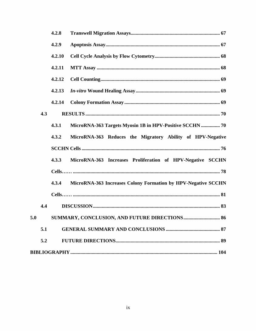
transformation, and immortalization (Figure 1). E6 prevents p53-mediated growth arrest by the
ubiquitination and subsequent proteasome-mediated degradation of p53 by the E6-E6AP-p53
tripartate complex (Huibregtse et al., 1991; Scheffner et al., 1990; Werness et al., 1990).
Although E6 reduces the net levels of p53, additional p53 may be activated in response to stress
including DNA damage. E6 binds to the histone acetyltransferases p300 and CBP and prevents
their ability to acetylate and stabilize p53 (Kumar et al., 2002; Patel et al., 1999; Zimmermann et
5
al., 1999). E6 also targets ADA3, another histone acetyltransferase, for ubiquitin-mediated
degradation thereby preventing it from acetylating and stabilizing p53 (Kumar et al., 2002; Patel
et al., 1999; Zimmermann et al., 1999). High-risk E6 prevents cell death by directly binding to
the TNF receptor 1 (Filippova et al., 2002) and by interacting with FADD, caspase 8, BAK,
BAX, and IRF3 (Filippova et al., 2004; Garnett et al., 2006; Ronco et al., 1998; Thomas and
Banks, 1998, 1999). To help immortalize cells, E6 binds E6AP and activates the telomerase
promoter via interacting with repressors (USF1/2, NFX1-91) and activators (c-myc/max, Sp1,
NFX1-123, and histone acetyltransferase complexes) (Howie et al., 2009). The high-risk E6
protein interacts with and causes the proteasome-mediated degradation of PDZ domain-
containing proteins, including hDlg and hScrib, which disrupt cell adhesion and polarity
(Thomas et al., 2008).
Figure 1: HPV-16 E6 Cellular Interactions
(Moody and Laimins, 2010, with permission)

6
High-risk HPV E7 functions in collaboration with E6 by targeting additional cellular
proteins and further promoting cellular proliferation and transformation (Figure 2). The
Retinoblastoma protein (pRb) is one of the main regulators of cellular entry into the S-phase. E7
binds to hypophosphorylated pRb, as well as to the other related pocket proteins p107 and p130.
The E7 binding mediates the degradation of pRb, thereby releasing the E2F transcription factors
to act on promoters of S-phase-specific genes (Boyer et al., 1996; Chellappan et al., 1992; Jones
et al., 1997b; Zerfass et al., 1995). High-risk E7 binds to and neutralizes the CDK inhibitors p21
and p27, thereby increasing the levels of cyclin A and cyclin E, which increases cellular levels of
CDK2 and promotes the entry of the cells into the S-phase (Funk et al., 1997; Jones et al., 1997a;
Zerfass-Thome et al., 1996). Furthermore, E7 binds to histone deacetylases (HDACs) which
results in increased E2F2-mediated transcription and entry of the cells into the S-phase
(Longworth et al., 2005). E7 also acts to prevent the interferon response by binding to IRF1 and
p48, thereby preventing activation of the Stat1-Stat2 transcription factors (Barnard and
McMillan, 1999; Park et al., 2000).
High-risk HPV E5 has emerged as a potential oncoprotein, and has functions that can
potentiate E6 and E7 mediated cellular transformation (Bouvard et al., 1994; Valle and Banks,
1995). The HPV-16 E5 gene can transform murine fibroblasts and human keratinocytes, and
contributes to skin carcinogenesis in mice (Maufort et al., 2007; Straight et al., 1993; Valle and
Banks, 1995). E5 activates the EGFR signaling pathway, which results in increased
angiogenesis and cell proliferation, and decreased apoptosis (DiMaio and Mattoon, 2001;
Genther Williams et al., 2005). Most studies of E5 have been in an overexpression model, and
its functional in vivo role is less understood.

7
Figure 2: HPV-16 E7 Cellular Interactions
(Moody and Laimins, 2010, with permission)
1.2.2 Human Papillomavirus-Positive Squamous Cell Carcinoma of the Head and Neck
HPV is considered a major cause of oropharyngeal cancer in developed countries because it is
detected in 45-90 percent of these cancers (D'Souza et al., 2007; Kreimer et al., 2005; Nasman et
al., 2009). HPVs have also been detected in 24 percent of laryngeal and 23 percent of oral cavity
cancers (Hobbs et al., 2006; Kreimer et al., 2005). High-risk HPV-16 accounts for 90-95 percent
of HPV-positive oropharyngeal cancers, while the remaining five percent are caused by high-risk
HPV-18, -31, -33, and -35 (Gillison et al., 2012; Gillison et al., 2000; Mork et al., 2001; Snijders
et al., 1996; Zhang et al., 2004). In the United States, oropharyngeal cancer rates are on the rise
despite a general decline in tobacco use, consistent with an increase in the number of HPV-
associated SCCHN cases (Chaturvedi et al., 2008; Marur et al., 2010; Nasman et al., 2009).

8
HPV-positive and HPV-negative SCCHN show different clinical and demographic
characteristics, leading some to classify them as distinct diseases (Vidal and Gillison, 2008). As
summarized in Table 1, HPV-positive SCCHN typically arise in the oropharynx area with
detection rates of up to 50 percent or more, while HPV-negative SCCHN arise in all head and
neck sites (Hammarstedt et al., 2006; Klussmann et al., 2001; Paz et al., 1997; Venuti et al.,
2004; Vidal and Gillison, 2008). Patients with HPV-positive SCCHN are diagnosed at a younger
age (40-60 years) compared to patients with HPV-negative SCCHN (> 60 years) (Cruz et al.,
1996; D'Souza et al., 2007; Fakhry et al., 2008; Mellin et al., 2000; Smith et al., 2004). SCCHN
is predominantly a male disease with a 3:1 ratio of men:women, and most often occurs in white
educated men who are frequently of higher socioeconomic status (D'Souza et al., 2007; Fakhry et
al., 2008; Gillison et al., 2008).
The emergence of and increase in HPV-associated SCCHN has led to new risk factors for
SCCHN. While the risk factors for HPV-negative SCCHN include alcohol consumption and/or
tobacco use, the risk factors for HPV-positive SCCHN include oral HPV infection caused by
sexual behavior (Table 1). A case-control study showed that a high number of vaginal or oral
sex partners was associated with high oropharyngeal cancer rates, with the majority of those
cancers testing positive for HPV (D'Souza et al., 2007). HPV-positive SCCHN tumors have
infrequent p53 mutations, but p53 expression is usually low due to inactivation of p53 by the
HPV E6 protein (Moody and Laimins, 2010; Poeta et al., 2007; van Houten et al., 2002). The E7
oncoprotein expressed in HPV-positive SCCHN reduces pRb and cyclin D levels. Since pRb
negatively regulates p16, the reduction/loss of pRb in HPV-positive SCCHN causes an increase
in p16 levels. However in HPV-negative SCCHN, p16 expression is reduced, usually due to

9
mutations, deletions, or methylation of the p16 gene (Table 1) (Olshan et al., 1997; Rischin et al.,
2010).
Table 1: HPV-positive and HPV-negative SCCHN characteristics
HPV-positive HPV-negative Anatomic site Oropharynx (tonsil, base of tongue) All sites
Age Younger Older Gender 3:1 men 3:1 men
Risk factors Sexual behavior Alcohol and tobacco Incidence Increasing Decreasing
p53 mutations Rare > 50 percent p16 expression High Low
Response to treatment Better Worse Survival High Low
A large meta-analysis of several published reports of SCCHN showed that patients with
HPV-positive SCCHN have a lower risk of recurrence and mortality compared to patients with
HPV-negative SCCHN (Ragin and Taioli, 2007). Several studies have shown that tumors
positive for HPV DNA responded better to chemotherapy, radiation, and surgery compared to
tumors lacking the HPV DNA (Fakhry et al., 2008; Tran et al., 2007b; Vidal and Gillison, 2008;
Worden et al., 2008). Additionally, individuals with HPV-positive SCCHN have a better
prognosis compared to those with HPV-negative SCCHN, with 95 percent of patients with HPV-
positive SCCHN still alive after two years, compared to only 62 percent of patients with HPV-
negative SCCHN (Fakhry et al., 2008).

10
1.3 MICRORNAS
MicroRNAs (miRNAs) are endogenously encoded single-stranded RNAs that most commonly
function as negative regulators of gene expression. MiRNAs were discovered in Caenorhabditis
elegans in 1993 by Victor Ambrose (Lee et al., 1993) and are predicted to regulate the
expression of up to a third of all genes (Lewis et al., 2005).
1.3.1 MicroRNA Biogenesis
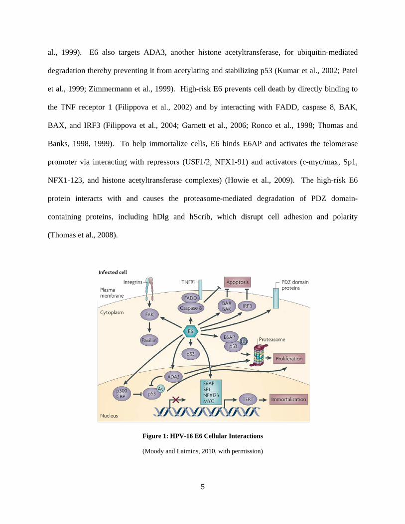
The majority of miRNAs are transcribed by RNA polymerase II into a primary miRNAs (pri-
miRNA) containing a stem-loop structure (Figure 3) (Lee et al., 2004). Pri-miRNAs are capped
and polyadenylated (Cai et al., 2004) and then processed into a 60-80 bp imperfectly paired
stem-loop precursor (pre) miRNA by the RNase III enzyme Drosha and its cofactor
Pasha/DGCR8 (Gregory et al., 2004). The pre-miRNA is actively transported out of the nucleus
to the cytoplasm by Ran-GTP and Exportin 5 (Lund et al., 2004) where the RNase III enzyme
Dicer recognizes the two-nucleotide 3’ overhang of the pre-miRNA and cleaves it into a 21-24
nucleotide miRNA duplex (Hutvagner et al., 2001). The miRNA duplex is loaded into the RNA-
Induced Silencing Complex (RISC), and only the mature miRNA strand is retained (Kawamata
et al., 2009). The RISC contains a member of the Argonaute protein family, a double-stranded
RNA binding protein TRBP, and Dicer (Chendrimada et al., 2005). The mature miRNA guides
the RISC to complementary sites in the 3’ untranslated region (UTR) of the target mRNA where
depending upon the degree of complementarity, the miRNA-RISC will either repress the
translation of the target mRNA or target the mRNA for degradation (Eulalio et al., 2008). The 5′
2-8 nt of the miRNA, known as the seed sequence, is usually complementary to the mRNA
11
target, and the remaining miRNA-mRNA interaction contains many mismatches. MiRNAs with
low complementarity to the target mRNA generally promote translational repression of the
mRNA whereas miRNAs with greater complementarity to the mRNA generally target the
mRNA for degradation (see Figure 3) (Eulalio et al., 2008; Martinez et al., 2002). Since
miRNAs do not have to be fully complementary to the target mRNA, one miRNA can regulate
many genes, and one gene can be regulated by many miRNAs (John et al., 2004; Kiriakidou et
al., 2004; Krek et al., 2005; Lewis et al., 2003; Lim et al., 2005).
Figure 3: MicroRNA Biogenesis
(Esquela-Kerscher and Slack, 2006, with permission)

12
1.3.2 MicroRNA Involvement in Cancer
The small percentage of miRNAs that have been characterized function in many cellular
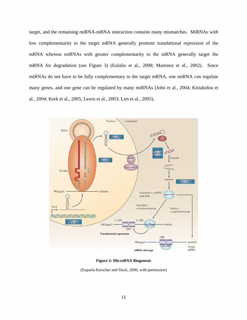
regulatory processes. Recently, many miRNAs have been shown to function as tumor
suppressors or oncogenes, and the dysregulation of such miRNAs has been shown to correlate
with tumor progression (Figure 4) (Esquela-Kerscher and Slack, 2006). Up to one-half of well-
studied miRNA genes are located in fragile sites of the genome, where genomic disruption often
leads to cancer (Calin et al., 2004). Low expression of a tumor suppressive miRNA can lead to
lack of repression of its target oncogenes, whereas high expression of an oncogenic miRNA can
lead to repression of its target tumor suppressor genes. Both types of such events can then lead
to cancer (Figure 4) (Esquela-Kerscher and Slack, 2006). Global miRNA dysregulation as well
as dysregulation of miRNA processing machinery is seen in many tumors (Caldas and Brenton,
2005; Esquela-Kerscher and Slack, 2006; Lu et al., 2005).
13
Figure 4: Tumor suppressive and Oncogenic MicroRNAs
(Esquela-Kerscher and Slack, 2006, with permission)
The first miRNA shown to act as a tumor suppressor was the miR-15a-miR-16-1 cluster
in B-cell chronic lymphocytic leukemia (CLL) (Calin et al., 2002). More than half of all CLL
cases have deletions in the miR-15a and miR-16 chromosomal regions, and these miRNAs are
downregulated or deleted in several other types of cancers. These tumor suppressor miRNAs are
known to target Bcl-2, cyclin D1, and Wnt3a (Bandi et al., 2009; Bonci et al., 2008; Cimmino et
al., 2005). MiR-143 and miR-145 are downregulated in colorectal and bladder tumors and
breast, cervical, lymphoid, and prostate cancer cell lines (Esquela-Kerscher and Slack, 2006;
Michael et al., 2003; Villadsen et al., 2012). The genes encoding the let-7 family of miRNAs are
14
located in fragile sites that are linked to breast, cervical, lung, and urothelial cancers (Calin et al.,
2004). The downregulation of these let-7 family members in lung cancer correlates with poor
prognosis and survival (Takamizawa et al., 2004). Furthermore, miRNAs in the let-7 family
target the Ras oncogene; therefore decreased expression of these miRNAs leads to increased cell
survival and cancer (Johnson et al., 2005).
One of the most well studied oncogenic miRNAs is miR-21. This was one of the first
miRNAs to be identified in humans and is overexpressed in almost all cancers, where it promotes
cell survival (Asangani et al., 2008; Chan et al., 2005; Fujita et al., 2008; Park et al., 2009a; Si et
al., 2007; Zhu et al., 2008). High levels of miR-21 have been found in breast, cervical, colon,
head and neck, hepatocellular, lung, ovarian, pancreatic, prostate, stomach, and thyroid cancers,
as well as glioblastoma and leukemia (Asangani et al., 2008; Frankel et al., 2008; Krichevsky
and Gabriely, 2009; Lu et al., 2008b; Meng et al., 2007; Si et al., 2007). The miR-17~92 cluster,
known as “oncomiR-1”, is often amplified and overexpressed in B cell lymphomas and small
cell lung cancer (Hayashita et al., 2005; Matsubara et al., 2007; Ota et al., 2004). The c-Myc
oncogene is often upregulated in many cancers, and it serves as a transcription factor for the
miR-17~92 miRNA cluster (He et al., 2005; O'Donnell et al., 2005; Sylvestre et al., 2007). The
c-Myc protein also represses miRNA expression by directly binding to the promoters of many
miRNAs, including let-7a/f/d, miR-15a, miR-16, miR-22, miR-26a/b, miR-29a/b, miR-30c/e,
miR-34a, and miR-146a (Chang et al., 2008b; Kleine-Kohlbrecher et al., 2006). There are two
other miRNA clusters (miR-106a~363 and miR-106b~25) that belong to the miR-17~92 family
of miRNA clusters, and these miRNA clusters are thought to have arisen through a series of
deletions and/or duplications, as many of the miRNAs have the same seed sequence and are

15
thought to target similar genes (Ventura et al., 2008). Thus, since miR-17~92 is an oncogenic
miRNA cluster, miR-106a~363 and miR-106b~25 are also potential oncogenic miRNA clusters.
MiRNAs are also involved in signaling pathways that are commonly disrupted in cancer.
The tumor suppressor protein p53 is mutated in up to 50 percent of cancers, and dysfunctional
p53 signaling is found in up to 80 percent of tumors (Levine et al., 2004; Levine et al., 2006;
Olivier et al., 2004). While p53 regulates several miRNAs, it is also targeted by some miRNAs.
MiR-504 and miR-125b directly regulate p53 (Hu et al., 2010). Additionally, miR-29, miR-34a,
and miR-122 positively regulate p53 through their targets p85α, SIRT1, and cyclin G1,
respectively (Fornari et al., 2009; Park et al., 2009b; Yamakuchi et al., 2008). The p53 protein is
a direct activator of the members of the miR-34 family through its binding to the promoters of
the miR-34a and miR-34b/c genes (Chang et al., 2007; Corney et al., 2007; He et al., 2007;
Raver-Shapira et al., 2007; Tazawa et al., 2007). The miR-34 family members target many
proteins that regulate the cell cycle, including cyclin E2, CDK4, CDK6, and BCL2. In cancers
where p53 is mutated/disrupted, the miR-34 family members are not expressed, and this leads to
cellular proliferation (Bommer et al., 2007; Chang et al., 2007; Tazawa et al., 2007).
Additionally, miR-34a is downregulated in cells expressing high-risk HPV E6, which targets p53
for ubiquitin-mediated degradation (Wang et al., 2009a). The p53 protein also regulates
hypoxia-induced signaling by activating miR-107, which targets the hypoxia inducible factor-1β
(Yamakuchi et al., 2010). The p53 protein activates the promoter of miR-145, which negatively
regulates the c-Myc oncogene (Sachdeva et al., 2009) and p53 activates the transcription of miR-
192 and miR-215, which target genes that regulate the cell cycle (Georges et al., 2008). The
miR-17~92, miR-106b~25, and miR-106a~92 clusters are regulated by p53 through its
repression of E2F1 (Brosh et al., 2008). Finally, p53 interacts with Drosha to enhance Drosha-

16
mediated primary miRNA maturation and enhances the expression of miR-16, miR-143, miR-
145, and miR-206 (Suzuki et al., 2009).
High-risk HPV E6 and E7 proteins are known to interact with many cellular proteins and
promote cellular transformation (Moody and Laimins, 2010). Among these interactions, c-Myc
and p53 are known to be involved in complex networks that involve miRNAs. Thus, disruption
of c-Myc and/or p53 by the HPV oncogenes may alter the expression of miRNAs that are
regulated by or involved in the c-Myc or p53 networks, (Chang et al., 2008b; O'Donnell et al.,
2005; Suzuki et al., 2009).
1.3.3 MicroRNA Involvement in SCCHN
There have been many reports on dysregulated miRNAs in SCCHN cell lines and tissues.
Although miRNA expression can vary by site (tonsil, tongue, etc.), there are several commonly
dysregulated miRNAs in all SCCHN sites. Some commonly downregulated miRNAs in head
and neck cancer compared to adjacent normal tissue include miR-375, miR-133a, and miR-133b
(Avissar et al., 2009; Childs et al., 2009; Hui et al., 2010; Lajer et al., 2011; Tran et al., 2007a;
Wong et al., 2008a; Wong et al., 2008b). Known as a tumor suppressive miRNA, low
expression of miR-375 is associated with low survival of oral cancer patients, reduced cellular
apoptosis, and increased proliferation (Harris et al., 2012; Nohata et al., 2011b). The tumor
suppressive miR-133a is often downregulated in several cancers, including SCCHN, where it
targets genes that are involved in cell migration and invasion (Kinoshita et al., 2012a; Kinoshita
et al., 2012b; Nohata et al., 2011a).

17
One commonly overexpressed miRNA in head and neck cancer compared to adjacent
normal tissue is miR-155 (Chang et al., 2008a; Hui et al., 2010; Lajer et al., 2011; Ramdas et al.,
2009; Wong et al., 2008b). This miRNA is overexpressed in many cancers, but in Epstein-Barr-
virus-negative nasopharyngeal carcinoma, high expression of miR-155 is associated with poor
prognosis (Du et al., 2011).
1.4 PROJECT HYPOTHESIS
We hypothesized that infection with high-risk HPV would lead to overexpression of the HPV E6
and E7 oncogenes. Expression of these oncogenes in SCCHN is expected to disrupt cellular
miRNA expression differently than in HPV-negative SCCHN. The altered miRNA expression in
HPV-positive and HPV-negative SCCHN would lead to alterations in the expression of the target
gene(s), thus altering pathways involved in HPV-positive and HPV-negative SCCHN. The
altered pathways may provide insight into the different characteristics seen in HPV-positive and
HPV-negative SCCHN.

18
2.0 ALTERATION OF MICRORNA PROFILES IN SQUAMOUS CELL
CARCINOMA OF THE HEAD AND NECK CELL LINES BY HUMAN
PAPILLOMAVIRUS
Work described in this section was published in Head & Neck (Head Neck. 2011
Apr;33(4):504-12) with authors Abigail I. Wald, Elizabeth E. Hoskins, Susanne I. Wells, Robert
L. Ferris, and Saleem A. Khan.

19
2.1 INTRODUCTION
Squamous cell carcinoma of the head and neck (SCCHN) ranks sixth among cancers
worldwide (Tran et al., 2007b). Many of these cases are associated with heavy consumption of
alcohol and/or tobacco use, which over time induce mutations in essential genetic pathways that
regulate the cell cycle. However, human papillomavirus (HPV) type 16 DNA has been found in
up to 30 percent of these cancers, most often in the oropharynx region, and such cases of
SCCHN are often found in individuals without the risk factors of alcohol and tobacco use
(Chaturvedi et al., 2008; Tran et al., 2007b). The HPV-positive SCCHN subset have increased in
the past 10 years (Chaturvedi et al., 2008). Because of this demographic shift and distinct
clinical behavior, the association and relevance of HPV in SCCHN is under intense
investigation.
Characteristics of HPV-associated SCCHN are very different from HPV-negative
SCCHN, causing disputes whether these cancers should be classified as distinct tumors (Vidal
and Gillison, 2008). HPV-positive oral tumors often exhibit loss of cell cycle control proteins,
including pRb and cyclin D1, whereas these two proteins are commonly overexpressed in HPV-
negative oral tumors (Tran et al., 2007b; Vidal and Gillison, 2008). One of the most common
tumor suppressor proteins, p53, is mutated in up to half of oral cancers, but is very rarely
mutated in HPV-positive SCCHN, and tumors with a high viral load have a better prognosis
compared to tumors with a low viral load or tumors that are HPV-negative (Tran et al., 2007b;
Vidal and Gillison, 2008). Patients with HPV-positive oral tumors have a better response to
chemotherapy, radiation, and surgery (Vidal and Gillison, 2008), and have evidence of immune
20
activation against viral antigens (Albers et al., 2005), despite having frequent metastasis to
regional lymph nodes (Vidal and Gillison, 2008). The biological basis for the differential
behavior of HPV-positive SCCHN is not understood.
Micro (mi) RNAs are small, ~22 nt long, chromosome-encoded single-stranded RNAs
that are commonly associated with negative regulation of gene expression (Bartel, 2004).
MiRNAs are transcribed and exported to the cytoplasm where further processing takes place, and
the mature miRNA strand is incorporated into the RNA-induced silencing complex (Bartel,
2004). The miRNA guides the RNA-induced silencing complex to the 3′ untranslated region of
its target mRNA where, depending upon the degree of complementarity, the miRNA either
translationally represses the mRNA or targets it for degradation (Bartel, 2004). MiRNA
dysregulation has been implicated in many different types of human cancers (Esquela-Kerscher
and Slack, 2006; Tong and Nemunaitis, 2008).
Previous reports have shown altered miRNA profiles in head and neck cancers compared
to the normal oral tissue (Childs et al., 2009; Hui et al., 2010; Ramdas et al., 2009; Wong et al.,
2008a). MiRNAs with high expression in the tumors compared to the normal oral tissue
included miR-21, whereas miR-125b was downregulated (Childs et al., 2009; Hui et al., 2010;
Ramdas et al., 2009). Basal miRNA expression in 9 head and neck cancer cell lines found that
33 miRNAs were expressed at a high level and 22 miRNAs were expressed at a low level (Tran
et al., 2007a). Interestingly, one of these cell lines UM-SCC47, is HPV-16-positive (Bradford et
al., 2003). In all 9 cell lines, let-7a, miR-16, miR-21, and miR-205 were highly expressed, and
miR-342, miR-346, and miR-373* were expressed at low levels (Tran et al., 2007a). Although
these studies show alterations in miRNA levels in head and neck cancer, they do not address the
role of HPVs. Because the number of cases of HPV-16-positive SCCHN have been increasing in

21
the past 10 years (Chaturvedi et al., 2008), and the characteristics of HPV-positive and HPV-
negative SCCHN support distinction between these cancers (Vidal and Gillison, 2008), we
sought to analyze the miRNA profiles in HPV-positive and HPV-negative SCCHN cell lines.
In this study, we demonstrate that miRNA expression profiles in HPV-16-positive
SCCHN cells are distinctly different from those in HPV-negative SCCHN cells and in normal
oral keratinocytes (NOKs) that have been immortalized by activation of h-TERT. Using human
foreskin keratinocytes (HFKs) expressing either the HPV-16 E6 or E7 oncogene, we also
demonstrate that expression of the E6 oncogene results in upregulation of miR-363 and
downregulation of miR-181a, mR-218 and miR-29a. Furthermore, siRNA knockdown of HPV-
16 E6 in the HPV-positive SCCHN cell line SCC2 reduces the expression of miR-363.
2.2 MATERIALS AND METHODS
2.2.1 Cell Lines
The cell lines used in this study are described in Table 2. Two HPV-16-positive SCCHN cell
lines, UD-SCC-2 (gift from Dr. Henning Bier, University of Dusseldorf) (Gwosdz et al., 2005),
and UPCI:SCC90 (Ferris et al., 2005), and two HPV-negative SCCHN cell lines, PCI-13 and
PCI-30 (gifts from Dr. Theresa Whiteside, UPCI) (Chikamatsu et al., 1999) were used for
miRNA expression profile analysis. The HPV-16-positive SCCHN cell lines UM-SCC47 (gift
from Dr. Thomas Carey, University of Michigan) (Bradford et al., 2003; Brenner et al., 2010)
and 93-VU-147T (gift from Dr. Hans Joenje, VU Medical Center Van der Boechorststraat 7, The
Netherlands) (Steenbergen et al., 1995) were used with the above cells lines for validation of the

22
miRNA microarrays. The UPCI:SCC90, UM-SCC-47, ad 93-VU-147T cell lines contain
integrated HPV-16 DNA, whereas the HPV-16 status (integrated vs. episomal) of the UD-SCC-2
cell line is not known (Ferris et al., 2005; Gupta et al., 2009; Steenbergen et al., 1995).
However, all the HPV-16-positive cell lines were shown to express the viral E6 and E7 genes
(Figure 5B). The UD-SCC-2, UPCI:SCC90, UM-SCC47, PCI-13 and PCI-30 cell lines were
grown in Dulbecco’s modified Eagle’s medium (Lonza, Walkersville, MD) supplemented with
10% fetal bovine serum, 1% penicillin/streptomycin, and 2% L-glutamine at 37°C in the
presence of 5% CO2. The 93-VU-147T cell line was grown in Dulbecco’s modified Eagle’s
medium/F12 medium (MediaTech, Manassas, VA) supplemented with 10% fetal bovine serum,
1% penicillin/streptomycin, and 2% L-glutamine at 37°C in the presence of 5% CO2. NOKs that
were immortalized by activation of h-TERT (Piboonniyom et al., 2003) were grown in defined
keratinocytes serum-free medium (Gibco, Grand Island, NY) supplemented with bovine pituitary
extract and 1% penicillin/streptomycin at 37°C in the presence of 5% CO2. Primary HFKs that
were transduced with an LXSN-based retroviral vector expressing high-risk HPV-16 E6 or E7
were grown in EpiLife medium (Invitrogen, Carlsbad, CA) supplemented with human
keratinocytes growth supplements (Invitrogen) and 1% penicillin/streptomycin at 37°C in the
presence of 5% CO2.

23
Table 2: SCCHN cell line characteristics
SCCHN Sample TNM Classification Specimen Site Sex HPV-status p53 gene PCI-13 T4N1M0 Oral Cavity Male HPV-negative E286K PCI30 T3N1M0 Oral Cavity Male HPV-negative wt
UD-SCC2 T1N2M0 Hypopharynx Male HPV-16 wt UPCI:SCC90 T2N1M0 Base of Tongue Male HPV-16 wt UM-SCC47 T3N1M0 Lateral Tongue Male HPV-16 wt 93-VU-147T T4N2 Floor of Mouth Male HPV-16 wt
2.2.2 Human Papillomavirus Status of Samples
The SCCHN cell lines were confirmed to be either HPV-positive or HPV-negative by
polymerase chain reaction (PCR) analysis using the MY09/MY11 primer set, which amplifies a
conserved region of the HPV L1 gene (Ferris et al., 2005). Although the HPV-positive SCCHN
cell lines have previously been characterized (Bradford et al., 2003; Ferris et al., 2005; Gwosdz
et al., 2005), we confirmed the HPV status of these cells. UD-SCC-2, UPCI:SCC90, UM-
SCC47, and 93-VU-147T were further confirmed to contain HPV-16 DNA by PCR using
primers that amplify a 477-bp region of the HPV-16 E6 gene using 5′-
ATGCACCAAAAGAGAACTGC-3′ as the forward primer and 5′-
TTACAGCTGGGTTTCTCTAC-3′ as the reverse primer. The glyceraldehyde-3-phosphate
dehydrogenase (GAPDH) gene was used as a loading control using 5′-
AGGGGAGATTCAGTGTGGTG-3′ as the forward primer and 5′-
GGCCTCCAAGGAGTAAGACC-3′ as the reverse primer, amplifying a 122-bp region. All
PCR reactions were performed as described previously (Ferris et al., 2005). The PCR amplified
DNA was analyzed by agarose gel electrophoresis.
24
2.2.3 RNA Isolation and Reverse Transcriptase-Polymerase Chain Reaction Analysis
Total RNA was isolated from all the cell lines grown to 90% confluency using the Ultraspec™
RNA Isolation System (Biotecx, Houston, TX, USA). DNase-I-treated total RNA (1 µg) of UD-
SCC-2, UPCI:SCC90, UM-SCC47, and 93-VU-147T was subjected to reverse transcriptase-
polymerase chain reaction (RT-PCR) analysis for expression of the HPV-16 E6 and E7
oncogenes using the Advantage® Clontech RT-for-PCR Kit (Clontech, Mountain View, CA,
USA) according to the manufacturer’s instructions.
The HPV-16 E6 gene was amplified using the primer set described above, and expression
of the HPV-16 E7 gene was done using the forward primer 5′-
CAGCTCAGAGGAGGAGGATG-3′ and the reverse primer 5′-
GCACAACCGAAGCGTAGAGT-3′, amplifying a 115-bp region. Expression of the GAPDH
gene was used as a control, using the primer set described above. The PCR products were
analyzed by agarose gel electrophoresis (Fig. 5B). The HFK-16E6 and HFK-16E7 cell lines
were also confirmed to be expressing the intended oncogene via RT-PCR as described above.
2.2.4 MicroRNA Microarray Analysis
Small RNAs (<200 nt) were enriched from total RNA from 2 HPV-positive SCCHN cell lines
(UD-SCC-2 and UPCI:SCC90) and 2 HPV-negative SCCHN cell lines (PCI-13 and PCI-30)
using the RNeasy Mini Kit and the RNeasy MinElute Clean Up Kit (Qiagen, Valencia, CA).
The small RNA fractions were validated on a 15% acrylamide gel. Small RNA fractions
obtained from 5 µg of total RNA were labeled with AlexaFluor 647 (Invitrogen) using the
mirVana Labeling Kit and hybridized (in duplicate) to the mirVana miRNA Bioarrays V2

25
(Ambion, Austin, TX). These bioarrays contained 662 antisense oligonucleotides, spotted in
quadruplicate, which included 328 known human miRNAs, 152 predicted miRNAs (ambi-
miRNAs), 266 mouse miRNAs (114 unique miRNAs), and 238 rat miRNAs (46 unique
miRNAs). The arrays were hybridized with labeled miRNAs at 42°C overnight. Each array was
subsequently washed once in low stringency was solution followed by washing twice in high
stringency was solution, and dried by centrifugation. The arrays were immediately scanned
using the GenePix 4000B scanner and the median fluorescent intensities, minus the background
fluorescence, were obtained using the GenePix Pro 6.0 software. The median fluorescent
intensities of each spot on the bioarrays were log2 transformed and normalized using the mean
intensities within the array and the global mean adjustment between the arrays by the GEDA
program (http://bioinformatics.upmc.edu/GE2/GEDA.html). Significance Analysis of
Microarray program version 1.21 (http://www-stat.stanford.edu/~tibs/SAM/) was used to
perform a t test to obtain the differential miRNA expression patterns of each sample. MiRNAs
with at least a 2-fold change in expression with a q-value (false discovery rate) of zero were
considered to have significant changes in their expression between the samples.
2.2.5 SiRNA Knockdown of Human Papillomavirus-16 E6 and Transfection Assays
The role of HPV-16 E6 in altered miRNA expression in HPV-positive SCCHN cell lines was
analyzed using double-stranded siRNA against HPV-16 E6 (siRNA 209 complementary to E6
positions 277 to 298, sense sequence 5’-UCCAUAUGCUGUAUGUGAUTT-3’; Dharmacon,
Lafayette, CO) (Jiang and Milner, 2002; Tang et al., 2006). The HPV-positive SCCHN cell line
SCC2 was seeded (1.5 x 10⁵) into 6-well plates, and after 24 hours transfected with 125 nM
siRNA using Lipofectamine 2000 Reagent (Invitrogen) and OPTI-MEM I (Gibco). BLOCK-iT

26
fluorescent oligo (Invitrogen) was used as a negative control siRNA (it has no human
homologous sequences) and a transfection efficiency control. Cells were harvested after 72
hours, and RNA extractions were done as previously described.
2.2.6 Real-Time Quantitative Reverse Transcriptase Polymerase Chain Reaction
Array data were confirmed by real-time quantitative RT-PCR (qRT-PCR) using the TaqMan
MicroRNA Reverse Transcription Kit and the TaqMan MicroRNA Assays (Applied Biosystems,
Foster City, CA) and the Real-Time Thermocycler iQ5 (Bio-Rad, Hercules, CA). These assays
use stem-loop primers designed to amplify only the mature miRNA. DNase I-treated total RNA
(5 ng) was used for each reaction, and all reactions were done in triplicate. Assays were
performed in accord with the manufacturer’s instructions and the miRNA levels were normalized
to the small nucleolar RNU43 levels. Relative expression levels of the miRNAs were calculated
using the 2-∆∆CT values (Livak and Schmittgen, 2001). Statistical analysis was done via a 2-tailed
t test.
HPV-16 E6 expression in siRNA knockdown experiments was confirmed via qRT-PCR
using the QuantiTect SYBR Green PCR kit (Qiagen) in accord with the manufacturer’s
instructions. The E6 gene was amplified using the forward primer 5′-
AGCGACCCAGAAAGTTACCA-3′ and the reverse primer 5′-
GCATAAATCCCGAAAAGCAA-3′, amplifying a 134-bp region. The E6 mRNA levels were
normalized to the GAPDH gene, using the primer set described above. DNase I-treated total
RNA (1 µg) was used for each reaction, and all the reactions were done in triplicate. The E6
mRNA levels were normalized to the GAPDH levels, and relative expression levels were

27
calculated using the 2-∆∆CT values (Livak and Schmittgen, 2001). Statistical analysis was done
via a 2-tailed t test.
2.3 RESULTS
2.3.1 MicroRNA Expression in Squamous Cell Carcinoma of the Head and Neck Cell
Lines
The UD-SCC-2, UPCI:SCC90, UM-SCC47, and 93-VU-147T cell lines were confirmed to be
HPV-positive by PCR analysis using the MY09/MY11 primers (Figure 5A). These cell lines
were further confirmed to contain HPV-16 DNA by PCR using E6 gene primers (Figure 5A).
Finally, the expression of the HPV-16 E6 and E7 genes in these cell lines was confirmed by RT-
PCR analysis (Figure 5B). We then analyzed miRNA expression in 2 HPV-16-positive (UD-
SCC-2 and UPCI:SCC90) and 2 HPV-negative (PCI-13 and PCI-30) SCCHN cell lines utilizing
miRVana miRNA Bioarrays V2. MiRNA analysis showed that 129 human miRNAs were
expressed in both of the HPV-negative cell lines (PCI-13 and PCI-30), with miR-21, miR-16,
and miR-29a being the most highly expressed (Supplementary Table 1). The HPV-16-positive
cell lines (UD-SCC-2 and UPCI:SCC90) both expressed 216 human miRNAs, indicating a
general upregulation of miRNA expression in the presence of HPV-16 DNA. The miRNAs with
high basal expression included miR-205, miR-16, and miR-21 (Supplementary Table 2).

28
Figure 5: HPV status of SCCHN cell lines
(A) PCR of all SCCHN cell lines to check HPV status. (B) RT-PCR for HPV-16 E6 and E7 expression

29
2.3.2 MicroRNA Expression is Altered in Human Papillomavirus-16-Positive Squamous
Cell Carcinoma of the Head and Neck Cell Lines
We compared the miRNA expression profiles in 2 HPV-16-positive cell lines, SCC2 and SCC90,
with that of 2 HPV-negative cell lines, PCI13 and PCI30. Two human miRNAs, miR-363 and
miR-33, and the rat miRNA miR-497 (which differs from human miR-497 by just 1 nucleotide),
were upregulated in HPV-positive cell lines compared to HPV-negative cell lines (Table 3).
Eight human miRNAs and 1 predicted human miRNA were downregulated in HPV-positive cell
lines (Table 3). A comparison of miRNA expression between individual cell lines showed that 2
miRNAs were downregulated in SCC2 compared to PCI13 (Supplementary Table 3), whereas 6
miRNAs were overexpressed in SCC2 compared to PCI30 (Supplementary Table 4). When
SCC90 was compared to PCI13, 4 miRNAs were overexpressed and 3 miRNAs were
underexpressed (Supplementary Table 5). On the other hand, 10 miRNA were overexpressed in
SCC90 compared to PCI30 (Supplementary Table 6). MiR-363 was upregulated in SCC90
compared to both PCI13 and PCI30 cell lines, and in SCC2 compared to PCI30 (Supplementary
Tables 4-6). We also found that miR-181a was downregulated in both SCC2 and SCC90
compared to PCI13 (Supplementary Tables 3 and 5). Although there were differences in miRNA
expression in individual SCCHN cell lines, several miRNAs including miR-363 and miR-181a
were similarly altered in both the HPV-positive cell lines in individual and pair-wise
comparisons with the 2 HPV-negative cell lines.

30
Table 3: MiRNAs differentially expressed in HPV-16-positive SCCHN cell lines compared to HPV-negative SCCHN cell lines
MiRNA Fold Change * Overexpressed hsa_miR_363 rno_miR_497 hsa_miR_33
5.16 3.20 1.99
Underexpressed hsa_miR_155 hsa_miR_181a hsa_miR_181b hsa_miR_29a hsa_miR_218 hsa_miR_222 hsa_miR_221 hsa_miR_142_5p ambi_miR_13232
-7.60 -7.34 -6.76 -4.68 -4.22 -3.69 -3.38 -3.20 -3.10
Abbreviations: MiRNA, microRNA; HPV, human papillomavirus; SCCHN, squamous cell carcinoma of the head and neck; miR, microRNA; hsa, human; rno, rat; ambi, Ambion predicted. *Mean fold changes in HPV-16-positive SCCHN cell lines UD-SCC-2 and UPCI:SCC90 compared to HPV-negative SCCHN cell lines PCI-13 and PCI-30. The q-values of all miRNAs were 0.
2.3.3 Human Papillomavirus-16-Positive Squamous Cell Carcinoma of the Head and
Neck Cell Lines have Altered MicroRNA Expression as Compared to Both Human
Papillomavirus-Negative Squamous Cell Carcinoma of the Head and Neck Cell Lines and
Immortalized Normal Oral Keratinocytes
To validate the microarray data, we carried out qRT-PCR analysis for selected miRNAs that
were found to be differentially expressed in SCC2 and SCC90 (HPV-positive) cell lines
compared to PCI13 and PCI30 (HPV-negative). For this, we used 4 HPV-16-positive SCCHN
cell lines (2 that were included in the array analysis and 2 that were not) and 2 HPV-negative
SCCHN cell lines. To exclude miRNA profiles only associated with squamous differentiation or
immortalization, we also used an NOK cell line that has been immortalized by activation of h-
TERT (Piboonniyom et al., 2003). The most overexpressed miRNA in the HPV-positive cells
31
based on the array analysis was miR-363. The qRT-PCR results showed higher expression of
miR-363 in HPV-positive cell lines SCC2 (6.3-fold), SCC90 (28.9-fold), SCC47 (1.3-fold), and
93-VU-147T (5.5-fold) compared to NOK cells (Figure 6A). The expression of miR-363 in the
HPV-negative cell lines was reduced 12-fold in PCI13 and 7-fold in PCI30 cells compared to the
NOK cells (Figure 6A). MiR-363 was upregulated in the above 4 HPV-positive SCCHN cell
lines by 61.7-fold, 283.1-fold, 12.8-fold, and 54.4-fold, respectively, compared to the average
expression in PCI13 and PCI30, with p < .01 (Figure 6A). Because miR-363 is part of a cluster
of miRNAs, we carried out qRT-PCR analysis for 2 other miRNAs in the cluster, miR-106a and
miR-92a. There was no difference in expression of these miRNAs in the HPV-positive SCCHN
cell lines compared to the HPV-negative SCCHN cell lines (data not shown). The Ambion
miRVana miRNA microarray includes a probe for both rat and human miR-497. The rat miR-
497 differs from the human miR-497 by the addition of 1 adenosine at its 3′ end (Griffiths-Jones
et al., 2008). Because the rat miR-497, but not the human miR-497, was significantly altered in
the HPV-positive samples as compared to the HPV-negative samples in our array analysis, we
also carried out qRT-PCR analysis for this miRNA. These results showed that human miR-497
was upregulated in 3 HPV-positive cell lines, SCC2 (17.2-fold), SCC90 (6.8-fold), and 93-VU-
147T (1.61-fold) compared to the NOK cells (Figure 6B). This miRNA was slightly
downregulated in HPV-negative PCI13 (1.8-fold) and PCI30 (2.0-fold) cells relative to the NOK
cells (Figure 2B). MiR-497 was upregulated in the above 3 HPV-positive SCCHN cell lines by
32.6-fold, 13.0-fold, and 3.1-fold, respectively, compared to the average expression in PCI13 and
PCI30, with p < .01 (Figure 6B).
The array results also showed that 8 human miRNAs, and 1 predicted human miRNA
were downregulated in 2 HPV-positive cell lines, SCC2 and SCC90, compared to the HPV-
32
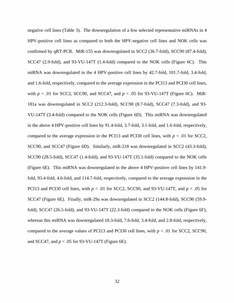
negative cell lines (Table 3). The downregulation of a few selected representative miRNAs in 4
HPV-positive cell lines as compared to both the HPV-negative cell lines and NOK cells was
confirmed by qRT-PCR. MiR-155 was downregulated in SCC2 (36.7-fold), SCC90 (87.4-fold),
SCC47 (2.9-fold), and 93-VU-147T (1.4-fold) compared to the NOK cells (Figure 6C). This
miRNA was downregulated in the 4 HPV-positive cell lines by 42.7-fold, 101.7-fold, 3.4-fold,
and 1.6-fold, respectively, compared to the average expression in the PCI13 and PCI30 cell lines,
with p < .01 for SCC2, SCC90, and SCC47, and p < .05 for 93-VU-147T (Figure 6C). MiR-
181a was downregulated in SCC2 (212.3-fold), SCC90 (8.7-fold), SCC47 (7.3-fold), and 93-
VU-147T (3.4-fold) compared to the NOK cells (Figure 6D). This miRNA was downregulated
in the above 4 HPV-positive cell lines by 91.4-fold, 3.7-fold, 3.1-fold, and 1.6-fold, respectively,
compared to the average expression in the PCI13 and PCI30 cell lines, with p < .01 for SCC2,
SCC90, and SCC47 (Figure 6D). Similarly, miR-218 was downregulated in SCC2 (43.3-fold),
SCC90 (28.5-fold), SCC47 (1.4-fold), and 93-VU-147T (35.1-fold) compared to the NOK cells
(Figure 6E). This miRNA was downregulated in the above 4 HPV-positive cell lines by 141.9-
fold, 93.4-fold, 4.6-fold, and 114.7-fold, respectively, compared to the average expression in the
PCI13 and PCI30 cell lines, with p < .01 for SCC2, SCC90, and 93-VU-147T, and p < .05 for
SCC47 (Figure 6E). Finally, miR-29a was downregulated in SCC2 (144.8-fold), SCC90 (59.9-
fold), SCC47 (26.5-fold), and 93-VU-147T (22.3-fold) compared to the NOK cells (Figure 6F),
whereas this miRNA was downregulated 18.3-fold, 7.6-fold, 3.4-fold, and 2.8-fold, respectively,
compared to the average values of PCI13 and PCI30 cell lines, with p < .01 for SCC2, SCC90,
and SCC47, and p < .05 for 93-VU-147T (Figure 6E).

33
Figure 6: QRT-PCR validation of microRNA expression data in 4 HPV-positive and 2 HPV-negative SCCHN
cell lines, and NOK cells
No RT, no reverse transcriptase added. Intensity values are relative to the NOK cells, which were arbitrarily
assigned a value of 1 or -1. The p values for the HPV-positive cell lines compared to the 2 HPV-negative SCCHN
cell lines are indicated by * (p < .05) and ** (p < .01).

34
2.3.4 Human Papillomavirus-16 E6 Oncogene Alters MicroRNA Expression
To test whether the altered miRNA expression in the HPV-positive SCCHN cell lines was due to
the expression of the HPV-16 oncogenes, we used primary HFKs transduced with either the
empty vector (LXSN) or vectors expressing the high-risk HPV-16 E6 or E7. Analysis of RNA
samples from HFKs containing the empty LXSN vector by RT-PCR showed that it did not
express the HPV-oncogenes while the HFK-16E6 and HFK-16E7 cell lines expressed the
appropriate oncogene (Figure 7A). We then tested the relative expression levels of 4 miRNAs,
miR-363, miR-181a, miR-218, and miR-29a that were significantly affected in HPV-positive cell
lines (Figure 6). QRT-PCR analysis showed that miR-363 (upregulated in HPV-positive
SCCHN cell lines compared to both HPV-negative SCCHN cell lines and NOK cells) was
upregulated in the HFK-16E6 cell line, with p < .01, but not in the HFK-16E7 cell line (Figure
7B). Similarly, miR-181a, miR-218, and miR-29a (downregulated in HPV-positive SCCHN cell
lines compared to both HPV-negative SCCHN cell lines and NOK cells) were downregulated in
the HFK-16E6 cell line, with p < .01. These miRNAs were either not affected or affected to a
lesser extent in the HFK-16E7 cell line (Figure 7B). These results showed that expression of the
E6 oncogene of HPV-16 is associated with upregulation of miR-363 and downregulation of miR-
181a, miR-218, and miR-29a.
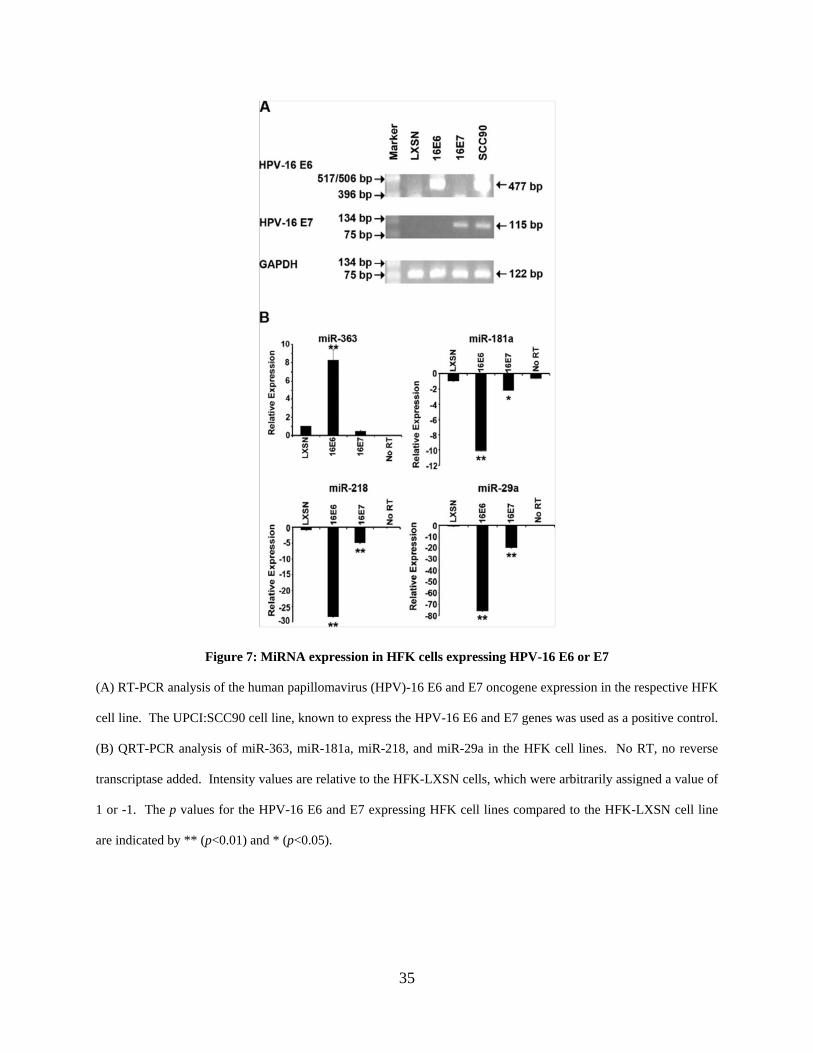
35
Figure 7: MiRNA expression in HFK cells expressing HPV-16 E6 or E7
(A) RT-PCR analysis of the human papillomavirus (HPV)-16 E6 and E7 oncogene expression in the respective HFK
cell line. The UPCI:SCC90 cell line, known to express the HPV-16 E6 and E7 genes was used as a positive control.
(B) QRT-PCR analysis of miR-363, miR-181a, miR-218, and miR-29a in the HFK cell lines. No RT, no reverse
transcriptase added. Intensity values are relative to the HFK-LXSN cells, which were arbitrarily assigned a value of
1 or -1. The p values for the HPV-16 E6 and E7 expressing HFK cell lines compared to the HFK-LXSN cell line
are indicated by ** (p<0.01) and * (p<0.05).

36
Because the expression of the HPV-16 E6 oncogene altered miRNA expression in the
HFK cells, siRNA knockdown of HPV-16 E6 was done in the HPV-positive SCCHN cell line
SCC2. The qRT-PCR results showed that reduction in the levels of E6 were accompanied by a
reduction in miR-363 levels (Figure 8). These results suggest that E6 is involved in the
upregulation of miR-363 in HPV-positive cell lines.
Figure 8: qRT-PCR analysis of the HPV-positive SCCHN cell line SCC2 transfected with siRNA against
HPV-16 E6
(A) QRT-PCR analysis of HPV-16 E6 oncogene expression in the HPV-positive cell line SCC2 upon transfection
with a negative control siRNA or with siRNA against E6. (B) QRT-PCR analysis of miR-363 expression in cells
transfected with a negative control siRNA or with siRNA against E6. No RT, no reverse transcriptase added.
Intensity values are relative to the SCC2 cells transfected with a negative control siRNA, which were arbitrarily
assigned a value of -1. The p values for the SCC2 + siRNA E6 cells compared to the SCC2 with the control siRNA
are indicated by ** (p<0.01).

37
2.4 DISCUSSION
HPV-positive and HPV-negative SCCHN have distinctly different clinical outcomes, and the
expression and mutation status of important cell cycle control proteins are very different (Tran et
al., 2007b; Vidal and Gillison, 2008). Our data show that several cellular miRNAs are also
differentially expressed in HPV-positive SCCHN cell lines as compared with HPV-negative
SCCHN cell lines. Many of these miRNAs were also found to be differentially expressed
between the HPV-positive SCCHN cell lines and transformed oral keratinocytes lacking HPV
DNA. Similarly, expression of the high-risk HPV-16 E6 oncogene in HFKs was strongly
associated with changes in miRNA expression similar to that seen in the HPV-positive SCCHN
cell lines, whereas siRNA knockdown of HPV-16 E6 reversed this effect for miR-363. These
results suggest that HPV-16, and in particular the E6 oncogene, may be involved in altering the
levels of several cellular miRNAs.
Altered regulation of cellular miRNAs has been observed in several types of human
cancers (Esquela-Kerscher and Slack, 2006; Lui et al., 2007; Tong and Nemunaitis, 2008) and
upon oncogenic viral infections, including hepatitis B and C (Jiang et al., 2008), Epstein-Barr
virus (Godshalk et al., 2008), human T-cell lymphotrophic virus 1 (Yeung et al., 2008), and
HPV-16 (Lui et al., 2007; Wang et al., 2008; Wang et al., 2009a). Recent studies have analyzed
miRNA expression in SCCHN (Chang et al., 2008a; Kozaki et al., 2008; Ramdas et al., 2009;
Tran et al., 2007a; Wong et al., 2008a; Wong et al., 2008b) and found that miRNA profiles in
SCCHN are different compared to normal oral tissue. However, currently there is no
information available on differential miRNA expression between HPV-positive and HPV-
negative SCCHN. Because HPV infection has been shown to play a significant role in the

38
etiology and prognosis of SCCHN (Tran et al., 2007b; Vidal and Gillison, 2008), we wanted to
study the effect of HPV-16 infection on cellular miRNA dysregulation in SCCHN.
Very few HPV-positive SCCHN cell lines have been described in the literature. Of 4
such cell lines that are available, we used 2 (UD-SCC-2 and UPCI:SCC90) to compare their
miRNA expression profiles to that of 2 HPV-negative SCCHN cell lines (PCI13 and PCI30) by
microarray analysis. We further used the 2 additional HPV-16-positive SCCHN cell lines (UM-
SCC47 and 93-VU-147T) to validate the miRNA data obtained in the above comparison.
MiRNA microarray analysis showed that 3 miRNAs (human miR-363 and miR-33, and rat miR-
497) were upregulated and 8 known and 1 predicted miRNAs were downregulated in the HPV-
positive SCCHN cell lines compared to the HPV-negative SCCHN cell lines (Table 3 and Figure
6). The miRNA microarray analysis was used as a screening tool, and the data were
subsequently validated via qRT-PCR. Similar to our current results, we have previously found
that qRT-PCR analysis is much more sensitive than the miRNA microarrays and the fold-
difference in qRT-PCR assays is usually much greater (Martinez et al., 2008). MiR-363 was
specifically upregulated in HPV-16-positive SCCHN cell lines compared to the HPV-negative
SCCHN cell lines and NOK cells (Table 3 and Figure 6A), suggesting a possible role of HPV-16
in altering the levels of this miRNA. Furthermore, experiments with HFKs showed that
expression of the HPV-16 E6 oncogene increased the levels of miR-363 (Figure 7B) and siRNA
knockdown of E6 reversed this effect (Figure 8). Interestingly, miR-363 is part of the oncogenic
miR-17~92 family of clusters, which is composed of 3 clusters of miRNAs, miR-17~92, miR-
106a~363, and miR-106b~25 and thought to have evolved through a series of deletions and
duplications (Ventura et al., 2008). Other members of this family of miRNA clusters have been
implicated in cancers, including small cell lung cancer (Hayashita et al., 2005), B-cell lymphoma
39
(Ota et al., 2004), and T-cell leukemia (Landais et al., 2007). MiRNAs in this family have
similar or identical seed sequences (Ventura et al., 2008), and because the seed sequence of a
mature miRNA contributes significantly to its specificity for its target mRNA (Bartel, 2004), it
has been hypothesized that miRNAs in the miR-17~92 family may have similar functions
(Ventura et al., 2008). MiR-363 has identical seed sequences to miR-92-1, miR-92-2, and miR-
25 (Griffiths-Jones et al., 2008). MiR-92-2 and miR-25 are also overexpressed in pancreatic,
prostate, and stomach cancers (Volinia et al., 2006). Recently, miR-25 has been shown to be
upregulated in gastric cancers where it targets p57, an essential tumor suppressor (Kim et al.,
2009). Because miR-363 and miR-25 have the same seed sequence, and miR-25 is involved in
cell cycle disruption (Kim et al., 2009), it is possible that miR-363 may also be involved in the
dysregulation of the cell cycle in HPV-associated SCCHN. The qRT-PCR analysis for miR-
106a and miR-92a did not show any differences in expression between the HPV-positive and
HPV-negative SCCHN cell lines (data not shown). This is not surprising because many
miRNAs in a cluster have independent promoters. Landais et. al. (Landais et al., 2007) has
shown that the miR-106a~363 cluster of miRNAs in mice is located downstream of the Kis2
gene. This gene has 3 different transcription start sites and it seems to encode the primary
miRNAs of the miR-106a~363 cluster. Also, the radiation leukemia virus is commonly
integrated close to the Kis2 locus. In mice, radiation leukemia virus-induced tumors had varied
expression of miRNAs in the miR-106a~363 cluster, indicating that they may not be transcribed
from the same promoter (Landais et al., 2007). Also, in gastric cancer, miR-363 was shown to
be downregulated compared to the normal tissue, whereas all of the other miRNAs in the miR-
106a~363 cluster were upregulated (Kim et al., 2009). Thus, while miR-363 is overexpressed in

40
HPV-positive SCCHN cells compared to HPV-negative SCCHN cells, it is not surprising that we
did not see a difference in expression of miR-106a and miR-92a between these cell lines.
Our results also show downregulation of several miRNAs in HPV-associated SCCHN
cell lines as compared to both HPV-negative SCCHN and NOK cell lines, including miR-155,
miR-181a, miR-218, and miR-29a (Table 3, Figure 6, and Figure 7B). We have recently
demonstrated that the HPV-16 E6 oncogene downregulates miR-218 expression in HPV-16
positive cervical carcinomas (Martinez et al., 2008). Furthermore, we showed that miR-218
targets LAMB3, and downregulation of miR-218 by the E6 oncogene results in overexpression of
LAMB3 in cervical carcinoma cells (Martinez et al., 2008). We found that expression of HPV-16
E6 in HFK cells also reduced the levels of miR-218 (Figure 7B). The downregulation of miR-
218 in both HPV-positive cervical and oropharyngeal cancer cell lines suggests that HPV-16
may target cellular pathways common to these 2 types of cancers. Although it is documented
that p53 expression activates miR-34a (He et al., 2007) and miR-34a levels are reduced in HPV-
16-positive cervical cancer (Wang et al., 2009a), we did not find a statistically significant
difference between miR-34a levels between HPV-positive and HPV-negative SCCHN cell lines.
Whereas all the HPV-positive cell lines used in our study are p53 wild-type (wt), the HPV-
negative cell line PCI-13 has a p53 mutation while PCI-30 has wt p53 (Table 2). There are
several possible reasons for our observations of miR-34a. For example, because the p53
pathway is complex, it is possible that a single miRNA may be subject to multiple regulatory
mechanisms.
Viral infections have been implicated in altered cellular miRNA expression. In human B
lymphocytes infected with the Epstein-Barr virus, elevated levels of miR-155 help in viral
persistence by reducing NF-κB signaling (Lu et al., 2008a). It is intriguing that miR-155 was

41
found to be downregulated in the presence of HPV-16 in our studies (Table 3, Figure 6C, and
Figure 7B). There have been other studies on miRNA expression in head and neck cancers that
have found miR-155 and miR-181a to be upregulated in oral cancer compared to normal oral
tissue (Chang et al., 2008a; Wong et al., 2008a; Wong et al., 2008b). However, when we
compared HPV-positive and HPV-negative SCCHN cell lines, these miRNAs were
downregulated in the presence of HPV-16 DNA. Future studies should define the relationship
between reduced levels of these miRNAs in HPV-positive SCCHN.
Our studies showed that miR-181a and miR-29a were downregulated in HPV-positive
SCCHN cells compared to HPV-negative SCCHN and NOK cells (Table 3, Figure 6D and 6F,
and Figure 7B). The levels of these 2 miRNAs also decreased upon expression of the HPV-16
E6 oncogene in HFKs (Figure 7B), suggesting a role for E6 in downregulation of these miRNAs.
The miR-181 family is known to be highly expressed in the brain (Miska et al., 2004) and is
involved in thymocyte development (Chen et al., 2004), but its role in other tissues is less well-
understood. Our data are the first to show a downregulation of miR-181a and miR-181b in
HPV-positive SCCHN cell lines compared to HPV-negative SCCHN cell lines (Table 3, Figure
6D, and Figure 7B). Overexpression of miR-181a and miR-181b have been shown to induce
apoptosis and inhibit growth and invasion in glioma cells (Shi et al., 2008). Further studies on
the roles of the miR-181 family may elucidate roles of these miRNAs in the different
characteristics seen in HPV-positive and HPV-negative SCCHN. MiR-29a has been shown to
interact with viral proteins. For example, miR-29a targets the HIV-1 Nef protein and interferes
with viral replication (Ahluwalia et al., 2008). MiR-29a also targets p85 and CDC42, which are
negative regulators of p53 (Park et al., 2009b). Because the HPV-16 E6 protein promotes the
degradation of the p53 protein (zur Hausen, 2002), it is possible that the downregulation of miR-

42
29a by E6 may further reduce p53 levels upon persistent HPV infection. The precise role of
HPV infection in cellular miRNA dysregulation, and the role of HPVs in SCCHN development,
which also contributes to a better prognosis for these cancers as compared to their HPV-negative
counterpart, will be the subject of future studies.

43
3.0 INCREASED EXPRESSION OF MICRORNA-363 IN HUMAN
PAPILLOMAVIRUS-ASSOCIATED SQUAMOUS CELL CARCINOMA OF THE HEAD
AND NECK TISSUES
44
3.1 INTRODUCTION
Squamous cell carcinoma of the head and neck (SCCHN) is the most common head and
neck malignancy and ranks sixth among cancers worldwide (Tran et al., 2007b). There are more
than 35,000 new cases of SCCHN in the United States and 500,000 new cases worldwide each
year (Greenlee et al., 2001). Although there have been advances in the detection and therapy of
SCCHN, the prognosis still remains poor, with a 5-year survival of approximately 50 percent
(Greenlee et al., 2001). The majority of SCCHN cases are associated with heavy alcohol
consumption and/or tobacco use, which over time induce mutations in essential genetic pathways
that regulate the cell cycle (Tran et al., 2007b). However, an emerging class of SCCHN has been
associated with high-risk human papillomavirus (HPV), where up to 30 percent of SCCHN
contain HPV-16 DNA, most often in the oropharynx region (oropharynx, base of tongue, and
tonsil), and often occurs in individuals without the major risk factors of alcohol and tobacco use
(D'Souza et al., 2007; Tran et al., 2007b).
HPVs are small double-stranded DNA viruses that infect the basal lamina of the skin or
mucous membranes (zur Hausen, 2002). High-risk HPVs have been associated with several
types of cancer, including cervical, anogenital, and oral cancers. In HPV-associated cancers,
expression of the E6 and E7 oncoproteins is essential for cellular transformation (Munger et al.,
2004). The high-risk HPV E6 and E7 proteins target many cellular proteins to induce cellular
transformation (for review on HPV oncogenes, see (Moody and Laimins, 2010)).
HPV-positive and HPV-negative SCCHN have different characteristics, leading many to
consider HPV-positive SCCHN a distinct tumor entity (Vidal and Gillison, 2008). The tumor

45
suppressor protein p53 is mutated in up to half of oral cancers, but is rarely mutated in HPV-
positive SCCHN (Agrawal et al., 2011; Stransky et al., 2011; Vidal and Gillison, 2008). Patients
with HPV-positive oral tumors have a much better prognosis compared to HPV-negative
SCCHN, with better response to chemotherapy, radiation, and surgery (Vidal and Gillison,
2008). It is also known that HPV-positive SCCHN cases have improved immune system
activation to viral antigens (Albers et al., 2005) and have a lower likelihood of metastasis than
patients with HPV-negative oral tumors (Fakhry and Gillison, 2006). A recent study (Gillison et
al., 2008) reported the association of HPV and oropharyngeal cancers with and without the
established risk factors of alcohol and tobacco use, suggesting that HPV-positive and HPV-
negative SCCHN are discrete molecular, clinical, and pathological diseases. Because of a recent
increase in HPV-positive SCCHN cases (Chaturvedi et al., 2008) and their distinct clinical
behavior, the molecular basis for the differences between HPV-positive and HPV-negative
SCCHN is under intense investigation.
Micro (mi) RNAs are small, endogenously encoded single-stranded RNAs that are most
commonly associated with post-transcriptional negative regulation of gene expression (Bartel,
2004). Some miRNAs have been shown to function as tumor suppressors or oncogenes, and the
dysregulation of such miRNAs have been observed in various cancers, including head and neck
cancers (Childs et al., 2009; Esquela-Kerscher and Slack, 2006; Hui et al., 2010; Ramdas et al.,
2009; Tran et al., 2007a; Volinia et al., 2006; Wong et al., 2008a).
We have previously reported differences in miRNA expression in HPV-positive and
HPV-negative SCCHN cell lines, and miR-363 was found to be the most overexpressed miRNA
in HPV-positive SCCHN cell lines (Wald et al., 2011). In this study, we sought to investigate
46
whether miR-363 is also overexpressed in HPV-positive SCCHN tumors compared to the HPV-
negative SCCHN tumors in patients from the western Pennsylvania area.
3.2 MATERIALS AND METHODS
3.2.1 SCCHN Cell Lines
Two SCCHN cell lines were used as positive and negative controls for HPV status and HPV
gene expression analysis. PCI-30 (gift from Dr. Theresa Whiteside, University of Pittsburgh
Cancer Institute) is an HPV-negative SCCHN cell line and UPCI:SCC90 is an HPV-16-positive
SCCHN cell line (previously described in (Wald et al., 2011)). These cell lines were maintained
in Dulbecco’s modified Eagle’s medium (Lonza, Walkersville, MD) supplemented with 10%
fetal bovine serum, 1% penicillin/streptomycin, and 2% L-glutamine at 37°C plus 5% CO2.
3.2.2 SCCHN Tissues
41 SCCHN tissues were obtained from patients according to Institutional Review Board protocol
99-069. Upon surgical removal, a portion of all tissues was sent to pathology for official tumor
staging and the remainder of all tissues was flash frozen until further processing.

47
3.2.3 DNA Isolation
DNA was isolated from all SCCHN tissues and cell lines using the DNeasy Blood & Tissue kit
(Qiagen, Valencia, CA) according to the manufacturer’s protocol.
3.2.4 RNA Isolation
Total RNA was isolated from all SCCHN tissues and cell lines using the Ultraspec RNA
Isolation System (Biotecx, Houston, TX) according to the manufacturer’s protocol.
3.2.5 PCR and RT-PCR
HPV status of all SCCHN tissues was analyzed using the MY09/MY11 primer set, which
amplifies a conserved region of the HPV L1 gene (Ferris et al., 2005). The glyceraldehyde-3-
phosphate dehydrogenase (GAPDH) gene was used as a loading control, using 5′-
CGACCACTTTGTCAAGCTCA-3′ as the forward primer and 5′-
AGGGGTCTACATGGCAACTG-3′ as the reverse primer, amplifying a 332-bp region. All
PCR reactions were performed using 20ng template DNA, 200µM of each deoxynucleoside
triphosphate (dNTP), 0.5µM of each primer, and 0.5 units of Taq polymerase and the associated
buffer (Promega, Madison, WI). Thermocycler conditions for all PCR reactions were 94°C for 5
minutes; 35 cycles of 94°C for 30 seconds, 57°C for 30 seconds, and 72°C for 30 seconds; and
72°C for 10 minutes. The PCR amplified DNA was visualized by agarose gel electrophoresis.
HPV-positive samples were tested for HPV type by amplifying the HPV-16 gene. HPV-16 E6

48
primers were forward 5′-AATGTTTCAGGACCCACAGG-3′ and 5′-
CAGCTGGGTTTCTCTACGTG-3′, amplifying a 454-bp region.
RT-PCR was done on HPV-16 E6 and E7 oncogenes to show expression in HPV-positive
SCCHN tumors. The HPV-16 E6 gene was amplified using the primer set described above and
the HPV-16 E7 gene was amplified using the forward primer 5′-
CATGGAGATACACCTACATTGCAT-3′ and the reverse primer 5′-
GAACAGATGGGGCACACAAT-3′, amplifying a 281-bp region. cDNA synthesis was done
using the iScript Select cDNA Synthesis Kit (Bio-Rad, Hercules, CA) according to the
manufacturer’s protocol using random primers. DNase I-treated RNA (60ng of total RNA) was
used for each cDNA synthesis reaction, and the cDNA was PCR amplified for HPV-16 E6,
HPV-16 E7, and GAPDH.
3.2.6 Determination of HPV DNA and p16 Expression in Tumor Tissue
Fluorescence in situ hybridization (FISH) for HPV-16 DNA was performed with a proprietary
pan selective probe set (Dako Cytomation, Carpinteria, CA). Immunohistochemical (IHC)
evaluation of the remaining deparaffinized sections was performed using immunoperoxidase
staining for p16 (p16INK4 mAb, BD Pharmingen, dilution 1:200) and scored semiquantitatively
for each core. Nuclear staining by HPV-16 FISH and diffuse p16 IHC (>80% cells) was
considered HPV-positive.

49
3.2.7 Real-Time Quantitative Reverse Transcriptase PCR (qRT-PCR)
Relative levels of miR-363 were measured using the TaqMan MicroRNA Reverse Transcription
Kit and the TaqMan MicroRNA Assay (Applied Biosystems, Foster City, CA) and the iQ5 Real-
Time Thermocycler (Bio-Rad). Twenty-five nanograms of total RNA was used for each
reaction, and all reactions were performed in triplicate. Assays were done according to the
manufacturer’s instructions, and the miRNA levels were normalized to the RNU43 small
nucleolar RNA levels. Relative miR-363 expression was calculated using the 2-∆∆CT values
(Livak and Schmittgen, 2001). Statistical analysis of miR-363 qRT-PCR expression was done
via a 2-tailed t test.
3.3 RESULTS AND DISCUSSION
3.3.1 HPV is Prevalent in Western PA SCCHN Cases
We studied a cohort of 41 SCCHN patients treated at the University of Pittsburgh Cancer
Institute between the years 2006 and 2009. The clinical and demographic characteristics of the
patients are detailed in Table 4 and summarized in Table 5. Upon receipt, all tissues were tested
for HPV positivity via PCR with the MY09/MY11 primers as described previously (Figure 9)
(Wald et al., 2011). Although HPV FISH was done for some SCCHN samples, DNA PCR was
used for all samples because of its high sensitivity of amplifying HPV DNA and to complete the
HPV testing on all samples. Of the 41 SCCHN samples, 24 (59%) were HPV-positive and 17
(41%) were HPV-negative. Fifteen of the 18 samples tested for HPV via FISH were HPV-

50
positive, confirming that testing for HPV via PCR has higher sensitivity than HPV testing via
FISH. All samples that were tested clinically for p16 (16 of the 24 HPV-positive samples) were
positive for p16. Only six HPV-negative SCCHN samples were tested for p16, two of which
were p16 positive, indicating that some HPV-negative SCCHN samples had high p16 expression.

51
Table 4: Detailed demographics of SCCHN tissues
Specimen Site TNM Stage Gender Race Age at diagnosis HPV status Tobacco
history Alcohol history Status Metastasis
Gum T1N0M0 Male Caucasian 78 HPV-negative never smoker never drinker Deceased cheek mucosa, mouth, tongue
Cheek T2N1M0 Male Caucasian 68 HPV-negative former smoker current drinker Deceased cheek mucosa, lip, soft palate,
oropharynx Mouth (1) T1N1M0 Male Caucasian 80 HPV-negative never smoker never drinker Alive none Mouth (2) T2N2CM0 Male Caucasian 56 HPV-16 former smoker former drinker Alive none Tongue (1) T3N2M0 Male Caucasian 64 HPV-16 current smoker current drinker Deceased mouth, lung Tongue (2) T3N2BM0 Male Caucasian 64 HPV-negative current smoker former drinker Deceased none Tongue (3) T4N2BM0 Female Caucasian 48 HPV-negative current smoker * current drinker * Deceased none Tongue (4) T4AN2CM0 Male Caucasian 65 HPV-negative former smoker current drinker Deceased none Vallecula T2BN2M0 Male African-American 51 HPV-16 never smoker never drinker Alive none
Base of tongue (1) T1N2BM0 Female Caucasian 84 HPV-negative current smoker * current drinker Deceased none Base of tongue (2) T1N2BM0 Male Caucasian 50 HPV-16 current smoker * former drinker Alive none
Base of tongue (3) T2N0 Female Caucasian 57 HPV-16 former smoker never drinker Deceased lymph node (tongue)
Base of tongue (4) T2N0M0 Male Caucasian 60 HPV-16 former smoker never drinker Deceased none Base of tongue (5) T2N0M0 Male Caucasian 60 HPV-negative former smoker current drinker * Deceased base of tongue Base of tongue (6) T2N1M0 Male Caucasian 51 HPV-16 current smoker * former drinker Alive none Base of tongue (7) T2N1M0 Female Caucasian 82 HPV-negative current smoker current drinker * Alive lung
Base of tongue (8) T2N2M0 Male Caucasian 70 HPV-16 unknown unknown Deceased recurrence - vallecula
Base of tongue (9) T2N2BM0 Male Caucasian 62 HPV-16 never smoker never drinker Alive none
Base of tongue (10) T2N2CM0 Male Caucasian 48 HPV-16 current smoker current drinker Deceased lymph nodes (head/face/neck)
Base of tongue (11) T2N3M0 Male African-American 68 HPV-16 current smoker * current drinker Deceased lymph nodes (intrathoracic)
Base of tongue (12) T3N0M0 Female Caucasian 59 HPV-negative never smoker never drinker Alive none Oropharynx (1) T2N0M0 Male Caucasian 48 HPV-negative current smoker former drinker Alive none
Oropharynx (2) T4BN2BM0 Male Caucasian 54 HPV-negative former smoker current drinker Deceased
tongue, lymph nodes
(head/face/neck) mouth

52
Oropharynx (3) recurrent -
stage unknown
Male Caucasian 59 HPV-negative current smoker * current drinker * Alive primary - gum, 2 recurrences oropharynx
Tonsil (1) T1N1M0 Male Caucasian 46 HPV-16 never smoker current drinker Alive met via PET
scan - Level II LN
Tonsil (2) T2N0M0 Female Caucasian 54 HPV-16 unknown unknown Alive none
Tonsil (3) T2N0M0 Male Caucasian 64 HPV-16 former smoker never drinker Alive recurrence - base of tongue
Tonsil (4) T2N0M0 Male Caucasian 60 HPV-16 never smoker never drinker Alive none
Tonsil (5) T2N1M0 Male Caucasian 49 HPV-16 snuff * current drinker Deceased
bones in skull/face, lung,
bones in pelvic/sacrum
Tonsil (6) T2N2 Male Caucasian 50 HPV-16 former smoker never drinker Alive none Tonsil (7) T2N2M0 Male Caucasian 48 HPV-16 current smoker current drinker Deceased none Tonsil (8) T2N2AM0 Male Caucasian 61 HPV-16 former smoker former drinker Alive none Tonsil (9) T2N2BM0 Male Caucasian 50 HPV-16 current smoker current drinker Alive none
Tonsil (10) T2N2BM0 Female Caucasian 56 HPV-16 current smoker * current drinker Alive none Tonsil (11) TXN2BM0 Female Caucasian 48 HPV-16 former smoker current drinker Alive none Tonsil (12) T2N2BM0 Female Caucasian 55 HPV-negative current smoker * current drinker * Alive none Tonsil (13) T2N2CM0 Male Caucasian 63 HPV-16 current smoker * current drinker * Alive none Tonsil (14) T2N3M0 Male Caucasian 49 HPV-16 current smoker * current drinker * Alive none Tonsil (15) T4N2BM0 Male Caucasian 65 HPV-negative current smoker * current drinker * Deceased none Tonsil (16) T4N3M0 Male Caucasian 65 HPV-negative current smoker * former drinker Deceased none Tonsil (17) T4AN2BM0 Male Caucasian 57 HPV-negative current smoker current drinker Deceased none
Abbreviations: * unknown if quit

53
Table 5: Summary of SCCHN tissue characteristics
HPV-16-positive HPV-negative Age, mean (SD) 55.6 (7.1) 64.2 (11.2) Gender, n (%) Males Females
20 (83%) 4 (17%)
12 (71%) 5 (29%)
Tumor location Mouth* Tongue Vallecula Base of tongue Oropharynx Tonsil
1 1 1 8 0 13
3 3 0 4 3 4
Tumor stage T1NXMX T2NXMX T3NXMX T4NXMX Unknown
2 21 1 0 0
3 5 2 6 1
* Mouth samples include mouth, gum, and cheek.
54
Figure 9: HPV status in SCCHN tissues
PCR analysis of all SCCHN tissues using the MY09/MY11 primer set to detect HPV positivity. SCC90, an HPV-
positive SCCHN cell line control. PCI30, an HPV-negative SCCHN cell line control.

55
All HPV-positive samples were subsequently tested for HPV type via DNA PCR for the
HPV-16 and HPV-18 E6 gene. All HPV-positive samples were found to contain HPV-16 DNA
(Figure 10A). All of the HPV-positive tumor tissues were found to express the HPV-16 E6 and
E7 oncogenes as determined by RT-PCR (Figure 10B).
Figure 10: HPV-16 PCR and RT-PCR in SCCHN tissues
(A) PCR of HPV-16 E6 gene in HPV-positive SCCHN tissues. (B) RT-PCR of HPV-16 E6 and E7 in HPV-positive
SCCHN tissues. SCC90, an HPV-16-positive SCCHN cell line positive control; PCI30, an HPV-negative SCCHN
cell line negative control; No RT, no reverse transcriptase added (used SCC90 sample).

56
As expected, the site with the highest HPV positivity rate (via PCR) in our cohort was the
tonsil, with 76 percent being HPV-positive, followed by tumors of the base of tongue, with 67
percent being HPV-positive (Table 5). The HPV-positive SCCHN cases of the tonsil and base of
tongue represented approximately 88 percent of all HPV-positive tumors. The HPV-negative
SCCHN cases had a more even distribution of the tumor sites, including the base of tongue,
oropharynx, tonsil, mouth, and tongue (Table 5). The average age at diagnosis for patients with
HPV-positive SCCHN was 55.6 years, or approximately nine years younger than for patients
with HPV-negative SCCHN tumors, similar to other studies (Chaturvedi et al., 2008; Tran et al.,
2007b). Ninety-six percent of HPV-positive SCCHN cases were classified as T1 or T2, whereas
only 50 percent of HPV-negative SCCHN cases manifested small primary tumors (Table 5).
These results are consistent with demographics and clinopathologic features reported for HPV-
positive disease, supporting the reflection of our cohort with those reported in the literature
(Chaturvedi et al., 2008).
3.3.2 MicroRNA-363 is Overexpressed in HPV-Positive SCCHN Tissues
We have previously reported that several miRNAs have altered expression in HPV-positive
SCCHN cell lines compared to HPV-negative SCCHN cell lines, including the upregulation of
miR-363 and downregulation of miR-181a, miR-218, and miR-29a (Wald et al., 2011). In that
study, we also showed that differential expression of these miRNAs was dependent upon the
expression of the HPV-16 E6 oncogene (Wald et al., 2011). We carried out qRT-PCR analysis
of miR-363 expression in the HPV-positive and HPV-negative SCCHN samples. HPV-positive
SCCHN samples expressed higher miR-363 levels compared to the HPV-negative SCCHN
samples, with p<0.01 (Figure 11). These data are consistent with those obtained with the

57
SCCHN cell lines (Wald et al., 2011), indicating that our work with SCCHN cell lines has
clinical relevance.
Figure 11: MicroRNA-363 expression in SCCHN tissues
QRT-PCR analysis of miR-363 in HPV-positive and HPV-negative SCCHN tissues. Black bars, HPV-16-positive
SCCHN samples; gray bars, HPV-negative SCCHN samples. Intensity values are relative to the HPV-negative
SCCHN sample with the lowest miR-363 expression, which was arbitrarily assigned a value of 1. The p-value for
the HPV-positive samples compared to the HPV-negative samples is indicated by ** (p<0.01). BOT, base of
tongue; no RT, no reverse transcriptase added.
58
MiR-363 has recently been shown to be overexpressed in HPV-positive pharyngeal
squamous cell carcinoma (PSCC) tissues in a Danish population (Lajer et al., 2011). Lajer and
colleagues found miR-363 to be the most overexpressed miRNA in 9 HPV-positive PSCC
compared to 11 HPV-negative PSCC (Lajer et al., 2011). Interestingly, in their study, PSCC had
an HPV-positivity rate of 42 percent, while OSCC (oral squamous cell carcinoma) had an HPV-
positivity rate of only 3 percent. The PSCC tumors came from the tonsil, base of tongue, and
other pharyngeal sites, whereas the OSCC samples came from the tongue, bucca, floor of mouth,
gingiva, and other oral cavity sites (Lajer et al., 2011). This supports our finding that HPV-
positivity rates are highest among the tonsil and base of tongue.
The association between high miR-363 expression in HPV-positive head and neck
cancers can be further explored for possible targets of therapeutic intervention or biomarkers for
prognosis.

59
4.0 MICRORNA-363 TARGETS MYSOIN 1B AND INCREASES THE
PROLIFERATION AND COLONY FORMATION OF SCCHN CELL LINES

60
4.1 INTRODUCTION
Squamous cell carcinoma of the head and neck (SCCHN) is the sixth most common cancer in the
United States (Jemal et al., 2010) and one of the leading cancers in developing countries (Parkin
et al., 1999). Along with the well-established risk factors of alcohol and tobacco use, infection
with high-risk human papillomavirus (HPV), most notably type-16, has emerged as a significant
risk factor for SCCHN (Chaturvedi et al., 2008; Tran et al., 2007b). Characteristics of HPV-
positive SCCHN differ from those of HPV-negative SCCHN, supporting the notion that these
cancers should be classified as distinct tumor entities (Vidal and Gillison, 2008). HPV-positive
SCCHN most often occurs in the oropharynx region (upwards of 50 percent or more), and is
diagnosed in younger patients compared to HPV-negative SCCHN. Moreover, SCCHN tumors
that are HPV-positive have infrequent p53 mutations while up to half of HPV-negative SCCHN
tumors have p53 mutations (Vidal and Gillison, 2008). Patients with HPV-positive SCCHN
tumors respond better to chemotherapy, radiation, and surgery, and have an overall 60 percent
reduction in death from SCCHN than patients with HPV-negative SCCHN tumors (Tran et al.,
2007b; Vidal and Gillison, 2008).
We have previously shown that micro (mi) RNA profiles in HPV-positive SCCHN cell
lines differ from HPV-negative SCCHN cell lines, and some miRNAs are dysregulated as a
direct result of the HPV-16 E6 and E7 oncogene expression (Wald et al., 2011). Furthermore
miR-363, which was the most highly overexpressed miRNA in HPV-positive SCCHN cell lines
compared to HPV-negative SCCHN cell lines, also shows this trend in a subset of HPV-positive
and HPV-negative SCCHN tumors from the western Pennsylvania population. In this study, we

61
sought to identify the possible mechanisms by which miR-363 may contribute to SCCHN
pathogenesis. We show that miR-363 targets myosin 1b, which reduces the migratory ability of
SCCHN cells. Additionally, miR-363 increases the proliferation and colony formation of
SCCHN cells.
4.2 MATERIALS AND METHODS
4.2.1 Cell Culture and Transfections
The HPV-negative SCCHN cell line PCI-30 was grown in Dulbecco’s modified Eagle’s medium
(Lonza, Walkersville, MD) supplemented with 10% fetal bovine serum, 1%
penicillin/streptomycin, and 2% L-glutamine at 37°C in the presence of 5% CO2. The HPV-
negative SCCHN cell line JHU-028 was grown in Roswell Park Memorial Institute medium
(Lonza) supplemented with 10% fetal bovine serum, 1% penicillin/streptomycin, and 2% L-
glutamine at 37°C in the presence of 5% CO2.
HPV-negative SCCHN cells PCI-30 or JHU-028 (1.5 x 10⁵) were seeded in 6-well plates
in antibiotic-free media. The following day, cells were transfected with 50 nM pre-miR (Applied
Biosystems, Foster City, CA) or 50 nM siRNA against MYO1B (ThermoFisher Scientific, San
Jose, CA) using Lipofectamine 2000 (Invitrogen, Carlsbad, CA) and Opti-mem (Gibco, Grand
Island, NY). A FAM-labeled negative control pre-miR (Applied Biosystems) or the Block-it
fluorescent oligo (Invitrogen) was used as the negative controls and to measure transfection
efficiency. Cells were harvested 48 hours after transfection and RNA and proteins were isolated
for various assays.

62
4.2.2 RNA Isolation
Total RNA was isolated from cell lines using the Ultraspec RNA Isolation System (Biotecx,
Houston, TX) according to the manufacturer’s instructions.
4.2.3 Quantitative Real Time RT-PCR
To confirm the efficiency of miR-363 transfections, miRNA expression was measured using the
TaqMan MicroRNA Reverse Transcription Kit and the TaqMan MicroRNA Assays (Applied
Biosystems) and the Real-Time Thermocycler iQ5 (Bio-Rad, Hercules, CA). Total RNA (50 µg)
was used for each reaction, and all of the reactions were performed in triplicate according to the
manufacturer’s instructions. MiRNA levels were normalized to small nucleolar RNU43 levels
and relative expression was calculated using the 2-∆∆CT values (Livak and Schmittgen, 2001).
Gene expression (mRNA) levels were measured using the iScript One-Step RT-PCR Kit with
SYBR Green (Bio-Rad). The MYO1B gene was amplified using the forward primer 5′-
GGTCTGGTGTGGAGGTCCTA-3′ and the reverse primer 5′-CGTTGCTTCCTCAGGTCTTC-
3′, amplifying a 127 bp region. The MYO1B mRNA levels were normalized to the GAPDH
mRNA levels, using the forward primer 5′-CAGCCTCAAGATCATCAGCA-3′ and the reverse
primer 5′-TGTGGTCATGAGTCCTTCCA-3′ which amplifies a 106 bp region. DNase-I-treated
total RNA (50 ng) was used for each reaction, and all the reactions were done in triplicate.
MYO1B mRNA was normalized to GAPDH, and the relative expression levels were calculated
using the 2-∆∆CT values (Livak and Schmittgen, 2001)

63
4.2.4 Spectral Counting Proteomics
The HPV-negative SCCHN cell line PCI-30 was transfected as described above with a negative
control pre-miR or pre-miR-363. After 48 hours, cells were lysed in 50 mM Tris-HCl, pH 7.4,
0.5% SDS, 1 mM ethylenediaminetetraacetic acid (EDTA), 1 mM phenylmethylsulfonyl fluoride
(PMSF) by boiling for 10 minutes, cooling to ambient temperature and then sonicating three
times. Protein quantitation was performed using the BCA Assay (Pierce, Rockford, IL) and 30
mg of protein was loaded onto a 10% Bis-Tris gel and run until the proteins entered the stacking
portion of the gel. The gel bands for each sample were excised and diced into approximately 1
mm2 sections and destained in 25 mM NH4HCO3/50% acetonitrile (ACN) with shaking. Gel
bands were dehydrated by addition of neat ACN and then dried in a 37°C incubator.
Proteins in the gel bands were reduced in 25 mM NH4HCO3, 10 mM dithiothreitol (DTT)
at 56°C for 1 hour followed by dehydration with ACN as before, and then alkylation was carried
out in 25 mM NH4HCO3, 55 mM iodoacetamide for 45 minutes in the dark at ambient
temperature followed by dehydration. Gel bands were rehydrated with trypsin (Promega,
porcine sequencing grade, 20 ng/ml) on ice for 45 minutes. Supernatant was removed and the
gel bands were washed once with 500 µl of 25 mM NH4HCO3, and then the gel bands were
covered with 25 mM NH4HCO3, (~100 µl) and incubated at 37°C overnight. The digest solution
was transferred into a clean microcentrifuge tube and peptides were extracted from the gel pieces
with sonication in a water bath for 10 minutes in 75 µl of 50% ACN/5% formic acid. The
supernatant was removed and added to the previous solution and this was repeated two additional
times. The combined extract was lyophilized to dryness and stored at -80°C until analysis.

64
Samples were analyzed by nanoflow reverse-phase liquid chromatography (Ultimate
3000, Dionex, Sunnyvale, CA) coupled online via electrospray ionization to a hybrid linear ion
trap-Orbitrap mass spectrometer (LTQ-Orbitrap, ThermoFisher Scientific, San Jose, CA). Five
replicate injections of peptide extracts were resolved on 100 µm i.d. by 360 µm o.d. by 200 mm
long fused silica capillary columns (Polymicro Technologies, Phoenix, AZ) slurrypacked in-
house with 5 µm, 300 Å pore size C-18 silica-bonded stationary phase (Jupiter, Phenomenex,
Torrance, CA). After sample injection, peptides were eluted from the column using a linear
gradient of 2% mobile phase B (0.1% formic acid in ACN) to 40% mobile phase B over 125
minutes at a constant flow rate of 200 nl/minutes followed by a column wash consisting of 95%
B for an additional 30 minutes at a constant flow rate of 400 nl/minute. The column was then re-
equilibrated in mobile phase A (0.1% formic acid in water) for 20 minutes prior to the next
injection. Eluting peptides were ionized using an electrospray voltage of 1.8 kV and analyzed in
the LTQ-Orbitrap MS which was configured to collect high resolution (R=60,000 at m/z 400)
broadband mass spectra (m/z 375-1800) from which the seven-most abundant peptide molecular
ions dynamically determined from the MS scan were selected for tandem MS using a relative
collision-induced dissociation (CID) energy of 30%. Dynamic exclusion was utilized to
minimize redundant selection of peptides for CID.
Peptide identifications were obtained by searching the LC-MS/MS data utilizing
SEQUEST (BioWorks, v3.2, ThermoFisher Scientific) on a 72-node Beowulf cluster against a
UniProt-derived human proteome database (11/2009 release) obtained from the European
Bioinformatics Institute (EBI) using the following parameters: trypsin (KR); full enzymatic
cleavage; two missed cleavage sites; 20 ppm peptide mass tolerance peptide tolerance, 0.5 amu
fragment ion tolerance; and variable modifications for methionine oxidation (m/z 15.99492) and

65
cysteine carboxyamidomethylation (m/z 57.02146). Resulting peptide identifications were
filtered according to specific SEQUEST scoring criteria: delta correlation (DCn) 3 0.08 and
charge state dependent cross correlation (XCorr) scores of 3 1.9 for [M+H]1+, 3 2.2 for [M+H]2+,
and 3 3.5 for [M+H]3+. These criteria resulted in a false discovery rate of <1% for all peptides
identified as determined by searching the data against a decoy human database where the protein
sequences were reversed. The differences in protein abundance between the samples were
derived by spectral counting.
4.2.5 Western Blotting
Proteins (25 µg) from total cell lysates from the HPV-negative SCCHN cell line PCI-30
transfected with pre-miR-363 were separated on a 10% SDS polyacrylamide gel and transferred
to Immobilion-P PVDF membrane (Millipore, Billerica, MA). The membrane was incubated
with a primary rabbit polyclonal antibody against MYO1B (Santa Cruz Biotechnology, Santa
Cruz, CA), and subsequently incubated with a 1:10,000 dilution of the secondary anti-rabbit
horseradish peroxidase antibody (Amersham Biosciences, Piscataway, NJ). Blots were
visualized using chemiluminescence with the ECL Plus Western Blotting Detection Reagents
(Amersham Biosciences), followed by film exposure. A murine monoclonal antibody against
GAPDH (Chemicon, Billerica, MA) was used to show equal sample loading.
4.2.6 Cloning of the MYO1B 3′ UTR into pMiR-Report Luciferase Vector
The MYO1B 3′ UTR (1429 bp) was PCR amplified from the HPV-positive SCCHN cell line
SCC90 using the forward primer 5′-GGACTAGTAACCGTCTCCTTGAAGTTGC-3′ and the

66
reverse primer 5′-GGAAGCTTGGCACAAGGCAAGAAGAATC-3′, amplifying a 1,725 bp
region. These primers were designed with an added SpeI restriction site on the forward primer
and a HindIII restriction site on the reverse primer to aid in directional cloning. PCR products
were separated on a 1% agarose gel, and the DNA was isolated using the QIAquick Gel
Extraction Kit (Qiagen, Valencia, CA). Three micrograms of each of the purified PCR products
and the pMiR-Report luciferase vector (Applied Biosystems) were digested with SpeI and
HindIII at 37°C for two hours. The digested DNA was separated on a 1% agarose gel, purified
using the QIAquick Gel Extraction Kit (Qiagen), and quantified using the Nanodrop. The
digested MYO1B 3′ UTR and pMiR-Report vector were ligated at a 1:3 (vector:insert) ratio at
16°C for 16 hours. DNA was precipitated and the ligated plasmid was electroporated into
competent DH5α cells. After a one-hour recovery, cells were plated on LB + ampicillin plates
and incubated at 37°C overnight. Ten colonies were patched on LB + ampicillin plates and
incubated at 37°C overnight. A lysates gel was run to determine the presence of the pMiR-
Report:MYO1B 3′ UTR plasmid. Selected colonies that contained the plasmid were grown in
LB broth + ampicillin at 37°C overnight and the pMiR-Report:MYO1B 3′ UTR plasmids were
extracted using the Wizard Plus Minipreps DNA Purification System (Promega, Madison, WI).
The plasmids were subsequently digested with HindIII and BamHI for 2 hours at 37°C and run
on a 1% agarose gel to confirm the insertion of the appropriate DNA fragment.
4.2.7 Luciferase Assays
HPV-negative JHU-028 SCCHN cells (10,000 cells per well) were seeded in a 96-well plate in
antibiotic free media. After 24 hours, cells were transfected using Lipofectamine 2000

67
(Invitrogen) and Opti-mem (Gibco) with the following nucleic acids: (A) 0.1 µg pMiR-Report +
2.5 ng phRL-TK, (B) 0.1 µg pMiR-Report + 2.5 ng phRL-TK + 50 nM negative pre-miR, (C)
0.1 µg pMiR-Report + 2.5 ng phRL-TK + 50 nM pre-miR-363, (D) 0.1 µg pMiR-
Report:MYO1B 3′ UTR + 2.5 ng phRL-TK, (E) 0.1 µg pMiR-Report:MYO1B 3′ UTR + 2.5 ng
phRL-TK + 50 nM negative pre-miR, or (F) 0.1 µg pMiR-Report:MYO1B 3′ UTR + 2.5 ng
phRL-TK + 50 nM pre-miR-363. After 48 hours, luciferase activity was measured using the
Dual-Luciferase Reporter Assay (Promega) and the Synergy 2 Luminometer.
4.2.8 Transwell Migration Assays
The HPV-negative SCCHN cell line JHU-028 was transfected with pre-miR-363 or an siRNA
against MYO1B as described above. Forty-eight hours after transfection, cells were harvested
and re-seeded into 24-well Transwell inserts, 8 µm pore size (Corning) in media depleted of
serum. The bottom chambers of the transwell plate were filled with media containing 20%
serum to serve as a chemoattractant. Transwells were stained with 0.1% crystal violet at 1, 3,
and 5 hours post-plating and the cells were counted and photographed under a microscope. The
mean of eight fields from four separate assays was used to determine the average number of
migratory cells.
4.2.9 Apoptosis Assay
The HPV-negative SCCHN cell line JHU-028 was transfected with pre-miR-363 as described
above. Forty-eight hours after transfection, live cells, dead cells, and apoptotic cells were
analyzed using the AlexaFluor 488 Annexin V/Dead Cell Apoptosis Kit with AlexaFluor 488

68
annexin V and PI for Flow Cytometry (Invitrogen) according to the manufacturer’s instructions
on the BD FACS Canto (BD Biosciences, San Jose, CA).
4.2.10 Cell Cycle Analysis by Flow Cytometry
The HPV-negative SCCHN cell line JHU-028 was transfected with pre-miR-363 as described
above. Forty-eight hours after transfection, cells were harvested and fixed in 70% ethanol
overnight at 4°C. Subsequently, the cells were washed once with 1X PBS and stained with
propidium iodide for four hours and subjected to flow cytometry using the BD FACS Canto (BD
Biosciences).
4.2.11 MTT Assay
The HPV-negative SCCHN cell line JHU-028 was transfected with pre-miR-363 as described
above. Twenty-four hours post-transfection, cells were harvested and re-seeded at 2,000 cells
per well of a 96-well plate. After an additional 24 hours (48 hours post-transfection), cells were
incubated with 20 µl of the MTT reagent (5mg/ml) at 37°C for 3 hours. Subsequently, growth
media and MTT reagent were removed from the wells, and 100 µl dimethyl sulfoxide was added
and the plate was incubated at 37°C for 5 minutes on a shaker. Absorbance was immediately
measured at 490 nm.

69
4.2.12 Cell Counting
The HPV-negative SCCHN cell line JHU-028 was transfected with pre-miR-363 as described
above. Cells were harvested and counted at 0, 24, 48, and 72 hours post-transfection. The mean
of three wells of cells (per condition) was used to determine the average number of live cells.
The number of live cells is relative to the zero hour time-point, which was set at 100%.
4.2.13 In-vitro Wound Healing Assay
The HPV-negative SCCHN cell line JHU-028 was transfected with pre-miR-363 as described
above. After 24 hours, the transfected cells were harvested and re-seeded to confluency in a 24-
well plate. After an additional 24 hours (48 hours post-transfection), a scratch/wound was made
using a sterile pipet tip that cleared ~500 µm wide gap in the confluent cells. The wound width
was measured and photographed every four hours until closed.
4.2.14 Colony Formation Assay
The HPV-negative SCCHN cell line PCI-30 was transfected with pre-miR-363 as described
above. Cells were harvested forty-eight hours post transfection and re-suspended at 20,000
cells/ml in 0.3% agarose/media solution. Two milliliters of the cell suspension was plated on top
of 2 ml of solidified 0.5% agarose/media solution in a 6-well plate. The 0.3% agarose/media/cell
suspension was incubated at room temperature for one hour to allow the agarose to solidify, and
then the 6-well plate was incubated at 37°C in 5% CO2. Every two days, 500 µl of media was
added to each well to prevent the gel from drying. After 21 days, cells were stained with 0.5%
70
crystal violet and visualized under a dissecting microscope. The mean number of colonies of
three wells of a six-well plate was used to determine the average number of colonies formed per
condition.
4.3 RESULTS
4.3.1 MicroRNA-363 Targets Myosin 1B in HPV-Positive SCCHN
Since a miRNA can potentially regulate dozens of genes (Lim et al., 2005), we used gene
expression profiling data (Martinez et al., 2007), spectral counting proteomics, and
computationally predicted miRNA target databases to identify the potential gene target(s) of
miR-363 in SCCHN. Since we have previously shown that miR-363 is overexpressed in HPV-
positive SCCHN compared to HPV-negative SCCHN ((Wald et al., 2011) and unpublished data),
the list of computationally predicted targets of miR-363 was compared with genes down-
regulated in HPV-positive oropharyngeal squamous cell carcinoma (OPSCC) compared to HPV-
negative OPSCC (Martinez et al., 2007), and with proteins downregulated by more than two-fold
in HPV-negative SCCHN cells overexpressing miR-363 compared to HPV-negative SCCHN
cells expressing a negative control miRNA.
The HPV-negative SCCHN cell line PCI-30 had 35 proteins downregulated by at least 50
percent upon exogenous overexpression of miR-363 compared to the same cell line expressing a
negative control miRNA (Table 6). These proteins were considered potential miR-363 targets
from the proteomics data, along with the 150 downregulated genes in HPV-positive OPSCC
compared to HPV-negative OPSCC that were identified in a previously published study
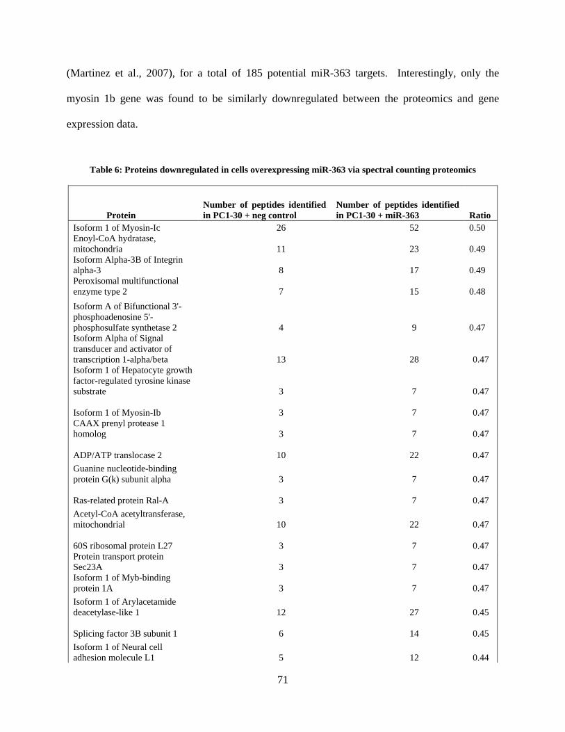
71
(Martinez et al., 2007), for a total of 185 potential miR-363 targets. Interestingly, only the
myosin 1b gene was found to be similarly downregulated between the proteomics and gene
expression data.
Table 6: Proteins downregulated in cells overexpressing miR-363 via spectral counting proteomics
Protein Number of peptides identified in PC1-30 + neg control
Number of peptides identified in PC1-30 + miR-363 Ratio
Isoform 1 of Myosin-Ic 26 52 0.50 Enoyl-CoA hydratase, mitochondria 11 23 0.49 Isoform Alpha-3B of Integrin alpha-3 8 17 0.49 Peroxisomal multifunctional enzyme type 2 7 15 0.48 Isoform A of Bifunctional 3'-phosphoadenosine 5'-phosphosulfate synthetase 2 4 9 0.47 Isoform Alpha of Signal transducer and activator of transcription 1-alpha/beta 13 28 0.47 Isoform 1 of Hepatocyte growth factor-regulated tyrosine kinase substrate 3 7 0.47
Isoform 1 of Myosin-Ib 3 7 0.47 CAAX prenyl protease 1 homolog 3 7 0.47
ADP/ATP translocase 2 10 22 0.47 Guanine nucleotide-binding protein G(k) subunit alpha 3 7 0.47
Ras-related protein Ral-A 3 7 0.47 Acetyl-CoA acetyltransferase, mitochondrial 10 22 0.47
60S ribosomal protein L27 3 7 0.47 Protein transport protein Sec23A 3 7 0.47 Isoform 1 of Myb-binding protein 1A 3 7 0.47 Isoform 1 of Arylacetamide deacetylase-like 1 12 27 0.45
Splicing factor 3B subunit 1 6 14 0.45 Isoform 1 of Neural cell adhesion molecule L1 5 12 0.44

72
Isoform 1 of ATPase family AAA domain-containing protein 3A 5 12 0.44 Isoform 2 of Microtubule-actin cross-linking factor 1, isoforms 1/2/3/5 7 17 0.43 Isoform 1 of Nuclear pore complex protein Nup155 3 8 0.41 Aconitate hydratase, mitochondrial 3 8 0.41 Probable ATP-dependent RNA helicase DDX47 3 8 0.41 Protein tyrosine phosphatase-like protein PTPLAD1 3 8 0.41
FACT complex subunit SPT16 7 18 0.41 Isoform 1 of Cytoplasmic FMR1-interacting protein 1 4 11 0.39 Isoform 1 of MAP7 domain-containing protein 1 3 9 0.37 Tyrosine-protein phosphatase non-receptor type 1 5 16 0.33 Isoform 1 of Fragile X mental retardation syndrome-related protein 1 4 13 0.33 Interferon-induced protein with tetratricopeptide repeats 1 3 12 0.28 Guanine nucleotide-binding protein G(i), alpha-1 subunit 4 16 0.27 Isoform 1 of CUB domain-containing protein 1 3 13 0.26 Inositol 1,4,5-trisphosphate receptor type 3 5 21 0.26 Isoform 3 of Ribosome-binding protein 1 5 25 0.22

73
The 35 proteins from Table 6 and 150 downregulated genes from our previous study
(Martinez et al., 2007) were cross-referenced with computationally predicted miR-363 targets
obtained from the miRanda and TargetScan databases. This cross-comparison resulted in six
potential targets based upon proteomics data and ten potential targets based upon the gene
expression data, with one potential miR-363 target in common, myosin 1b (MYO1B, Figure 12).
Figure 12: Venn diagram of potential miR-363 targets

74
The 3′ UTR of MYO1B contains two predicted binding sites for miR-363, and since
MYO1B expression at both the mRNA and protein levels was reduced upon miR-363
overexpression (Figure 13), it was considered a likely target of miR-363.
Figure 13: MYO1B expression is reduced upon miR-363 expression
HPV-negative SCCHN cell line PCI30 transfected with either a negative control miRNA or miR-363. (A) qRT-
PCR for MYO1B at mRNA level. (B) Western blot for MYO1B at protein level. The p value for PCI30 + miR-363
compared to PCI30 + neg. is indicated by ** (p < 0.01).

75
To test whether MYO1B was a direct target of miR-363, we cloned the 3′ UTR of
MYO1B downstream of the firefly luciferase gene in the pMiR-Report vector. A dual luciferase
assay, where the firefly luciferase was normalized to the renilla luciferase, showed that the firefly
luciferase activity was reduced when miR-363 was overexpressed but not when a control
miRNA that has no binding sites in the MYO1B 3′ UTR was expressed (Figure 14). These
results suggest that miR-363 binds directly to the 3′ UTR of MYO1B and represses its
expression.
Figure 14: MYO1B is a direct target of miR-363 in SCCHN
Dual luciferase assay of HPV-negative SCCHN cells transfected with reporter vectors with or without the MYO1B
3′UTR cloned downstream of firefly luciferase. The p value for pMiR-Report-MYO1B + miR-363 compared to
pMiR-Report-MYO1B or pMiR-Report-MYO1B + miR-363 compared to pMiR-Report-MYO1B + negative
miRNA is indicated by ** (p < 0.01).
76
4.3.2 MicroRNA-363 Reduces the Migratory Ability of HPV-Negative SCCHN Cells
Since myosin proteins are associated with cellular migration amongst other functions (Edgar et
al., 1996; Evangelista et al., 2000), we wished to investigate the effect of miR-363 on cellular
migration. Transwell migration assays showed that there was a significant decrease in the
number of migratory HPV-negative SCCHN cells when miR-363 was overexpressed compared
to the same cells that do not express miR-363 (Figure 15). Since miR-363 targets MYO1B in
SCCHN (Figure 14) and MYO1B is involved in cell migration (Edgar et al., 1996; Evangelista et
al., 2000), we wanted to test whether reduction in MYO1B levels directly reduce cell migration.
Knockdown of MYO1B via siRNA in HPV-negative SCCHN cells showed reduced cell
migration compared to cells expressing a negative control siRNA (Figure 16). These results
indicate that high miR-363 expression (which would reduce MYO1B levels) as well as reduction
in MYO1B levels by siRNA knockdown reduce cell migration (Figures 15 and 16).
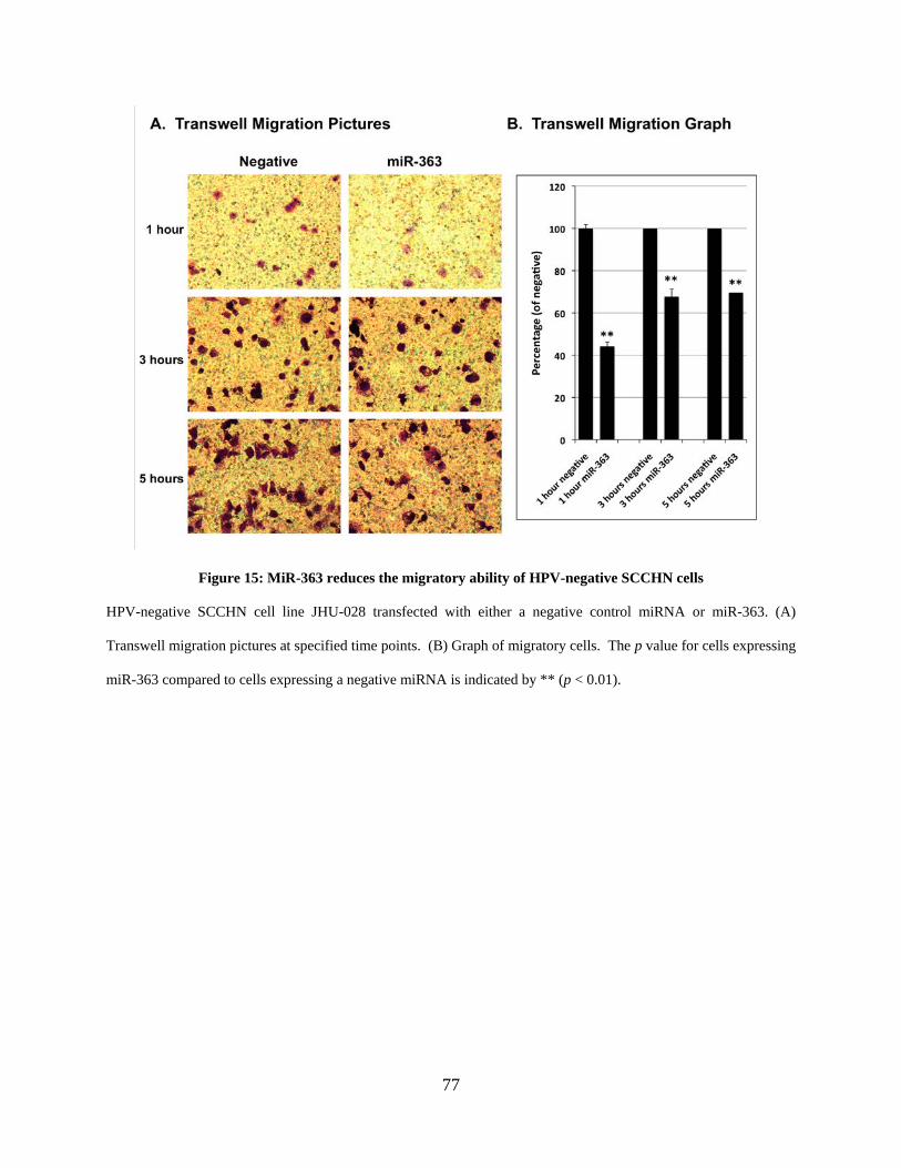
77
Figure 15: MiR-363 reduces the migratory ability of HPV-negative SCCHN cells
HPV-negative SCCHN cell line JHU-028 transfected with either a negative control miRNA or miR-363. (A)
Transwell migration pictures at specified time points. (B) Graph of migratory cells. The p value for cells expressing
miR-363 compared to cells expressing a negative miRNA is indicated by ** (p < 0.01).
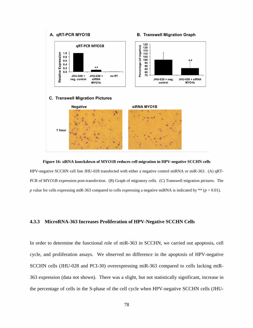
78
Figure 16: siRNA knockdown of MYO1B reduces cell migration in HPV-negative SCCHN cells
HPV-negative SCCHN cell line JHU-028 transfected with either a negative control miRNA or miR-363. (A) qRT-
PCR of MYO1B expression post-transfection. (B) Graph of migratory cells. (C) Transwell migration pictures. The
p value for cells expressing miR-363 compared to cells expressing a negative miRNA is indicated by ** (p < 0.01).
4.3.3 MicroRNA-363 Increases Proliferation of HPV-Negative SCCHN Cells
In order to determine the functional role of miR-363 in SCCHN, we carried out apoptosis, cell
cycle, and proliferation assays. We observed no difference in the apoptosis of HPV-negative
SCCHN cells (JHU-028 and PCI-30) overexpressing miR-363 compared to cells lacking miR-
363 expression (data not shown). There was a slight, but not statistically significant, increase in
the percentage of cells in the S-phase of the cell cycle when HPV-negative SCCHN cells (JHU-

79
028 and PCI-30) overexpressed miR-363 compared to the same cells expressing a negative
control miRNA (data not shown). However, miR-363 overexpression in HPV-negative SCCHN
cells (JHU-028) increased the proliferation capacity of these cells as measured via MTT assay
and cell counting (Figure 17). Furthermore, growth of miR-363-transfected cells in serum-
depleted media either had the same effect as cells grown in the normal media, or exacerbated the
effect seen in cells grown in the normal media (Figure 17).
Figure 17: MicroRNA-363 increases the proliferation of HPV-negative SCCHN cells
HPV-negative SCCHN cell line JHU-028 transfected with either a negative control miRNA or miR-363. (A) MTT
assay of transfected cells grown in normal media (left) or serum-depleted media (right). (B) Cell counting assay of
transfected cells grown in normal media (left) or serum-depleted media (right).

80
Furthermore, an in vitro wound healing assay showed that miR-363 enhanced the ability
of HPV-negative SCCHN cells to close the gap/lesion at a faster rate than in the presence of a
control miRNA (Figure 18).
Figure 18: MicroRNA-363 enhances the ability of HPV-negative SCCHN cells to close a gap/wound
HPV-negative SCCHN cell line JHU-028 transfected with either a negative control miRNA or miR-363. (A) Wound
healing assay pictures taken at 0 hours and 20 hours. (B) Graph of wound healing assay at designated time points.
The p value for cells expressing miR-363 compared to cells expressing a negative miRNA is indicated by ** (p <
0.01).

81
4.3.4 MicroRNA-363 Increases Colony Formation by HPV-Negative SCCHN Cells
Overexpression of miR-363 in HPV-negative SCCHN cell line PCI-30 showed an increase in the
number of colonies formed by cells expressing miR-363 compared to cells expressing a negative
control miRNA in a soft agar assay (Figure 19). In addition, the colony size was significantly
greater in cells expressing miR-363 compared to those expressing a negative control miRNA
(Figure 19). These results suggest that miR-363 may promote cell growth and colony formation.

82
Figure 19: MicroRNA-363 increases colony formation in HPV-negative SCCHN cells
HPV-negative SCCHN cell line PCI-30 transfected with either a negative control miRNA or miR-363. (A) Picture
of a representative well of a six-well plate, showing colonies formed, including representative diameter. (B) Graph
representing the number of colonies formed per condition. The p value for cells expressing miR-363 compared to
cells expressing a negative miRNA is indicated by ** (p < 0.01).
83
4.4 DISCUSSION
We have shown that miR-363 is overexpressed in HPV-positive SCCHN cell lines and tissue
samples compared to HPV-negative SCCHN cell lines and tissues (Wald et al., 2011).
Furthermore, expression of the HPV-16 E6 oncogene increases the expression of miR-363 while
siRNA knockdown of HPV-16 E6 reduces miR-363 expression (Wald et al., 2011). We sought
to characterize the functional role of miR-363 in SCCHN.
MiR-363 is part of the miR-106a~363 cluster of miRNAs, which is one of three miRNA
clusters in the oncogenic miR-17~92 family of clusters (Ventura et al., 2008). Several miRNAs
in these clusters are upregulated in other cancers, including T-cell leukemia (Landais et al.,
2007), colon cancer (Bandres et al., 2006; Tsuchida et al., 2011; Volinia et al., 2006; Yu et al.,
2011), and lung cancer (Hayashita et al., 2005; Osada and Takahashi, 2011). MiR-363, miR-25,
and miR-92 all belong to the miR-17~92 family and have identical seed sequences (Ventura et
al., 2008). Since the seed sequence of a mature miRNA helps determine its target mRNA
(Bartel, 2004), it is hypothesized that these three miRNAs could target similar (if not the same)
genes (Ventura et al., 2008). MiR-25 is overexpressed in bile duct, gastric, and ovarian cancers
where it targets the TRAIL Death Receptor 4 (Razumilava et al., 2012), p57 (Kim et al., 2009),
and BIM (Zhang et al., 2012), respectively. Additionally, miR-92 levels are increased in colon
cancer and small cell lung cancer where it targets BIM (Tsuchida et al., 2011) and RAB14
(Kanzaki et al., 2011), respectively. Moreover, in myeloid cells miR-92 targets p63 (Manni et
al., 2009). Because miR-363 has the same seed sequence as miR-25 and miR-92, and miR-25
and miR-92 have been implicated in cell cycle regulation, it is possible that miR-363 has a
similar function.

84
Since miRNAs can either target mRNA transcripts for degradation or promote their
translational repression, a decrease in the levels of their target mRNA is not always observed
(Eulalio et al., 2008). We were successfully able to narrow the potential targets of miR-363 by
utilizing spectral counting proteomics (Table 6), gene expression profiling data previously
generated in our laboratory (Martinez et al., 2007), and computationally predicted miRNA target
databases. Myosin 1B (MYO1B) showed a significant decrease at the mRNA and protein levels
when miR-363 was overexpressed (Figure 13), and was confirmed to be a direct target of miR-
363 by the luciferase assay (Figure 14). Myosins are common motor proteins involved in many
cellular processes, including cell motility (Edgar et al., 1996; Evangelista et al., 2000). MiR-363
overexpression caused by HPV-16 E6 in HPV-positive SCCHN cell lines results in a decrease in
MYO1B expression (Figure 13) and reduces cell migration (Figure 15). Additionally, when
MYO1B levels were reduced via siRNA knockdown, cell migration also decreased (Figure 16).
These data show that miR-363 expression causes a decrease in cell migration in SCCHN by
reducing MYO1B expression.
To test a more comprehensive role for miR-363 in SCCHN, we analyzed its possible
roles via several functional assays. Addition of exogenous miR-363 to HPV-negative SCCHN
cells that do not express miR-363 caused an increase in cell proliferation, as measured by cell
counting, MTT, wound healing, and colony formation assays (Figures 17-19). The HPV E6 and
E7 oncogenes are known to promote cell proliferation as well as viral replication (Moody and
Laimins, 2010). Overexpression of miR-363 by the HPV-16 E6 oncogene may be an additional
mechanism by which HPVs could promote cell proliferation. We were unable to significantly
knock-down miR-363 levels in HPV-positive SCCHN cell lines by using either an antagomir to
85
miR-363 or by siRNA knockdown of E6 (data not shown). Therefore, the direct effect of
reducing miR-363 expression in HPV-positive SCCHN cell lines could not be tested.
We also carried out colony formation assays in soft agar using the HPV-negative SCCHN
cell line PCI-30. The colony forming ability of these cells has not been previously characterized.
We found that overexpression of miR-363 in this cell line increased both the number and size of
the colonies (Figure 19). This was particularly striking because in these cells, very few colonies
were observed when a negative control miRNA was expressed but many colonies were seen in
the presence of miR-363 (Figure 19). Thus, our observation that HPV-negative SCCHN cells
form larger colonies when expressing miR-363 compared to a negative control miRNA supports
our results that miR-363 promotes the proliferative ability of SCCHN cells.
The data presented in this study show that miR-363 targets MYO1B which reduces cell
migration. Furthermore, miR-363 increases proliferation and colony formation of HPV-negative
SCCHN cells. This data could give more insight and help lead to a better understanding of the
differences between HPV-positive and HPV-negative SCCHN. Based on our study, we envision
that infection with HPV-16 causes overexpression of the E6 and E7 oncogenes and E6
expression results in an increase in miR-363 levels. High expression of miR-363 may then
reduce MYO1B levels, thereby reducing the migratory ability of these cells. Furthermore, high
levels of miR-363 may also promote cellular proliferation and colony formation.

86
5.0 SUMMARY, CONCLUSION, AND FUTURE DIRECTIONS
87
5.1 GENERAL SUMMARY AND CONCLUSIONS
High-risk HPV infection is associated with several cancers, including cervical, anogenital and a
subset of head and neck cancer (De Vuyst et al., 2009; Frisch et al., 1997; Pascual et al., 2007;
Tran et al., 2007b; Walboomers et al., 1999). The number of HPV-positive SCCHN cases has
been increasing in the past ten years, and the characteristics of HPV-positive and HPV-negative
SCCHN are different, leading some to treat them as distinct tumor entities (Chaturvedi et al.,
2008; D'Souza et al., 2007). The role of miRNAs in the regulation of gene expression has
provided insights into the dysregulation of miRNA expression profiles in human diseases,
including cancers (Esquela-Kerscher and Slack, 2006). We hypothesized that overexpression of
the E6 and E7 oncogenes in HPV-positive SCCHN would alter miRNA profiles in SCCHN
differently than in HPV-negative SCCHN. Differences between miRNA profiles in HPV-
positive and HPV-negative SCCHN would lead to specific alterations in the targets of these
miRNAs, giving rise to the different characteristics observed for HPV-positive and HPV-
negative SCCHN.
These studies are amongst the first to show a role for HPV in altering cellular miRNA
profiles in SCCHN. The miRNA microarray analysis showed dysregulation of several miRNAs
in the HPV-positive SCCHN cell lines compared to the HPV-negative SCCHN cell lines.
Several of these miRNAs were altered specifically in the presence of HPV-16 E6 and/or E7,
including the upregulation of miR-363, and the downregulation of miR-181a, miR-218, and
miR-29a. Additionally, siRNA knockdown of HPV-16 E6 in HPV-positive SCCHN cell lines
showed reduced expression of miR-363, indicating that miR-363 expression was dependent upon
HPV-16 E6 expression (Wald et al., 2011).

88
We further showed that in addition to the HPV-positive SCCHN cell lines, miR-363 was
also overexpressed in tissue samples from HPV-positive SCCHN patients from Western
Pennsylvania compared to the HPV-negative SCCHN tissues. These results suggest that miR-
363 overexpression may have clinical relevance. Additionally, the characteristics of our SCCHN
tissues were similar to a previously published study (Chaturvedi et al., 2008). The tonsil and
base of tongue had the highest rate of HPV-positivity, and represented 88% of HPV-positive
SCCHN tissues in our cohort, while the HPV-negative SCCHN tumors were evenly distributed
between the various head and neck sites. Patients with HPV-positive SCCHN were about nine
years younger than those with HPV-negative SCCHN, and a vast majority of the HPV-positive
SCCHN tissues compared to only one-half of the HPV-negative SCCHN tissues were classified
as T1 or T2 tumors.
Since miRNAs act to repress their target mRNAs (Bartel, 2004), we utilized gene
expression data, spectral counting proteomics, and computationally predicted miRNA target
databases to show that miR-363 targets MYO1B in SCCHN. Furthermore, high levels of miR-
363, and low levels of MYO1B reduced cell migration in SCCHN cells. To further elucidate the
role of miR-363 in SCCHN, we carried out several studies which suggested that miR-363
increases proliferation and colony formation of SCCHN cells.
It is not surprising that miRNA expression profiles are different in HPV-positive and
HPV-negative SCCHN. HPV alters miRNA profiles in HaCaT cells that were transfected with
the HPV-11, -16, or -45 whole genomes (Dreher et al., 2011), as well as HaCaT cells transfected
with only the HPV-16 E5 gene (Greco et al., 2011). Although transfection with one gene or the
entire HPV genome does not mimic natural HPV infection, it confirms that the HPV viral genes
can alter cellular miRNA profiles. There are many reports on dysregulated miRNAs in cervical

89
cancer (99% of which are caused by high-risk HPV) and head and neck cancer, but our studies
are amongst the first to show that miRNAs are dysregulated differently in HPV-positive and
HPV-negative SCCHN.
5.2 FUTURE DIRECTIONS
The data presented here show that miRNA profiles in HPV-positive and HPV-negative SCCHN
are different. Furthermore, miR-363 is overexpressed in HPV-positive SCCHN compared to
HPV-negative SCCHN, targets MYO1B to reduce cellular migration, and increases cellular
proliferation and colony formation. Additional studies are necessary to further analyze the role
of miRNAs in the development of HPV-associated SCCHN. For example, identification of the
mechanism by which the E6 oncogene of HPV-16 promotes the overexpression of miR-363
would be of great interest. The cellular functions and domains of HPV E6 are well
characterized. We have used three HPV-16 E6 mutants (F2V, L50G, and L110Q) that are
defective in their interaction with specific cellular targets, but we were unable to identify a direct
mechanism by which E6 regulates cellular miRNAs. Future studies would include the use of
additional E6 mutants to study the mechanism of upregulation of miR-363 by the E6 oncogene.
MiR-363 is part of a cluster of miR-106a~363 miRNAs, however only miR-363 was
found to be overexpressed in our studies. A cluster of miRNAs is defined by the proximity to
other miRNAs and can either have one promoter for the transcription of polycistronic miRNAs,
or a miRNA cluster can have multiple promoters (Monteys et al., 2010; Ozsolak et al., 2008;
Wang et al., 2009b). The promoter(s) of the miR-106a~363 cluster are not known, and since
only miR-363 is overexpressed in HPV-positive SCCHN, it is likely that either miR-363 has its

90
own promoter or it is processed differently from a polycistronic RNA than other miRNAs in the
cluster. Identifying the miR-363 (or cluster) promoter, and the mechanism of its upregulation in
HPV-positive SCCHN should provide valuable insight into the development of this disease. A
previous report predicted the transcription start site for miR-363 (Ozsolak et al., 2008), and
provides a good starting point for such studies. The cloning of such regions into a promoter-less
luciferase vector should lead to the identification of the miR-363 promoter. Once a promoter
region for miR-363 has been identified, chromatin immunoprecipitation (ChIP) assays could be
done to investigate whether E6 (or one of its downstream targets) binds to the miR-363
promoter. The E6 protein could also be targeted to the miR-363 promoter through its interaction
with a cellular transcription factor that acts at this promoter. In addition, epigenetic mechanisms
may also be involved in the regulation of miR-363 expression. There have been previous reports
demonstrating silencing of miRNAs by DNA methylation in oral cancer (Kozaki et al., 2008;
Langevin et al., 2011). In vitro analysis of SCCHN cells treated with 5-azacytidine which
inhibits DNA methylation, or trichostatin A which inhibits histone deacetylation, may provide
insight into the possible regulation of miR-363 by epigenetic mechanisms.
Additionally, since we have shown that expression of HPV-16 E6 in primary human
keratinocytes increases miR-363 levels (Figure 7), gene expression analysis of the E6-expressing
keratinocytes may provide clues to cellular factors which may be involved in the upregulation of
miR-363.
Another direction for future experiments could involve the ability of miR-363 to induce
or prevent tumor formation. We have shown that HPV-negative SCCHN cells expressing
exogenous miR-363 form more colonies (via soft agar assay, Figure 19). This initial finding

91
could be further explored in animal model systems, which may give insight into the in vivo role
of miR-363 in HPV-positive SCCHN.
This dissertation details the role of one altered miRNA, miR-363, in HPV-positive
SCCHN. There were several other miRNAs whose levels were altered in HPV-positive
compared to HPV-negative SCCHN cells that could be investigated for their role in the
development of HPV-positive SCCHN as well as for diagnosis and developing new treatment
approaches for SCCHN.

92
SUPPLEMENTARY TABLES
Supplementary Table 1 Basal expression of miRNAs in HPV-16 positive SCCHN cell linesName Median hsa_miR_21 15166.0 hsa_miR_16 9969.5 hsa_miR_29a 9183.5 hsa_miR_23b 5818.5 hsa_miR_221 5180.5 hsa_miR_24 5157.0 hsa_miR_23a 4930.0 hsa_let_7a 4129.5 hsa_miR_31 4102.0 hsa_miR_222 3579.5 hsa_miR_30a_5p 2689.0 hsa_miR_26a 2684.5 hsa_miR_29b 2591.5 hsa_miR_107 2116.5 hsa_miR_15b 2051.5 hsa_miR_103 2039.5 hsa_miR_106a 2014.5 hsa_miR_30d 1795.5 hsa_miR_27b 1665.5 hsa_let_7d 1655.0 hsa_miR_93 1534.5 hsa_miR_106b 1533.5 hsa_miR_205 1515.5 hsa_miR_125b 1407.5 hsa_miR_100 1324.0 hsa_miR_182 1264.0 hsa_miR_17_5p 1250.5 hsa_miR_130a 1176.0 hsa_miR_30c 1086.0 hsa_let_7c 1054.5 hsa_miR_22 1054.0 hsa_let_7i 1031.0 hsa_miR_181a 991.0 hsa_miR_200c 965.5 hsa_let_7b 862.0 hsa_miR_27a 856.0 hsa_miR_25 842.5 hsa_let_7e 826.0
hsa_let_7f 807.0 hsa_miR_181b 791.0 hsa_miR_99a 791.0 hsa_miR_19b 719.5 hsa_miR_20a 682.5 hsa_miR_15a 598.0 hsa_let_7g 592.5 hsa_miR_20b 562.5 hsa_miR_30b 560.0 hsa_miR_155 540.5 hsa_miR_191 508.5 hsa_miR_210 405.5 hsa_miR_30e_5p 394.0 hsa_miR_200b 324.0 hsa_miR_125a 318.5 hsa_miR_361 313.0 hsa_miR_320 278.0 hsa_miR_203 207.5 hsa_miR_138 202.5 hsa_miR_185 183.0 hsa_miR_130b 170.0 hsa_miR_181d 138.0 hsa_miR_7 123.0 hsa_miR_96 122.0 hsa_miR_92 121.5 hsa_miR_422b 110.5 hsa_miR_99b 107.5 hsa_miR_18a 98.0 hsa_miR_494 97.0 hsa_miR_193b 91.5 hsa_miR_224 85.5 hsa_miR_151 82.5 hsa_miR_152 80.5 hsa_miR_128a 76.0 hsa_miR_196a 69.0 hsa_miR_200a 68.5 hsa_miR_128b 67.0 hsa_miR_423 63.5 hsa_miR_136 63.0
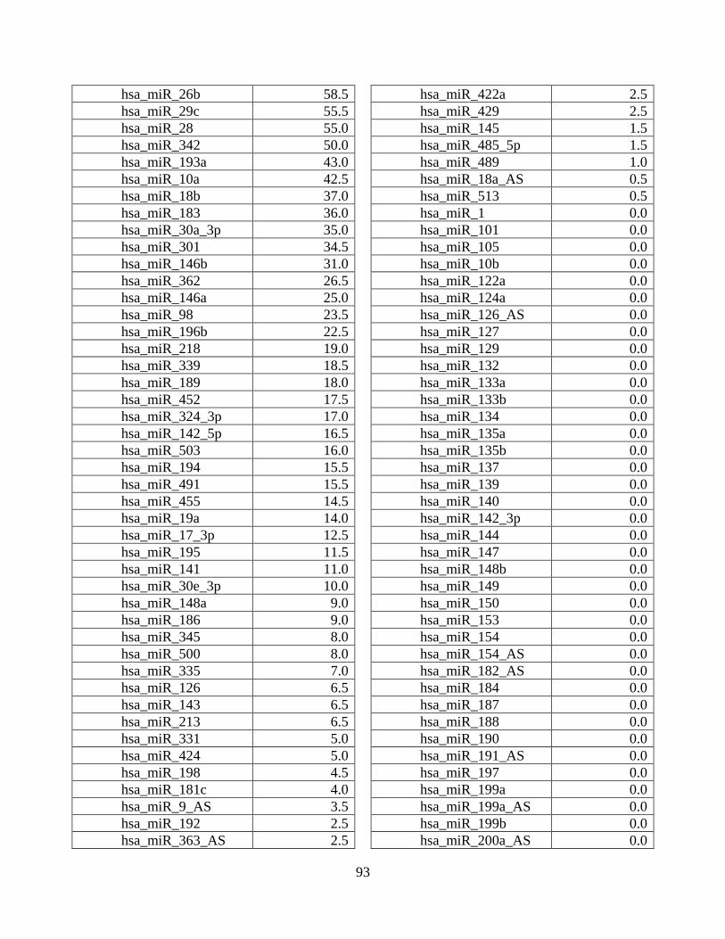
93
hsa_miR_26b 58.5 hsa_miR_29c 55.5 hsa_miR_28 55.0 hsa_miR_342 50.0 hsa_miR_193a 43.0 hsa_miR_10a 42.5 hsa_miR_18b 37.0 hsa_miR_183 36.0 hsa_miR_30a_3p 35.0 hsa_miR_301 34.5 hsa_miR_146b 31.0 hsa_miR_362 26.5 hsa_miR_146a 25.0 hsa_miR_98 23.5 hsa_miR_196b 22.5 hsa_miR_218 19.0 hsa_miR_339 18.5 hsa_miR_189 18.0 hsa_miR_452 17.5 hsa_miR_324_3p 17.0 hsa_miR_142_5p 16.5 hsa_miR_503 16.0 hsa_miR_194 15.5 hsa_miR_491 15.5 hsa_miR_455 14.5 hsa_miR_19a 14.0 hsa_miR_17_3p 12.5 hsa_miR_195 11.5 hsa_miR_141 11.0 hsa_miR_30e_3p 10.0 hsa_miR_148a 9.0 hsa_miR_186 9.0 hsa_miR_345 8.0 hsa_miR_500 8.0 hsa_miR_335 7.0 hsa_miR_126 6.5 hsa_miR_143 6.5 hsa_miR_213 6.5 hsa_miR_331 5.0 hsa_miR_424 5.0 hsa_miR_198 4.5 hsa_miR_181c 4.0 hsa_miR_9_AS 3.5 hsa_miR_192 2.5 hsa_miR_363_AS 2.5
hsa_miR_422a 2.5 hsa_miR_429 2.5 hsa_miR_145 1.5 hsa_miR_485_5p 1.5 hsa_miR_489 1.0 hsa_miR_18a_AS 0.5 hsa_miR_513 0.5 hsa_miR_1 0.0 hsa_miR_101 0.0 hsa_miR_105 0.0 hsa_miR_10b 0.0 hsa_miR_122a 0.0 hsa_miR_124a 0.0 hsa_miR_126_AS 0.0 hsa_miR_127 0.0 hsa_miR_129 0.0 hsa_miR_132 0.0 hsa_miR_133a 0.0 hsa_miR_133b 0.0 hsa_miR_134 0.0 hsa_miR_135a 0.0 hsa_miR_135b 0.0 hsa_miR_137 0.0 hsa_miR_139 0.0 hsa_miR_140 0.0 hsa_miR_142_3p 0.0 hsa_miR_144 0.0 hsa_miR_147 0.0 hsa_miR_148b 0.0 hsa_miR_149 0.0 hsa_miR_150 0.0 hsa_miR_153 0.0 hsa_miR_154 0.0 hsa_miR_154_AS 0.0 hsa_miR_182_AS 0.0 hsa_miR_184 0.0 hsa_miR_187 0.0 hsa_miR_188 0.0 hsa_miR_190 0.0 hsa_miR_191_AS 0.0 hsa_miR_197 0.0 hsa_miR_199a 0.0 hsa_miR_199a_AS 0.0 hsa_miR_199b 0.0 hsa_miR_200a_AS 0.0

94
hsa_miR_202 0.0 hsa_miR_202_AS 0.0 hsa_miR_204 0.0 hsa_miR_206 0.0 hsa_miR_208 0.0 hsa_miR_211 0.0 hsa_miR_212 0.0 hsa_miR_214 0.0 hsa_miR_215 0.0 hsa_miR_216 0.0 hsa_miR_217 0.0 hsa_miR_219 0.0 hsa_miR_220 0.0 hsa_miR_223 0.0 hsa_miR_296 0.0 hsa_miR_299_3p 0.0 hsa_miR_299_5p 0.0 hsa_miR_302a 0.0 hsa_miR_302a_AS 0.0 hsa_miR_302b 0.0 hsa_miR_302b_AS 0.0 hsa_miR_302c 0.0 hsa_miR_302c_AS 0.0 hsa_miR_302d 0.0 hsa_miR_32 0.0 hsa_miR_323 0.0 hsa_miR_324_5p 0.0 hsa_miR_325 0.0 hsa_miR_326 0.0 hsa_miR_328 0.0 hsa_miR_329 0.0 hsa_miR_33 0.0 hsa_miR_330 0.0 hsa_miR_337 0.0 hsa_miR_338 0.0 hsa_miR_340 0.0 hsa_miR_346 0.0 hsa_miR_34a 0.0 hsa_miR_34b 0.0 hsa_miR_34c 0.0 hsa_miR_363 0.0 hsa_miR_365 0.0 hsa_miR_367 0.0 hsa_miR_368 0.0 hsa_miR_369_3p 0.0
hsa_miR_369_5p 0.0 hsa_miR_370 0.0 hsa_miR_371 0.0 hsa_miR_372 0.0 hsa_miR_373 0.0 hsa_miR_373_AS 0.0 hsa_miR_374 0.0 hsa_miR_375 0.0 hsa_miR_376a 0.0 hsa_miR_376a_AS 0.0 hsa_miR_376b 0.0 hsa_miR_377 0.0 hsa_miR_378 0.0 hsa_miR_379 0.0 hsa_miR_380_3p 0.0 hsa_miR_380_5p 0.0 hsa_miR_381 0.0 hsa_miR_382 0.0 hsa_miR_383 0.0 hsa_miR_384 0.0 hsa_miR_409_3p 0.0 hsa_miR_409_5p 0.0 hsa_miR_410 0.0 hsa_miR_412 0.0 hsa_miR_425 0.0 hsa_miR_431 0.0 hsa_miR_432 0.0 hsa_miR_432_AS 0.0 hsa_miR_433 0.0 hsa_miR_448 0.0 hsa_miR_449 0.0 hsa_miR_450 0.0 hsa_miR_451 0.0 hsa_miR_452_AS 0.0 hsa_miR_453 0.0 hsa_miR_483 0.0 hsa_miR_484 0.0 hsa_miR_485_3p 0.0 hsa_miR_486 0.0 hsa_miR_487a 0.0 hsa_miR_487b 0.0 hsa_miR_488 0.0 hsa_miR_490 0.0 hsa_miR_492 0.0 hsa_miR_493_3p 0.0
95
hsa_miR_493_5p 0.0 hsa_miR_495 0.0 hsa_miR_496 0.0 hsa_miR_497 0.0 hsa_miR_498 0.0 hsa_miR_499 0.0 hsa_miR_501 0.0 hsa_miR_502 0.0 hsa_miR_504 0.0 hsa_miR_505 0.0 hsa_miR_506 0.0 hsa_miR_507 0.0 hsa_miR_508 0.0 hsa_miR_509 0.0 hsa_miR_510 0.0 hsa_miR_511 0.0 hsa_miR_512_3p 0.0 hsa_miR_512_5p 0.0 hsa_miR_514 0.0 hsa_miR_515_3p 0.0 hsa_miR_515_5p 0.0 hsa_miR_516_3p 0.0 hsa_miR_516_5p 0.0 hsa_miR_517_AS 0.0 hsa_miR_517a 0.0 hsa_miR_517b 0.0 hsa_miR_517c 0.0 hsa_miR_518a 0.0 hsa_miR_518a_2_AS 0.0 hsa_miR_518b 0.0 hsa_miR_518c 0.0 hsa_miR_518c_AS 0.0 hsa_miR_518d 0.0 hsa_miR_518e 0.0 hsa_miR_518f 0.0 hsa_miR_518f_AS 0.0 hsa_miR_519a 0.0
hsa_miR_519b 0.0 hsa_miR_519c 0.0 hsa_miR_519d 0.0 hsa_miR_519e 0.0 hsa_miR_519e_AS 0.0 hsa_miR_520a 0.0 hsa_miR_520a_AS 0.0 hsa_miR_520b 0.0 hsa_miR_520c 0.0 hsa_miR_520d 0.0 hsa_miR_520d_AS 0.0 hsa_miR_520e 0.0 hsa_miR_520f 0.0 hsa_miR_520g 0.0 hsa_miR_520h 0.0 hsa_miR_521 0.0 hsa_miR_522 0.0 hsa_miR_523 0.0 hsa_miR_524 0.0 hsa_miR_524_AS 0.0 hsa_miR_525 0.0 hsa_miR_525_AS 0.0 hsa_miR_526a 0.0 hsa_miR_526b 0.0 hsa_miR_526b_AS 0.0 hsa_miR_526c 0.0 hsa_miR_527 0.0 hsa_miR_539 0.0 hsa_miR_542_3p 0.0 hsa_miR_542_5p 0.0 hsa_miR_544 0.0 hsa_miR_545 0.0 hsa_miR_9 0.0 hsa_miR_95 0.0
The basal expression of miRNAs in the HPV-negative cell lines PCI-13 and PCI-30 was identified using miRNA microarrays. We obtained the median fluorescent intensity for each miRNA, and miRNAs having values of zero in at least four of their corresponding spots (4 spots per array and two arrays per sample) were considered to be non-expressing.

96
Supplementary Table 2 Basal expression of miRNAs in HPV-16 positive SCCHN cell lines Name Median hsa_miR_205 25541.5 hsa_miR_16 22308.0 hsa_miR_24 7394.0 hsa_miR_23b 6441.5 hsa_miR_141 6103.0 hsa_miR_26a 5640.0 hsa_miR_21 5434.0 hsa_let_7a 5364.5 hsa_miR_23a 4327.0 hsa_miR_27b 4024.0 hsa_miR_203 3831.5 hsa_miR_200b 3692.0 hsa_miR_106a 3677.0 hsa_miR_106b 3634.0 hsa_miR_107 3458.0 hsa_miR_93 3143.5 hsa_let_7i 3062.0 hsa_miR_15b 2664.5 hsa_miR_17_5p 2634.0 hsa_miR_200c 2559.5 hsa_let_7b 2537.5 hsa_let_7c 2512.5 hsa_let_7d 2493.5 hsa_miR_103 2413.0 hsa_miR_19b 2231.5 hsa_miR_200a 2214.0 hsa_miR_25 2114.0 hsa_miR_31 1904.5 hsa_miR_99a 1839.5 hsa_miR_30a_5p 1630.5 hsa_miR_130a 1534.5 hsa_let_7f 1483.5 hsa_miR_27a 1469.0 hsa_miR_15a 1458.5 hsa_miR_30d 1436.0 hsa_miR_30b 1379.0 hsa_miR_221 1369.5 hsa_miR_125b 1243.0 hsa_miR_182 1236.5 hsa_miR_20b 1213.0 hsa_miR_34a 1188.0 hsa_miR_29a 1083.0 hsa_miR_22 1010.5
hsa_let_7g 1005.5 hsa_miR_20a 981.0 hsa_miR_494 944.5 hsa_miR_30e_5p 933.0 hsa_miR_191 814.0 hsa_let_7e 802.5 hsa_miR_30c 765.5 hsa_miR_222 744.0 hsa_miR_422b 678.0 hsa_miR_361 659.5 hsa_miR_363 636.5 hsa_miR_125a 572.0 hsa_miR_92 517.0 hsa_miR_320 471.5 hsa_miR_18a 441.0 hsa_miR_210 423.0 hsa_miR_195 414.5 hsa_miR_99b 377.5 hsa_miR_148a 282.5 hsa_miR_342 274.5 hsa_miR_130b 273.0 hsa_miR_151 269.0 hsa_miR_224 259.0 hsa_miR_18b 245.5 hsa_miR_128a 226.0 hsa_miR_152 213.0 hsa_miR_96 183.5 hsa_miR_128b 182.5 hsa_miR_100 169.0 hsa_miR_423 156.0 hsa_miR_7 151.5 hsa_miR_19a 146.0 hsa_miR_29b 145.0 hsa_miR_183 140.5 hsa_miR_185 139.5 hsa_miR_26b 125.5 hsa_miR_28 114.0 hsa_miR_301 111.0 hsa_miR_429 99.0 hsa_miR_29c 98.0 hsa_miR_193a 84.0 hsa_miR_17_3p 83.5 hsa_miR_339 79.5 hsa_miR_362 78.5
97
hsa_miR_138 78.0 hsa_miR_196a 72.5 hsa_miR_189 63.0 hsa_miR_98 53.0 hsa_miR_324_3p 42.0 hsa_miR_335 42.0 hsa_miR_422a 41.5 hsa_miR_331 38.0 hsa_miR_186 35.5 hsa_miR_491 35.5 hsa_miR_136 32.5 hsa_miR_194 31.5 hsa_miR_30e_3p 30.5 hsa_miR_345 29.5 hsa_miR_452 29.0 hsa_miR_181b 26.5 hsa_miR_192 26.0 hsa_miR_484 23.5 hsa_miR_363_AS 23.0 hsa_miR_503 22.5 hsa_miR_196b 21.5 hsa_miR_497 21.5 hsa_miR_193b 20.0 hsa_miR_378 19.0 hsa_miR_149 18.5 hsa_miR_511 18.5 hsa_miR_379 18.0 hsa_miR_202 17.5 hsa_miR_146b 16.5 hsa_miR_500 16.5 hsa_miR_198 16.0 hsa_miR_132 15.5 hsa_miR_513 15.5 hsa_miR_370 15.0 hsa_miR_432 15.0 hsa_miR_521 14.5 hsa_miR_148b 14.0 hsa_miR_18a_AS 14.0 hsa_miR_197 13.0 hsa_miR_200a_AS 13.0 hsa_miR_453 13.0 hsa_miR_181a 12.0 hsa_miR_101 11.5 hsa_miR_30a_3p 11.5 hsa_miR_33 11.5
hsa_miR_126 9.5 hsa_miR_188 9.5 hsa_miR_425 9.5 hsa_miR_330 8.5 hsa_miR_213 8.0 hsa_miR_373_AS 8.0 hsa_miR_9_AS 8.0 hsa_miR_374 7.5 hsa_miR_485_5p 7.5 hsa_miR_505 7.0 hsa_miR_542_5p 7.0 hsa_miR_181d 6.5 hsa_miR_518c_AS 6.0 hsa_miR_155 5.5 hsa_miR_182_AS 5.5 hsa_miR_324_5p 5.5 hsa_miR_409_5p 5.5 hsa_miR_122a 5.0 hsa_miR_181c 5.0 hsa_miR_184 5.0 hsa_miR_302c_AS 5.0 hsa_miR_346 5.0 hsa_miR_34b 5.0 hsa_miR_492 4.5 hsa_miR_299_3p 4.0 hsa_miR_129 3.5 hsa_miR_142_3p 3.5 hsa_miR_365 3.5 hsa_miR_483 3.5 hsa_miR_519e_AS 3.5 hsa_miR_10a 3.0 hsa_miR_206 3.0 hsa_miR_212 3.0 hsa_miR_424 3.0 hsa_miR_134 2.5 hsa_miR_140 2.5 hsa_miR_368 2.5 hsa_miR_373 2.5 hsa_miR_450 2.5 hsa_miR_452_AS 2.5 hsa_miR_455 2.5 hsa_miR_517a 2.5 hsa_miR_519d 2.5 hsa_miR_544 2.5 hsa_miR_10b 2.0
98
hsa_miR_127 2.0 hsa_miR_143 2.0 hsa_miR_328 2.0 hsa_miR_499 2.0 hsa_miR_518a_2_AS 2.0 hsa_miR_145 1.5 hsa_miR_187 1.5 hsa_miR_199a 1.5 hsa_miR_220 1.5 hsa_miR_371 1.5 hsa_miR_381 1.5 hsa_miR_431 1.5 hsa_miR_485_3p 1.5 hsa_miR_493_3p 1.5 hsa_miR_495 1.5 hsa_miR_501 1.5 hsa_miR_519c 1.5 hsa_miR_9 1.5 hsa_miR_32 1.0 hsa_miR_486 1.0 hsa_miR_487a 1.0 hsa_miR_489 1.0 hsa_miR_502 1.0 hsa_miR_515_5p 1.0 hsa_miR_520d 1.0 hsa_miR_520d_AS 1.0 hsa_miR_526b 1.0 hsa_miR_527 1.0 hsa_miR_542_3p 1.0 hsa_miR_133b 0.5 hsa_miR_146a 0.5 hsa_miR_199b 0.5 hsa_miR_380_3p 0.5 hsa_miR_449 0.5 hsa_miR_487b 0.5 hsa_miR_510 0.5 hsa_miR_517b 0.5 hsa_miR_518f 0.5 hsa_miR_525 0.5 hsa_miR_1 0.0 hsa_miR_105 0.0 hsa_miR_124a 0.0 hsa_miR_126_AS 0.0 hsa_miR_133a 0.0 hsa_miR_135a 0.0
hsa_miR_135b 0.0 hsa_miR_137 0.0 hsa_miR_139 0.0 hsa_miR_142_5p 0.0 hsa_miR_144 0.0 hsa_miR_147 0.0 hsa_miR_150 0.0 hsa_miR_153 0.0 hsa_miR_154 0.0 hsa_miR_154_AS 0.0 hsa_miR_190 0.0 hsa_miR_191_AS 0.0 hsa_miR_199a_AS 0.0 hsa_miR_202_AS 0.0 hsa_miR_204 0.0 hsa_miR_208 0.0 hsa_miR_211 0.0 hsa_miR_214 0.0 hsa_miR_215 0.0 hsa_miR_216 0.0 hsa_miR_217 0.0 hsa_miR_218 0.0 hsa_miR_219 0.0 hsa_miR_223 0.0 hsa_miR_296 0.0 hsa_miR_299_5p 0.0 hsa_miR_302a 0.0 hsa_miR_302a_AS 0.0 hsa_miR_302b 0.0 hsa_miR_302b_AS 0.0 hsa_miR_302c 0.0 hsa_miR_302d 0.0 hsa_miR_323 0.0 hsa_miR_325 0.0 hsa_miR_326 0.0 hsa_miR_329 0.0 hsa_miR_337 0.0 hsa_miR_338 0.0 hsa_miR_340 0.0 hsa_miR_34c 0.0 hsa_miR_367 0.0 hsa_miR_369_3p 0.0 hsa_miR_369_5p 0.0 hsa_miR_372 0.0 hsa_miR_375 0.0

99
hsa_miR_376a 0.0 hsa_miR_376a_AS 0.0 hsa_miR_376b 0.0 hsa_miR_377 0.0 hsa_miR_380_5p 0.0 hsa_miR_382 0.0 hsa_miR_383 0.0 hsa_miR_384 0.0 hsa_miR_409_3p 0.0 hsa_miR_410 0.0 hsa_miR_412 0.0 hsa_miR_432_AS 0.0 hsa_miR_433 0.0 hsa_miR_448 0.0 hsa_miR_451 0.0 hsa_miR_488 0.0 hsa_miR_490 0.0 hsa_miR_493_5p 0.0 hsa_miR_496 0.0 hsa_miR_498 0.0 hsa_miR_504 0.0 hsa_miR_506 0.0 hsa_miR_507 0.0 hsa_miR_508 0.0 hsa_miR_509 0.0 hsa_miR_512_3p 0.0 hsa_miR_512_5p 0.0 hsa_miR_514 0.0 hsa_miR_515_3p 0.0 hsa_miR_516_3p 0.0 hsa_miR_516_5p 0.0 hsa_miR_517_AS 0.0
hsa_miR_517c 0.0 hsa_miR_518a 0.0 hsa_miR_518b 0.0 hsa_miR_518c 0.0 hsa_miR_518d 0.0 hsa_miR_518e 0.0 hsa_miR_518f_AS 0.0 hsa_miR_519a 0.0 hsa_miR_519b 0.0 hsa_miR_519e 0.0 hsa_miR_520a 0.0 hsa_miR_520a_AS 0.0 hsa_miR_520b 0.0 hsa_miR_520c 0.0 hsa_miR_520e 0.0 hsa_miR_520f 0.0 hsa_miR_520g 0.0 hsa_miR_520h 0.0 hsa_miR_522 0.0 hsa_miR_523 0.0 hsa_miR_524 0.0 hsa_miR_524_AS 0.0 hsa_miR_525_AS 0.0 hsa_miR_526a 0.0 hsa_miR_526b_AS 0.0 hsa_miR_526c 0.0 hsa_miR_539 0.0 hsa_miR_545 0.0 hsa_miR_95 0.0
The basal expression of miRNAs in the HPV-16 positive cell lines UD-SCC-2 and UPCI:SCC90 was identified using miRNA microarrays. We obtained the median fluorescent intensity for each miRNA, and miRNAs having values of zero in at least four of their corresponding spots (4 spots per array and two arrays per sample) were considered to be non-expressing.

100
Supplementary Table 3 MiRNAs Differentially Expressed in HPV-16-positive SCCHN cell line UD-SCC-2 compared to HPV-negative SCCHN cell line PCI-13
MiRNA Fold Change Underexpressed hsa_miR_181a -13.72 hsa_miR_181b -13.37
miR, microRNA; hsa, human; The q-values of all miRNAs were 0.

101
Supplementary Table 4 MiRNAs Differentially Expressed in HPV-16-positive SCCHN cell line UD-SCC-2 compared to HPV-negative SCCHN cell line PCI-30
MiRNA Fold Change Overexpressed hsa_miR_205 31.66 hsa_miR_141 14.18 hsa_miR_200c 11.39 hsa_miR_34a 8.60 hsa_miR_200b 7.69 hsa_miR_363 4.85
miR, microRNA; hsa, human; The q-values of all miRNAs were 0.

102
Supplementary Table 5 MiRNAs Differentially Expressed in HPV-16-positive SCCHN cell line UPCI:SCC90 compared to HPV-negative SCCHN cell line PCI-13
MiRNA Fold Change Overexpressed hsa_miR_148a 5.56 hsa_miR_363 5.47 hsa_miR_186 4.69 hsa_miR_101 3.54 Underexpressed hsa_miR_155 -11.36 hsa_miR_181a -5.63 ambi_miR_13143 -5.43
miR, microRNA; hsa, human; ambi, Ambion predicted; The q-values of all miRNAs were 0.

103
Supplementary Table 6 MiRNAs Differentially Expressed in HPV-16-positive SCCHN cell line UPCI:SCC90 compared to HPV-negative SCCHN cell line PCI-30
MiRNA Fold Change Overexpressed hsa_miR_205 22.92 hsa_miR_203 15.85 hsa_miR_141 15.34 hsa_miR_200c 11.47 hsa_miR_34a 10.50 hsa_miR_200b 9.47 hsa_miR_200a 7.79 hsa_miR_429 6.29 hsa_miR_363 5.41 hsa_miR_200a_AS 2.68
miR, microRNA; hsa, human; The q-values of all miRNAs were 0.

104
BIBLIOGRAPHY
Agrawal, N., Frederick, M.J., Pickering, C.R., Bettegowda, C., Chang, K., Li, R.J., Fakhry, C., Xie, T.X., Zhang, J., Wang, J., et al. (2011). Exome sequencing of head and neck squamous cell carcinoma reveals inactivating mutations in NOTCH1. Science 333, 1154-1157.
Ahluwalia, J.K., Khan, S.Z., Soni, K., Rawat, P., Gupta, A., Hariharan, M., Scaria, V., Lalwani, M., Pillai, B., Mitra, D., et al. (2008). Human cellular microRNA hsa-miR-29a interferes with viral nef protein expression and HIV-1 replication. Retrovirology 5, 117.
Albers, A., Abe, K., Hunt, J., Wang, J., Lopez-Albaitero, A., Schaefer, C., Gooding, W., Whiteside, T.L., Ferrone, S., DeLeo, A., et al. (2005). Antitumor activity of human papillomavirus type 16 E7-specific T cells against virally infected squamous cell carcinoma of the head and neck. Cancer Res 65, 11146-11155.
Asangani, I.A., Rasheed, S.A., Nikolova, D.A., Leupold, J.H., Colburn, N.H., Post, S., and Allgayer, H. (2008). MicroRNA-21 (miR-21) post-transcriptionally downregulates tumor suppressor Pdcd4 and stimulates invasion, intravasation and metastasis in colorectal cancer. Oncogene 27, 2128-2136.
Avissar, M., Christensen, B.C., Kelsey, K.T., and Marsit, C.J. (2009). MicroRNA expression ratio is predictive of head and neck squamous cell carcinoma. Clin Cancer Res 15, 2850-2855.
Balz, V., Scheckenbach, K., Gotte, K., Bockmuhl, U., Petersen, I., and Bier, H. (2003). Is the p53 inactivation frequency in squamous cell carcinomas of the head and neck underestimated? Analysis of p53 exons 2-11 and human papillomavirus 16/18 E6 transcripts in 123 unselected tumor specimens. Cancer Res 63, 1188-1191.
Bandi, N., Zbinden, S., Gugger, M., Arnold, M., Kocher, V., Hasan, L., Kappeler, A., Brunner, T., and Vassella, E. (2009). miR-15a and miR-16 are implicated in cell cycle regulation in a Rb-dependent manner and are frequently deleted or down-regulated in non-small cell lung cancer. Cancer Res 69, 5553-5559.
Bandres, E., Cubedo, E., Agirre, X., Malumbres, R., Zarate, R., Ramirez, N., Abajo, A., Navarro, A., Moreno, I., Monzo, M., et al. (2006). Identification by Real-time PCR of 13 mature microRNAs differentially expressed in colorectal cancer and non-tumoral tissues. Mol Cancer 5, 29.
105
Barnard, P., and McMillan, N.A. (1999). The human papillomavirus E7 oncoprotein abrogates signaling mediated by interferon-alpha. Virology 259, 305-313.
Bartel, D.P. (2004). MicroRNAs: genomics, biogenesis, mechanism, and function. Cell 116, 281-297.
Blot, W.J., McLaughlin, J.K., Winn, D.M., Austin, D.F., Greenberg, R.S., Preston-Martin, S., Bernstein, L., Schoenberg, J.B., Stemhagen, A., and Fraumeni, J.F., Jr. (1988). Smoking and drinking in relation to oral and pharyngeal cancer. Cancer Res 48, 3282-3287.
Bommer, G.T., Gerin, I., Feng, Y., Kaczorowski, A.J., Kuick, R., Love, R.E., Zhai, Y., Giordano, T.J., Qin, Z.S., Moore, B.B., et al. (2007). p53-mediated activation of miRNA34 candidate tumor-suppressor genes. Curr Biol 17, 1298-1307.
Bonci, D., Coppola, V., Musumeci, M., Addario, A., Giuffrida, R., Memeo, L., D'Urso, L., Pagliuca, A., Biffoni, M., Labbaye, C., et al. (2008). The miR-15a-miR-16-1 cluster controls prostate cancer by targeting multiple oncogenic activities. Nat Med 14, 1271-1277.
Bouvard, V., Matlashewski, G., Gu, Z.M., Storey, A., and Banks, L. (1994). The human papillomavirus type 16 E5 gene cooperates with the E7 gene to stimulate proliferation of primary cells and increases viral gene expression. Virology 203, 73-80.
Boyer, S.N., Wazer, D.E., and Band, V. (1996). E7 protein of human papilloma virus-16 induces degradation of retinoblastoma protein through the ubiquitin-proteasome pathway. Cancer Res 56, 4620-4624.
Bradford, C.R., Zhu, S., Ogawa, H., Ogawa, T., Ubell, M., Narayan, A., Johnson, G., Wolf, G.T., Fisher, S.G., and Carey, T.E. (2003). P53 mutation correlates with cisplatin sensitivity in head and neck squamous cell carcinoma lines. Head Neck 25, 654-661.
Brenner, J.C., Graham, M.P., Kumar, B., Saunders, L.M., Kupfer, R., Lyons, R.H., Bradford, C.R., and Carey, T.E. (2010). Genotyping of 73 UM-SCC head and neck squamous cell carcinoma cell lines. Head Neck 32, 417-426.
Brosh, R., Shalgi, R., Liran, A., Landan, G., Korotayev, K., Nguyen, G.H., Enerly, E., Johnsen, H., Buganim, Y., Solomon, H., et al. (2008). p53-Repressed miRNAs are involved with E2F in a feed-forward loop promoting proliferation. Mol Syst Biol 4, 229.
Cai, X., Hagedorn, C.H., and Cullen, B.R. (2004). Human microRNAs are processed from capped, polyadenylated transcripts that can also function as mRNAs. RNA 10, 1957-1966.
Caldas, C., and Brenton, J.D. (2005). Sizing up miRNAs as cancer genes. Nat Med 11, 712-714.
Calin, G.A., Dumitru, C.D., Shimizu, M., Bichi, R., Zupo, S., Noch, E., Aldler, H., Rattan, S., Keating, M., Rai, K., et al. (2002). Frequent deletions and down-regulation of micro- RNA genes miR15 and miR16 at 13q14 in chronic lymphocytic leukemia. Proc Natl Acad Sci U S A 99, 15524-15529.

106
Calin, G.A., Sevignani, C., Dumitru, C.D., Hyslop, T., Noch, E., Yendamuri, S., Shimizu, M., Rattan, S., Bullrich, F., Negrini, M., et al. (2004). Human microRNA genes are frequently located at fragile sites and genomic regions involved in cancers. Proc Natl Acad Sci U S A 101, 2999-3004.
Chan, J.A., Krichevsky, A.M., and Kosik, K.S. (2005). MicroRNA-21 is an antiapoptotic factor in human glioblastoma cells. Cancer Res 65, 6029-6033.
Chang, S.S., Jiang, W.W., Smith, I., Poeta, L.M., Begum, S., Glazer, C., Shan, S., Westra, W., Sidransky, D., and Califano, J.A. (2008a). MicroRNA alterations in head and neck squamous cell carcinoma. Int J Cancer 123, 2791-2797.
Chang, T.C., Wentzel, E.A., Kent, O.A., Ramachandran, K., Mullendore, M., Lee, K.H., Feldmann, G., Yamakuchi, M., Ferlito, M., Lowenstein, C.J., et al. (2007). Transactivation of miR-34a by p53 broadly influences gene expression and promotes apoptosis. Mol Cell 26, 745-752.
Chang, T.C., Yu, D., Lee, Y.S., Wentzel, E.A., Arking, D.E., West, K.M., Dang, C.V., Thomas-Tikhonenko, A., and Mendell, J.T. (2008b). Widespread microRNA repression by Myc contributes to tumorigenesis. Nat Genet 40, 43-50.
Chaturvedi, A.K., Engels, E.A., Anderson, W.F., and Gillison, M.L. (2008). Incidence trends for human papillomavirus-related and -unrelated oral squamous cell carcinomas in the United States. J Clin Oncol 26, 612-619.
Chellappan, S., Kraus, V.B., Kroger, B., Munger, K., Howley, P.M., Phelps, W.C., and Nevins, J.R. (1992). Adenovirus E1A, simian virus 40 tumor antigen, and human papillomavirus E7 protein share the capacity to disrupt the interaction between transcription factor E2F and the retinoblastoma gene product. Proc Natl Acad Sci U S A 89, 4549-4553.
Chen, C.Z., Li, L., Lodish, H.F., and Bartel, D.P. (2004). MicroRNAs modulate hematopoietic lineage differentiation. Science 303, 83-86.
Chendrimada, T.P., Gregory, R.I., Kumaraswamy, E., Norman, J., Cooch, N., Nishikura, K., and Shiekhattar, R. (2005). TRBP recruits the Dicer complex to Ago2 for microRNA processing and gene silencing. Nature 436, 740-744.
Chikamatsu, K., Nakano, K., Storkus, W.J., Appella, E., Lotze, M.T., Whiteside, T.L., and DeLeo, A.B. (1999). Generation of anti-p53 cytotoxic T lymphocytes from human peripheral blood using autologous dendritic cells. Clin Cancer Res 5, 1281-1288.
Childs, G., Fazzari, M., Kung, G., Kawachi, N., Brandwein-Gensler, M., McLemore, M., Chen, Q., Burk, R.D., Smith, R.V., Prystowsky, M.B., et al. (2009). Low-level expression of microRNAs let-7d and miR-205 are prognostic markers of head and neck squamous cell carcinoma. Am J Pathol 174, 736-745.
107
Cimmino, A., Calin, G.A., Fabbri, M., Iorio, M.V., Ferracin, M., Shimizu, M., Wojcik, S.E., Aqeilan, R.I., Zupo, S., Dono, M., et al. (2005). miR-15 and miR-16 induce apoptosis by targeting BCL2. Proc Natl Acad Sci U S A 102, 13944-13949.
Corney, D.C., Flesken-Nikitin, A., Godwin, A.K., Wang, W., and Nikitin, A.Y. (2007). MicroRNA-34b and MicroRNA-34c are targets of p53 and cooperate in control of cell proliferation and adhesion-independent growth. Cancer Res 67, 8433-8438.
Cruz, I.B., Snijders, P.J., Steenbergen, R.D., Meijer, C.J., Snow, G.B., Walboomers, J.M., and van der Waal, I. (1996). Age-dependence of human papillomavirus DNA presence in oral squamous cell carcinomas. Eur J Cancer B Oral Oncol 32B, 55-62.
D'Souza, G., Kreimer, A.R., Viscidi, R., Pawlita, M., Fakhry, C., Koch, W.M., Westra, W.H., and Gillison, M.L. (2007). Case-control study of human papillomavirus and oropharyngeal cancer. N Engl J Med 356, 1944-1956.
de Villiers, E.M., Fauquet, C., Broker, T.R., Bernard, H.U., and zur Hausen, H. (2004). Classification of papillomaviruses. Virology 324, 17-27.
De Vuyst, H., Clifford, G.M., Nascimento, M.C., Madeleine, M.M., and Franceschi, S. (2009). Prevalence and type distribution of human papillomavirus in carcinoma and intraepithelial neoplasia of the vulva, vagina and anus: a meta-analysis. Int J Cancer 124, 1626-1636.
DiMaio, D., and Mattoon, D. (2001). Mechanisms of cell transformation by papillomavirus E5 proteins. Oncogene 20, 7866-7873.
Dreher, A., Rossing, M., Kaczkowski, B., Andersen, D.K., Larsen, T.J., Christophersen, M.K., Nielsen, F.C., and Norrild, B. (2011). Differential expression of cellular microRNAs in HPV 11, -16, and -45 transfected cells. Biochem Biophys Res Commun 412, 20-25.
Du, Z.M., Hu, L.F., Wang, H.Y., Yan, L.X., Zeng, Y.X., Shao, J.Y., and Ernberg, I. (2011). Upregulation of MiR-155 in nasopharyngeal carcinoma is partly driven by LMP1 and LMP2A and downregulates a negative prognostic marker JMJD1A. PLoS One 6, e19137.
Edgar, A.J., Knight, A.E., and Bennett, J.P. (1996). Chicken myosin IB mRNA is highly expressed in lymphoid tissues. J Anat 189 ( Pt 2), 451-456.
Esquela-Kerscher, A., and Slack, F.J. (2006). Oncomirs - microRNAs with a role in cancer. Nat Rev Cancer 6, 259-269.
Eulalio, A., Huntzinger, E., and Izaurralde, E. (2008). Getting to the root of miRNA-mediated gene silencing. Cell 132, 9-14.
Evangelista, M., Klebl, B.M., Tong, A.H., Webb, B.A., Leeuw, T., Leberer, E., Whiteway, M., Thomas, D.Y., and Boone, C. (2000). A role for myosin-I in actin assembly through interactions with Vrp1p, Bee1p, and the Arp2/3 complex. J Cell Biol 148, 353-362.
108
Fakhry, C., and Gillison, M.L. (2006). Clinical implications of human papillomavirus in head and neck cancers. J Clin Oncol 24, 2606-2611.
Fakhry, C., Westra, W.H., Li, S., Cmelak, A., Ridge, J.A., Pinto, H., Forastiere, A., and Gillison, M.L. (2008). Improved survival of patients with human papillomavirus-positive head and neck squamous cell carcinoma in a prospective clinical trial. J Natl Cancer Inst 100, 261-269.
Ferris, R.L., Martinez, I., Sirianni, N., Wang, J., Lopez-Albaitero, A., Gollin, S.M., Johnson, J.T., and Khan, S. (2005). Human papillomavirus-16 associated squamous cell carcinoma of the head and neck (SCCHN): a natural disease model provides insights into viral carcinogenesis. Eur J Cancer 41, 807-815.
Filippova, M., Parkhurst, L., and Duerksen-Hughes, P.J. (2004). The human papillomavirus 16 E6 protein binds to Fas-associated death domain and protects cells from Fas-triggered apoptosis. J Biol Chem 279, 25729-25744.
Filippova, M., Song, H., Connolly, J.L., Dermody, T.S., and Duerksen-Hughes, P.J. (2002). The human papillomavirus 16 E6 protein binds to tumor necrosis factor (TNF) R1 and protects cells from TNF-induced apoptosis. J Biol Chem 277, 21730-21739.
Fornari, F., Gramantieri, L., Giovannini, C., Veronese, A., Ferracin, M., Sabbioni, S., Calin, G.A., Grazi, G.L., Croce, C.M., Tavolari, S., et al. (2009). MiR-122/cyclin G1 interaction modulates p53 activity and affects doxorubicin sensitivity of human hepatocarcinoma cells. Cancer Res 69, 5761-5767.
Frankel, L.B., Christoffersen, N.R., Jacobsen, A., Lindow, M., Krogh, A., and Lund, A.H. (2008). Programmed cell death 4 (PDCD4) is an important functional target of the microRNA miR-21 in breast cancer cells. J Biol Chem 283, 1026-1033.
Frisch, M., Glimelius, B., van den Brule, A.J., Wohlfahrt, J., Meijer, C.J., Walboomers, J.M., Goldman, S., Svensson, C., Adami, H.O., and Melbye, M. (1997). Sexually transmitted infection as a cause of anal cancer. N Engl J Med 337, 1350-1358.
Fujita, S., Ito, T., Mizutani, T., Minoguchi, S., Yamamichi, N., Sakurai, K., and Iba, H. (2008). miR-21 Gene expression triggered by AP-1 is sustained through a double-negative feedback mechanism. J Mol Biol 378, 492-504.
Funk, J.O., Waga, S., Harry, J.B., Espling, E., Stillman, B., and Galloway, D.A. (1997). Inhibition of CDK activity and PCNA-dependent DNA replication by p21 is blocked by interaction with the HPV-16 E7 oncoprotein. Genes Dev 11, 2090-2100.
Garnett, T.O., Filippova, M., and Duerksen-Hughes, P.J. (2006). Accelerated degradation of FADD and procaspase 8 in cells expressing human papilloma virus 16 E6 impairs TRAIL-mediated apoptosis. Cell Death Differ 13, 1915-1926.
Genther Williams, S.M., Disbrow, G.L., Schlegel, R., Lee, D., Threadgill, D.W., and Lambert, P.F. (2005). Requirement of epidermal growth factor receptor for hyperplasia induced by E5, a high-risk human papillomavirus oncogene. Cancer Res 65, 6534-6542.

109
Georges, S.A., Biery, M.C., Kim, S.Y., Schelter, J.M., Guo, J., Chang, A.N., Jackson, A.L., Carleton, M.O., Linsley, P.S., Cleary, M.A., et al. (2008). Coordinated regulation of cell cycle transcripts by p53-Inducible microRNAs, miR-192 and miR-215. Cancer Res 68, 10105-10112.
Gillison, M.L., Broutian, T., Pickard, R.K., Tong, Z.Y., Xiao, W., Kahle, L., Graubard, B.I., and Chaturvedi, A.K. (2012). Prevalence of oral HPV infection in the United States, 2009-2010. JAMA 307, 693-703.
Gillison, M.L., D'Souza, G., Westra, W., Sugar, E., Xiao, W., Begum, S., and Viscidi, R. (2008). Distinct risk factor profiles for human papillomavirus type 16-positive and human papillomavirus type 16-negative head and neck cancers. J Natl Cancer Inst 100, 407-420.
Gillison, M.L., Koch, W.M., Capone, R.B., Spafford, M., Westra, W.H., Wu, L., Zahurak, M.L., Daniel, R.W., Viglione, M., Symer, D.E., et al. (2000). Evidence for a causal association between human papillomavirus and a subset of head and neck cancers. J Natl Cancer Inst 92, 709-720.
Godshalk, S.E., Bhaduri-McIntosh, S., and Slack, F.J. (2008). Epstein-Barr virus-mediated dysregulation of human microRNA expression. Cell Cycle 7, 3595-3600.
Grandis, J.R., and Tweardy, D.J. (1993). Elevated levels of transforming growth factor alpha and epidermal growth factor receptor messenger RNA are early markers of carcinogenesis in head and neck cancer. Cancer Res 53, 3579-3584.
Greco, D., Kivi, N., Qian, K., Leivonen, S.K., Auvinen, P., and Auvinen, E. (2011). Human papillomavirus 16 E5 modulates the expression of host microRNAs. PLoS One 6, e21646.
Greenlee, R.T., Hill-Harmon, M.B., Murray, T., and Thun, M. (2001). Cancer statistics, 2001. CA Cancer J Clin 51, 15-36.
Gregory, R.I., Yan, K.P., Amuthan, G., Chendrimada, T., Doratotaj, B., Cooch, N., and Shiekhattar, R. (2004). The Microprocessor complex mediates the genesis of microRNAs. Nature 432, 235-240.
Griffiths-Jones, S., Saini, H.K., van Dongen, S., and Enright, A.J. (2008). miRBase: tools for microRNA genomics. Nucleic Acids Res 36, D154-158.
Gupta, A.K., Lee, J.H., Wilke, W.W., Quon, H., Smith, G., Maity, A., Buatti, J.M., and Spitz, D.R. (2009). Radiation response in two HPV-infected head-and-neck cancer cell lines in comparison to a non-HPV-infected cell line and relationship to signaling through AKT. Int J Radiat Oncol Biol Phys 74, 928-933.
Gwosdz, C., Balz, V., Scheckenbach, K., and Bier, H. (2005). p53, p63 and p73 expression in squamous cell carcinomas of the head and neck and their response to cisplatin exposure. Adv Otorhinolaryngol 62, 58-71.
110
Hammarstedt, L., Lindquist, D., Dahlstrand, H., Romanitan, M., Dahlgren, L.O., Joneberg, J., Creson, N., Lindholm, J., Ye, W., Dalianis, T., et al. (2006). Human papillomavirus as a risk factor for the increase in incidence of tonsillar cancer. Int J Cancer 119, 2620-2623.
Harris, T., Jimenez, L., Kawachi, N., Fan, J.B., Chen, J., Belbin, T., Ramnauth, A., Loudig, O., Keller, C.E., Smith, R., et al. (2012). Low-Level Expression of miR-375 Correlates with Poor Outcome and Metastasis While Altering the Invasive Properties of Head and Neck Squamous Cell Carcinomas. Am J Pathol 180, 917-928.
Hayashita, Y., Osada, H., Tatematsu, Y., Yamada, H., Yanagisawa, K., Tomida, S., Yatabe, Y., Kawahara, K., Sekido, Y., and Takahashi, T. (2005). A polycistronic microRNA cluster, miR-17-92, is overexpressed in human lung cancers and enhances cell proliferation. Cancer Res 65, 9628-9632.
He, L., He, X., Lim, L.P., de Stanchina, E., Xuan, Z., Liang, Y., Xue, W., Zender, L., Magnus, J., Ridzon, D., et al. (2007). A microRNA component of the p53 tumour suppressor network. Nature 447, 1130-1134.
He, L., Thomson, J.M., Hemann, M.T., Hernando-Monge, E., Mu, D., Goodson, S., Powers, S., Cordon-Cardo, C., Lowe, S.W., Hannon, G.J., et al. (2005). A microRNA polycistron as a potential human oncogene. Nature 435, 828-833.
Hebner, C.M., and Laimins, L.A. (2006). Human papillomaviruses: basic mechanisms of pathogenesis and oncogenicity. Rev Med Virol 16, 83-97.
Hobbs, C.G., Sterne, J.A., Bailey, M., Heyderman, R.S., Birchall, M.A., and Thomas, S.J. (2006). Human papillomavirus and head and neck cancer: a systematic review and meta-analysis. Clin Otolaryngol 31, 259-266.
Howie, H.L., Katzenellenbogen, R.A., and Galloway, D.A. (2009). Papillomavirus E6 proteins. Virology 384, 324-334.
Howlader, N., Noone, A.M., Krapcho, M., Neyman, N., Aminou, R., Waldron, W., Altekruse, S.F., Kosary, C.L., Ruhl, J., Tatalovich, Z., et al. (2011). SEER Cancer Statistics Review, 1975-2008, National Cancer Institute. Bethesda, MD.
Hu, W., Zhang, C., Wu, R., Sun, Y., Levine, A., and Feng, Z. (2010). Glutaminase 2, a novel p53 target gene regulating energy metabolism and antioxidant function. Proc Natl Acad Sci U S A 107, 7455-7460.
Hui, A.B., Lenarduzzi, M., Krushel, T., Waldron, L., Pintilie, M., Shi, W., Perez-Ordonez, B., Jurisica, I., O'Sullivan, B., Waldron, J., et al. (2010). Comprehensive MicroRNA profiling for head and neck squamous cell carcinomas. Clin Cancer Res 16, 1129-1139.
Huibregtse, J.M., Scheffner, M., and Howley, P.M. (1991). A cellular protein mediates association of p53 with the E6 oncoprotein of human papillomavirus types 16 or 18. EMBO J 10, 4129-4135.

111
Hutvagner, G., McLachlan, J., Pasquinelli, A.E., Balint, E., Tuschl, T., and Zamore, P.D. (2001). A cellular function for the RNA-interference enzyme Dicer in the maturation of the let-7 small temporal RNA. Science 293, 834-838.
Jemal, A., Siegel, R., Xu, J., and Ward, E. (2010). Cancer statistics, 2010. CA Cancer J Clin 60, 277-300.
Jiang, J., Gusev, Y., Aderca, I., Mettler, T.A., Nagorney, D.M., Brackett, D.J., Roberts, L.R., and Schmittgen, T.D. (2008). Association of MicroRNA expression in hepatocellular carcinomas with hepatitis infection, cirrhosis, and patient survival. Clin Cancer Res 14, 419-427.
Jiang, M., and Milner, J. (2002). Selective silencing of viral gene expression in HPV-positive human cervical carcinoma cells treated with siRNA, a primer of RNA interference. Oncogene 21, 6041-6048.
John, B., Enright, A.J., Aravin, A., Tuschl, T., Sander, C., and Marks, D.S. (2004). Human MicroRNA targets. PLoS Biol 2, e363.
Johnson, S.M., Grosshans, H., Shingara, J., Byrom, M., Jarvis, R., Cheng, A., Labourier, E., Reinert, K.L., Brown, D., and Slack, F.J. (2005). RAS is regulated by the let-7 microRNA family. Cell 120, 635-647.
Jones, D.L., Alani, R.M., and Munger, K. (1997a). The human papillomavirus E7 oncoprotein can uncouple cellular differentiation and proliferation in human keratinocytes by abrogating p21Cip1-mediated inhibition of cdk2. Genes Dev 11, 2101-2111.
Jones, D.L., Thompson, D.A., and Munger, K. (1997b). Destabilization of the RB tumor suppressor protein and stabilization of p53 contribute to HPV type 16 E7-induced apoptosis. Virology 239, 97-107.
Kamangar, F., Dores, G.M., and Anderson, W.F. (2006). Patterns of cancer incidence, mortality, and prevalence across five continents: defining priorities to reduce cancer disparities in different geographic regions of the world. J Clin Oncol 24, 2137-2150.
Kanzaki, H., Ito, S., Hanafusa, H., Jitsumori, Y., Tamaru, S., Shimizu, K., and Ouchida, M. (2011). Identification of direct targets for the miR-17-92 cluster by proteomic analysis. Proteomics 11, 3531-3539.
Kawamata, T., Seitz, H., and Tomari, Y. (2009). Structural determinants of miRNAs for RISC loading and slicer-independent unwinding. Nat Struct Mol Biol 16, 953-960.
Kim, Y.K., Yu, J., Han, T.S., Park, S.Y., Namkoong, B., Kim, D.H., Hur, K., Yoo, M.W., Lee, H.J., Yang, H.K., et al. (2009). Functional links between clustered microRNAs: suppression of cell-cycle inhibitors by microRNA clusters in gastric cancer. Nucleic Acids Res 37, 1672-1681.
Kinoshita, T., Nohata, N., Fuse, M., Hanazawa, T., Kikkawa, N., Fujimura, L., Watanabe-Takano, H., Yamada, Y., Yoshino, H., Enokida, H., et al. (2012a). Tumor suppressive microRNA-133a regulates novel targets: moesin contributes to cancer cell proliferation and
112
invasion in head and neck squamous cell carcinoma. Biochem Biophys Res Commun 418, 378-383.
Kinoshita, T., Nohata, N., Watanabe-Takano, H., Yoshino, H., Hidaka, H., Fujimura, L., Fuse, M., Yamasaki, T., Enokida, H., Nakagawa, M., et al. (2012b). Actin-related protein 2/3 complex subunit 5 (ARPC5) contributes to cell migration and invasion and is directly regulated by tumor-suppressive microRNA-133a in head and neck squamous cell carcinoma. Int J Oncol.
Kiriakidou, M., Nelson, P.T., Kouranov, A., Fitziev, P., Bouyioukos, C., Mourelatos, Z., and Hatzigeorgiou, A. (2004). A combined computational-experimental approach predicts human microRNA targets. Genes Dev 18, 1165-1178.
Kleine-Kohlbrecher, D., Adhikary, S., and Eilers, M. (2006). Mechanisms of transcriptional repression by Myc. Curr Top Microbiol Immunol 302, 51-62.
Klussmann, J.P., Weissenborn, S.J., Wieland, U., Dries, V., Kolligs, J., Jungehuelsing, M., Eckel, H.E., Dienes, H.P., Pfister, H.J., and Fuchs, P.G. (2001). Prevalence, distribution, and viral load of human papillomavirus 16 DNA in tonsillar carcinomas. Cancer 92, 2875-2884.
Kozaki, K., Imoto, I., Mogi, S., Omura, K., and Inazawa, J. (2008). Exploration of tumor-suppressive microRNAs silenced by DNA hypermethylation in oral cancer. Cancer Res 68, 2094-2105.
Kreimer, A.R., Clifford, G.M., Boyle, P., and Franceschi, S. (2005). Human papillomavirus types in head and neck squamous cell carcinomas worldwide: a systematic review. Cancer Epidemiol Biomarkers Prev 14, 467-475.
Krek, A., Grun, D., Poy, M.N., Wolf, R., Rosenberg, L., Epstein, E.J., MacMenamin, P., da Piedade, I., Gunsalus, K.C., Stoffel, M., et al. (2005). Combinatorial microRNA target predictions. Nat Genet 37, 495-500.
Krichevsky, A.M., and Gabriely, G. (2009). miR-21: a small multi-faceted RNA. J Cell Mol Med 13, 39-53.
Kumar, A., Zhao, Y., Meng, G., Zeng, M., Srinivasan, S., Delmolino, L.M., Gao, Q., Dimri, G., Weber, G.F., Wazer, D.E., et al. (2002). Human papillomavirus oncoprotein E6 inactivates the transcriptional coactivator human ADA3. Mol Cell Biol 22, 5801-5812.
Lajer, C.B., Nielsen, F.C., Friis-Hansen, L., Norrild, B., Borup, R., Garnaes, E., Rossing, M., Specht, L., Therkildsen, M.H., Nauntofte, B., et al. (2011). Different miRNA signatures of oral and pharyngeal squamous cell carcinomas: a prospective translational study. Br J Cancer 104, 830-840.
Landais, S., Landry, S., Legault, P., and Rassart, E. (2007). Oncogenic potential of the miR-106-363 cluster and its implication in human T-cell leukemia. Cancer Res 67, 5699-5707.
Langevin, S.M., Stone, R.A., Bunker, C.H., Lyons-Weiler, M.A., LaFramboise, W.A., Kelly, L., Seethala, R.R., Grandis, J.R., Sobol, R.W., and Taioli, E. (2011). MicroRNA-137 promoter

113
methylation is associated with poorer overall survival in patients with squamous cell carcinoma of the head and neck. Cancer 117, 1454-1462.
Lee, R.C., Feinbaum, R.L., and Ambros, V. (1993). The C. elegans heterochronic gene lin-4 encodes small RNAs with antisense complementarity to lin-14. Cell 75, 843-854.
Lee, Y., Kim, M., Han, J., Yeom, K.H., Lee, S., Baek, S.H., and Kim, V.N. (2004). MicroRNA genes are transcribed by RNA polymerase II. EMBO J 23, 4051-4060.
Levine, A.J., Finlay, C.A., and Hinds, P.W. (2004). P53 is a tumor suppressor gene. Cell 116, S67-69, 61 p following S69.
Levine, A.J., Hu, W., and Feng, Z. (2006). The P53 pathway: what questions remain to be explored? Cell Death Differ 13, 1027-1036.
Lewis, B.P., Burge, C.B., and Bartel, D.P. (2005). Conserved seed pairing, often flanked by adenosines, indicates that thousands of human genes are microRNA targets. Cell 120, 15-20.
Lewis, B.P., Shih, I.H., Jones-Rhoades, M.W., Bartel, D.P., and Burge, C.B. (2003). Prediction of mammalian microRNA targets. Cell 115, 787-798.
Lim, L.P., Lau, N.C., Garrett-Engele, P., Grimson, A., Schelter, J.M., Castle, J., Bartel, D.P., Linsley, P.S., and Johnson, J.M. (2005). Microarray analysis shows that some microRNAs downregulate large numbers of target mRNAs. Nature 433, 769-773.
Livak, K.J., and Schmittgen, T.D. (2001). Analysis of relative gene expression data using real-time quantitative PCR and the 2(-Delta Delta C(T)) Method. Methods 25, 402-408.
Longworth, M.S., Wilson, R., and Laimins, L.A. (2005). HPV31 E7 facilitates replication by activating E2F2 transcription through its interaction with HDACs. EMBO J 24, 1821-1830.
Lu, F., Weidmer, A., Liu, C.G., Volinia, S., Croce, C.M., and Lieberman, P.M. (2008a). Epstein-Barr virus-induced miR-155 attenuates NF-kappaB signaling and stabilizes latent virus persistence. J Virol 82, 10436-10443.
Lu, J., Getz, G., Miska, E.A., Alvarez-Saavedra, E., Lamb, J., Peck, D., Sweet-Cordero, A., Ebert, B.L., Mak, R.H., Ferrando, A.A., et al. (2005). MicroRNA expression profiles classify human cancers. Nature 435, 834-838.
Lu, Z., Liu, M., Stribinskis, V., Klinge, C.M., Ramos, K.S., Colburn, N.H., and Li, Y. (2008b). MicroRNA-21 promotes cell transformation by targeting the programmed cell death 4 gene. Oncogene 27, 4373-4379.
Lui, W.O., Pourmand, N., Patterson, B.K., and Fire, A. (2007). Patterns of known and novel small RNAs in human cervical cancer. Cancer Res 67, 6031-6043.
Lund, E., Guttinger, S., Calado, A., Dahlberg, J.E., and Kutay, U. (2004). Nuclear export of microRNA precursors. Science 303, 95-98.

114
Manni, I., Artuso, S., Careccia, S., Rizzo, M.G., Baserga, R., Piaggio, G., and Sacchi, A. (2009). The microRNA miR-92 increases proliferation of myeloid cells and by targeting p63 modulates the abundance of its isoforms. FASEB J 23, 3957-3966.
Mao, L., Lee, J.S., Fan, Y.H., Ro, J.Y., Batsakis, J.G., Lippman, S., Hittelman, W., and Hong, W.K. (1996). Frequent microsatellite alterations at chromosomes 9p21 and 3p14 in oral premalignant lesions and their value in cancer risk assessment. Nat Med 2, 682-685.
Martinez, I., Gardiner, A.S., Board, K.F., Monzon, F.A., Edwards, R.P., and Khan, S.A. (2008). Human papillomavirus type 16 reduces the expression of microRNA-218 in cervical carcinoma cells. Oncogene 27, 2575-2582.
Martinez, I., Wang, J., Hobson, K.F., Ferris, R.L., and Khan, S.A. (2007). Identification of differentially expressed genes in HPV-positive and HPV-negative oropharyngeal squamous cell carcinomas. Eur J Cancer 43, 415-432.
Martinez, J., Patkaniowska, A., Urlaub, H., Luhrmann, R., and Tuschl, T. (2002). Single-stranded antisense siRNAs guide target RNA cleavage in RNAi. Cell 110, 563-574.
Marur, S., D'Souza, G., Westra, W.H., and Forastiere, A.A. (2010). HPV-associated head and neck cancer: a virus-related cancer epidemic. Lancet Oncol 11, 781-789.
Matsubara, H., Takeuchi, T., Nishikawa, E., Yanagisawa, K., Hayashita, Y., Ebi, H., Yamada, H., Suzuki, M., Nagino, M., Nimura, Y., et al. (2007). Apoptosis induction by antisense oligonucleotides against miR-17-5p and miR-20a in lung cancers overexpressing miR-17-92. Oncogene 26, 6099-6105.
Maufort, J.P., Williams, S.M., Pitot, H.C., and Lambert, P.F. (2007). Human papillomavirus 16 E5 oncogene contributes to two stages of skin carcinogenesis. Cancer Res 67, 6106-6112.
McCaul, J.A., Gordon, K.E., Clark, L.J., and Parkinson, E.K. (2002). Telomerase inhibition and the future management of head-and-neck cancer. Lancet Oncol 3, 280-288.
Mellin, H., Friesland, S., Lewensohn, R., Dalianis, T., and Munck-Wikland, E. (2000). Human papillomavirus (HPV) DNA in tonsillar cancer: clinical correlates, risk of relapse, and survival. Int J Cancer 89, 300-304.
Meng, F., Henson, R., Wehbe-Janek, H., Ghoshal, K., Jacob, S.T., and Patel, T. (2007). MicroRNA-21 regulates expression of the PTEN tumor suppressor gene in human hepatocellular cancer. Gastroenterology 133, 647-658.
Michael, M.Z., SM, O.C., van Holst Pellekaan, N.G., Young, G.P., and James, R.J. (2003). Reduced accumulation of specific microRNAs in colorectal neoplasia. Mol Cancer Res 1, 882-891.
Miska, E.A., Alvarez-Saavedra, E., Townsend, M., Yoshii, A., Sestan, N., Rakic, P., Constantine-Paton, M., and Horvitz, H.R. (2004). Microarray analysis of microRNA expression in the developing mammalian brain. Genome Biol 5, R68.

115
Monteys, A.M., Spengler, R.M., Wan, J., Tecedor, L., Lennox, K.A., Xing, Y., and Davidson, B.L. (2010). Structure and activity of putative intronic miRNA promoters. RNA 16, 495-505.
Moody, C.A., and Laimins, L.A. (2010). Human papillomavirus oncoproteins: pathways to transformation. Nat Rev Cancer 10, 550-560.
Mork, J., Lie, A.K., Glattre, E., Hallmans, G., Jellum, E., Koskela, P., Moller, B., Pukkala, E., Schiller, J.T., Youngman, L., et al. (2001). Human papillomavirus infection as a risk factor for squamous-cell carcinoma of the head and neck. N Engl J Med 344, 1125-1131.
Munger, K., Baldwin, A., Edwards, K.M., Hayakawa, H., Nguyen, C.L., Owens, M., Grace, M., and Huh, K. (2004). Mechanisms of human papillomavirus-induced oncogenesis. J Virol 78, 11451-11460.
Nasman, A., Attner, P., Hammarstedt, L., Du, J., Eriksson, M., Giraud, G., Ahrlund-Richter, S., Marklund, L., Romanitan, M., Lindquist, D., et al. (2009). Incidence of human papillomavirus (HPV) positive tonsillar carcinoma in Stockholm, Sweden: an epidemic of viral-induced carcinoma? Int J Cancer 125, 362-366.
Nohata, N., Hanazawa, T., Kikkawa, N., Mutallip, M., Fujimura, L., Yoshino, H., Kawakami, K., Chiyomaru, T., Enokida, H., Nakagawa, M., et al. (2011a). Caveolin-1 mediates tumor cell migration and invasion and its regulation by miR-133a in head and neck squamous cell carcinoma. Int J Oncol 38, 209-217.
Nohata, N., Hanazawa, T., Kikkawa, N., Mutallip, M., Sakurai, D., Fujimura, L., Kawakami, K., Chiyomaru, T., Yoshino, H., Enokida, H., et al. (2011b). Tumor suppressive microRNA-375 regulates oncogene AEG-1/MTDH in head and neck squamous cell carcinoma (HNSCC). J Hum Genet 56, 595-601.
O'Donnell, K.A., Wentzel, E.A., Zeller, K.I., Dang, C.V., and Mendell, J.T. (2005). c-Myc-regulated microRNAs modulate E2F1 expression. Nature 435, 839-843.
Olivier, M., Hussain, S.P., Caron de Fromentel, C., Hainaut, P., and Harris, C.C. (2004). TP53 mutation spectra and load: a tool for generating hypotheses on the etiology of cancer. IARC Sci Publ, 247-270.
Olshan, A.F., Weissler, M.C., Pei, H., Conway, K., Anderson, S., Fried, D.B., and Yarbrough, W.G. (1997). Alterations of the p16 gene in head and neck cancer: frequency and association with p53, PRAD-1 and HPV. Oncogene 14, 811-818.
Osada, H., and Takahashi, T. (2011). let-7 and miR-17-92: small-sized major players in lung cancer development. Cancer Sci 102, 9-17.
Ota, A., Tagawa, H., Karnan, S., Tsuzuki, S., Karpas, A., Kira, S., Yoshida, Y., and Seto, M. (2004). Identification and characterization of a novel gene, C13orf25, as a target for 13q31-q32 amplification in malignant lymphoma. Cancer Res 64, 3087-3095.

116
Ozsolak, F., Poling, L.L., Wang, Z., Liu, H., Liu, X.S., Roeder, R.G., Zhang, X., Song, J.S., and Fisher, D.E. (2008). Chromatin structure analyses identify miRNA promoters. Genes Dev 22, 3172-3183.
Park, J.K., Lee, E.J., Esau, C., and Schmittgen, T.D. (2009a). Antisense inhibition of microRNA-21 or -221 arrests cell cycle, induces apoptosis, and sensitizes the effects of gemcitabine in pancreatic adenocarcinoma. Pancreas 38, e190-199.
Park, J.S., Kim, E.J., Kwon, H.J., Hwang, E.S., Namkoong, S.E., and Um, S.J. (2000). Inactivation of interferon regulatory factor-1 tumor suppressor protein by HPV E7 oncoprotein. Implication for the E7-mediated immune evasion mechanism in cervical carcinogenesis. J Biol Chem 275, 6764-6769.
Park, S.Y., Lee, J.H., Ha, M., Nam, J.W., and Kim, V.N. (2009b). miR-29 miRNAs activate p53 by targeting p85 alpha and CDC42. Nat Struct Mol Biol 16, 23-29.
Parkin, D.M., Pisani, P., and Ferlay, J. (1999). Global cancer statistics. CA Cancer J Clin 49, 33-64, 31.
Pascual, A., Pariente, M., Godinez, J.M., Sanchez-Prieto, R., Atienzar, M., Segura, M., and Poblet, E. (2007). High prevalence of human papillomavirus 16 in penile carcinoma. Histol Histopathol 22, 177-183.
Patel, D., Huang, S.M., Baglia, L.A., and McCance, D.J. (1999). The E6 protein of human papillomavirus type 16 binds to and inhibits co-activation by CBP and p300. EMBO J 18, 5061-5072.
Paz, I.B., Cook, N., Odom-Maryon, T., Xie, Y., and Wilczynski, S.P. (1997). Human papillomavirus (HPV) in head and neck cancer. An association of HPV 16 with squamous cell carcinoma of Waldeyer's tonsillar ring. Cancer 79, 595-604.
Piboonniyom, S.O., Duensing, S., Swilling, N.W., Hasskarl, J., Hinds, P.W., and Munger, K. (2003). Abrogation of the retinoblastoma tumor suppressor checkpoint during keratinocyte immortalization is not sufficient for induction of centrosome-mediated genomic instability. Cancer Res 63, 476-483.
Poeta, M.L., Manola, J., Goldwasser, M.A., Forastiere, A., Benoit, N., Califano, J.A., Ridge, J.A., Goodwin, J., Kenady, D., Saunders, J., et al. (2007). TP53 mutations and survival in squamous-cell carcinoma of the head and neck. N Engl J Med 357, 2552-2561.
Ragin, C.C., and Taioli, E. (2007). Survival of squamous cell carcinoma of the head and neck in relation to human papillomavirus infection: review and meta-analysis. Int J Cancer 121, 1813-1820.
Ramdas, L., Giri, U., Ashorn, C.L., Coombes, K.R., El-Naggar, A., Ang, K.K., and Story, M.D. (2009). miRNA expression profiles in head and neck squamous cell carcinoma and adjacent normal tissue. Head Neck 31, 642-654.
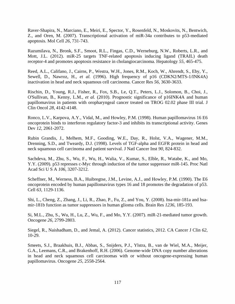
117
Raver-Shapira, N., Marciano, E., Meiri, E., Spector, Y., Rosenfeld, N., Moskovits, N., Bentwich, Z., and Oren, M. (2007). Transcriptional activation of miR-34a contributes to p53-mediated apoptosis. Mol Cell 26, 731-743.
Razumilava, N., Bronk, S.F., Smoot, R.L., Fingas, C.D., Werneburg, N.W., Roberts, L.R., and Mott, J.L. (2012). miR-25 targets TNF-related apoptosis inducing ligand (TRAIL) death receptor-4 and promotes apoptosis resistance in cholangiocarcinoma. Hepatology 55, 465-475.
Reed, A.L., Califano, J., Cairns, P., Westra, W.H., Jones, R.M., Koch, W., Ahrendt, S., Eby, Y., Sewell, D., Nawroz, H., et al. (1996). High frequency of p16 (CDKN2/MTS-1/INK4A) inactivation in head and neck squamous cell carcinoma. Cancer Res 56, 3630-3633.
Rischin, D., Young, R.J., Fisher, R., Fox, S.B., Le, Q.T., Peters, L.J., Solomon, B., Choi, J., O'Sullivan, B., Kenny, L.M., et al. (2010). Prognostic significance of p16INK4A and human papillomavirus in patients with oropharyngeal cancer treated on TROG 02.02 phase III trial. J Clin Oncol 28, 4142-4148.
Ronco, L.V., Karpova, A.Y., Vidal, M., and Howley, P.M. (1998). Human papillomavirus 16 E6 oncoprotein binds to interferon regulatory factor-3 and inhibits its transcriptional activity. Genes Dev 12, 2061-2072.
Rubin Grandis, J., Melhem, M.F., Gooding, W.E., Day, R., Holst, V.A., Wagener, M.M., Drenning, S.D., and Tweardy, D.J. (1998). Levels of TGF-alpha and EGFR protein in head and neck squamous cell carcinoma and patient survival. J Natl Cancer Inst 90, 824-832.
Sachdeva, M., Zhu, S., Wu, F., Wu, H., Walia, V., Kumar, S., Elble, R., Watabe, K., and Mo, Y.Y. (2009). p53 represses c-Myc through induction of the tumor suppressor miR-145. Proc Natl Acad Sci U S A 106, 3207-3212.
Scheffner, M., Werness, B.A., Huibregtse, J.M., Levine, A.J., and Howley, P.M. (1990). The E6 oncoprotein encoded by human papillomavirus types 16 and 18 promotes the degradation of p53. Cell 63, 1129-1136.
Shi, L., Cheng, Z., Zhang, J., Li, R., Zhao, P., Fu, Z., and You, Y. (2008). hsa-mir-181a and hsa-mir-181b function as tumor suppressors in human glioma cells. Brain Res 1236, 185-193.
Si, M.L., Zhu, S., Wu, H., Lu, Z., Wu, F., and Mo, Y.Y. (2007). miR-21-mediated tumor growth. Oncogene 26, 2799-2803.
Siegel, R., Naishadham, D., and Jemal, A. (2012). Cancer statistics, 2012. CA Cancer J Clin 62, 10-29.
Smeets, S.J., Braakhuis, B.J., Abbas, S., Snijders, P.J., Ylstra, B., van de Wiel, M.A., Meijer, G.A., Leemans, C.R., and Brakenhoff, R.H. (2006). Genome-wide DNA copy number alterations in head and neck squamous cell carcinomas with or without oncogene-expressing human papillomavirus. Oncogene 25, 2558-2564.
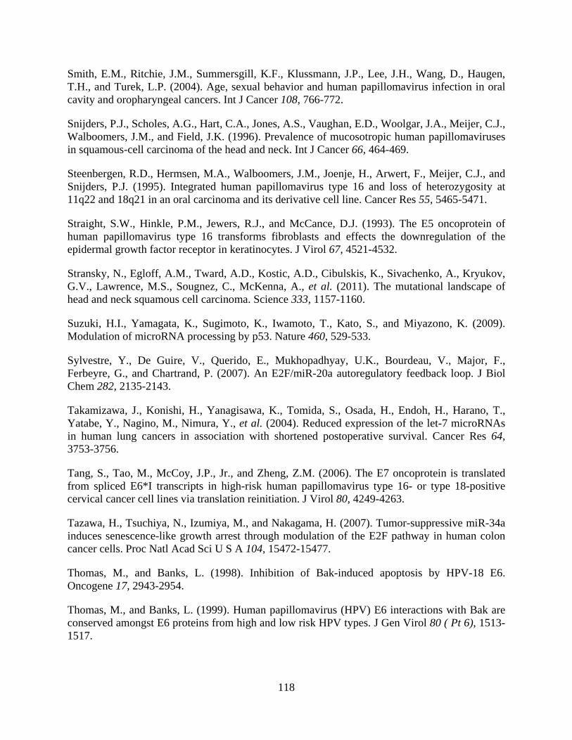
118
Smith, E.M., Ritchie, J.M., Summersgill, K.F., Klussmann, J.P., Lee, J.H., Wang, D., Haugen, T.H., and Turek, L.P. (2004). Age, sexual behavior and human papillomavirus infection in oral cavity and oropharyngeal cancers. Int J Cancer 108, 766-772.
Snijders, P.J., Scholes, A.G., Hart, C.A., Jones, A.S., Vaughan, E.D., Woolgar, J.A., Meijer, C.J., Walboomers, J.M., and Field, J.K. (1996). Prevalence of mucosotropic human papillomaviruses in squamous-cell carcinoma of the head and neck. Int J Cancer 66, 464-469.
Steenbergen, R.D., Hermsen, M.A., Walboomers, J.M., Joenje, H., Arwert, F., Meijer, C.J., and Snijders, P.J. (1995). Integrated human papillomavirus type 16 and loss of heterozygosity at 11q22 and 18q21 in an oral carcinoma and its derivative cell line. Cancer Res 55, 5465-5471.
Straight, S.W., Hinkle, P.M., Jewers, R.J., and McCance, D.J. (1993). The E5 oncoprotein of human papillomavirus type 16 transforms fibroblasts and effects the downregulation of the epidermal growth factor receptor in keratinocytes. J Virol 67, 4521-4532.
Stransky, N., Egloff, A.M., Tward, A.D., Kostic, A.D., Cibulskis, K., Sivachenko, A., Kryukov, G.V., Lawrence, M.S., Sougnez, C., McKenna, A., et al. (2011). The mutational landscape of head and neck squamous cell carcinoma. Science 333, 1157-1160.
Suzuki, H.I., Yamagata, K., Sugimoto, K., Iwamoto, T., Kato, S., and Miyazono, K. (2009). Modulation of microRNA processing by p53. Nature 460, 529-533.
Sylvestre, Y., De Guire, V., Querido, E., Mukhopadhyay, U.K., Bourdeau, V., Major, F., Ferbeyre, G., and Chartrand, P. (2007). An E2F/miR-20a autoregulatory feedback loop. J Biol Chem 282, 2135-2143.
Takamizawa, J., Konishi, H., Yanagisawa, K., Tomida, S., Osada, H., Endoh, H., Harano, T., Yatabe, Y., Nagino, M., Nimura, Y., et al. (2004). Reduced expression of the let-7 microRNAs in human lung cancers in association with shortened postoperative survival. Cancer Res 64, 3753-3756.
Tang, S., Tao, M., McCoy, J.P., Jr., and Zheng, Z.M. (2006). The E7 oncoprotein is translated from spliced E6*I transcripts in high-risk human papillomavirus type 16- or type 18-positive cervical cancer cell lines via translation reinitiation. J Virol 80, 4249-4263.
Tazawa, H., Tsuchiya, N., Izumiya, M., and Nakagama, H. (2007). Tumor-suppressive miR-34a induces senescence-like growth arrest through modulation of the E2F pathway in human colon cancer cells. Proc Natl Acad Sci U S A 104, 15472-15477.
Thomas, M., and Banks, L. (1998). Inhibition of Bak-induced apoptosis by HPV-18 E6. Oncogene 17, 2943-2954.
Thomas, M., and Banks, L. (1999). Human papillomavirus (HPV) E6 interactions with Bak are conserved amongst E6 proteins from high and low risk HPV types. J Gen Virol 80 ( Pt 6), 1513-1517.

119
Thomas, M., Narayan, N., Pim, D., Tomaic, V., Massimi, P., Nagasaka, K., Kranjec, C., Gammoh, N., and Banks, L. (2008). Human papillomaviruses, cervical cancer and cell polarity. Oncogene 27, 7018-7030.
Tong, A.W., and Nemunaitis, J. (2008). Modulation of miRNA activity in human cancer: a new paradigm for cancer gene therapy? Cancer Gene Ther 15, 341-355.
Tran, N., McLean, T., Zhang, X., Zhao, C.J., Thomson, J.M., O'Brien, C., and Rose, B. (2007a). MicroRNA expression profiles in head and neck cancer cell lines. Biochem Biophys Res Commun 358, 12-17.
Tran, N., Rose, B.R., and O'Brien, C.J. (2007b). Role of human papillomavirus in the etiology of head and neck cancer. Head Neck 29, 64-70.
Tsuchida, A., Ohno, S., Wu, W., Borjigin, N., Fujita, K., Aoki, T., Ueda, S., Takanashi, M., and Kuroda, M. (2011). miR-92 is a key oncogenic component of the miR-17-92 cluster in colon cancer. Cancer Sci 102, 2264-2271.
Valle, G.F., and Banks, L. (1995). The human papillomavirus (HPV)-6 and HPV-16 E5 proteins co-operate with HPV-16 E7 in the transformation of primary rodent cells. J Gen Virol 76 ( Pt 5), 1239-1245.
van Houten, V.M., Tabor, M.P., van den Brekel, M.W., Kummer, J.A., Denkers, F., Dijkstra, J., Leemans, R., van der Waal, I., Snow, G.B., and Brakenhoff, R.H. (2002). Mutated p53 as a molecular marker for the diagnosis of head and neck cancer. J Pathol 198, 476-486.
Ventura, A., Young, A.G., Winslow, M.M., Lintault, L., Meissner, A., Erkeland, S.J., Newman, J., Bronson, R.T., Crowley, D., Stone, J.R., et al. (2008). Targeted deletion reveals essential and overlapping functions of the miR-17 through 92 family of miRNA clusters. Cell 132, 875-886.
Venuti, A., Badaracco, G., Rizzo, C., Mafera, B., Rahimi, S., and Vigili, M. (2004). Presence of HPV in head and neck tumours: high prevalence in tonsillar localization. J Exp Clin Cancer Res 23, 561-566.
Vidal, L., and Gillison, M.L. (2008). Human papillomavirus in HNSCC: recognition of a distinct disease type. Hematol Oncol Clin North Am 22, 1125-1142, vii.
Villadsen, S.B., Bramsen, J.B., Ostenfeld, M.S., Wiklund, E.D., Fristrup, N., Gao, S., Hansen, T.B., Jensen, T.I., Borre, M., Orntoft, T.F., et al. (2012). The miR-143/-145 cluster regulates plasminogen activator inhibitor-1 in bladder cancer. Br J Cancer 106, 366-374.
Volinia, S., Calin, G.A., Liu, C.G., Ambs, S., Cimmino, A., Petrocca, F., Visone, R., Iorio, M., Roldo, C., Ferracin, M., et al. (2006). A microRNA expression signature of human solid tumors defines cancer gene targets. Proc Natl Acad Sci U S A 103, 2257-2261.
Walboomers, J.M., Jacobs, M.V., Manos, M.M., Bosch, F.X., Kummer, J.A., Shah, K.V., Snijders, P.J., Peto, J., Meijer, C.J., and Munoz, N. (1999). Human papillomavirus is a necessary cause of invasive cervical cancer worldwide. J Pathol 189, 12-19.

120
Wald, A.I., Hoskins, E.E., Wells, S.I., Ferris, R.L., and Khan, S.A. (2011). Alteration of microRNA profiles in squamous cell carcinoma of the head and neck cell lines by human papillomavirus. Head Neck 33, 504-512.
Wang, X., Tang, S., Le, S.Y., Lu, R., Rader, J.S., Meyers, C., and Zheng, Z.M. (2008). Aberrant expression of oncogenic and tumor-suppressive microRNAs in cervical cancer is required for cancer cell growth. PLoS One 3, e2557.
Wang, X., Wang, H.K., McCoy, J.P., Banerjee, N.S., Rader, J.S., Broker, T.R., Meyers, C., Chow, L.T., and Zheng, Z.M. (2009a). Oncogenic HPV infection interrupts the expression of tumor-suppressive miR-34a through viral oncoprotein E6. RNA 15, 637-647.
Wang, X., Xuan, Z., Zhao, X., Li, Y., and Zhang, M.Q. (2009b). High-resolution human core-promoter prediction with CoreBoost_HM. Genome Res 19, 266-275.
Werness, B.A., Levine, A.J., and Howley, P.M. (1990). Association of human papillomavirus types 16 and 18 E6 proteins with p53. Science 248, 76-79.
Wong, T.S., Liu, X.B., Chung-Wai Ho, A., Po-Wing Yuen, A., Wai-Man Ng, R., and Ignace Wei, W. (2008a). Identification of pyruvate kinase type M2 as potential oncoprotein in squamous cell carcinoma of tongue through microRNA profiling. Int J Cancer 123, 251-257.
Wong, T.S., Liu, X.B., Wong, B.Y., Ng, R.W., Yuen, A.P., and Wei, W.I. (2008b). Mature miR-184 as Potential Oncogenic microRNA of Squamous Cell Carcinoma of Tongue. Clin Cancer Res 14, 2588-2592.
Worden, F.P., Kumar, B., Lee, J.S., Wolf, G.T., Cordell, K.G., Taylor, J.M., Urba, S.G., Eisbruch, A., Teknos, T.N., Chepeha, D.B., et al. (2008). Chemoselection as a strategy for organ preservation in advanced oropharynx cancer: response and survival positively associated with HPV16 copy number. J Clin Oncol 26, 3138-3146.
Yamakuchi, M., Ferlito, M., and Lowenstein, C.J. (2008). miR-34a repression of SIRT1 regulates apoptosis. Proc Natl Acad Sci U S A 105, 13421-13426.
Yamakuchi, M., Lotterman, C.D., Bao, C., Hruban, R.H., Karim, B., Mendell, J.T., Huso, D., and Lowenstein, C.J. (2010). P53-induced microRNA-107 inhibits HIF-1 and tumor angiogenesis. Proc Natl Acad Sci U S A 107, 6334-6339.
Yeung, M.L., Yasunaga, J., Bennasser, Y., Dusetti, N., Harris, D., Ahmad, N., Matsuoka, M., and Jeang, K.T. (2008). Roles for microRNAs, miR-93 and miR-130b, and tumor protein 53-induced nuclear protein 1 tumor suppressor in cell growth dysregulation by human T-cell lymphotrophic virus 1. Cancer Res 68, 8976-8985.
Yu, G., Tang, J.Q., Tian, M.L., Li, H., Wang, X., Wu, T., Zhu, J., Huang, S.J., and Wan, Y.L. (2011). Prognostic values of the miR-17-92 cluster and its paralogs in colon cancer. J Surg Oncol.

121
Zerfass, K., Schulze, A., Spitkovsky, D., Friedman, V., Henglein, B., and Jansen-Durr, P. (1995). Sequential activation of cyclin E and cyclin A gene expression by human papillomavirus type 16 E7 through sequences necessary for transformation. J Virol 69, 6389-6399.
Zerfass-Thome, K., Zwerschke, W., Mannhardt, B., Tindle, R., Botz, J.W., and Jansen-Durr, P. (1996). Inactivation of the cdk inhibitor p27KIP1 by the human papillomavirus type 16 E7 oncoprotein. Oncogene 13, 2323-2330.
Zhang, H., Zuo, Z., Lu, X., Wang, L., Wang, H., and Zhu, Z. (2012). MiR-25 regulates apoptosis by targeting Bim in human ovarian cancer. Oncol Rep 27, 594-598.
Zhang, Z.Y., Sdek, P., Cao, J., and Chen, W.T. (2004). Human papillomavirus type 16 and 18 DNA in oral squamous cell carcinoma and normal mucosa. Int J Oral Maxillofac Surg 33, 71-74.
Zhu, S., Wu, H., Wu, F., Nie, D., Sheng, S., and Mo, Y.Y. (2008). MicroRNA-21 targets tumor suppressor genes in invasion and metastasis. Cell Res 18, 350-359.
Zimmermann, H., Degenkolbe, R., Bernard, H.U., and O'Connor, M.J. (1999). The human papillomavirus type 16 E6 oncoprotein can down-regulate p53 activity by targeting the transcriptional coactivator CBP/p300. J Virol 73, 6209-6219.
zur Hausen, H. (2002). Papillomaviruses and cancer: from basic studies to clinical application. Nat Rev Cancer 2, 342-350.