Role of intra-articular platelet-rich plasma in sacroiliac ...
6
www.elsevier.com/locate/trap Available online at www.sciencedirect.com Role of intra-articular platelet-rich plasma in sacroiliac joint pain Annu Navani, MD a,b,n , Deepak Gupta, BA, MS-II c a Department of Regenerative Medicine, Comprehensive Spine and Sports Center, Campbell, California b Division of Pain Medicine, Stanford University School of Medicine, Stanford, California c Department of Regenerative Medicine, Comprehensive Spine and Sports Center, Campbell, California article info Keywords: Sarco-iliac joint Platelet-rich plasma Regeneration abstract The goal of this case review is to evaluate safety and efficacy with the use of intra-articular platelet-rich plasma (PRP) in patients with sacroiliac (SI) joint (SIJ) pain. The secondary outcomes include additional medical treatments, hospitalization, and surgery. SIJ pain contributes significantly to the social and economic burden due to its long-standing and debilitating course. Current treatments include either interventional procedures with transient benefits or invasive surgical options. PRP has been used clinically in various settings for its anti-inflammatory and tissue repair properties attributed to growth factors. Ten patients with chronic SIJ pain who tried and failed conservative treatments were administered a single injection of 4 mm autologous PRP into the joint under fluoroscopic guidance after careful clinical and imaging evaluation. The patients were followed up at 1, 3, 6, and 12 months postinjection and primary and secondary outcomes were recorded. Verbal analog scale score for pain of all patients decreased more than 50% and their function increased for the period of 12 months. None of the patients presented to the hospital or clinic or received any treatments or surgery after the PRP injection. There were no adverse reactions, side effects, or complications. PRP presents as a promising option based on our preliminary observation. Larger, well-designed randomized controlled trials are warranted to understand the full breath of the efficacy, risks, and complications from the use of PRP for SIJ pain. & 2016 Elsevier Inc. All rights reserved. Introduction Low back pain is the greatest contributor of disability worldwide and a significant burden to society being respon- sible for an estimated 83 million years lived with disability in 2010. 1,2 In those with low back pain, the prevalence of sacroiliac (SI) joint (SIJ)-mediated pain ranges from 13%- 30%. 3 There is an even higher prevalence in patients who have had lumbar fusion surgery, with one study showing 35% of patients with back pain after a technically successful fusion exhibiting SIJ-mediated pain. 3 The vast prevalence of SIJ mediated pain is responsible for the increase in SIJ interventions from 46,940 in 2000 to 231,800 in 2008 thereby raising the combined Medicare payments for low back interventions from $362 million in 2000 to $1.231 billion to 2008. 4 There is an immediate need for long-lasting, minimally invasive and cost-effective treatments for SIJ- mediated pain. http://dx.doi.org/10.1053/j.trap.2016.09.010 1084-208X/& 2016 Elsevier Inc. All rights reserved. n Correspondence to: Annu H. Navani, MD, Medical Director, Comprehensive Spine and Sports Center, 3425 South Bascom Avenue, Suite 200, Campbell, California 95008. E-mail address: [email protected] (A. Navani). T ECHNIQUES IN R EGIONAL A NESTHESIA AND P AIN M ANAGEMENT 19(2015) 54 – 59
Transcript of Role of intra-articular platelet-rich plasma in sacroiliac ...

www.elsevier.com/locate/trap
Available online at www.sciencedirect.com
Role of intra-articular platelet-rich plasma insacroiliac joint pain
Annu Navani, MDa,b,n, Deepak Gupta, BA, MS-IIc
aDepartment of Regenerative Medicine, Comprehensive Spine and Sports Center, Campbell, CaliforniabDivision of Pain Medicine, Stanford University School of Medicine, Stanford, CaliforniacDepartment of Regenerative Medicine, Comprehensive Spine and Sports Center, Campbell, California
a r t i c l e i n f o
Keywords:Sarco-iliac jointPlatelet-rich plasmaRegeneration
a b s t r a c t
The goal of this case review is to evaluate safety and efficacy with the use of intra-articularplatelet-rich plasma (PRP) in patients with sacroiliac (SI) joint (SIJ) pain. The secondaryoutcomes include additional medical treatments, hospitalization, and surgery. SIJ paincontributes significantly to the social and economic burden due to its long-standing anddebilitating course. Current treatments include either interventional procedures withtransient benefits or invasive surgical options. PRP has been used clinically in varioussettings for its anti-inflammatory and tissue repair properties attributed to growth factors.Ten patients with chronic SIJ pain who tried and failed conservative treatments wereadministered a single injection of 4 mm autologous PRP into the joint under fluoroscopicguidance after careful clinical and imaging evaluation. The patients were followed up at 1,3, 6, and 12 months postinjection and primary and secondary outcomes were recorded.Verbal analog scale score for pain of all patients decreased more than 50% and theirfunction increased for the period of 12 months. None of the patients presented to thehospital or clinic or received any treatments or surgery after the PRP injection. There wereno adverse reactions, side effects, or complications. PRP presents as a promising optionbased on our preliminary observation. Larger, well-designed randomized controlled trialsare warranted to understand the full breath of the efficacy, risks, and complications fromthe use of PRP for SIJ pain.
& 2016 Elsevier Inc. All rights reserved.
Introduction
Low back pain is the greatest contributor of disabilityworldwide and a significant burden to society being respon-sible for an estimated 83 million years lived with disability in2010.1,2 In those with low back pain, the prevalence ofsacroiliac (SI) joint (SIJ)-mediated pain ranges from 13%-30%.3 There is an even higher prevalence in patients whohave had lumbar fusion surgery, with one study showing 35%
of patients with back pain after a technically successfulfusion exhibiting SIJ-mediated pain.3 The vast prevalenceof SIJ mediated pain is responsible for the increase inSIJ interventions from 46,940 in 2000 to 231,800 in 2008thereby raising the combined Medicare payments for lowback interventions from $362 million in 2000 to $1.231 billionto 2008.4 There is an immediate need for long-lasting,minimally invasive and cost-effective treatments for SIJ-mediated pain.
http://dx.doi.org/10.1053/j.trap.2016.09.0101084-208X/& 2016 Elsevier Inc. All rights reserved.
nCorrespondence to: Annu H. Navani, MD, Medical Director, Comprehensive Spine and Sports Center, 3425 South Bascom Avenue,Suite 200, Campbell, California 95008.
E-mail address: [email protected] (A. Navani).
T E C H N I Q U E S I N R E G I O N A L A N E S T H E S I A A N D P A I N M A N A G E M E N T 1 9 ( 2 0 1 5 ) 5 4 – 5 9

Regenerative medicine is a rapidly growing field with apromise for tissue repair and restoration. The benefits of thisfield extend to a number of medical disciplines, includingcardiology, neurology, vascular, plastic surgery, orthopedics,and spine. The use of biologics for low back pain has beenpreviously described in discs, facet joints, lumbar ligamentsand muscles, and epidural space. To our knowledge therehave been no reported cases of intra-articular injection ofplatelet-rich plasma (PRP) injection for SIJ pain. In thisarticle we have discussed safety and efficacy with the useof intra-articular PRP for SIJ-mediated pain.
Biologics background
There are several autologous and allogenic biologic prepara-tions from a variety of sources such as fetal, embryonic,amniotic, adult skin, peripheral blood, adipose tissue, andbone marrow. The autologous products, PRP, obtainedfrom centrifugation of human peripheral blood and themesenchymal stem cells from human adipose tissue or bonemarrow are most commonly used in clinical practice.PRP is the plasma fraction rich in platelets that package a
variety of growth factors in their alpha granules, which alongwith cytokines modulate the anti-inflammatory pathway andultimately decrease inflammation and repair the damagedtissue. PRP has been shown to promote macrophage activa-tion, collagen proliferation, cellular differentiation, vasodila-tion, and vascularization. The rationale behind the injectionof PRP is to deliver a high concentration of autologous growthfactors, and cytokines to areas with poor vascularization orpoor inherent healing potential.5 This makes it a worthwhilesolution for SIJ related inflammation and pain.
Clinical presentation
The SIJ is the largest axial joint in the human body. It is weightbearing, diarthrodial synovial joint that transfers energy, andforce from the spine to the pelvis.6-8 A healthy SIJ is stabilizedby the SI, iliolumbar, sacrotuberous, and sacrospinal liga-ments.7 The specific innervation of SIJ is controversial.9 Theinnervation from the ventral rami of L4, L5, the dorsal rami ofL5, S1, and S2 is most commonly accepted. SIJ-mediated paincan be caused by osteoarthritic degeneration, inflammatorydisease, tumor, infection, or disruption of the joint, which canoccur via trauma or pregnancy.10 Pain from the SIJ mostcommonly presents in the gluteal region, but can be referredto a variety of other sites, including the lumbar region, abdo-men, groin, lower limbs, and foot.6
Diagnosing SIJ-mediated pain can be difficult because thepresenting pain is similar in both character and location toother sources of back pain.6,7 Imaging studies such as X-rays,computed tomography or magnetic resonance imagings do not
serve as a conclusive diagnostic tool except in case of signifi-cant structural joint alteration. The current assessment fordiagnosing SIJ-mediated pain is single or double diagnosticblock of the heavily innervated SIJ through the injection of localanesthetic. The 2013 American Society of Interventional PainPhysicians (ASIPP) guidelines describe the evidence for single ordouble blocks with 75%-100% pain relief as good.11 To helpclinicians decide which patients warrant further study anddiagnostic blocks, provocative maneuvers, including the dis-traction, compression, thigh thrust, Gaenslen's and sacralthrust test, can be useful if three or more are positive.6,11
Current treatments
As with most musculoskeletal conditions, conservative andinterventional management options are available for SIJ-mediated pain. Conservative options include exercise therapyand chiropractic manipulation, but no high-quality evidencesupports their efficacy.10
Nonsurgical interventions to manage SIJ-mediated pain areintra-articular and peri-articular injections as well as neurol-ysis of the SIJ. The efficacy of intra-articular steroid injectionshas been evaluated in many clinical and systematic reviews,but the evidence for the effectiveness is limited for short- andlong-term relief.11 ASIPP guidelines recognize the effective-ness of conventional radiofrequency neurotomy of SIJ
Table 1 – Age distribution categorized by age groups.
Age range 30-40 40-50 50-60Number of patients 5 3 2
Fig. 1 – Pain range (VAS) and years of pain distribution. VAS,visual analog scale. (Color version of figure is availableonline.)
Fig. 2 – Age distribution of patients, years. (Color version offigure is available online.)
T E C H N I Q U E S I N R E G I O N A L A N E S T H E S I A A N D P A I N M A N A G E M E N T 1 9 ( 2 0 1 5 ) 5 4 – 5 9 55
innervation as limited.11 In fact, the only therapy describedby ASIPP to have a better than limited efficacy was cooledradiofrequency neurotomy.11
Methods
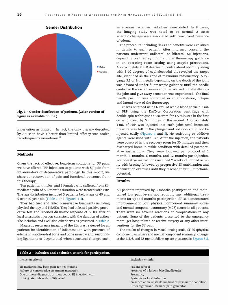
Given the lack of effective, long-term solutions for SIJ pain,we have offered PRP injections to patients with SIJ pain frominflammatory or degenerative pathology. In this report, weshare our observation of pain and functional outcomes fromthis therapy.Ten patients, 4 males, and 6 females who suffered from SIJ-
mediated pain of 46 months duration were treated with PRP.The age distribution included 5 patients below age of 40 and5 over 40-year old (Table 1 and Figures 1-3).They had tried and failed conservative treatments including
physical therapy and NSAIDs. They had at least 1 positive provo-cative test and reported diagnostic response of 450% after oflocal anesthetic injection consistent with the duration of action.The inclusion and exclusion criteria was as presented in Table 2.Magnetic resonance imaging of the SIJs was reviewed for all
patients for identification of inflammation with presence ofedema in subchondral bone and bone marrow and surround-ing ligaments or degenerated when structural changes such
as erosions, sclerosis, ankylosis were noted. In 8 cases,the imaging study was noted to be normal, 2 casessclerotic changes were associated with concurrent presenceof edema.The procedure including risks and benefits were explained
in details to each patient. After informed consent, thepatients underwent unilateral or bilateral SIJ injections,depending on their symptoms under fluoroscopy guidancein an operating room setting using aseptic precautions.Approximately 20-30 degrees of contralateral obliquity alongwith 5-10 degrees of cephalocaudal tilt revealed the targetsite, identified as the zone of maximum radiolucency. A 22-gauge 3.5 or 5-in. needle depending on the depth of the jointwas advanced under fluoroscopic guidance until the needlecontacted the sacral lamina and then walked off laterally intothe joint and give away sensation was experienced. The finalneedle position was confirmed in anteroposterior, obliqueand lateral view of the fluoroscopy.PRP was obtained using 60 mL of whole blood to yield 7 mL
of PRP using the EmCyte Corporation centrifuge withdouble spin technique at 3800 rpm for 1.5 minutes in the firstcycle followed by 5 minutes in the second. Approximately4 mL of PRP was injected into each joint until increasedpressure was felt in the plunger and solution could not beinjected easily (Figures 4 and 5). No activating or additiveagents were used with PRP. After the injection, the patientswere observed in the recovery room for 30 minutes and thendischarged home in stable condition with detailed postoper-ative instructions. They were followed per protocol at 1month, 3 months, 6 months, and 12 months postinjection.Postoperative instructions included 2 weeks of limited activ-ity with bracing followed by progressive SIJ stabilization andmobilization exercises until they reached their full functionalpotential.
Results
All patients improved by 3 months postinjection and main-tained low pain levels not requiring any additional treat-ments for up to 6 months postinjection. SF-36 demonstratedimprovement in both physical component summary scoresand mental component summary (MCS) scores in all patients.There were no adverse reactions or complications in anypatient. None of the patients presented to the emergencyroom, get hospitalized or receive surgery or any other inter-ventions for the SIJ pain.The results of changes in visual analog scale, SF-36 (physical
component summary andmental component summary) changesat the 1, 3, 6, and 12-month follow-up are presented in Figures 6-8.
Fig. 3 – Gender distribution of patients. (Color version offigure is available online.)
Table 2 – Inclusion and exclusion criteria for participation.
Inclusion criteria Exclusion criteria
SIJ-mediated low back pain for Z6 months Patient refusalFailure of conservative treatment measures Presence of a known bleedingdisorderOne or more diagnostic or therapeutic SIJ injection with
LA 7 steroids with 450% reliefPregnancySystemic or local infectionPresence of an unstable medical or psychiatric conditionOther significant low back pain generator
T E C H N I Q U E S I N R E G I O N A L A N E S T H E S I A A N D P A I N M A N A G E M E N T 1 9 ( 2 0 1 5 ) 5 4 – 5 956

DiscussionPRP has been extensively studied in spine and orthopedics incontext of intervertebral disc degeneration, spinal fusion, andosteoarthritis and cartilage repair of major joints. Despite itsextensive use in major joints, there have been no reports ofits use in sacroiliac joints. There is extensive literature onprolotherapy over the sacroiliac ligaments including a recentcase study of four patients with series of 2 PRP injections viaprolotherapy technique at the Hackett points A-C underultrasound guidance.12 The study by Ko et al12 reportedstatistically significant reduction in pain, and improvementin quality of life at 12 months and at follow up 4-yearsposttreatment. The PRP injection, however, was injected atthe ligament bone junction at the Hackett points A-C and
not injected directly into the joint. Here, we have discussedthe results in the first ever reported use of intra-articularSIJ PRP.The essence of PRP is to boost the damaged tissue's own
repair processes by delivering a concentrated dose of autol-ogous growth factors and thereby activating local mesenchy-mal stem cells at the site of injury. Some of the specificgrowth factors released such as platelet-derived growthfactor, transforming growth factor-beta 1, insulin-like growthfactor-1, vascular endothelial growth factor, fibroblasticgrowth factor, epidermal growth factor have been shown tocontrol the mechanism of tissue repair and restoration.13
Through carefully orchestrated chemotaxis, angiogenesis,cellular migration, proliferation and differentiation and extracellular matrix production, regeneration is brought forth.14
Fig. 4 – Left sacroiliac joint; contralateral oblique view at 201 (A) and 71 (B) with needle tip in between the posterior and anteriorjoint lines.
Fig. 5 – Right sacroiliac joint; contralateral oblique view at 221 (A) and 51 (B) with needle tip in between the posterior andanterior joint lines. (Color version of figure is available online.)
T E C H N I Q U E S I N R E G I O N A L A N E S T H E S I A A N D P A I N M A N A G E M E N T 1 9 ( 2 0 1 5 ) 5 4 – 5 9 57

This process seems particularly relevant in the setting ofinflamed or degenerated SIJ-mediated pain.Similarly to PRP therapy, there is a breadth of evidence
supporting the use of MSCs in musculoskeletal, orthopedic,and spine conditions. MSCs are self-renewing and undiffer-entiated and upon induction by certain growth factors, thesecells can differentiate into osteoblast, chondroblasts, andadipocytes.15 They have demonstrated secretion of growthfactors, cell proliferation, angiogenesis, anti-inflammatoryeffects, antiapoptic effects, and immunomodulation. The
exploitation of these properties has shown promise in regen-erating the tissue of degenerated IVDs through increasedproteoglycan synthesis and type II collagen production16 andproviding pain relief.17 There are no published reports on theuse of bone marrow concentrate for SIJ-mediated pain to ourknowledge. Our preliminary results of safety and efficacy withintra-articular SIJ bone marrow concentrate look promising.Based on our observation earlier, PRP appears to be an
assuring option for SIJ mediated pain. This is noted improve-ment in pain and function in patients with chronic SIJ pain
Fig. 6 – Changes in VAS score for minimum, average, and maximum VAS along y-axis and time interval along x-axis. VAS,visual analog scale. (Color version of figure is available online.)
Fig. 7 – Changes in SF-36 PCS for minimum, average, and maximum PCS along y-axis and time interval along x-axis. PCS,physical component summary scores. (Color version of figure is available online.)
T E C H N I Q U E S I N R E G I O N A L A N E S T H E S I A A N D P A I N M A N A G E M E N T 1 9 ( 2 0 1 5 ) 5 4 – 5 958

for as long as 8 years at various points along the spectrum ofSIJ inflammation and degeneration. We recognize the short-comings of our report including the small sample size andshort duration of observation; however, this does provide aninaugural consideration for biologics for SIJ mediated pain.Larger well-designed, randomized clinical trials are needed tounderstand the full effect of PRP and bone marrow concen-trate for SIJ-mediated pain.
Conclusion
SIJ is a strong contributor of low back pain, the number onecause of disability in United States. Currently availableinterventional and surgical therapeutic options are not suffi-cient for long-term pain relief. We report the first case seriesof intra-articular use of PRP for SIJ-mediated pain. PRPprovides a natural, nonpharmacological, minimally invasiveoption that has the potential for repair and restoration. Thereis need for larger well-designed randomized controlledstudies to understand the full breath of its application toSIJ-mediated pain.
r e f e r e n c e s
1. Buchbinder R, Blyth F, March L, Brooks P, Woolf A, Hoy D.Placing the global burden of low back pain in context. BestPract Res Clin Rheumatol. 2013;27(5):575–589.
2. Hoy D, March L, Brooks P, et al. The global burden of low backpain: estimates from the Global Burden of Disease 2010 study.Ann Rheum Dis. 2014;73(6):968–974.
3. Boswell MV, Trescot AM, Datta S, et al. Interventional techni-ques. Evidence-based practice guidelines in the managementof chronic spinal pain. Pain Phys. 2007;10(1):7–111.
4. Shaffrey C, Smith J. Nonoperative care to manage the sacroil-iac joint. J Neurosurg Spine. 2014;20(4):351–352.
5. Tuakli-Wosornu Y, Terry A, Lutz G, et al. Original Research—CME: lumbar intradiskal platelet-rich plasma (PRP) injections:a prospective, double-blind, randomized controlled study.PM&R. 2016;8:1–10.
6. Vanelderen P, Szadek K, Van Zundert J, et al. Sacroiliac jointpain. Pain Pract. 2010;10(5):470–478.
7. Dreyfuss P, Dreyer S, Cole A, Mayo K. Sacroiliac joint pain.J Am Acad Orthop Surg. 2004;12(4):255–265.
8. Hoek van Dijke G, Snijders CJ, Stoeckart R, Stam H. A bio-mechanical model on muscle forces in the transfer of spinalload to the pelvis and legs. J Biomech. 1999;32(9):927–933.
9. Forst S, Wheeler M, Fortin J, et al. The sacroiliac joint: anatomy,physiology, and clinical significance. Pain Phys. 2006;9(1):61–68.
10. Polly D, Cher D, Sembrano J, et al. Randomized controlled trialof minimally invasive sacroiliac joint fusion using triangulartitanium implants vs nonsurgical management for sacroiliacjoint dysfunction: 12-month outcomes. Neurosurgery. 2015;77(5):674–690.
11. Manchikanti L, Abdi S, Atluri S, et al. An update ofcomprehensive evidence-based guidelines for interventionaltechniques in chronic spinal pain. Part II: Guidance andrecommendations. Pain Phys. 2013;16(suppl 2):S49–S283.
12. Ko G, Mindra S, Lawson G, Whitmore S, Arseneau L. Case seriesof ultrasound-guided platelet-rich plasma injections for sac-roiliac joint dysfunction. J Back Musculoskeletal Rehabil. 2016:1–8.
13. Wang S, Rui Y, Tan Q, Wang C. Enhancing intervertebral discrepair and regeneration through biology: platelet-rich plasmaas an alternative strategy. Arthritis Res Ther. 2013;15(5):220.
14. Bennett N, Schultz G. Growth factors and wound healing:biochemical properties of the growth factors and their recep-tors. Am J Surg. 1993;165(6):728–737.
15. DePalma M, Gasper J. Regenerative medicine: cellular supple-mentation technologies for painful spine disorders. PM&R.2015;7(4):S19–S25.
16. Vasiliadis E, Pneumaticos S, Evangelopoulos D, PapavassiliouA. Biologic treatment of mild and moderate intervertebraldisc degeneration. MolMed. 2014;20:400–409.
17. Pettine K, Suzuki R, Sand T, Murphy M. Treatment of disco-genic back pain with autologous bone marrow concentrateinjection with minimum two year follow-up. Int Orthop. 2016;40(1):135–140.
Fig. 8 – Changes in SF-36 MCS for minimum, average, and maximum with MCS along y-axis and time interval along x-axis.MCS, mental component summary scores. (Color version of figure is available online.)
T E C H N I Q U E S I N R E G I O N A L A N E S T H E S I A A N D P A I N M A N A G E M E N T 1 9 ( 2 0 1 5 ) 5 4 – 5 9 59