Role of CT and MRCP in Evaluation of Biliary Tract Obstruction · PDF fileRole of CT and MRCP...
13
ABDOMINAL CT IMAGING (A JOSHI, SECTION EDITOR) Role of CT and MRCP in Evaluation of Biliary Tract Obstruction Anagha Joshi • Kishore Rajpal • Ketan Kakadiya • Ashank Bansal Published online: 26 September 2014 Ó Springer Science+Business Media New York 2014 Abstract Obstructive jaundice which is caused by bile duct obstruction can be clinically and biochemically indistinguishable from cholestatic jaundice caused by hepatocellular disease. The management of both these conditions being radically different, the principle task of the radiologist is to differentiate between hepatocellular and obstructive jaundice, using available imaging modality and help in further management. With the availability of non-invasive modality like computed tomography and magnetic resonance imaging (MRI), it is possible to diag- nose obstructive jaundice early and accurately without any patient discomfort. The purpose of this article is to describe the protocol for evaluation of obstructive jaundice with use of MDCT and magnetic resonance cholangio pancreatog- raphy sequence of MRI and to describe the imaging fea- tures of the most common causes of obstructive jaundice like biliary calculi, bile duct strictures, choledochal cyst, gall bladder carcinoma, cholangiocarcinoma, primary sclerosing cholangitis, and pancreatic head carcinoma. Keywords CT Á MRCP Á Obstructive jaundice Á Biliary stricture Á Cholangiocarcinoma Á Choledocholithiasis Introduction Obstructive jaundice is a type of jaundice in which there is blockage of flow of bile from the liver to the intestine resulting in redirection of excess bile and its by products like bilirubin into the blood. It can lead to complications like ascending cholangitis, hepatorenal syndrome, and malabsorbtion and hence requires urgent surgical inter- vention. The role of radiologist therefore is important in early diagnosis and in accurately delineating the level and the cause of obstruction, thus helping in staging as well as preoperative assessment of tumor respectability. For long, Endoscopic Retrograde Cholangio Pancrea- tography (ERCP) was the standard established procedure for evaluation of patients with obstructive jaundice. How- ever, being an invasive procedure, it has an inherent complication rate of 3–9 % and mortality rate of 0.2–0.5 % [1]. Due to significant advances in cross-sectional imaging, in particular the advent of magnetic resonance cholangio pancreatography (MRCP), ERCP currently has an almost exclusively therapeutic role. Endoscopic Ultrasound (EUS), an adjunct procedure to ERCP, can be used to detect small distal biliary ductal calculi, for local staging of periampullary neoplasm and for guided fine-needle aspiration. Ultrasonography (USG) can be used as initial imaging modality to diagnose the biliary obstruction but artifacts due to bowel gas, gall bladder and bile duct calculi, breathing artifacts and obesity brings it down in the list of imaging modality of choice in obstructive jaundice. With recent advances in MDCT technology enabling faster acquisition in a single breath hold and upgraded software techniques for image reconstruction, MDCT is now more sensitive for determining the preliminary level and cause of obstruction. Due to sub-second acquisition and multiphasic approach, newer studies have further refined the role of MDCT in terms of specific diagnosis and staging of the pathology. Thus, single breath hold, multi- phasic, sub centimeter iso-voxel scanning with post This article is part of topical collection on Abdominal CT Imaging. A. Joshi (&) Á K. Rajpal Á K. Kakadiya Á A. Bansal Lokmanya Tilak Munincipal Medical College & Lokmanya Tilak Munincipal General Hospital, Sion, Mumbai, India e-mail: [email protected] 123 Curr Radiol Rep (2014) 2:72 DOI 10.1007/s40134-014-0072-x
Transcript of Role of CT and MRCP in Evaluation of Biliary Tract Obstruction · PDF fileRole of CT and MRCP...
ABDOMINAL CT IMAGING (A JOSHI, SECTION EDITOR)
Role of CT and MRCP in Evaluation of Biliary Tract Obstruction
Anagha Joshi • Kishore Rajpal • Ketan Kakadiya •
Ashank Bansal
Published online: 26 September 2014
� Springer Science+Business Media New York 2014
Abstract Obstructive jaundice which is caused by bile
duct obstruction can be clinically and biochemically
indistinguishable from cholestatic jaundice caused by
hepatocellular disease. The management of both these
conditions being radically different, the principle task of
the radiologist is to differentiate between hepatocellular
and obstructive jaundice, using available imaging modality
and help in further management. With the availability of
non-invasive modality like computed tomography and
magnetic resonance imaging (MRI), it is possible to diag-
nose obstructive jaundice early and accurately without any
patient discomfort. The purpose of this article is to describe
the protocol for evaluation of obstructive jaundice with use
of MDCT and magnetic resonance cholangio pancreatog-
raphy sequence of MRI and to describe the imaging fea-
tures of the most common causes of obstructive jaundice
like biliary calculi, bile duct strictures, choledochal cyst,
gall bladder carcinoma, cholangiocarcinoma, primary
sclerosing cholangitis, and pancreatic head carcinoma.
Keywords CT � MRCP � Obstructive jaundice � Biliary
stricture � Cholangiocarcinoma � Choledocholithiasis
Introduction
Obstructive jaundice is a type of jaundice in which there is
blockage of flow of bile from the liver to the intestine
resulting in redirection of excess bile and its by products
like bilirubin into the blood. It can lead to complications
like ascending cholangitis, hepatorenal syndrome, and
malabsorbtion and hence requires urgent surgical inter-
vention. The role of radiologist therefore is important in
early diagnosis and in accurately delineating the level and
the cause of obstruction, thus helping in staging as well as
preoperative assessment of tumor respectability.
For long, Endoscopic Retrograde Cholangio Pancrea-
tography (ERCP) was the standard established procedure
for evaluation of patients with obstructive jaundice. How-
ever, being an invasive procedure, it has an inherent
complication rate of 3–9 % and mortality rate of 0.2–0.5 %
[1]. Due to significant advances in cross-sectional imaging,
in particular the advent of magnetic resonance cholangio
pancreatography (MRCP), ERCP currently has an almost
exclusively therapeutic role.
Endoscopic Ultrasound (EUS), an adjunct procedure to
ERCP, can be used to detect small distal biliary ductal
calculi, for local staging of periampullary neoplasm and for
guided fine-needle aspiration.
Ultrasonography (USG) can be used as initial imaging
modality to diagnose the biliary obstruction but artifacts
due to bowel gas, gall bladder and bile duct calculi,
breathing artifacts and obesity brings it down in the list of
imaging modality of choice in obstructive jaundice.
With recent advances in MDCT technology enabling
faster acquisition in a single breath hold and upgraded
software techniques for image reconstruction, MDCT is
now more sensitive for determining the preliminary level
and cause of obstruction. Due to sub-second acquisition
and multiphasic approach, newer studies have further
refined the role of MDCT in terms of specific diagnosis and
staging of the pathology. Thus, single breath hold, multi-
phasic, sub centimeter iso-voxel scanning with post
This article is part of topical collection on Abdominal CT Imaging.
A. Joshi (&) � K. Rajpal � K. Kakadiya � A. Bansal
Lokmanya Tilak Munincipal Medical College & Lokmanya
Tilak Munincipal General Hospital, Sion, Mumbai, India
e-mail: [email protected]
123
Curr Radiol Rep (2014) 2:72
DOI 10.1007/s40134-014-0072-x
processing through volume acquisition, maximum intensity
projections (MIPs), and multiplanar reformations (MPR)
all help to increase the diagnostic accuracy, which is
immensely helpful in
a) Differentiating benign from malignant stricture.
b) Staging complex biliary malignancies in terms of
involvement of biliary confluence, invasion, and
encasement of the adjacent major arterial and venous
channel rendering it inoperable, as well as regional
lymphadenopathy and hepatic metastasis.
Hence, Multiphasic MDCT of the abdomen with pan-
creatic and venous phase through the hepatic-biliary-pan-
creatic region is now the accepted worldwide protocol for
pancreatico-biliary malignancies.
Magnetic resonance (MR) imaging besides being non-
invasive has the advantages of allowing detailed evaluation
of the pancreatico-biliary tract with a large field of view
(FOV), excellent patient tolerance, and three-dimensional
(3D) data sets that can be cholangio-pancreatico graphi-
cally displayed. Two unique properties of bile that are
exploited to produce MRCP images are its relatively high
water content & stasis of bile, in comparison with the blood
flowing through adjacent vessels in the portal tract [2•],
producing projection images like ERCP. Hence, MRCP has
virtually replaced ERCP as the primary investigative
modality in all cases of obstructive jaundice not requiring
early endoscopic intervention.
Method of Evaluation
MRI
In newer MRI machines, with state of the art imaging
capability, MRCP can be performed either without the use
of contrast agent or with the usage of contrast agent
showing biliary excretion.
Non-contrast MRCP
Non-contrast MRCP technique is based on heavily T2
weighted sequence which can be performed in two-
dimensional (2D) or 3D modes [3•]. It shows increased
signal from bile and pancreatic duct (PD) fluid and sup-
presses signal from background tissues such as solid organs
and moving blood [4, 5]. The imaging obstacles of long
acquisition times and respiratory motion artifacts have
largely been overcome with technical innovations such as
short-breath-hold T2-weighted acquisitions, parallel imag-
ing, and sophisticated respiratory triggering mechanisms.
The parameters we used for non-contrast MRCP sequence
in our institute are as follows (Table 1).
Contrast MRCP
Contrast-enhanced MRCP is based on the principle of
selective excretion of liver specific, gadolinium-, and
magnesium-based MR contrast media by the liver into the
biliary system, in 10–60 min [6]. These excreted contrast
media cause T1 shortening of the bile, resulting in hyper-
intensity on T1w sequences. The liver-specific contrast
agents are gadobenate dimeglumine (Gd-BOPTA), gadox-
etic acid disodium (gadoxetate disodium, or Gd-EOB-
DTPA), and mangafodipir trisodium (Mn-DPDP). In our
institute, the most commonly used sequence for contrast
MRCP is e-THRIVE (fat-saturated T1 weighted fast gra-
dient sequence), which can be acquired in a single breath
hold of 20–22 s, reducing the breathing-related artifact.
The main indication of contrast MRCP is in evaluation of
biliary leak, which can occur either due to iatrogenic
trauma or at the site of anastomosis in case of liver trans-
plantation. Contrast MRCP gives advantage over non-
contrast MRCP in determining the exact site of leak and in
giving information about the communication of this leak
with surrounding collection, if any. However, being gad-
olinium-based technique, it suffers from the inherent
complications related to gadolinium like risk of nephro-
genic systemic fibrosis in patients of renal failure and
allergic reactions [7]. The other disadvantage is longer scan
duration to obtain significant concentration of gadolinium
in the biliary system.
Advantage of 3 T Over 1.5 T
The signal to noise ratio (SNR) at 3.0 T is twice that at
1.5 T, producing higher-resolution image data sets with
reduced acquisition times. The visualization of arborization
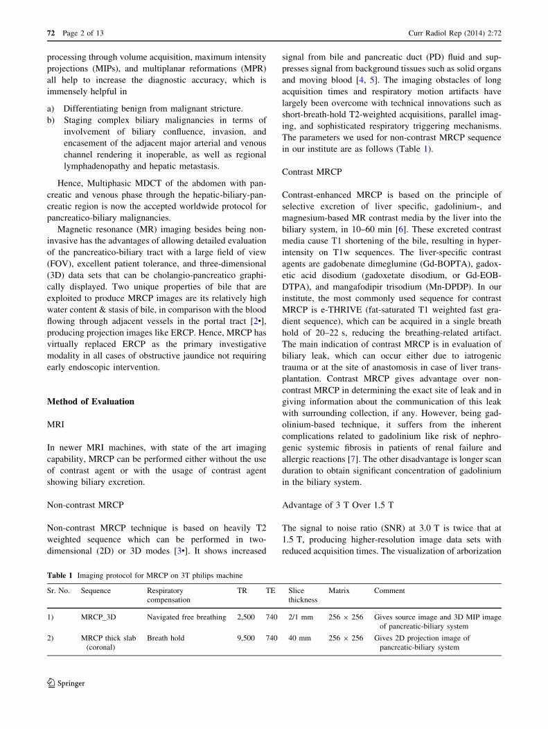
Table 1 Imaging protocol for MRCP on 3T philips machine
Sr. No. Sequence Respiratory
compensation
TR TE Slice
thickness
Matrix Comment
1) MRCP_3D Navigated free breathing 2,500 740 2/1 mm 256 9 256 Gives source image and 3D MIP image
of pancreatic-biliary system
2) MRCP thick slab
(coronal)
Breath hold 9,500 740 40 mm 256 9 256 Gives 2D projection image of
pancreatic-biliary system
72 Page 2 of 13 Curr Radiol Rep (2014) 2:72
123
of intrahepatic ducts, bile duct variations, delineation of the
PD particularly its side branches, and relation of the pan-
creatic parenchyma to the ductal system is better obtained
at 3.0 T than at 1.5 T. Also signal from fluid in the gas-
trointestinal tract is more effectively suppressed at 3.0 T.
Use of parallel imaging reduces acquisition time and hence
reduces breathing-related artifacts. All this encourages the
use of this modality for specific diagnostic applications like
in diagnosis of primary sclerosing cholangitis [8].
CT
In our institute, plain abdominal CT is obtained at 120 kV,
200 mAs to determine baseline HU value and to visualize
any biliary calculi containing calcium. To determine post
contrast enhancement, 90 ml of non-ionic iodinated con-
trast (350 mg % v/w) is injected intravenously at a flow
rate of 3 ml/sec with the aid of pressure injector (Mal-
linckrodt). Arterial and venous phase are taken at an
interval of 18–22 & 60–65 s, respectively, from the time of
contrast injection. Delayed phase is taken after 10–15 min.
Delayed phase is particularly important in cases of chol-
angiocarcinoma which show increased contrast enhance-
ment as compared to normal surrounding parenchyma. 3D
reconstruction with thin planar (1 mm) and MPR is per-
formed in coronal and sagittal planes for better depiction of
intraluminal and wall lesions of biliary tract.
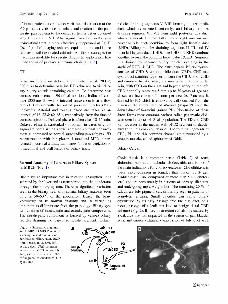
Normal Anatomy of Pancreato-Biliary System
in MRCP (Fig. 1)
Bile plays an important role in intestinal absorption. It is
secreted by the liver and is transported into the duodenum
through the biliary system. There is significant variation
seen in the biliary tree, with normal biliary anatomy seen
only in 50–60 % of the population. Hence, the basic
knowledge of its normal anatomy and its variant is
important to differentiate from the pathology. Biliary sys-
tem consists of intrahepatic and extrahepatic components.
The intrahepatic component is formed by various biliary
radicles draining the respective hepatic segments. Biliary
radicles draining segments V, VIII form right anterior bile
duct which is oriented vertically, and biliary radicles
draining segment VI, VII form right posterior bile duct
which is oriented horizontally. These right anterior and
posterior bile ducts combine to form right hepatic duct
(RHD). Biliary radicles draining segments II, III, and IV
form left hepatic duct (LHD). The LHD and RHD combine
together to form the common hepatic duct (CHD). Segment
I is drained by separate biliary radicles draining in the
angle of RHD & LHD. The extra-hepatic biliary system
consists of CHD & common bile duct (CBD). CHD and
cystic duct combine together to form the CBD. Both CBD
and common hepatic artery are seen anterior to the portal
vein, with CBD on the right and hepatic artery on the left.
CBD normally measures 5 mm up to 50 years of age and
shows an increment of 1 mm per decade. Pancreas is
drained by PD which is embryologically derived from the
fusion of the ventral duct of Wirsung (major PD) and the
dorsal duct of Santorini (minor PD). Non-fusion of these
ducts forms most common variant called pancreatic divi-
sum seen in up to 14 % of population. The PD and CBD
join together in the medial wall of D2 segment of duode-
num forming a common channel. The terminal segments of
CBD, PD, and this common channel are surrounded by a
smooth muscle, called sphincter of Oddi.
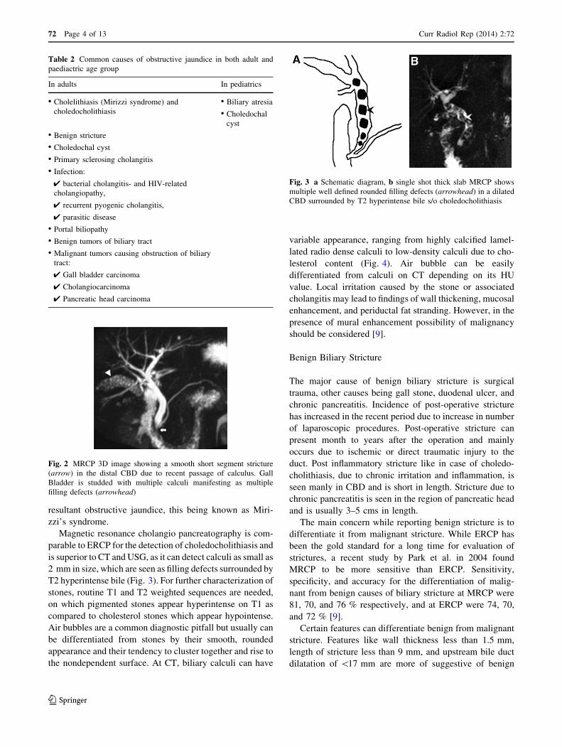
Biliary Calculi
Cholelithiasis is a common cause (Table 2) of acute
abdominal pain due to calculus cholecystitis and is one of
the main indications for cholecystectomy. Cholelithiasis is
twice more common in females than males. 80 % gall
bladder calculi are composed of more than 50 % choles-
terol and are seen mainly in patients of obesity, diabetes,
and undergoing rapid weight loss. The remaining 20 % of
calculi are bile pigment calculi mainly seen in patients of
hemolytic anemia. Small calculus can cause biliary
obstruction by its easy passage into the bile duct, or a
recent passage of calculi can lead to benign distal CBD
stricture (Fig. 2). Biliary obstruction can also be caused by
a calculus that has impacted in the region of gall bladder
neck and causes extrinsic compression of bile duct with
Fig. 1 a Schematic diagram
and b MIP 3D MRCP sequence
showing normal anatomy of
pancreatico-biliary tract. RHD
right hepatic duct, LHD left
hepatic duct, CHD common
hepatic duct, CBD common bile
duct, PD pancreatic duct, D2
2nd segment of duodenum, CD
cystic duct
Curr Radiol Rep (2014) 2:72 Page 3 of 13 72
123
resultant obstructive jaundice, this being known as Miri-
zzi’s syndrome.
Magnetic resonance cholangio pancreatography is com-
parable to ERCP for the detection of choledocholithiasis and
is superior to CT and USG, as it can detect calculi as small as
2 mm in size, which are seen as filling defects surrounded by
T2 hyperintense bile (Fig. 3). For further characterization of
stones, routine T1 and T2 weighted sequences are needed,
on which pigmented stones appear hyperintense on T1 as
compared to cholesterol stones which appear hypointense.
Air bubbles are a common diagnostic pitfall but usually can
be differentiated from stones by their smooth, rounded
appearance and their tendency to cluster together and rise to
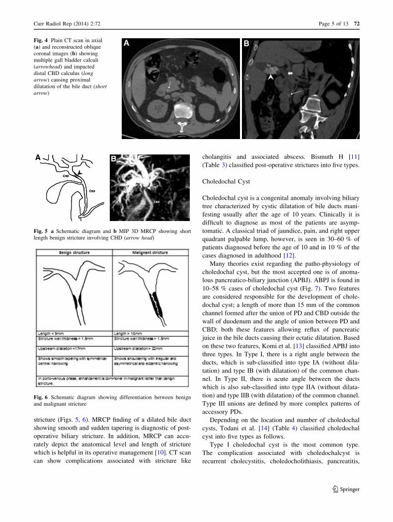
the nondependent surface. At CT, biliary calculi can have
variable appearance, ranging from highly calcified lamel-
lated radio dense calculi to low-density calculi due to cho-
lesterol content (Fig. 4). Air bubble can be easily
differentiated from calculi on CT depending on its HU
value. Local irritation caused by the stone or associated
cholangitis may lead to findings of wall thickening, mucosal
enhancement, and periductal fat stranding. However, in the
presence of mural enhancement possibility of malignancy
should be considered [9].
Benign Biliary Stricture
The major cause of benign biliary stricture is surgical
trauma, other causes being gall stone, duodenal ulcer, and
chronic pancreatitis. Incidence of post-operative stricture
has increased in the recent period due to increase in number
of laparoscopic procedures. Post-operative stricture can
present month to years after the operation and mainly
occurs due to ischemic or direct traumatic injury to the
duct. Post inflammatory stricture like in case of choledo-
cholithiasis, due to chronic irritation and inflammation, is
seen manly in CBD and is short in length. Stricture due to
chronic pancreatitis is seen in the region of pancreatic head
and is usually 3–5 cms in length.
The main concern while reporting benign stricture is to
differentiate it from malignant stricture. While ERCP has
been the gold standard for a long time for evaluation of
strictures, a recent study by Park et al. in 2004 found
MRCP to be more sensitive than ERCP. Sensitivity,
specificity, and accuracy for the differentiation of malig-
nant from benign causes of biliary stricture at MRCP were
81, 70, and 76 % respectively, and at ERCP were 74, 70,
and 72 % [9].
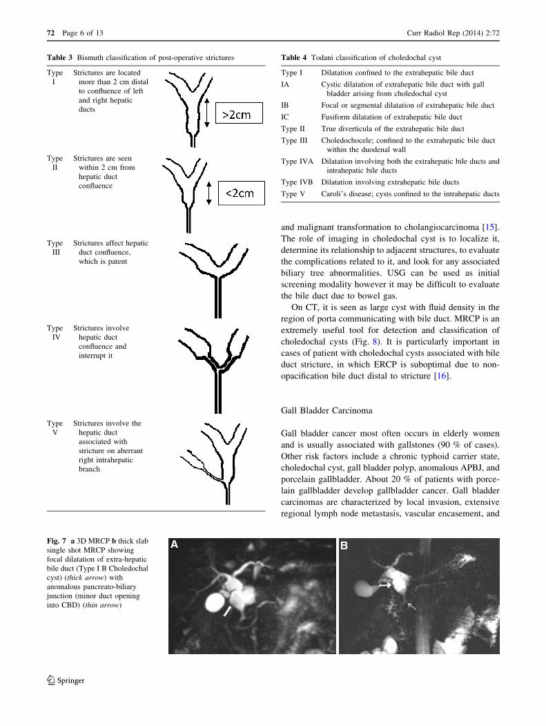
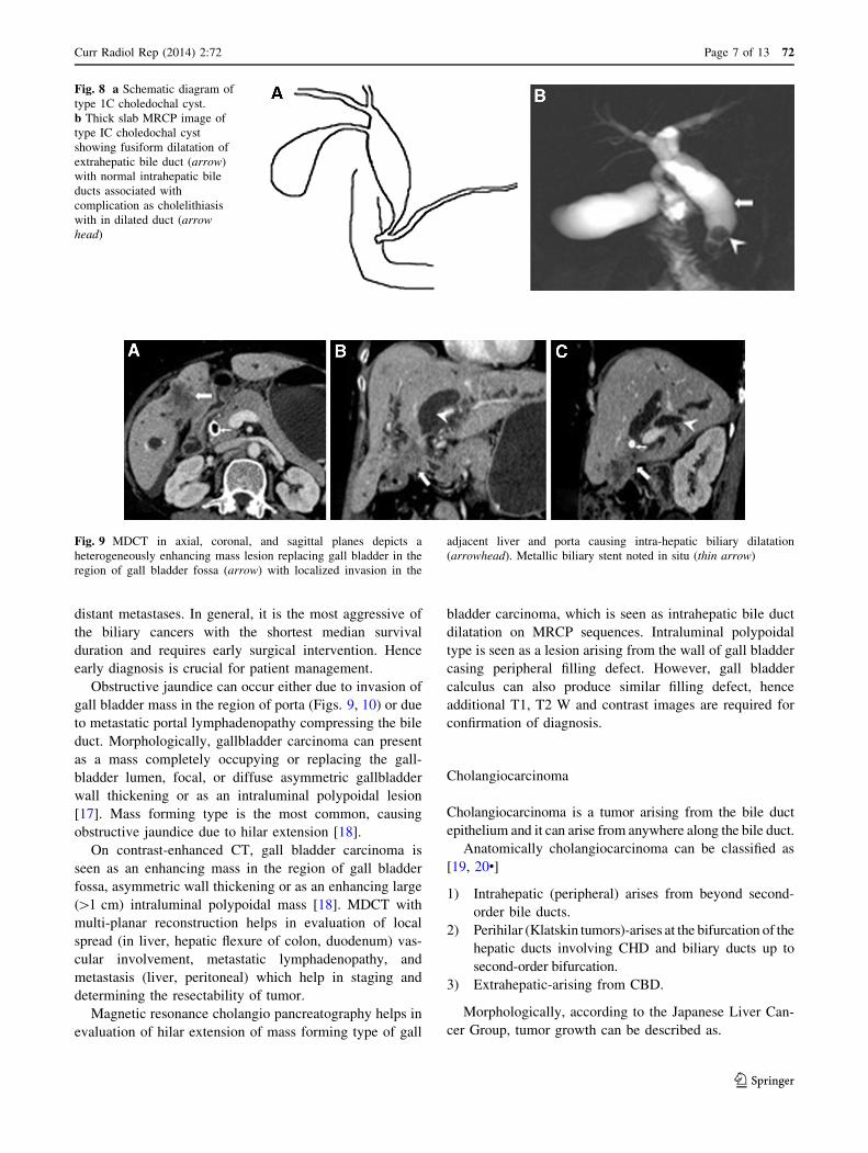
Certain features can differentiate benign from malignant
stricture. Features like wall thickness less than 1.5 mm,
length of stricture less than 9 mm, and upstream bile duct
dilatation of \17 mm are more of suggestive of benign
Table 2 Common causes of obstructive jaundice in both adult and
paediactric age group
In adults In pediatrics
• Cholelithiasis (Mirizzi syndrome) and
choledocholithiasis
• Biliary atresia
• Choledochal
cyst
• Benign stricture
• Choledochal cyst
• Primary sclerosing cholangitis
• Infection:
4 bacterial cholangitis- and HIV-related
cholangiopathy,
4 recurrent pyogenic cholangitis,
4 parasitic disease
• Portal biliopathy
• Benign tumors of biliary tract
• Malignant tumors causing obstruction of biliary
tract:
4 Gall bladder carcinoma
4 Cholangiocarcinoma
4 Pancreatic head carcinoma
Fig. 2 MRCP 3D image showing a smooth short segment stricture
(arrow) in the distal CBD due to recent passage of calculus. Gall
Bladder is studded with multiple calculi manifesting as multiple
filling defects (arrowhead)
Fig. 3 a Schematic diagram, b single shot thick slab MRCP shows
multiple well defined rounded filling defects (arrowhead) in a dilated
CBD surrounded by T2 hyperintense bile s/o choledocholithiasis
72 Page 4 of 13 Curr Radiol Rep (2014) 2:72
123
stricture (Figs. 5, 6). MRCP finding of a dilated bile duct
showing smooth and sudden tapering is diagnostic of post-
operative biliary stricture. In addition, MRCP can accu-
rately depict the anatomical level and length of stricture
which is helpful in its operative management [10]. CT scan
can show complications associated with stricture like
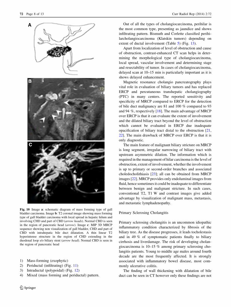
cholangitis and associated abscess. Bismuth H [11]
(Table 3) classified post-operative strictures into five types.
Choledochal Cyst
Choledochal cyst is a congenital anomaly involving biliary
tree characterized by cystic dilatation of bile ducts mani-
festing usually after the age of 10 years. Clinically it is
difficult to diagnose as most of the patients are asymp-
tomatic. A classical triad of jaundice, pain, and right upper
quadrant palpable lump, however, is seen in 30–60 % of
patients diagnosed before the age of 10 and in 10 % of the
cases diagnosed in adulthood [12].
Many theories exist regarding the patho-physiology of
choledochal cyst, but the most accepted one is of anoma-
lous pancreatico-biliary junction (APBJ). ABPJ is found in
10–58 % cases of choledochal cyst (Fig. 7). Two features
are considered responsible for the development of chole-
dochal cyst; a length of more than 15 mm of the common
channel formed after the union of PD and CBD outside the
wall of duodenum and the angle of union between PD and
CBD; both these features allowing reflux of pancreatic
juice in the bile ducts causing their ectatic dilatation. Based
on these two features, Komi et al. [13] classified APBJ into
three types. In Type I, there is a right angle between the
ducts, which is sub-classified into type IA (without dila-
tation) and type IB (with dilatation) of the common chan-
nel. In Type II, there is acute angle between the ducts
which is also sub-classified into type IIA (without dilata-
tion) and type IIB (with dilatation) of the common channel.
Type III unions are defined by more complex patterns of
accessory PDs.
Depending on the location and number of choledochal
cysts, Todani et al. [14] (Table 4) classified choledochal
cyst into five types as follows.
Type I choledochal cyst is the most common type.
The complication associated with choledochalcyst is
recurrent cholecystitis, choledocholithiasis, pancreatitis,
Fig. 4 Plain CT scan in axial
(a) and reconstructed oblique
coronal images (b) showing
multiple gall bladder calculi
(arrowhead) and impacted
distal CBD calculus (long
arrow) causing proximal
dilatation of the bile duct (short
arrow)
Fig. 6 Schematic diagram showing differentiation between benign
and malignant stricture
Fig. 5 a Schematic diagram and b MIP 3D MRCP showing short
length benign stricture involving CHD (arrow head)
Curr Radiol Rep (2014) 2:72 Page 5 of 13 72
123
and malignant transformation to cholangiocarcinoma [15].
The role of imaging in choledochal cyst is to localize it,
determine its relationship to adjacent structures, to evaluate
the complications related to it, and look for any associated
biliary tree abnormalities. USG can be used as initial
screening modality however it may be difficult to evaluate
the bile duct due to bowel gas.
On CT, it is seen as large cyst with fluid density in the
region of porta communicating with bile duct. MRCP is an
extremely useful tool for detection and classification of
choledochal cysts (Fig. 8). It is particularly important in
cases of patient with choledochal cysts associated with bile
duct stricture, in which ERCP is suboptimal due to non-
opacification bile duct distal to stricture [16].
Gall Bladder Carcinoma
Gall bladder cancer most often occurs in elderly women
and is usually associated with gallstones (90 % of cases).
Other risk factors include a chronic typhoid carrier state,
choledochal cyst, gall bladder polyp, anomalous APBJ, and
porcelain gallbladder. About 20 % of patients with porce-
lain gallbladder develop gallbladder cancer. Gall bladder
carcinomas are characterized by local invasion, extensive
regional lymph node metastasis, vascular encasement, and
Table 3 Bismuth classification of post-operative strictures
Type
I
Strictures are located
more than 2 cm distal
to confluence of left
and right hepatic
ducts
Type
II
Strictures are seen
within 2 cm from
hepatic duct
confluence
Type
III
Strictures affect hepatic
duct confluence,
which is patent
Type
IV
Strictures involve
hepatic duct
confluence and
interrupt it
Type
V
Strictures involve the
hepatic duct
associated with
stricture on aberrant
right intrahepatic
branch
Fig. 7 a 3D MRCP b thick slab
single shot MRCP showing
focal dilatation of extra-hepatic
bile duct (Type I B Choledochal
cyst) (thick arrow) with
anomalous pancreato-biliary
junction (minor duct opening
into CBD) (thin arrow)
Table 4 Todani classification of choledochal cyst
Type I Dilatation confined to the extrahepatic bile duct
IA Cystic dilatation of extrahepatic bile duct with gall
bladder arising from choledochal cyst
IB Focal or segmental dilatation of extrahepatic bile duct
IC Fusiform dilatation of extrahepatic bile duct
Type II True diverticula of the extrahepatic bile duct
Type III Choledochocele; confined to the extrahepatic bile duct
within the duodenal wall
Type IVA Dilatation involving both the extrahepatic bile ducts and
intrahepatic bile ducts
Type IVB Dilatation involving extrahepatic bile ducts
Type V Caroli’s disease; cysts confined to the intrahepatic ducts
72 Page 6 of 13 Curr Radiol Rep (2014) 2:72
123
distant metastases. In general, it is the most aggressive of
the biliary cancers with the shortest median survival
duration and requires early surgical intervention. Hence
early diagnosis is crucial for patient management.
Obstructive jaundice can occur either due to invasion of
gall bladder mass in the region of porta (Figs. 9, 10) or due
to metastatic portal lymphadenopathy compressing the bile
duct. Morphologically, gallbladder carcinoma can present
as a mass completely occupying or replacing the gall-
bladder lumen, focal, or diffuse asymmetric gallbladder
wall thickening or as an intraluminal polypoidal lesion
[17]. Mass forming type is the most common, causing
obstructive jaundice due to hilar extension [18].
On contrast-enhanced CT, gall bladder carcinoma is
seen as an enhancing mass in the region of gall bladder
fossa, asymmetric wall thickening or as an enhancing large
([1 cm) intraluminal polypoidal mass [18]. MDCT with
multi-planar reconstruction helps in evaluation of local
spread (in liver, hepatic flexure of colon, duodenum) vas-
cular involvement, metastatic lymphadenopathy, and
metastasis (liver, peritoneal) which help in staging and
determining the resectability of tumor.
Magnetic resonance cholangio pancreatography helps in
evaluation of hilar extension of mass forming type of gall
bladder carcinoma, which is seen as intrahepatic bile duct
dilatation on MRCP sequences. Intraluminal polypoidal
type is seen as a lesion arising from the wall of gall bladder
casing peripheral filling defect. However, gall bladder
calculus can also produce similar filling defect, hence
additional T1, T2 W and contrast images are required for
confirmation of diagnosis.
Cholangiocarcinoma
Cholangiocarcinoma is a tumor arising from the bile duct
epithelium and it can arise from anywhere along the bile duct.
Anatomically cholangiocarcinoma can be classified as
[19, 20•]
1) Intrahepatic (peripheral) arises from beyond second-
order bile ducts.
2) Perihilar (Klatskin tumors)-arises at the bifurcation of the
hepatic ducts involving CHD and biliary ducts up to
second-order bifurcation.
3) Extrahepatic-arising from CBD.
Morphologically, according to the Japanese Liver Can-
cer Group, tumor growth can be described as.
Fig. 8 a Schematic diagram of
type 1C choledochal cyst.
b Thick slab MRCP image of
type IC choledochal cyst
showing fusiform dilatation of
extrahepatic bile duct (arrow)
with normal intrahepatic bile
ducts associated with
complication as cholelithiasis
with in dilated duct (arrow
head)
Fig. 9 MDCT in axial, coronal, and sagittal planes depicts a
heterogeneously enhancing mass lesion replacing gall bladder in the
region of gall bladder fossa (arrow) with localized invasion in the
adjacent liver and porta causing intra-hepatic biliary dilatation
(arrowhead). Metallic biliary stent noted in situ (thin arrow)
Curr Radiol Rep (2014) 2:72 Page 7 of 13 72
123
1) Mass-forming (exophytic)
2) Periductal (infiltrating) (Fig. 11)
3) Intraductal (polypoidal) (Fig. 12)
4) Mixed (mass forming and periductal) pattern.
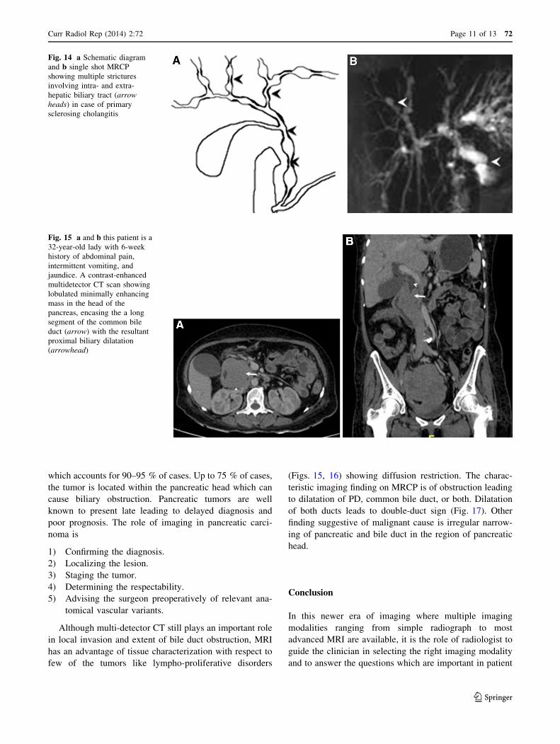
Out of all the types of cholangiocarcinoma, perihilar is
the most common type, presenting as jaundice and shows
infiltrating pattern. Bismuth and Corlette classified perihi-
larcholangiocarcinoma (Klatskin tumors) depending on
extent of ductal involvement (Table 5) (Fig. 13).
Apart from localization of level of obstruction and cause
of obstruction, contrast-enhanced CT scan helps in deter-
mining the morphological type of cholangiocarcinoma,
local spread, vascular involvement and determining stage
and resectability of tumor. In cases of cholangiocarcinoma,
delayed scan at 10–15 min is particularly important as it is
shows delayed enhancement.
Magnetic resonance cholangio pancreatography plays
vital role in evaluation of biliary tumors and has replaced
ERCP and percutaneous transhepatic cholangiography
(PTC) in many centers. The reported sensitivity and
specificity of MRCP compared to ERCP for the detection
of bile duct malignancy are 81 and 100 % compared to 93
and 94 %, respectively [18]. The main advantage of MRCP
over ERCP is that it can evaluate the extent of involvement
and the dilated biliary tract beyond the level of obstruction
which cannot be evaluated in ERCP due inadequate
opacification of biliary tract distal to the obstruction [21,
22]. The main drawback of MRCP over ERCP is that it is
only diagnostic.
The main feature of malignant biliary stricture on MRCP
is long segment, irregular narrowing of biliary tract with
upstream asymmetric dilation. The information which is
required in the management of hilar carcinoma is the level of
obstruction, extent of involvement, whether the involvement
is up to primary or second-order branches and associated
choledocholithiasis [23]; all can be obtained from MRCP
images [22]. MRCP provides only endoluminal images from
fluid, hence sometimes it could be inadequate to differentiate
between benign and malignant stricture. In such cases,
conventional T2, T1 W and contrast images give added
advantage by visualization of malignant mass, metastasis,
and metastatic lymphadenopathy.
Primary Sclerosing Cholangitis
Primary sclerosing cholangitis is an uncommon idiopathic
inflammatory condition characterized by fibrosis of the
biliary tree. As the disease progresses, it leads tocholestasis
and in 49 % of symptomatic patients finally to biliary
cirrhosis and liverdamage. The risk of developing cholan-
giocarcinoma is 10–15 % among primary sclerosing cho-
langitis patients. Young to middle age males around fourth
decade are the most frequently affected. It is strongly
associated with inflammatory bowel disease, most com-
monly ulcerative colitis.
The finding of wall thickening with dilatation of bile
duct can be seen in CT however only these findings are not
Fig. 10 Image a: schematic diagram of mass forming type of gall
bladder carcinoma. Image b: T2 coronal image showing mass forming
type of gall bladder carcinoma with local spread in hepatic hilum and
involving CHD and part of CBD (arrow heads). Normal CBD is seen
in the region of pancreatic head (arrow). Image c: MIP 3D MRCP
sequence showing non visualization of gall bladder, CHD and part of
CBD with intrahepatic bile duct dilatation. A thin linear T2
hyperintense structure in the region of CHD extending in the
duodenal loop s/o biliary stent (arrow head). Normal CBD is seen in
the region of pancreatic head
72 Page 8 of 13 Curr Radiol Rep (2014) 2:72
123
sufficient for diagnosis of PSC. In normal subjects on
MRCP, only the central ducts containing adequate bile can
be seen. Peripheral small biliary radicles are not seen as
there is insufficient pressure to distend them. Multiple
stricturous involvement of central bile ducts in PSC pre-
vents contrast opacification of peripheral bile ducts, pro-
ducing prune tree appearance on ERCP. However, because
of generation of back pressure in these peripheral small bile
ducts, they can be appreciated on MRCP giving an added
advantage over ERCP. A study done by Fulcher et al. [24],
found sensitivity of 88 % for MRCP as compared to ERCP
in diagnosis of PSC.
The characteristic finding of PSC is multiple alternat-
ing short segment strictures and slightly dilated inter-
vening bile duct segments leading to bead on string
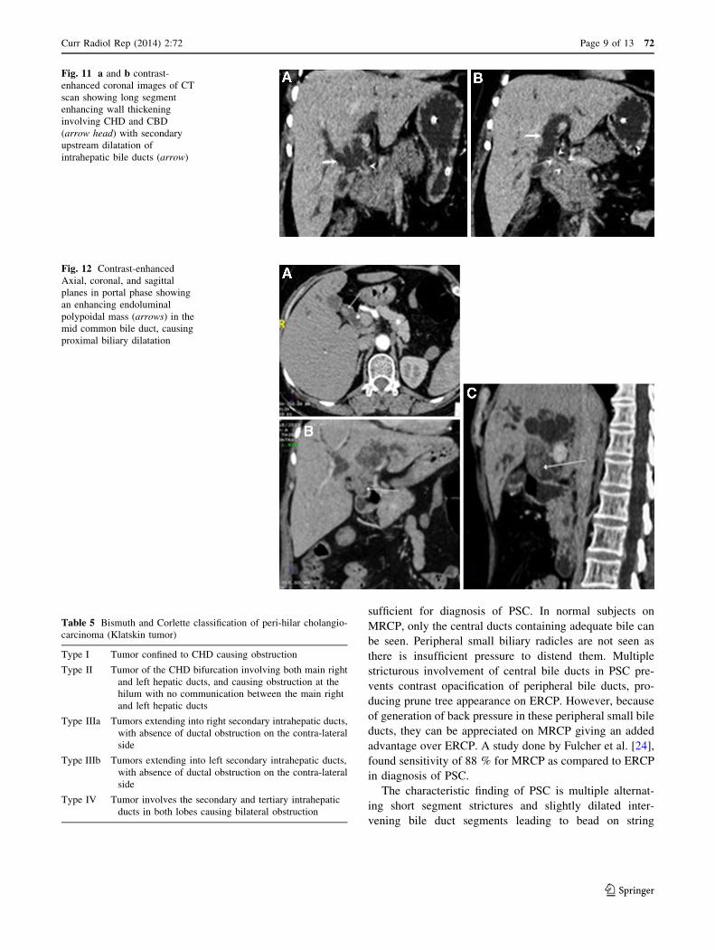
Fig. 11 a and b contrast-
enhanced coronal images of CT
scan showing long segment
enhancing wall thickening
involving CHD and CBD
(arrow head) with secondary
upstream dilatation of
intrahepatic bile ducts (arrow)
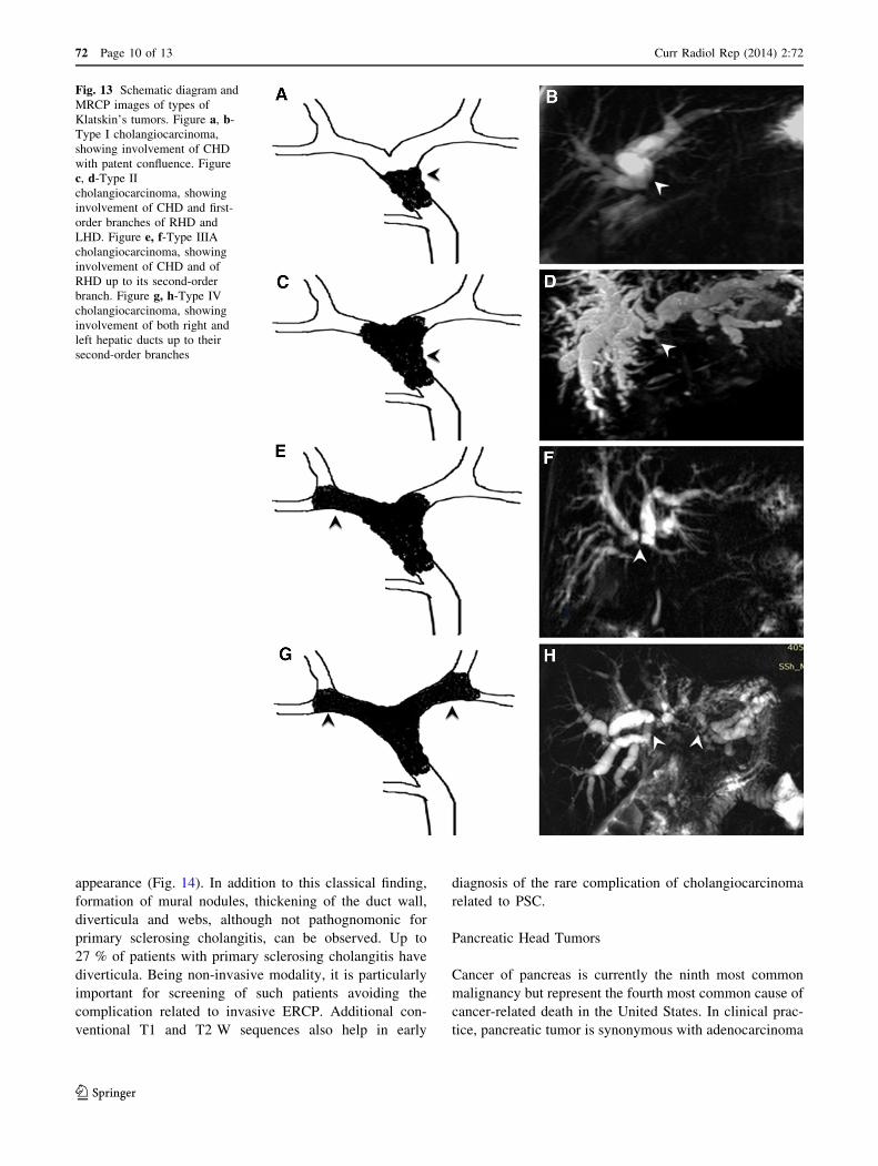
Fig. 12 Contrast-enhanced
Axial, coronal, and sagittal
planes in portal phase showing
an enhancing endoluminal
polypoidal mass (arrows) in the
mid common bile duct, causing
proximal biliary dilatation
Table 5 Bismuth and Corlette classification of peri-hilar cholangio-
carcinoma (Klatskin tumor)
Type I Tumor confined to CHD causing obstruction
Type II Tumor of the CHD bifurcation involving both main right
and left hepatic ducts, and causing obstruction at the
hilum with no communication between the main right
and left hepatic ducts
Type IIIa Tumors extending into right secondary intrahepatic ducts,
with absence of ductal obstruction on the contra-lateral
side
Type IIIb Tumors extending into left secondary intrahepatic ducts,
with absence of ductal obstruction on the contra-lateral
side
Type IV Tumor involves the secondary and tertiary intrahepatic
ducts in both lobes causing bilateral obstruction
Curr Radiol Rep (2014) 2:72 Page 9 of 13 72
123
appearance (Fig. 14). In addition to this classical finding,
formation of mural nodules, thickening of the duct wall,
diverticula and webs, although not pathognomonic for
primary sclerosing cholangitis, can be observed. Up to
27 % of patients with primary sclerosing cholangitis have
diverticula. Being non-invasive modality, it is particularly
important for screening of such patients avoiding the
complication related to invasive ERCP. Additional con-
ventional T1 and T2 W sequences also help in early
diagnosis of the rare complication of cholangiocarcinoma
related to PSC.
Pancreatic Head Tumors
Cancer of pancreas is currently the ninth most common
malignancy but represent the fourth most common cause of
cancer-related death in the United States. In clinical prac-
tice, pancreatic tumor is synonymous with adenocarcinoma
Fig. 13 Schematic diagram and
MRCP images of types of
Klatskin’s tumors. Figure a, b-
Type I cholangiocarcinoma,
showing involvement of CHD
with patent confluence. Figure
c, d-Type II
cholangiocarcinoma, showing
involvement of CHD and first-
order branches of RHD and
LHD. Figure e, f-Type IIIA
cholangiocarcinoma, showing
involvement of CHD and of
RHD up to its second-order
branch. Figure g, h-Type IV
cholangiocarcinoma, showing
involvement of both right and
left hepatic ducts up to their
second-order branches
72 Page 10 of 13 Curr Radiol Rep (2014) 2:72
123
which accounts for 90–95 % of cases. Up to 75 % of cases,
the tumor is located within the pancreatic head which can
cause biliary obstruction. Pancreatic tumors are well
known to present late leading to delayed diagnosis and
poor prognosis. The role of imaging in pancreatic carci-
noma is
1) Confirming the diagnosis.
2) Localizing the lesion.
3) Staging the tumor.
4) Determining the respectability.
5) Advising the surgeon preoperatively of relevant ana-
tomical vascular variants.
Although multi-detector CT still plays an important role
in local invasion and extent of bile duct obstruction, MRI
has an advantage of tissue characterization with respect to
few of the tumors like lympho-proliferative disorders
(Figs. 15, 16) showing diffusion restriction. The charac-
teristic imaging finding on MRCP is of obstruction leading
to dilatation of PD, common bile duct, or both. Dilatation
of both ducts leads to double-duct sign (Fig. 17). Other
finding suggestive of malignant cause is irregular narrow-
ing of pancreatic and bile duct in the region of pancreatic
head.
Conclusion
In this newer era of imaging where multiple imaging
modalities ranging from simple radiograph to most
advanced MRI are available, it is the role of radiologist to
guide the clinician in selecting the right imaging modality
and to answer the questions which are important in patient
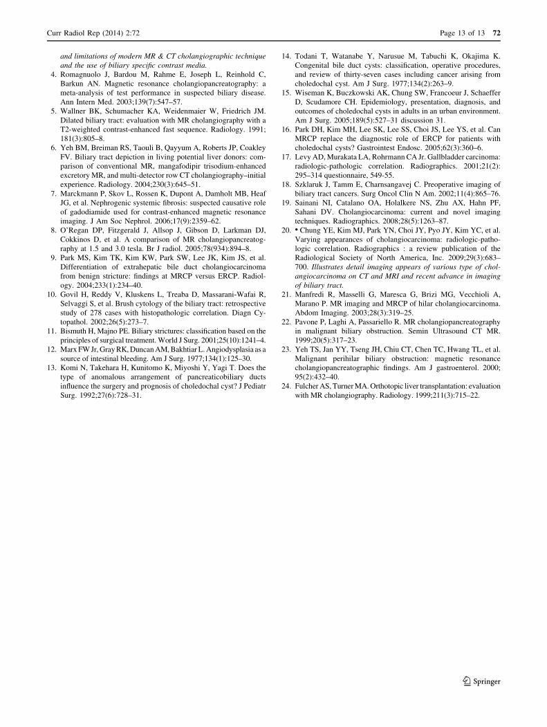
Fig. 14 a Schematic diagram
and b single shot MRCP
showing multiple strictures
involving intra- and extra-
hepatic biliary tract (arrow
heads) in case of primary
sclerosing cholangitis
Fig. 15 a and b this patient is a
32-year-old lady with 6-week
history of abdominal pain,
intermittent vomiting, and
jaundice. A contrast-enhanced
multidetector CT scan showing
lobulated minimally enhancing
mass in the head of the
pancreas, encasing the a long
segment of the common bile
duct (arrow) with the resultant
proximal biliary dilatation
(arrowhead)
Curr Radiol Rep (2014) 2:72 Page 11 of 13 72
123

management. Although ultrasound remains a screening
modality for diagnosis of biliary obstruction, it is unable to
answer the true extent and cause of obstructive jaundice
requiring the use of another imaging modality like CECT
and MRCP which scores over ultrasound in the diagnostic
accuracy. MRCP can be considered as the new gold stan-
dard for the investigation of biliary obstruction and permits
reservation of ERCP to patients with a high probability of
therapeutic intervention.
Compliance with Ethics Guidelines
Conflict of Interest Dr. Anagha Joshi is a section editor for Current
Radiology Reports. Dr. Kishore Rajpal, Dr. Ketan Kakadiya, and Dr.
Ashank Bansal each declare no potential conflicts of interest.
Human and Animal Rights and Informed Consent This article
does not contain any studies with human or animal subjects
performed by any of the authors.
References
Recently published papers of particular interest have been
highlighted as:• Of importance
1. Rana SS, Bhasin DK, Sharma V, Rao C, Gupta R, Singh K. Role
of endoscopic ultrasound in evaluation of unexplained common
bile duct dilatation on magnetic resonance cholangiopancrea-
tography. Ann Gastroenterol. 2013;26(1):66–70.
2. • Patel HT, Shah AJ, Khandelwal SR, Patel HF, Patel MD. MR
cholangiopancreatography at 3.0 T. Radiographics: a review
publication of the Radiological Society of North America, Inc.
2009;29(6):1689–706. Describes and illustrates in detail 3.0T
MRCP appearances of normal biliary tract anatomy, variants
and benign and malignant pathologies.
3. • Yeh BM, Liu PS, Soto JA, Corvera CA, Hussain HK. MR
imaging and CT of the biliary tract. Radiographics : a review
publication of the Radiological Society of North America, Inc.
2009;29(6):1669–88. Gives good knowledge about the benefits
Fig. 16 a, b, and c T2 weighted
fat-suppressed images in axial
and coronal planes of the same
patient in Fig. 16, showing
intermediate signal intensity
inhomogeneous soft tissue mass
in the head of pancreas with
encasement and narrowing of
common bile duct (arrow)
causing proximal duct
dilatation. DWI images showing
foci of diffusion restriction of
the pancreatic mass
(arrowhead) with renal deposits
(small arrow) representing a
lymphoproliferative disorder. A
lobulated soft tissue bowel mass
is seen in the pelvis showing
intermediate to hyperintense
signal intensity on T2 W fat-
suppressed images (star)
Fig. 17 a Schematic diagram
and b single shot MRCP
showing dilated CBD and PD
(double-duct sign) in their entire
extent with abrupt cutoff in the
region of pancreatic head due to
peri-ampullary tumor (arrow
head) associated with over
distended gall bladder(black
star)
72 Page 12 of 13 Curr Radiol Rep (2014) 2:72
123
and limitations of modern MR & CT cholangiographic technique
and the use of biliary specific contrast media.
4. Romagnuolo J, Bardou M, Rahme E, Joseph L, Reinhold C,
Barkun AN. Magnetic resonance cholangiopancreatography: a
meta-analysis of test performance in suspected biliary disease.
Ann Intern Med. 2003;139(7):547–57.
5. Wallner BK, Schumacher KA, Weidenmaier W, Friedrich JM.
Dilated biliary tract: evaluation with MR cholangiography with a
T2-weighted contrast-enhanced fast sequence. Radiology. 1991;
181(3):805–8.
6. Yeh BM, Breiman RS, Taouli B, Qayyum A, Roberts JP, Coakley
FV. Biliary tract depiction in living potential liver donors: com-
parison of conventional MR, mangafodipir trisodium-enhanced
excretory MR, and multi-detector row CT cholangiography–initial
experience. Radiology. 2004;230(3):645–51.
7. Marckmann P, Skov L, Rossen K, Dupont A, Damholt MB, Heaf
JG, et al. Nephrogenic systemic fibrosis: suspected causative role
of gadodiamide used for contrast-enhanced magnetic resonance
imaging. J Am Soc Nephrol. 2006;17(9):2359–62.
8. O’Regan DP, Fitzgerald J, Allsop J, Gibson D, Larkman DJ,
Cokkinos D, et al. A comparison of MR cholangiopancreatog-
raphy at 1.5 and 3.0 tesla. Br J radiol. 2005;78(934):894–8.
9. Park MS, Kim TK, Kim KW, Park SW, Lee JK, Kim JS, et al.
Differentiation of extrahepatic bile duct cholangiocarcinoma
from benign stricture: findings at MRCP versus ERCP. Radiol-
ogy. 2004;233(1):234–40.
10. Govil H, Reddy V, Kluskens L, Treaba D, Massarani-Wafai R,
Selvaggi S, et al. Brush cytology of the biliary tract: retrospective
study of 278 cases with histopathologic correlation. Diagn Cy-
topathol. 2002;26(5):273–7.
11. Bismuth H, Majno PE. Biliary strictures: classification based on the
principles of surgical treatment. World J Surg. 2001;25(10):1241–4.
12. Marx FW Jr, Gray RK, Duncan AM, Bakhtiar L. Angiodysplasia as a
source of intestinal bleeding. Am J Surg. 1977;134(1):125–30.
13. Komi N, Takehara H, Kunitomo K, Miyoshi Y, Yagi T. Does the
type of anomalous arrangement of pancreaticobiliary ducts
influence the surgery and prognosis of choledochal cyst? J Pediatr
Surg. 1992;27(6):728–31.
14. Todani T, Watanabe Y, Narusue M, Tabuchi K, Okajima K.
Congenital bile duct cysts: classification, operative procedures,
and review of thirty-seven cases including cancer arising from
choledochal cyst. Am J Surg. 1977;134(2):263–9.
15. Wiseman K, Buczkowski AK, Chung SW, Francoeur J, Schaeffer
D, Scudamore CH. Epidemiology, presentation, diagnosis, and
outcomes of choledochal cysts in adults in an urban environment.
Am J Surg. 2005;189(5):527–31 discussion 31.
16. Park DH, Kim MH, Lee SK, Lee SS, Choi JS, Lee YS, et al. Can
MRCP replace the diagnostic role of ERCP for patients with
choledochal cysts? Gastrointest Endosc. 2005;62(3):360–6.
17. Levy AD, Murakata LA, Rohrmann CA Jr. Gallbladder carcinoma:
radiologic-pathologic correlation. Radiographics. 2001;21(2):
295–314 questionnaire, 549-55.
18. Szklaruk J, Tamm E, Charnsangavej C. Preoperative imaging of
biliary tract cancers. Surg Oncol Clin N Am. 2002;11(4):865–76.
19. Sainani NI, Catalano OA, Holalkere NS, Zhu AX, Hahn PF,
Sahani DV. Cholangiocarcinoma: current and novel imaging
techniques. Radiographics. 2008;28(5):1263–87.
20. • Chung YE, Kim MJ, Park YN, Choi JY, Pyo JY, Kim YC, et al.
Varying appearances of cholangiocarcinoma: radiologic-patho-
logic correlation. Radiographics : a review publication of the
Radiological Society of North America, Inc. 2009;29(3):683–
700. Illustrates detail imaging appears of various type of chol-
angiocarcinoma on CT and MRI and recent advance in imaging
of biliary tract.
21. Manfredi R, Masselli G, Maresca G, Brizi MG, Vecchioli A,
Marano P. MR imaging and MRCP of hilar cholangiocarcinoma.
Abdom Imaging. 2003;28(3):319–25.
22. Pavone P, Laghi A, Passariello R. MR cholangiopancreatography
in malignant biliary obstruction. Semin Ultrasound CT MR.
1999;20(5):317–23.
23. Yeh TS, Jan YY, Tseng JH, Chiu CT, Chen TC, Hwang TL, et al.
Malignant perihilar biliary obstruction: magnetic resonance
cholangiopancreatographic findings. Am J gastroenterol. 2000;
95(2):432–40.
24. Fulcher AS, Turner MA. Orthotopic liver transplantation: evaluation
with MR cholangiography. Radiology. 1999;211(3):715–22.
Curr Radiol Rep (2014) 2:72 Page 13 of 13 72
123