Role of Coronin 1B in PDGF-induced Migration of Vascular ...
23
Role of Coronin 1B in PDGF-induced Migration of Vascular Smooth Muscle Cells Holly Ann Williams, Emory University Alejandra San Martin Almeyda, Emory University Candace M. Adamo, Emory University Bonnie Seidel-Rogol, Emory University Lily Pounkova, Emory University Srinivasan Raju Datla, Emory University Bernard P Lassegue, Emory University James E. Bear, University of North Carolina-Chapel Hill Kathy Griendling, Emory University Journal Title: Circulation Research Volume: Volume 111, Number 1 Publisher: American Heart Association | 2012-06-22, Pages 56-65 Type of Work: Article | Post-print: After Peer Review Publisher DOI: 10.1161/CIRCRESAHA.111.255745 Permanent URL: http://pid.emory.edu/ark:/25593/cx590 Final published version: http://dx.doi.org/10.1161%2FCIRCRESAHA.111.255745 Copyright information: © 2012 American Heart Association, Inc. Accessed February 7, 2022 2:51 AM EST
Transcript of Role of Coronin 1B in PDGF-induced Migration of Vascular ...

Role of Coronin 1B in PDGF-induced Migration ofVascular Smooth Muscle CellsHolly Ann Williams, Emory UniversityAlejandra San Martin Almeyda, Emory UniversityCandace M. Adamo, Emory UniversityBonnie Seidel-Rogol, Emory UniversityLily Pounkova, Emory UniversitySrinivasan Raju Datla, Emory UniversityBernard P Lassegue, Emory UniversityJames E. Bear, University of North Carolina-Chapel HillKathy Griendling, Emory University
Journal Title: Circulation ResearchVolume: Volume 111, Number 1Publisher: American Heart Association | 2012-06-22, Pages 56-65Type of Work: Article | Post-print: After Peer ReviewPublisher DOI: 10.1161/CIRCRESAHA.111.255745Permanent URL: http://pid.emory.edu/ark:/25593/cx590
Final published version:http://dx.doi.org/10.1161%2FCIRCRESAHA.111.255745
Copyright information:© 2012 American Heart Association, Inc.
Accessed February 7, 2022 2:51 AM EST

Role of Coronin 1B in PDGF-induced Migration of VascularSmooth Muscle Cells
Holly C. Williams1, Alejandra San Martín1, Candace M. Adamo1, Bonnie Seidel-Rogol1, LilyPounkova1, Srinivasan Raju Datla1, Bernard Lassègue1, James E. Bear2, and KathyGriendling1
1Department of Medicine, Division of Cardiology, Emory University School of Medicine. Atlanta.Georgia 30322, U.S.A2Howard Hughes Medical Institute and Lineberger Comprehensive Cancer Center, University ofNorth Carolina-Chapel Hill, Chapel Hill, North Carolina 27599
AbstractRationale—The type I subclass of Coronins, a family of actin binding proteins, regulates variousactin dependent cellular processes including migration. However, the existence and role ofcoronins in vascular smooth muscle cell (VSMC) migration has yet to be determined.
Objective—The goal of the present study was to define the mechanism by which coroninsregulate platelet-derived growth factor (PDGF)-induced VSMC migration.
Methods and Results—Coronin 1B (Coro1B) and 1C (Coro1C) were both found to beexpressed in VSMCs at the mRNA and protein levels. Down regulation of Coro1B by siRNAincreases PDGF-induced migration, while down regulation of Coro1C has no effect. Weconfirmed through kymograph analysis that the Coro1B-downregulation-mediated increase inmigration is directly linked to increased lamellipodial protraction rate and protrusion distance inVSMC. In other cell types, coronins exert their effects on lamellipodia dynamics by an inhibitoryinteraction with the ARP2/3 complex, which is disrupted by the phosphorylation of Coro1B. Wefound that PDGF induces phosphorylation of Coro1B on serine-2 via PKCε, leading to a decreasein the interaction of Coro1B with the ARP2/3 complex. VSMCs transfected with a phospho-deficient S2A Coro1B mutant showed decreased migration in response to PDGF, suggesting thatthe phosphorylation of Coro1B is required for the promotion of migration by PDGF. In both therat and mouse Coro1B phosphorylation was increased in response to vessel injury in vivo.
Conclusions—Our data support that phosphorylation of Coro1B and the subsequent reducedinteraction with ARP2/3 complex participate in PDGF-induced VSMC migration, an importantstep in vascular lesion formation.
Keywordsvascular smooth muscle; coronin 1B; migration; platelet-derived growth factor
Address correspondence to: Kathy K. Griendling, Emory University, Division of Cardiology, 319 WMB, 1639 Pierce Dr. Atlanta, GA30322, Telephone: 404-727-3364, Fax: 404-727-3585 [email protected].
DisclosuresNone
NIH Public AccessAuthor ManuscriptCirc Res. Author manuscript; available in PMC 2012 December 22.
Published in final edited form as:Circ Res. 2012 June 22; 111(1): . doi:10.1161/CIRCRESAHA.111.255745.
NIH
-PA Author Manuscript
NIH
-PA Author Manuscript
NIH
-PA Author Manuscript

IntroductionThe migration of cells is important in multiple facets of normal and aberrant cell behavior.In the vasculature, the migration of vascular smooth muscle cells (VSMC) is vital forphysiological processes such as angiogenesis and vessel remodeling, and is a criticalcomponent of pathophysiological lesion formation occurring during atherosclerosis andrestenosis after percutaneous coronary intervention (PCI).1, 2 In these latter diseases, one ofthe major characteristic changes is the accumulation of VSMCs within the intimal layer ofthe blood vessel.3, 4 Narrowing of the blood vessels caused by these events can ultimatelylead to thrombosis or embolus formation, both of which remain a significant clinicalproblem. Genetic manipulation of vascular cells combined with multiple inhibitorystrategies have provided strong evidence that platelet-derived growth factor (PDGF) playsan important role in the migration of VSMCs into the neointima following acute injury andin atherosclerosis.5 Although PDGF has been shown to be the primary regulator of VSMCmigration in vivo, little is understood about the molecular mechanisms and intracellularsignaling pathways that contribute to this migration.
Migration is a dynamic and cyclic process that generally begins with the extension of actinrich protrusions called lamellipodia.6 The formation of these structures at the leading edgeof the cell requires the protrusive forces that are generated by actin polymerization andincreased actin branching.7 Lamellipodia formation is regulated by the actin related protein2/3 complex (ARP2/3 complex), which participates concomitantly in actin nucleation andfilament branching. The Arp2/3 complex binds to the side of an existing parent filament andnucleates the formation of new actin filaments at a distinctive 70° angle, leading to theformation of branched filament networks8 that are required for efficient cell migration. Forfull activation, the ARP2/3 complex must bind with actin filaments and activator proteinssuch as Wiskott–Aldrich Syndrome protein (WASp) and suppressor of cAR /WASP familyVerprolin-homologous protein (SCAR/WAVE).9 Conversely, recent studies havedemonstrated that the ARP2/3 actin nucleation activity can be negatively regulated by aninteraction with actin binding proteins known as coronins.10, 11
Coronins are a family of evolutionarily conserved WD-repeat actin-binding proteins knownto control a variety of cellular processes involving actin dynamics.12 The coronin proteinfamily includes 7 proteins in mammals, separated into three subclasses (type I, II, and III)based on phylogenetic similarity.13, 1415 The type I coronins consist of coronin 1A(Coro1A), 1B (Coro1B), and 1C (Coro1C) and are the most studied coronin subfamily.Coro1A is highly expressed in cells of hematopoietic lineage in addition to various tissues ofthe nervous system, while having significantly lower expression in other tissues of thebody.16 On the other hand Coro1B and Coro1C are more ubiquitously expressed at higherlevels in most tissues. 11 Coro1B localizes to the plasma membrane, regulates lamellipodiaformation, and when phosphorylated is no longer able to bind ARP2/3 and inhibit its actinnucleation abilities.11 In contrast, in epithelial cells, Coro1C was demonstrated to regulatefocal adhesion dynamics and increase wound closure.17 Thus, Coro1B and 1C canpotentially influence migration via several different mechanisms, and their overall impact islikely to be a function of the complement of coronins expressed in a given cell type.
Although there have been significant advances in identifying the functions of coronins, notmuch is known about physiological regulators or upstream signaling pathways involved incoronin activation, and virtually nothing is known about their role in VSMCs. Therefore, thegoal of the present study was to determine if Coro1B and 1C are present in VSMCs and toexplore the mechanism by which they might regulate PDGF-induced VSMC migration.Here, we provide data that demonstrate that Coro1B and 1C are expressed in VSMCs, andthat Coro1B is highly phosphorylated after stimulation by the physiological agonist PDGF
Williams et al. Page 2
Circ Res. Author manuscript; available in PMC 2012 December 22.
NIH
-PA Author Manuscript
NIH
-PA Author Manuscript
NIH
-PA Author Manuscript

in VSMC. The consequence of this phosphorylation is a decreased interaction of Coro1Bwith the ARP2/3 complex, leading to an increase in PDGF-induced VSMC migration. Wealso found that Coro1B phosphorylation is induced in the rat carotid balloon injury andmouse carotid wire injury model of neointimal formation. These data suggest that PDGFsignals to Coro1B to coordinate lamellipodia formation and thus migration in VSMCs, andsuggests a new therapeutic target for vasculopathies with a significant migratory component.
MethodsMaterials
Recombinant human PDGF-BB was purchased from R&D Systems Inc. Rö-32-0423 andGö-6796 PKC inhibitors were purchased from Calbiochem. Primary antibodies werepurchased from Sigma (α-smooth muscle actin and β-actin), ECM Bioscience (phospho-Coro1B Ser-2), Santa Cruz Biotechnology (Coro1B M-80, Coro1B S-20, cyclin-dependentkinase 4 (CDK4), and actin-related protein 2/3 complex subunit 2 (ArpC2)), Cell SignalingTechnologies (Myc (9B11 and 71D10) and PKCε)and Millipore (ArpC2). Mousemonoclonal antibody against coronin 1c was created in the laboratory of Dr. James Bear aspreviously described.11 Streptadividin conjugated Quantum dots with a 603 fluorescent labelwere purchased from Invitrogen. siGlo Red transfection indicator was purchased fromDharmacon RNAI Technologies.
Cell CultureVascular smooth muscle cells (VSMC) were isolated from rat thoracic aorta by enzymaticdigestion as previously described.18 Isolated VSMCs were grown in Dulbecco’s ModifiedEagle’s Media supplemented with 10% calf serum. For experiments, cells between passages7 and 15 were plated and allowed to grow until they reached 60% confluence. Cells werethen serum starved for 24–48 hrs prior to PDGF stimulation.
Reverse Transcriptase PCR (RT-PCR)RNA was purified from rat lung tissue, and VSMCs using the RNeasy kit (Qiagen),following the manufacturer’s instructions. The purified RNA was reverse-transcribed withSuperscript II reverse transcriptase (Invitrogen) using random nanomers. TheresultingcDNA samples were amplified by non-quantitative PCR using recombinantPlatinum Taq DNA polymerase (Invitrogen). Amplification conditions were as follows: 300nmol/L primers, 1.5 mmol/LMgCl2, 200 μmol/L dNTPs, with an annealing temperature of55°C. The following primer sequences were created for amplification of: Coro1B (upstreamprimer, 5′-ACA TGT CCT TCC GAA AAG TTG TGC-3′; downstream primer, 5′-CTGATC CAC TGG CAA TGA CTT CGT -3′), and Coro1C (upstream primer, 5′-TGT CTTCAC TAC TGG TTT TAG CCG TA-3′; downstream primer, 5′-TCT AGC TTT GAA ATGCGC TCG TCT-3′). GAPDH (upstream primer, 5′-AAT GGG GTG ATG CTG GTG CTGAGT A-3′; downstream primer, 5′-GGA AGA ATG GGA GTT GCT GTT GAA G-3′)
Western BlottingAfter treatment with PDGF, cells were washed twice with phosphate buffered saline andthen lysed in Hunters buffer (25 mmol/L HEPES, 150 mmol/L KCl, 1.5 mmol/L MgCl2, 1mmol/L EGTA, 10 mmol/L Na-pyrophosphate, 10 mmol/L NaF, 1% Na deoxycholate, 1%Triton X 100, 0.1% SDS, 10% Glycerol, Na-orthovanadate and protease inhibitors). Lysateswere then sonicated and cleared at 13,000 × g for 5 minutes. Proteins were separated usingSDS-PAGE and transferred to Immobilon-P polyvinylidene difluoride (PVDF) membranes(Millipore), blocked with 5% non-fat dairy milk, and incubated with appropriate primaryantibodies. Subsequently, blots were incubated with horseradish peroxidase-conjugated
Williams et al. Page 3
Circ Res. Author manuscript; available in PMC 2012 December 22.
NIH
-PA Author Manuscript
NIH
-PA Author Manuscript
NIH
-PA Author Manuscript

secondary antibodies and proteins were detected by enhanced chemiluminescence (ECL,GE). Band intensity was quantified by densitometry using ImageJ (NIH) or CarestreamMolecular Imaging (Carestream) software.
Co-immunoprecipitationCells were washed twice with phosphate buffered saline and lysed with a KCl buffer (20mmol/L HEPES, pH 7.0, 100 mmol/L KCl, 0.5% Nonidet P-40, 1 mmol/L EDTA, andprotease inhibitors). Lysates were cleared at 13,000 × g for 5 min. 500 μg of protein lysatewas incubated with 1 μg of primary antibody for one hour at 4 °C, followed by the additionof 30 μl of Protein A beads (Santa Cruz Biotechnology) for another hour. Beads wereblocked with 1 mg/ml bovine serum albumin for one hour before use. Immunoprecipitatedproteins were collected by centrifugation, washed three times with KCl buffer, separated bySDS-PAGE, and transferred to PVDF membranes for Western blotting.
Plasmid Construction and Site- directed MutagenesisC-terminal Myc-tagged Coro1B-WT pCDNA3 was constructed by amplifying the openreading frame of human Coro1B from pCMV6-AC-Coro1B (Origene) using Phusion High-Fidelity DNA polymerase (Thermo-Fisher Scientific) as well as primers that contained theMyc coding sequence. Amplification conditions were as follows: 300 nmol/L primers, 1.5mmol/LMgCl2, 200 μmol/L dNTPs, and 3% dimethyl sulfoxide (DMSO) with an annealingtemperature of 72°C. Primer sequences were as follows: upstream primer -5′ TAC GGATCC GCC ACC ATG TCC TTC CGC AAA GTG GTC CGG CAG AGC A -3′ downstreamprimer -5′ GTA TCT AGA TCA GAA TTC CAG ATC CTC TTC TGA GAT GAG TTTTTG TTC CGC ATC CCC GTT CTC CAT GCG GCC CAG CT -3′. Myc-tagged Coro1B-S2A pCDNA3 and Myc-tagged Coro1B-S2D were generated from Myc-tagged Coro1B-WTpCDNA3 by Quick Change site-directed mutagenesis kit (Strategene) using mutation-encoding primers. Primer sequences were as follows: S2A primers (upstream primer 5′-GATCCG CCA CCA TGG CCT TCC GCA AAG TG -3′; downstream primer 5′-CAC TTTGCG GAA GGC CAT GGT GGC GGA TC-3′) and S2D primers (upstream primer 5′-CGGATC CGC CAC CAT GGA CTT CCG CAA AGT GGT C-3′; downstream primer 5′-GACCAC TTT GCG GAA GTC CAT GGT GGC GG ATC CG -3′).
Small Interfering RNA and Plasmid Transfection ExperimentsCells were transfected by electroporation using the Amaxa Nucleofector system(Lonza AG)set to the U25 program with 5 μg of plasmid per 1.5×10⁶ cells, or with 3 μg of annealedsiRNA duplexes for Coro1B, Coro1C, PKCε or nonsilencing control sequence no. 1 fromQiagen per 1.5×10⁶ cells. siRNA target sequences were as follows: Coro1b (5′-CAG CACCTT CTG CGC AGT CAA -3′), Coro1c (5′-ACG AGA GAA AGT GTG AAC CTA -3′)and PKCε (5′-CCC GGG AAG AGC CAA TAC TTA -3′). The cells were transfected,allowed to attach and recover for 24 hours and then serum starved for 24–48 hours. Cellsthat were used for single cell tracking and kymography experiments were also co-transfectedwith 1 μg siGlo to visually detect siRNA transfection.
Modified Boyden Chamber AssayMigration was measured using a modified Boyden chamber assay as previouslydescribed.19, 20 Briefly, cells were grown to 60%confluence and then made quiescent inserum-free media for 48hours before migration. Membrane inserts were coated with 5 μg/cm2 of type I rat tail collagen (BD Bioscience). VSMC were added at a density of 5×10⁴
cells/well to the upper chamber of a Transwell dish with a 6.5-mm polycarbonate membraneinsert containing 8-μm pores (Costar). VSMC were then exposed to PDGF (10 ng/mL) inthe lower chamber and allowed to migrate for 4 hours. Nonmigrated cellswere removed
Williams et al. Page 4
Circ Res. Author manuscript; available in PMC 2012 December 22.
NIH
-PA Author Manuscript
NIH
-PA Author Manuscript
NIH
-PA Author Manuscript

from the upper membrane using a cotton swab. The remaining cells were methanol fixed andfluorescently stained with 4′, 6-diamidino-2-phenylindole (DAPI) (1 μg/mL). Membraneswere removed from the insert and mounted on slides with Fluoromount-G (SouthernBiotech). Migrated cells were visualized using a Zeiss Axioskop microscope and fiveimages from five random fields per membrane were quantified from three independentexperiments. Images were quantified using ImageJ software.
Single Cell TrackingCells were plated on 5 μg/cm2 collagen coated MatTek dishes (MatTek Corp.), allowed toattach to the dishes for 3 hrs and then serum starved for 24 hrs. Cells were stimulated with10 ng/ml PDGF and monitored for 12 hrs using the Olympus Viva View live cell imagingmicroscope system. Ten viewing fields were chosen from each dish and images were takenof each field every 15 minutes for 12 hrs. Images were taken at a magnification of 20x andwere converted to stacks using Image J software. Single cell velocity and distance traveledwere obtained from the aforementioned stacks using Image J tracking software. To avoidbias in the analysis, only cells that did not divide, remained within the field of view for theentire duration of the experiment, did not touch other cells more than transiently, and werefluorescently labeled by siGLO were tracked. Quantification of individual cell speed andtotal distance traveled were obtained. Cells were transfected and migration was observed intwo independent experiments.
Lamellipodia KymographyCells were plated on 5 μg/cm2 collagen coated MatTek dishes (MatTek Corp.) and allowedto attach to the dishes for 3 hrs then serum starved for 24 hrs. Cells were then stimulatedwith 10 ng/ml PDGF for 20 minutes. Cells with lamellipodia (broad thin protrusions) andlabeled with siGLO were identified and images were taken every 4s for 4 min at 80x on theNikon BioStation IM. Images were converted to stacks and kymographs were created usingImage J software. Lamellipodial protrusion rate, protrusion distance and protrusion durationwere quantified as previously described.21, 22
Rat Carotid Balloon Injury and Mouse Carotid Artery Wire InjurySprague-Dawley rats (375 to 400 g) subjected to left common carotid artery injury by meansof a 2F arterial embolectomy balloon catheter introduced into the external branch werepurchased from Zivic-Miller Laboratories. Carotid arteries were harvested 7 and 10 aftersurgery. Arteries were embedded in OCT (Tissue-Tek) and cut into 7μm sections.
Wild type C57BL/6 mice were subjected to wire injury of the carotid artery as previouslyreported.23 At 7 and 14 days after injury, the mice were sacrificed. Six animals were usedper time point. Carotids were pooled together into groups of two before protein extractionand analysis by SDS-PAGE gel.
ImmunocytochemistryVSMCs were plated onto 22-mm diameter round No. 1 German glass coverslips coated withcollagen (BD Bioscience) and serum starved for 24 to 48 hrs. Before fixation cells wererinsed with ice-cold PBS, then fixed in 10% formaldehyde for 10 min at room temperature,permeabilized in 0.2% Triton X-100 in PBS for 5 minutes. Subsequent incubation in 50mmol/L NH4Cl for 10 minutes was used to quench free aldehydes. After 1 hour of blockingin 3% bovine serum albumin (BSA) in PBS, the cells were incubated with antibodiesovernight, and incubated for 1 hour with secondary antibody conjugated to Rhodamine RedX (Jackson ImmnoResearch). Actin filaments were stained with phalloidin Alexa-488(Molecular Probes) and nuclei were stained with DAPI. Coverslips were mounted with
Williams et al. Page 5
Circ Res. Author manuscript; available in PMC 2012 December 22.
NIH
-PA Author Manuscript
NIH
-PA Author Manuscript
NIH
-PA Author Manuscript

Vectashield mounting medium (Vector Laboratories, Inc.). Images were acquired with aZeiss LSM 510 META Laser Scanning Confocal Microscope System using a 63x oilobjective lens (numerical aperture: 1.40) and Zeiss ZEN acquisition software. Controls withrabbit IgG antibody showed no fluorescence. When comparing cells from different treatmentgroups, all image threshold settings of the confocal microscope remained constant. Allimages are maximum intensity projections of Z-series from the base through the top of thecell.
ImmunohistochemistrySections were incubated with primary antibodies overnight at 4°C. The sections werewashed and incubated with secondary streptavidin-labeled antibody for 30 minutes at roomtemperature. Sections were then washed again and incubated with anti-streptavidin 633fluorescently labeled Qdots from Invitrogen. Cells were then counterstained with DAPI fornuclear localization. Sections treated with secondary antibodies alone did not show specificstaining. Carotid arteries from 3 animals per treatment group were analyzed and 2 to 3sections were stained per animal. Images were acquired with a Zeiss LSM 510 META LaserScanning Confocal Microscope System using a 20x air objective lens and Zeiss ZENacquisition software. When comparing sections from different experimental groups, allimage threshold settings of the confocal microscope remained constant.
StatisticsResults are expressed as means ± SEM. Differences amonggroups were analyzed usingstudent’s t-test as well as one-way and two-way analysis of variance (ANOVA), followed bythe Bonferroni post hoc test. A value of P<0.05 was considered to be statistically significant.
ResultsExpression Pattern and Role of Type I Coronins in VSMC Migration
There have been no studies to date examining coronin expression or function in VSMCs. Toelucidate if Coro1B and Coro1C are expressed in VSMCs, we used reverse transcriptionPCR. Using RNA from rat lung tissue homogenates as a positive control, we discovered thatboth proteins are expressed in VSMCs (Figure 1A.) To determine if VSMCs from othervascular beds express a similar complement of coronins, we tested human coronary arterysmooth muscle cells (HCoASMC)and found that these cells also express Coro1B and 1CmRNA (Online Figure I A). Both, Coro1B and 1C proteins were detectable by western blot,and we were able to specifically knock down each protein (Online Figure II and Figure 1B).On average Coro1B was down regulated by 87 ± 3% and Coro1C by 68 ± 5%.
As previously stated, coronins have been shown to serve a significant function in actindependent processes such as migration. To determine if either Coro1B or 1C plays a role inVSMC motility stimulated by PDGF, we used siRNA against either protein. Single celltracking was used to measure the distance the cells traveled and to calculate cell velocity inresponse to PDGF treatment. As expected, in all samples stimulated with PDGF, there was asignificant increase in the total distance traveled that mirrored changes in velocity (Figure1C and Online Figure III). Cells in which Coro1B was down regulated, showed increasedPDGF-induced VSMC motility compared to siNegative control (siNeg Ctrl) transfectedsamples (210.3 ± 8.2 μm v. 166.6 ± 8.2 μm, p< 0.001) (Figure 1C, Video File I, Video FileII and Online Figure IV). However, the down-regulation of Coro1C had no effect on PDGF-induced VSMC motility when compared to siNeg Ctrl (170.0 ± 6.3 μm v. 166.6 ± 8.2 μm,p>0.05) (Figure 1C). These data suggest that Coro1B plays a negative regulatory role inVSMC motility and that Coro1C does not modulate PDGF-induced motility in these cells.
Williams et al. Page 6
Circ Res. Author manuscript; available in PMC 2012 December 22.
NIH
-PA Author Manuscript
NIH
-PA Author Manuscript
NIH
-PA Author Manuscript

Coronin 1B Down Regulation Modifies PDGF-induced Changes in Lamellipodia DynamicsTo gain insight into how Coro1B might regulate migration, we examined its localization inVSMCs. We observed specific immunofluorescent staining at the cell periphery, in thecytosol and in the peri-nuclear region (Figure 2A). Coro1B also co-localizes with actinstress fibers when over expressed (Figure 2B).
Because of its membrane localization and the previous demonstration that Coro1Bmodulates lamellipodia dynamics in fibroblasts,21 we next examined the effects of siRNAagainst Coro1B on PDGF-induced VSMC lamellipodia dynamics using Kymography(Figure 3A). siCoro1B transfected samples showed increased PDGF-induced lamellipodiaprotrusion rate (4.9 ± 0.2 μm/min v. 3.7 ± 0.2 μm/min, p<0.0001) (Figure 3B), andprotrusion distance (1.4 ± 0.04 μm v. 1.1 ± 0.03 μm, p< 0.0001) (Figure 3C) when comparedwith siNeg Ctrl transfected samples. However, inhibition of Coro1B expression did notsignificantly affect PDGF-induced changes in lamellipodia persistence (Figure 3D). Thesechanges in lamellipodia dynamics correspond to the changes that were observed in VSMCdistance traveled and velocity (Figure 1C and Online Figure IV), supporting the notion thatCoro1B may normally play an inhibitory role in VSMC migration.
PDGF Stimulation of VSMCs Induces Coronin 1B Serine 2 Phosphorylation via PKCεTo begin to understand how PDGF might regulate Coro1B activity, we focused onphosphorylation. Data presented by Cai et al. and others11, 24 demonstrate thatphosphorylation of Coro1B on Ser-2 can be stimulated by phorbol esters, although whetherthis occurs in response to a physiological agonist is not known. We hypothesized that PDGFwould stimulate Coro1B phosphorylation on Ser-2 (p-Ser2 Coro1B) in a protein kinase C(PKC)-dependent manner. In initial studies we detected the serine phosphorylation ofCoro1B in VSMCs stimulated with PDGF as early as 1 min (data not shown). Thephosphorylation response peaked at 5 minutes (1009 ± 95% above basal) and wasmaintained for at least 30 minutes (Figure 4A). We also examined Coro1b phosphorylationin HCoASMC and found a similar pattern of phosphorylation (Online Figure IB). Next weexamined the localization of phosphorylated Coro1B. When cells were stimulated withPDGF for 15 minutes there was an increase in p-Ser2 Coro1B staining when compared tounstimulated cells (Figure 4B). When siCoro1B transfected cells were stimulated withPDGF, p-Ser2 Coro1B detection was decreased by western blot and immunofluorescence(Online Figure V A and B). Ser-2 phosphorylated Coro1B stained in a stress fiber pattern aswell as the cell periphery and lamellipodia. This pattern of staining was similar to that oftotal Coro1B (Figure 2A and B).
To determine if the phosphorylation of Coro1B is mediated by PKC, we used the pan-PKCinhibitor Rö-32-0432 (0.5 μmol/L). Rö-32-0432 reduced PDGF- induced Coro1Bphosphorylation by 88 ± 10% compared to control treated samples (Figure 5A). Next weemployed the use of classical PKC inhibitor Gö-6976 (0.5 μmol/L) and specific PKCsiRNAs to narrow down the identity of possible PKC isoforms. Use of Gö-6976 and siRNAagainst PKCα had no effect on PDGF-induced Coro1B phosphorylation (Online Figure VIA and B). The lack of an effect of these reagents suggested that classical PKC isoforms werenot responsible for Coro1B Ser-2 phosphorylation. Thus, we examined novel PKCs that hadknown cytoskeletal substrates and found PKCε to be a viable candidate.25, 26 When siRNAagainst PKCε was transfected into VSMCs there was a significant decrease in PDGF-induced Coro1B phosphorylation (40 ± 3.7%) compared to PDGF stimulated siNeg Ctrl(Figure 5B). These data strongly suggest that PKCε phosphorylates Coro1B in response toPDGF stimulation in VSMCs.
Williams et al. Page 7
Circ Res. Author manuscript; available in PMC 2012 December 22.
NIH
-PA Author Manuscript
NIH
-PA Author Manuscript
NIH
-PA Author Manuscript

Coronin Over Expression and Phosphorylation Deficient Mutant Decrease MigrationNext we assessed the importance of Coro1B over expression and phosphorylation state onPDGF-induced VSMC migration. We transfected wild type Coro1B (WT) or the phospho-deficient S2A mutant into VSMCs and measured their effects on PDGF-induced cellmigration. Using the modified Boyden chamber assay, we observed a decrease in cellmigration when cells were transfected with Coro1B WT (44 ± 11%), this decrease wasexacerbated in the S2A mutant (71 ± 0.8%) when compared to empty control vectortransfected cells (Figure 6A). To confirm expression and correct targeting and localizationof Coro1B WT and mutant Coro1B S2A, we used western analysis andimmunocytochemistry. We observed that Coro1B WT and S2A were expressed at or aboveendogenous Coro1B levels (Figure 6B) and Coro1b S2A had a similar localization pattern asendogenous Coro1B (compare Figure 6C and Figure 2), suggesting that mutant Coro1B S2Awas being targeted to similar areas of the cell as the endogenous protein. Together, thesedata suggest that the phosphorylation state and expression of Coro1B is an important factorin the induction of PDGF mediated VSMC migration.
PDGF Stimulation Disrupts Coronin1B and ARP2/3 Complex InteractionPrevious data11, 21 suggest that Coro1B and the ARP2/3 complex interact and that thiscomplex can be dissociated by Coro1B phosphorylation.11, 16 The interaction of Coro1Bwith the ARP2/3 complex inhibits ARP2/3 actin nucleation activity21 which can possiblynegatively affect lamellipodia formation and migration. We therefore tested whether Coro1Band ARP2/3 interact in VSMCs, and if this interaction could be disrupted by PDGFstimulation. Using co-immunoprecipitation, we observed a 45 ± 5 % and 51 ± 11% decreasein ARPC2 (a subunit of the ARP2/3 complex) and Coro1B interaction after 5 and 10minutes of PDGF stimulation, respectively (Figure 7A). This inhibition of interactionoccurred during the time of peak PDGF induced Coro1B phosphorylation (Figure 4). Todetermine whether Coro1B phosphorylation is important for its interaction with the Arp2/3complex in VSMC, we transfected cells with Myc tagged Coro1B WT, phospho-deficientCoro1B S2A mutant and phospho-mimetic Coro1B S2D. The phospho-mimetic S2D mutantof Coro1B showed less co-immunoprecipitation with the ARP 2/3 complex subunit ARPC2,than the phospho-deficient mutant S2A by 48 ± 7% (Figure 7B). Over expression of Coro1BWT trended toward a decrease in interaction with the ARP2/3 complex when compared tothe Coro1b S2A mutant, although it did not meet statistical significance. We found this wasmost likely due to the increased basal phosphorylation of the over expressed protein (OnlineFigure VII). These data suggest that the function of Coro1B phosphorylation is to negativelyregulate its interaction with the ARP2/3 complex, the most important modulator of actinprotrusion in lamellipodia. These data also explain why the Coro1B S2A mutant inhibitsPDGF-induced VSMC migration
Coronin 1B is Phosphorylated in the Neointima After Vascular InjuryLastly, we examined the expression pattern of Coro1B phosphorylation in vivo in responseto arterial injury to verify that the changes we observed in vitro reflect those that occur whencells are induced to migrate and proliferate in vivo. In Figure 8A, we examined the level ofCoro1B phosphorylation in response to carotid wire injury in C57BL/6 mice. Mice weresubjected to left carotid wire injury and then sacrificed 7 or 14 days post surgery. Rightcarotids were used as a control. At 7 days after surgery there was no significant change in p-Ser2 Coro1B levels in injured vessels compared to uninjured vessels. However, at 14 dayspost surgery, there was a significant 63±11% increase in p-Ser2 Coro1B in injured vessels.This suggests that pathways that lead to Coro1B phosphorylation are active in vivo. Next weexamined the localization of phospho-Coro1B in the blood vessel. Sprague-Dawley ratswere subjected to left common carotid artery injury or sham surgery and arteries werecollected 10 days post injury, at which time VSMC are actively migrating.27 In the mouse,
Williams et al. Page 8
Circ Res. Author manuscript; available in PMC 2012 December 22.
NIH
-PA Author Manuscript
NIH
-PA Author Manuscript
NIH
-PA Author Manuscript

migration and proliferation begin 7 to 14 days post injury and are generally complete at 21days.23 In the rat, neointimal formation is generally complete at 14 days. We chose 10 daysin the rat to capture a similar condition. P-Ser2 Coro1B was observed at low levels in themedia of uninjured vessels (Figure 8B, panel a). In vessels from injured animals, there wassignificant staining in the neointima (Figure 8B, panel b). This staining pattern correlatedclosely to that of α-smooth muscle actin (Figure 8B panels c and d). These data suggest thatCoro1B phosphorylation in vivo occurs in cells that are phenotypically modulated to migrateand proliferate.
DiscussionVSMC migration is important in both physiological and pathophysiological cellularprocesses. In atherosclerosis and restenosis after PCI, VSMCs migrate from the media to theintima of the blood vessel and this can eventually lead to occlusion. In vivo this process hasbeen attributed to PDGF receptor-β activation by PDGF-BB. Here we describe a mechanismby which PDGF, acting through PKCε, regulates the phosphorylation of the actin bindingprotein Coro1B and its interaction with the ARP2/3 complex to modulate lamellipodiaformation and migration. We demonstrate for the first time that both Coro1B and 1C areexpressed in VSMCs of various lineages and that Coro1B is expressed in vivo in thevascular wall. In addition, we show that PDGF, a strong physiological migratory agonist,phosphorylates Coro1B on Ser-2, leading to a decrease in the interaction of Coro1B with theARP2/3 complex. Dissociation of phospho-Coro1B from ARP2/3 releases an inhibitoryeffect on actin polymerization, resulting in an overall increase in lamellipodial protrusionand eventually migration.
Although the mechanisms regulating migration in different cell types are often similarinvolving lamellipodia formation and protrusion, focal adhesion turnover and contraction ofthe cell body, regulation of the actin cytoskeleton is complex. Actin polymerization in thelamellipodium is controlled by cofilin/slingshot-mediated actin depolymerization andARP2/3/WAVE/WASP-stimulated actin extension and branching.7 A role for type Icoronins in fine-tuning this process was only recently recognized. Originally identified inDictyostelium discoideum, coronins make up a family of actin binding proteins that werefound to be necessary for cytokinesis and cell migration in this organism.28, 29 In Rat-2fibroblasts, Coro1B has been demonstrated to regulate actin polymerization via binding toand inhibiting ARP2/3 activity, and actin depolymerization by directing SSH1L tolamellipodia where SSH1L dephosphorylates and activates cofilin.21 SSH1L has beenshown to dephosphorylate Coro1B as well.21 However, in contrast to the presentobservations showing that Coro1B negatively regulates PDGF-induced lamellipodialformation and migration, previous investigators using Rat-2 fibroblasts and Dictyosteliumdiscoideum21, 28 found that Coro1B expression actually promotes migration, and that cellsdeficient in Coro1B showed impaired migratory responses. In Rat-2 cells, Coro1B inhibitedthe generation of free barbed end of actin filaments and coordinated actin filament assemblyat the front of the lamellipodium. Using an in vitro actin polymerization activity, the authorsshowed that Coro1B WT and S2A, but not S2D, could inhibit ARP2/3 nucleation activity.They also found that Coro1B formed a complex with ARP2/3 and slingshot. This work wasperformed in unstimulated cells or in vitro. While our results support the idea that Coro1Bphosphorylation regulates its interaction with ARP2/3 (Figure 7), we were unable to detectan interaction with slingshot phosphatase (unpublished observations). This difference mighthelp to explain why we observed a negative regulation of migration by Coro1B, while itappears to be promigratory in Rat-2 cells. Experiments in our own laboratory haveconfirmed the positive role of Coro1B in transformed fibroblasts, but in general support anegative role for Coro1B in primary cells (both VSMCs and human dermal fibroblasts(Online Figure VIII). It should be noted that Rat-2 cells are an immortalized cell line with
Williams et al. Page 9
Circ Res. Author manuscript; available in PMC 2012 December 22.
NIH
-PA Author Manuscript
NIH
-PA Author Manuscript
NIH
-PA Author Manuscript

limited tumorgenicity,30 while the primary cells used here have no invasive potential. Thissuggests that cell transformation may influence the effects that Coro1B has on migration,and underlines the importance of investigating the molecular mechanisms in migration inmultiple cell types, as they are obviously not identical. Moreover, in our studies westimulated migration with PDGF, while motility in the Rat-2 cells was observed under basalconditions. PDGF activates multiple signaling pathways in addition to PKC, includingcalcium mobilization and activation of tyrosine kinase cascades, and it is the integration ofthose pathways that stimulate migration. Additional work will be necessary to identify allthe factors that impinge on Coro1B to regulate the actin cytoskeleton.
Our examination of PDGF–induced changes in lamellipodia dynamics demonstrated thatinhibition of Coro1B expression increases lamellipodial protrusion rate and protrusiondistance, while not altering lamellipodial persistence (Figure 3). The amplitude of thesechanges were consistent with the amplitude of the change in velocity and migration distance,suggesting the changes in migration were due to a direct effect of Coro1B inhibition onlamellipodia formation. These data suggest that Coro1B acts by regulating the protrusionrate and distance that the lamellipodia protrude in VSMCs. The Arp2/3 complex is a knownregulator of lamellipodia formation and is thought to regulate this structure by initiatingactin polymerization and branching. Coro1B has been shown to a negative regulator ofARP2/3 complex activity,10, 11 and our data support that notion.
Previously, the phosphorylation of Coro1B at Ser-2 was only observed after PMA treatmentof cells. Here, we describe a physiological agonist that induces this phosphorylation (Figure4). We have also shown that Coro1B phosphorylation and expression is regulated in vivo(Figure 8). Importantly, we identified PKCε as a PKC that phosphorylates Coro1B inresponse to PDGF. It is noteworthy that the data presented here identify a new PKCεsubstrate. Deuse et al.31 demonstrated in a rat model of balloon injury with stenting thatrestenosis was significantly reduced after treatment with a PKCε inhibitor (εV1–2)compared with saline. While PKCε has been shown to play a pivotal role in migration ofVSMCs,32 how it does so remains to be fully explored. Of relevance, PKCε has been shownto phosphorylate other cytoskeleton associated proteins such as connexin 43 incardiomyocytes,25 cytokeratins 8 and 18, 26 and diacylglycerol kinase.33 Our study showsthat PKCε may also act through Coro1B to regulate cytoskeletal remodeling.
In in vivo studies, we demonstrated that Coro1B phosphorylation is stimulated in response tovessel injury in both the rat and mouse. These data show that Coro1B phosphorylationoccurs under pathophysiological conditions associated with cell migration. In both the ratcarotid balloon and mouse carotid wire injury models, Coro1B was phosphorylated at timepoints during which cells are actively migrating into the intima. This increasedphosphorylation of Coro1B is a positive stimulus for migration by decreasing its inhibitoryinteraction with the ARP2/3 complex. These data support the idea that Coro1Bphosphorylation may be necessary for VSMC migration and neointimal formation.However, further investigation will be necessary to establish a causal role for Coro1B inneointimal formation. In future studies the effects of Coro1B knockout and overexpressionshould be evaluated to more thoroughly investigate the role of Coro1B in neointimalformation and vessel remodeling.
In summary our data demonstrate a role for Coro1B in PDGF-induced VSMC migration.This is the first report of coronin expression in VSMCs, and the apparent functionalimportance of Coro1B in the response to PDGF makes it clear that further investigation ofthis family of proteins is warranted. By understanding the intracellular signalingmechanisms by which PDGF induces migration, therapeutic options to combat lesionformation may be expanded.
Williams et al. Page 10
Circ Res. Author manuscript; available in PMC 2012 December 22.
NIH
-PA Author Manuscript
NIH
-PA Author Manuscript
NIH
-PA Author Manuscript

Supplementary MaterialRefer to Web version on PubMed Central for supplementary material.
AcknowledgmentsWe thank Emir Veledar for his assistance with statistical analysis of Figure 1. Confocal microscopy data for thisstudy were acquired in the Microscopy in Medicine Core (MiM Core) at Emory University. Live cell imaging datawere acquired in the UNC Olympus Imaging Research Center. We would like to thank Giji Joseph for herassistance with immunohistochemistry.
Sources of Funding
This work was supported by NIH grants HL38206, HL092120 and HL058863 to KKG, F31-HL941023 to HCW,HL093115 to ASM, GM083035 to JEB and P01 HL095070 to MiM Core.
Non-standard Abbreviations and Acronyms
VSMC Vascular smooth muscle
cell Coro1A coronin 1a
Coro1B coronin 1b
Coro1C coronin 1c
PDGF platelet-derived growth factor
PKCε protein kinase C ε
ARP2/3 complex actin related protein 2 and 3 complex
PCI percutaneous coronary intervention
WASp Wiskott–Aldrich Syndrome protein
SCAR suppressor of cAR
WAVE WASP family Verprolin homologous protein
DAPI 4′,6-diamidino-2-phenylindole
NegCtrl Negative Control
and SSH1L slingshot phosphatase 1L
References1. Gerthoffer WT. Mechanisms of vascular smooth muscle cell migration. Circ Res. 2007; 100:607–
621. [PubMed: 17363707]
2. Schwartz SM. Smooth muscle migration in vascular development and pathogenesis. TransplImmunol. 1997; 5:255–260. [PubMed: 9504144]
3. Schwartz SM. Smooth muscle migration in atherosclerosis and restenosis. J Clin Invest. 1997;100:S87–89. [PubMed: 9413408]
4. Clowes AW. Regulation of smooth muscle cell proliferation and migration. Transplant Proc. 1999;31:810–811. [PubMed: 10083350]
5. Raines EW. Pdgf and cardiovascular disease. Cytokine Growth Factor Rev. 2004; 15:237–254.[PubMed: 15207815]
6. Li S, Guan JL, Chien S. Biochemistry and biomechanics of cell motility. Annu Rev Biomed Eng.2005; 7:105–150. [PubMed: 16004568]
7. Pollard TD, Borisy GG. Cellular motility driven by assembly and disassembly of actin filaments.Cell. 2003; 112:453–465. [PubMed: 12600310]
Williams et al. Page 11
Circ Res. Author manuscript; available in PMC 2012 December 22.
NIH
-PA Author Manuscript
NIH
-PA Author Manuscript
NIH
-PA Author Manuscript

8. Mullins RD, Heuser JA, Pollard TD. The interaction of arp2/3 complex with actin: Nucleation, highaffinity pointed end capping, and formation of branching networks of filaments. Proc Natl Acad SciU S A. 1998; 95:6181–6186. [PubMed: 9600938]
9. Firat-Karalar EN, Welch MD. New mechanisms and functions of actin nucleation. Curr Opin CellBiol. 2011; 23:4–13. [PubMed: 21093244]
10. Humphries CL, Balcer HI, D’Agostino JL, Winsor B, Drubin DG, Barnes G, Andrews BJ, GoodeBL. Direct regulation of arp2/3 complex activity and function by the actin binding protein coronin.J Cell Biol. 2002; 159:993–1004. [PubMed: 12499356]
11. Cai L, Holoweckyj N, Schaller MD, Bear JE. Phosphorylation of coronin 1b by protein kinase cregulates interaction with arp2/3 and cell motility. J Biol Chem. 2005; 280:31913–31923.[PubMed: 16027158]
12. Gandhi M, Goode BL. Coronin: The double-edged sword of actin dynamics. Subcell Biochem.2008; 48:72–87. [PubMed: 18925372]
13. Xavier CP, Eichinger L, Fernandez MP, Morgan RO, Clemen CS. Evolutionary and functionaldiversity of coronin proteins. Subcell Biochem. 2008; 48:98–109. [PubMed: 18925374]
14. Morgan RO, Fernandez MP. Molecular phylogeny and evolution of the coronin gene family.Subcell Biochem. 2008; 48:41–55. [PubMed: 18925370]
15. Chan KT, Creed SJ, Bear JE. Unraveling the enigma: Progress towards understanding the coroninfamily of actin regulators. Trends Cell Biol. 2011
16. Nal B, Carroll P, Mohr E, Verthuy C, Da Silva MI, Gayet O, Guo XJ, He HT, Alcover A, FerrierP. Coronin-1 expression in t lymphocytes: Insights into protein function during t cell developmentand activation. Int Immunol. 2004; 16:231–240. [PubMed: 14734608]
17. Samarin SN, Koch S, Ivanov AI, Parkos CA, Nusrat A. Coronin 1c negatively regulates cell-matrixadhesion and motility of intestinal epithelial cells. Biochem Biophys Res Commun. 2010;391:394–400. [PubMed: 19913511]
18. Travo P, Barrett G, Burnstock G. Differences in proliferation of primary cultures of vascularsmooth muscle cells taken from male and female rats. Blood Vessels. 1980; 17:110–116.[PubMed: 7362876]
19. Brown C, Pan X, Hassid A. Nitric oxide and c-type atrial natriuretic peptide stimulate primaryaortic smooth muscle cell migration via a cgmp-dependent mechanism: Relationship tomicrofilament dissociation and altered cell morphology. Circ Res. 1999; 84:655–667. [PubMed:10189353]
20. Weber DS, Taniyama Y, Rocic P, Seshiah PN, Dechert MA, Gerthoffer WT, Griendling KK.Phosphoinositide-dependent kinase 1 and p21-activated protein kinase mediate reactive oxygenspecies-dependent regulation of platelet-derived growth factor-induced smooth muscle cellmigration. Circ Res. 2004; 94:1219–1226. [PubMed: 15059930]
21. Cai L, Marshall TW, Uetrecht AC, Schafer DA, Bear JE. Coronin 1b coordinates arp2/3 complexand cofilin activities at the leading edge. Cell. 2007; 128:915–929. [PubMed: 17350576]
22. Hinz B, Alt W, Johnen C, Herzog V, Kaiser HW. Quantifying lamella dynamics of cultured cellsby saced, a new computer-assisted motion analysis. Exp Cell Res. 1999; 251:234–243. [PubMed:10438589]
23. Lindner V, Fingerle J, Reidy MA. Mouse model of arterial injury. Circ Res. 1993; 73:792–796.[PubMed: 8403250]
24. Yan M, Di Ciano-Oliveira C, Grinstein S, Trimble WS. Coronin function is required forchemotaxis and phagocytosis in human neutrophils. J Immunol. 2007; 178:5769–5778. [PubMed:17442961]
25. Doble BW, Ping P, Kardami E. The epsilon subtype of protein kinase c is required forcardiomyocyte connexin-43 phosphorylation. Circ Res. 2000; 86:293–301. [PubMed: 10679481]
26. Omary MB, Baxter GT, Chou CF, Riopel CL, Lin WY, Strulovici B. Pkc epsilon-related kinaseassociates with and phosphorylates cytokeratin 8 and 18. J Cell Biol. 1992; 117:583–593.[PubMed: 1374067]
27. Jawien A, Bowen-Pope DF, Lindner V, Schwartz SM, Clowes AW. Platelet-derived growth factorpromotes smooth muscle migration and intimal thickening in a rat model of balloon angioplasty. JClin Invest. 1992; 89:507–511. [PubMed: 1531345]
Williams et al. Page 12
Circ Res. Author manuscript; available in PMC 2012 December 22.
NIH
-PA Author Manuscript
NIH
-PA Author Manuscript
NIH
-PA Author Manuscript

28. de Hostos EL, Rehfuess C, Bradtke B, Waddell DR, Albrecht R, Murphy J, Gerisch G.Dictyostelium mutants lacking the cytoskeletal protein coronin are defective in cytokinesis andcell motility. J Cell Biol. 1993; 120:163–173. [PubMed: 8380174]
29. de Hostos EL, Bradtke B, Lottspeich F, Guggenheim R, Gerisch G. Coronin, an actin bindingprotein of dictyostelium discoideum localized to cell surface projections, has sequence similaritiesto g protein beta subunits. EMBO J. 1991; 10:4097–4104. [PubMed: 1661669]
30. Reynolds VL, DiPietro M, Lebovitz RM, Lieberman MW. Inherent tumorigenic and metastaticproperties of rat-1 and rat-2 cells. Cancer Res. 1987; 47:6384–6387. [PubMed: 3677083]
31. Deuse T, Koyanagi T, Erben RG, Hua X, Velden J, Ikeno F, Reichenspurner H, Robbins RC,Mochly-Rosen D, Schrepfer S. Sustained inhibition of epsilon protein kinase c inhibits vascularrestenosis after balloon injury and stenting. Circulation. 2010; 122:S170–178. [PubMed:20837910]
32. Quintavalle M, Elia L, Condorelli G, Courtneidge SA. Microrna control of podosome formation invascular smooth muscle cells in vivo and in vitro. J Cell Biol. 2010; 189:13–22. [PubMed:20351064]
33. Schaap D, van der Wal J, van Blitterswijk WJ, van der Bend RL, Ploegh HL. Diacylglycerolkinase is phosphorylated in vivo upon stimulation of the epidermal growth factor receptor andserine/threonine kinases, including protein kinase c-epsilon. Biochem J. 1993; 289 ( Pt 3):875–881. [PubMed: 7679574]
Williams et al. Page 13
Circ Res. Author manuscript; available in PMC 2012 December 22.
NIH
-PA Author Manuscript
NIH
-PA Author Manuscript
NIH
-PA Author Manuscript

Novelty and Significance
What is Known?
• Coronins are a family of evolutionarily conserved WD-repeat actin-bindingproteins known to control a variety of cellular processes involved in actindynamics.
• Coronin 1B (Coro1B) and 1C (Coro1C) and are the most widely expressedmembers of the coronin family.
• Coro1B localizes to the plasma membrane, regulates lamellipodia formation,and when phosphorylated is no longer able to bind ARP2/3 and inhibit its actinnucleation abilities in transformed cells. In contrast, Coro1C regulates focaladhesion dynamics and increases wound closure.
• The expression and the role of coronins in vascular smooth muscle cell (VSMC)migration has yet to be determined.
What New Information Does This Article Contribute?
• Coro1B and 1C are expressed in VSMCs
• Downregulation of Coro1B increases PDGF-induced VSMC migration, whiledownregulation of Coro1C has no effect.
• Downregulation of Coro1B is directly linked to an increase in lamellipodialprotrusion rate and protrusion distance
• PDGF induces the phosphorylation of Coro1B in a PKCε-dependent manner.This event decreases the interaction of Coro1B with the Arp2/3 complex,promoting PDGF-induced migration.
• Vessel injury increases Coro1B phosphorylation in vivo
Although several function of coronins have been identified, little is known about thephysiological mechanisms that regulate activation of coronins and their role in VSMCshas not been examined. Here, we show that Coro1B and 1C are expressed in VSMCs,and that Coro1B downregulation increases PDGF-induced VSMC migration. Thisincrease in migration is in direct contrast to what is observed in transformed cells, wheremigration is decreased when Coro1B is downregulated. We also demonstrate for the firsttime that in VSMCs Coro1B is highly phosphorylated after stimulation by PDGF. Thisphosphorylation decreases the interaction of Coro1B with the ARP2/3 complex, leadingto an increase in PDGF-induced VSMC migration. Coro1B phosphorylation is stimulatedin the rat carotid balloon injury and mouse carotid wire injury model of neointimalformation, suggesting that pathways that lead to Coro1B phosphorylation are active invivo. These data demonstrate a novel role for Coro1B in PDGF-induced VSMCmigration. Further understanding of the intracellular signaling mechanisms by whichPDGF induces migration can expand therapeutic options to combat lesion formation.
Williams et al. Page 14
Circ Res. Author manuscript; available in PMC 2012 December 22.
NIH
-PA Author Manuscript
NIH
-PA Author Manuscript
NIH
-PA Author Manuscript

Figure 1. Coro1B and 1C are Expressed in VSMCs, but Only Coro1B Modulates PDGF-inducedChanges in Cell Velocity(A) VSMCs were grown in serum for 48 hours and then harvested for RNA. Coro1B,1C,and GAPDH mRNA was detected using sequence specific PCR primers. Rat lung cDNAwas used as a positive control. (B) VSMCs were transfected with siNegCtrl, siCoro1B orsiCoro1C with the Amaxa electroporation system. Cell lysates were harvested andimmunoblotted for Coro1B and Coro1C and CDK4 as a loading control. On averageCoro1B was down regulated by 87 ± 3% (N= 10) and Coro1C by 68 ± 5% (N=5). (C) Livecell imaging was used to measure the distance traveled by VSMCs transfected withsiNegCtrl, siCoro1B, or Coro 1C, cells were serum starved for 24hrs and then stimulatedwith 10ng/ml of PDGF for 12hrs. The graph represents the mean ± SEM of the distancetraveled by each cell. The N (number of cells) is indicated within each column. (*p< 0.001,PDGF treated siNegCtrl compared to siCoro1B).
Williams et al. Page 15
Circ Res. Author manuscript; available in PMC 2012 December 22.
NIH
-PA Author Manuscript
NIH
-PA Author Manuscript
NIH
-PA Author Manuscript

Figure 2. Coro1B Localizes to the Periphery of VSMCs(A) Confocal images were acquired after transfecting VSMCs with siNegCtrl or siCoro1Busing the Amaxa electroporation system. Cells were serum starved for 48 hrs and then fixedand permeabilized. Immunofluorescence of Coro1B (red), DAPI (blue) and phalloidin(green) was then detected using the Zeiss LSM 510 META Laser Scanning ConfocalMicroscope System. (B) VSMCs were transfected with empty vector or Myc- Coro1B withthe Amaxa electroporation system, serum starved for 48 hrs and then fixed andpermeabilized. Immunofluorescent images using DAPI (blue), phalloidin (green), Coro1B(red) and Myc (magenta) antibodies were then acquired.
Williams et al. Page 16
Circ Res. Author manuscript; available in PMC 2012 December 22.
NIH
-PA Author Manuscript
NIH
-PA Author Manuscript
NIH
-PA Author Manuscript

Figure 3. Down Regulation of Coro1B Increases Lamellipodia Protrusion Rate and Distance inVSMCs(A) Kymographs of cells transfected with siNegCtrl or siCoro1B and stimulated with 10 ng/ml PDGF were generated by taking images of cells containing lamellipodia every 4 secondsfor 4 minutes. Images were stacked into movies and then converted to minimal intensityprojections. Pixel intensities along a 1-pixel width line drawn through the lamellipodia wereused to create kymographs. X-axis represents distance and y-axis represents time. Graphsrepresent protrusion rates (B), persistence (C), and distance (D) of lamellipodia in VSMCstransfected with siNegCtrl or siCoro1B. The graphs represent the mean ± SEM values. TheN (number of events measured) is indicated within each column. (*p< 0.001, siCoro1Bcompared to siNegCtrl).
Williams et al. Page 17
Circ Res. Author manuscript; available in PMC 2012 December 22.
NIH
-PA Author Manuscript
NIH
-PA Author Manuscript
NIH
-PA Author Manuscript
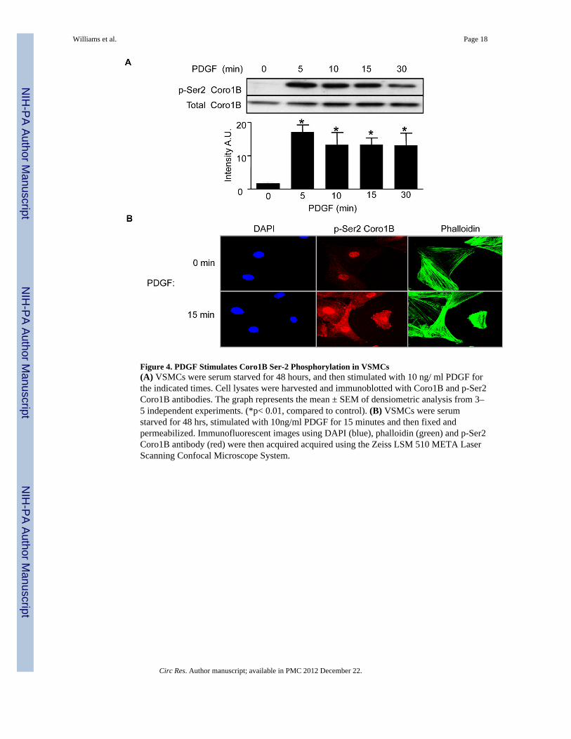
Figure 4. PDGF Stimulates Coro1B Ser-2 Phosphorylation in VSMCs(A) VSMCs were serum starved for 48 hours, and then stimulated with 10 ng/ ml PDGF forthe indicated times. Cell lysates were harvested and immunoblotted with Coro1B and p-Ser2Coro1B antibodies. The graph represents the mean ± SEM of densiometric analysis from 3–5 independent experiments. (*p< 0.01, compared to control). (B) VSMCs were serumstarved for 48 hrs, stimulated with 10ng/ml PDGF for 15 minutes and then fixed andpermeabilized. Immunofluorescent images using DAPI (blue), phalloidin (green) and p-Ser2Coro1B antibody (red) were then acquired acquired using the Zeiss LSM 510 META LaserScanning Confocal Microscope System.
Williams et al. Page 18
Circ Res. Author manuscript; available in PMC 2012 December 22.
NIH
-PA Author Manuscript
NIH
-PA Author Manuscript
NIH
-PA Author Manuscript

Figure 5. siRNA Against PKCε Attenuates Coro1B Five Minute Phosphorylation(A) VSMCs were serum starved for 48 hours, and then pre-incubated with 0.5 μMRo-32-0432 (PKC inhibitor) or vehicle for 30 min. The cells were then stimulated with 10ng/ml PDGF for 5 minutes. Cell lysates were harvested and immunoblotted with Coro1Band p-Ser2 Coro1B antibody. The graph represents the mean ± SEM of densiometricanalysis from 4 independent experiments. (*p< 0.01 compared to control, = p<0.0001compared to control PDGF treated). (B) VSMCs were transfected with siNegCtrl or siPKCεusing the Amaxa electroporation system. Cells were serum starved for 48 hours and thenstimulated with 10 ng/ml PDGF for 5 minutes. Cell protein lysates were harvested andimmunoblotted with p-Ser2 Coro1B, Coro1B and PKCε antibodies. Blots presented are fromthe same gel, but unrelated lanes were removed for clarity. The graph represents the mean ±SEM of densiometric analysis from 4 independent experiments. (*p< 0.0001, siNeg Ctrl vs.PDGF stimulated siNegCtrl, and = p< 0.01, PDGF stimulated siNegCtrl vs, PDGFstimulated siPKCε transfected).
Williams et al. Page 19
Circ Res. Author manuscript; available in PMC 2012 December 22.
NIH
-PA Author Manuscript
NIH
-PA Author Manuscript
NIH
-PA Author Manuscript

Figure 6. Coronin Over Expression and Phosphorylation Deficient Mutant Decrease MigrationVSMCs were transfected with the Myc tagged Coro1B WT or S2A phospho-deficientmutant using the Amaxa electroporation system and then serum starved for 24hrs. Cellswere harvested and migration was assessed via modified Boyden chamber assay (A). Thisgraph represents the mean ± SEM of the number of cells migrated per field (*p< 0.001,pcDNA3 vs pcDNA3 with PDGF stimulation, = p < 0.01, PDGF stimulated pcDNA3 vs.PDGF stimulated Coro1B WT transfected cells, and # p < 0.001, PDGF stimulated pcDNA3vs. PDGF stimulated Coro1B S2A mutant transfected cells N=4). (B) Protein lysates fromexcess cells not used in the migration assay were harvested and immunoblotted with Mycand Coro1B to verify Coro1B WT and S2A protein expression. (C) VSMCs weretransfected with Myc tagged S2A Coro1B, serum starved for 24 hrs, then fixed andpermeabilized. Immunofluorescent images using DAPI (blue), phalloidin (green), Coro1Bantibody (red) and Myc antibody (magenta) were then acquired acquired using the ZeissLSM 510 META Laser Scanning Confocal Microscope System.
Williams et al. Page 20
Circ Res. Author manuscript; available in PMC 2012 December 22.
NIH
-PA Author Manuscript
NIH
-PA Author Manuscript
NIH
-PA Author Manuscript

Figure 7. ARP2/3 subunit ARPC2 and Coro1B Interactions are Negatively Regulated by PDGFand are Dependent on the Phosphorylation State of Coro1B in VSMCs(A) VSMCs were serum starved for 24 hrs and then treated with 10 ng/ml PDGF for theindicated times. Cell lysates were harvested and immunoprecipitated with Coro1B antibody.Membranes were immunoblotted with Coro1B and ARPC2. The graph represents the mean± SEM of densiometric analysis from 3–4 independent experiments. Data are plotted as totalARPC2/ Coro1B. (*p< 0.05 compared to control). (B) VSMCs were transfected with theMyc tagged Coro1B WT, the S2A Coro1B phospho-deficient mutant, or the S2D Coro1Bphospho-mimetic mutant using the Amaxa electroporation system and then serum starvedfor 24hrs. Cell lysates were harvested and immunoprecipitated with Myc antibody.Membranes were immunoblotted with anti-Myc and ARPC2. The graph represents the mean± SEM of densiometric analysis from 4 independent experiments. Data are plotted as totalARPC2/Myc. (+ p< 0.05 compared to S2A).
Williams et al. Page 21
Circ Res. Author manuscript; available in PMC 2012 December 22.
NIH
-PA Author Manuscript
NIH
-PA Author Manuscript
NIH
-PA Author Manuscript

Figure 8. Coro1B is Phosphorylated in Response to Wire Injury in the Mouse Carotid Arteryand in the Neointima After Rat Carotid Balloon Injury(A) Mice were subjected to left carotid wire injury and then sacrificed 7 or 14 days postsurgery. The left carotids were collected and the right carotid was used as a control. Tissuewas harvested for protein and immunoblotted with p-Ser2 Coro1B and Coro1B antibodies(N=3 *p< 0.05 compared to control). (B) Sprague-Dawley rats were subjected to ballooninjury of the left common carotid artery. Ten days after injury, arteries were harvested,embedded in OTC and then cut into 7μm sections. Sections were stained for p-Ser2 Coro1B(magenta, panels a and b) or α smooth muscle actin (red, panels c and d) and nuclei (DAPI,blue, all panels) Green represents the autofluorescence of the internal elastic lamina. Theblood vessel is labeled as follows; I denotes intimal region, M denotes medial region, and Adenotes the adventitia.
Williams et al. Page 22
Circ Res. Author manuscript; available in PMC 2012 December 22.
NIH
-PA Author Manuscript
NIH
-PA Author Manuscript
NIH
-PA Author Manuscript