Roche Tissue Diagnostics Featuring the VENTANA … Tissue Diagnostics Featuring the VENTANA Total...
164
Roche Tissue Diagnostics Featuring the VENTANA Total Solution Product Catalog 2017
Transcript of Roche Tissue Diagnostics Featuring the VENTANA … Tissue Diagnostics Featuring the VENTANA Total...
Roche Tissue Diagnostics Featuring the VENTANA Total Solution
Product Catalog 2017
Roc
he T
issu
e D
iagn
ostic
s •
Pro
duct
Cat
alog
201
7


Table of Contents Introduction
Roche Diagnostics . . . . . . . . . . . . . . . . . . . . . . . . . . . . . . . . . . . . . . . . 3-5
What’s New . . . . . . . . . . . . . . . . . . . . . . . . . . . . . . . . . . . . . . . . . . . . . . 6-8
Antibodies
Companion and Complementary Diagnostics . . . . . . . . . . . . . . . . . 9-12
Primary Antibodies . . . . . . . . . . . . . . . . . . . . . . . . . . . . . . . . . . . . . 13-108
IHC/ISH Detection . . . . . . . . . . . . . . . . . . . . . . . . . . . . . . . . . . . . . 109-114
Special Staining Kits . . . . . . . . . . . . . . . . . . . . . . . . . . . . . . . . . . . 115-126
Instruments
H&E Staining . . . . . . . . . . . . . . . . . . . . . . . . . . . . . . . . . . . . . . . . . 128-129
IHC/ISH Automation . . . . . . . . . . . . . . . . . . . . . . . . . . . . . . . . . . . 130-133
Research Platforms . . . . . . . . . . . . . . . . . . . . . . . . . . . . . . . . . . . . 134-135
Special Stains . . . . . . . . . . . . . . . . . . . . . . . . . . . . . . . . . . . . . . . . . 136-137
Digital Pathology . . . . . . . . . . . . . . . . . . . . . . . . . . . . . . . . . . . . . . 138-144
Workflow Solutions . . . . . . . . . . . . . . . . . . . . . . . . . . . . . . . . . . . . 145-146
Customer Experience
Workflow Consulting . . . . . . . . . . . . . . . . . . . . . . . . . . . . . . . . . . . . . . . 148
Roche Diagnostics University . . . . . . . . . . . . . . . . . . . . . . . . . . . . . . . . 149
Roche Support Network . . . . . . . . . . . . . . . . . . . . . . . . . . . . . . . . . . . . 150
CareGiver Remote Support . . . . . . . . . . . . . . . . . . . . . . . . . . . . . . . . . . 151
Reimbursement . . . . . . . . . . . . . . . . . . . . . . . . . . . . . . . . . . . . . . . . . . . 152
Appendices
Index . . . . . . . . . . . . . . . . . . . . . . . . . . . . . . . . . . . . . . . . . . . . . . . . 154-157
Trademarks and Disclaimers . . . . . . . . . . . . . . . . . . . . . . . . . . . . . . . . 158


INTRODUCTION
1
Legacy Dedicated to improving
the lives of all patients afflicted with cancer
Roche Tissue Diagnostics

INTRODUCTION
2

INTRODUCTION
3
Roche Diagnostics Delivering medical value and testing efficiency
Research Clinical Applications
Academia
Sequencing Solutions DiabetesCare
CoagulationMonitoring
Molecular Diagnostics
Tissue Diagnostics
Professional Diagnostics
Pharma Reference Lab Hospital Doctor’s Office Patient Homes
Spanning the entire spectrum of diagnostics users
Roche is a leader in research-focused healthcare with combined strengths in diagnostics and pharmaceuticals . The world’s largest biotech company, Roche is the world leader in in vitro diagnostics and tissue-based cancer diagnostics and is a frontrunner in diabetes management .
Leadership in DiagnosticsLeadership in Pharma
Combined strengths of the Roche Group

INTRODUCTION
4
Roche Tissue Diagnostics Providing what your lab needs to advance patient care
Like you, we’re passionate about the patient behind every slide . And we’re committed to empowering your lab with advanced solutions to transform patient care . As part of the world’s leading healthcare company, we’re dedicated to helping pathology professionals deliver accurate, reliable results that help inform decision making to advance patient care .
Ensuring the identity of David’s specimen
VENTANA VANTAGE Workflow SolutionComplete chain of custody to mitigate risk of misidentification
Protecting the integrity of Sue’s slide
VENTANA HE 600 systemIndividual H&E slide staining to help you safeguard your patients
Steering Leslie’stherapy in the right direction
VENTANA BenchMark Series Staining PlatformsBroad assay menu to help you deliver diagnostic confidence
Pathologist founded, patient focused
Building on the vision of Dr . Thomas Grogan, Roche Tissue Diagnostics continues to drive diagnostic innovation that transforms the practice of medicine to improve patients’ lives .

INTRODUCTION
5
Roche Tissue Diagnostics is transforming cancer diagnosis with the VENTANA total solution, addressing the needs of anatomic pathology labs and healthcare professionals to help them improve the lives of the patients they serve . Our broad portfolio includes integrated staining and workflow management platforms, digital pathology solutions and a robust menu of high- medical-value assays that help labs increase efficiency, improve patient safety and deliver medical value .
From the time a patient specimen arrives to delivery of test results, the VENTANA total solution enables your lab to provide accurate and reliable results in a timely manner . This helps patients get the right test, for the right treatment, at the right time .
The VENTANA total solution Empowering your lab from sample accessioning through case sign-out
Helping Sonia get the right test at the right time
VENTANA Companion DiagnosticsSo you can deliver the promise of personalized healthcare
Bringingconfidence to Sam’s diagnosis
VENTANA Digital Pathology SolutionsTransforming tissue diagnostics to help you improve patient care
Giving Robert hope for a breakthrough
VENTANA DISCOVERY Automated Staining Platforms Flexibility and innovation to accelerate your IHC/ISH research
DISCOVERY products are for research use only. Not for use in diagnostic procedures.

INTRODUCTION
6
What’s New Improving lives through ongoing innovation
VENTANA PD-L1 (SP142) Assay Guiding immunotherapy The first FDA-approved test predictive for TECENTRIQ® (atezolizumab) in urothelial carcinoma (UC) patients . Now also FDA approved to assess non-small cell lung cancer (NSCLC) patient treatment benefit from TECENTRIQ.
Learn more at PDL1ihc.com
Hiker’s path: VENTANA PD-L1 (SP142) Assay on non-small cell lung cancer tissue Location: Point Conception, CA
PD-L1
VENTANA PD-L1 (SP142) AssayGuiding immunotherapy in NSCLC
VENTANA PD-L1 (SP142) Assay50 tests Ordering Code Cat. No. 07709374001 740-4859
See complete product details on page 91.
As part of Roche, we’re continuously investing in diagnostic tools that address unmet medical needs and provide information to help advance patient care.

INTRODUCTION
7
VENTANA ALK (D5F3) CDx Assay 50 tests Ordering Code Cat. No. 06687199001 790-4796
See complete product details on page 19.
anti-p40 (BC28) Mouse Monoclonal Primary Antibody
50 tests Ordering Code Cat. No. 07394420001 790-4950
See complete product details on page 87.
SOX-10 (SP267) Rabbit Monoclonal Primary Antibody 50 tests Ordering Code Cat. No. 07560389001 760-4968
See complete product details on page 98.
Desmoglein 3 (5G11) Mouse Monoclonal Primary Antibody50 tests Ordering Code Cat. No. 07709447001 760-5060
See complete product details on page 51.
NKX3.1 (EP356) Rabbit Monoclonal Primary Antibody
50 tests Ordering Code Cat. No. 07859759001 760-5086
See complete product details on page 85.
NOW
FDA Approved
for use on BenchMark
ULTRA system
For current product information, including the newest assays, visit usdiagnostics.roche.com/en/tissue/overview.html

INTRODUCTION
8
Olig2 (EP112) Rabbit Monoclonal Primary Antibody50 tests Ordering Code Cat. No. 07667973001 760-5050
See complete product details on page 86.
VENTANA PTEN (SP218) Rabbit Monoclonal Primary Antibody
50 tests Ordering Code Cat. No. 07970200001 790-5097
See complete product details on page 95.
VENTANA PD-L1 (SP263) Rabbit Monoclonal Primary Antibody50 tests Ordering Code Cat. No. 07494190001 790-4905
See complete product details on page 91.
HPV mRNA ISH Probes
RNA probes for detection of specific HPV mRNA localization in formalin-fixed, paraffin-embedded (FFPE) tissue, for use in laboratory developed tests .
Ordering Code Cat. No. HPV 6 mRNA Probe 07658877001 760-1239 HPV 11 mRNA Probe 07658885001 760-1240 HPV 16 mRNA Probe 07658834001 760-1236 HPV 18 mRNA Probe 07658842001 760-1237 HPV 33 mRNA Probe 07658869001 760-1238
For current product information, including the newest assays, visit usdiagnostics.roche.com/en/tissue/overview.html
HPV 16 mRNA Probe, 40x magnification
�Analyte�Specific�Reagents.�Analytical�and� performance characteristics are not established.

Companion and Complementary Diagnostics
Transformative Pioneering solutions that drive
personalized healthcare

10
COMPANION AND COMPLEMENTARY DIAGNOSTICS
Companion Diagnostics Selective and predictive assays essential for personalized healthcare
A call to personalize the practice of medicine
“ Personalized healthcare is not holding a patient’s hand. It’s holding the intimate details of a patient’s test results to a brighter light and asking, ‘What am I missing?’”
— Tom Grogan, M.D., pathologist and founder of Ventana Medical Systems, Inc.
Linking the most accurate diagnosis with the most targeted and relevant therapeutic is the essence of personalized healthcare, offering the potential for more positive outcomes for the patient—saving lives and improving the quality of life.
Our commitment to helping you deliver personalized healthcare
In our mission to improve the lives of all patients afflicted with cancer, our goal is to empower you with the highest quality, most innovative tools to help deliver on the promise of personalized diagnostics and treat-ment. We’re committed to accelerating the development of new cancer tests that can help improve and extend patients’ lives by spanning the gap between generic and personalized treatment.
“ Targeted diagnostic tests that help to improve medical decision-making not only offer clinical benefits for patients but are also attractive through health economic benefits to regulatory authorities and payers.”
— Severin Schwan, CEO, Roche
Helping Sonia get the right test at the right time
Fast, reliable and standardized diagnostic tests
can help physicians assess treatment options
for cancer patients like Sonia. By identifying
patients who express specific biomarkers,
VENTANA companion diagnostic assays
provide predictive information that aids in
the choice of therapies. Targeted diagnostics
and treatments offer cancer patients the
potential for fewer side effects, improved
quality of life, and better outcomes. With
companion diagnostics helping patients get
the right treatment at the right time, the
promise of personalized healthcare becomes
a reality.
———————— ~ ————————
11
COMPANION AND COMPLEMENTARY DIAGNOSTICS
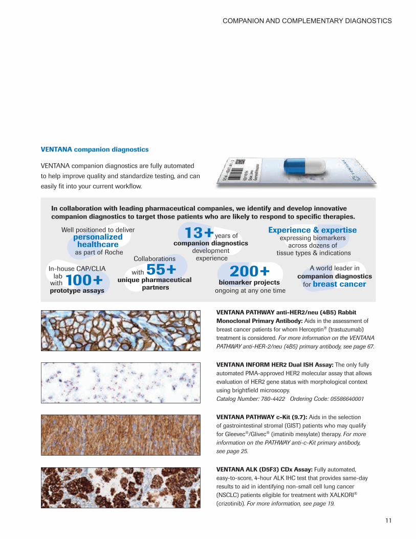
VENTANA companion diagnostics
VENTANA companion diagnostics are fully automated to help improve quality and standardize testing, and can easily fit into your current workflow.
VENTANA PATHWAY anti-HER2/neu (4B5) Rabbit Monoclonal Primary Antibody: Aids in the assessment of breast cancer patients for whom Herceptin® (trastuzumab) treatment is considered. For more information on the VENTANA PATHWAY anti-HER-2/neu (4B5) primary antibody, see page 67.
VENTANA INFORM HER2 Dual ISH Assay: The only fully automated PMA-approved HER2 molecular assay that allows evaluation of HER2 gene status with morphological context using brightfield microscopy. Catalog Number: 780-4422 Ordering Code: 05586640001
VENTANA PATHWAY c-Kit (9.7): Aids in the selection of gastrointestinal stromal (GIST) patients who may qualify for Gleevec®/Glivec® (imatinib mesylate) therapy. For more information on the PATHWAY anti-c-Kit primary antibody, see page 25.
VENTANA ALK (D5F3) CDx Assay: Fully automated, easy-to-score, 4-hour ALK IHC test that provides same-day results to aid in identifying non-small cell lung cancer (NSCLC) patients eligible for treatment with XALKORI® (crizotinib). For more information, see page 19.
A world leader in companion diagnostics
for breast cancer
In collaboration with leading pharmaceutical companies, we identify and develop innovative companion diagnostics to target those patients who are likely to respond to specific therapies.
Well positioned to deliver personalized healthcare
as part of RocheCollaborations
with 55+ unique pharmaceutical
partners
In-house CAP/CLIA lab
with 100+ prototype assays
13+years of companion diagnostics
development experience
200+biomarker projects
ongoing at any one time
Experience & expertise expressing biomarkers
across dozens of tissue types & indications

12
COMPANION AND COMPLEMENTARY DIAGNOSTICS
Complementary Diagnostics Important information for drug-benefit decisions
Insights to guide immunotherapy decisions
Complementary diagnostic assays expand the array of tools available to help drive personalized healthcare and enable better outcomes for patients.
While not required for safe and effective use of a therapeutic drug, complementary diagnostic assays may provide additional information that helps physicians make informed treatment decisions.
VENTANA complementary diagnostics
The VENTANA PD-L1 (SP142) Assay is fully automated to help provide reliable, accurate results to enable timely diagnostic decisions and therapeutic choices.
VENTANA PD-L1 (SP142) Assay: The first and only FDA-approved test predictive for TECENTRIQ® (atezolizumab) in urothelial carcinoma (UC) patients. Now also FDA approved to assess non-small cell lung cancer (NSCLC) patient treat-ment benefit from TECENTRIQ®. Catalog Number: 740-4859 Ordering Code: 07709374001For more information on the VENTANA PD-L1 (SP142) Assay, see page 91.
Right therapy, for the right patient, at the right time
By helping identify patients that are more likely to benefit from a specific therapy, complementary diagnostics aid in the choice of specific therapies for each patient.

Primary Antibodies
Breadth A vast product portfolio
supported by the resources and global reach of a leading healthcare company

PRIMARY ANTIBODIES
14
Ready-to-Use Primary Antibodies More than 250 prediluted antibodies, optimized for use on VENTANA staining platforms
Featuring a world-class breast panel as well as corner-stone and emerging markers for lung diagnostics and other disease states, our comprehensive portfolio of high-value assays empowers pathology labs to deliver accurate, timely results with clinical confidence.
• Improvetestingefficiency:Our prediluted antibodies are optimized for use on the fully automated VENTANA BenchMark staining platforms, reducing time-to-result and the resources required with manual or semi-automated solutions.
• Countonconsistentperformance:Roche antibodies have consistently demonstrated (via in-house validation) superior performance when compared to concentrated antibodies that require manual dilution.1
• Choosefromacomprehensiveportfolio: We offer more than 250 ready-to-use Class I assays, including the largest menu of rabbit monoclonal IVD antibodies available.
Breast
n ER (SP1)n PR (1E2)n HER-2/neu (4B5)n Ki-67 (30-9)n p53 (DO-7) n HER2 Dual ISH
Lung
n TTF-1 (SP141)n Cytokeratin 5/6 (D5/16B4)n Napsin A (MRQ-60)n p40 (BC28)
Colorectal
n MLH-1 (M1)n MSH2 (G219-1129)n MSH6 (44)n PMS2 (EPR3947)n BRAF-V600E (VE1)n CDX-2 (EPR2764Y)
Ourbroadportfolioincludessensitive,specificassaysforkeymarkers.Turnthepageforacompletelistingofour primary antibodies and other IHC/ISH reagents by disease state.

PRIMARY ANTIBODIES
15
TherabbitmonoclonaldifferenceSuperior sensitivity and specificity for consistent, high-quality staining
Demonstrating a better signal-to-noise ratio compared to mouse monoclonal and rabbit polyclonal antibodies,2 rabbit monoclonals:
• Target a specific epitope
• Provide greater diagnostic specificity
• Reduce background for cleaner slides
• Provide greater consistency from lot to lot
References:
1. Assessment Run B13 2012. Estrogen receptor (ER). NordiQC. http://www.nordiqc.org/Run-35-B13-H1/ Assessment/assessment-B13-ER.htm. Updated July 10, 2012. Accessed May 23, 2013.
2. Spieker-Polet H, Sethupathi P, Yam PC, Knight KL. Rabbit monoclonal antibodies: Generating a fusion partner to produce rabbit-rabbit hybridomas. Proc Natl Acad Sci U S A. 1995; 92(20):9348-9352.
SteeringLeslie’stherapy intherightdirection
VENTANA high-medical-value assays
provide biomarker analysis information
that can help drive targeted treatment
for cancer patients. Our assays empower
pathology labs to deliver accurate, high-
quality results that help clinicians gain
insights for diagnosis and treatment
decisions. Every cancer patient’s journey
is unique. Our advanced diagnostics can
lead to personalized treatment that helps
give patients like Leslie a fighting chance.
———————— ~ ————————

PRIMARY ANTIBODIES
16
Breast
TypicalBreast PanelAssays
Estrogen Receptor (ER) (SP1), CONFIRMHER-2/neu (4B5), PATHWAYHER2 Dual ISH DNA Probe Cocktail, INFORMKi-67 (30-9), CONFIRMp53 (DO-7), CONFIRMProgesterone Receptor (PR) (1E2), CONFIRM
Calponin-1 (EP798Y)Cytokeratin 14 (SP53)Cytokeratin 5/6 (D5/16B4)Cytokeratin 5/14 (EP1601Y/LL002)E-cadherin (36), CONFIRME-cadherin (EP700Y)FOXA1 (2F83)GATA3 (L50-823)GCDFP-15 (EP1582Y)p120 (98)Topoisomerase IIa (JS5B4), CONFIRM
ColorectalandGastrointestinal
TypicalColorectal PanelAssays
BRAF-V600E (VE1)CDX-2 (EPR2764Y)MLH-1 (M1)MSH2 (G219-1129)MSH6 (44), CONFIRMPMS2 (EPR3947)
TypicalGastrointestinalPanelAssaysBRAF-V600E (VE1)c-KIT (9.7), PATHWAYCDX-2 (EPR2764Y)DOG1 (SP31)Helicobacter pylori (SP48), VENTANAMLH-1 (M1)MSH2 (G219-1129)MSH6 (44), CONFIRMPMS2 (EPR3947)
Beta-catenin (14)Cadherin 17 (SP183)CEA (TF3H8-1)COX-2 (SP21)Cytokeratin 7 (SP52), CONFIRMCytokeratin 19 (A53-B/A2.26)Cytokeratin 20 (SP33), CONFIRMGlutamine Synthetase (GS-6)MUC1 (H23)MUC2 (MRQ-18)
Dermatopathology
TypicalMelanoma PanelAssays
MART-1/melan A (A103), CONFIRMMelanoma Associated Antigen (KBA.62)Melanoma Associated Antigen (PNL2)Melanosome (HMB45), CONFIRMMITF (C5/D5), CONFIRM S100 (4C4.9), CONFIRMS100 (Polyclonal), CONFIRMSOX-10 (SP267) Albumin, FITCa-1-Antichymotrypsin (ACT)a-1-Antitrypsin (AAT)CEA (TF3H8-1)CD2 (MRQ-11)
CD3 (2GV6), CONFIRMCD31 (JC70)CD34 (QBEnd/10), CONFIRMCD63 (NKI/C3)Cytokeratin (AE1), CONFIRMCytokeratin 8 & 18 (B22.1 & B23.1)Desmin (DE-R-11), CONFIRMEMA (E29), CONFIRMEp-CAM (Epithelial Specific Antigen) (Ber-EP4)Factor VIII Related AntigenFactor XIIIa (AC-1A1)Factor XIIIa (EP3372)C1q, FITCC3, FITCC4, FITCFibrinogen, FITCKappa, FITCLambda, FITCHHV-8 (Human Herpes Virus Type 8) (13B10)
IgA (Immunoglobulin A)IgA (Immunoglobulin A), FITCIgG (Immunoglobulin G)IgG (Immunoglobulin G), FITCIgM (Immunoglobulin M)IgM (Immunoglobulin M), FITCMacrophage (HAM-56)Melanoma Triple Cocktail (A103, HMB45, T311)Neurofilament (2F11)p53 (DO-7), CONFIRMp53 (Bp53-11)Podoplanin (D2-40)Synaptophysin (MRQ-40)Synaptophysin (SP11), CONFIRMTryptase (G3)Tyrosinase (T311), CONFIRMVimentin (V9), CONFIRMVimentin (Vim 3B4), CONFIRM
IHC/ISH Reagents by Disease State Extensive menu of ready-to-use clinical reagents, optimized for use on VENTANA BenchMark IHC/ISH staining platforms

PRIMARY ANTIBODIES
17
Hematopathology
TypicalHematopathology PanelAssays
bcl-2 (SP66)c-Myc (Y69)CD3 (2GV6), CONFIRMCD10 (SP67), VENTANACD20 (L26), CONFIRMCD30 (Ber-H2)Cyclin D1 (SP4-R) ALK1 (ALK01), CONFIRMAnnexin A1 (MRQ-3) bcl-2 (124), CONFIRMbcl-6 (GI191E/A8)BOB.1 (SP92)CD1a (EP3622)CD2 (MRQ-11)CD4 (SP35), CONFIRMCD5 (SP19), CONFIRMCD7 (SP94)CD8 (SP57), CONFIRMCD13 (SP187)CD14 (EPR3653)
CD15 (MMA), CONFIRMCD16 (SP175)CD22 (SP104)CD23 (SP23), CONFIRMCD25 (4C9)CD31 (JC70)CD33 (SP266)CD34 (QBEnd/10), CONFIRMCD43 (L60)CD45 (LCA) (2B11 & PD7/26)CD45 (LCA) (RP2/18), CONFIRMCD45R (MB1)CD45RO (UCHL-1), CONFIRMCD56 (123C3), CONFIRMCD56 (MRQ-42)CD57 (NK-1)CD61 (2f2)CD68 (KP-1), CONFIRMCD71 (MRQ-48)CD79a (SP18), CONFIRMCD99 (O13), CONFIRMCD138 (Syndecan-1) (B-A38)Fascin (55k-2)Galectin-3 (9C4)Glycophorin A (GA-R2)
Granzyme BHemoglobin A (SP212)HGAL (MRQ-49)IgA (Immunoglobulin A)IgD (Immunoglobulin D)IgG (Immunoglobulin G)IgM (Immunoglobulin M)Kappa, CONFIRMLambda, CONFIRMLMO2 (1A9-1), CONFIRMLMO2 (SP51)LysozymeMUM1 (MRQ-43)MyeloperoxidaseNeutrophil Elastase (SP203)Oct-2 (MRQ-2)PAX5 (SP34), CONFIRMPD-1 (NAT-105)SOX-11 (MRQ-58)Spectrin (RBC2/3D5)T-bet (MRQ-46)TdTTRAcP (9C5)ZAP-70 (2F3.2)
Lung
TypicalLung PanelAssays
Cytokeratin 5/6 (D5/16B4)Napsin A (MRQ-60)p40 (BC28)Thyroid Transcription Factor-1 (SP141) ALK (D5F3)c-MET Total (SP44), CONFIRMCalretinin (SP65), CONFIRMCarcinoembryonic Antigen (CEA) (TF3H8-1)Caveolin-1 (SP43)CD56 (123C3), CONFIRM
CD56 (123C3), CONFIRMCD56 (MRQ-42)Chromogranin A (LK2H10)Cytokeratin (CAM 5.2)Cytokeratin 5 (SP27)Cytokeratin 5/14 (EP1601Y/LL002)Cytokeratin 7 (SP52), CONFIRMCytokeratin 17 (SP95)Cytokeratin 20 (SP33), CONFIRMDesmoglein 3 (5G11) E-cadherin (36), VENTANAE-cadherin (EP700Y)EGFR E746-A750 del (SP111)EGFR (5B7), CONFIRMEGFR (3C6), CONFIRM
EGFR L858R (SP125)EMA (E29), CONFIRMEpithelial-Related Antigen (MOC-31)Ep-CAM (Epithelial Specific Antigen) (Ber-EP4)IGF-1R(G11), CONFIRMMesothelial Cell HBME-1 (HBME-1)MUC1 (H23)NSE (MRQ-55)p63 (4A4), VENTANAPD-L1 (SP263)SOX-2 (SP76)Synaptophysin (MRQ-40)Synaptophysin (SP11), CONFIRMTAG-72 (B72.3)WT1 (6F-H2)
Prostate
TypicalProstate PanelAssays
Basal Cell Cocktail (34βE12+p63), VENTANACytokeratin 5/6 (D5/16B4)p63 (4A4), VENTANAPSA, CONFIRMPSAP (PASE/4LJ)
Androgen Receptor (SP107)Cytokeratin (34βE12)Cytokeratin 7 (SP52), CONFIRMCytokeratin 20 (SP33), CONFIRMERG (EPR3864)EZH2 (SP129)NKX3.1 (EP356) PTEN (SP218)
The identified typical antibodies and panels listed for disease state stratification are for informational use only and no medical value or intended use claims are being made. For that information, please refer to the specific product package insert.

PRIMARY ANTIBODIES
18
A-1-Antichymotrypsin(polyclonal)
CatalogNumber: 760-2604 OrderingCode: 05267196001 Quantity: 50 tests Controls: Histiocytic Tumors CloneName: Polyclonal Species: Rabbit Localization: Cytoplasmic RegulatoryStatus: IVD
INTENDEDUSE
Alpha-1-Antichymotrypsin Primary Antibody reacts with histiocytes and histiocytic neoplasms. Its major application is defining the presence of Alpha-1-Antichymotrypsin in histiocytes and tumors derived from them. In eosinophilic granuloma and malignant histiocytosis, the reaction for this marker is heterogeneous in intensity and distribution. In fibrous histiocytomas on the other hand, a diffuse homogeneous reaction may be observed. Acinar tumors of the pancreas and salivary gland may also exhibit Alpha-1-Antichymotrypsin positivity.
A-1-Antitrypsin(polyclonal)
CatalogNumber: 760-2605OrderingCode: 05267200001Quantity: 50 testsControls: Appendix, Lymph Node, TonsilCloneName: PolyclonalSpecies: RabbitLocalization: CytoplasmicRegulatoryStatus: IVD
INTENDEDUSE
The immunohistochemical staining of Alpha-1-Antitrypsin is considered to be very useful in the study of inherited AAT deficiency, benign and malignant hepatic tumors and yolk sac carcinomas. Positive staining for A-1-Antitrypsin may also be used in detection of benign and malignant lesions of an histiocytic nature. Sensitivity and specificity of the results have made this antibody a useful tool in the screening of patients with cryptogenic cirrhosis or other forms of liver disease with portal fibrosis of uncertain etiology.
ACTH(polyclonal)
CatalogNumber: 760-2708OrderingCode: 05268176001Quantity: 50 testsControls: Normal PituitaryCloneName: PolyclonalSpecies: RabbitLocalization: CytoplasmicRegulatoryStatus: IVD
INTENDEDUSE
Anti-ACTH is a useful marker in classification of pituitary tumors and the study of pituitary disease. It reacts with ACTH-producing cells (corti-cotrophs). It also may react with other tumors (e.g., some small cell carcinomas of the lung) causing paraneoplastic syndromes by secreting ACTH.
Actin,MuscleSpecific(HHF35)
CatalogNumber: 760-2601OrderingCode: 05267161001Quantity: 50 testsControls: Skeletal MuscleIsotypes: IgG1/KCloneName: HHF35Species: MouseLocalization: CytoplasmicRegulatoryStatus: IVD
INTENDEDUSE
Actin is a major component of the cytoskeleton. This antibody recognizes actin of skeletal, cardiac, and smooth muscle cells. It is not reactive with other mesenchymal cells except for myoepithelium. Actin can be resolved on the basis of its isoelectric points into three distinctive components: alpha, beta, and gamma in order of increasing isoelectric point. Anti-Muscle-Specific Actin recognizes alpha and gamma isotypes of all muscle groups. Non-muscle cells such as vascular endothelial cells and connective tissues are non-reactive. Also, neoplastic cells of non-muscle-derived tissue such as carcinomas, melanomas, and lymphomas are negative. This antibody is useful in the identification of rhabdoid cellular elements.

PRIMARY ANTIBODIES
19
Actin,SmoothMuscle(1A4)Mouse MonoclonalAntibody
CatalogNumber: 760-2833OrderingCode: 05268303001Quantity: 50 testsControls: Appendix, UterusIsotypes: IgGCloneName: 1A4Species: MouseLocalization: CytoplasmicRegulatoryStatus: IVD
INTENDEDUSE
This antibody is intended for in vitro diagnostic (IVD) use. The Cell Marque Actin, Smooth Muscle (1A4) antibody is intended for qualified laboratories to qualitatively identify by light microscopy the presence of associated antigens in sections of formalin-fixed, paraffin-embedded tissue sections using IHC test methods. Use of this antibody is indicated as an aid in the identification and diagnosis of smooth muscle and myoepithelial tumors within the context of an antibody panel, the patient’s clinical history, and other diagnostic tests evaluated by a qualified pathologist.
anti-Actin,Muscle(HUC1-1) Primary Antibody
CatalogNumber: 760-2502OrderingCode: 05266882001Quantity: 50 testsControls: Skeletal MuscleIsotypes: IgG1CloneName: HUC1-1Species: MouseLocalization: CytoplasmicRegulatoryStatus: IVD
INTENDEDUSE
This antibody is intended for in vitro diagnostic (IVD) use. Ventana Medical Systems’ (Ventana) anti-Actin, Muscle (HUC 1-1) may be used to aid in the identification of cells of normal and abnormal myocytic lineage as an aid in diagnosis of anaplastic tumors. Anti-Actin, Muscle (HUC 1-1) specifically binds to antigens located in the cytoplasm of myocytic cells. This antibody is intended for laboratory use to qualitatively identify by light microscopy muscle actin in sections of formalin-fixed, paraffin-embedded tissue on a VENTANA automated slide stainer. The clinical interpretation of any staining, or the absence of staining, must be complemented by morphological studies and evaluation of proper controls. Evaluation must be made by a qualified pathologist within the context of the patient’s clinical history and other diagnostic tests. Prescription only.
VENTANAALK(D5F3)CDxAssay
CatalogNumber: 790-4796OrderingCode: 06687199001Quantity: 50 testsControls: Appendix Isotypes: IgGCloneName: D5F3Species: Rabbit Monoclonal Localization: Cytoplasmic RegulatoryStatus: IVD, FDA Approved (PMA)
INTENDEDUSE
VENTANA ALK (D5F3) CDx Assay is intended for the qualitative detection of the anaplastic lymphoma kinase (ALK) protein in formalin-fixed, paraffin-embedded (FFPE) non-small cell lung carcinoma (NSCLC) tissue stained with a BenchMark XT or BenchMark ULTRA automated staining instrument. It is indicated as an aid in identifying patients eligible for treatment with XALKORI® (crizotinib). This product should be interpreted by a qualified pathologist in conjunction with histological examination, relevant clinical information, and proper controls. This product is intended for in vitro diagnostic (IVD) use.

PRIMARY ANTIBODIES
20
CONFIRManti-ALK1(ALK01) Primary Antibody
CatalogNumber: 790-2918OrderingCode: 05278147001Quantity: 50 testsControls: Anaplastic Large Cell LymphomaIsotypes: IgG3/KCloneName: ALK01Species: MouseLocalization: Nuclear, CytoplasmicRegulatoryStatus: IVD
INTENDEDUSE
This antibody is intended for in vitro diagnostic (IVD) use. Ventana Medical Systems’ (Ventana) CONFIRM anti-ALK1 (ALK01) Primary Antibody is a mouse monoclonal antibody directed against p80 protein present in human tissue. This antibody is intended for laboratory use to qualitatively stain sections of formalin-fixed, paraffin-embedded tissue on a VENTANA automated slide stainer. The clinical interpretation of any staining, or the absence of staining, must be complemented by morphological studies and evaluation of proper controls. Evaluation must be made by a qualified pathologist within the context of the patient’s clinical history and other diagnostic tests. Prescription only.
Alpha-FetoproteinRabbit PolyclonalAntibody
CatalogNumber: 760-2603OrderingCode: 05267188001Quantity: 50 testsControls: Fetal LiverCloneName: PolyclonalSpecies: RabbitLocalization: CytoplasmicRegulatoryStatus: IVD
INTENDEDUSE
This antibody is intended for in vitro diagnostic (IVD) use. The Cell Marque Alpha-Fetoprotein (Polyclonal) antibody is intended for qualified laborato-ries to qualitatively identify by light microscopy the presence of associated antigens in sections of formalin-fixed, paraffin-embedded tissue sections using IHC test methods. Use of this antibody is indicated, subsequent to clinical differential diagnoses of diseases, as an aid in the identification of liver disease, yolk sac and mixed germ cell tumors within the context of antibody panels, the patient’s clinical history, and other diagnostic tests evaluated by a qualified pathologist.
anti-AndrogenReceptor(SP107)Rabbit MonoclonalPrimaryAntibody
CatalogNumber: 760-4605OrderingCode: 06523838001Quantity: 50 testsControls: ProstateIsotypes: IgGCloneName: SP107Species: RabbitLocalization: NuclearRegulatoryStatus: IVD
INTENDEDUSE
This antibody is intended for in vitro diagnostic (IVD) use. The Cell Marque Androgen Receptor (SP107) antibody is intended for qualified laboratories to qualitatively identify by light microscopy the presence of associated antigens in sections of formalin-fixed, paraffin-embedded tissue sections using IHC test methods. This antibody is used as an aid in the identification of Androgen Receptor positivity in human tissues within the context of an antibody panel, the patient’s clinical history, and other diagnostic tests evaluated by a qualified pathologist.
PRIMARY ANTIBODIES
21
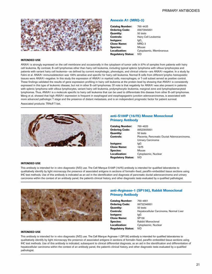
AnnexinA1(MRQ-3)
CatalogNumber: 760-4435OrderingCode: 05973945001Quantity: 50 testsControls: Hairy Cell LeukemiaIsotypes: IgG1CloneName: MRQ-3Species: MouseLocalization: Cytoplasmic, MembranousRegulatoryStatus: IVD
INTENDEDUSE
ANXA1 is strongly expressed on the cell membrane and occasionally in the cytoplasm of tumor cells in 97% of samples from patients with hairy cell leukemia. By contrast, B-cell lymphomas other than hairy cell leukemia, including typical splenic lymphoma with villous lymphocytes and patients with variant hairy cell leukemia—as defined by current morphologic, phenotypic, and clinical criteria—are ANXA1-negative. In a study by Falini et al. ANXA1 immunodetection was 100% sensitive and specific for hairy cell leukemia. Normal B cells from different lympho-hemopoietic tissues were ANXA1-negative. In this study the expression of ANXA1 in myeloid cells, macrophages, or T-cell subset served as positive control. These findings validated the results of gene expression profiling in hairy cell leukemia at the protein level by showing that ANXA1 is consistently expressed in this type of leukemic disease, but not in other B-cell lymphomas. Of note is that negativity for ANXA1 was also present in patients with splenic lymphoma with villous lymphocytes, variant hairy cell leukemia, prolymphocytic leukemia, marginal zone and lymphoplasmacytoid lymphomas. Thus, ANXA1 is a molecule specific to hairy cell leukemia that can be used to differentiate this disease from other B-cell lymphomas. Wang et al. showed that high ANXA1 expression is frequent in esophageal and esophagogastric junction adenocarcinomas, is associated with more advanced pathologic T stage and the presence of distant metastasis, and is an independent prognostic factor for patient survival.
Associated products: TRAcP, T-bet.
anti-S100P(16/f5)MouseMonoclonal Primary Antibody
CatalogNumber: 760-4620OrderingCode: 06523935001Quantity: 50 testsControls: Placenta, Pancreatic Ductal Adenocarcinoma,
Urinary CarcinomaIsotypes: IgGCloneName: 16/f5Species: MouseLocalization: Cytoplasmic, NuclearRegulatoryStatus: IVD
INTENDEDUSE
This antibody is intended for in vitro diagnostic (IVD) use. The Cell Marque S100P (16/f5) antibody is intended for qualified laboratories to qualitatively identify by light microscopy the presence of associated antigens in sections of formalin-fixed, paraffin-embedded tissue sections using IHC test methods. Use of this antibody is indicated as an aid in the identification and diagnosis of pancreatic ductal adenocarcinoma and urinary carcinoma within the context of an antibody panel, the patient’s clinical history, and other diagnostic tests evaluated by a qualified pathologist.
anti-Arginase-1(SP156),RabbitMonoclonalPrimary Antibody
CatalogNumber: 760-4801OrderingCode: 06732348001Quantity: 50 testsControls: Hepatocellular Carcinoma, Normal LiverIsotypes: IgGCloneName: SP156Species: Rabbit MonoclonalLocalization: Cytoplasmic, NuclearRegulatoryStatus: IVD
INTENDEDUSE
This antibody is intended for in vitro diagnostic (IVD) use. The Cell Marque Arginase-1 (SP156) antibody is intended for qualified laboratories to qualitatively identify by light microscopy the presence of associated antigens in sections of formalin-fixed, paraffin-embedded tissue sections using IHC test methods. Use of this antibody is indicated, subsequent to clinical differential diagnosis, as an aid in the identification and differentiation of hepatocellular carcinoma within the context of an antibody panel, the patient’s clinical history, and other diagnostic tests evaluated by a qualified pathologist.

PRIMARY ANTIBODIES
22
VENTANABasalCellCocktail(34βE12+p63)
CatalogNumber: 790-4536 790-1010OrderingCode: 06364497001 06419445001Quantity: 50 tests 250 testsControls: Normal Basal Cells in Prostate TissueIsotypes: IgG2a/KCloneName: 34βE12 + 4A4Species: MouseLocalization: Nuclear (4A4), Cytoplasmic (34βE12)RegulatoryStatus: IVD
INTENDEDUSE
VENTANA Basal Cell Cocktail (34βE12+p63) is an antibody cocktail of anti-p63 (4A4) and anti-keratin (34βE12). Anti-p63 (4A4) reacts with the p63 molecule in the nuclei of human prostatic basal cells and urothelial tissues. Anti-keratin (34βE12) reacts with cytokeratins 1, 5, 10 and 14, and stains the cytoplasm of human prostatic basal cells. This antibody cocktail may be used to aid in the differentiation of benign and malignant prostatic lesions. The antibody is intended for qualitative staining in sections of formalin-fixed, paraffin-embedded tissue. This product should be interpreted by a qualified pathologist in conjunction with histological examination, relevant clinical information, and proper controls. This antibody is intended for in vitro diagnostic (IVD) use.
BCA-225(Cu-18)
CatalogNumber: 760-2607OrderingCode: 05267226001Quantity: 50 testsControls: Breast CarcinomaIsotypes: IgG1CloneName: Cu-18Species: MouseLocalization: CytoplasmicRegulatoryStatus: IVD
INTENDEDUSE
Anti-BCA-225 antibody recognizes a human breast carcinoma associated glycoprotein BCA-225 (220-225kD). This protein differs in size and distribution from other breast carcinoma antigens. Unlike other antibodies against breast carcinoma antigens, this antibody does not react with benign or malignant colonic, stomach, prostate, liver, pancreas, thyroid, or parotid tissues. Adenocarcinomas of the lung, ovary and endometrium also stain with this antibody.
CONFIRManti-bcl-2(124)MouseMonoclonalPrimary Antibody
CatalogNumber: 790-4464OrderingCode: 05986826001Quantity: 50 testsControls: Interfollicular T Cells in Tonsil,
B Cells of the Mantle ZoneIsotypes: IgG1CloneName: 124Species: MouseLocalization: CytoplasmicRegulatoryStatus: IVD
INTENDEDUSE
Ventana Medical Systems’ (Ventana) CONFIRM anti-bcl-2 (124) Mouse Monoclonal Primary Antibody (CONFIRM anti-bcl-2 (124)) is directed against human bcl-2 expressed by B cells of the mantle zone and interfollicular T cells. This antibody exhibits a cytoplasmic staining pattern and may be used to aid in the identification of follicular lymphomas and diffuse large cell lymphomas, and to differentiate follicular lymphomas from reactive lymph nodes. CONFIRM anti-bcl-2 (124) is designed to qualitatively detect the presence of cells expressing bcl-2 protein via light microscopy in formalin-fixed, paraffin-embedded tissue. This product should be interpreted by a qualified pathologist in conjunction with histological examination, relevant clinical information, and proper controls. This antibody is intended for in vitro diagnostic (IVD) use.

PRIMARY ANTIBODIES
23
anti-bcl-2(SP66)RabbitMonoclonal Primary Antibody
CatalogNumber: 790-4604OrderingCode: 06446329001Quantity: 50 testsControls: Interfollicular T Cells in Tonsil,
B Cells of the Mantle ZoneCloneName: SP66Species: RabbitLocalization: CytoplasmicRegulatoryStatus: IVD
INTENDEDUSE
Anti-bcl-2 (SP66) Rabbit Monoclonal Primary Antibody is directed against human bcl-2 expressed by B-cells of the mantle zone and interfollicular T cells. This antibody exhibits a cytoplasmic staining pattern and may be used to aid in the identification of follicular lymphomas and diffuse large cell lymphomas. The antibody is intended for qualitative staining in sections of formalin-fixed, paraffin-embedded tissue. This product should be interpreted by a qualified pathologist in conjunction with histological examination, relevant clinical information, and proper controls. This antibody is intended for in vitro diagnostic (IVD) use.
bcl-6(GI191E/A8)
CatalogNumber: 760-4241OrderingCode: 05269008001Quantity: 50 testsControls: Lymph Node, TonsilIsotypes: IgG1CloneName: GI191E/A8Species: MouseLocalization: NuclearRegulatoryStatus: IVD
INTENDEDUSE
Anti-bcl-6 (GI191E/A8) Mouse Monoclonal Antibody is intended for laboratory use in the detection of the bcl6 protein in formalin-fixed, paraffin-embedded tissue stained on VENTANA BenchMark immunohistochemical (IHC) automated slide stainers. This product should be interpreted by a qualified pathologist in conjunction with histological examination, relevant clinical information, and proper controls. This antibody is intended for in vitro diagnostic (IVD) use.
Beta-Catenin(14)
CatalogNumber: 760-4242OrderingCode: 05269016001Quantity: 50 testsControls: Fibromatosis of Breast or AbdomenIsotypes: IgG1CloneName: 14Species: MouseLocalization: NuclearRegulatoryStatus: IVD
INTENDEDUSE
Beta-Catenin is a 92 kD protein normally found in the cytoplasm of the cell in the submembranous location. This protein is associated with E-Cadherin and may be essential for the function of E-Cadherin. Mutations in the Beta-Catenin gene result in nuclear accumulation of this protein. Nuclear accumulation of this protein has been demonstrated in fibromatosis lesions of the breast, and abdomen and therefore is useful in differentiating this lesion from other spindle cell lesions that may occur in these locations. Nuclear accumulation of Beta-Catenin has also been demonstrated in colorectal carcinoma.

PRIMARY ANTIBODIES
24
BG8,Lewisy(F3)
CatalogNumber: 760-4450OrderingCode: 05973767001Quantity: 50 testsControls: Lung AdenocarcinomaIsotypes: IgG1CloneName: F3Species: MouseLocalization: CytoplasmicRegulatoryStatus: IVD
INTENDEDUSE
Blood group antigens have been examined as potential descriminators between pulmonary adenocarcinoma (PACA) and epithelioid mesotheloma (EM). Lewisy is the only one of these that appears to have some merit. BG8 is raised from the SK-LU-3 lung cancer line and its ability to distinguish between PACA and EM was first reported by Jordon and colleagues in 1989. Three groups have since reported their results. These studies included 231 cases of PACA and 197 cases of EM. Sensitivity and specificity for PACA were both 93%. Yaziji H et al. reported a sensitivity of nonmesothelial antigens for adenocarcinoma as organ dependent, with BG8 performing at 98% in the breast cancer group, and 100% in the lung cancer group. The specificity of the nonmesothelial (non-EM) antigens for adenocarcinoma was 98% for BG8. They concluded using logical regression analysis that a three-antibody immunohistochemical panel including calretinin, BG8, and MOC-31 would provide 96% sensitivity and specificity for distinguishing EM from adenocarcinoma from a variety of sources (lung, ovary, breast, stomach).
BOB.1(SP92)
CatalogNumber: 760-4593OrderingCode: 06433308001Quantity: 50 testsControls: TonsilCloneName: SP92Species: Rabbit MonoclonalLocalization: Nuclear, CytoplasmicRegulatoryStatus: IVD
INTENDEDUSE
This antibody is intended for in vitro diagnostic (IVD) use. The Cell Marque BOB.1 (SP92) antibody is intended for qualified laboratories to qualitatively identify by light microscopy the presence of associated antigens in sections of formalin-fixed, paraffin-embedded tissue sections using IHC test methods. Use of this antibody is indicated as an aid in the identification of B-cells in germinal centers, mantle cells, and differentiation of lymphomas within the context of an antibody panel, the patient’s clinical history, and other diagnostic tests evaluated by a qualified pathologist.
anti-BRAFV600E(VE1)MouseMonoclonal Primary Antibody
CatalogNumber: 790-4855OrderingCode: 06918727001Quantity: 50 testsControls: Appropriate positive tissue controls include
colorectal adenocarcinoma or thyroid papillary carcinoma with the BRAF V600E mutation
Isotypes: IgG2aCloneName: VE1Species: Mouse monoclonal antibodyLocalization: CytoplasmicRegulatoryStatus: IVD
INTENDEDUSE
Anti-BRAF V600E (VE1) Mouse Monoclonal Primary Antibody (antiBRAF V600E (VE1)) may be used to aid in the identification of the mutant protein, BRAF V600E. The antibody is intended for qualitative staining in sections of formalin-fixed, paraffin-embedded tissue. This product should be interpreted by a qualified pathologist in conjunction with histological examination, relevant clinical information and proper controls. This antibody is intended for in vitro diagnostic (IVD) use.

PRIMARY ANTIBODIES
25
PATHWAYAnti-c-KIT(9.7) Primary Antibody
CatalogNumber: 790-2951OrderingCode: 05278317001Quantity: 50 testsControls: Breast, GIST, SkinIsotypes: IgGCloneName: 9.7Species: RabbitLocalization: Cytoplasmic, MembranousRegulatoryStatus: IVD - FDA Approved
INTENDEDUSE
This antibody is intended for in vitro diagnostic (IVD) use.
Ventana® Medical Systems’ PATHWAY Anti-c-KIT (9.7) Primary Antibody is intended for laboratory use, via light microscopy, for the qualitative detection of KIT protein in formalin-fixed, paraffin-embedded gastrointestinal stromal tumors (GISTs) using either an automated immunohisto-chemistry staining system or a manual assay. It is indicated as an aid in the diagnosis of GIST within the context of the patient’s clinical history, tumor morphology, and other diagnostic tests evaluated by a qualified pathologist. It may be used after the diagnosis of GIST as an aid in the selection of GIST patients who may qualify for imatinib mesylate (Gleevec®/Glivec®) therapy.
PATHWAY Anti-c-KIT (9.7) Primary Antibody is optimized for use on Ventana Automated Slide Stainer and for manual application in combination with Ventana Medical Systems’ iVIEW™ DAB Detection Kit and accessories. The clinical interpretation of any staining, or the absence of staining, must be complemented by morphological studies and evaluation of proper controls. Evaluation must be made by a qualified pathologist within the context of the patient’s clinical history and other diagnostic tests.
Note: The test is not intended as the sole basis for making the diagnosis of GIST and is not intended as the sole basis for selecting Gleevec/Glivec therapy. The proportion of c-KIT negative GIST patients who can respond to Gleevec/Glivec has not been established. A negative result would not necessarily exclude the diagnosis of GIST, nor should it preclude treatment with Gleevec/Glivec.14, 34, 39
All of the subjects in the Novartis Gleevec/Glivec clinical trials were selected using an investigational immunocytochemical antibody (ICA). None of the subjects in those trials were selected using the Ventana PATHWAY Anti-c-KIT (9.7) Primary Antibody. The Ventana PATHWAY Anti-c-KIT (9.7) Primary Antibody was compared to the ICA on independent sets of samples and found to provide acceptably concordant results. 14. Heinrich, M., Corless, C., Duensing, A., McGreevey, L., Chen, C-J., Joseph, N., Singer, S., Griffith, D., Haley, A., Town, A., Demetri, G., Fletcher, C.,
and Fletcher, J. PDGFRA Activating Mutations in Gastrointestinal Stromal Tumors. Science. 229:708-710, 2003.34. Taniguchi, M., Nishida, T., Hirota, S., Isozaki, K., Ito, T., Nomura, T., Matsuda, H., and Kitamura, Y. Effect of c-KIT mutation on prognosis of
gastrointestinal stromal tumors, Cancer Res. 59: 4297-300, 1999. 39. Medeiros, F., Corless, C.L., Duensing, A., Hornick, J.L., Oliveira, A.M., Heinrich, M.C., Fletcher, J.A., Fletcher, C.D. KIT-negative gastrointestinal
stromal tumors: Proof of concept and therapeutic implications. Am J Surg Pathol. Jul 28(7): 889-894, 2004.
anti-c-MYC(Y69)RabbitMonoclonal Primary Antibody
CatalogNumber: 790-4628OrderingCode: 06504612001Quantity: 50 testsControls: SkinIsotypes: IgGCloneName: Y69Species: RabbitLocalization: NuclearRegulatoryStatus: IVD
INTENDEDUSE
Anti-c-MYC (Y69) Rabbit Monoclonal Primary Antibody (anti-c-MYC (Y69)) is directed against the transcription factor c-MYC, an important factor in cell cycle regulation. The anti-c-MYC (Y69) antibody exhibits a nuclear staining pattern and may be used to aid in the characterization of lymphoma. This antibody is intended for qualitative staining of sections of formalin-fixed, paraffin-embedded tissue. This product should be interpreted by a qualified pathologist in conjunction with histological examination, relevant clinical information, and proper controls. This antibody is intended for in vitro diagnostic (IVD) use.

PRIMARY ANTIBODIES
26
C3dRabbitPolyclonalAntibody
CatalogNumber: 760-4522OrderingCode: 06419143001Quantity: 50 testsControls: Acute Rejected Kidney TransplantIsotypes: IgGCloneName: PolyclonalSpecies: RabbitLocalization: Membranous, CytoplasmicRegulatoryStatus: IVD
INTENDEDUSE
This antibody is intended for in vitro diagnostic (IVD) use. The Cell Marque anti-C3d (polyclonal) antibody is intended for qualified laboratories to qualitatively identify by light microscopy the presence of associated antigens in sections of formalin-fixed, paraffin-embedded tissue sections using VENTANA BenchMark automated IHC test methods. Use of this antibody is indicated as an aid in the identification of C3d and the diagnostic determination of acute kidney rejection within the context of the patient’s clinical history and other diagnostic tests evaluated by a qualified pathologist or physician.
C4d(polyclonal)
CatalogNumber: 760-4436OrderingCode: 05973937001Quantity: 50 testsControls: Lymph Node, Tonsil, Kidney (Reject Transplant)Isotypes: IgGCloneName: PolyclonalSpecies: RabbitLocalization: Membranous, CytoplasmicRegulatoryStatus: IVD
INTENDEDUSE
C4d is a stable split product remnant of classical complement activation which becomes covalently bound to endothelium and basement membrane, after induction of the classical antibody-induced pathway. As an established marker of antibody-mediated acute renal allograft rejection and its proclivity for endothelium, this component can be detected in peritubular capillaries in both chronic renal allograft rejection as well as hyperacute rejection, acute vascular rejection, acute cellular rejection, and borderline rejection. It has been shown to be a significant predictor of transplant kidney graft survival and is an aid in treating acute rejection.
C4d(SP91)
CatalogNumber: 760-4803OrderingCode: 06732364001Quantity: 50 testsControls: Acute Rejected Kidney Transplant,
Tonsil/Lymph NodeIsotypes: IgGCloneName: SP91Species: Rabbit MonoclonalLocalization: MembranousRegulatoryStatus: IVD
INTENDEDUSE
This antibody is intended for in vitro diagnostic (IVD) use. The Cell Marque C4d (SP91) antibody is intended for qualified laboratories to qualita-tively identify by light microscopy the presence of associated antigens in sections of formalin-fixed, paraffin-embedded tissue sections using IHC test methods. Use of this antibody is indicated, subsequent to clinical differential diagnosis, as an aid in the identification and differentiation of C4d within the context of an antibody panel, the patient’s clinical history, and other diagnostic tests evaluated by a qualified pathologist.

PRIMARY ANTIBODIES
27
CA-125(OC125)
CatalogNumber: 760-2610OrderingCode: 05267269001Quantity: 50 testsControls: Ovarian CarcinomaIsotypes: IgG1/KCloneName: OC125Species: MouseLocalization: Membranous, CytoplasmicRegulatoryStatus: IVD
INTENDEDUSE
Anti-CA 125 (OC125) Mouse Monoclonal Primary Antibody is intended for laboratory use in the detection of the CA 125 protein in formalin-fixed, paraffin-embedded neoplastic tissue stained on VENTANA BenchMark immunohistochemical (IHC) automated slide stainers. This product should be interpreted by a qualified pathologist in conjunction with histological examination, relevant clinical information, and proper controls. This antibody is intended for in vitro diagnostic (IVD) use.
CA19-9(121SLE)
CatalogNumber: 760-2609OrderingCode: 05267242001Quantity: 50 testsControls: Colon, Salivary GlandIsotypes: IgMCloneName: 121SLESpecies: MouseLocalization: CytoplasmicRegulatoryStatus: IVD
INTENDEDUSE
CA19-9 antigen is highly expressed in gastrointestinal (gastric, pancreatic, and colonic) adenocarcinomas and salivary gland mucoepidermoid carcinomas. Anti-CA19-9 antibody is usually not reactive with breast, kidney, and prostate carcinomas.
Cadherin17(SP183)RabbitMonoclonal Primary Antibody
CatalogNumber: 760-4865OrderingCode: 07047703001Quantity: 50 testsControls: Colorectal CarcinomaIsotypes: IgGCloneName: SP183Species: Rabbit MonoclonalLocalization: Membranous, Cytoplasmic RegulatoryStatus: IVD
INTENDEDUSE
This antibody is intended for in vitro diagnostic (IVD) use. The Cell Marque Cadherin 17 (SP183) antibody is intended for qualified laboratories to qualitatively identify by light microscopy the presence of associated antigens in sections of formalin-fixed, paraffin-embedded tissue sections using IHC test methods. Use of this antibody is indicated, subsequent to clinical differential diagnosis, as an aid in the identification of gastrointestinal carcinoma within the context of an antibody panel, the patient’s clinical history, and other diagnostic tests evaluated by a qualified pathologist.

PRIMARY ANTIBODIES
28
Calcitonin(polyclonal)
CatalogNumber: 760-2611OrderingCode: 05267277001Quantity: 50 testsControls: Medullary Carcinoma of ThyroidCloneName: PolyclonalSpecies: RabbitLocalization: CytoplasmicRegulatoryStatus: IVD
INTENDEDUSE
Immunohistochemical staining with anti-calcitonin antibody has proven to be an effective way of demonstrating calcitonin-producing cells in the thyroid. C-cell hyperplasia and medullary thyroid carcinomas stain positive for calcitonin. Studies of calcitonin have resulted in the identification of a wide spectrum of C-cell proliferative abnormalities.
anti-Calcitonin(SP17),RabbitMonoclonal Primary Antibody
CatalogNumber: 760-4705OrderingCode: 06586554001Quantity: 50 testsControls: Medullary Carcinoma of ThyroidIsotypes: IgGCloneName: SP17Species: RabbitLocalization: CytoplasmicRegulatoryStatus: IVD
INTENDEDUSE
This antibody is intended for in vitro diagnostic (IVD) use. The Cell Marque Calcitonin (SP17) antibody is intended for qualified laboratories to qualitatively identify by light microscopy the presence of associated antigens in sections of formalin-fixed, paraffin-embedded tissue sections using IHC test methods. This antibody is used as an aid in the identification of thyroid C-cells and diagnosis of thyroid medullary carcinoma within the context of an antibody panel, patient’s clinical history, and other diagnostic tests determined by a qualified pathologist.
Caldesmon(E89)
CatalogNumber: 760-4375OrderingCode: 05463459001Quantity: 50 testsControls: Esophagus, Smooth Muscle from Bowel, UterusIsotypes: IgGCloneName: E89Species: RabbitLocalization: CytoplasmicRegulatoryStatus: IVD
INTENDEDUSE
Anti-Caldesmon is a regulatory protein found in smooth muscle and other tissues which interacts with actin, myosin, tropomyosin, and calmodulin. Anti-Caldesmon antibody labels smooth muscle and tumors of smooth muscle, myofibroblastic, and myoepithelial differentiation. Anti-Caldesmon has also been used to differentiate epithelioid mesothelioma from serous papillary carcinoma of the ovary.

PRIMARY ANTIBODIES
29
Calponin-1(EP798Y)
CatalogNumber: 760-4376OrderingCode: 05435684001Quantity: 50 testsControls: Esophagus, Smooth Muscle from Bowel, UterusIsotypes: IgGCloneName: EP798YSpecies: RabbitLocalization: CytoplasmicRegulatoryStatus: IVD
INTENDEDUSE
This antibody is intended for in vitro diagnostic (IVD) use. The Cell Marque Calponin-1 (EP798Y) antibody is intended for qualified laboratories to qualitatively identify by light microscopy the presence of associated antigens in sections of formalin-fixed, paraffin-embedded tissue sections using IHC test methods. Use of this antibody is indicated, subsequent to clinical differential diagnoses of diseases, as an aid in the identification of smooth muscle cell tumors within the context of antibody panels, the patient’s clinical history, and other diagnostic tests evaluated by a qualified pathologist.
CONFIRMCalretinin(SP65)RabbitMonoclonalPrimary Antibody
CatalogNumber: 790-4467OrderingCode: 05992184001Quantity: 50 testsControls: Mesothelioma, Normal AppendixCloneName: SP65Species: RabbitLocalization: Cytoplasmic, NuclearRegulatoryStatus: IVD
INTENDEDUSE
Ventana Medical Systems’ (Ventana) CONFIRM anti-Calretinin (SP65) Rabbit Monoclonal Primary Antibody (CONFIRM anti-Calretinin (SP65)) is directed against the calcium binding protein, calretinin, expressed by normal and reactive mesothelium, eccrine glands of skin, Sertoli cells of the testis, ovarian stromal cells and adrenal cortical cells. This antibody exhibits a nuclear and cytoplasmic staining pattern and may be used to aid in the identification of mesothelioma, and in distinguishing mesothelioma from adenocarcinoma. The antibody is intended for qualitative staining in sections of formalin-fixed, paraffin-embedded tissue. This product should be interpreted by a qualified pathologist in conjunction with histological examination, relevant clinical information, and proper controls. This antibody is intended for in vitro diagnostic (IVD) use.
Caveolin-1(SP43)RabbitMonoclonal Primary Antibody
CatalogNumber: 760-4951OrderingCode: 07391439001Quantity: 50 testsControls: Ewings Sarcoma, MesotheliomaIsotypes: IgGCloneName: SP43Species: RabbitLocalization: MembranousRegulatoryStatus: IVD
INTENDEDUSE
This antibody is intended for in vitro diagnostic (IVD) use. The Cell Marque Caveolin-1 (SP43) antibody is intended for qualified laboratories to qualitatively identify by light microscopy the presence of associated antigens in sections of formalin-fixed, paraffin-embedded tissue sections using IHC test methods. Use of this antibody is indicated, subsequent to clinical differential diagnosis, as an aid in the differentiation of mesothelioma and Ewing sarcoma from other tumors within the context of an antibody panel, the patient’s clinical history, and other diagnostic tests evaluated by a qualified pathologist.
PRIMARY ANTIBODIES
30
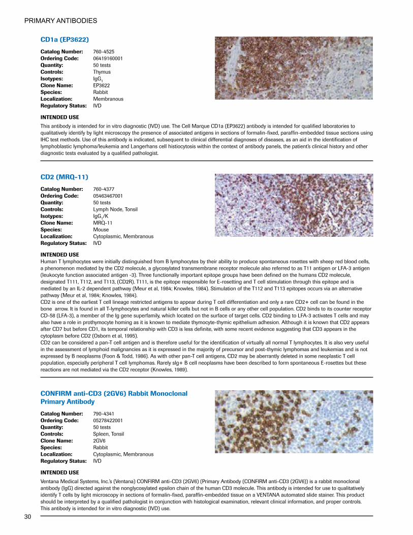
CD1a(EP3622)
CatalogNumber: 760-4525OrderingCode: 06419160001Quantity: 50 testsControls: ThymusIsotypes: IgG1CloneName: EP3622Species: RabbitLocalization: MembranousRegulatoryStatus: IVD
INTENDEDUSE
This antibody is intended for in vitro diagnostic (IVD) use. The Cell Marque CD1a (EP3622) antibody is intended for qualified laboratories to qualitatively identify by light microscopy the presence of associated antigens in sections of formalin-fixed, paraffin-embedded tissue sections using IHC test methods. Use of this antibody is indicated, subsequent to clinical differential diagnoses of diseases, as an aid in the identification of lymphoblastic lymphoma/leukemia and Langerhans cell histiocytosis within the context of antibody panels, the patient’s clinical history and other diagnostic tests evaluated by a qualified pathologist.
CD2(MRQ-11)
CatalogNumber: 760-4377OrderingCode: 05463467001Quantity: 50 testsControls: Lymph Node, TonsilIsotypes: IgG1/KCloneName: MRQ-11Species: MouseLocalization: Cytoplasmic, MembranousRegulatoryStatus: IVD
INTENDEDUSEHuman T lymphocytes were initially distinguished from B lymphocytes by their ability to produce spontaneous rosettes with sheep red blood cells, a phenomenon mediated by the CD2 molecule, a glycosylated transmembrane receptor molecule also referred to as T11 antigen or LFA-3 antigen (leukocyte function associated antigen -3). Three functionally important epitope groups have been defined on the humans CD2 molecule, designated T111, T112, and T113, (CD2R). T111, is the epitope responsible for E-rosetting and T cell stimulation through this epitope and is mediated by an IL-2 dependent pathway (Meur et al, 1984; Knowles, 1984). Stimulation of the T112 and T113 epitopes occurs via an alternative pathway (Meur et al, 1984; Knowles, 1984).CD2 is one of the earliest T cell lineage restricted antigens to appear during T cell differentiation and only a rare CD2+ cell can be found in the bone arrow. It is found in all T-lymphocytes and natural killer cells but not in B cells or any other cell population. CD2 binds to its counter receptor CD-58 (LFA-3), a member of the Ig gene superfamily, which located on the surface of target cells. CD2 binding to LFA-3 activates T cells and may also have a role in prothymocyte homing as it is known to mediate thymocyte-thymic epithelium adhesion. Although it is known that CD2 appears after CD7 but before CD1, its temporal relationship with CD3 is less definite, with some recent evidence suggesting that CD3 appears in the cytoplasm before CD2 (Osborn et al, 1995).CD2 can be considered a pan-T cell antigen and is therefore useful for the identification of virtually all normal T lymphocytes. It is also very useful in the assessment of lymphoid malignancies as it is expressed in the majority of precursor and post-thymic lymphomas and leukemias and is not expressed by B neoplasms (Foon & Todd, 1986). As with other pan-T cell antigens, CD2 may be aberrantly deleted in some neoplastic T cell population, especially peripheral T cell lymphomas. Rarely slg+ B cell neoplasms have been described to form spontaneous E-rosettes but these reactions are not mediated via the CD2 receptor (Knowles, 1989).
CONFIRManti-CD3(2GV6)RabbitMonoclonalPrimary Antibody
CatalogNumber: 790-4341OrderingCode: 05278422001Quantity: 50 testsControls: Spleen, TonsilCloneName: 2GV6Species: RabbitLocalization: Cytoplasmic, MembranousRegulatoryStatus: IVD
INTENDEDUSE
Ventana Medical Systems, Inc.’s (Ventana) CONFIRM anti-CD3 (2GV6) (Primary Antibody (CONFIRM anti-CD3 (2GV6)) is a rabbit monoclonal antibody (IgG) directed against the nonglycosylated epsilon chain of the human CD3 molecule. This antibody is intended for use to qualitatively identify T cells by light microscopy in sections of formalin-fixed, paraffin-embedded tissue on a VENTANA automated slide stainer. This product should be interpreted by a qualified pathologist in conjunction with histological examination, relevant clinical information, and proper controls. This antibody is intended for in vitro diagnostic (IVD) use.

PRIMARY ANTIBODIES
31
CONFIRManti-CD4(SP35)RabbitMonoclonalPrimary Antibody
CatalogNumber: 790-4423OrderingCode: 05552737001Quantity: 50 testsControls: Liver, TonsilCloneName: SP35Species: RabbitLocalization: MembranousRegulatoryStatus: IVD
INTENDEDUSE
This antibody is intended for in vitro diagnostic (IVD) use. Ventana Medical Systems’ (Ventana) CONFIRM anti-CD4 (SP35) Rabbit Monoclonal Primary Antibody is intended for the qualitative detection of CD4 in sections of formalin-fixed, paraffin-embedded normal and neoplastic human tissues. CD4 is present on helper-inducer T lymphocytes that recognize antigen in the context of MHC class 2 molecules. CD4 positive staining results may aid in identifying T-cell lymphomas and in identifying the T helper-inducer cell subset of T lymphocytes in normal tissues. The clinical interpretation of any staining, or the absence of staining, must be complemented by morphological studies and evaluation of proper controls. Evaluation must be made by a qualified pathologist within the context of the patient’s clinical history and other diagnostic tests.
CONFIRManti-CD5(SP19)RabbitMonoclonalPrimary Antibody
CatalogNumber: 790-4451OrderingCode: 05929903001Quantity: 50 testsControls: Tonsil, LymphomaCloneName: SP19Species: RabbitLocalization: Cell Membrane, CytoplasmicRegulatoryStatus: IVD
INTENDEDUSE
Ventana Medical Systems’ (Ventana) CONFIRM anti-CD5 (SP19) Rabbit Monoclonal Primary Antibody is directed against human CD5 expressed on the plasma membrane of virtually all human T cells and the B1a subset of human B cells found in the follicular mantle zones, bone marrow and peripheral blood. Staining CD5 is commonly used as part of several IHC panels to determine T cell and B cell subclassifications. CD5 may be used to aid in the identification of T cell lymphomas, and certain B cell lymphomas, including mantle cell lymphoma. The antibody is intended for qualitative staining in sections of formalin-fixed, paraffin-embedded tissue. This product should be interpreted by a qualified pathologist in conjunction with histological examination, relevant clinical information and proper controls. This antibody is intended for in vitro diagnostic (IVD) use.
anti-CD7(SP94)RabbitMonoclonal Primary Antibody
CatalogNumber: 790-4558OrderingCode: 06537847001Quantity: 50 testsControls: Thymus, TonsilIsotypes: Rabbit IgCloneName: SP94Species: RabbitLocalization: MembranousRegulatoryStatus: IVD
INTENDEDUSE
Anti-CD7 (SP94) Rabbit Monoclonal Primary Antibody (anti-CD7 (SP94)) is directed against the 40kD transmembrane glycoprotein, CD7 which is present in thymocytes and mature T cells. Anti-CD7 (SP94) may be used to aid in the identification of T cell lymphomas. This antibody is intended for qualitative staining of sections of formalin-fixed, paraffin-embedded tissue. This product should be interpreted by a qualified pathologist in conjunction with histological examination, relevant clinical information, and proper controls. This antibody is intended for in vitro diagnostic (IVD) use.

PRIMARY ANTIBODIES
32
CONFIRManti-CD8(SP57)RabbitMonoclonalPrimary Antibody
CatalogNumber: 790-4460OrderingCode: 05937248001Quantity: 50 testsControls: Tonsil, T Cell LymphomaCloneName: SP57Species: RabbitLocalization: Cell MembraneRegulatoryStatus: IVD
INTENDEDUSE
Ventana Medical Systems’ (Ventana) CONFIRM anti-CD8 (SP57) Rabbit Monoclonal Primary Antibody (CONFIRM anti-CD8 (SP57)) is intended for the qualitative detection of CD8 in sections of normal and neoplastic human tissues. The CD8 glycoprotein is present on cytotoxic/suppressor T lymphocytes that recognize antigen in the context of MHC class 1 molecules. CD8 positive staining results may aid in identifying T-cell lymphomas and in identifying the T cytotoxic/suppressor cell subset of T lymphocytes in normal tissues. The antibody is intended for qualitative staining in sections of formalin-fixed, paraffin-embedded tissue. This product should be interpreted by a qualified pathologist in conjunction with histological examination, relevant clinical information and proper controls. This antibody is intended for in vitro diagnostic (IVD) use.
VENTANAanti-CD10(SP67)RabbitMonoclonalPrimary Antibody
CatalogNumber: 790-4506OrderingCode: 05857856001Quantity: 50 testsControls: TonsilIsotypes: Rabbit IgCloneName: SP67Species: RabbitLocalization: Membranous, CytoplasmicRegulatoryStatus: IVD
INTENDEDUSE
VENTANA anti-CD10 (SP67) Rabbit Monoclonal Primary Antibody is directed against the CD10 molecule, or common acute lymphoblastic leukemia antigen (CALLA), expressed on the surface of early lymphoid cells, and on various nonlymphoid tissues including breast myoepithelial cells, bile canaliculi, fibroblasts, kidney tubular brush borders and small intestine epithelium. This antibody exhibits a cell membranous and/or cytoplasmic staining pattern and may be used to aid in the identification of Burkitt’s lymphoma and follicular germ cell lymphoma, and in the classification of some carcinomas such as renal cell carcinoma. The antibody is intended for qualitative staining in sections of formalin-fixed, paraffin-embedded tissue. This product should be interpreted by a qualified pathologist in conjunction with histological examination, relevant clinical information, and proper controls. This antibody is intended for in vitro diagnostic (IVD) use.
CD13(SP187)RabbitMonoclonal Primary Antibody
CatalogNumber: 760-4862OrderingCode: 07047673001Quantity: 50 testsControls: LiverIsotypes: IgGCloneName: SP187Species: Rabbit MonoclonalLocalization: Cytoplasmic, Membranous RegulatoryStatus: IVD
INTENDEDUSE
This antibody is intended for in vitro diagnostic (IVD) use. The Cell Marque CD13 (SP187) antibody is intended for qualified laboratories to qualitatively identify by light microscopy the presence of associated antigens in sections of formalin-fixed, paraffin-embedded tissue sections using IHC test methods. Use of this antibody is indicated as an aid in the identification of acute myeloid leukemia and hepatocellular carcinomas within the context of an antibody panel, the patient’s clinical history, and other diagnostic tests evaluated by a qualified pathologist.

PRIMARY ANTIBODIES
33
CD14(EPR3653)RabbitMonoclonalAntibody
CatalogNumber: 760-4523OrderingCode: 06419151001Quantity: 50 testsControls: TonsilIsotypes: IgGCloneName: EPR3653Species: RabbitLocalization: Membranous, CytoplasmicRegulatoryStatus: IVD
INTENDEDUSE
This antibody is intended for in vitro diagnostic (IVD) use. The Cell Marque CD14 (EPR3653) antibody is intended for qualified laboratories to qualitatively identify by light microscopy the presence of associated antigens in sections of formalin-fixed, paraffin-embedded tissue sections using IHC test methods. Use of this antibody is indicated as an aid in the identification and diagnosis of myeloid leukemia and histiocytic neoplasms within the context of an antibody panel, the patient’s clinical history, and other diagnostic tests evaluated by a qualified pathologist.
CONFIRManti-CD15(MMA)MouseMonoclonalPrimary Antibody
CatalogNumber: 760-2504OrderingCode: 05266904001Quantity: 50 testsControls: Hodgkin’s LymphomaIsotypes: IgMCloneName: MMASpecies: MouseLocalization: Cytoplasmic, MembranousRegulatoryStatus: IVD
INTENDEDUSE
This antibody is intended for in vitro diagnostic use. Ventana Medical Systems’ CONFIRM anti-CD15 (MMA) Mouse Monoclonal Primary Antibody is a mouse monoclonal antibody (IgM, kappa) directed against a carbohydrate epitope present on most granulocytic cells.1 This antibody is intended for use to qualitatively identify CD15 by light microscopy in sections of formalin-fixed, paraffin-embedded tissue stained on the Ventana automated slide stainer. The clinical interpretation of any staining, or the absence of staining, must be complemented by morphological studies and evaluation of proper controls. Evaluation must be made by a qualified pathologist within the context of the patient’s clinical history and other diagnostic tests. 1. Pinkus GS., P. Thomas, and J.W. Said. Leu-M1--A Marker for Reed-Sternberg Cells in Hodgkin's Disease: An Immunoperoxidase Study of
Paraffin-Embedded Tissues. Am. J. Pathol. 1985; Vol. 119, p. 244-252.
CD16(SP175)RabbitMonoclonal Primary Antibody
CatalogNumber: 760-4863OrderingCode: 07047681001Quantity: 50 testsControls: TonsilIsotypes: IgGCloneName: SP175Species: Rabbit MonoclonalLocalization: Membranous, Cytoplasmic RegulatoryStatus: IVD
INTENDEDUSE
This antibody is intended for in vitro diagnostic (IVD) use. The Cell Marque CD16 (SP175) antibody is intended for qualified laboratories to qualitatively identify by light microscopy the presence of associated antigens in sections of formalin-fixed, paraffin-embedded tissue sections using IHC test methods. Use of this antibody is indicated as an aid in the identification of lymphomas and leukemias in the tissue within the context of an antibody panel, the patient’s clinical history, and other diagnostic tests evaluated by a qualified pathologist.

PRIMARY ANTIBODIES
34
CONFIRManti-CD20(L26) Primary Antibody
CatalogNumber: 760-2531OrderingCode: 05267099001Quantity: 50 testsControls: Lymph Node, TonsilIsotypes: IgG2a/KCloneName: L26Species: MouseLocalization: MembranousRegulatoryStatus: IVD
INTENDEDUSE
This antibody is intended for in vitro diagnostic (IVD) use. Ventana Medical Systems’ (Ventana) CONFIRM anti-CD20 (L26) Primary Antibody is a mouse monoclonal antibody (IgG2a, kappa) directed against an epitope present on human B lymphocytes. This antibody is intended for use to qualitatively identify cells of B lymphocytic lineage1 by light microscopy in sections of formalin-fixed, paraffin-embedded tissue on a VENTANA automated slide stainer. The clinical interpretation of any staining, or the absence of staining, must be complemented by morphological studies and evaluation of proper controls. Evaluation must be made by a qualified pathologist within the context of the patient’s clinical history and other diagnostic tests. 1. Mason DY, Comans-Bitter WM, Cordell JL, Verhoeven MA, van Dongen JJ. Antibody L26 recognizes an intracellular epitope on the B-cell-associated
CD20 antigen. Am J Clin Pathol 136(6):, 1215-1222, 1990.
CD21(2G9)
CatalogNumber: 760-4245OrderingCode: 05269059001Quantity: 50 testsControls: Lymph Node, TonsilIsotypes: IgG2aCloneName: 2G9Species: MouseLocalization: MembranousRegulatoryStatus: IVD
INTENDEDUSE
Anti-CD21 Mouse Monoclonal Primary Antibody is intended for laboratory use in the detection of the CD21 protein in formalin-fixed, paraffin- embedded tissue stained on VENTANA BenchMark immunohistochemical (IHC) automated slide stainers. This product should be interpreted by a qualified pathologist in conjunction with histological examination, relevant clinical information, and proper controls. This antibody is intended for in vitro diagnostic (IVD) use.
CD21(EP3093)
CatalogNumber: 760-4438OrderingCode: 05973902001Quantity: 50 testsControls: Lymph Node, TonsilIsotypes: IgG1CloneName: EP3093Species: RabbitLocalization: Cytoplasmic, MembranousRegulatoryStatus: IVD
INTENDEDUSE
CD21 antigen is an integral membrane glycoprotein of molecular weight 140 kD. Anti-CD21 is useful in the identification of follicular dendriditic cell matrix found in normal lymph nodes and tonsillar tissue. This antibody also labels follicular dendritic cell tumor/sarcomas. The antigen is absent on T-lymphocytes, monocytes, and granulocytes.
Associated products: CD35, Follicular Dendritic Cell
PRIMARY ANTIBODIES
35
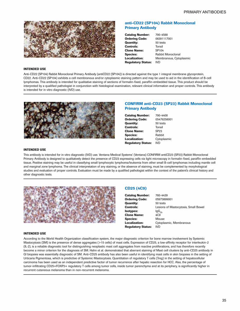
anti-CD22(SP104)RabbitMonoclonal Primary Antibody
CatalogNumber: 790-4588OrderingCode: 06391117001Quantity: 50 testsControls: TonsilCloneName: SP104Species: Rabbit MonoclonalLocalization: Membranous, CytoplasmicRegulatoryStatus: IVD
INTENDEDUSE
Anti-CD22 (SP104) Rabbit Monoclonal Primary Antibody (antiCD22 (SP104)) is directed against the type 1 integral membrane glycoprotein, CD22. Anti-CD22 (SP104) exhibits a cell membranous and/or cytoplasmic staining pattern and may be used to aid in the identification of B-cell lymphomas. This antibody is intended for qualitative staining of sections of formalin-fixed, paraffin-embedded tissue. This product should be interpreted by a qualified pathologist in conjunction with histological examination, relevant clinical information and proper controls. This antibody is intended for in vitro diagnostic (IVD) use.
CONFIRManti-CD23(SP23)RabbitMonoclonalPrimary Antibody
CatalogNumber: 790-4408OrderingCode: 05479258001Quantity: 50 testsControls: TonsilCloneName: SP23Species: RabbitLocalization: CytoplasmicRegulatoryStatus: IVD
INTENDEDUSE
This antibody is intended for in vitro diagnostic (IVD) use. Ventana Medical Systems’ (Ventana) CONFIRM antiCD23 (SP23) Rabbit Monoclonal Primary Antibody is designed to qualitatively detect the presence of CD23 expressing cells via light microscopy in formalin-fixed, paraffin-embedded tissue. Positive staining may be useful in classifying small lymphocytic lymphoma/leukemia from other small B-cell lymphomas including mantle cell and marginal zone lymphoma. The clinical interpretation of any staining, or the absence of staining, must be complemented by morphological studies and evaluation of proper controls. Evaluation must be made by a qualified pathologist within the context of the patient’s clinical history and other diagnostic tests.
CD25(4C9)
CatalogNumber: 760-4439OrderingCode: 05973899001Quantity: 50 testsControls: Lesions of Mastocytosis, Small BowelIsotypes: IgG2bCloneName: 4C9Species: MouseLocalization: Cytoplasmic, MembranousRegulatoryStatus: IVD
INTENDEDUSE
According to the World Health Organization classification system, the major diagnostic criterion for bone marrow involvement by Systemic Mastocytosis (SM) is the presence of dense aggregates (>15 cells) of mast cells. Expression of CD25, a low-affinity receptor for interleukin-2 (IL-2), is a reliable diagnostic tool for distinguishing neoplastic mast cell aggregates from reactive proliferations, and has therefore recently become a minor criterion for the diagnosis of SM. Hahn et al. demonstrated that aberrant staining of Mast cell clusters by anti-CD25 antibody in GI biopsies was essentially diagnostic of SM. Anti-CD25 antibody has also been useful in identifying mast cells in skin biopsies in the setting of Urticaria Pigmentosa, which is predictive of Systemic Mastocytosis. Quantitation of regulatory T cells (Treg) in the setting of hepatocellular carcinoma has been used as an independent predictive factor of tumor recurrence after hepatic resection for HCC. Also, the percentage of tumor-infiltrating CD25+FOXP3+ regulatory T cells among tumor cells, inside tumor parenchyma and at its periphery, is significantly higher in recurrent cutaneous melanoma than in non-recurrent melanoma.

PRIMARY ANTIBODIES
36
anti-CD30(Ber-H2)MouseMonoclonal Primary Antibody
CatalogNumber: 790-4858OrderingCode: 07007841001Quantity: 50 testsControls: Positive control tissue: Lymphoma or Tonsil
Negative control tissue: Tonsil Germinal Center B-cells
Isotypes: IgG1/KCloneName: Ber-H2Species: MouseLocalization: Cell Membrane, Cytoplasmic, GolgiRegulatoryStatus: IVD
INTENDEDUSE
The anti-CD30 (Ber-H2) Mouse Monoclonal Primary Antibody (antiCD30 (Ber-H2)) is intended for laboratory use in the detection of the CD30 protein in formalin-fixed, paraffin-embedded tissue stained with a VENTANA BenchMark series immunohistochemical (IHC) automated slide stainer. CD30 positive staining results may aid in the identification of classical Hodgkin lymphoma and anaplastic large cell lymphoma. This product should be interpreted by a qualified pathologist in conjunction with histological examination, relevant clinical information, and proper controls. This antibody is intended for in vitro diagnostic (IVD) use.
CONFIRManti-CD30(Ber-H2)PrimaryAntibody
CatalogNumber: 790-2926OrderingCode: 05278201001Quantity: 50 testsControls: Hodgkins LymphomaIsotypes: IgG1/KCloneName: Ber-H2Species: MouseLocalization: MembranousRegulatoryStatus: IVD
INTENDEDUSE
This antibody is intended for in vitro diagnostic use. Ventana Medical Systems' CONFIRM anti-CD30 (Ber-H2) Primary Antibody contains a mouse monoclonal antibody (IgG1, kappa) directed against CD30. The antibody is intended for use to qualitatively identify CD30 by light microscopy in sections of formalin-fixed, paraffin-embedded tissue stained on a Ventana automated slide staining device. The clinical interpretation of any staining, or the absence of staining, must be complemented by morphological studies and evaluation of proper controls. Evaluation must be made by a qualified pathologist within the context of the patient’s clinical history and other diagnostic tests.
CD31(JC70)
CatalogNumber: 760-4378OrderingCode: 05463475001Quantity: 50 testsControls: Appendix, Placenta, TonsilIsotypes: IgG1/KCloneName: JC70Species: MouseLocalization: Cytoplasmic, MembranousRegulatoryStatus: IVD
INTENDEDUSE
Anti-CD31 (JC70) Mouse Monoclonal Primary Antibody is intended for laboratory use in the detection of the CD31 protein in formalin-fixed, paraffin-embedded neoplastic tissue stained on VENTANA BenchMark immunohistochemical (IHC) automated slide stainers. This product should be interpreted by a qualified pathologist in conjunction with histological examination, relevant clinical information, and proper controls. This antibody is intended for in vitro diagnostic (IVD) use.

PRIMARY ANTIBODIES
37
CD33(SP266)RabbitMonoclonal Primary Antibody
CatalogNumber: 760-4952OrderingCode: 07391447001Quantity: 50 testsControls: Acute Leukemia, Placenta
SyncytiotrophoblastsIsotypes: IgGCloneName: SP266Species: RabbitLocalization: MembranousRegulatoryStatus: IVD
INTENDEDUSE
This antibody is intended for in vitro diagnostic (IVD) use. The Cell Marque CD33 (SP266) antibody is intended for qualified laboratories to qualitatively identify by light microscopy the presence of associated antigens in sections of formalin-fixed, paraffin-embedded tissue sections using IHC test methods. Use of this antibody is indicated, subsequent to clinical differential diagnoses of diseases, as an aid in the identification of acute leukemia within the context of antibody panels, the patient’s clinical history, and other diagnostic tests evaluated by a qualified pathologist.
CONFIRManti-CD34(QBEnd/10) Primary Antibody
CatalogNumber: 790-2927OrderingCode: 05278210001Quantity: 50 testsControls: Appendix, Placenta, TonsilIsotypes: IgG1CloneName: QBEnd/10Species: MouseLocalization: MembranousRegulatoryStatus: IVD
INTENDEDUSE
This antibody is intended for in vitro diagnostic (IVD) use. Ventana Medical Systems’ (Ventana) CONFIRM anti-CD34 (QBEnd/10) Primary Antibody is a mouse monoclonal antibody (IgG1) directed against human CD34. This antibody is intended for use to qualitatively identify CD34 by light microscopy in sections of formalin-fixed, paraffin-embedded tissue stained on a VENTANA automated slide stainer. The clinical interpretation of any staining, or the absence of staining, must be complemented by morphological studies and evaluation of proper controls. Evaluation must be made by a qualified pathologist within the context of the patient’s clinical history and other diagnostic tests.
anti-CD43(L60)MouseMonoclonal Primary Antibody
CatalogNumber: 760-2511OrderingCode: 05266980001Quantity: 50 testsControls: Lymph Node, TonsilIsotypes: IgG1CloneName: L60Species: MouseLocalization: MembranousRegulatoryStatus: IVD
INTENDEDUSE
This antibody is intended for in vitro diagnostic (IVD) use. Ventana Medical Systems’ (Ventana) Anti-CD43 (L60) Mouse Monoclonal Primary Antibody is a mouse monoclonal antibody directed against an epitope present on human monocytes, granulocytes and lymphocytes. This reagent may be used to aid in the identification of cells of lymphoid and myelomonocytic lineage.1 Anti-CD43 (L60) is intended for qualitative staining in sections of formalin-fixed, paraffin-embedded tissue on a VENTANA automated slide stainer. The clinical interpretation of any staining, or the absence of staining, must be complemented by morphological studies and evaluation of proper controls. Evaluation must be made by a qualified pathologist within the context of the patient’s clinical history and other diagnostic tests. 1. Strickler JG, Weiss LM, Copenhaver CM, Bindl J, McDaid R, Buck D, Warnke R. Monoclonal antibodies reactive in routinely processed tissue
sections of malignant lymphoma, with emphasis on T cell lymphomas. Hum Pathol. Aug; 18(8):808-14, 1987.

PRIMARY ANTIBODIES
38
anti-CD44(SP37)RabbitMonoclonal Primary Antibody
CatalogNumber: 790-4537OrderingCode: 06364985001Quantity: 50 testsControls: Normal TonsilIsotypes: IgGCloneName: SP37Species: RabbitLocalization: MembranousRegulatoryStatus: IVD
INTENDEDUSE
Anti-CD44 (SP37) Rabbit Monoclonal Primary Antibody is directed against the CD44 protein. CD44 is a type I transmembrane receptor protein expressed in several cell types including T and B lymphocytes, monocytes, granulocytes, erythrocytes, fibroblasts, epithelial, and mast cells. This antibody exhibits a membraneous staining pattern and may be used to detect the CD44 standard and variant proteins in a variety of neoplastic tissues, including squamous cell and urothelial carcinoma. The antibody is intended for qualitative staining of sections of formalin-fixed, paraffin-embedded tissue. This product should be interpreted by a qualified pathologist in conjunction with histological examination, relevant clinical information, and proper controls. This antibody is intended for in vitro diagnostic (IVD) use.
CD45(LCA)(2B11&PD7/26)
CatalogNumber: 760-4279OrderingCode: 05269423001Quantity: 50 testsControls: Lymph Node, TonsilIsotypes: IgG1/KCloneName: 2B11 & PD7/26Species: MouseLocalization: MembranousRegulatoryStatus: IVD
INTENDEDUSE
This antibody is intended for in vitro diagnostic (IVD) use. The Cell Marque CD45 (LCA) (2B11 & PD7/26) antibody is intended for qualified laboratories to qualitatively identify by light microscopy the presence of associated antigens in sections of formalin-fixed, paraffin-embedded tissue sections using IHC test methods. Use of this antibody is indicated, subsequent to clinical differential diagnosis, as an aid in the identification of lymphoid neoplasms within the context of an antibody panel, the patient’s clinical history, and other diagnostic tests evaluated by a qualified pathologist.
CONFIRManti-CD45,LCA(RP2/18) Primary Antibody
CatalogNumber: 760-2505OrderingCode: 05266912001Quantity: 50 testsControls: TonsilIsotypes: IgG1/KCloneName: RP2/18Species: MouseLocalization: MembranousRegulatoryStatus: IVD
INTENDEDUSE
This antibody is intended for in vitro diagnostic (IVD) use. Ventana Medical Systems’ (Ventana) CONFIRM anti-CD45, LCA (RP2/18) Primary Antibody is a mouse monoclonal antibody (IgG1) directed against CD45. CONFIRM anti-CD45, LCA (RP2/18) may be used in immunocytochemical methods to aid in the identification of normal and abnormal cells of leukocytic lineage and as an aid in the diagnosis of anaplastic tumors. CONFIRM anti-CD45, LCA (RP2/18) specifically binds to antigens located on the membrane of leukocytes. Staining is most consistent for lymphocytic cells with less consistent reaction in other leukocytes (macrophages/histiocytes/myeloid cells). The epitope recognized by the antibody is LCA-RB. CONFIRM anti-CD45, LCA (RP2/18) is intended for laboratory use to qualitatively stain leukocytes in sections of formalin-fixed, paraffin-embedded tissue on a VENTANA automated slide stainer. Light microscopy is used to detect the staining of cell components. The clinical interpretation of any staining, or the absence of staining, must be complemented by morphological studies and evaluation of proper controls. Evaluation must be made by a qualified pathologist within the context of the patient’s clinical history and other diagnostic tests. Prescription only.

PRIMARY ANTIBODIES
39
CD45R(MB1)
CatalogNumber: 760-2624OrderingCode: 05267340001Quantity: 50 testsControls: Lymph Node, TonsilIsotypes: IgG1CloneName: MB1Species: MouseLocalization: MembranousRegulatoryStatus: IVD
INTENDEDUSE
MB1 antibody stains preferentially B-cells and their neoplasms but is less specific as it will also react with some T-cell lymphomas and non- lymphoid tumors. The antigen for this antibody is in the membrane of all B-cells with the exception of plasma cells and some mature T-cells.
CONFIRManti-CD45RO(UCHL-1) Primary Antibody
CatalogNumber: 790-2930OrderingCode: 05278244001Quantity: 50 testsControls: Lymph Node, TonsilIsotypes: IgMCloneName: UCHL-1Species: MouseLocalization: MembranousRegulatoryStatus: IVD
INTENDEDUSE
This antibody is intended for in vitro diagnostic (IVD) use. Ventana Medical Systems’ (Ventana) CONFIRM anti-CD45RO (UCHL-1) Primary Antibody is a mouse monoclonal antibody (IgG2a) directed against CD45RO. This antibody is intended for use to qualitatively identify CD45RO by light microscopy in sections of formalin-fixed, paraffin-embedded tissue on a VENTANA automated slide stainer. The clinical interpretation of any staining, or the absence of staining, must be complemented by morphological studies and evaluation of proper controls. Evaluation must be made by a qualified pathologist within the context of the patient’s clinical history and other diagnostic tests. Prescription only.
CONFIRManti-CD56(123C3)MouseMonoclonalPrimary Antibody
CatalogNumber: 790-4465OrderingCode: 05878519001Quantity: 50 testsControls: Brain, Normal PancreasIsotypes: IgG1CloneName: 123C3Species: MouseLocalization: Membrane, CytoplasmicRegulatoryStatus: IVD
INTENDEDUSE
Ventana Medical Systems’ (Ventana) CONFIRM anti-CD56 (123C3) Mouse Monoclonal Primary Antibody (CONFIRM anti-CD56 (123C3)) is directed against the CD56 molecule or neural cell adhesion molecule expressed by most neuroectodermally derived cell lines, tissues and neoplasms. It is also expressed on some mesodermally derived tumors, on natural killer cells, and NK cell lymphomas. This antibody exhibits a membrane/cytoplasmic staining pattern and may be used to aid in the identification of small cell carcinomas, neuroblastoma, and rhabdomyosarcoma. The antibody is intended for qualitative staining in sections of formalin-fixed, paraffin-embedded tissue. This product should be interpreted by a qualified pathologist in conjunction with histological examination, relevant clinical information and proper controls. This antibody is intended for in vitro diagnostic (IVD) use.

PRIMARY ANTIBODIES
40
CD56(MRQ-42)
CatalogNumber: 760-4596OrderingCode: 06433359001Quantity: 50 testsControls: Pancreatic Islet Cells, Pancreatic Endocrine
Cells, NeuroblastomaCloneName: MRQ-42Species: Rabbit MonoclonalLocalization: Cytoplasmic, MembranousRegulatoryStatus: IVD
INTENDEDUSE
This antibody is intended for in vitro diagnostic (IVD) use. The Cell Marque CD56 (MRQ-42) antibody is intended for qualified laboratories to qualitatively identify by light microscopy the presence of associated antigens in sections of formalin-fixed, paraffin-embedded tissue sections using IHC test methods. Use of this antibody is indicated as an aid in the identification of neural/neurodendocrine tissues, natural killer (NK) cells, NK-like T-cells, and diagnosis of related neoplasms within the context of an antibody panel, the patient’s clinical history, and other diagnostic tests evaluated by a qualified pathologist.
CD57(NK-1)
CatalogNumber: 760-2626OrderingCode: 05267366001Quantity: 50 testsControls: Lymph Node, TonsilIsotypes: IgM/KCloneName: NK-1Species: MouseLocalization: MembranousRegulatoryStatus: IVD
INTENDEDUSE
This antibody is intended for in vitro diagnostic (IVD) use. The Cell Marque CD57 (NK-1) antibody is intended for qualified laboratories to qualitatively identify by light microscopy the presence of associated antigens in sections of formalin-fixed, paraffin-embedded tissue sections using IHC test methods. Use of this antibody is indicated, subsequent to clinical differential diagnoses of diseases, as an aid in the identification of T-cell large granular lymphocyte disorders, oligodendrogliomas, and neuroendocrine tumors within the context of antibody panels, the patient’s clinical history, and other diagnostic tests evaluated by a qualified pathologist.
CD61(2f2)
CatalogNumber: 760-4249OrderingCode: 05269091001Quantity: 50 testsControls: Bone MarrowIsotypes: IgG1CloneName: 2f2Species: MouseLocalization: CytoplasmicRegulatoryStatus: IVD
INTENDEDUSE
Antibody to CD61 labels the IIIa sub-unit of the non-covalently linked glycoprotein heterodimer IIb/IIIa complex present on human platelets and their precursors. This antibody is useful in identifying megakaryoblastic differentiation as seen in megakaryoblastic leukemia.

PRIMARY ANTIBODIES
41
CD63(NKI/C3)
CatalogNumber: 760-4379OrderingCode: 05463483001Quantity: 50 testsControls: Malignant MelanomaIsotypes: IgG1/KCloneName: NKI/C3Species: MouseLocalization: Cytoplasmic, MembranousRegulatoryStatus: IVD
INTENDEDUSE
This antibody reacts with a 53 kDa protein which forms a part of the family of tetraspan moieties. The antigen was originally designated as a lysosomal membrane protein characterized as an activation dependent platelet surface antigen. In fact the CD63 antigen has a diverse distribution on the surface and in the cytoplasm of many cell types including lymphoid, myeloid, endothelial cells and melanoma. It has been quite useful in identifying malignant melanoma. Mete et al. found it useful in differentiating renal oncocytomas (RO) from eosinophilic renal cell carcinomas (eRCC). In his series of 35 ROs, 94% demonstrated apical and/or polar CD63 positivity. Of his 77 eRCCs, 96% showed diffuse cytoplasmic staining.
CONFIRManti-CD68(KP-1)PrimaryAntibody
CatalogNumber: 790-2931OrderingCode: 05278252001Quantity: 50 testsControls: Lymph Node, TonsilIsotypes: IgG1/KCloneName: KP-1Species: MouseLocalization: Cytoplasmic, MembranousRegulatoryStatus: IVD
INTENDEDUSE
This antibody is intended for in vitro diagnostic use. Ventana Medical Systems’ (Ventana®) CONFIRM anti-CD68 (KP-1) Primary Antibody is a mouse monoclonal antibody, class IgG1 kappa. The antibody is intended for qualitative staining in sections of formalin-fixed, paraffin-embedded tissue on a VENTANA automated slide stainer. CONFIRM anti-CD68 reacts with a 110 KD intracellular glycoprotein associated with the cell membrane of macrophages and some myeloid elements.1,2,3,4 Unexpected antigen expression or loss of expression may occur, especially in neoplasms. Occasionally stromal elements surrounding heavily stained tissue or cells will show immunoreactivity. The clinical interpretation of any staining, or the absence of staining, must be complemented by morphological studies and evaluation of proper controls. Evaluation must be made by a qualified pathologist within the context of the patient’s clinical history and other diagnostic tests. Caution: U.S. Federal law restricts this device to sale by or on the order of a physician. 1. Herman GE, Elfont EA. The taming of immunohistochemistry: the new era of quality control. Biotech Histochem 66(4): 194-199, 1991.2. Omata M, Liew CT, Ashcavai M, Peters RL. Nonimmunologic binding of horseradish peroxidase to hepatitis B surface antigen. A possible source
of error in immunohistochemistry. Am J Clin Pathol 73(5): 626-32, 1980.3. Nadji M, Morales AR. Immunoperoxidase: part 1. The technique and its pitfalls. Lab Med 14: 767, 1983.4. Taylor RT, Cote RJ. Immunomicroscopy: A Diagnostic Tool For The Surgical Pathologist, 2nd Edition. W.B. Saunders Company, Philadelphia, 1994.
CD71(MRQ-48)MouseMonoclonalAntibody
CatalogNumber: 760-4589OrderingCode: 06433162001Quantity: 50 testsControls: Bone MarrowIsotypes: IgGCloneName: MRQ-48Species: Mouse MonoclonalLocalization: Membranous, CytoplasmicRegulatoryStatus: IVD
INTENDEDUSE
This antibody is intended for in vitro diagnostic (IVD) use. The Cell Marque CD71 (MRQ-48) antibody is intended for qualified laboratories to qualitatively identify by light microscopy the presence of associated antigens in sections of formalin-fixed, paraffin-embedded tissue sections using IHC test methods. Use of this antibody is indicated as an aid in the identification of the erythroid precursors in bone marrow and in the determination of acute myeloid leukemia and myelodysplastic syndrome (MDS) within the context of an antibody panel, the patient’s clinical history, and other diagnostic tests evaluated by a qualified pathologist.
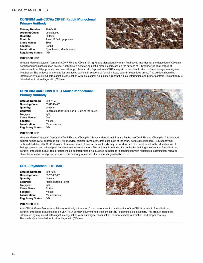
PRIMARY ANTIBODIES
42
CONFIRManti-CD79a(SP18)RabbitMonoclonalPrimary Antibody
CatalogNumber: 790-4432OrderingCode: 05640296001Quantity: 50 testsControls: Tonsil, B Cell LymphomaCloneName: SP18Species: RabbitLocalization: Cytoplasmic, MembranousRegulatoryStatus: IVD
INTENDEDUSE
Ventana Medical Systems’ (Ventana) CONFIRM anti-CD79a (SP18) Rabbit Monoclonal Primary Antibody is intended for the detection of CD79a in normal and neoplastic human tissues. AntiCD79a is directed against a protein expressed on the surface of B lymphocytes at all stages of maturation, from B lymphocyte precursors through plasma cells. Expression of CD79a may aid in the identification of B cell lineage in malignant lymphomas. The antibody is intended for qualitative staining in sections of formalin-fixed, paraffin-embedded tissue. This product should be interpreted by a qualified pathologist in conjunction with histological examination, relevant clinical information and proper controls. This antibody is intended for in vitro diagnostic (IVD) use.
CONFIRManti-CD99(O13)MouseMonoclonalPrimary Antibody
CatalogNumber: 790-4452OrderingCode: 05913594001Quantity: 50 testsControls: Pancreatic Islet Cells, Sertoli Cells of the TestisIsotypes: IgG1CloneName: O13Species: MouseLocalization: MembranousRegulatoryStatus: IVD
INTENDEDUSE
Ventana Medical Systems’ (Ventana) CONFIRM anti-CD99 (O13) Mouse Monoclonal Primary Antibody (CONFIRM anti-CD99 (O13)) is directed against human CD99 expressed on T lymphocytes, cortical thymocytes, granulosa cells of the ovary, pancreatic islet cells, CNS ependymal cells and Sertoli’s cells. CD99 shows a plasma membrane location. This antibody may be used as part of a panel to aid in the identification of Ewing’s sarcoma and related peripheral neuroectodermal tumors. The antibody is intended for qualitative staining in sections of formalin-fixed, paraffin-embedded tissue. This product should be interpreted by a qualified pathologist in conjunction with histological examination, relevant clinical information, and proper controls. This antibody is intended for in vitro diagnostic (IVD) use.
CD138/syndecan-1(B-A38)
CatalogNumber: 760-4248OrderingCode: 05269083001Quantity: 50 testsControls: Plasmacytoma, TonsilIsotypes: IgGCloneName: B-A38Species: MouseLocalization: MembranousRegulatoryStatus: IVD
INTENDEDUSE
Anti-CD138 Mouse Monoclonal Primary Antibody is intended for laboratory use in the detection of the CD138 protein in formalin-fixed, paraffin-embedded tissue stained on VENTANA BenchMark immunohistochemical (IHC) automated slide stainers. This product should be interpreted by a qualified pathologist in conjunction with histological examination, relevant clinical information, and proper controls. This antibody is intended for in vitro diagnostic (IVD) use.

PRIMARY ANTIBODIES
43
CD163(MRQ-26)
CatalogNumber: 760-4437OrderingCode: 05973929001Quantity: 50 testsControls: Inflamed TissueIsotypes: IgG1CloneName: MRQ-26Species: MouseLocalization: Cytoplasmic, MembranousRegulatoryStatus: IVD
INTENDEDUSE
CD163 was recently identified as an acute phase-regulated transmembrane protein whose function is to mediate the endocytosis of haptoglobin- hemoglobin complexes. This receptor is expressed on the surface of monocytes (low expression) and tissue macrophages [also known as histiocytes] (high expression). It is a member of the cysteine-rich scavenger receptor superfamily, encoded by a gene localized on human chromosome 12p13.3. Solubilized in plasma, CD163 functions as an anti-inflammatory signal and has many roles in disease processes that range from autoimmune conditions such as rheumatoid arthritis to atherosclerosis. Previous work has shown that the CD163 gene can be regulated by glucocorticoids, IL-10, and other inflammatory modulators, and is highly expressed in inflamed tissues, consistent with its role in the resolution of inflammation. Staining for CD163 has been helpful in distinguishing synovial macrophages from synovial intimal fibroblasts in the setting of rheumatoid arthritis, where its specificity for macrophages was found to be superior to that of CD68, which does not discriminate between these cell types. Increased levels of CD163 were also detected in patients with microbial infections and myelomonocytic leukemias by an enzyme-linked immunosorbent assay. Flow cytometry studies have confirmed that CD163 expression is limited to leukemias with monocytic differentiation. Another recent study showed that all 5 cases of synovial-type giant cell tumors of the vertebral column stained for CD163.
CDX-2(EPR2764Y)
CatalogNumber: 760-4380OrderingCode: 05463491001Quantity: 50 testsControls: Adenocarcinoma of Colon, Normal ColonIsotypes: IgG1/KCloneName: EPR2764YSpecies: RabbitLocalization: NuclearRegulatoryStatus: IVD
INTENDEDUSE
Anti-CDX-2 Rabbit Monoclonal Primary Antibody is intended for laboratory use in the detection of the CDX-2 protein in formalin-fixed, paraffin- embedded tissue stained on VENTANA BenchMark immunohistochemical (IHC) automated slide stainers. This product should be interpreted by a qualified pathologist in conjunction with histological examination, relevant clinical information, and proper controls. This antibody is intended for in vitro diagnostic (IVD) use.
CEA(CEA31)MouseMonoclonalAntibody
CatalogNumber: 760-4594OrderingCode: 06433316001Quantity: 50 testsControls: Adenocarcinoma of Colon, Colon MucosaIsotypes: IgG1CloneName: CEA31Species: Mouse MonoclonalLocalization: CytoplasmicRegulatoryStatus: IVD
INTENDEDUSE
This antibody is intended for in vitro diagnostic (IVD) use. The Cell Marque CEA (CEA31) antibody is intended for qualified laboratories to qualitatively identify by light microscopy the presence of associated antigens in sections of formalin-fixed, paraffin-embedded tissue sections using IHC test methods. Use of this antibody is indicated as an aid in the identification and diagnosis of adenocarcinomas within the context of an antibody panel, the patient’s clinical history, and other diagnostic tests evaluated by a qualified pathologist.

PRIMARY ANTIBODIES
44
Anti-CEA(TF3H8-1)PrimaryAntibody
CatalogNumber: 760-2507OrderingCode: 05266947001Quantity: 50 testsControls: ColonIsotypes: IgG1CloneName: TF3H8-1Species: MouseLocalization: CytoplasmicRegulatoryStatus: IVD
INTENDEDUSE
This antibody is intended for in vitro diagnostic use. Ventana Medical Systems’ (Ventana) CEA (TF 3H8-1) Primary Antibody may be used to aid in the identification of cells of normal and abnormal epithelial lineage as an aid in diagnosis of anaplastic tumors. CEA (TF 3H8-1) Primary Antibody specifically binds to antigens located in the plasma membrane and cytoplasmic regions of normal simple epithelial cells. CEA (TF 3H8-1) is a mouse monoclonal antibody (IgG1) directed against carcinoembryonic antigen (CEA). The clinical interpretation of any staining, or the absence of staining, must be complemented by morphological studies and evaluation of proper controls. Evaluation must be made by a qualified pathologist within the context of the patient’s clinical history and other diagnostic tests. Caution: U.S. Federal law restricts this device to sale by or on the order of a physician.
anti-ChromograninA(LK2H10)PrimaryAntibody
CatalogNumber: 760-2519OrderingCode: 05267056001Quantity: 50 testsControls: PancreasIsotypes: IgG1/KCloneName: LK2H10Species: MouseLocalization: CytoplasmicRegulatoryStatus: IVD
INTENDEDUSE
This antibody is intended for in vitro diagnostic use. Ventana Medical Systems’ (Ventana) anti-Chromogranin A (LK2H10) Primary Antibody contains a mouse monoclonal antibody (IgG1) directed against a protein found in the secretory granules of most endocrine cells.1 The Chromogr-anin A protein is widely expressed in neuronal tissues and in the secretory granules of human endocrine cells. Chromogranin A is also expressed by adrenal (medulla), parathyroid, anterior pituitary cells, islet cells of the pancreas, gastrointestinal cells, bronchial endocrine cells, and thyroid c-cells. Chromogranin A is expressed in tumors of neuroendocrine origin, including pheochromocytomas, pituitary adenomas, islet cell tumors, medullary thyroid carcinomas, carcinoids and Merkel cell tumors. This reagent may be used to aid in the identification of cells of normal or neoplastic neuroendocrine lineage. The antibody is intended for qualitative staining in sections of formalin-fixed, paraffin-embedded tissue on a Ventana automated slide stainer. Anti-Chromogranin A (LK2H10) specifically binds to the chromogranin A protein located in the secretory granules of normal and neoplastic neuroendocrine cells. Unexpected antigen expression or loss of expression may occur, especially in neoplasms. Occasionally stromal elements surrounding heavily stained tissue and/or cells will show immunoreactivity. The clinical interpretation of any staining, or the absence of staining, must be complemented by morphological studies and evaluation of proper controls. Evaluation must be made by a qualified pathologist within the context of the patient’s clinical history and other diagnostic tests. Caution: U.S. Federal law restricts this device to sale by or on the order of a physician.1. Wilson BS, Lloyd RV. Detection of Chromogranin in Neuroendocrine cells with a monoclonal antibody. Am J Clin Pathol 115(3): 458-468, 1984.
CINtec®HistologyKit
CatalogNumber: 9517OrderingCode: 06594450001Quantity: 50 testsControls: Normal Pancreas, Normal Tonsil,
Cervical CarcinomaIsotypes: IgG2aCloneName: E6H4™Species: Mouse monoclonalRegulatoryStatus: IVD
INTENDEDUSE
For in vitro diagnostic use. The CINtec® Histology Kit is an immunohistochemistry assay for the qualitative detection of the p16INK4a antigen on formalin-fixed, paraffin-embedded tissue sections prepared from cervical biopsies. It is intended for use in medical pathology laboratories to provide adjunctive information after initial diagnosis has been made by established diagnostic methods. Interpretation of the test results may only be made by a certified professional in conjunction with the patient´s clinical history and any additional diagnostic tests that have been performed.
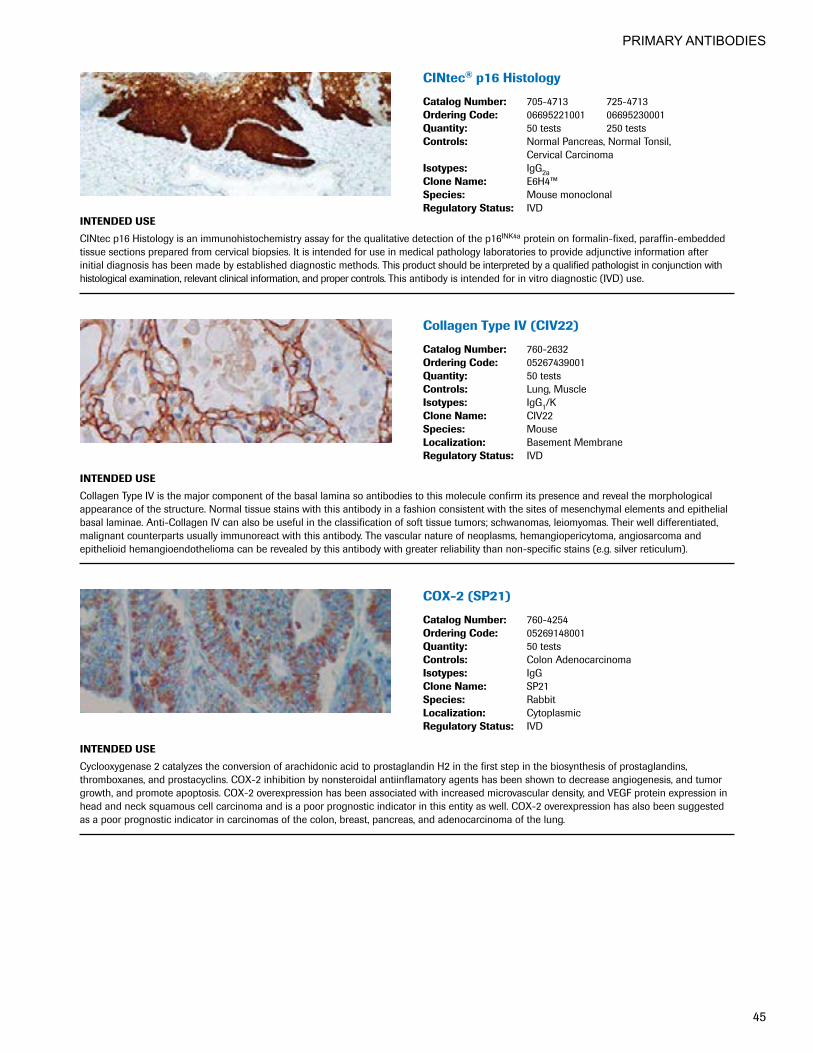
PRIMARY ANTIBODIES
45
CINtec®p16Histology
CatalogNumber: 705-4713 725-4713OrderingCode: 06695221001 06695230001Quantity: 50 tests 250 tests Controls: Normal Pancreas, Normal Tonsil,
Cervical CarcinomaIsotypes: IgG2aCloneName: E6H4™Species: Mouse monoclonalRegulatoryStatus: IVD
INTENDEDUSE
CINtec p16 Histology is an immunohistochemistry assay for the qualitative detection of the p16INK4a protein on formalin-fixed, paraffin-embedded tissue sections prepared from cervical biopsies. It is intended for use in medical pathology laboratories to provide adjunctive information after initial diagnosis has been made by established diagnostic methods. This product should be interpreted by a qualified pathologist in conjunction with histological examination, relevant clinical information, and proper controls. This antibody is intended for in vitro diagnostic (IVD) use.
CollagenTypeIV(CIV22)
CatalogNumber: 760-2632OrderingCode: 05267439001Quantity: 50 testsControls: Lung, MuscleIsotypes: IgG1/KCloneName: CIV22Species: MouseLocalization: Basement MembraneRegulatoryStatus: IVD
INTENDEDUSE
Collagen Type IV is the major component of the basal lamina so antibodies to this molecule confirm its presence and reveal the morphological appearance of the structure. Normal tissue stains with this antibody in a fashion consistent with the sites of mesenchymal elements and epithelial basal laminae. Anti-Collagen IV can also be useful in the classification of soft tissue tumors; schwanomas, leiomyomas. Their well differentiated, malignant counterparts usually immunoreact with this antibody. The vascular nature of neoplasms, hemangiopericytoma, angiosarcoma and epithelioid hemangioendothelioma can be revealed by this antibody with greater reliability than non-specific stains (e.g. silver reticulum).
COX-2(SP21)
CatalogNumber: 760-4254OrderingCode: 05269148001Quantity: 50 testsControls: Colon AdenocarcinomaIsotypes: IgGCloneName: SP21Species: RabbitLocalization: CytoplasmicRegulatoryStatus: IVD
INTENDEDUSE
Cyclooxygenase 2 catalyzes the conversion of arachidonic acid to prostaglandin H2 in the first step in the biosynthesis of prostaglandins, thromboxanes, and prostacyclins. COX-2 inhibition by nonsteroidal antiinflamatory agents has been shown to decrease angiogenesis, and tumor growth, and promote apoptosis. COX-2 overexpression has been associated with increased microvascular density, and VEGF protein expression in head and neck squamous cell carcinoma and is a poor prognostic indicator in this entity as well. COX-2 overexpression has also been suggested as a poor prognostic indicator in carcinomas of the colon, breast, pancreas, and adenocarcinoma of the lung.

PRIMARY ANTIBODIES
46
VENTANAanti-CyclinD1(SP4-R)Rabbit MonoclonalPrimaryAntibody
CatalogNumber: 790-4508OrderingCode: 05862949001Quantity: 50 testsControls: Normal TonsilIsotypes: IgGCloneName: SP4-RSpecies: RabbitLocalization: NuclearRegulatoryStatus: IVD
INTENDEDUSE
VENTANA anti-Cyclin D1 (SP4-R) Rabbit Monoclonal Primary Antibody (VENTANA anti-Cyclin D1 (SP4-R)) is directed against the Cyclin D1 molecule, a key cell cycle regulator, expressed primarily during the G1 phase of the cell cycle. This antibody exhibits a nuclear staining pattern, and may be used to aid in the identification of mantle cell lymphoma, and breast carcinoma. The antibody is intended for qualitative staining in sections of formalin-fixed, paraffin-embedded tissue. This product should be interpreted by a qualified pathologist in conjunction with histological examination, relevant clinical information and proper controls. This antibody is intended for in vitro diagnostic (IVD) use.
Cytokeratin(35betaH11)
CatalogNumber: 760-2637OrderingCode: 05267471001Quantity: 50 testsControls: ProstateIsotypes: IgM/KCloneName: 35betaH11Species: MouseLocalization: CytoplasmicRegulatoryStatus: IVD
INTENDEDUSE
Anti-Cytokeratin 8 stains most non-squamous epithelial tumors, squamous tumors are negative for this antibody as a rule. This antibody stains adenocarcinomas of the breast, ovary, gastrointestinal tract, thyroid, pancreas, bile duct, and salivary glands. This antibody does not react with skeletal muscle or nerve cells.
CONFIRManti-Cytokeratin(AE1) Primary Antibody
CatalogNumber: 760-2521OrderingCode: 05267064001Quantity: 50 testsControls: Prostate, Salivary GlandIsotypes: IgG1CloneName: AE1Species: MouseLocalization: CytoplasmicRegulatoryStatus: IVD
INTENDEDUSE
This antibody is intended for in vitro diagnostic (IVD) use. Ventana® Medical Systems’ (Ventana) CONFIRM anti-Cytokeratin (AE1) is intended for laboratory use to qualitatively stain cytokeratin in sections of formalin-fixed, paraffin-embedded tissue on a VENTANA automated slide stainer. CONFIRM anti-Cytokeratin (AE1) may be used to aid in the identification of cells of normal and abnormal epithelial lineage as an aid in diagnosis of anaplastic tumors. CONFIRM anti-Cytokeratin (AE1) specifically binds to antigens located in the cytoplasmic regions of normal epithelial cells. The clinical interpretation of any staining, or the absence of staining, must be complemented by morphological studies and evaluation of proper controls. Evaluation must be made by a qualified pathologist within the context of the patient’s clinical history and other diagnostic tests. Prescription only.
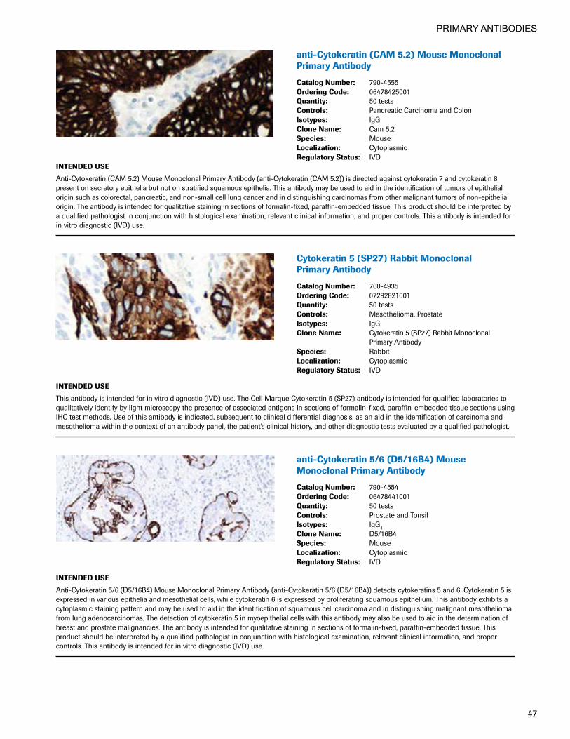
PRIMARY ANTIBODIES
47
anti-Cytokeratin(CAM5.2)MouseMonoclonalPrimary Antibody
CatalogNumber: 790-4555OrderingCode: 06478425001Quantity: 50 testsControls: Pancreatic Carcinoma and ColonIsotypes: IgGCloneName: Cam 5.2Species: MouseLocalization: CytoplasmicRegulatoryStatus: IVD
INTENDEDUSE
Anti-Cytokeratin (CAM 5.2) Mouse Monoclonal Primary Antibody (anti-Cytokeratin (CAM 5.2)) is directed against cytokeratin 7 and cytokeratin 8 present on secretory epithelia but not on stratified squamous epithelia. This antibody may be used to aid in the identification of tumors of epithelial origin such as colorectal, pancreatic, and non-small cell lung cancer and in distinguishing carcinomas from other malignant tumors of non-epithelial origin. The antibody is intended for qualitative staining in sections of formalin-fixed, paraffin-embedded tissue. This product should be interpreted by a qualified pathologist in conjunction with histological examination, relevant clinical information, and proper controls. This antibody is intended for in vitro diagnostic (IVD) use.
Cytokeratin5(SP27)RabbitMonoclonal Primary Antibody
CatalogNumber: 760-4935OrderingCode: 07292821001Quantity: 50 testsControls: Mesothelioma, ProstateIsotypes: IgGCloneName: Cytokeratin 5 (SP27) Rabbit Monoclonal
Primary AntibodySpecies: RabbitLocalization: CytoplasmicRegulatoryStatus: IVD
INTENDEDUSE
This antibody is intended for in vitro diagnostic (IVD) use. The Cell Marque Cytokeratin 5 (SP27) antibody is intended for qualified laboratories to qualitatively identify by light microscopy the presence of associated antigens in sections of formalin-fixed, paraffin-embedded tissue sections using IHC test methods. Use of this antibody is indicated, subsequent to clinical differential diagnosis, as an aid in the identification of carcinoma and mesothelioma within the context of an antibody panel, the patient’s clinical history, and other diagnostic tests evaluated by a qualified pathologist.
anti-Cytokeratin5/6(D5/16B4)Mouse MonoclonalPrimaryAntibody
CatalogNumber: 790-4554OrderingCode: 06478441001Quantity: 50 testsControls: Prostate and TonsilIsotypes: IgG1CloneName: D5/16B4Species: MouseLocalization: CytoplasmicRegulatoryStatus: IVD
INTENDEDUSE
Anti-Cytokeratin 5/6 (D5/16B4) Mouse Monoclonal Primary Antibody (anti-Cytokeratin 5/6 (D5/16B4)) detects cytokeratins 5 and 6. Cytokeratin 5 is expressed in various epithelia and mesothelial cells, while cytokeratin 6 is expressed by proliferating squamous epithelium. This antibody exhibits a cytoplasmic staining pattern and may be used to aid in the identification of squamous cell carcinoma and in distinguishing malignant mesothelioma from lung adenocarcinomas. The detection of cytokeratin 5 in myoepithelial cells with this antibody may also be used to aid in the determination of breast and prostate malignancies. The antibody is intended for qualitative staining in sections of formalin-fixed, paraffin-embedded tissue. This product should be interpreted by a qualified pathologist in conjunction with histological examination, relevant clinical information, and proper controls. This antibody is intended for in vitro diagnostic (IVD) use.
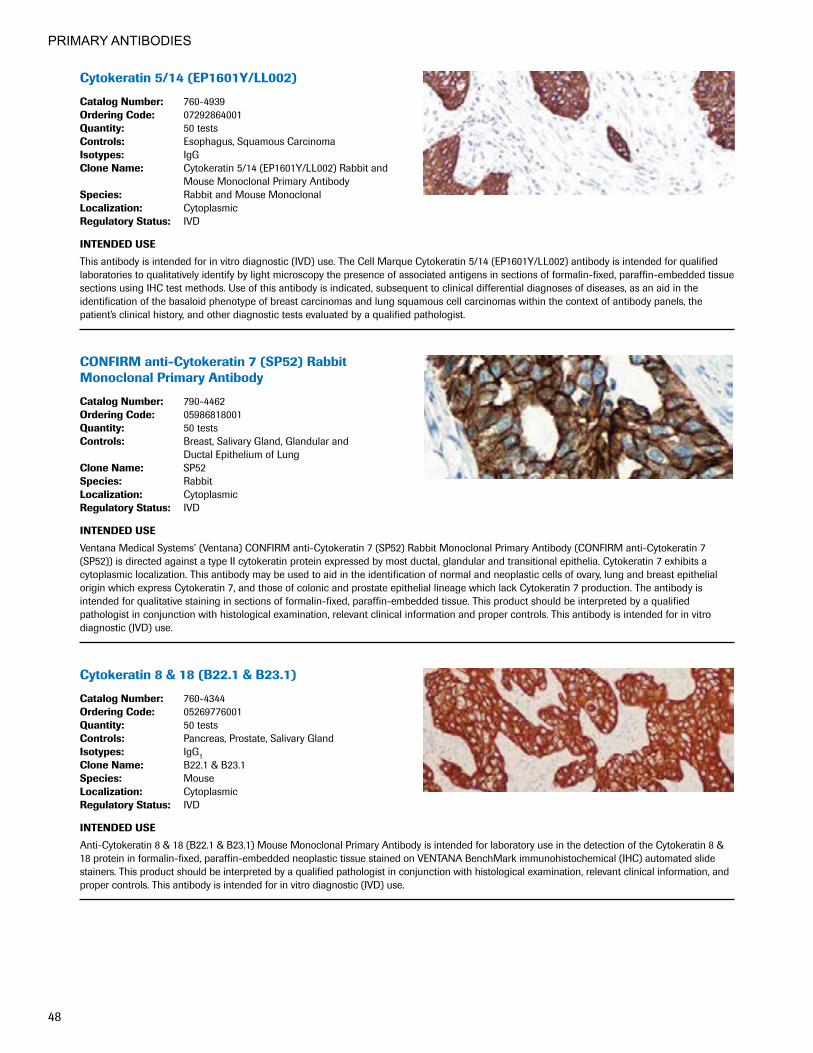
PRIMARY ANTIBODIES
48
Cytokeratin5/14(EP1601Y/LL002)
CatalogNumber: 760-4939OrderingCode: 07292864001Quantity: 50 testsControls: Esophagus, Squamous CarcinomaIsotypes: IgGCloneName: Cytokeratin 5/14 (EP1601Y/LL002) Rabbit and
Mouse Monoclonal Primary AntibodySpecies: Rabbit and Mouse MonoclonalLocalization: CytoplasmicRegulatoryStatus: IVD
INTENDEDUSE
This antibody is intended for in vitro diagnostic (IVD) use. The Cell Marque Cytokeratin 5/14 (EP1601Y/LL002) antibody is intended for qualified laboratories to qualitatively identify by light microscopy the presence of associated antigens in sections of formalin-fixed, paraffin-embedded tissue sections using IHC test methods. Use of this antibody is indicated, subsequent to clinical differential diagnoses of diseases, as an aid in the identification of the basaloid phenotype of breast carcinomas and lung squamous cell carcinomas within the context of antibody panels, the patient’s clinical history, and other diagnostic tests evaluated by a qualified pathologist.
CONFIRManti-Cytokeratin7(SP52)RabbitMonoclonalPrimaryAntibody
CatalogNumber: 790-4462OrderingCode: 05986818001Quantity: 50 testsControls: Breast, Salivary Gland, Glandular and
Ductal Epithelium of LungCloneName: SP52Species: RabbitLocalization: CytoplasmicRegulatoryStatus: IVD
INTENDEDUSE
Ventana Medical Systems’ (Ventana) CONFIRM anti-Cytokeratin 7 (SP52) Rabbit Monoclonal Primary Antibody (CONFIRM anti-Cytokeratin 7 (SP52)) is directed against a type II cytokeratin protein expressed by most ductal, glandular and transitional epithelia. Cytokeratin 7 exhibits a cytoplasmic localization. This antibody may be used to aid in the identification of normal and neoplastic cells of ovary, lung and breast epithelial origin which express Cytokeratin 7, and those of colonic and prostate epithelial lineage which lack Cytokeratin 7 production. The antibody is intended for qualitative staining in sections of formalin-fixed, paraffin-embedded tissue. This product should be interpreted by a qualified pathologist in conjunction with histological examination, relevant clinical information and proper controls. This antibody is intended for in vitro diagnostic (IVD) use.
Cytokeratin8&18(B22.1&B23.1)
CatalogNumber: 760-4344OrderingCode: 05269776001Quantity: 50 testsControls: Pancreas, Prostate, Salivary GlandIsotypes: IgG1CloneName: B22.1 & B23.1Species: MouseLocalization: CytoplasmicRegulatoryStatus: IVD
INTENDEDUSE
Anti-Cytokeratin 8 & 18 (B22.1 & B23.1) Mouse Monoclonal Primary Antibody is intended for laboratory use in the detection of the Cytokeratin 8 & 18 protein in formalin-fixed, paraffin-embedded neoplastic tissue stained on VENTANA BenchMark immunohistochemical (IHC) automated slide stainers. This product should be interpreted by a qualified pathologist in conjunction with histological examination, relevant clinical information, and proper controls. This antibody is intended for in vitro diagnostic (IVD) use.
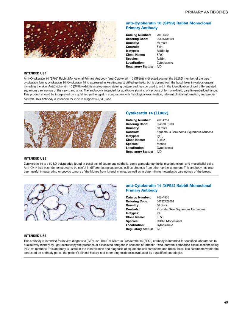
PRIMARY ANTIBODIES
49
anti-Cytokeratin10(SP99)RabbitMonoclonalPrimary Antibody
CatalogNumber: 790-4562OrderingCode: 06425135001Quantity: 50 testsControls: SkinIsotypes: Rabbit IgCloneName: SP99Species: RabbitLocalization: CytoplasmicRegulatoryStatus: IVD
INTENDEDUSE
Anti-Cytokeratin 10 (SP99) Rabbit Monoclonal Primary Antibody (anti-Cytokeratin 10 (SP99)) is directed against the 56.5kD member of the type 1 cytokeratin family, cytokeratin 10. Cytokeratin 10 is expressed in keratinizing stratified epithelia, but is absent from the basal layer, in various organs including the skin. AntiCytokeratin 10 (SP99) exhibits a cytoplasmic staining pattern and may be used to aid in the identification of well differentiated squamous carcinomas of the cervix and anus. The antibody is intended for qualitative staining of sections of formalin-fixed, paraffin-embedded tissue. This product should be interpreted by a qualified pathologist in conjunction with histological examination, relevant clinical information, and proper controls. This antibody is intended for in vitro diagnostic (IVD) use.
Cytokeratin14(LL002)
CatalogNumber: 760-4251OrderingCode: 05269113001Quantity: 50 testsControls: Squamous Carcinoma, Squamous MucosaIsotypes: IgG3CloneName: LL002Species: MouseLocalization: CytoplasmicRegulatoryStatus: IVD
INTENDEDUSE
Cytokeratin 14 is a 50 kD polypeptide found in basal cell of squamous epithelia, some glandular epithelia, myoepithelium, and mesothelial cells. Anti-CK14 has been demonstrated to be useful in differentiating squamous cell carcinomas from other epithelial tumors. This antibody has also been useful in separating oncocytic tumors of the kidney from it renal mimics, as well as in determining metaplastic carcinomas of the breast.
anti-Cytokeratin14(SP53)RabbitMonoclonalPrimary Antibody
CatalogNumber: 760-4805OrderingCode: 06732429001Quantity: 50 testsControls: Prostate, Skin, Squamous CarcinomaIsotypes: IgGCloneName: SP53Species: Rabbit MonoclonalLocalization: CytoplasmicRegulatoryStatus: IVD
INTENDEDUSE
This antibody is intended for in vitro diagnostic (IVD) use. The Cell Marque Cytokeratin 14 (SP53) antibody is intended for qualified laboratories to qualitatively identify by light microscopy the presence of associated antigens in sections of formalin-fixed, paraffin-embedded tissue sections using IHC test methods. This antibody is useful in the identification and diagnosis of squamous cell carcinoma and breast basal like carcinoma within the context of an antibody panel, the patient’s clinical history, and other diagnostic tests evaluated by a qualified pathologist.
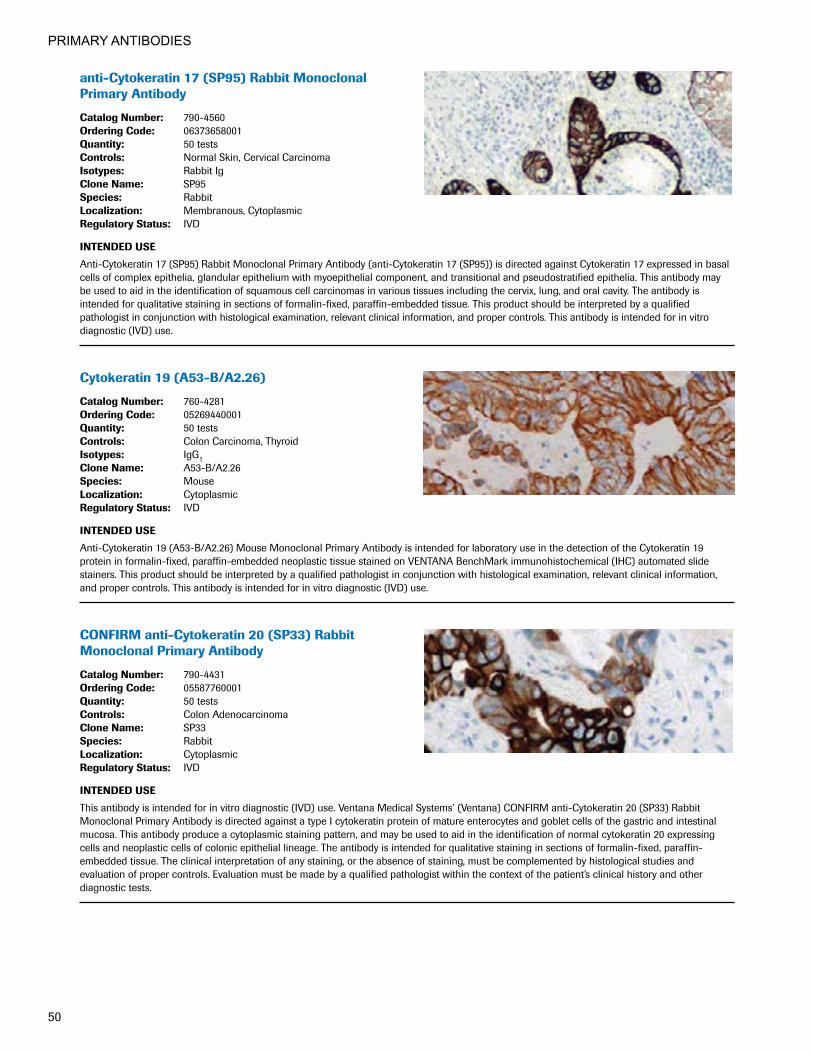
PRIMARY ANTIBODIES
50
anti-Cytokeratin17(SP95)RabbitMonoclonalPrimary Antibody
CatalogNumber: 790-4560OrderingCode: 06373658001Quantity: 50 testsControls: Normal Skin, Cervical CarcinomaIsotypes: Rabbit IgCloneName: SP95Species: RabbitLocalization: Membranous, CytoplasmicRegulatoryStatus: IVD
INTENDEDUSE
Anti-Cytokeratin 17 (SP95) Rabbit Monoclonal Primary Antibody (anti-Cytokeratin 17 (SP95)) is directed against Cytokeratin 17 expressed in basal cells of complex epithelia, glandular epithelium with myoepithelial component, and transitional and pseudostratified epithelia. This antibody may be used to aid in the identification of squamous cell carcinomas in various tissues including the cervix, lung, and oral cavity. The antibody is intended for qualitative staining in sections of formalin-fixed, paraffin-embedded tissue. This product should be interpreted by a qualified pathologist in conjunction with histological examination, relevant clinical information, and proper controls. This antibody is intended for in vitro diagnostic (IVD) use.
Cytokeratin19(A53-B/A2.26)
CatalogNumber: 760-4281OrderingCode: 05269440001Quantity: 50 testsControls: Colon Carcinoma, ThyroidIsotypes: IgG1CloneName: A53-B/A2.26Species: MouseLocalization: CytoplasmicRegulatoryStatus: IVD
INTENDEDUSE
Anti-Cytokeratin 19 (A53-B/A2.26) Mouse Monoclonal Primary Antibody is intended for laboratory use in the detection of the Cytokeratin 19 protein in formalin-fixed, paraffin-embedded neoplastic tissue stained on VENTANA BenchMark immunohistochemical (IHC) automated slide stainers. This product should be interpreted by a qualified pathologist in conjunction with histological examination, relevant clinical information, and proper controls. This antibody is intended for in vitro diagnostic (IVD) use.
CONFIRManti-Cytokeratin20(SP33)RabbitMonoclonalPrimaryAntibody
CatalogNumber: 790-4431OrderingCode: 05587760001Quantity: 50 testsControls: Colon AdenocarcinomaCloneName: SP33Species: RabbitLocalization: CytoplasmicRegulatoryStatus: IVD
INTENDEDUSE
This antibody is intended for in vitro diagnostic (IVD) use. Ventana Medical Systems’ (Ventana) CONFIRM anti-Cytokeratin 20 (SP33) Rabbit Monoclonal Primary Antibody is directed against a type I cytokeratin protein of mature enterocytes and goblet cells of the gastric and intestinal mucosa. This antibody produce a cytoplasmic staining pattern, and may be used to aid in the identification of normal cytokeratin 20 expressing cells and neoplastic cells of colonic epithelial lineage. The antibody is intended for qualitative staining in sections of formalin-fixed, paraffin- embedded tissue. The clinical interpretation of any staining, or the absence of staining, must be complemented by histological studies and evaluation of proper controls. Evaluation must be made by a qualified pathologist within the context of the patient’s clinical history and other diagnostic tests.
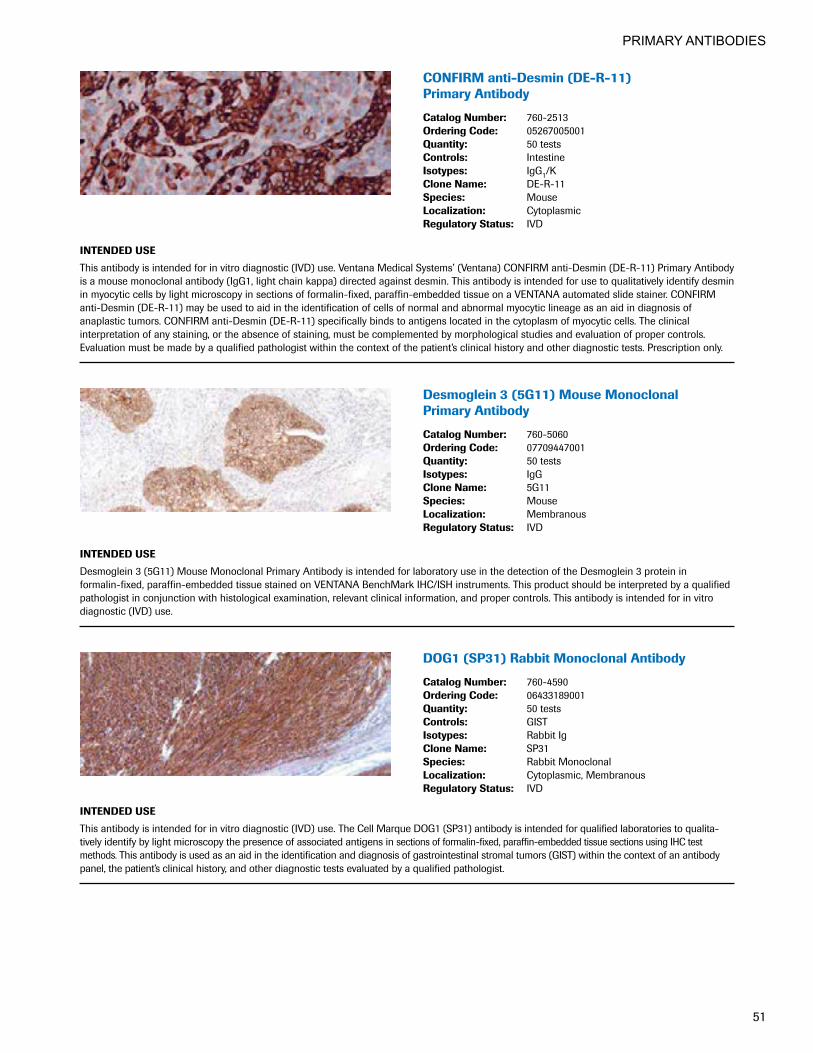
PRIMARY ANTIBODIES
51
CONFIRManti-Desmin(DE-R-11) Primary Antibody
CatalogNumber: 760-2513OrderingCode: 05267005001Quantity: 50 testsControls: IntestineIsotypes: IgG1/KCloneName: DE-R-11Species: MouseLocalization: CytoplasmicRegulatoryStatus: IVD
INTENDEDUSE
This antibody is intended for in vitro diagnostic (IVD) use. Ventana Medical Systems’ (Ventana) CONFIRM anti-Desmin (DE-R-11) Primary Antibody is a mouse monoclonal antibody (IgG1, light chain kappa) directed against desmin. This antibody is intended for use to qualitatively identify desmin in myocytic cells by light microscopy in sections of formalin-fixed, paraffin-embedded tissue on a VENTANA automated slide stainer. CONFIRM anti-Desmin (DE-R-11) may be used to aid in the identification of cells of normal and abnormal myocytic lineage as an aid in diagnosis of anaplastic tumors. CONFIRM anti-Desmin (DE-R-11) specifically binds to antigens located in the cytoplasm of myocytic cells. The clinical interpretation of any staining, or the absence of staining, must be complemented by morphological studies and evaluation of proper controls. Evaluation must be made by a qualified pathologist within the context of the patient’s clinical history and other diagnostic tests. Prescription only.
Desmoglein3(5G11)MouseMonoclonal Primary Antibody
CatalogNumber: 760-5060OrderingCode: 07709447001Quantity: 50 testsIsotypes: IgGCloneName: 5G11Species: MouseLocalization: MembranousRegulatoryStatus: IVD
INTENDEDUSE
Desmoglein 3 (5G11) Mouse Monoclonal Primary Antibody is intended for laboratory use in the detection of the Desmoglein 3 protein in formalin-fixed, paraffin-embedded tissue stained on VENTANA BenchMark IHC/ISH instruments. This product should be interpreted by a qualified pathologist in conjunction with histological examination, relevant clinical information, and proper controls. This antibody is intended for in vitro diagnostic (IVD) use.
DOG1(SP31)RabbitMonoclonalAntibody
CatalogNumber: 760-4590OrderingCode: 06433189001Quantity: 50 testsControls: GISTIsotypes: Rabbit IgCloneName: SP31Species: Rabbit MonoclonalLocalization: Cytoplasmic, MembranousRegulatoryStatus: IVD
INTENDEDUSE
This antibody is intended for in vitro diagnostic (IVD) use. The Cell Marque DOG1 (SP31) antibody is intended for qualified laboratories to qualita-tively identify by light microscopy the presence of associated antigens in sections of formalin-fixed, paraffin-embedded tissue sections using IHC test methods. This antibody is used as an aid in the identification and diagnosis of gastrointestinal stromal tumors (GIST) within the context of an antibody panel, the patient’s clinical history, and other diagnostic tests evaluated by a qualified pathologist.

PRIMARY ANTIBODIES
52
VENTANAanti-E-cadherin(36)MouseMonoclonalPrimary Antibody
CatalogNumber: 790-4497OrderingCode: 05905290001Quantity: 50 testsControls: Ductal Breast CarcinomaIsotypes: IgG2a/KCloneName: 36Species: MouseLocalization: Membranous RegulatoryStatus: IVD
INTENDEDUSE
VENTANA anti-E-cadherin (36) Mouse Monoclonal Primary Antibody (VENTANA anti-E-cadherin (36)) is directed against the cytoplasmic domain of the human transmembrane protein E-cadherin expressed as a part of the cell-cell adhesion complex in epithelial tissues. Reduction or loss of expression is associated with invasive carcinoma and possibly metastasis in a variety of carcinomas. This antibody may be used to aid in the differentiation of in-situ and/or invasive lobular carcinoma from in situ and/or invasive ductal carcinoma of the breast. This antibody is intended for qualitative staining in sections of formalin-fixed, paraffin-embedded tissue. Test results should be interpreted by a qualified pathologist in conjunc-tion with histological examination, relevant clinical information, and proper controls. This antibody is intended for in vitro diagnostic (IVD) use.
E-Cadherin(EP700Y)
CatalogNumber: 760-4440OrderingCode: 05973872001Quantity: 50 testsControls: Breast, Lung Adenocarcinoma, PancreasIsotypes: IgGCloneName: EP700YSpecies: RabbitLocalization: MembranousRegulatoryStatus: IVD
INTENDEDUSE
E-Cadherin is an adhesion protein that is expressed in cells of epithelial lineage. Anti-E-Cadherin stains positively in glandular epithelium as well as adenocarcinomas of the lung and G.I. tract, and ovary. It has been useful in distinguishing adenocarcinoma from mesothelioma. Another application involves the differentiation of ductal (which stains positive) vs. lobular cancer of the breast. It has also been shown to be positive in some thyroid carcinomas. Loss of E-Cadherin expression has been suggested as a poor prognostic sign in breast carcinoma and non-small cell lung carcinoma.
CONFIRManti-EpidermalGrowthFactorReceptor(3C6)PrimaryAntibody
CatalogNumber: 790-2988OrderingCode: 05278341001Quantity: 50 testsControls: Colorectal Adenocarcinoma, SkinIsotypes: IgG1CloneName: 3C6Species: MouseLocalization: MembranousRegulatoryStatus: IVD
INTENDEDUSE
This antibody is intended for in vitro diagnostic (IVD) use. Ventana Medical Systems’ (Ventana) CONFIRM anti-Epidermal Growth Factor Receptor (EGFR) (3C6) Primary Antibody is a mouse monoclonal antibody (IgG1) directed against the extracellular domain of human EGFR protein of 170kD. The antibody also recognizes the type III variant of 145kD. This antibody is intended for laboratory use, via light microscopy, for the qualitative detection of EGFR in formalin-fixed, paraffin-embedded tissue on a VENTANA automated slide stainer. The clinical interpretation of any staining, or the absence of staining, must be complemented by morphological studies and evaluation of proper controls. Evaluation must be made by a qualified pathologist within the context of the patient’s clinical history and other diagnostic tests. Prescription only.
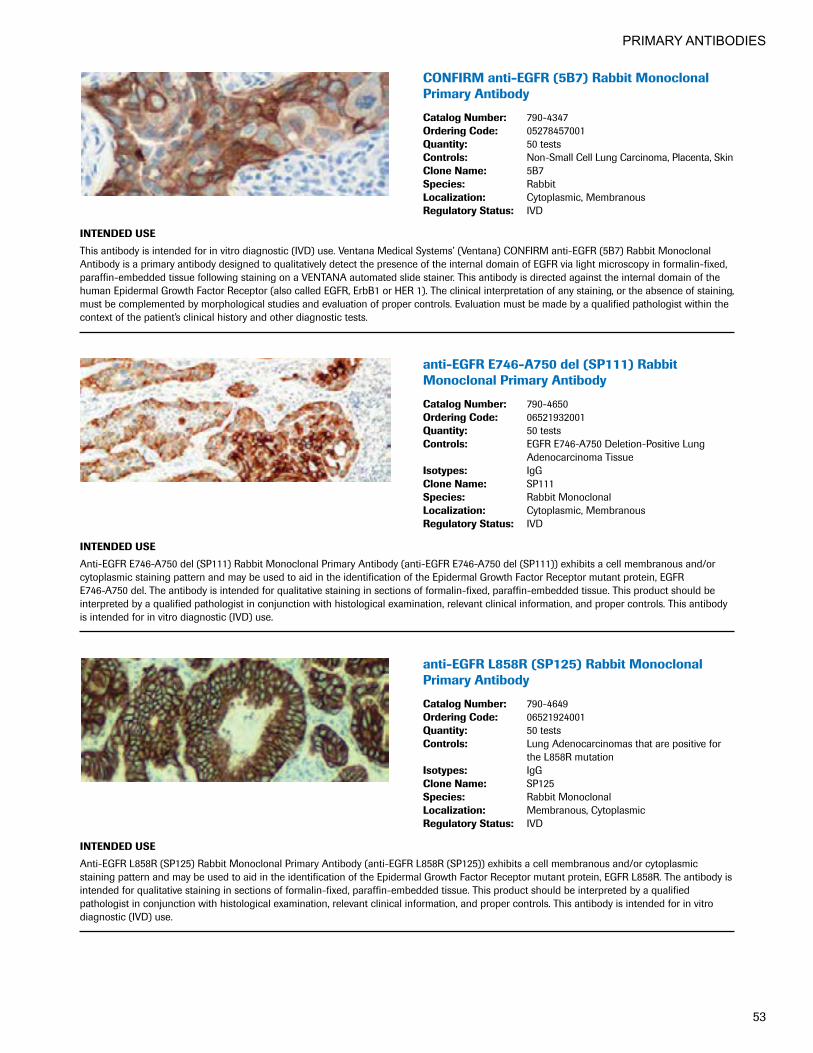
PRIMARY ANTIBODIES
53
CONFIRManti-EGFR(5B7)RabbitMonoclonalPrimary Antibody
CatalogNumber: 790-4347OrderingCode: 05278457001Quantity: 50 testsControls: Non-Small Cell Lung Carcinoma, Placenta, SkinCloneName: 5B7Species: RabbitLocalization: Cytoplasmic, MembranousRegulatoryStatus: IVD
INTENDEDUSE
This antibody is intended for in vitro diagnostic (IVD) use. Ventana Medical Systems’ (Ventana) CONFIRM anti-EGFR (5B7) Rabbit Monoclonal Antibody is a primary antibody designed to qualitatively detect the presence of the internal domain of EGFR via light microscopy in formalin-fixed, paraffin-embedded tissue following staining on a VENTANA automated slide stainer. This antibody is directed against the internal domain of the human Epidermal Growth Factor Receptor (also called EGFR, ErbB1 or HER 1). The clinical interpretation of any staining, or the absence of staining, must be complemented by morphological studies and evaluation of proper controls. Evaluation must be made by a qualified pathologist within the context of the patient’s clinical history and other diagnostic tests.
anti-EGFRE746-A750del(SP111)Rabbit MonoclonalPrimaryAntibody
CatalogNumber: 790-4650OrderingCode: 06521932001Quantity: 50 testsControls: EGFR E746-A750 Deletion-Positive Lung
Adenocarcinoma TissueIsotypes: IgGCloneName: SP111Species: Rabbit MonoclonalLocalization: Cytoplasmic, MembranousRegulatoryStatus: IVD
INTENDEDUSE
Anti-EGFR E746-A750 del (SP111) Rabbit Monoclonal Primary Antibody (anti-EGFR E746-A750 del (SP111)) exhibits a cell membranous and/or cytoplasmic staining pattern and may be used to aid in the identification of the Epidermal Growth Factor Receptor mutant protein, EGFR E746-A750 del. The antibody is intended for qualitative staining in sections of formalin-fixed, paraffin-embedded tissue. This product should be interpreted by a qualified pathologist in conjunction with histological examination, relevant clinical information, and proper controls. This antibody is intended for in vitro diagnostic (IVD) use.
anti-EGFRL858R(SP125)RabbitMonoclonalPrimary Antibody
CatalogNumber: 790-4649OrderingCode: 06521924001Quantity: 50 testsControls: Lung Adenocarcinomas that are positive for
the L858R mutationIsotypes: IgGCloneName: SP125Species: Rabbit MonoclonalLocalization: Membranous, CytoplasmicRegulatoryStatus: IVD
INTENDEDUSE
Anti-EGFR L858R (SP125) Rabbit Monoclonal Primary Antibody (anti-EGFR L858R (SP125)) exhibits a cell membranous and/or cytoplasmic staining pattern and may be used to aid in the identification of the Epidermal Growth Factor Receptor mutant protein, EGFR L858R. The antibody is intended for qualitative staining in sections of formalin-fixed, paraffin-embedded tissue. This product should be interpreted by a qualified pathologist in conjunction with histological examination, relevant clinical information, and proper controls. This antibody is intended for in vitro diagnostic (IVD) use.
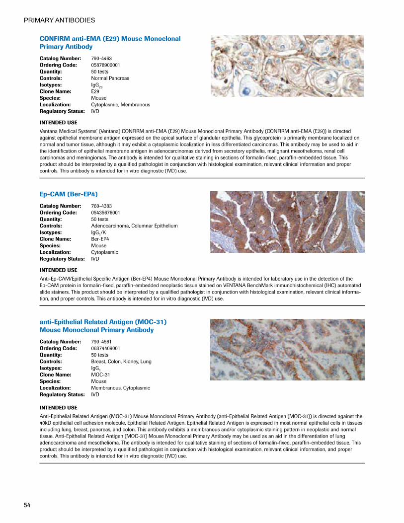
PRIMARY ANTIBODIES
54
CONFIRManti-EMA(E29)MouseMonoclonalPrimary Antibody
CatalogNumber: 790-4463OrderingCode: 05878900001Quantity: 50 testsControls: Normal PancreasIsotypes: IgG2aCloneName: E29Species: MouseLocalization: Cytoplasmic, MembranousRegulatoryStatus: IVD
INTENDEDUSE
Ventana Medical Systems’ (Ventana) CONFIRM anti-EMA (E29) Mouse Monoclonal Primary Antibody (CONFIRM anti-EMA (E29)) is directed against epithelial membrane antigen expressed on the apical surface of glandular epithelia. This glycoprotein is primarily membrane localized on normal and tumor tissue, although it may exhibit a cytoplasmic localization in less differentiated carcinomas. This antibody may be used to aid in the identification of epithelial membrane antigen in adenocarcinomas derived from secretory epithelia, malignant mesothelioma, renal cell carcinomas and meningiomas. The antibody is intended for qualitative staining in sections of formalin-fixed, paraffin-embedded tissue. This product should be interpreted by a qualified pathologist in conjunction with histological examination, relevant clinical information and proper controls. This antibody is intended for in vitro diagnostic (IVD) use.
Ep-CAM(Ber-EP4)
CatalogNumber: 760-4383OrderingCode: 05435676001Quantity: 50 testsControls: Adenocarcinoma, Columnar EpitheliumIsotypes: IgG1/KCloneName: Ber-EP4Species: MouseLocalization: CytoplasmicRegulatoryStatus: IVD
INTENDEDUSE
Anti-Ep-CAM/Epithelial Specific Antigen (Ber-EP4) Mouse Monoclonal Primary Antibody is intended for laboratory use in the detection of the Ep-CAM protein in formalin-fixed, paraffin-embedded neoplastic tissue stained on VENTANA BenchMark immunohistochemical (IHC) automated slide stainers. This product should be interpreted by a qualified pathologist in conjunction with histological examination, relevant clinical informa-tion, and proper controls. This antibody is intended for in vitro diagnostic (IVD) use.
anti-EpithelialRelatedAntigen(MOC-31) MouseMonoclonalPrimaryAntibody
CatalogNumber: 790-4561OrderingCode: 06374409001Quantity: 50 testsControls: Breast, Colon, Kidney, LungIsotypes: IgG1CloneName: MOC-31Species: MouseLocalization: Membranous, CytoplasmicRegulatoryStatus: IVD
INTENDEDUSE
Anti-Epithelial Related Antigen (MOC-31) Mouse Monoclonal Primary Antibody (anti-Epithelial Related Antigen (MOC-31)) is directed against the 40kD epithelial cell adhesion molecule, Epithelial Related Antigen. Epithelial Related Antigen is expressed in most normal epithelial cells in tissues including lung, breast, pancreas, and colon. This antibody exhibits a membranous and/or cytoplasmic staining pattern in neoplastic and normal tissue. Anti-Epithelial Related Antigen (MOC-31) Mouse Monoclonal Primary Antibody may be used as an aid in the differentiation of lung adenocarcinoma and mesothelioma. The antibody is intended for qualitative staining of sections of formalin-fixed, paraffin-embedded tissue. This product should be interpreted by a qualified pathologist in conjunction with histological examination, relevant clinical information, and proper controls. This antibody is intended for in vitro diagnostic (IVD) use.

PRIMARY ANTIBODIES
55
anti-ERG(EPR3864)RabbitMonoclonal Primary Antibody
CatalogNumber: 790-4576OrderingCode: 06478450001Quantity: 50 testsControls: SpleenCloneName: EPR3864Species: RabbitLocalization: Nuclear, CytoplasmicRegulatoryStatus: IVD
INTENDEDUSE
Anti-ERG (EPR3864) Rabbit Monoclonal Primary Antibody (anti-ERG (EPR3864)) is directed against the C-terminus of the ETS transcription regulator, ERG, and is capable of detecting both wildtype ERG, and truncated ERG resulting from ERG gene rearrangement. This antibody exhibits a nuclear staining pattern and may be used to aid in the identification of prostate adenocarcinomas through the detection of truncated ERG. The antibody is intended for qualitative staining in sections of formalin-fixed, paraffin-embedded tissue. This product should be interpreted by a qualified pathologist in conjunction with histological examination, relevant clinical information, and proper controls. This antibody is intended for in vitro diagnostic (IVD) use.
CONFIRManti-EstrogenReceptor(ER)(SP1)RabbitMonoclonalPrimaryAntibody
CatalogNumber: 790-4325 790-4324OrderingCode: 5278414001 05278406001Quantity: 250 tests 50 testsControls: BreastIsotypes: IgGCloneName: SP1Species: RabbitLocalization: NuclearRegulatoryStatus: IVD
INTENDEDUSE
This antibody is intended for in vitro diagnostic (IVD) use. CONFIRM anti-Estrogen Receptor (ER) (SP1) Rabbit Monoclonal Primary Antibody is intended for laboratory use for the qualitative detection of estrogen receptor (ER) antigen in sections of formalin-fixed, paraffin-embedded breast tissue on a VENTANA automated slide stainer with VENTANA detection kits and ancillary reagents. CONFIRM anti-ER (SP1) is directed against an epitope present on human ER alpha protein located in the nucleus of ER positive normal and neoplastic cells. CONFIRM anti-ER (SP1) is indicated as an aid in the management, prognosis, and prediction of hormone therapy for breast carcinoma. This product should be interpreted by a qualified pathologist in conjunction with histological examination, relevant clinical information, and proper controls. Prescription use only.
anti-EZH2(SP129)RabbitMonoclonal Primary Antibody
CatalogNumber: 790-4651OrderingCode: 06522858001Quantity: 50 testsControls: TonsilIsotypes: IgGCloneName: SP129Species: Rabbit MonoclonalLocalization: NuclearRegulatoryStatus: IVD
INTENDEDUSE
Anti-EZH2 (SP129) Rabbit Monoclonal Primary Antibody (antiEZH2 (SP129)) is directed against human EZH2 expressed by cancer cells and lymphocytes. This antibody exhibits a nuclear staining pattern and may be used to aid in the identification of prostate cancer, breast cancer and bladder cancer. The antibody is intended for qualitative staining in sections of formalin-fixed, paraffin-embedded tissue. This product should be interpreted by a qualified pathologist in conjunction with histological examination, relevant clinical information, and proper controls. This antibody is intended for in vitro diagnostic (IVD) use.
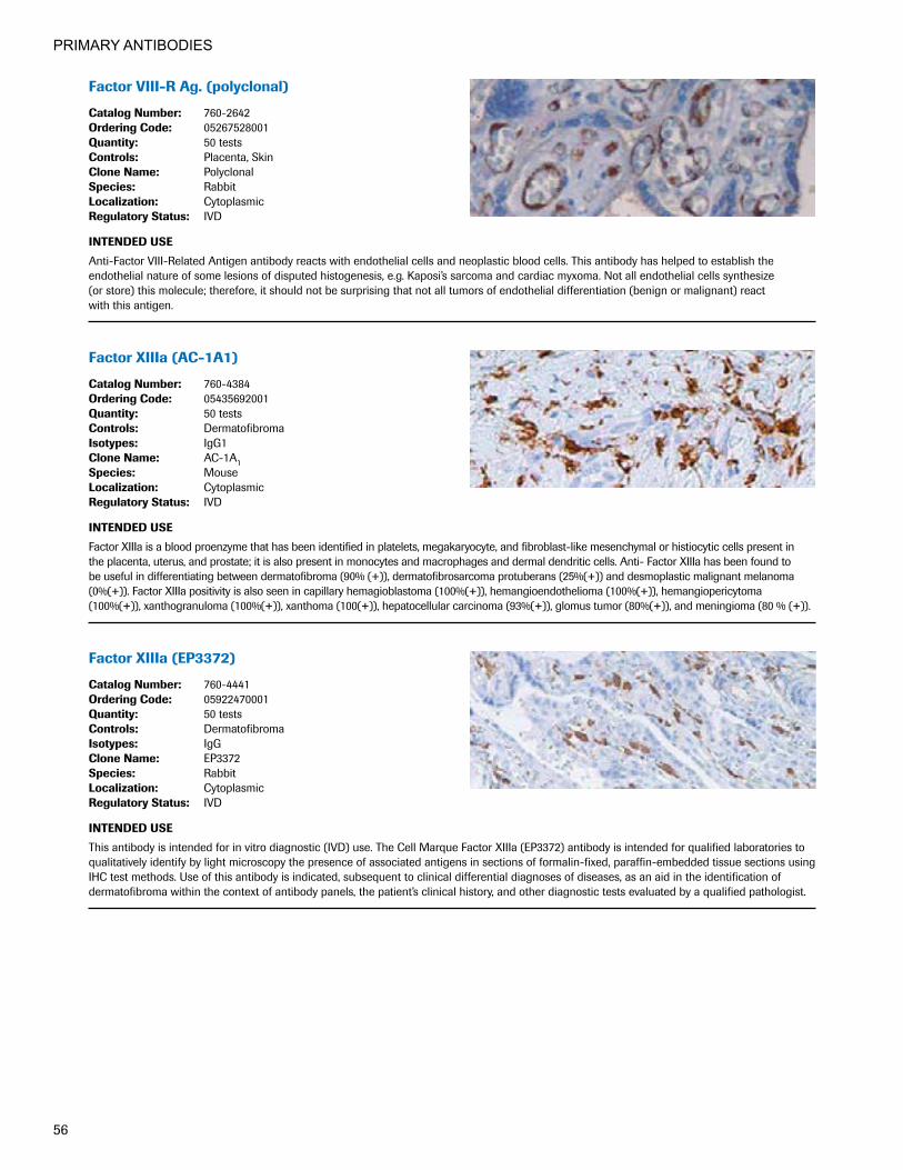
PRIMARY ANTIBODIES
56
FactorVIII-RAg.(polyclonal)
CatalogNumber: 760-2642OrderingCode: 05267528001Quantity: 50 testsControls: Placenta, SkinCloneName: PolyclonalSpecies: RabbitLocalization: CytoplasmicRegulatoryStatus: IVD
INTENDEDUSE
Anti-Factor VIII-Related Antigen antibody reacts with endothelial cells and neoplastic blood cells. This antibody has helped to establish the endothelial nature of some lesions of disputed histogenesis, e.g. Kaposi’s sarcoma and cardiac myxoma. Not all endothelial cells synthesize (or store) this molecule; therefore, it should not be surprising that not all tumors of endothelial differentiation (benign or malignant) react with this antigen.
FactorXIIIa(AC-1A1)
CatalogNumber: 760-4384OrderingCode: 05435692001Quantity: 50 testsControls: DermatofibromaIsotypes: IgG1CloneName: AC-1A1Species: MouseLocalization: CytoplasmicRegulatoryStatus: IVD
INTENDEDUSE
Factor XIIIa is a blood proenzyme that has been identified in platelets, megakaryocyte, and fibroblast-like mesenchymal or histiocytic cells present in the placenta, uterus, and prostate; it is also present in monocytes and macrophages and dermal dendritic cells. Anti- Factor XIIIa has been found to be useful in differentiating between dermatofibroma (90% (+)), dermatofibrosarcoma protuberans (25%(+)) and desmoplastic malignant melanoma (0%(+)). Factor XIIIa positivity is also seen in capillary hemagioblastoma (100%(+)), hemangioendothelioma (100%(+)), hemangiopericytoma (100%(+)), xanthogranuloma (100%(+)), xanthoma (100(+)), hepatocellular carcinoma (93%(+)), glomus tumor (80%(+)), and meningioma (80 % (+)).
FactorXIIIa(EP3372)
CatalogNumber: 760-4441OrderingCode: 05922470001Quantity: 50 testsControls: DermatofibromaIsotypes: IgGCloneName: EP3372Species: RabbitLocalization: CytoplasmicRegulatoryStatus: IVD
INTENDEDUSE
This antibody is intended for in vitro diagnostic (IVD) use. The Cell Marque Factor XIIIa (EP3372) antibody is intended for qualified laboratories to qualitatively identify by light microscopy the presence of associated antigens in sections of formalin-fixed, paraffin-embedded tissue sections using IHC test methods. Use of this antibody is indicated, subsequent to clinical differential diagnoses of diseases, as an aid in the identification of dermatofibroma within the context of antibody panels, the patient’s clinical history, and other diagnostic tests evaluated by a qualified pathologist.

PRIMARY ANTIBODIES
57
Fascin(55k-2)
CatalogNumber: 760-2702OrderingCode: 05268117001Quantity: 50 testsControls: Hodgkin's Lymphoma, Lymph NodeIsotypes: IgG1CloneName: 55k-2Species: MouseLocalization: CytoplasmicRegulatoryStatus: IVD
INTENDEDUSE
Anti-Fascin is a very sensitive marker for Reed-Sternberg cells and variants in nodular sclerosis, mixed cellularity, and lymphocyte depletion Hodgkin’s disease. It is uniformly negative in lymphoid cells, plasma cells and myeloid cells. Anti-Fascin is positive in dendritic cells. This marker may be helpful in distinguishing between Hodgkin’s disease and non-Hodgkin’s lymphoma in difficult cases. Also, the lack of expression of Fascin in the neoplastic follicles in follicular lymphoma can be helpful in distinguishing these lymphomas from reactive follicular hyperplasia in which the number of follicular dendritic cells is normal or increased. Anti-Fascin has been suggested as a prognostic marker in neuroendocrine neoplasms of the lung, as well as ovarian cancer.
FITCanti-AlbuminPrimaryAntibody
CatalogNumber: 760-2699OrderingCode: 05268087001Quantity: 50 testsControls: Frozen, Diseased Skin or Diseased Renal TissueCloneName: PolyclonalSpecies: GoatRegulatoryStatus: IVD
INTENDEDUSE
This antibody is intended for in vitro diagnostic (IVD) use. Ventana® Medical Systems’ (Ventana) Systems FITC anti-Albumin Primary Antibody is a goat derived polyclonal antibody labeled with fluorescein and specifically directed against human albumin. This reagent should be used in conjunction with a panel of antibodies to aid in the identification of human albumin in target tissue (e.g., in the diagnosis of renal or dermal pathologies). FITC anti-Albumin is intended for laboratory use to qualitatively stain sections of frozen tissue on a VENTANA automated slide stainer. The clinical interpretation of any staining, or the absence of staining, must be complemented by morphological studies and evaluation of proper controls. Evaluation must be made by a qualified pathologist within the context of the patient’s clinical history and other diagnostic tests. Prescription only.
FITCanti-C1qPrimaryAntibody
CatalogNumber: 760-2688OrderingCode: 05267994001Quantity: 50 testsControls: Frozen, Diseased Skin or Diseased Renal TissueCloneName: PolyclonalSpecies: GoatRegulatoryStatus: IVD
INTENDEDUSE
This antibody is intended for in vitro diagnostic (IVD) use. Ventana Medical Systems’ (Ventana) Systems FITC anti-C1q (complement 1q) Primary Antibody is a goat derived polyclonal antibody labeled with fluorescein and specifically directed against human C1q. This reagent should be used in conjunction with a panel of antibodies to aid in the identification of complement 1q in C1q target tissue (e.g., in the diagnosis of renal or dermal pathologies). FITC anti-C1q is intended for laboratory use to qualitatively stain sections of frozen tissue on a VENTANA automated slide stainer. The clinical interpretation of any staining, or the absence of staining, must be complemented by morphological studies and evaluation of proper controls. Evaluation must be made by a qualified pathologist within the context of the patient’s clinical history and other diagnostic tests. Prescription only.
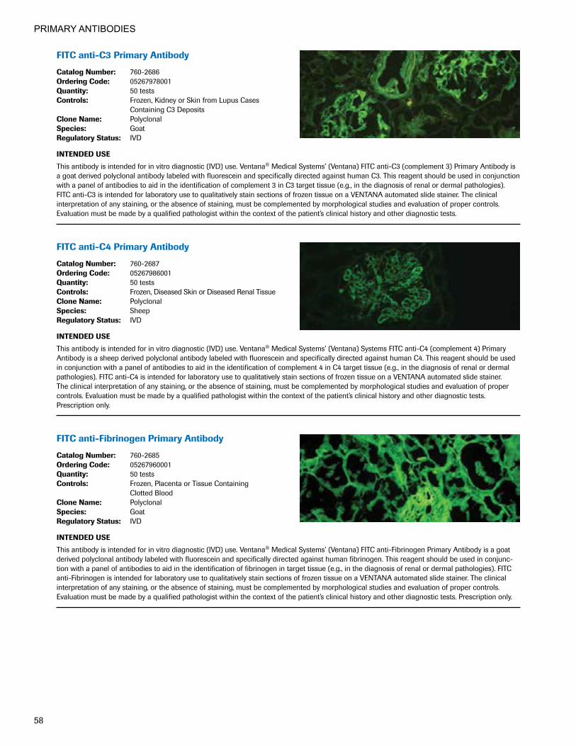
PRIMARY ANTIBODIES
58
FITCanti-C3PrimaryAntibody
CatalogNumber: 760-2686OrderingCode: 05267978001Quantity: 50 testsControls: Frozen, Kidney or Skin from Lupus Cases
Containing C3 DepositsCloneName: PolyclonalSpecies: GoatRegulatoryStatus: IVD
INTENDEDUSE
This antibody is intended for in vitro diagnostic (IVD) use. Ventana® Medical Systems’ (Ventana) FITC anti-C3 (complement 3) Primary Antibody is a goat derived polyclonal antibody labeled with fluorescein and specifically directed against human C3. This reagent should be used in conjunction with a panel of antibodies to aid in the identification of complement 3 in C3 target tissue (e.g., in the diagnosis of renal or dermal pathologies). FITC anti-C3 is intended for laboratory use to qualitatively stain sections of frozen tissue on a VENTANA automated slide stainer. The clinical interpretation of any staining, or the absence of staining, must be complemented by morphological studies and evaluation of proper controls. Evaluation must be made by a qualified pathologist within the context of the patient’s clinical history and other diagnostic tests.
FITCanti-C4PrimaryAntibody
CatalogNumber: 760-2687OrderingCode: 05267986001Quantity: 50 testsControls: Frozen, Diseased Skin or Diseased Renal TissueCloneName: PolyclonalSpecies: SheepRegulatoryStatus: IVD
INTENDEDUSE
This antibody is intended for in vitro diagnostic (IVD) use. Ventana® Medical Systems’ (Ventana) Systems FITC anti-C4 (complement 4) Primary Antibody is a sheep derived polyclonal antibody labeled with fluorescein and specifically directed against human C4. This reagent should be used in conjunction with a panel of antibodies to aid in the identification of complement 4 in C4 target tissue (e.g., in the diagnosis of renal or dermal pathologies). FITC anti-C4 is intended for laboratory use to qualitatively stain sections of frozen tissue on a VENTANA automated slide stainer. The clinical interpretation of any staining, or the absence of staining, must be complemented by morphological studies and evaluation of proper controls. Evaluation must be made by a qualified pathologist within the context of the patient’s clinical history and other diagnostic tests. Prescription only.
FITCanti-FibrinogenPrimaryAntibody
CatalogNumber: 760-2685OrderingCode: 05267960001Quantity: 50 testsControls: Frozen, Placenta or Tissue Containing
Clotted BloodCloneName: PolyclonalSpecies: GoatRegulatoryStatus: IVD
INTENDEDUSE
This antibody is intended for in vitro diagnostic (IVD) use. Ventana® Medical Systems’ (Ventana) FITC anti-Fibrinogen Primary Antibody is a goat derived polyclonal antibody labeled with fluorescein and specifically directed against human fibrinogen. This reagent should be used in conjunc-tion with a panel of antibodies to aid in the identification of fibrinogen in target tissue (e.g., in the diagnosis of renal or dermal pathologies). FITC anti-Fibrinogen is intended for laboratory use to qualitatively stain sections of frozen tissue on a VENTANA automated slide stainer. The clinical interpretation of any staining, or the absence of staining, must be complemented by morphological studies and evaluation of proper controls. Evaluation must be made by a qualified pathologist within the context of the patient’s clinical history and other diagnostic tests. Prescription only.

PRIMARY ANTIBODIES
59
FITCanti-IgAPrimaryAntibody
CatalogNumber: 760-2681OrderingCode: 05267927001Quantity: 50 testsControls: Frozen, Tonsil or Lymph NodeCloneName: PolyclonalSpecies: GoatRegulatoryStatus: IVD
INTENDEDUSE
This antibody is intended for in vitro diagnostic (IVD) use. Ventana® Medical Systems’ (Ventana) Systems FITC anti-IgA (immunoglobulin A) Primary Antibody is a goat derived polyclonal antibody labeled with fluorescein and specifically directed against human IgA. This reagent should be used in conjunction with a panel of antibodies to aid in the identification of immunoglobulin A in IgA target tissue (e.g., in the diagnosis of renal or dermal pathologies). FITC anti-IgA is intended for laboratory use to qualitatively stain sections of frozen tissue on a VENTANA automated slide stainer. The clinical interpretation of any staining, or the absence of staining, must be complemented by morphological studies and evaluation of proper controls. Evaluation must be made by a qualified pathologist within the context of the patient’s clinical history and other diagnostic tests. Prescription only.
FITCanti-IgGPrimaryAntibody
CatalogNumber: 760-2680OrderingCode: 05267919001Quantity: 50 testsControls: Frozen, Tonsil or Lymph Node Clotted BloodCloneName: PolyclonalSpecies: GoatRegulatoryStatus: IVD
INTENDEDUSE
This antibody is intended for in vitro diagnostic (IVD) use. Ventana® Medical Systems’ (Ventana) Systems FITC anti-IgG (immunoglobulin G) Primary Antibody is a goat derived polyclonal antibody labeled with fluorescein and specifically directed against human IgG. This reagent should be used in conjunction with a panel of antibodies to aid in the identification of immunoglobulin G in IgG target tissue (e.g., in the diagnosis of renal or dermal pathologies). FITC anti-IgG is intended for laboratory use to qualitatively stain sections of frozen tissue on a VENTANA automated slide stainer. The clinical interpretation of any staining, or the absence of staining, must be complemented by morphological studies and evaluation of proper controls. Evaluation must be made by a qualified pathologist within the context of the patient’s clinical history and other diagnostic tests. Prescription only.
FITCanti-IgMPrimaryAntibody
CatalogNumber: 760-2682OrderingCode: 05267935001Quantity: 50 testsControls: Frozen, Tonsil or Lymph NodeCloneName: PolyclonalSpecies: GoatRegulatoryStatus: IVD
INTENDEDUSE
This antibody is intended for in vitro diagnostic (IVD) use. Ventana® Medical Systems’ (Ventana) Systems FITC anti-IgM (immunoglobulin M) Primary Antibody is a goat derived polyclonal antibody labeled with fluorescein and specifically directed against human IgM. This reagent should be used in conjunction with a panel of antibodies to aid in the identification of immunoglobulin M in IgM target tissue (e.g., in the diagnosis of renal or dermal pathologies). FITC anti-IgM is intended for laboratory use to qualitatively stain sections of frozen tissue on a VENTANA automated slide stainer. The clinical interpretation of any staining, or the absence of staining, must be complemented by morphological studies and evaluation of proper controls. Evaluation must be made by a qualified pathologist within the context of the patient’s clinical history and other diagnostic tests. Prescription only.

PRIMARY ANTIBODIES
60
FITCanti-KappaPrimaryAntibody
CatalogNumber: 760-2683OrderingCode: 05267943001Quantity: 50 testsControls: Frozen, Tonsil or Lymph Node Clotted BloodCloneName: PolyclonalSpecies: GoatRegulatoryStatus: IVD
INTENDEDUSE
This antibody is intended for in vitro diagnostic (IVD) use. Ventana® Medical Systems’ (Ventana) Systems FITC anti-Kappa Primary Antibody is a goat derived polyclonal antibody labeled with fluorescein and specifically directed against human kappa immunoglobulin light chains. This reagent should be used in conjunction with a panel of antibodies to aid in the identification of kappa in target tissue (e.g., in the diagnosis of renal or dermal pathologies). FITC anti-Kappa is intended for laboratory use to qualitatively stain sections of frozen tissue on a VENTANA automated slide stainer. The clinical interpretation of any staining, or the absence of staining, must be complemented by morphological studies and evaluation of proper controls. Evaluation must be made by a qualified pathologist within the context of the patient’s clinical history and other diagnostic tests. Prescription only.
FITCanti-LambdaPrimaryAntibody
CatalogNumber: 760-2684OrderingCode: 05267951001Quantity: 50 testsControls: Frozen, Tonsil or Lymph NodeCloneName: PolyclonalSpecies: BurroRegulatoryStatus: IVD
INTENDEDUSE
This antibody is intended for in vitro diagnostic (IVD) use. Ventana® Medical Systems’ (Ventana) Systems FITC anti-Lambda Primary Antibody is a burro derived polyclonal antibody labeled with fluorescein and specifically directed against human lambda light chains. This reagent should be used in conjunction with a panel of antibodies to aid in the identification of lambda light chains in target tissue (e.g., in the diagnosis of renal or dermal pathologies). FITC anti-Lambda is intended for laboratory use to qualitatively stain sections of frozen tissue on a VENTANA automated slide stainer. The clinical interpretation of any staining, or the absence of staining, must be complemented by morphological studies and evaluation of proper controls. Evaluation must be made by a qualified pathologist within the context of the patient’s clinical history and other diagnostic tests. Prescription only.
FOXA1(2F83)MouseMonoclonal Primary Antibody
CatalogNumber: 760-4937OrderingCode: 07292848001Quantity: 50 testsControls: Breast CarcinomaIsotypes: IgGCloneName: FOXA1 (2F83) Mouse Monoclonal
Primary AntibodySpecies: MouseLocalization: NuclearRegulatoryStatus: IVDINTENDEDUSE
This antibody is intended for in vitro diagnostic (IVD) use. The Cell Marque FOXA1 (2F83) antibody is intended for qualified laboratories to qualitatively identify by light microscopy the presence of associated antigens in sections of formalin-fixed, paraffin-embedded tissue sections using IHC test methods. Use of this antibody is indicated, subsequent to clinical differential diagnoses of diseases, as an aid in the identification of breast carcinomas within the context of antibody panels, the patient’s clinical history, and other diagnostic tests evaluated by a qualified pathologist.

PRIMARY ANTIBODIES
61
FSH(polyclonal)
CatalogNumber: 760-2710OrderingCode: 05268192001Quantity: 50 testsControls: Normal PituitaryCloneName: PolyclonalSpecies: RabbitLocalization: CytoplasmicRegulatoryStatus: IVD
INTENDEDUSE
Anti-FSH is a useful marker in classification of pituitary tumors and the study of pituitary disease. It reacts with FSH-producing cells (gonadotrophs).
Galectin-3(9C4)
CatalogNumber: 760-4256OrderingCode: 05269164001Quantity: 50 testsControls: Papillary or Follicular Carcinoma of ThyroidIsotypes: IgG1CloneName: 9C4Species: MouseLocalization: CytoplasmicRegulatoryStatus: IVD
INTENDEDUSE
Galectin-3 is a 31 kD beta-galactosidase binding lectin. It has been associated with binding to the basement membrane glycoprotein laminin. Anti-Galectin-3 has been demonstrated to be valuable in differentiating between benign and malignant thyroid neoplasms in both histologic sections and fine needle aspiration biopsy material. Anti-Galectin-3 antibody has also been useful in identifying anaplastic large cell lymphoma.
Gastrin(polyclonal)
CatalogNumber: 760-2643OrderingCode: 05267536001Quantity: 50 testsControls: StomachCloneName: PolyclonalSpecies: RabbitLocalization: CytoplasmicRegulatoryStatus: IVD
INTENDEDUSE
Anti-Gastrin antibody gives positive staining of G-cells of human antral/pyloric mucosa and cells producing gastrin or a structural gastrin analogue as is seen in stomach; no staining of other cells or tissue types has been observed. This antibody may react with sulfated and non-sulfated forms of gastrin. The antibody cross-reacts with more than 50% of the present choleocystokinin octapeptide.
GATA3(L50-823)MouseMonoclonal Primary Antibody
CatalogNumber: 760-4897OrderingCode: 07107749001Quantity: 50 testsControls: Ductal Carcinoma in situIsotypes: IgGCloneName: L50-823Species: MouseLocalization: NuclearRegulatoryStatus: IVD
INTENDEDUSE
This antibody is intended for in vitro diagnostic (IVD) use. The Cell Marque GATA3 (L50-823) antibody is intended for qualified laboratories to qualitatively identify by light microscopy the presence of associated antigens in sections of formalin-fixed, paraffin-embedded tissue sections using IHC test methods. Use of this antibody is indicated, subsequent to clinical differential diagnosis, as an aid in the identification of breast carcinomas and urothelial carcinomas within the context of an antibody panel, the patient’s clinical history, and other diagnostic tests evaluated by a qualified pathologist.

PRIMARY ANTIBODIES
62
GCDFP-15(EP1582Y)RabbitMonoclonalAntibody
CatalogNumber: 760-4386OrderingCode: 05463530001Quantity: 50 testsControls: Breast, Breast CarcinomaIsotypes: IgGCloneName: EP1582YSpecies: RabbitLocalization: CytoplasmicRegulatoryStatus: IVD
INTENDEDUSE
This antibody is intended for in vitro diagnostic (IVD) use. The Cell Marque GCDFP-15 (EP1582Y) antibody is intended for qualified laboratories to qualitatively identify by light microscopy the presence of associated antigens in sections of formalin-fixed, paraffin-embedded tissue sections using IHC test methods. Use of this antibody is indicated, subsequent to clinical differential diagnosis, as an aid in the identification of GCDFP-15 within the context of an antibody panel, the patient’s clinical history, and other diagnostic tests evaluated by a qualified pathologist.
GH(polyclonal)
CatalogNumber: 760-2804OrderingCode: 05268257001Quantity: 50 testsControls: Normal PituitaryCloneName: PolyclonalSpecies: RabbitLocalization: CytoplasmicRegulatoryStatus: IVD
INTENDEDUSE
Anti-GH is a useful marker in classification of pituitary tumors and the study of pituitary disease (acromegaly). It reacts with GH-producing cells. Growth hormone receptors have been found in various non-pituitary cells, including that from hepatocellular carcinoma and various benign and malignant cutaneous lesions.
GlialFibrillaryAcidicProtein(GFAP)(EP672Y)
CatalogNumber: 760-4345OrderingCode: 05269784001Quantity: 50 testsControls: BrainIsotypes: IgGCloneName: EP672YSpecies: RabbitLocalization: CytoplasmicRegulatoryStatus: IVD
INTENDEDUSE
This antibody is intended for in vitro diagnostic (IVD) use. The Cell Marque Glial Fibrillary Acidic Protein (EP672Y) antibody is intended for qualified laboratories to qualitatively identify by light microscopy the presence of associated antigens in sections of formalinfixed, paraffin-embedded tissue sections using IHC test methods. Use of this antibody is indicated, subsequent to clinical differential diagnoses of diseases, as an aid in the identification of astrocytes and glial lesions in the central nervous system under normal and pathological conditions within the context of antibody panels, the patient’s clinical history, and other diagnostic tests evaluated by a qualified pathologist.
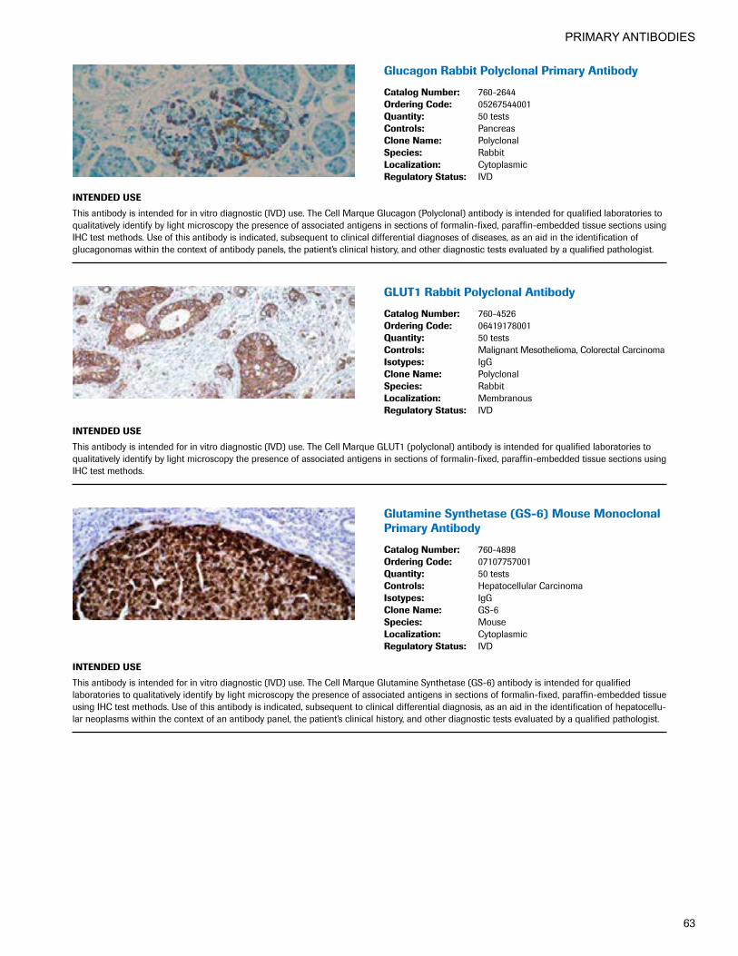
PRIMARY ANTIBODIES
63
GlucagonRabbitPolyclonalPrimaryAntibody
CatalogNumber: 760-2644OrderingCode: 05267544001Quantity: 50 testsControls: PancreasCloneName: PolyclonalSpecies: RabbitLocalization: CytoplasmicRegulatoryStatus: IVD
INTENDEDUSE
This antibody is intended for in vitro diagnostic (IVD) use. The Cell Marque Glucagon (Polyclonal) antibody is intended for qualified laboratories to qualitatively identify by light microscopy the presence of associated antigens in sections of formalin-fixed, paraffin-embedded tissue sections using IHC test methods. Use of this antibody is indicated, subsequent to clinical differential diagnoses of diseases, as an aid in the identification of glucagonomas within the context of antibody panels, the patient’s clinical history, and other diagnostic tests evaluated by a qualified pathologist.
GLUT1RabbitPolyclonalAntibody
CatalogNumber: 760-4526OrderingCode: 06419178001Quantity: 50 testsControls: Malignant Mesothelioma, Colorectal CarcinomaIsotypes: IgGCloneName: PolyclonalSpecies: RabbitLocalization: MembranousRegulatoryStatus: IVD
INTENDEDUSE
This antibody is intended for in vitro diagnostic (IVD) use. The Cell Marque GLUT1 (polyclonal) antibody is intended for qualified laboratories to qualitatively identify by light microscopy the presence of associated antigens in sections of formalin-fixed, paraffin-embedded tissue sections using IHC test methods.
GlutamineSynthetase(GS-6)MouseMonoclonalPrimary Antibody
CatalogNumber: 760-4898OrderingCode: 07107757001Quantity: 50 testsControls: Hepatocellular CarcinomaIsotypes: IgGCloneName: GS-6Species: MouseLocalization: CytoplasmicRegulatoryStatus: IVD
INTENDEDUSE
This antibody is intended for in vitro diagnostic (IVD) use. The Cell Marque Glutamine Synthetase (GS-6) antibody is intended for qualified laboratories to qualitatively identify by light microscopy the presence of associated antigens in sections of formalin-fixed, paraffin-embedded tissue using IHC test methods. Use of this antibody is indicated, subsequent to clinical differential diagnosis, as an aid in the identification of hepatocellu-lar neoplasms within the context of an antibody panel, the patient’s clinical history, and other diagnostic tests evaluated by a qualified pathologist.

PRIMARY ANTIBODIES
64
GlycophorinA(GA-R2)
CatalogNumber: 760-4257OrderingCode: 05269172001Quantity: 50 testsControls: Bone MarrowIsotypes: IgG2a/KCloneName: GA-R2Species: MouseLocalization: MembranousRegulatoryStatus: IVD
INTENDEDUSE
This antibody is intended for in vitro diagnostic (IVD) use. The Cell Marque Glycophorin A (GA-R2) antibody is intended for qualified laboratories to qualitatively identify by light microscopy the presence of associated antigens in sections of formalin-fixed, paraffin-embedded tissue sections using IHC test methods. Use of this antibody is indicated, subsequent to clinical differential diagnoses of diseases, as an aid in the identification of benign erythroid proliferation and neoplasms derived from erythroid lineage within the context of antibody panels, the patient’s clinical history, and other diagnostic tests evaluated by a qualified pathologist.
Glypican-3(1G12)
CatalogNumber: 760-4442OrderingCode: 05973864001Quantity: 50 testsControls: Hepatocellular CarcinomaIsotypes: IgG1CloneName: 1G12Species: MouseLocalization: CytoplasmicRegulatoryStatus: IVD
INTENDEDUSE
This antibody is intended for in vitro diagnostic (IVD) use. The Cell Marque Glypican-3 (1G12) antibody is intended for qualified laboratories to qualitatively identify by light microscopy the presence of associated antigens in sections of formalin-fixed, paraffin-embedded tissue sections using IHC test methods. Use of this antibody is indicated, subsequent to clinical differential diagnoses of diseases, as an aid in the identification of hepatocellular carcinoma (HCC) within the context of antibody panels, the patient’s clinical history, and other diagnostic tests evaluated by a qualified pathologist.
anti-Glypican3(GC33)MouseMonoclonalPrimary Antibody
CatalogNumber: 790-4564OrderingCode: 06483186001Quantity: 50 testsControls: Placenta, Hepatocellular CarcinomaIsotypes: IgG2aCloneName: GC33Species: MouseLocalization: Granular Cytoplasmic/MembraneRegulatoryStatus: IVD
INTENDEDUSE
Anti-Glypican 3 (GC33) Mouse Monoclonal Primary Antibody (antiGlypican 3 (GC33)) is directed against the heparan sulfate proteoglycan, glypican 3. This antibody may be used to aid in the differentiation of hepatocellular carcinoma from normal liver or benign lesions. The antibody is intended for qualitative staining in sections of formalin-fixed, paraffin-embedded tissue. This product should be interpreted by a qualified pathologist in conjunction with histological examination, relevant clinical information and proper controls. This antibody is intended for in vitro diagnostic (IVD) use.

PRIMARY ANTIBODIES
65
GranzymeB(polyclonal)
CatalogNumber: 760-4283OrderingCode: 05269466001Quantity: 50 testsControls: TonsilCloneName: PolyclonalSpecies: RabbitLocalization: Granular 2, Cytotoxic Thymphocytes,
Natural Killer CellsRegulatoryStatus: IVD
INTENDEDUSE
Granzymes are serine proteases which are stored in specialized lytic granules of cytotoxic T lymphocytes and in natural killer cells. Anti-Granzyme B has been useful in diagnosing Natural killer/T cell lymphoma, as well as anaplastic large cell lymphoma. High percentages of cytotoxic T cells have been shown to be an unfavorable prognostic indicator in Hodgkin’s disease.
HBME-1(HBME-1)
CatalogNumber: 760-4445OrderingCode: 05973813001Quantity: 50 testsControls: Mesothelioma, Thyroid CarcinomaIsotypes: IgM/KCloneName: HBME-1Species: MouseLocalization: Cytoplasmic, MembranousRegulatoryStatus: IVD
INTENDEDUSE
Anti-HBME-1 has been demonstrated to label mesothelial cells, both benign and malignant (malignant mesothelioma) and thus has been used in distinguishing mesothelioma from adenocarcinomas of various origins. More recently this antibody has been used to distinguish Thyroid carcinomas (both Follicular and Papillary) from benign thyroid lesions.
hCG(polyclonal)
CatalogNumber: 760-2650OrderingCode: 05267617001Quantity: 50 testsControls: PlacentaCloneName: PolyclonalSpecies: RabbitLocalization: CytoplasmicRegulatoryStatus: IVD
INTENDEDUSE
hCG is a protein secreted in large quantities by normal trophoblasts; the antibody detects cells and tumors of trophoblastic origin such as Choriocarcinoma. Large Cell Carcinoma and Adenocarcinoma of Lung demonstrate hCG positivity in 90% and 60% of cases respectively. 20% of Squamous Cell Lung Carcinomas are positive for hCG. hCG expression by non-trophoblastic tumors may indicate aggressive behavior since it has been observed that hCG may play a role in the host response to a given tumor.

PRIMARY ANTIBODIES
66
VENTANAanti-Helicobacter pylori (SP48) RabbitMonoclonalPrimaryAntibody
CatalogNumber: 790-1014 790-1015OrderingCode: 06425607001 6425623001Quantity: 50 tests 250 testsControls: H. pylori-Infected StomachCloneName: SP48Species: RabbitLocalization: Whole H. pylori OrganismRegulatoryStatus: 510(k)
INTENDEDUSE
VENTANA anti-Helicobacter pylori (SP48) Rabbit Monoclonal Primary Antibody (VENTANA anti-H. pylori (SP48)) is designed to qualitatively detect the presence of H. pylori in formalin-fixed, paraffin-embedded gastric biopsy tissue via light microscopy. Immunohistochemical staining with this antibody product may aid in the diagnosis of H. pylori infection. This product should be interpreted by a qualified pathologist in conjunction with histological examination, relevant clinical information and proper controls. This antibody is intended for in vitro diagnostic (IVD) use.
HemoglobinA(SP212)RabbitMonoclonal Primary Antibody
CatalogNumber: 760-4936OrderingCode: 07292830001Quantity: 50 testsControls: Bone MarrowIsotypes: IgGCloneName: Hemoglobin A (SP212) Rabbit Monoclonal
Primary AntibodySpecies: RabbitLocalization: CytoplasmicRegulatoryStatus: IVD
INTENDEDUSE
This antibody is intended for in vitro diagnostic (IVD) use. The Cell Marque Hemoglobin A (SP212) antibody is intended for qualified laboratories to qualitatively identify by light microscopy the presence of associated antigens in sections of formalin-fixed, paraffin-embedded tissue sections using IHC test methods. Use of this antibody is indicated, subsequent to clinical differential diagnosis, as an aid in the identification of erythrocytes and precursors within the context of an antibody panel, the patient’s clinical history, and other diagnostic tests evaluated by a qualified pathologist.
HepatocyteSpecificAntigen(OCH1E5)
CatalogNumber: 760-4350OrderingCode: 05269792001Quantity: 50 testsControls: LiverIsotypes: IgG1/KCloneName: 0CH1E5Species: MouseLocalization: CytoplasmicRegulatoryStatus: IVD
INTENDEDUSE
Anti-Hepatocyte Specific Antigen recognizes both benign and malignant liver derived tissues including such tumors as hepatoblastoma, hepatocellular carcinoma, and hepatic adenoma. It recognizes both normal adult and fetal liver tissue. The typical pattern is a granular cytoplasmic staining. This antibody is useful in differentiating hepatocellular carcinomas with adenoid features from adenocarcinomas, either primary in the liver or metastatic lesions to the liver. In recognizing hepatoblastoma, it is useful in differentiating this entity from other small round cell tumors.

PRIMARY ANTIBODIES
67
PATHWAYanti-HER-2/neu(4B5) RabbitMonoclonalPrimaryAntibody
CatalogNumber: 790-2991OrderingCode: 05278368001Quantity: 50 testsControls: Breast CarcinomaCloneName: 4B5Species: RabbitLocalization: MembranousRegulatoryStatus: IVD
INTENDEDUSE
This antibody is intended for in vitro diagnostic use. Ventana Medical Systems, Inc.’s (Ventana) PATHWAY anti-HER-2/neu (4B5) Rabbit Monoclonal Primary Antibody (PATHWAY HER2 (4B5)) is a rabbit monoclonal antibody intended for laboratory use for the semi-quantitative detection of HER2 antigen in sections of formalin-fixed, paraffin-embedded normal and neoplastic tissue on a VENTANA automated immunohistochemistry slide staining device. It is indicated as an aid in the assessment of breast cancer patients for whom Herceptin treatment is considered.
Note: All of the patients in the Herceptin clinical trials were selected using a clinical trial assay. None of the patients in those trials were selected using PATHWAY anti-HER-2/neu (4B5). PATHWAY anti-HER-2/neu (4B5) was compared to PATHWAY HER-2 (clone CB11) Primary Antibody on an independent sample set and found to provide acceptably concordant results. The actual correlation of PATHWAY anti-HER-2/neu (4B5) to clinical outcome has not been established.
The VIAS Image Analysis System is an adjunctive optional computer-assisted image analysis system functionally connected to an interactive microscope. It is intended for use as an aid to the pathologist in the detection, classification and counting of cells of interest based on marker intensity, size and shape using appropriate controls to assure the validity of the VIAS scores. Prescription use only.
HGAL(MRQ-49)MouseMonoclonalAntibody
CatalogNumber: 760-4597OrderingCode: 06433375001Quantity: 50 testsControls: Lymph Node, TonsilIsotypes: IgG2a/KCloneName: MRQ-49Species: Mouse MonoclonalLocalization: CytoplasmicRegulatoryStatus: IVD
INTENDEDUSE
This antibody is intended for in vitro diagnostic (IVD) use. The Cell Marque HGAL (MRQ-49) antibody is intended for qualified laboratories to qualitatively identify by light microscopy the presence of associated antigens in sections of formalin-fixed, paraffin-embedded tissue sections using IHC test methods. Use of this antibody is indicated as an aid in the identification of follicular lymphoma within the context of an antibody panel, the patient’s clinical history, and other diagnostic tests evaluated by a qualified pathologist.
HHV-8(13B10)
CatalogNumber: 760-4260OrderingCode: 05269229001Quantity: 50 testsControls: Kaposis SarcomaIsotypes: IgG1CloneName: 13B10Species: MouseLocalization: NuclearRegulatoryStatus: IVD
INTENDEDUSE
Human herpesvirus type 8 (HHV-8) is the likely etiological agent of Kaposi’s sarcoma (KS). HHV-8 DNA sequences have been found in Kaposi’s sarcoma lesions, primary effusion lymphoma, and multicentric Castleman’s disease via polymerase chain reaction and in situ hybridization. Latent nuclear antigen (LNA-1, LNA, LANA-1), also known as ORF73, is a 222- or 234 kD protein that is consistently expressed in HHV-8 infected cells. Anti-HHV-8 labels the latent nuclear antigen protein via immunohistochemistry.

PRIMARY ANTIBODIES
68
HumanEquilibrativeNucleosideTransporter1(SP120)RabbitMonoclonalPrimaryAntibody
CatalogNumber: 790-4852OrderingCode: 06903444001Quantity: 50 testsControls: Adrenal Cortex, Kidney, Thyroid, Tonsil,
Normal PancreasIsotypes: IgGCloneName: SP120Species: Rabbit MonoclonalLocalization: Membranous, Cytoplasmic RegulatoryStatus: IVD
INTENDEDUSE
Anti-hENT1 (SP120) Rabbit Monoclonal Primary Antibody is intended for laboratory use in the detection of hENT1 in formalin-fixed, paraffin- embedded tissue. This antibody exhibits a membranous and/or cytoplasmic staining pattern and may be used to detect hENT1 protein in benign and neoplastic human pancreatic, breast, lymphoid or lung tissue stained with a VENTANA immunohistochemical automated slide stainer. This product should be interpreted by a qualified pathologist in conjunction with histological examination, relevant clinical information, and proper controls. This antibody is intended for in vitro diagnostic (IVD) use.
HumanPlacentalLactogen(hPL)(polyclonal)
CatalogNumber: 760-4443OrderingCode: 05973830001Quantity: 50 testsControls: PlacentaIsotypes: IgGCloneName: PolyclonalSpecies: RabbitLocalization: CytoplasmicRegulatoryStatus: IVD
INTENDEDUSE
Human placental lactogen (hPL), also previously known as ‘human chorionic somatomammotropin’, is a 22 kD protein with partial homology to growth hormone. hPL is first detectable in the maternal serum in the fifth week of gestation and reaches a plateau by the thirty-fourth week. hPL has been demonstrated by immunochemistry in the syncytiotrophoblastic cells of choriocarcinoma. A rare variant of trophoblastic tumor has been reported in the testis with resemblance to uterine placental site trophoblastic tumor. It consisted purely of intermediate trophoblasts, which was diffusely positive for hPL and focally for Β-hCG.
IgA(polyclonal)
CatalogNumber: 760-2652OrderingCode: 05267625001Quantity: 50 testsControls: Lymph Node, TonsilCloneName: PolyclonalSpecies: RabbitLocalization: CytoplasmicRegulatoryStatus: IVD
INTENDEDUSE
Anti-IgA antibody reacts with surface immunoglobulin IgA alpha chains. It is useful when identifying leukemias, plasmacytomas, and B-cell lineage derived Hodgkin’s lymphomas. Due to the restricted expression of heavy and light chains in these diseases, demonstration of B-cell lymphoma/ plasmacytoma is aided with this antibody.

PRIMARY ANTIBODIES
69
IgD(polyclonal)
CatalogNumber: 760-4444OrderingCode: 05973821001Quantity: 50 testsControls: Lymph Node, TonsilIsotypes: IgGCloneName: PolyclonalSpecies: RabbitLocalization: CytoplasmicRegulatoryStatus: IVD
INTENDEDUSE
Anti-IgD antibody reacts with surface immunoglobulin IgD delta chains. This antibody is useful when identifying leukemias, plasmacytomas, and B-cell lineage derived lymphomas (in particular Marginal Zone Lymphoma). Cytoplasmic staining is easily identified on paraffin tissue. Surface immunoglobulin is difficult to demonstrate on paraffin sections but can be demonstrated on frozen sections.
CONFIRManti-IGF-1R(G11)RabbitMonoclonalPrimary Antibody
CatalogNumber: 790-4346OrderingCode: 05278449001Quantity: 50 testsControls: Breast Carcinoma, PlacentaCloneName: G11Species: RabbitLocalization: MembranousRegulatoryStatus: IVD
INTENDEDUSE
This antibody is intended for in vitro diagnostic (IVD) use. Ventana Medical Systems’ (Ventana) CONFIRM anti-IGF-1R (G11) Primary Antibody is a rabbit monoclonal antibody directed against IGF-1R. This antibody is intended for use to qualitatively identify IGF-1R positive cells by light microscopy in sections of formalin-fixed, paraffin-embedded tissue on a Ventana automated slide stainer. The clinical interpretation of any staining, or the absence of staining, must be complemented by morphological studies and evaluation of proper controls. Evaluation must be made by a qualified pathologist within the context of the patient’s clinical history and other diagnostic tests.
IgG(polyclonal)
CatalogNumber: 760-2653OrderingCode: 05267633001Quantity: 50 testsControls: Lymph Node, TonsilCloneName: PolyclonalSpecies: RabbitLocalization: CytoplasmicRegulatoryStatus: IVD
INTENDEDUSE
Anti-IgG Polyclonal Primary Antibody is intended for laboratory use in the detection of the IgG protein in formalin-fixed, paraffin-embedded tissue stained on VENTANA BenchMark immunohistochemical (IHC) automated slide stainers. This product should be interpreted by a qualified pathologist in conjunction with histological examination, relevant clinical information, and proper controls. This antibody is intended for in vitro diagnostic (IVD) use.

PRIMARY ANTIBODIES
70
IgG4(MRQ-44)MouseMonoclonalAntibody
CatalogNumber: 760-4614OrderingCode: 06523854001Quantity: 50 testsControls: Tonsil, Plasma CellsIsotypes: IgG1/KCloneName: MRQ-44Species: MouseLocalization: CytoplasmicRegulatoryStatus: IVD
INTENDEDUSE
This antibody is intended for in vitro diagnostic (IVD) use. The Cell Marque IgG4 (MRQ-44) antibody is intended for qualified laboratories to qualitatively identify by light microscopy the presence of associated antigens in sections of formalin-fixed, paraffin-embedded tissue sections using IHC test methods. Use of this antibody is indicated, subsequent to clinical differential diagnoses of diseases, as an aid in the identification of IgG4-related sclerosing diseases within the context of antibody panels, the patient’s clinical history, and other diagnostic tests evaluated by a qualified pathologist.
IgM(polyclonal)
CatalogNumber: 760-2654OrderingCode: 05267641001Quantity: 50 testsControls: Lymph Node, TonsilCloneName: PolyclonalSpecies: RabbitLocalization: CytoplasmicRegulatoryStatus: IVD
INTENDEDUSE
Anti-IgM antibody reacts with surface immunoglobulin IgM mu chains. IgM is one of the predominant surface immunoglobulins on B-lymphocytes. This antibody is useful when identifying lymphomas, plasmacytomas, and B-cell lineage derived Hodgkin’s lymphomas. Due to the restricted expression of heavy and light chains in these diseases, demonstration of B-cell lymphomas is possible with clonal gene rearrangement studies.
Inhibin,alpha(R1)
CatalogNumber: 760-2834OrderingCode: 05268311001Quantity: 50 testsControls: Adrenal Cortex, Placenta, TestisIsotypes: IgG2aCloneName: R1Species: MouseLocalization: CytoplasmicRegulatoryStatus: IVD
INTENDEDUSE
Anti-Inhibin, alpha Mouse Monoclonal Primary Antibody is intended for laboratory use in the detection of the Inhibin, alpha protein in formalin-fixed, paraffin-embedded tissue stained on VENTANA BenchMark immunohistochemical (IHC) automated slide stainers. This product should be interpreted by a qualified pathologist in conjunction with histological examination, relevant clinical information, and proper controls. This antibody is intended for in vitro diagnostic (IVD) use.
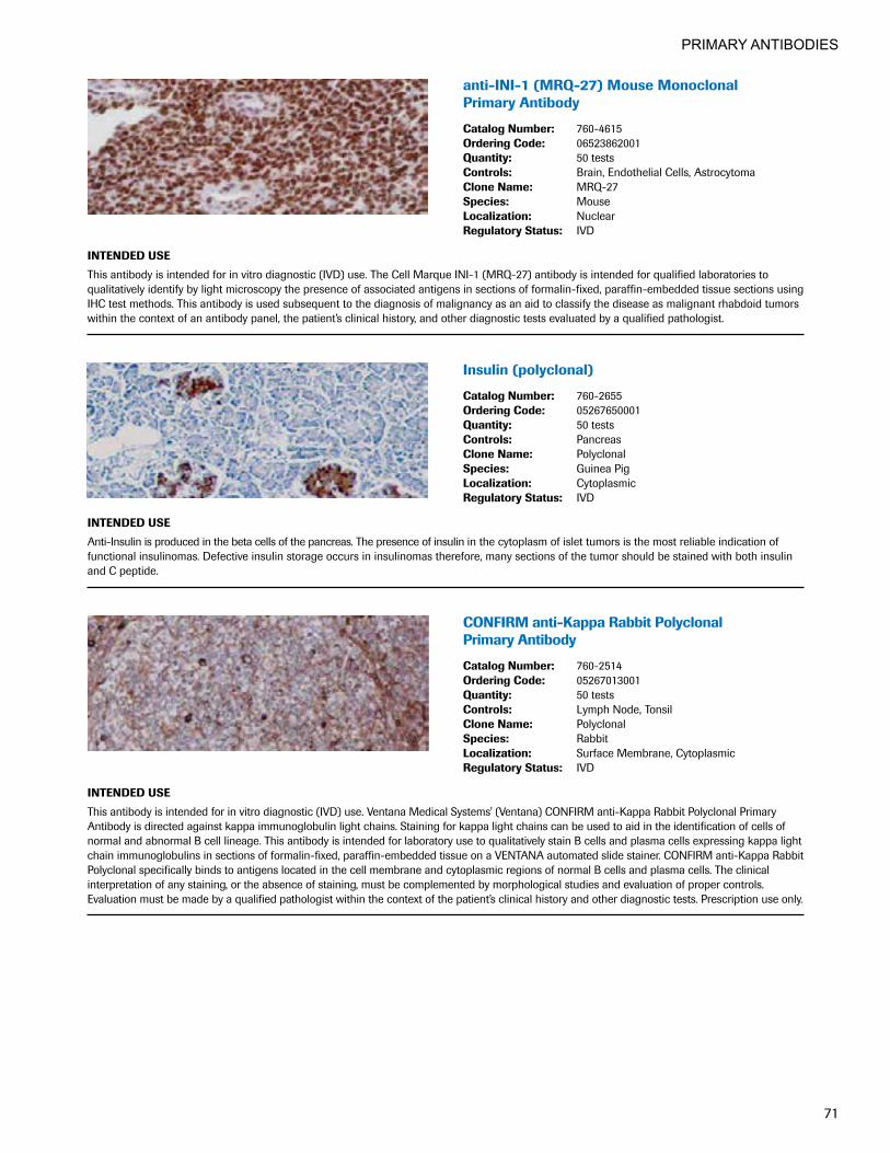
PRIMARY ANTIBODIES
71
anti-INI-1(MRQ-27)MouseMonoclonal Primary Antibody
CatalogNumber: 760-4615OrderingCode: 06523862001Quantity: 50 testsControls: Brain, Endothelial Cells, AstrocytomaCloneName: MRQ-27Species: MouseLocalization: NuclearRegulatoryStatus: IVD
INTENDEDUSE
This antibody is intended for in vitro diagnostic (IVD) use. The Cell Marque INI-1 (MRQ-27) antibody is intended for qualified laboratories to qualitatively identify by light microscopy the presence of associated antigens in sections of formalin-fixed, paraffin-embedded tissue sections using IHC test methods. This antibody is used subsequent to the diagnosis of malignancy as an aid to classify the disease as malignant rhabdoid tumors within the context of an antibody panel, the patient’s clinical history, and other diagnostic tests evaluated by a qualified pathologist.
Insulin(polyclonal)
CatalogNumber: 760-2655OrderingCode: 05267650001Quantity: 50 testsControls: PancreasCloneName: PolyclonalSpecies: Guinea PigLocalization: CytoplasmicRegulatoryStatus: IVD
INTENDEDUSE
Anti-Insulin is produced in the beta cells of the pancreas. The presence of insulin in the cytoplasm of islet tumors is the most reliable indication of functional insulinomas. Defective insulin storage occurs in insulinomas therefore, many sections of the tumor should be stained with both insulin and C peptide.
CONFIRManti-KappaRabbitPolyclonal Primary Antibody
CatalogNumber: 760-2514OrderingCode: 05267013001Quantity: 50 testsControls: Lymph Node, TonsilCloneName: PolyclonalSpecies: RabbitLocalization: Surface Membrane, CytoplasmicRegulatoryStatus: IVD
INTENDEDUSE
This antibody is intended for in vitro diagnostic (IVD) use. Ventana Medical Systems' (Ventana) CONFIRM anti-Kappa Rabbit Polyclonal Primary Antibody is directed against kappa immunoglobulin light chains. Staining for kappa light chains can be used to aid in the identification of cells of normal and abnormal B cell lineage. This antibody is intended for laboratory use to qualitatively stain B cells and plasma cells expressing kappa light chain immunoglobulins in sections of formalin-fixed, paraffin-embedded tissue on a VENTANA automated slide stainer. CONFIRM anti-Kappa Rabbit Polyclonal specifically binds to antigens located in the cell membrane and cytoplasmic regions of normal B cells and plasma cells. The clinical interpretation of any staining, or the absence of staining, must be complemented by morphological studies and evaluation of proper controls. Evaluation must be made by a qualified pathologist within the context of the patient’s clinical history and other diagnostic tests. Prescription use only.

PRIMARY ANTIBODIES
72
CONFIRManti-Keratin(34βE12)Mouse MonoclonalPrimaryAntibody
CatalogNumber: 790-4373OrderingCode: 05479266001Quantity: 50 testsControls: ProstateIsotypes: IgG1CloneName: 34bE12Species: MouseLocalization: CytoplasmicRegulatoryStatus: IVD
INTENDEDUSE
This antibody is intended for in vitro diagnostic use. Ventana Medical Systems' CONFIRM® anti-Keratin (34βE12) Mouse Monoclonal Primary Antibody recognizes cytokeratins 1, 5, 10, and 14 and is used as an aid in the identification of basal cells in prostate, and myoepithelial cells in breast, via light microscopy in formalin-fixed, paraffin-embedded tissue following staining on a VENTANA automated slide stainer. The clinical interpretation of any staining, or the absence of staining, must be complemented by morphological studies and evaluation of proper controls. Evaluation must be made by a qualified pathologist within the context of the patient’s clinical history and other diagnostic tests.
CONFIRManti-Keratin(AE3) Primary Antibody
CatalogNumber: 790-2909OrderingCode: 05278040001Quantity: 50 testsControls: Bladder, Prostate, Salivary GlandIsotypes: IgG1CloneName: AE3Species: MouseLocalization: CytoplasmicRegulatoryStatus: IVD
INTENDEDUSE
This antibody is intended for in vitro diagnostic use. Ventana Medical Systems' (Ventana) CONFIRM anti-Keratin (AE3) is a mouse monoclonal antibody (IgG1, kappa) directed against basic cytokeratins of h 65-67, 64, 59, 56, and 52 kD.1,2,3,4 Unexpected antigen expression or loss of expression may occur, especially in neoplasms. Occasionally stromal elements surrounding heavily stained tissue or cells will show immunoreactiv-ity. The clinical interpretation of any staining, or the absence of staining, must be complemented by morphological studies and evaluation of proper controls. Evaluation must be made by a qualified pathologist within the context of the patient’s clinical history and other diagnostic tests. Caution: U.S. Federal law restricts this device to sale by or on the order of a physician. 1. Pinkus GS, O'Connor EM, Etheridge CL, Corson JM.. Optimal immunoreactivity of keratin proteins in formol fixed, paraffin-embedded tissue
requires preliminary trypsinizination. An immunoperoxidase study of various tumors using polyclonal and monoclonal antibodies. The Journal of Histochemistry and Cytochemistry 33(5): 465-473, 1985.
2. Weiss RA, Eichner R, Sun TT. Monoclonal antibody analysis of keratin expression in epidermal diseases: a 48- and 56-KD keratin as molecular markers for hyperproliferative keratinocytes. The Journal of Cell Biology 98(4): 1397-1406, 1984.
3. Tseng SC, Jarvinen MJ, Nelson WG, Huang JW, Woodcock-Mitchell J, Sun TT. Correlation of specific keratins with different types of epithelial differentiation; monoclonal antibody studies. Cell 30(2): 361-372, 1982.
4. Woodcock-Mitchell J, Eichner R, Nelson WG, Sun TT. Immunolocalization of keratin polypeptides in human epidermis using monoclonal antibodies. The Journal of Cell Biology 95(2 pt 1): 580-588, 1982.

PRIMARY ANTIBODIES
73
CONFIRManti-Ki-67(30-9)RabbitMonoclonalPrimary Antibody
CatalogNumber: 790-4286OrderingCode: 05278384001Quantity: 50 testsControls: Lymph Node, TonsilCloneName: 30-9Species: RabbitLocalization: NuclearRegulatoryStatus: IVD
INTENDEDUSE
This antibody is intended for in vitro diagnostic (IVD) use. Ventana Medical Systems' (Ventana) CONFIRM anti-Ki-67 (30-9) Primary Antibody is a rabbit monoclonal antibody (IgG) directed against C-terminal portion of Ki-67 antigen. Staining for Ki-67 can be used to aid in assessing the proliferative activity of normal and neoplastic tissue.1,2,3 This antibody is intended for use to identify stained proliferating cells by light microscopy in sections of formalin-fixed, paraffin-embedded tissue on a VENTANA automated slide stainer. The clinical interpretation of any staining, or the absence of staining, must be complemented by morphological studies and evaluation of proper controls. Evaluation must be made by a qualified pathologist within the context of the patient’s clinical history and other diagnostic tests. 1. Keng PC, Siemann DW. Measurement of proliferation activities in human tumor models: a comparison of flow cytometric methods. Radiatn Oncol
Investig, 6(3):120-7, 1998. 2. Rey A Lara PC. Ki67 proliferation index in tumors of the upper urinary tract as related to established prognostic factors and long-term survival.
Arch Esp Urol, 51(2): 204-10, 1998.3. Bacchi CE, Gown AM. Detection of cell proliferation in tissue sections. Braz J Med Biol Res, 26(7): 677-87, 1993.
Ksp-cadherin(MRQ-33)
CatalogNumber: 760-4387OrderingCode: 05463548001Quantity: 50 testsControls: Chromophobe Renal Cell Carcinoma, KidneyIsotypes: IgG1CloneName: MRQ-33Species: MouseLocalization: Cytoplasmic, MembranousRegulatoryStatus: IVD
INTENDEDUSE
Kidney-specific cadherin (Ksp-Cadherin) is a novel, kidney-specific member of the cadherin family of cell adhesion molecules. Within the kidney, Ksp-Cadherin is found exclusively in the basolateral membrane of renal tubular epithelial cells and collecting duct cells, and not in glomeruli, renal interstitial cells, or blood vessels. Different cadherins, including E-cadherin, cadherin-6, and N-cadherin, have been investigated in renal cell cancers, demonstrating possible correlations of tumor dedifferentiation and the presence of lymph node metastasis with loss of cadherins. Mazal et al. investigated the utility of using Ksp-Cadherin to distinguish chromophobe renal cell carcinoma from oncocytoma. He found a membranous pattern of staining in 96% of 30 chromophobe carcinomas, and in only 6% of 31 oncocytomas leading them to conclude that this is a useful antibody in differentiating these two lesions. Shen et al., on the other hand, found Ksp-Cadherin positivity in 100% of 13 chromophobe RCCs, and 95% of 20 oncocytomas.

PRIMARY ANTIBODIES
74
CONFIRManti-LambdaRabbitPolyclonal Primary Antibody
CatalogNumber: 760-2515OrderingCode: 05267021001Quantity: 50 testsControls: TonsilCloneName: PolyclonalSpecies: RabbitLocalization: Surface Membrane, CytoplasmicRegulatoryStatus: IVD
INTENDEDUSE
This antibody is intended for in vitro diagnostic (IVD) use. Ventana Medical Systems' (Ventana) CONFIRM anti-Lambda Rabbit Polyclonal Primary Antibody is directed against lambda immunoglobulin light chains. Staining for lambda light chains can be used to aid in the identification of cells of normal and abnormal B cell lineage This antibody is intended for laboratory use to qualitatively stain B cells and plasma cells expressing lambda light chain immunoglobulins in sections of formalin-fixed, paraffin-embedded tissue on a VENTANA automated slide stainer. CONFIRM anti-Lambda Rabbit Polyclonal specifically binds to antigens located in the cell membrane and cytoplasmic regions of normal B cells and plasma cells. The clinical interpretation of any staining, or the absence of staining, must be complemented by morphological studies and evaluation of proper controls. Evaluation must be made by a qualified pathologist within the context of the patient’s clinical history and other diagnostic tests. Prescription use only.
LH(polyclonal)
CatalogNumber: 760-2802OrderingCode: 05268222001Quantity: 50 testsControls: Lymph Node, TonsilCloneName: PolyclonalSpecies: RabbitLocalization: CytoplasmicRegulatoryStatus: IVD
INTENDEDUSE
Anti-LH is a useful marker in classification of pituitary tumors and the study of pituitary disease. It reacts with LH-producing cells (gonadotrophs).
CONFIRManti-LMO2(1A9-1)MouseMonoclonalPrimary Antibody
CatalogNumber: 790-4368OrderingCode: 05479274001Quantity: 50 testsControls: Lymph Node, TonsilIsotypes: IgG1/KCloneName: 1A9-1Species: MouseLocalization: NuclearRegulatoryStatus: IVD
INTENDEDUSE
This antibody is intended for in vitro diagnostic (IVD) use. Ventana Medical Systems' (Ventana) CONFIRM anti-LMO2 (1A9-1) Mouse Monoclonal Primary Antibody is designed to qualitatively detect the presence of LMO2 expressing cells via light microscopy in formalin-fixed, paraffin-embedded tissue. Positive staining results may aid in the classification of B-cell lymphomas. The clinical interpretation of any staining, or the absence of staining, must be complemented by morphological studies and evaluation of proper controls. Evaluation must be made by a qualified pathologist within the context of the patient’s clinical history and other diagnostic tests.
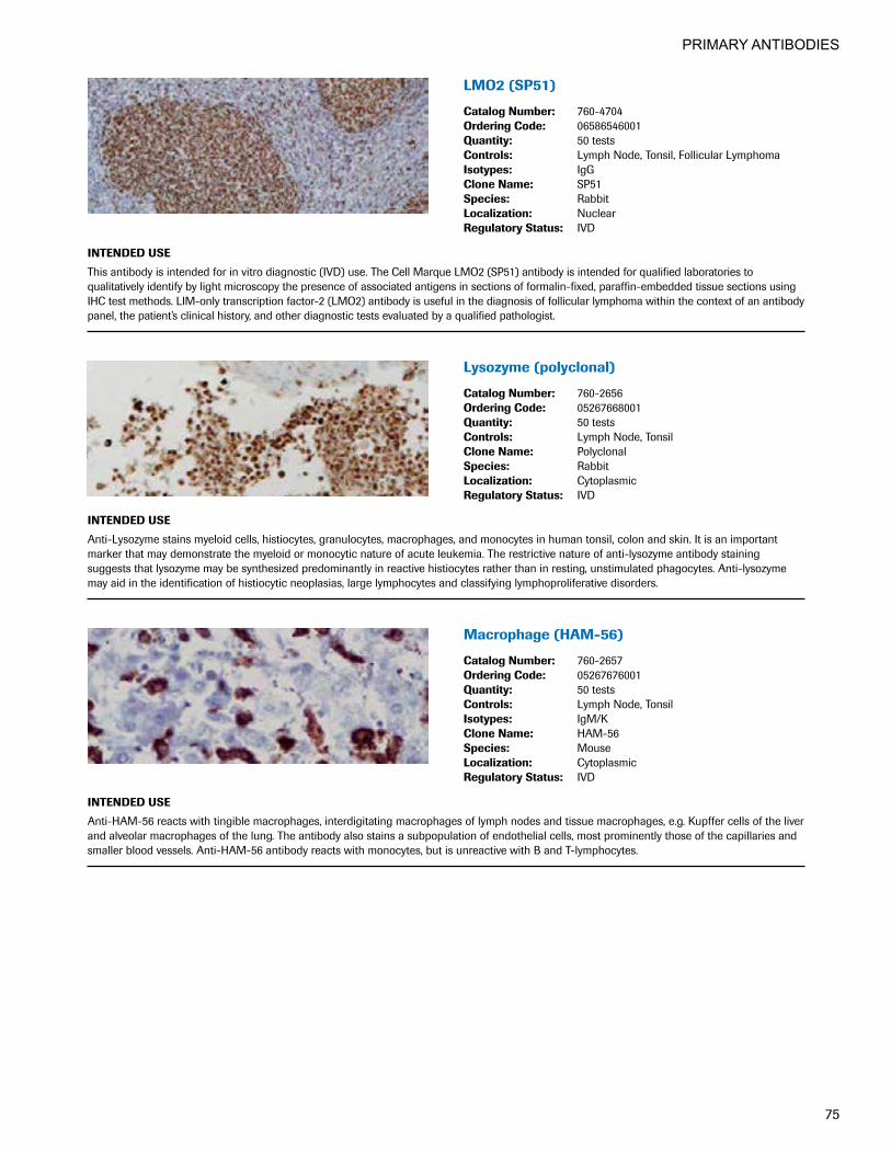
PRIMARY ANTIBODIES
75
LMO2(SP51)
CatalogNumber: 760-4704OrderingCode: 06586546001Quantity: 50 testsControls: Lymph Node, Tonsil, Follicular LymphomaIsotypes: IgGCloneName: SP51Species: RabbitLocalization: NuclearRegulatoryStatus: IVD
INTENDEDUSE
This antibody is intended for in vitro diagnostic (IVD) use. The Cell Marque LMO2 (SP51) antibody is intended for qualified laboratories to qualitatively identify by light microscopy the presence of associated antigens in sections of formalin-fixed, paraffin-embedded tissue sections using IHC test methods. LIM-only transcription factor-2 (LMO2) antibody is useful in the diagnosis of follicular lymphoma within the context of an antibody panel, the patient’s clinical history, and other diagnostic tests evaluated by a qualified pathologist.
Lysozyme(polyclonal)
CatalogNumber: 760-2656OrderingCode: 05267668001Quantity: 50 testsControls: Lymph Node, TonsilCloneName: PolyclonalSpecies: RabbitLocalization: CytoplasmicRegulatoryStatus: IVD
INTENDEDUSE
Anti-Lysozyme stains myeloid cells, histiocytes, granulocytes, macrophages, and monocytes in human tonsil, colon and skin. It is an important marker that may demonstrate the myeloid or monocytic nature of acute leukemia. The restrictive nature of anti-lysozyme antibody staining suggests that lysozyme may be synthesized predominantly in reactive histiocytes rather than in resting, unstimulated phagocytes. Anti-lysozyme may aid in the identification of histiocytic neoplasias, large lymphocytes and classifying lymphoproliferative disorders.
Macrophage(HAM-56)
CatalogNumber: 760-2657OrderingCode: 05267676001Quantity: 50 testsControls: Lymph Node, TonsilIsotypes: IgM/KCloneName: HAM-56Species: MouseLocalization: CytoplasmicRegulatoryStatus: IVD
INTENDEDUSE
Anti-HAM-56 reacts with tingible macrophages, interdigitating macrophages of lymph nodes and tissue macrophages, e.g. Kupffer cells of the liver and alveolar macrophages of the lung. The antibody also stains a subpopulation of endothelial cells, most prominently those of the capillaries and smaller blood vessels. Anti-HAM-56 antibody reacts with monocytes, but is unreactive with B and T-lymphocytes.

PRIMARY ANTIBODIES
76
Mammaglobin(31A5)
CatalogNumber: 760-4263OrderingCode: 5269253001Quantity: 50 testsControls: Breast CarcinomaCloneName: 31A5Species: RabbitLocalization: CytoplasmicRegulatoryStatus: IVD
INTENDEDUSE
This antibody is intended for in vitro diagnostic (IVD) use. The Cell Marque Mammaglobin (31A5) antibody is intended for qualified laboratories to qualitatively identify by light microscopy the presence of associated antigens in sections of formalin-fixed, paraffin-embedded tissue sections using IHC test methods. Use of this antibody is indicated as an aid in identification of substantial proportion of primary and metastatic breast carcinomas within the context of an antibody panel, the patient’s clinical history and other diagnostic tests evaluated by a qualified pathologist.
CONFIRManti-MART-1/melanA(A103) MouseMonoclonalPrimaryAntibody
CatalogNumber: 790-2990OrderingCode: 05278350001Quantity: 50 testsControls: Melanoma, Normal SkinIsotypes: IgG1CloneName: A103Species: MouseLocalization: CytoplasmicRegulatoryStatus: IVD
INTENDEDUSE
This antibody is intended for in vitro diagnostic (IVD) use. Ventana Medical Systems, Inc.'s (Ventana) CONFIRM anti-MART-1/melan A (A103) Mouse Monoclonal Primary Antibody is a mouse monoclonal antibody (IgG1) directed against melan A. This antibody is designed to qualitatively detect the presence of melan A by light microscopy in sections of formalin-fixed, paraffin-embedded tissue. Positive staining may aid in the classification of melanoma. The clinical interpretation of any staining, or the absence of staining, must be complemented by morphological studies and evaluation of proper controls. Evaluation must be made by a qualified pathologist within the context of the patient’s clinical history and other diagnostic tests.
MelanomaAssociatedAntigen(KBA.62) MouseMonoclonalAntibody
CatalogNumber: 760-4527OrderingCode: 06419186001Quantity: 50 testsControls: MelanomaIsotypes: IgG1CloneName: KBA.62Species: MouseLocalization: CytoplasmicRegulatoryStatus: IVD
INTENDEDUSE
This antibody is intended for in vitro diagnostic (IVD) use. The Cell Marque anti-Melanoma Associated Antigen (KBA.62) antibody is intended for qualified laboratories to qualitatively identify by light microscopy the presence of associated antigens in sections of formalin-fixed, paraffin-embed-ded tissue sections using VENTANA BenchMark® automated IHC test methods. Use of this antibody is indicated as an aid in the diagnosis of primary and metastatic melanoma within the context of an antibody panel, the patient’s clinical history and other diagnostic tests evaluated by a qualified pathologist or physician.

PRIMARY ANTIBODIES
77
MelanomaAssociatedAntigen(PNL2) MouseMonoclonalAntibody
CatalogNumber: 760-4528OrderingCode: 06419194001Quantity: 50 testsControls: MelanomaIsotypes: IgG1CloneName: PNL2Species: MouseLocalization: CytoplasmicRegulatoryStatus: IVD
INTENDEDUSE
This antibody is intended for in vitro diagnostic (IVD) use. The Cell Marque anti-Melanoma Associated Antigen (PNL2) antibody is intended for qualified laboratories to qualitatively identify by light microscopy the presence of associated antigens in sections of formalin-fixed, paraffin- embedded tissue sections using VENTANA BenchMark® automated IHC test methods. Use of this antibody is indicated as an aid in diagnostic determination of the diagnosis of primary and metastatic melanoma within the context of an antibody panel, the patient’s clinical history and other diagnostic tests evaluated by a qualified pathologist or physician.
MelanomaTripleCocktail(HMB45+A103+T311)Primary Antibody
CatalogNumber: 790-4677OrderingCode: 06527787001Quantity: 50 testsControls: MelanomaCloneName: HMB45+A103+T311Species: N/ALocalization: CytoplasmicRegulatoryStatus: IVD
INTENDEDUSE
Melanoma Triple Cocktail (HMB45+A103+T311) is an antibody cocktail of anti-Melanosome (HMB45), anti-MART-1/melan A (A103), and anti-Tyrosinase (T311) mouse monoclonal antibodies. This antibody cocktail demonstrates cytoplasmic staining and may be used to aid in the identification of melanoma. The antibody is intended for qualitative staining in sections of formalin-fixed, paraffin-embedded tissue. This product should be interpreted by a qualified pathologist in conjunction with histological examination, relevant clinical information, and proper controls. This antibody is intended for in vitro diagnostic (IVD) use.
CONFIRManti-Melanosome(HMB45) MouseMonoclonalPrimaryAntibody
CatalogNumber: 790-4366OrderingCode: 05479282001Quantity: 50 testsControls: Melanoma, SkinIsotypes: IgG1/KCloneName: HMB45Species: MouseLocalization: CytoplasmicRegulatoryStatus: IVD
INTENDEDUSE
This antibody is intended for in vitro diagnostic use. Ventana Medical Systems' (Ventana) CONFIRM anti-Melanosome (HMB45) is a mouse monoclonal antibody (IgG1, kappa) directed against an oncofetal antigen present within immature melanosomes in the cytoplasm of activated and neoplastic melanocytes. CONFIRM anti-Melanosome (HMB45) is intended for laboratory use to qualitatively detect, via light microscopy, cells containing immature melanosomes in sections of formalin-fixed, paraffin-embedded tissue following staining on a VENTANA automated slide stainer. The clinical interpretation of any staining, or the absence of staining, must be complemented by morphological studies and evaluation of proper controls. Evaluation must be made by a qualified pathologist within the context of the patient’s clinical history and other diagnostic tests.

PRIMARY ANTIBODIES
78
CONFIRManti-MITF(C5/D5)MouseMonoclonalPrimary Antibody
CatalogNumber: 790-4367OrderingCode: 05549175001Quantity: 50 testsControls: MelanomaIsotypes: IgG1CloneName: C5/D5Species: MouseLocalization: NuclearRegulatoryStatus: IVD
INTENDEDUSE
This antibody is intended for in vitro diagnostic (IVD) use. Ventana Medical Systems’ (Ventana) CONFIRM anti-MITF (C5/D5) Mouse Monoclonal Primary Antibody is a mouse monoclonal cocktail. This antibody cocktail reagent detects microphthalmia transcription factor (MITF) proteins 52-56KD of MITF expressing cells in normal and neoplastic tissues. This antibody is used in the identification of melanotic lesions such as malignant melanoma and melanotic neurofibroma. The antibody is intended for qualitative staining in sections of formalin-fixed, paraffin-embedded tissue. The clinical interpretation of any staining or absence of staining, must be complemented by morphological studies and evaluation of proper controls. Evaluation must be made by a qualified pathologist within the context of the patient’s clinical history and other diagnostic tests.
anti-MLH-1(M1)MouseMonoclonal Primary Antibody
CatalogNumber: 790-4535OrderingCode: 06472966001Quantity: 50 testsControls: Appendix, Breast, Colon, Testis, OvaryIsotypes: IgGCloneName: M1Species: MouseLocalization: NuclearRegulatoryStatus: IVD
INTENDEDUSE
Anti-MLH-1 (M1) Mouse Monoclonal Primary Antibody (anti-MLH-1 (M1)) is used to qualitatively identify human DNA mismatch repair (MMR) protein MLH1, expressed in the nucleus of normal proliferating cells. Deficient or low levels of MLH1 are associated with colorectal and other cancers. The antibody is intended for qualitative staining in sections of formalin-fixed, paraffin-embedded tissue. This product should be interpreted by a qualified pathologist in conjunction with histological examination, relevant clinical information, and proper controls. This antibody is intended for in vitro diagnostic (IVD) use.
MSH2(G219-1129)MouseMonoclonalAntibody
CatalogNumber: 760-4265OrderingCode: 05269270001Quantity: 50 testsControls: Colon Carcinoma, Colon MucosaIsotypes: IgG1CloneName: G219-1129Species: MouseLocalization: NuclearRegulatoryStatus: IVD
INTENDEDUSE
This antibody is intended for in vitro diagnostic (IVD) use. The Cell Marque MSH2 (G219-1129) antibody is intended for qualified laboratories to qualitatively identify by light microscopy the presence of associated antigens in sections of formalin-fixed, paraffin-embedded tissue sections using IHC test methods. Use of this antibody is indicated, subsequent to clinical differential diagnoses of diseases, as an aid in the identification of microsatellite instability within the context of antibody panels, the patient’s clinical history, and other diagnostic tests evaluated by a qualified pathologist.
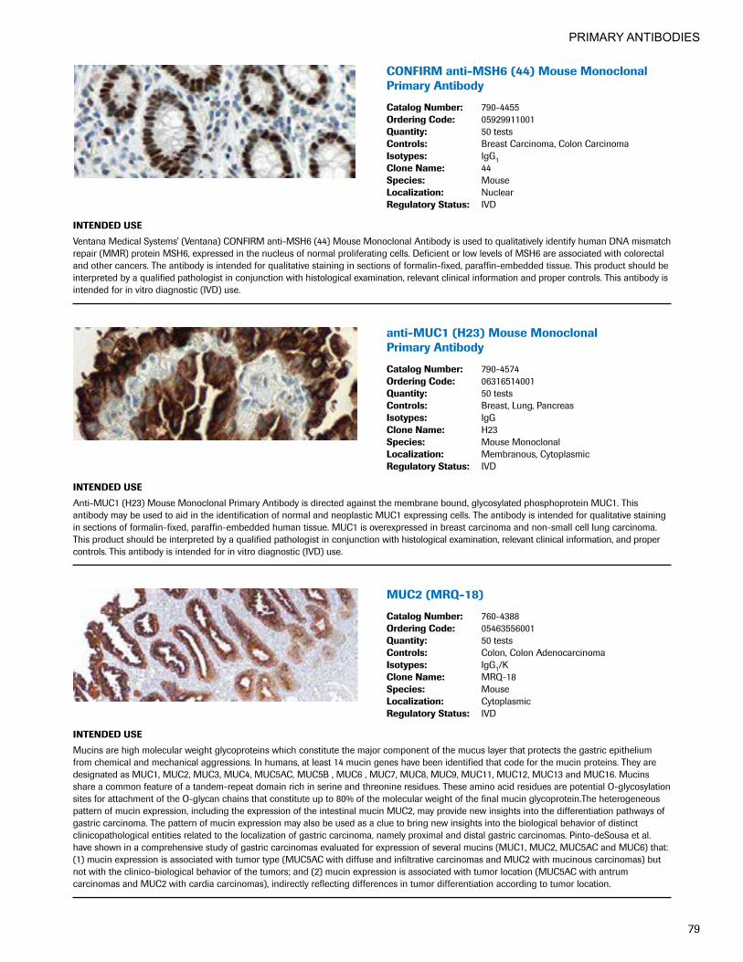
PRIMARY ANTIBODIES
79
CONFIRManti-MSH6(44)MouseMonoclonalPrimary Antibody
CatalogNumber: 790-4455OrderingCode: 05929911001Quantity: 50 testsControls: Breast Carcinoma, Colon CarcinomaIsotypes: IgG1CloneName: 44Species: MouseLocalization: NuclearRegulatoryStatus: IVD
INTENDEDUSE
Ventana Medical Systems' (Ventana) CONFIRM anti-MSH6 (44) Mouse Monoclonal Antibody is used to qualitatively identify human DNA mismatch repair (MMR) protein MSH6, expressed in the nucleus of normal proliferating cells. Deficient or low levels of MSH6 are associated with colorectal and other cancers. The antibody is intended for qualitative staining in sections of formalin-fixed, paraffin-embedded tissue. This product should be interpreted by a qualified pathologist in conjunction with histological examination, relevant clinical information and proper controls. This antibody is intended for in vitro diagnostic (IVD) use.
anti-MUC1(H23)MouseMonoclonal Primary Antibody
CatalogNumber: 790-4574OrderingCode: 06316514001Quantity: 50 testsControls: Breast, Lung, PancreasIsotypes: IgGCloneName: H23Species: Mouse MonoclonalLocalization: Membranous, CytoplasmicRegulatoryStatus: IVD
INTENDEDUSE
Anti-MUC1 (H23) Mouse Monoclonal Primary Antibody is directed against the membrane bound, glycosylated phosphoprotein MUC1. This antibody may be used to aid in the identification of normal and neoplastic MUC1 expressing cells. The antibody is intended for qualitative staining in sections of formalin-fixed, paraffin-embedded human tissue. MUC1 is overexpressed in breast carcinoma and non-small cell lung carcinoma. This product should be interpreted by a qualified pathologist in conjunction with histological examination, relevant clinical information, and proper controls. This antibody is intended for in vitro diagnostic (IVD) use.
MUC2(MRQ-18)
CatalogNumber: 760-4388OrderingCode: 05463556001Quantity: 50 testsControls: Colon, Colon AdenocarcinomaIsotypes: IgG1/KCloneName: MRQ-18Species: MouseLocalization: CytoplasmicRegulatoryStatus: IVD
INTENDEDUSE
Mucins are high molecular weight glycoproteins which constitute the major component of the mucus layer that protects the gastric epithelium from chemical and mechanical aggressions. In humans, at least 14 mucin genes have been identified that code for the mucin proteins. They are designated as MUC1, MUC2, MUC3, MUC4, MUC5AC, MUC5B , MUC6 , MUC7, MUC8, MUC9, MUC11, MUC12, MUC13 and MUC16. Mucins share a common feature of a tandem-repeat domain rich in serine and threonine residues. These amino acid residues are potential O-glycosylation sites for attachment of the O-glycan chains that constitute up to 80% of the molecular weight of the final mucin glycoprotein.The heterogeneous pattern of mucin expression, including the expression of the intestinal mucin MUC2, may provide new insights into the differentiation pathways of gastric carcinoma. The pattern of mucin expression may also be used as a clue to bring new insights into the biological behavior of distinct clinicopathological entities related to the localization of gastric carcinoma, namely proximal and distal gastric carcinomas. Pinto-deSousa et al. have shown in a comprehensive study of gastric carcinomas evaluated for expression of several mucins (MUC1, MUC2, MUC5AC and MUC6) that: (1) mucin expression is associated with tumor type (MUC5AC with diffuse and infiltrative carcinomas and MUC2 with mucinous carcinomas) but not with the clinico-biological behavior of the tumors; and (2) mucin expression is associated with tumor location (MUC5AC with antrum carcinomas and MUC2 with cardia carcinomas), indirectly reflecting differences in tumor differentiation according to tumor location.
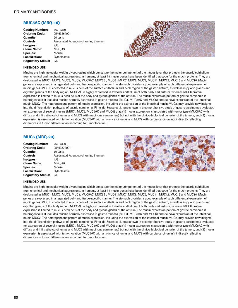
PRIMARY ANTIBODIES
80
MUC5AC(MRQ-19)
CatalogNumber: 760-4389OrderingCode: 05463564001Quantity: 50 testsControls: Associated Adenocarcinomas, StomachIsotypes: IgG1CloneName: MRQ-19Species: MouseLocalization: CytoplasmicRegulatoryStatus: IVD
INTENDEDUSE
Mucins are high molecular weight glycoproteins which constitute the major component of the mucus layer that protects the gastric epithelium from chemical and mechanical aggressions. In humans, at least 14 mucin genes have been identified that code for the mucin proteins. They are designated as MUC1, MUC2, MUC3, MUC4, MUC5AC, MUC5B , MUC6 , MUC7, MUC8, MUC9, MUC11, MUC12, MUC13 and MUC16. Mucin genes are expressed in a regulated cell- and tissue-specific manner. The stomach provides a good example of such differential expression of mucin genes. MUC1 is detected in mucus cells of the surface epithelium and neck region of the gastric antrum, as well as in pyloric glands and oxynthic glands of the body region. MUC5AC is highly expressed in foveolar epithelium of both body and antrum, whereas MUC6 protein expression is limited to mucus neck cells of the body and pyloric glands of the antrum. The mucin expression pattern of gastric carcinoma is heterogeneous. It includes mucins normally expressed in gastric mucosa (MUC1, MUC5AC and MUC6) and de novo expression of the intestinal mucin MUC2. The heterogeneous pattern of mucin expression, including the expression of the intestinal mucin MUC2, may provide new insights into the differentiation pathways of gastric carcinoma. Pinto-de-Sousa et al. have shown in a comprehensive study of gastric carcinomas evaluated for expression of several mucins (MUC1, MUC2, MUC5AC and MUC6) that: (1) mucin expression is associated with tumor type (MUC5AC with diffuse and infiltrative carcinomas and MUC2 with mucinous carcinomas) but not with the clinico-biological behavior of the tumors; and (2) mucin expression is associated with tumor location (MUC5AC with antrum carcinomas and MUC2 with cardia carcinomas), indirectly reflecting differences in tumor differentiation according to tumor location.
MUC6(MRQ-20)
CatalogNumber: 760-4390OrderingCode: 05463572001Quantity: 50 testsControls: Associated Adenocarcinomas, StomachIsotypes: IgG1CloneName: MRQ-20Species: MouseLocalization: CytoplasmicRegulatoryStatus: IVD
INTENDEDUSE
Mucins are high molecular weight glycoproteins which constitute the major component of the mucus layer that protects the gastric epithelium from chemical and mechanical aggressions. In humans, at least 14 mucin genes have been identified that code for the mucin proteins. They are designated as MUC1, MUC2, MUC3, MUC4, MUC5AC, MUC5B , MUC6 , MUC7, MUC8, MUC9, MUC11, MUC12, MUC13 and MUC16. Mucin genes are expressed in a regulated cell- and tissue-specific manner. The stomach provides a good example of such differential expression of mucin genes. MUC1 is detected in mucus cells of the surface epithelium and neck region of the gastric antrum, as well as in pyloric glands and oxynthic glands of the body region. MUC5AC is highly expressed in foveolar epithelium of both body and antrum, whereas MUC6 protein expression is limited to mucus neck cells of the body and pyloric glands of the antrum. The mucin expression pattern of gastric carcinoma is heterogeneous. It includes mucins normally expressed in gastric mucosa (MUC1, MUC5AC and MUC6) and de novo expression of the intestinal mucin MUC2. The heterogeneous pattern of mucin expression, including the expression of the intestinal mucin MUC2, may provide new insights into the differentiation pathways of gastric carcinoma. Pinto-de-Sousa et al. have shown in a comprehensive study of gastric carcinomas evaluated for expression of several mucins (MUC1, MUC2, MUC5AC and MUC6) that: (1) mucin expression is associated with tumor type (MUC5AC with diffuse and infiltrative carcinomas and MUC2 with mucinous carcinomas) but not with the clinico-biological behavior of the tumors; and (2) mucin expression is associated with tumor location (MUC5AC with antrum carcinomas and MUC2 with cardia carcinomas), indirectly reflecting differences in tumor differentiation according to tumor location.

PRIMARY ANTIBODIES
81
MUM1(MRQ-43)
CatalogNumber: 760-4529OrderingCode: 06419208001Quantity: 50 testsControls: TonsilIsotypes: IgG1CloneName: MRQ-43Species: RabbitLocalization: Nuclear, CytoplasmicRegulatoryStatus: IVD
INTENDEDUSE
This antibody is intended for in vitro diagnostic (IVD) use. The Cell Marque MUM1 (MRQ-43) antibody is intended for qualified laboratories to qualitatively identify by light microscopy the presence of associated antigens in sections of formalin-fixed, paraffin-embedded tissue using IHC test methods. Use of this antibody is indicated as an aid in the identification of plasma cells, B-cells in the light zone of the germinal center and activated T-cells, and in the diagnosis of classic Hodgkin disease, subtype of B-cell lymphomas, and plasma cell tumors within the context of antibody panels, the patient’s clinical history, and other diagnostic tests evaluated by a qualified pathologist.
Myeloperoxidase(polyclonal)
CatalogNumber: 760-2659OrderingCode: 05267692001Quantity: 50 testsControls: Bone MarrowCloneName: PolyclonalSpecies: RabbitLocalization: CytoplasmicRegulatoryStatus: IVD
INTENDEDUSE
Anti-Myeloperoxidase Polyclonal Primary Antibody is intended for laboratory use in the detection of the myeloperoxidase protein in formalin-fixed, paraffin-embedded tissue stained on VENTANA BenchMark immunohistochemical (IHC) automated slide stainers. This product should be interpreted by a qualified pathologist in conjunction with histological examination, relevant clinical information, and proper controls. This antibody is intended for in vitro diagnostic (IVD) use.
MyoD1(EP212)RabbitMonoclonal Primary Antibody
CatalogNumber: 760-4900OrderingCode: 07107773001Quantity: 50 testsControls: RhabdomyosarcomaIsotypes: IgGCloneName: EP212Species: RabbitLocalization: NuclearRegulatoryStatus: IVD
INTENDEDUSE
This antibody is intended for in vitro diagnostic (IVD) use. The Cell Marque MyoD1 (EP212) antibody is intended for qualified laboratories to qualitatively identify by light microscopy the presence of associated antigens in sections of formalin-fixed, paraffin-embedded tissue using IHC test methods. Use of this antibody is indicated, subsequent to clinical differential diagnosis, as an aid in the identification of rhabdomyosarcoma within the context of an antibody panel, the patient’s clinical history, and other diagnostic tests evaluated by a qualified pathologist.
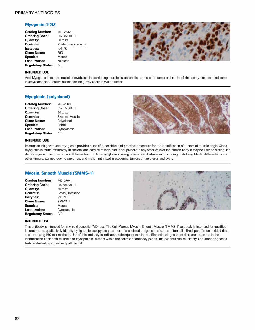
PRIMARY ANTIBODIES
82
Myogenin(F5D)
CatalogNumber: 760-2832OrderingCode: 05268290001Quantity: 50 testsControls: RhabdomyosarcomaIsotypes: IgG1/KCloneName: F5DSpecies: MouseLocalization: NuclearRegulatoryStatus: IVD
INTENDEDUSE
Anti-Myogenin labels the nuclei of myoblasts in developing muscle tissue, and is expressed in tumor cell nuclei of rhabdomyosarcoma and some leiomyosarcomas. Positive nuclear staining may occur in Wilm’s tumor.
Myoglobin(polyclonal)
CatalogNumber: 760-2660OrderingCode: 05267706001Quantity: 50 testsControls: Skeletal MuscleCloneName: PolyclonalSpecies: RabbitLocalization: CytoplasmicRegulatoryStatus: IVD
INTENDEDUSE
Immunostaining with anti-myoglobin provides a specific, sensitive and practical procedure for the identification of tumors of muscle origin. Since myoglobin is found exclusively in skeletal and cardiac muscle and is not present in any other cells of the human body, it may be used to distinguish rhabdomyosarcoma from other soft tissue tumors. Anti-myoglobin staining is also useful when demonstrating rhabdomyoblastic differentiation in other tumors, e.g. neurogenic sarcomas, and malignant mixed mesodermal tumors of the uterus and ovary.
Myosin,SmoothMuscle(SMMS-1)
CatalogNumber: 760-2704OrderingCode: 05268133001Quantity: 50 testsControls: Breast, IntestineIsotypes: IgG1/KCloneName: SMMS-1Species: MouseLocalization: CytoplasmicRegulatoryStatus: IVD
INTENDEDUSE
This antibody is intended for in vitro diagnostic (IVD) use. The Cell Marque Myosin, Smooth Muscle (SMMS-1) antibody is intended for qualified laboratories to qualitatively identify by light microscopy the presence of associated antigens in sections of formalin-fixed, paraffin-embedded tissue sections using IHC test methods. Use of this antibody is indicated, subsequent to clinical differential diagnoses of diseases, as an aid in the identification of smooth muscle and myoepithelial tumors within the context of antibody panels, the patient’s clinical history, and other diagnostic tests evaluated by a qualified pathologist.

PRIMARY ANTIBODIES
83
NapsinA(MRQ-60)MouseMonoclonal Primary Antibody
CatalogNumber: 760-4867OrderingCode: 07047720001Quantity: 50 testsControls: Lung Adenocarcinoma, Renal Cell CarcinomaIsotypes: IgG1/KCloneName: MRQ-60Species: Mouse MonoclonalLocalization: Cytoplasmic RegulatoryStatus: IVD
INTENDEDUSE
This antibody is intended for in vitro diagnostic (IVD) use. The Cell Marque Napsin A (MRQ-60) antibody is intended for qualified laboratories to qualitatively identify by light microscopy the presence of associated antigens in sections of formalin-fixed, paraffin-embedded tissue sections using IHC test methods. Use of this antibody is indicated, subsequent to clinical differential diagnosis, as an aid in the identification of napsin A protein over-expression in various lung adenocarcinomas within the context of an antibody panel, the patient’s clinical history, and other diagnostic tests evaluated by a qualified pathologist.
NapsinA(polyclonal)
CatalogNumber: 760-4446OrderingCode: 05973805001Quantity: 50 testsControls: Lung Adenocarcinoma Isotypes: IgGCloneName: PolyclonalSpecies: RabbitLocalization: Cytoplasmic RegulatoryStatus: IVD
INTENDEDUSE
Napsin is a pepsin-like aspartic proteinase, in the A1 clan of the AA clade of proteinases. There are two closely related napsins, napsin A and napsin B. Napsin A is expressed as a single chain protein with the molecular weight of approximately 38 kDa. Immunohistochemical studies revealed high expression levels of napsin A in human lung and kidney but low expression in spleen. Napsin A is expressed in type II pneumocytes and in adenocarcinomas of lung. The high specificity expression of napsin A in adenocarcinomas of lung is useful to distinguish primary lung adenocarcinomas from adenocarcinomas of other organs.
NegativeControl(Monoclonal) NegativeControl(Monoclonal)
CatalogNumber: 760-2014OrderingCode: 05266670001Quantity: 250 testsControls: KidneyCloneName: MOPC21Species: MouseLocalization: CytoplasmicRegulatoryStatus: IVD
INTENDEDUSE
This antibody is intended for in vitro diagnostic (IVD) use. Ventana® Medical Systems' (Ventana) Negative Control (Monoclonal) is a mouse monoclonal antibody (MOPC-211) which is not directed against a known epitope present in human tissue. The reagent is intended for use as a negative control in qualitative staining of sections of routine formalin-fixed, paraffin-embedded tissue on a VENTANA automated slide stainer. The clinical interpretation of any staining, or the absence of staining, must be complemented by morphological studies and evaluation of proper controls. Evaluation must be made by a qualified pathologist within the context of the patient’s clinical history and other diagnostic tests. Prescription only.
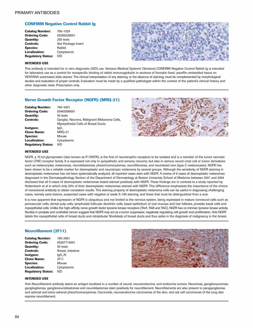
PRIMARY ANTIBODIES
84
CONFIRMNegativeControlRabbitIg
CatalogNumber: 760-1029OrderingCode: 05266238001Quantity: 250 testsControls: See Package InsertSpecies: RabbitLocalization: CytoplasmicRegulatoryStatus: IVD
INTENDEDUSE
This antibody is intended for in vitro diagnostic (IVD) use. Ventana Medical Systems' (Ventana) CONFIRM Negative Control Rabbit Ig is intended for laboratory use as a control for nonspecific binding of rabbit immunoglobulin in sections of formalin-fixed, paraffin-embedded tissue on VENTANA automated slide stainer. The clinical interpretation of any staining, or the absence of staining, must be complemented by morphological studies and evaluation of proper controls. Evaluation must be made by a qualified pathologist within the context of the patient’s clinical history and other diagnostic tests. Prescription only.
NerveGrowthFactorReceptor(NGFR)(MRQ-21)
CatalogNumber: 760-4391OrderingCode: 05463599001Quantity: 50 testsControls: Ganglia, Neurons, Malignant Melanoma Cells,
Myoepithelial Cells of Breast DuctsIsotypes: IgG1CloneName: MRQ-21Species: MouseLocalization: CytoplasmicRegulatoryStatus: IVD
INTENDEDUSE
NGFR, a 75 kd glycoprotein (also known as P-75NTR), is the first of neurotrophin receptors to be isolated and is a member of the tumor necrosis factor (TNF) receptor family. It is expressed not only in sympathetic and sensory neurons, but also in various neural crest cell or tumor derivatives such as melanocytes, melanomas, neuroblastomas, pheochromocytomas, neurofibromas, and neurotized nevi (type C melanocytes). NGFR has been shown to be a reliable marker for desmoplastic and neurotropic melanoma by several groups. Although the sensitivity of NGFR staining in desmoplastic melanomas has not been systematically analyzed, all reported cases stain with NGFR. A review of 9 cases of desmoplastic melanomas diagnosed in the Dermatopathology Section of the Department of Dermatology at Boston University School of Medicine between 2001 and 2004 disclosed that all 9 cases of desmoplastic melanomas tested stained positively with NGFR. These findings are in contrast to a study reported by Huttenbach et al in which only 33% of their desmoplastic melanomas stained with NGFR. This difference emphasizes the importance of the choice of monoclonal antibody to obtain consistent results. This staining property of desmoplastic melanoma cells can be useful in diagnosing challenging cases, namely early lesions, suspected cases with negative or weak S-100 staining, and those that must be distinguished from a scar.
It is now apparent that expression of NGFR is ubiquitous and not limited to the nervous system, being expressed in mature nonneural cells such as perivascular cells, dental pulp cells, lymphoidal follicular dendritic cells, basal epithelium of oral mucosa and hair follicles, prostate basal cells and myoepithelial cells. Unlike the high-affinity nerve growth factor tyrosine kinase receptors (TrkA, TrkB and TrkC), NGFR has no intrinsic tyrosine kinase activity. Studies in prostate and urothelial cancer suggest that NGFR may act as a tumor suppressor, negatively regulating cell growth and proliferation. Anti-NGFR labels the myoepithelial cells of breast ducts and intralobular fibroblasts of breast ducts and thus aides in the diagnosis of malignancy in the breast.
Neurofilament(2F11)
CatalogNumber: 760-2661OrderingCode: 05267714001Quantity: 50 testsControls: Breast, IntestineIsotypes: IgG1/KCloneName: 2F11Species: MouseLocalization: CytoplasmicRegulatoryStatus: IVD
INTENDEDUSE
Anti-Neurofilament antibody stains an antigen localized in a number of neural, neuroendocrine, and endocrine tumors. Neuromas, ganglioneuromas, gangliogliomas, ganglioneuroblastomas and neuroblastomas stain positively for neurofilament. Neurofilaments are also present in paragangliomas and adrenal and extra-adrenal pheochromocytomas. Carcinoids, neuroendocrine carcinomas of the skin, and oat cell carcinomas of the lung also express neurofilament.

PRIMARY ANTIBODIES
85
NeutrophilElastase(SP203)RabbitMonoclonalPrimary Antibody
CatalogNumber: 760-4953OrderingCode: 07391455001Quantity: 50 testsControls: Bone MarrowIsotypes: IgGCloneName: SP203Species: RabbitLocalization: CytoplasmicRegulatoryStatus: IVD
INTENDEDUSE
This antibody is intended for in vitro diagnostic (IVD) use. The Cell Marque Neutrophil Elastase (SP203) antibody is intended for qualified laboratories to qualitatively identify by light microscopy the presence of associated antigens in sections of formalin-fixed, paraffin-embedded tissue sections using IHC test methods. Use of this antibody is indicated, subsequent to clinical differential diagnosis, as an aid in the identification of neutrophils and myeloid precursors within the context of an antibody panel, the patient’s clinical history, and other diagnostic tests evaluated by a qualified pathologist.
NKX3.1(EP356)RabbitMonoclonalPrimaryAntibody
CatalogNumber: 760-5086OrderingCode: 07859759001Quantity: 50 testsControls: Prostate, Prostate AdenocarcinomaIsotypes: IgGCloneName: EP356Species: RabbitLocalization: NuclearRegulatoryStatus: IVD
INTENDEDUSE
NKX3.1 (EP356) Rabbit Monoclonal Primary Antibody is intended for laboratory use in the detection of the NKX3.1 protein in formalin-fixed, paraffin-embedded tissue stained on VENTANA BenchMark IHC/ISH instruments. This product should be interpreted by a qualified pathologist in conjunction with histological examination, relevant clinical information, and proper controls. This antibody is intended for in vitro diagnostic (IVD) use.
anti-NSE(MRQ-55)MouseMonoclonal Primary Antibody
CatalogNumber: 760-4786OrderingCode: 06648568001Quantity: 50 testsControls: Pancreas, Carcinoid TumorCloneName: MRQ-55Species: Mouse MonoclonalLocalization: CytoplasmicRegulatoryStatus: IVD
INTENDEDUSE
This antibody is intended for in vitro diagnostic (IVD) use. The Cell Marque NSE (MRQ-55) antibody is intended for qualified laboratories to qualitatively identify by light microscopy the presence of associated antigens in sections of formalin-fixed, paraffin-embedded tissue sections using IHC test methods. Use of this antibody is indicated, subsequent to clinical differential diagnoses of diseases, as an aid in the identification of neuroendocrine neoplasms within the context of antibody panels, the patient’s clinical history, and other diagnostic tests evaluated by a qualified pathologist.
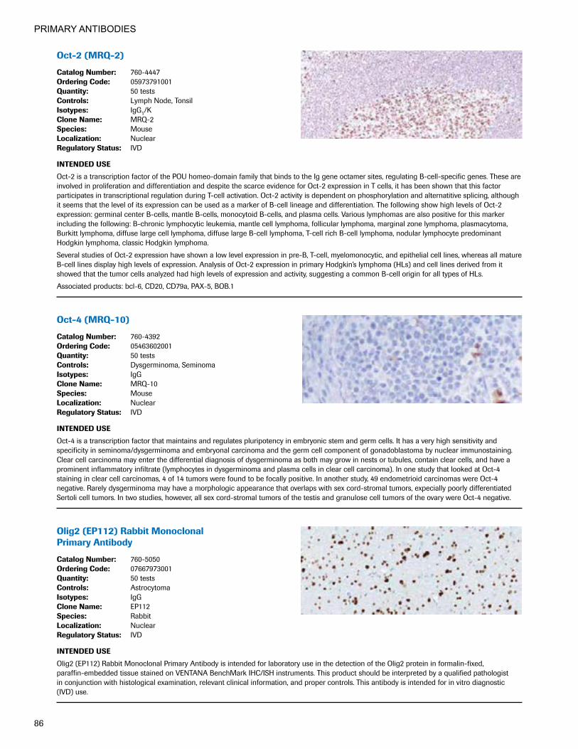
PRIMARY ANTIBODIES
86
Oct-2(MRQ-2)
CatalogNumber: 760-4447OrderingCode: 05973791001Quantity: 50 testsControls: Lymph Node, TonsilIsotypes: IgG1/KCloneName: MRQ-2Species: MouseLocalization: NuclearRegulatoryStatus: IVD
INTENDEDUSE
Oct-2 is a transcription factor of the POU homeo-domain family that binds to the Ig gene octamer sites, regulating B-cell-specific genes. These are involved in proliferation and differentiation and despite the scarce evidence for Oct-2 expression in T cells, it has been shown that this factor participates in transcriptional regulation during T-cell activation. Oct-2 activity is dependent on phosphorylation and alternatitive splicing, although it seems that the level of its expression can be used as a marker of B-cell lineage and differentiation. The following show high levels of Oct-2 expression: germinal center B-cells, mantle B-cells, monocytoid B-cells, and plasma cells. Various lymphomas are also positive for this marker including the following: B-chronic lymphocytic leukemia, mantle cell lymphoma, follicular lymphoma, marginal zone lymphoma, plasmacytoma, Burkitt lymphoma, diffuse large cell lymphoma, diffuse large B-cell lymphoma, T-cell rich B-cell lymphoma, nodular lymphocyte predominant Hodgkin lymphoma, classic Hodgkin lymphoma.
Several studies of Oct-2 expression have shown a low level expression in pre-B, T-cell, myelomonocytic, and epithelial cell lines, whereas all mature B-cell lines display high levels of expression. Analysis of Oct-2 expression in primary Hodgkin’s lymphoma (HLs) and cell lines derived from it showed that the tumor cells analyzed had high levels of expression and activity, suggesting a common B-cell origin for all types of HLs.
Associated products: bcl-6, CD20, CD79a, PAX-5, BOB.1
Oct-4(MRQ-10)
CatalogNumber: 760-4392OrderingCode: 05463602001Quantity: 50 testsControls: Dysgerminoma, SeminomaIsotypes: IgGCloneName: MRQ-10Species: MouseLocalization: NuclearRegulatoryStatus: IVD
INTENDEDUSE
Oct-4 is a transcription factor that maintains and regulates pluripotency in embryonic stem and germ cells. It has a very high sensitivity and specificity in seminoma/dysgerminoma and embryonal carcinoma and the germ cell component of gonadoblastoma by nuclear immunostaining. Clear cell carcinoma may enter the differential diagnosis of dysgerminoma as both may grow in nests or tubules, contain clear cells, and have a prominent inflammatory infiltrate (lymphocytes in dysgerminoma and plasma cells in clear cell carcinoma). In one study that looked at Oct-4 staining in clear cell carcinomas, 4 of 14 tumors were found to be focally positive. In another study, 49 endometrioid carcinomas were Oct-4 negative. Rarely dysgerminoma may have a morphologic appearance that overlaps with sex cord-stromal tumors, expecially poorly differentiated Sertoli cell tumors. In two studies, however, all sex cord-stromal tumors of the testis and granulose cell tumors of the ovary were Oct-4 negative.
Olig2(EP112)RabbitMonoclonal Primary Antibody
CatalogNumber: 760-5050OrderingCode: 07667973001Quantity: 50 testsControls: AstrocytomaIsotypes: IgGCloneName: EP112Species: RabbitLocalization: NuclearRegulatoryStatus: IVD
INTENDEDUSE
Olig2 (EP112) Rabbit Monoclonal Primary Antibody is intended for laboratory use in the detection of the Olig2 protein in formalin-fixed, paraffin-embedded tissue stained on VENTANA BenchMark IHC/ISH instruments. This product should be interpreted by a qualified pathologist in conjunction with histological examination, relevant clinical information, and proper controls. This antibody is intended for in vitro diagnostic (IVD) use.

PRIMARY ANTIBODIES
87
p21WAF1(DCS-60.2)
CatalogNumber: 760-4453OrderingCode: 05973961001Quantity: 50 testsControls: Colon, Non-Small Cell Lung CarcinomaIsotypes: IgG2aCloneName: DCS-60.2Species: ColonLocalization: NuclearRegulatoryStatus: IVD
INTENDEDUSE
p21 is a nuclear 21kD protein, a product of WAF1/CIP1 gene. It is a cyclin-dependent kinase inhibitor 1A (p21, Cip1), also known as CDKN1A, which in humans is encoded by the CDKN1A gene located on chromosome (6p21.2). It is a constituent of a large complex of nuclear proteins, including cyclins, cyclin dependent kinases, and PCNA. Cell cycle progression is regulated by cyclins and their cognate Cdks. p21 inhibits the activity of each member of the cyclin/Cdk family. The expression of this gene acts as an inhibitor of the cell cycle during G1 phase and is tightly controlled by the tumor suppressor protein p53. Normal cells generally display a rather intense nuclear p21 expression. Loss of p21 expression has been associated with poor prognosis in several carcinomas (gastric carcinoma, non-small cell lung carcinoma, thyroid carcinoma).
p27Kip1(SX53G8)
CatalogNumber: 760-4268OrderingCode: 05269300001Quantity: 50 testsControls: Colon, Non-Small Cell Lung CarcinomaIsotypes: IgG1/KCloneName: SX53G8Species: MouseLocalization: CytoplasmicRegulatoryStatus: IVD
INTENDEDUSE
p27 is a cyclin-dependent kinase inhibitor which regulates progression from G1 to S phase by binding and inhibiting cyclin-dependent kinases. Low p27 expression has been associated with unfavorable prognosis in renal cell carcinoma, colon carcinoma, small breast carcinomas, non-small cell lung carcinoma, hepatocellular carcinoma, multiple myeloma, lymph node metastases in Papillary carcinoma of the Thyroid, and more aggressive phenotype in carcinoma of the cervix.
anti-p40(BC28)MouseMonoclonal Primary Antibody
CatalogNumber: 790-4950OrderingCode: 07394420001Quantity: 50 testsControls: Normal basal cells in prostate tissue,
Lung squamous cell carcinomaIsotypes: (IgG, IgA…) : IgG1CloneName: BC28Species: MouseLocalization: Nuclear RegulatoryStatus: IVD
INTENDEDUSE
Anti-p40 (BC28) Mouse Monoclonal Primary Antibody [anti-p40 (BC28)] is intended for laboratory use in the detection of the p40 protein in formalin-fixed, paraffin-embedded tissue. This product should be interpreted by a qualified pathologist in conjunction with histological examination, relevant clinical information, and proper controls. This antibody is intended for in vitro diagnostic (IVD) use.

PRIMARY ANTIBODIES
88
anti-p53(Bp53-11)PrimaryAntibody
CatalogNumber: 760-2542OrderingCode: 05267102001Quantity: 50 testsControls: Colon AdenocarcinomaIsotypes: IgG2aCloneName: Bp-53-11Species: MouseLocalization: NuclearRegulatoryStatus: IVD
INTENDEDUSE
This antibody is intended for in vitro diagnostic (IVD) use. Ventana® Medical Systems' (Ventana) anti-p53 Primary Antibody (Bp53-11) is a mouse monoclonal antibody directed against both the mutant and wild type of the p53 nuclear phosphoprotein. Very rare normal cells express p53, but alterations in the p53 suppressor gene result in an overproduction of this protein in malignancies. This reagent may be used to aid in the identification of abnormally proliferating cells in neoplastic cell populations. The antibody is intended for qualitative staining in sections of formalin-fixed, paraffin-embedded tissue on a VENTANA automated slide stainer. The clinical interpretation of any staining, or the absence of staining, must be complemented by morphological studies and evaluation of proper controls. Evaluation must be made by a qualified pathologist within the context of the patient’s clinical history and other diagnostic tests. Prescription only.
CONFIRManti-p53(DO-7)PrimaryAntibody
CatalogNumber: 790-2912OrderingCode: 05278074001Quantity: 50 testsControls: Colon AdenocarcinomaIsotypes: IgG1/KCloneName: DO-7Species: MouseLocalization: NuclearRegulatoryStatus: IVD
INTENDEDUSE
This antibody is intended for in vitro diagnostic (IVD) use. Ventana® Medical Systems' (Ventana) CONFIRM anti-p53 (DO-7) primary antibody is a mouse monoclonal antibody (IgG1, kappa) directed against human p53. The antibody is intended for laboratory use to qualitatively identify by light microscopy wild type and mutant p53 in sections of formalin-fixed, paraffin-embedded tissue on a VENTANA automated slide stainer. The clinical interpretation of any staining, or the absence of staining, must be complemented by morphological studies and evaluation of proper controls. Evaluation must be made by a qualified pathologist within the context of the patient’s clinical history and other diagnostic tests. Caution: U.S. Federal law restricts this device to sale by or on the order of a physician.
anti-p57Kip2(Kp10)MouseMonoclonal Primary Antibody
CatalogNumber: 760-4617OrderingCode: 06523897001Quantity: 50 testsControls: PlacentaIsotypes: IgG2b/KCloneName: Kp10Species: MouseLocalization: NuclearRegulatoryStatus: IVD
INTENDEDUSE
This antibody is intended for in vitro diagnostic (IVD) use. The Cell Marque p57Kip2 (Kp10) antibody is intended for qualified laboratories to qualitatively identify by light microscopy the presence of associated antigens in sections of formalin-fixed, paraffin-embedded tissue sections using IHC test methods. This antibody is used, subsequent to the clinical differential diagnoses of molar pregnancies, as an aid in the recognition of the diseases partial hydatidiform mole, complete hydatidiform, and hydropic abortuses within the context of an antibody panel, the patient’s clinical history, and other diagnostic tests evaluated by a qualified pathologist.

PRIMARY ANTIBODIES
89
VENTANAanti-p63(4A4)MouseMonoclonalPrimary Antibody
CatalogNumber: 790-4509OrderingCode: 05867061001Quantity: 50 testsControls: Prostate, TonsilIsotypes: IgG2a/KCloneName: 4A4Species: MouseLocalization: NuclearRegulatoryStatus: IVD
INTENDEDUSE
VENTANA anti-p63 (4A4) Mouse Monoclonal Primary Antibody (VENTANA anti-p63 (4A4)) is directed against the p63 protein. This antibody exhibits a nuclear staining pattern and may be used to aid in the differentiation of benign and malignant prostatic or breast lesions, and in the differentiation of lung squamous cell carcinoma and lung adenocarcinoma. The antibody is intended for qualitative staining in sections of formalin-fixed, paraffin-embedded tissue. This product should be interpreted by a qualified pathologist in conjunction with histological examina-tion, relevant clinical information, and proper controls. This antibody is intended for in vitro diagnostic (IVD) use.
VENTANAanti-p120catenin(98) MouseMonoclonalPrimaryAntibody
CatalogNumber: 790-4517OrderingCode: 05867088001Quantity: 50 testsControls: Normal Ducts of the BreastIsotypes: IgG1CloneName: 98Species: MouseLocalization: Membraneous (ductal), Cytoplasmic (lobular)RegulatoryStatus: IVD
INTENDEDUSE
VENTANA anti-p120 catenin (98) Mouse Monoclonal Primary Antibody is directed against human juxtamembrane protein p120 expressed as a part of the cell-cell adhesion complex in epithelial tissues. Cytoplasmic accumulation of p120 is associated with lobular breast carcinoma whereas ductal neoplasms retain membranous localization. This antibody is intended for qualitative staining in sections of formalin-fixed, paraffin-embedded tissue. This product should be interpreted by a qualified pathologist in conjunction with histological examination, relevant clinical information, and proper controls. This antibody is intended for in vitro diagnostic (IVD) use.
anti-PanKeratin(AE1/AE3/PCK26) Primary Antibody
CatalogNumber: 760-2135 760-2595OrderingCode: 05266840001 5267145001Quantity: 250 tests 50 testsControls: Intestine, LiverIsotypes: IgG1CloneName: AE1/AE3 & PCK26Species: MouseLocalization: CytoplasmicRegulatoryStatus: IVD
INTENDEDUSE
Anti-Pan Keratin (AE1/AE3/PCK26) Primary Antibody (Anti-Pan Keratin (AE1/AE3/PCK26) is a cocktail of antibodies directed against keratins present in simple and complex epithelial cells. This product exhibits a cytoplasmic staining pattern and may be used to aid in the identification of normal and abnormal epithelial cells, and as an aid in determining tumor lineage. The antibody is intended for qualitative staining in sections of formalin-fixed, paraffin-embedded tissue. This product should be interpreted by a qualified pathologist in conjunction with histological examina-tion, relevant clinical information and proper controls. This antibody is intended for in vitro diagnostic (IVD) use.

PRIMARY ANTIBODIES
90
ParathyroidHormone(PTH)(MRQ-31) MouseMonoclonalAntibody
CatalogNumber: 760-4532OrderingCode: 06419224001Quantity: 50 testsControls: ParathyroidIsotypes: IgG2aCloneName: MRQ-31Species: MouseLocalization: Cytoplasmic, MembranousRegulatoryStatus: IVD
INTENDEDUSE
This antibody is intended for in vitro diagnostic (IVD) use. The Cell Marque anti-Parathyroid Hormone (PTH) (MRQ-31) antibody is intended for qualified laboratories to qualitatively identify by light microscopy the presence of associated antigens in sections of formalin-fixed, paraffin-embedded tissue sections using VENTANA BenchMark® automated IHC test methods. Use of this antibody is indicated as an aid in diagnostic determination of the identification of parathyroid glands within the context of an antibody panel, the patient’s clinical history and other diagnostic tests evaluated by a qualified pathologist or physician.
PAX-8(MRQ-50)
CatalogNumber: 760-4618OrderingCode: 06523927001Quantity: 50 testsControls: Thyroid Carcinoma, Ovarian Carcinoma
(Non-Mucinous Carcinoma)Isotypes: IgGCloneName: MRQ-50Species: MouseLocalization: NuclearRegulatoryStatus: IVD
INTENDEDUSE
This antibody is intended for in vitro diagnostic (IVD) use. TThe Cell Marque PAX8 (MRQ-50) antibody is intended for qualified laboratories to qualitatively identify by light microscopy the presence of associated antigens in sections of formalin-fixed, paraffin-embedded tissue sections using IHC test methods. Use of this antibody is indicated, subsequent to clinical differential diagnoses of diseases, as an aid in the identification of renal cell carcinoma, thyroid carcinoma, and ovarian carcinoma within the context of antibody panels, the patient’s clinical history and other diagnostic tests evaluated by a qualified pathologist.
CONFIRManti-PAX5(SP34)RabbitMonoclonalPrimary Antibody
CatalogNumber: 790-4420OrderingCode: 05552729001Quantity: 50 testsControls: Spleen, TonsilCloneName: SP34Species: RabbitLocalization: NuclearRegulatoryStatus: IVD
INTENDEDUSE
This antibody is intended for in vitro diagnostic (IVD) use. Ventana Medical Systems’ (Ventana) CONFIRM antiPAX5 (SP34) Rabbit Monoclonal Primary Antibody is directed against a nuclear epitope present in human B lymphocytes. This antibody may be used to aid in the identification of normal and neoplastic cells of B-lymphocytic lineage. The antibody is intended for qualitative staining in sections of formalin-fixed, paraffin- embedded tissue. The clinical interpretation of any staining, or the absence of staining, shall be complemented by morphological studies and evaluation of proper controls. Evaluation shall be made by a qualified pathologist within the context of the patient’s clinical history and other diagnostic tests.

PRIMARY ANTIBODIES
91
PD-1(NAT105)MouseMonoclonalAntibody
CatalogNumber: 760-4895OrderingCode: 07099029001Quantity: 50 testsControls: Lymph Node, TonsilIsotypes: IgG1CloneName: NAT105Species: MouseLocalization: CytoplasmicRegulatoryStatus: IVD
INTENDEDUSE
This antibody is intended for in vitro diagnostic (IVD) use. The Cell Marque PD-1 (NAT105) antibody is intended for qualified laboratories to qualitatively identify by light microscopy the presence of associated antigens in sections of formalin-fixed, paraffin-embedded tissue sections using IHC test methods. Use of this antibody is indicated, subsequent to clinical differential diagnoses of diseases, as an aid in the identification of T-cell lymphoma within the context of antibody panels, the patient’s clinical history, and other diagnostic tests evaluated by a qualified pathologist.
VENTANAPD-L1(SP142)Assay
CatalogNumber: 740-4859OrderingCode: 07709374001Quantity: 50 testsControls: TonsilCloneName: SP142Species: RabbitLocalization: Membranous and/or Cytoplasmic StainingRegulatoryStatus: IVD
INTENDEDUSE
VENTANA PD-L1 (SP142) Assay is a qualitative immunohistochemical assay using rabbit monoclonal anti-PD-L1 clone SP142 intended for use in the assessment of the PD-L1 protein in formalin-fixed, paraffin-embedded (FFPE) urothelial carcinoma and non-small cell lung cancer (NSCLC) tissue stained with OptiView DAB IHC Detection Kit and OptiView Amplification Kit on a VENTANA BenchMark ULTRA instrument. Determination of PD-L1 status is indication-specific, and evaluation is based on either the proportion of tumor area occupied by PD-L1 expressing tumor-infiltrat-ing immune cells (% IC) of any intensity or the percentage of PD-L1 expressing tumor cells (% TC) of any intensity. PD-L1 expression in ≥ 5% IC determined by VENTANA PD-L1 (SP142) Assay in urothelial carcinoma tissue is associated with increased objective response rate (ORR) in a nonrandomized study of TECENTRIQ (atezolizumab). PD-L1 expression in ≥ 50% TC or ≥ 10% IC determined by VENTANA PD-L1 (SP142) Assay in NSCLC tissue may be associated with enhanced overall survival from TECENTRIQ (atezolizumab). This product is intended for in vitro diagnostic (IVD) use.
VENTANAPD-L1(SP263)RabbitMonoclonalPrimary Antibody
CatalogNumber: 790-4905OrderingCode: 07494190001Quantity: 50 testsControls: PlacentaIsotypes: IgGCloneName: SP263Species: RabbitLocalization: Membranous and/or Cytoplasmic StainingRegulatoryStatus: IVD
INTENDEDUSE
VENTANA PD-L1 (SP263) Rabbit Monoclonal Primary Antibody is intended for laboratory use in the detection of the PD-L1 protein in formalin-fixed, paraffin-embedded tissue. It is intended to be stained with the VENTANA BenchMark ULTRA immunohistochemical automated slide stainer. It is indicated as an aid in the assessment of PD-L1 expression in non-small cell lung cancer (NSCLC) and other tumor types. This product should be interpreted by a qualified pathologist in conjunction with histological examination, relevant clinical information, and proper controls. This antibody is intended for in vitro diagnostic (IVD) use.
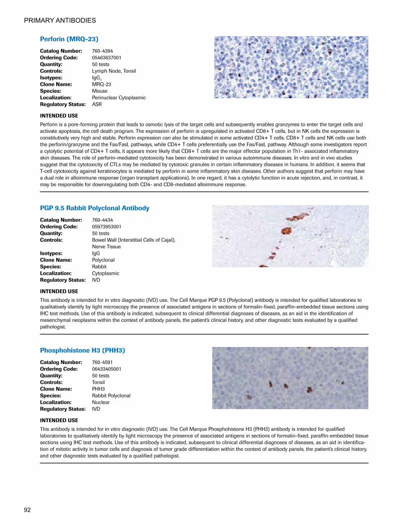
PRIMARY ANTIBODIES
92
Perforin(MRQ-23)
CatalogNumber: 760-4394OrderingCode: 05463637001Quantity: 50 testsControls: Lymph Node, TonsilIsotypes: IgG1CloneName: MRQ-23Species: MouseLocalization: Perinuclear CytoplasmicRegulatoryStatus: ASR
INTENDEDUSE
Perforin is a pore-forming protein that leads to osmotic lysis of the target cells and subsequently enables granzymes to enter the target cells and activate apoptosis, the cell death program. The expression of perforin is upregulated in activated CD8+ T cells, but in NK cells the expression is constitutively very high and stable. Perforin expression can also be stimulated in some activated CD4+ T cells. CD8+ T cells and NK cells use both the perforin/granzyme and the Fas/FasL pathways, while CD4+ T cells preferentially use the Fas/FasL pathway. Although some investigators report a cytolytic potential of CD4+ T cells, it appears more likely that CD8+ T cells are the major effector population in Th1- associated inflammatory skin diseases. The role of perforin-mediated cytotoxicity has been demonstrated in various autoimmune diseases. In vitro and in vivo studies suggest that the cytotoxicity of CTLs may be mediated by cytotoxic granules in certain inflammatory diseases in humans. In addition, it seems that T-cell cytotoxicity against keratinocytes is mediated by perforin in some inflammatory skin diseases. Other authors suggest that perforin may have a dual role in alloimmune response (organ transplant applications). In one regard, it has a cytolytic function in acute rejection, and, in contrast, it may be responsible for downregulating both CD4- and CD8-mediated alloimmune response.
PGP9.5RabbitPolyclonalAntibody
CatalogNumber: 760-4434OrderingCode: 05973953001Quantity: 50 testsControls: Bowel Wall (Interstitial Cells of Cajal),
Nerve TissueIsotypes: IgGCloneName: PolyclonalSpecies: RabbitLocalization: CytoplasmicRegulatoryStatus: IVD
INTENDEDUSE
This antibody is intended for in vitro diagnostic (IVD) use. The Cell Marque PGP 9.5 (Polyclonal) antibody is intended for qualified laboratories to qualitatively identify by light microscopy the presence of associated antigens in sections of formalin-fixed, paraffin-embedded tissue sections using IHC test methods. Use of this antibody is indicated, subsequent to clinical differential diagnoses of diseases, as an aid in the identification of mesenchymal neoplasms within the context of antibody panels, the patient’s clinical history, and other diagnostic tests evaluated by a qualified pathologist.
PhosphohistoneH3(PHH3)
CatalogNumber: 760-4591OrderingCode: 06433405001Quantity: 50 testsControls: TonsilCloneName: PHH3Species: Rabbit PolyclonalLocalization: NuclearRegulatoryStatus: IVD
INTENDEDUSE
This antibody is intended for in vitro diagnostic (IVD) use. The Cell Marque Phosphohistone H3 (PHH3) antibody is intended for qualified laboratories to qualitatively identify by light microscopy the presence of associated antigens in sections of formalin-fixed, paraffin-embedded tissue sections using IHC test methods. Use of this antibody is indicated, subsequent to clinical differential diagnoses of diseases, as an aid in identifica-tion of mitotic activity in tumor cells and diagnosis of tumor grade differentiation within the context of antibody panels, the patient’s clinical history, and other diagnostic tests evaluated by a qualified pathologist.

PRIMARY ANTIBODIES
93
PLAP(NB10)
CatalogNumber: 760-2664OrderingCode: 05267757001Quantity: 50 testsControls: PlacentaIsotypes: IgGCloneName: NB10Species: MouseLocalization: CytoplasmicRegulatoryStatus: IVD
INTENDEDUSE
Anti-PLAP antibody immunoreacts with germ cell tumors and can discriminate between these and other neoplasms. Somatic neoplasms e.g. breast, gastrointestinal, prostatic and urinary cancers may also immunoreact with antibodies to PLAP. Anti-PLAP positivity in conjunction with keratin negativity favors seminoma over carcinoma. Germ cell tumors are usually keratin positive, but they regularly fail to stain with anti-EMA, whereas most carcinomas stain with anti-EMA. Anti-PLAP has been useful in the diagnosis of gestational trophoblastic disease. This antibody has shown cross-reaction with human intestinal alkaline phosphatase.
PMS2(EPR3947)RabbitMonoclonalAntibody
CatalogNumber: 760-4531OrderingCode: 06419216001Quantity: 50 testsControls: ColonIsotypes: IgGCloneName: EPR3947Species: RabbitLocalization: NuclearRegulatoryStatus: IVD
INTENDEDUSE
This antibody is intended for in vitro diagnostic (IVD) use. The Cell Marque PMS2 (EPR3947) antibody is intended for qualified laboratories to qualitatively identify by light microscopy the presence of associated antigens in sections of formalin-fixed, paraffin-embedded tissue sections using IHC test methods. Use of this antibody is indicated, subsequent to clinical differential diagnoses of diseases, as an aid in the identification of MSI-H tumors within the context of antibody panels, the patient’s clinical history, and other diagnostic tests evaluated by a qualified pathologist.
Podoplanin(D2-40)MouseMonoclonalAntibody
CatalogNumber: 760-4395OrderingCode: 05463645001Quantity: 50 testsControls: Lymph Node, LymphangiomaIsotypes: IgG1CloneName: D2-40Species: MouseLocalization: CytoplasmicRegulatoryStatus: IVD
INTENDEDUSE
This antibody is intended for in vitro diagnostic (IVD) use. The Cell Marque Podoplanin (D2-40) antibody is intended for qualified laboratories to qualitatively identify by light microscopy the presence of associated antigens in sections of formalin-fixed, paraffin-embedded tissue sections using IHC test methods. Use of this antibody is indicated, subsequent to clinical differential diagnoses of diseases, as an aid in the identification of lymphatic invasion of a variety of tumors and between epitheloid and mesothelioma and lung adenocarcinoma within the context of antibody panels, the patient’s clinical history, and other diagnostic tests evaluated by a qualified pathologist.

PRIMARY ANTIBODIES
94
CONFIRManti-ProgesteroneReceptor(PR)(1E2)RabbitMonoclonalPrimaryAntibody
CatalogNumber: 790-2223 790-4296OrderingCode: 05277990001 5278392001Quantity: 50 tests 250 testsControls: BreastIsotypes: IgGCloneName: 1E2Species: RabbitLocalization: NuclearRegulatoryStatus: IVD
INTENDEDUSE
This antibody is intended for in vitro diagnostic (IVD) use. CONFIRM anti-Progesterone Receptor (PR) (1E2) Rabbit Monoclonal (IgG) is intended for laboratory use for the qualitative detection of progesterone receptor (PR) antigen in sections of formalin-fixed, paraffin-embedded tissue on a VENTANA automated slide stainer with VENTANA detection kits and ancillary reagents. CONFIRM antiPR (1E2) is directed against an epitope present on human progesterone receptor protein located in the nucleus of PR positive normal and neoplastic cells. CONFIRM anti-PR (1E2) is indicated as an aid in the management, prognosis, and prediction of therapy outcome of breast carcinoma. This product should be interpreted by a qualified pathologist in conjunction with histological examination, relevant clinical information, and proper controls. Prescription use only.
Prolactin(polyclonal)
CatalogNumber: 760-2803OrderingCode: 05268249001Quantity: 50 testsControls: Normal PituitaryCloneName: PolyclonalSpecies: RabbitLocalization: CytoplasmicRegulatoryStatus: IVD
INTENDEDUSE
Anti-Prolactin is a useful marker in classification of pituitary tumors and the study of pituitary disease. It reacts with prolactin-producing cells. Such prolactin-producing cells can also be found in prostate epithelium.
ƒ
CONFIRManti-ProstateSpecificAntigen(PSA)RabbitPolyclonalPrimaryAntibody
CatalogNumber: 760-2506OrderingCode: 05266939001Quantity: 50 testsControls: ProstateIsotypes: IgGCloneName: PolyclonalSpecies: RabbitLocalization: CytoplasmicRegulatoryStatus: IVD
INTENDEDUSE
This antibody is intended for in vitro diagnostic (IVD) use. Ventana Medical Systems' (Ventana) CONFIRM anti-Prostate Specific Antigen (PSA)Primary Antibody is a rabbit polyclonal antibody (lg) directed against both free and bound human prostate specific antigen (PSA). This antibody is intended for use to qualitatively identify PSA by light microscopy in sections of formalin-fixed, paraffin-embedded tissue following staining on a VENTANA automated slide stainer. The clinical interpretation of any staining, or the absence of staining, must be complemented by morphological studies and evaluation of proper controls. Evaluation must be made by a qualified pathologist within the context of the patient’s clinical history and other diagnostic tests.
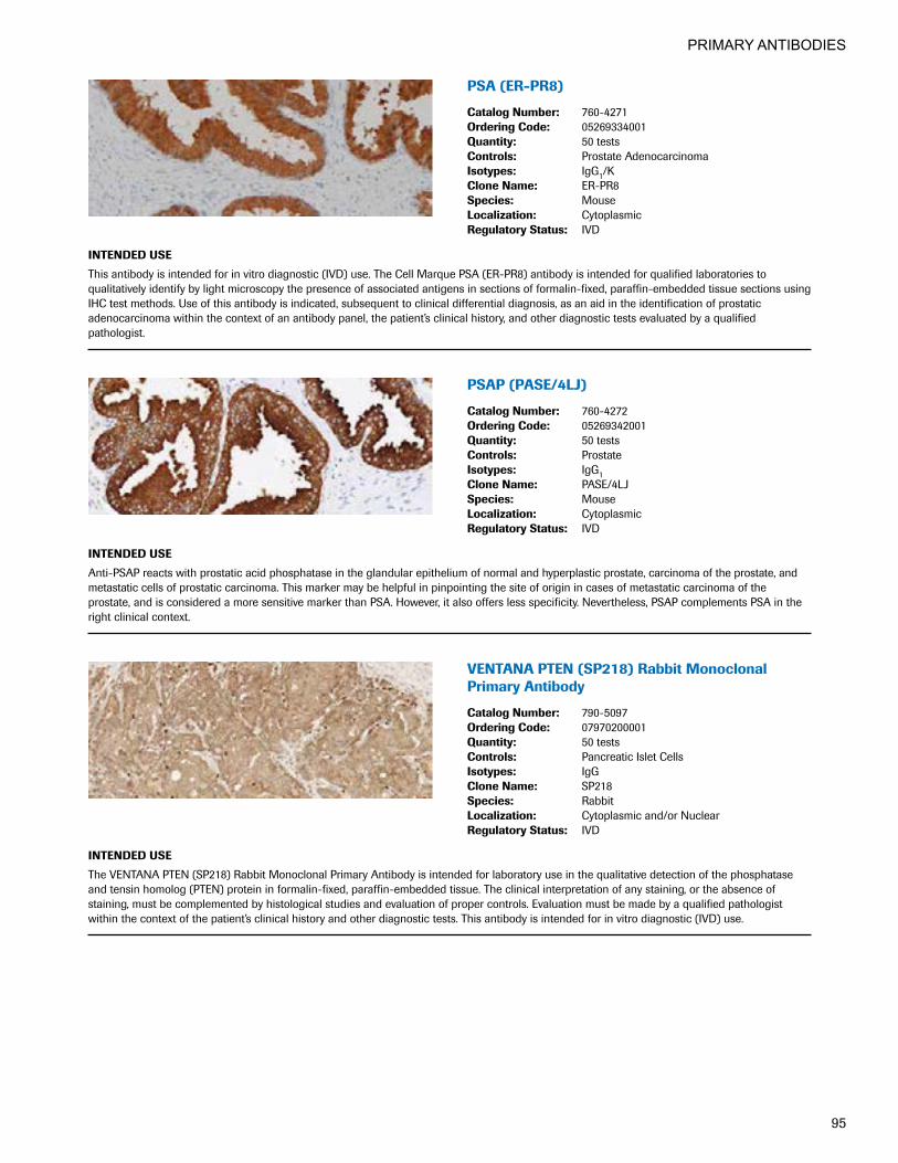
PRIMARY ANTIBODIES
95
PSA(ER-PR8)
CatalogNumber: 760-4271OrderingCode: 05269334001Quantity: 50 testsControls: Prostate AdenocarcinomaIsotypes: IgG1/KCloneName: ER-PR8Species: MouseLocalization: CytoplasmicRegulatoryStatus: IVD
INTENDEDUSE
This antibody is intended for in vitro diagnostic (IVD) use. The Cell Marque PSA (ER-PR8) antibody is intended for qualified laboratories to qualitatively identify by light microscopy the presence of associated antigens in sections of formalin-fixed, paraffin-embedded tissue sections using IHC test methods. Use of this antibody is indicated, subsequent to clinical differential diagnosis, as an aid in the identification of prostatic adenocarcinoma within the context of an antibody panel, the patient’s clinical history, and other diagnostic tests evaluated by a qualified pathologist.
PSAP(PASE/4LJ)
CatalogNumber: 760-4272OrderingCode: 05269342001Quantity: 50 testsControls: ProstateIsotypes: IgG1CloneName: PASE/4LJSpecies: MouseLocalization: CytoplasmicRegulatoryStatus: IVD
INTENDEDUSE
Anti-PSAP reacts with prostatic acid phosphatase in the glandular epithelium of normal and hyperplastic prostate, carcinoma of the prostate, and metastatic cells of prostatic carcinoma. This marker may be helpful in pinpointing the site of origin in cases of metastatic carcinoma of the prostate, and is considered a more sensitive marker than PSA. However, it also offers less specificity. Nevertheless, PSAP complements PSA in the right clinical context.
VENTANAPTEN(SP218)RabbitMonoclonalPrimary Antibody
CatalogNumber: 790-5097OrderingCode: 07970200001Quantity: 50 testsControls: Pancreatic Islet CellsIsotypes: IgGCloneName: SP218 Species: RabbitLocalization: Cytoplasmic and/or NuclearRegulatoryStatus: IVD
INTENDEDUSE
The VENTANA PTEN (SP218) Rabbit Monoclonal Primary Antibody is intended for laboratory use in the qualitative detection of the phosphatase and tensin homolog (PTEN) protein in formalin-fixed, paraffin-embedded tissue. The clinical interpretation of any staining, or the absence of staining, must be complemented by histological studies and evaluation of proper controls. Evaluation must be made by a qualified pathologist within the context of the patient’s clinical history and other diagnostic tests. This antibody is intended for in vitro diagnostic (IVD) use.
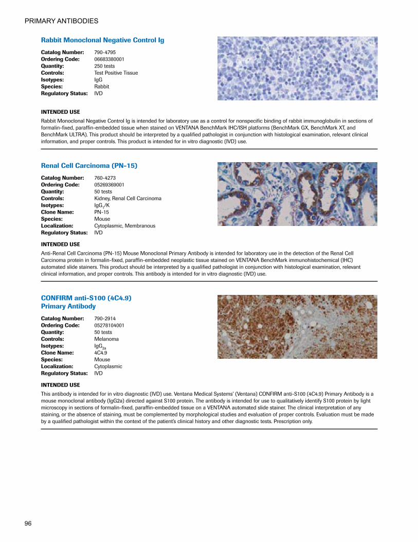
PRIMARY ANTIBODIES
96
RabbitMonoclonalNegativeControlIg
CatalogNumber: 790-4795OrderingCode: 06683380001Quantity: 250 testsControls: Test Positive TissueIsotypes: IgGSpecies: RabbitRegulatoryStatus: IVD
INTENDEDUSE
Rabbit Monoclonal Negative Control Ig is intended for laboratory use as a control for nonspecific binding of rabbit immunoglobulin in sections of formalin-fixed, paraffin-embedded tissue when stained on VENTANA BenchMark IHC/ISH platforms (BenchMark GX, BenchMark XT, and BenchMark ULTRA). This product should be interpreted by a qualified pathologist in conjunction with histological examination, relevant clinical information, and proper controls. This product is intended for in vitro diagnostic (IVD) use.
RenalCellCarcinoma(PN-15)
CatalogNumber: 760-4273OrderingCode: 05269369001Quantity: 50 testsControls: Kidney, Renal Cell CarcinomaIsotypes: IgG1/KCloneName: PN-15Species: MouseLocalization: Cytoplasmic, MembranousRegulatoryStatus: IVD
INTENDEDUSE
Anti-Renal Cell Carcinoma (PN-15) Mouse Monoclonal Primary Antibody is intended for laboratory use in the detection of the Renal Cell Carcinoma protein in formalin-fixed, paraffin-embedded neoplastic tissue stained on VENTANA BenchMark immunohistochemical (IHC) automated slide stainers. This product should be interpreted by a qualified pathologist in conjunction with histological examination, relevant clinical information, and proper controls. This antibody is intended for in vitro diagnostic (IVD) use.
CONFIRManti-S100(4C4.9) Primary Antibody
CatalogNumber: 790-2914OrderingCode: 05278104001Quantity: 50 testsControls: MelanomaIsotypes: IgG2aCloneName: 4C4.9Species: MouseLocalization: CytoplasmicRegulatoryStatus: IVD
INTENDEDUSE
This antibody is intended for in vitro diagnostic (IVD) use. Ventana Medical Systems’ (Ventana) CONFIRM anti-S100 (4C4.9) Primary Antibody is a mouse monoclonal antibody (IgG2a) directed against S100 protein. The antibody is intended for use to qualitatively identify S100 protein by light microscopy in sections of formalin-fixed, paraffin-embedded tissue on a VENTANA automated slide stainer. The clinical interpretation of any staining, or the absence of staining, must be complemented by morphological studies and evaluation of proper controls. Evaluation must be made by a qualified pathologist within the context of the patient’s clinical history and other diagnostic tests. Prescription only.

PRIMARY ANTIBODIES
97
CONFIRManti-S100(Polyclonal) Primary Antibody
CatalogNumber: 760-2523OrderingCode: 05267072001Quantity: 50 testsControls: MelanomaCloneName: PolyclonalSpecies: RabbitLocalization: CytoplasmicRegulatoryStatus: IVD
INTENDEDUSE
Ventana Medical Systems, Inc.’s (Ventana) CONFIRM anti-S100 (Polyclonal) Primary Antibody contains rabbit antiserum directed against an epitope found on S100 protein and may be used to aid in the identification of cells of normal and abnormal neuronal and neuroendocrine lineage and as an aid in the diagnosis of anaplastic tumors. The antibody is intended for qualitative staining in section of formalin-fixed, paraffin-embedded tissue on a VENTANA automated slide stainer. This product should be interpreted by a qualified pathologist in conjunction with histological examination, relevant clinical information, and proper controls. This antibody is intended for in vitro diagnostic (IVD) use.
SALL4(6E3)MouseMonoclonal Primary Antibody
CatalogNumber: 760-4864OrderingCode: 07047690001Quantity: 50 testsControls: SeminomaIsotypes: IgGCloneName: 6E3Species: MouseLocalization: NuclearRegulatoryStatus: IVD
INTENDEDUSE
This antibody is intended for in vitro diagnostic (IVD) use. The Cell Marque SALL4 (6E3) antibody is intended for qualified laboratories to qualitatively identify by light microscopy the presence of associated antigens in sections of formalin-fixed, paraffin-embedded tissue using IHC test methods. Use of this antibody is indicated as an aid in the identification of germ cell neoplasms within the context of an antibody panel, the patient’s clinical history, and other diagnostic tests evaluated by a qualified pathologist.
Smoothelin(R4A)MouseMonoclonalAntibody
CatalogNumber: 760-4592OrderingCode: 06433278001Quantity: 50 testsControls: Bladder, LeiomyosarcomaIsotypes: IgG1CloneName: R4ASpecies: Mouse MonoclonalLocalization: Cytoplasmic (MP), NuclearRegulatoryStatus: IVD
INTENDEDUSE
This antibody is intended for in vitro diagnostic (IVD) use. The Cell Marque Smoothelin (R4A) antibody is intended for qualified laboratories to qualitatively identify by light microscopy the presence of associated antigens in sections of formalin-fixed, paraffin-embedded tissue sections using IHC test methods. Use of this antibody is indicated as an aid in the distinction of terminally differentiated smooth muscle cells, smooth muscle neoplasms of the gastrointestinal tract and other organs, and staging of bladder carcinoma within the context of an antibody panel, the patient’s clinical history, and other diagnostic tests evaluated by a qualified pathologist.

PRIMARY ANTIBODIES
98
Somatostatin(polyclonal)
CatalogNumber: 760-2667OrderingCode: 05267781001Quantity: 50 testsControls: PancreasCloneName: PolyclonalSpecies: RabbitLocalization: CytoplasmicRegulatoryStatus: IVD
INTENDEDUSE
Anti-Somatostatin is a useful marker of D-cells of pancreatic islet cells. D-cells identification can be used to identify hyperplasia of the pancreatic islets as well as islet cell tumors such as somatostatinomas. Somatostain can also be found in gastrointestinal and bronchopulmonary endocrine cells, thymic endocrine cells, and thyroid C cells. Somatostatin suppresses gastric acid secretion, gallbladder contractions, and pancreatic enzyme secretion.
SOX-2(SP76)
CatalogNumber: 760-4621OrderingCode: 06523943001Quantity: 50 testsControls: Squamous EpitheliumIsotypes: IgGCloneName: SP76Species: RabbitLocalization: NuclearRegulatoryStatus: IVD
INTENDEDUSE
This antibody is intended for in vitro diagnostic (IVD) use. The Cell Marque SOX-2 (SP76) antibody is intended for qualified laboratories to qualitatively identify by light microscopy the presence of associated antigens in sections of formalin-fixed, paraffin-embedded tissue sections using IHC test methods. Use of this antibody is indicated, subsequent to clinical differential diagnoses of diseases, as an aid in the identification of lung squamous cell carcinoma and germ cell tumors within the context of antibody panels, the patient’s clinical history, and other diagnostic tests evaluated by a qualified pathologist.
SOX-10(SP267)RabbitMonoclonal Primary Antibody
CatalogNumber: 760-4968OrderingCode: 07560389001Quantity: 50 testsControls: Melanoma, Skin Melanocytes Isotypes: IgG CloneName: SP267Species: RabbitLocalization: Nuclear RegulatoryStatus: IVD
INTENDEDUSE
SOX-10 (SP267) Rabbit Monoclonal Primary Antibody is intended for laboratory use in the detection of the SOX-10 protein in formalin-fixed, paraffin-embedded tissue stained on BenchMark immunohistochemical (IHC/ISH) automated slide stainers. This product should be interpreted by a qualified pathologist in conjunction with histological examination, relevant clinical information, and proper controls. This antibody is intended for in vitro diagnostic (IVD) use.

PRIMARY ANTIBODIES
99
SOX-11(MRQ-58)MouseMonoclonal Primary Antibody
CatalogNumber: 760-4868OrderingCode: 07047738001Quantity: 50 testsControls: Mantle Cell LymphomaIsotypes: IgG1CloneName: MRQ-58Species: Mouse MonoclonalLocalization: Nuclear RegulatoryStatus: IVD
INTENDEDUSE
This antibody is intended for in vitro diagnostic (IVD) use. The Cell Marque SOX-11 (MRQ-58) antibody is intended for qualified laboratories to qualitatively identify by light microscopy the presence of associated antigens in sections of formalin-fixed, paraffin-embedded tissue sections using IHC test methods. Use of this antibody is indicated, subsequent to clinical differential diagnosis, as an aid in the identification and differentiation of mantle cell lymphomas within the context of an antibody panel, the patient’s clinical history, and other diagnostic tests evaluated by a qualified pathologist.
Spectrin(RBC2/3D5)
CatalogNumber: 760-4274OrderingCode: 05269377001Quantity: 50 testsControls: Bone MarrowIsotypes: IgG2a/KCloneName: RBC2/3D5Species: MouseLocalization: MembranousRegulatoryStatus: IVD
INTENDEDUSE
Spectrin is a cytoskeletal protein which is found in muscles, red blood cells and red cell precursors. Anti-Spectrin antibody is useful in the diagnosis of erythroid leukemias.
Synaptophysin(MRQ-40) RabbitMonoclonalAntibody
CatalogNumber: 760-4595OrderingCode: 06433324001Quantity: 50 testsControls: Pancreatic Islet CellsIsotypes: IgG1CloneName: MRQ-40Species: Rabbit MonoclonalLocalization: CytoplasmicRegulatoryStatus: IVD
INTENDEDUSE
This antibody is intended for in vitro diagnostic (IVD) use. The Cell Marque Synaptophysin (MRQ-40) antibody is intended for qualified laboratories to qualitatively identify by light microscopy the presence of associated antigens in sections of formalin-fixed, paraffin-embedded tissue sections using IHC test methods. This antibody is used as an aid in the identification of neuroendocrine neoplasms within the context of an antibody panel, the patient’s clinical history, and other diagnostic tests evaluated by a qualified pathologist.

PRIMARY ANTIBODIES
100
CONFIRManti-Synaptophysin(SP11) RabbitMonoclonalPrimaryAntibody
CatalogNumber: 790-4407OrderingCode: 05479304001Quantity: 50 testsControls: PancreasCloneName: SP11Species: RabbitLocalization: CytoplasmicRegulatoryStatus: IVD
INTENDEDUSE
This antibody is intended for in vitro diagnostic (IVD) use. Ventana Medical Systems' (Ventana) CONFIRM antiSynaptophysin (SP11) Rabbit Monoclonal Primary Antibody is designed to qualitatively detect the presence of synaptophysin expressing cells via light microscopy in formalin- fixed, paraffin-embedded tissue. Positive staining results may aid in the classification of neuroendocrine tumors. The clinical interpretation of any staining, or the absence of staining, must be complemented by morphological studies and evaluation of proper controls. Evaluation must be made by a qualified pathologist within the context of the patient’s clinical history and other diagnostic tests.
T-bet(MRQ-46)RabbitMonoclonalAntibody
CatalogNumber: 760-4598OrderingCode: 06433391001Quantity: 50 testsControls: Hairy Cell Leukemia, TonsilIsotypes: IgG1CloneName: MRQ-46Species: Rabbit MonoclonalLocalization: NuclearRegulatoryStatus: IVD
INTENDEDUSE
This antibody is intended for in vitro diagnostic (IVD) use. The Cell Marque T-bet (MRQ-46) antibody is intended for qualified laboratories to qualitatively identify by light microscopy the presence of associated antigens in sections of formalin-fixed, paraffin-embedded tissue sections using IHC test methods. Use of this antibody is indicated, subsequent to clinical differential diagnoses of diseases, as an aid in the identification of hairy cell leukemia, Hodgkin lymphoma, and precursor B-cell lymphoblastic leukemia/lymphoma within the context of an antibody panel, the patient’s clinical history, and other diagnostic tests evaluated by a qualified pathologist.
TAG-72(B72.3)
CatalogNumber: 760-2669OrderingCode: 05267803001Quantity: 50 testsControls: Breast CarcinomaIsotypes: IgG1/KCloneName: B72.3Species: MouseLocalization: CytoplasmicRegulatoryStatus: IVD
INTENDEDUSE
Anti-TAG-72 (B72.3) Mouse Monoclonal Primary Antibody is intended for laboratory use in the detection of the TAG-72 protein in formalin-fixed, paraffin-embedded neoplastic tissue stained on VENTANA BenchMark immunohistochemical (IHC) automated slide stainers. This product should be interpreted by a qualified pathologist in conjunction with histological examination, relevant clinical information, and proper controls. This antibody is intended for in vitro diagnostic (IVD) use.

PRIMARY ANTIBODIES
101
TdT(polyclonal)
CatalogNumber: 760-2670OrderingCode: 05267811001Quantity: 50 testsControls: TdT Positive Lymphomas, ThymusCloneName: PolyclonalSpecies: RabbitLocalization: NuclearRegulatoryStatus: IVD
INTENDEDUSE
This antibody is intended for in vitro diagnostic (IVD) use. The Cell Marque TdT antibody is intended for qualified laboratories to qualitatively identify by light microscopy the presence of associated antigens in sections of formalin-fixed, paraffin-embedded tissue sections using IHC test methods. Use of this antibody is indicated, subsequent to clinical differential diagnosis, as an aid in the identification of lymphomas within the context of antibody panels, the patient’s clinical history, and other diagnostic tests evaluated by a qualified pathologist.
anti-TFE3(MRQ-37)RabbitMonoclonal Primary Antibody
CatalogNumber: 760-4622OrderingCode: 06523951001Quantity: 50 testsControls: Testis (Germ Cells), XP11.2 Translocation
Renal CellIsotypes: IgGCloneName: MRQ-37Species: RabbitLocalization: NuclearRegulatoryStatus: IVD
INTENDEDUSE
This antibody is intended for in vitro diagnostic (IVD) use. The Cell Marque TFE3 (MRQ-37) antibody is intended for qualified laboratories to qualitatively identify by light microscopy the presence of associated antigens in sections of formalin-fixed, paraffin-embedded tissue sections using IHC test methods. Use of this antibody is indicated subsequent to the clinical diagnosis of malignancy as an aid in the recognition of Xp11 translocation renal cell carcinoma and alveolar soft part sarcoma within the context of an antibody panel, the patient’s clinical history, and other diagnostic tests evaluated by a qualified pathologist.
CONFIRManti-ThymidinePhosphorylase(P-GF.44C)MouseMonoclonalPrimaryAntibody
CatalogNumber: 790-4454OrderingCode: 05976502001Quantity: 50 testsControls: See Package InsertIsotypes: IgG1CloneName: P-GF.44CSpecies: MouseLocalization: Nuclear, CytoplasmicRegulatoryStatus: IVD
INTENDEDUSE
Ventana Medical Systems’ (Ventana) CONFIRM anti-Thymidine Phosphorylase (PGF.44C) Mouse Monoclonal Primary Antibody recognizes human thymidine phosphorylase, an angiogenic factor, expressed in varying levels by macrophages, stromal cells, glial cells and some epithelial cells. Thymidine phosphorylase shows a cytoplasmic and/or nuclear location. This antibody exhibits a nuclear and/or cytoplasmic staining pattern and may be used to aid in the detection of thymidine phosphorylase in carcinoma of the breast, esophagus, stomach, colon and rectum, pancreas, head and neck, ovaries and lung. The antibody is intended for qualitative staining in sections of formalin-fixed, paraffin-embedded tissue. This product should be interpreted by a qualified pathologist in conjunction with histological examination, relevant clinical information and proper controls. This antibody is intended for in vitro diagnostic (IVD) use.

PRIMARY ANTIBODIES
102
Thyroglobulin(2H11+6E1)
CatalogNumber: 760-2671OrderingCode: 05267820001Quantity: 50 testsControls: ThyroidIsotypes: IgG1CloneName: 2H11+6E1Species: MouseLocalization: CytoplasmicRegulatoryStatus: IVD
INTENDEDUSE
Anti-Thyroglobulin antibody reacts with human thyroglobulin as demonstrated by a single band of immunoblotting in a lysate of human thyroid tissue. The vast majority of follicular carcinomas of the thyroid will give positive immunoreactivity for thyroglobulin even though sometimes only focal. Poorly differentiated carcinomas of the thyroid are frequently thyroglobulin negative. Adenocarcinomas of other than thyroid origin do not react with this antibody.
CONFIRManti-ThyroidTranscriptionFactor-1(8G7G3/1)MouseMonoclonalPrimaryAntibody
CatalogNumber: 790-4398OrderingCode: 05479312001Quantity: 50 testsControls: Lung, Thyroid, Lung CarcinomaIsotypes: IgG1CloneName: 8G7G3/1Species: MouseLocalization: NuclearRegulatoryStatus: IVD
INTENDEDUSE
CONFIRM anti-Thyroid Transcription Factor-1 (8G7G3/1) Mouse Monoclonal Primary Antibody (CONFIRM anti-Thyroid Transcription Factor-1 (8G7G3/1)) is designed to qualitatively detect the presence of thyroid transcription factor-1 (TTF-1) expressing cells. TTF-1 positive staining results may aid in the classification of neoplasms of the lung and thyroid. The clinical interpretation of any staining, or the absence of staining, must be complemented by morphological studies and evaluation of proper controls. The antibody is intended for qualitative staining of sections of formalin-fixed, paraffin-embedded tissue. This product should be interpreted by a qualified pathologist in conjunction with histological examina-tion, relevant clinical information, and proper controls. This antibody is intended for in vitro diagnostic (IVD) use.
anti-ThyroidTranscriptionFactor-1(SP141)RabbitMonoclonalPrimaryAntibody
CatalogNumber: 790-4756OrderingCode: 06640613001Quantity: 50 testsControls: Normal Thyroid, Lung Carcinoma, Normal LungIsotypes: IgGCloneName: SP141Species: RabbitLocalization: NuclearRegulatoryStatus: IVDAlternateNames: Thyroid Transcription Factor - 1
INTENDEDUSE
Anti-Thyroid Transcription Factor-1 (SP141) Rabbit Monoclonal Primary Antibody (anti-TTF-1 (SP141)) is directed against the thyroid transcription factor-1 (TTF-1) protein. This antibody exhibits a nuclear staining pattern and may be used to aid in the classification of neoplasms of the lung and thyroid. This antibody is intended for qualitative staining in sections of formalin-fixed, paraffin-embedded tissue. This product should be inter-preted by a qualified pathologist in conjunction with histological examination, relevant clinical information, and proper controls. This antibody is intended for in vitro diagnostic (IVD) use.

PRIMARY ANTIBODIES
103
CONFIRManti-TopoisomeraseIIα(JS5B4) RabbitMonoclonalPrimaryAntibody
CatalogNumber: 790-4371OrderingCode: 05479339001Quantity: 50 testsControls: Spleen, TonsilCloneName: JS5B4Species: RabbitLocalization: NuclearRegulatoryStatus: IVD
INTENDEDUSE
This antibody is intended for in vitro diagnostic (IVD) use. Ventana Medical Systems' CONFIRM anti-Topoisomerase IIα (JS5B4) Rabbit Monoclonal Primary Antibody is designed to qualitatively detect the presence of topoisomerase IIα expressing cells via light microscopy in formalin-fixed, paraffin-embedded tissue following staining on a Ventana BenchMark XT instrument. During the cell cycle, topoisomerase IIα is expressed in the G1, S, G2, and M phase but is absent in the G0 (quiescent) phase.1 Assessment of the proportion of positive staining cells for topoisomerase IIα may aid in evaluation of the proliferative activity of normal and/or neoplastic tissue.2,3,4 The clinical interpretation of any staining, or the absence of staining, must be complemented by morphological studies and evaluation of proper controls. Evaluation must be made by a qualified pathologist within the context of the patient’s clinical history and other diagnostic tests. 1. Kreipe H, Heidebrecht HJ, Hansen S, Röhlk W, Kubbres M, Wacker HH, et al. A new proliferation associated nuclear antigen detectable in
paraffin-embedded tissues by monoclonal antibody Ki-S1. AM J. Pathol 1993; 142:3-9.2. Rudolph P, Lappe T, Scubert C, Schmidt D, Parwaresch RM, Christophers E. Diagnostic assessment of two novel proliferation–specific antigens in
benign and malignant melanocytic lesions. AM J. Pathol 1995; 147:1615-25.3. Kreipe H, Alm P, Olsson H, Hauberg M, Fisher L, Parwaresch RM. Prognostic significance of a formalin resistance nuclear proliferation antigen in
mammary carcinoma as determined by the monoclonal antibody Ki-S1. AM J Pathol 1993; 142:651-657.4. Kellner U, Heidebrecht HJ, Rudolph P, et al. Detection of human Topoisomerase IIα in cell lines and tissue: Characterization of five novel
monoclonal antibodies. J of Histochem and Cytochem 1997; 45:251-264.
ƒ
CONFIRManti-Totalc-MET(SP44) RabbitMonoclonalPrimaryAntibody
CatalogNumber: 790-4430OrderingCode: 05571219001Quantity: 50 testsControls: Kidney, Non-Small Cell Lung Carcinoma,
Gastric CarcinomaCloneName: SP44Species: RabbitLocalization: Cytoplasmic, MembranousRegulatoryStatus: IVD
INTENDEDUSE
Ventana Medical Systems' (Ventana) CONFIRM anti-Total c-MET (SP44) Rabbit Monoclonal Primary Antibody is directed against a membranous and/or cytoplasmic epitope present in human normal epithelial or tumor cells. This antibody may be used to aid in the identification of normal and neoplastic c-MET expressing cells. The antibody is intended for qualitative staining in sections of formalin-fixed, paraffin-embedded tissue. The clinical interpretation of any staining, or the absence of staining, must be complemented by histological studies and evaluation of proper controls. Evaluation must be made by a qualified pathologist within the context of the patient’s clinical history and other diagnostic tests. This antibody is intended for in vitro diagnostic (IVD) use.

PRIMARY ANTIBODIES
104
TRAcP(9C5)MouseMonoclonal Primary Antibody
CatalogNumber: 760-4275OrderingCode: 05269385001Quantity: 50 testsControls: Hairy Cell LeukemiaIsotypes: IgG2bCloneName: 9C5Species: MouseLocalization: CytoplasmicRegulatoryStatus: IVD
INTENDEDUSE
This antibody is intended for in vitro diagnostic (IVD) use. The Cell Marque TRAcP (9C5) antibody is intended for qualified laboratories to qualitatively identify by light microscopy the presence of associated antigens in sections of formalin-fixed, paraffin-embedded tissue sections using IHC test methods. Use of this antibody is indicated, subsequent to clinical differential diagnoses of diseases, as an aid in the identification of Hairy Cell Leukemia within the context of antibody panels, the patient’s clinical history, and other diagnostic tests evaluated by a qualified pathologist.
Tryptase(G3)
CatalogNumber: 760-4276OrderingCode: 05269393001Quantity: 50 testsControls: Mast Cell-Containing TissueIsotypes: IgG1CloneName: G3Species: MouseLocalization: CytoplasmicRegulatoryStatus: IVD
INTENDEDUSE
Tryptases constitute a subfamily of trypsin-like proteinases, stored in the mast cell secretory granules and basophils. Upon cellular activation, these enzymes are released into the extracellular environment. Anti-tryptase is a good marker for mast cells, basophils, and their derivatives.
ƒ
TSH(polyclonal)
CatalogNumber: 760-2709OrderingCode: 05268184001Quantity: 50 testsControls: Normal PituitaryCloneName: PolyclonalSpecies: RabbitLocalization: CytoplasmicRegulatoryStatus: IVD
INTENDEDUSE
Anti-TSH is a useful marker in classification of pituitary tumors and the study of pituitary disease. It reacts with TSH-producing cells (thyrotrophs).

PRIMARY ANTIBODIES
105
CONFIRManti-Tyrosinase(T311) MouseMonoclonalPrimaryAntibody
CatalogNumber: 790-4365OrderingCode: 05479347001Quantity: 50 testsControls: Lung, MelanomaIsotypes: IgG2aCloneName: T311Species: MouseLocalization: CytoplasmicRegulatoryStatus: IVD
INTENDEDUSE
This antibody is intended for in vitro diagnostic use. Ventana Medical Systems’ CONFIRM® anti-Tyrosinase (T311) Mouse Monoclonal Primary Antibody is designed to qualitatively detect the presence of tyrosinase expressing cells via light microscopy in formalin-fixed, paraffin-embedded tissue on a VENTANA automated slide stainer. Positive results aid in the classification of melanoma. The clinical interpretation of any staining, or the absence of staining, must be complemented by morphological studies and evaluation of proper controls. Evaluation must be made by a qualified pathologist within the context of the patient’s clinical history and other diagnostic tests.
UroplakinIII(SP73)RabbitMonoclonalAntibody
CatalogNumber: 760-4533OrderingCode: 06419232001Quantity: 50 testsControls: BladderIsotypes: IgG1CloneName: SP73Species: RabbitLocalization: Membranous, CytoplasmicRegulatoryStatus: IVD
INTENDEDUSE
This antibody is intended for in vitro diagnostic (IVD) use. The Cell Marque anti-Uroplakin III (SP73) antibody is intended for qualified laboratories to qualitatively identify by light microscopy the presence of associated antigens in sections of formalin-fixed, paraffin-embedded tissue sections using VENTANA BenchMark® automated IHC test methods. Use of this antibody is indicated, subsequent to clinical differential diagnoses of diseases as an aid in the identification of transitional cell carcinoma within the context of antibody panels, the patient’s clinical history, and other diagnostic tests evaluated by a qualified pathologist.
Villin(CWWB1)
CatalogNumber: 760-4277OrderingCode: 05269407001Quantity: 50 testsControls: Colonic Mucosa, Small Bowel MucosaIsotypes: IgG1CloneName: CWWB1Species: MouseLocalization: Cytoplasmic, MembranousRegulatoryStatus: IVD
INTENDEDUSE
Villin is a 95-kD glycoprotein of microvilli associated with rootlet formation in gastrointestinal mucosal epithelium. Anti-Villin labels the brush border area in the gastrointestinal mucosal epithelium. This antibody has been useful in differentiating gastrointestinal adenocarcinoma, neuroendocrine carcinomas and ovarian adenocarcinomas from adenocarcinomas from other organs. Also labeled by this antibody are Merkel cells of the skin.
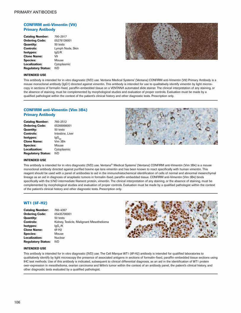
PRIMARY ANTIBODIES
106
CONFIRManti-Vimentin(V9) Primary Antibody
CatalogNumber: 790-2917OrderingCode: 05278139001Quantity: 50 testsControls: Lymph Node, SkinIsotypes: IgG/KCloneName: V9Species: MouseLocalization: CytoplasmicRegulatoryStatus: IVD
INTENDEDUSE
This antibody is intended for in vitro diagnostic (IVD) use. Ventana Medical Systems' (Ventana) CONFIRM anti-Vimentin (V9) Primary Antibody is a mouse monoclonal antibody (IgG1) directed against vimentin. This antibody is intended for use to qualitatively identify vimentin by light micros-copy in sections of formalin-fixed, paraffin-embedded tissue on a VENTANA automated slide stainer. The clinical interpretation of any staining, or the absence of staining, must be complemented by morphological studies and evaluation of proper controls. Evaluation must be made by a qualified pathologist within the context of the patient’s clinical history and other diagnostic tests. Prescription only.
CONFIRManti-Vimentin(Vim3B4) Primary Antibody
CatalogNumber: 760-2512OrderingCode: 05266998001Quantity: 50 testsControls: Intestine, LiverIsotypes: IgG2aCloneName: Vim 3B4Species: MouseLocalization: CytoplasmicRegulatoryStatus: IVD
INTENDEDUSE
This antibody is intended for in vitro diagnostic (IVD) use. Ventana® Medical Systems' (Ventana) CONFIRM anti-Vimentin (Vim 3B4) is a mouse monoclonal antibody directed against purified bovine eye lens vimentin and has been known to react specifically with human vimentin. This reagent should be used with a panel of antibodies to aid in the immunohistochemical identification of cells of normal and abnormal mesenchymal lineage as an aid in diagnosis of anaplastic tumors in formalin-fixed, paraffin-embedded tissue. CONFIRM anti-Vimentin (Vim 3B4) binds specifically with the 57kD intermediate filament protein, vimentin. The clinical interpretation of any staining, or the absence of staining, must be complemented by morphological studies and evaluation of proper controls. Evaluation must be made by a qualified pathologist within the context of the patient’s clinical history and other diagnostic tests. Prescription only.
WT1(6F-H2)
CatalogNumber: 760-4397OrderingCode: 05435706001Quantity: 50 testsControls: Kidney, Testicle, Malignant MesotheliomaIsotypes: IgG1/KCloneName: 6F-H2Species: MouseLocalization: NuclearRegulatoryStatus: IVD
INTENDEDUSE
This antibody is intended for in vitro diagnostic (IVD) use. The Cell Marque WT1 (6F-H2) antibody is intended for qualified laboratories to qualitatively identify by light microscopy the presence of associated antigens in sections of formalin-fixed, paraffin-embedded tissue sections using IHC test methods. Use of this antibody is indicated, subsequent to clinical differential diagnosis, as an aid in the identification of WT1 protein over-expression in mesothelioma, ovarian carcinoma and Wilm’s tumor within the context of an antibody panel, the patient’s clinical history, and other diagnostic tests evaluated by a qualified pathologist.
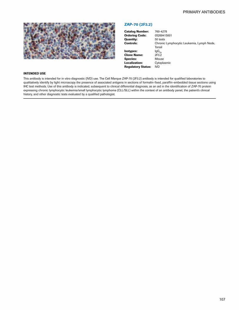
PRIMARY ANTIBODIES
107
ZAP-70(2F3.2)
CatalogNumber: 760-4278OrderingCode: 05269415001Quantity: 50 testsControls: Chronic Lymphocytic Leukemia, Lymph Node,
TonsilIsotypes: IgG2aCloneName: 2F3.2Species: MouseLocalization: CytoplasmicRegulatoryStatus: IVD
INTENDEDUSE
This antibody is intended for in vitro diagnostic (IVD) use. The Cell Marque ZAP-70 (2F3.2) antibody is intended for qualified laboratories to qualitatively identify by light microscopy the presence of associated antigens in sections of formalin-fixed, paraffin-embedded tissue sections using IHC test methods. Use of this antibody is indicated, subsequent to clinical differential diagnosis, as an aid in the identification of ZAP-70 protein expressing chronic lymphocytic leukemia/small lymphocytic lymphoma (CLL/SLL) within the context of an antibody panel, the patient’s clinical history, and other diagnostic tests evaluated by a qualified pathologist.

PRIMARY ANTIBODIES
108

Detection Systems
Innovation Delivering solutions that change
the practice of medicine and improve patient care

110
DETECTION SYSTEMS
OptiView DAB IHC Detection Innovative detection for enhanced stain quality
Powered by proprietary hapten technology, the dynamic range of VENTANA OptiView DAB IHC detection delivers unparalleled sensitivity and specificity so you can detect antigens across a wide range of expression levels. Virtually eliminating background even as signal intensity increases, OptiView chemistry provides the quality, flexibility and speed you need to deliver results quickly and confidently for enhanced patient care.
• Increase sensitivity: By increasing the numbers of HRP enzymes at each primary antibody site, OptiView chemistry provides unparalleled signal intensity, empowering you to visualize even low-level markers.
• Improvespecificity:Our patented synthetic, non-endogenous hapten system eliminates the potential for species-to-species interactions, which frequently cause background.
• Customize intensity: Unique chemistry and flexibility in protocol optimization enable greater control to meet your preferred staining intensity.
• Improve turnaround time: Enhanced sensitivity and software flexibility allow you to reduce turn-around time by 30 minutes or more for most assays, giving you the ability to process more slides in less time and deliver results sooner.
OptiView detection chemistry: our unique formula for success
Featuring a two-step design and unique synthetic, non-endogenous hapten technology on the secondary antibody, OptiView detection provides excellent background reduction. In addition, it delivers increased sensitivity through a higher number of multimers per primary antibody. The reaction is further enhanced by a proprietary reagent that accelerates DAB color development in the presence of the HRP enzyme.
Feature stains: Cyclin D1 (SP4) on mantle cell lymphoma with OptiView DAB IHC Detection Kit (left) and with OptiView detection plus OptiView Amplification Kit (right).

111
DETECTION SYSTEMS
HRP
HRP
HRP
HRP
HRP
HRP
HRP
HRP
HRP
HRP
HRP
HRP
Primary antibody
Tissue
OptiView HQ Universal Linker
HQ
HQHQ
HQ
HQHQ
1. Primary antibody binds to target antigen in tissue.
2. OptiView HQ Universal Linker, which has numerous non-endogenous HQ haptens, binds to the primary antibody.
3. OptiView HRP Multimer binds to the HQ haptens. The number of multimer molecules is multiplied in this way, resulting in increased staining intensity without increased background.
4. DAB chromogen reacts with HRP and H2O2 to generate a clean, crisp signal.
DAB
OptiView HRP Multimer
H2O2
AmplificationKit Catalog Number: 760-080 Ordering Code: 05266114001 Quantity: 100 tests Regulatory Status: IVD
Ventana Medical Systems, Inc.’s (Ventana) Amplification Kit may be used in conjunction with VENTANA detection kits to increase the signal intensity of weak staining mouse and rabbit primary antibodies. The kit is to be used for qualitative staining of formalin-fixed, paraffin-embedded tissue, frozen tissue or cytological preparations on VENTANA automated side stainers with VENTANA primary antibodies, detection kits and ancillary reagents.
The clinical interpretation of any staining, or the absence of staining, must be complemented by morphological studies and evaluation of proper controls. Evaluation must be made by a qualified pathologist within the context of the patient’s clinical history and other diagnostic tests.
This product is intended for in vitro diagnostic (IVD) use.
OptiViewAmplificationKit(50) OptiViewAmplificationKit(250) Catalog Number: 760-099 860-099 Ordering Code: 06396518001 06718663001 Quantity: 50 tests 250 tests Regulatory Status: IVD IVD
OptiView Amplification Kit may be used in conjunction with the OptiView DAB IHC Detection Kit (OptiView Detection Kit) to increase the staining intensity of mouse and rabbit primary antibodies. The kit is to be used for qualitative staining of formalin-fixed, paraffin- embedded tissue, frozen tissue, or cytological preparations on VENTANA BenchMark Advanced Staining automated slide stainers with VENTANA primary antibodies and ancillary reagents, visualized by light microscopy.
The clinical interpretation of any staining, or the absence of staining, must be complemented by morphological studies and evaluation of proper controls. Evaluation must be made by a qualified pathologist within the context of the patient’s clinical history and other diagnostic tests.
This product is intended for in vitro diagnostic (IVD) use.
VENTANAancillaryproductsforamplification

112
DETECTION SYSTEMS
iVIEWDABDetectionKit
Catalog Number: 760-091Ordering Code: 05266157001Quantity: 250 testsRegulatory Status: IVD
INTENDED USE
Ventana Medical Systems' (Ventana) iVIEW DAB Detection Kit is an indirect biotin streptavidin system for detecting mouse IgG, mouse IgM and rabbit primary antibodies. The kit is intended for laboratory use to identify targets by immunohistochemistry (IHC) in sections of formalin-fixed, paraffin-embedded and frozen tissue on BenchMark and BenchMark XT instruments by light microscopy.
This product should be interpreted by a qualified pathologist in conjunction with histological examination, relevant clinical information, and proper controls.
This product is intended for in vitro diagnostic (IVD) use.
OptiViewDABIHCDetectionKit
Catalog Number: 760-700Ordering Code: 06396500001Quantity: 250 testsRegulatory Status: IVD
INTENDED USE
OptiView DAB IHC Detection Kit (OptiView) is an indirect, biotin-free system for detecting mouse IgG, mouse IgM and rabbit primary antibodies. The kit is intended to identify targets by immunohistochemistry (IHC) in sections of formalin-fixed, paraffin-embedded and frozen tissue that are stained on the VENTANA automated slide stainers and visualized by light microscopy. The clinical interpretation of any staining, or the absence of staining, must be complemented by morphological studies and evaluation of proper controls.
This product should be interpreted by a qualified pathologist in conjunction with histological examination, relevant clinical information, and proper controls.
This product is intended for in vitro diagnostic (IVD) use.
ultraView Universal Alkaline Phosphatase Red DetectionKit
Catalog Number: 760-501Ordering Code: 05269814001Quantity: 250 testsRegulatory Status: IVD
INTENDED USE
Ventana Medical Systems' (Ventana) ultraView Universal Alkaline Phosphatase Red Detection Kit is an indirect, biotin-free system for detecting mouse IgG, mouse IgM and rabbit primary antibodies. The kit is intended to identify targets by immunohistochemistry (IHC) in sections of formalin-fixed, paraffin-embedded and frozen tissue that are stained on the Ventana BenchMark Series instruments including BenchMark, BenchMark XT, and BenchMark ULTRA automated slide stainers. The clinical interpretation of any staining, or the absence of staining, must be complemented by morphological studies and evaluation of proper controls. Evaluation must be made by a qualified pathologist within the context of the patient’s clinical history and other diagnostic tests.
This detection kit is intended for in vitro diagnostic use.

113
DETECTION SYSTEMS
ultraViewUniversalDABDetectionKit
Catalog Number: 760-500Ordering Code: 05269806001Quantity: 250 testsRegulatory Status: IVD
INTENDED USE
Ventana Medical Systems' (Ventana) ultraView Universal DAB Detection Kit is an indirect, biotin-free system for detecting mouse IgG, mouse IgM and rabbit primary antibodies. The kit is intended to identify targets by immunohistochemistry (IHC) in sections of formalin-fixed, paraffin-embedded and frozen tissue that are stained on the BenchMark, BenchMark XT, and BenchMark ULTRA instruments.
This product should be interpreted by a qualified pathologist in conjunction with histological examination, relevant clinical information, and proper controls.
This product is intended for in vitro diagnostic (IVD) use.
ultraVIEWSISHDetectionKit
Catalog Number: 780-001Ordering Code: 05271967001Quantity: 100 testsRegulatory Status: IVD
INTENDED USE
This detection kit is intended for in vitro diagnostic use.
Ventana Medical Systems' (Ventana) ultraView SISH Detection Kit is an indirect, biotin-free system for detecting rabbit primary antibodies. The kit is intended to identify targets by in situ hybridization (ISH) in sections of formalin-fixed, paraffin-embedded tissue that are stained on the BenchMark Series automated slide stainers. The clinical interpretation of any staining, or the absence of staining, must be complemented by morphological studies and evaluation of proper controls. Evaluation must be made by a qualified pathologist within the context of the patient’s clinical history and other diagnostic tests.
ISH iVIEWBluePlusDetectionKit
Catalog Number: 760-097Ordering Code: 05266181001Quantity: 100 testsRegulatory Status: IVD
INTENDED USE
Ventana Medical Systems, Inc.’s (Ventana) ISH iVIEWBlue Plus Detection Kit consists of a primary rabbit anti-DNP antibody followed by an indirect biotin streptavidin system for detecting VENTANA DNP labeled probes and antibodies. This kit is intended for staining sections of routinely fixed (neutral buffered formalin), paraffin-embedded tissue on the BenchMark Series automated slide stainers.
Light microscopy is used to detect the staining intensity. Positive results aid in the classification of normal and abnormal cells and tissues and serve as an adjunct to conventional histopathology tests.
This product should be interpreted by a qualified pathologist in conjunction with histological examination, relevant clinical information, and proper controls.
This reagent is intended for in vitro diagnostic (IVD) use.

114
DETECTION SYSTEMS
ultraViewRedISHDIGDetectionKit
Catalog Number: 760-505Ordering Code: 05587778001Quantity: 100 testsRegulatory Status: IVD
INTENDED USE
Ventana Medical Systems, Inc’s (Ventana) ultraView Red ISH DIG Detection Kit is an indirect, biotin-free system for detecting DIG labeled probes. The kit is intended to identify targets by chromogenic red in situ hybridization (ISH) in sections of formalin-fixed, paraffin-embedded tissue that are stained on VENTANA BenchMark XT automated slide stainer or VENTANA BenchMark ULTRA automated slide stainer.
The clinical interpretation of any staining, or the absence of staining, must be complemented by histological examination and evaluation of proper controls. Evaluation must be made by a qualified reader within the context of the patient’s clinical history and other diagnostic tests.
This detection kit is intended for in vitro diagnostic (IVD) use.
ultraViewSISHDNPDetectionKit
Catalog Number: 760-098Ordering Code: 05572037001Quantity: 100 testsRegulatory Status: IVD
INTENDED USE
Ventana Medical Systems, Inc.’s (Ventana) ultraView SISH DNP Detection Kit is an indirect, biotin-free system for detecting DNP labeled probes. The kit is intended to identify targets by silver in situ hybridization (ISH) in sections of formalin-fixed, paraffin-embedded tissue that are stained on VENTANA BenchMark XT automated slide stainer or VENTANA BenchMark ULTRA automated slide stainer instruments.
The clinical interpretation of any staining, or the absence of staining, must be complemented by histological examination and evaluation of proper controls. Evaluation must be made by a qualified reader within the context of the patient’s clinical history and other diagnostic tests.
This detection kit is intended for in vitro diagnostic (IVD) use.

Special Stains
Dedication Committed to providing
automated solutions for consistent high-quality stains
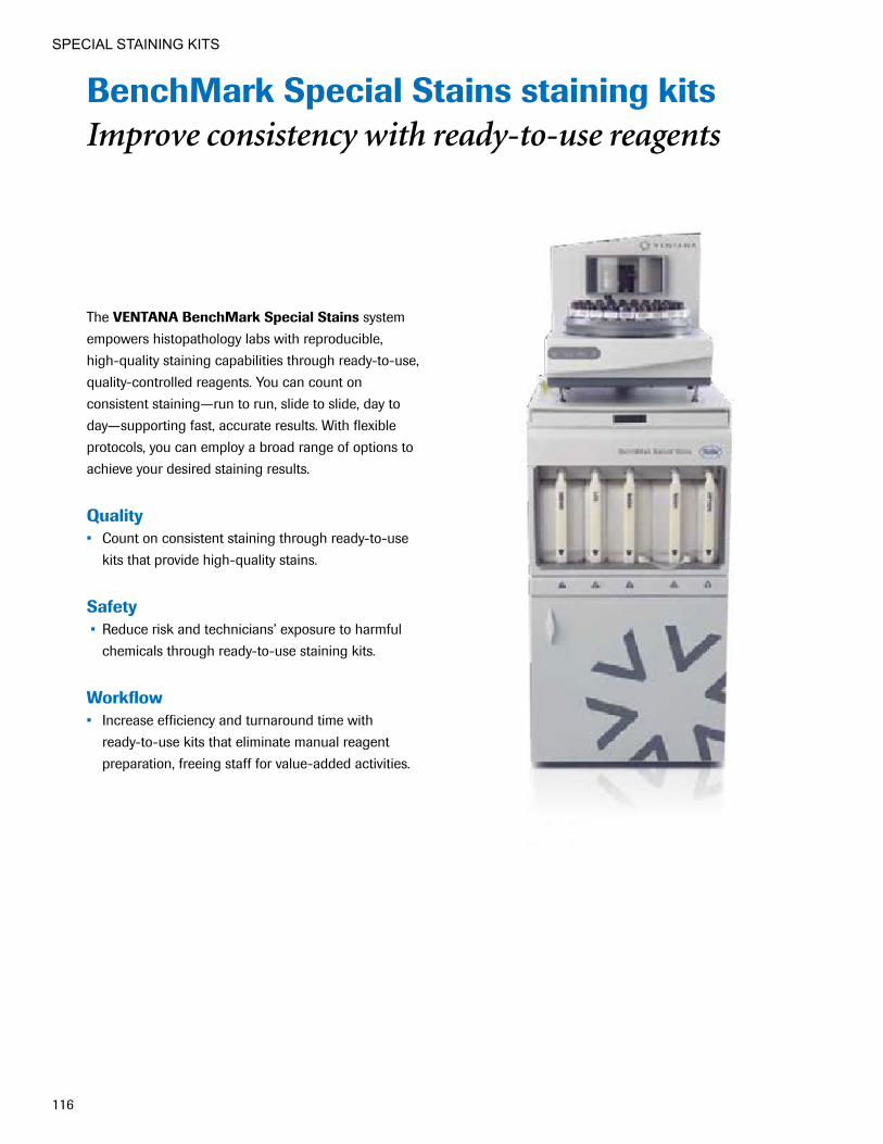
116
SPECIAL STAINING KITS
BenchMark Special Stains staining kits Improve consistency with ready-to-use reagents
The VENTANA BenchMark Special Stains system empowers histopathology labs with reproducible, high-quality staining capabilities through ready-to-use, quality-controlled reagents. You can count on consistent staining—run to run, slide to slide, day to day—supporting fast, accurate results. With flexible protocols, you can employ a broad range of options to achieve your desired staining results.
Quality • Count on consistent staining through ready-to-use
kits that provide high-quality stains.
Safety • Reduce risk and technicians’ exposure to harmful
chemicals through ready-to-use staining kits.
�Workflow� • Increase efficiency and turnaround time with
ready-to-use kits that eliminate manual reagent preparation, freeing staff for value-added activities.

117
SPECIAL STAINING KITS
VENTANA Special Stains System Selection GuideSpecial Stains for Mucin
Alcian Blue for PAS Light Green for PAS Alcian Blue Staining Kit Mucicarmine Staining Kit Diastase Kit PAS Staining Kit
Special Stains for Connective TissueElastic Staining Kit Trichrome II Blue Staining Kit Green for Trichrome Trichrome III Blue Staining Kit Jones H&E Staining Kit Trichrome III Green Staining Kit Jones Light Green Staining Kit Trichrome Staining Kit Reticulum II Staining Kit
Special Stains for MicroorganismsAFB III Staining Kit Gram Staining Kit Alcian Yellow Staining Kit Steiner Staining Kit GMS II Staining Kit Steiner II Staining Kit
Other Special Stains Congo Red Staining Kit Iron Staining Kit Giemsa Staining Kit
n For use on the VENTANA BenchMark Special Stains system.
n For use on the VENTANA NexES Special Stains system. Instrument no longer available.

118
SPECIAL STAINING KITS
Special Staining Kits for Mucin
Alcian Blue for PAS
Catalog Number: 860-003Ordering Code: 05279194001Quantity: 75 testsControls: Colon, Salivary Gland, Small Intestine
For use on the BenchMark Special Stains and NexES Special Stains systems.
INTENDED USE
Alcian Blue for PAS Staining Kit, in conjunction with the PAS Staining Kit (860-014 / 05279291001), is intended for use as a qualitative histologic stain to differentiate acid mucin from neutral polysaccharides in formalin-fixed, paraffin-embedded tissue. This product should be interpreted by a qualified pathologist in conjunction with histological examination, relevant clinical information, and proper controls. This product is intended for in vitro diagnostic (IVD) use.
Alcian Blue Staining Kit
Catalog Number: 860-002Ordering Code: 05279186001Quantity: 75 testsControls: Colon, Small Intestine
For use on the BenchMark Special Stains and NexES Special Stains systems.
INTENDED USE
Alcian Blue Staining Kit is intended for in vitro diagnostic (IVD) use as a qualitative histologic stain to demonstrate weakly acidic mucopolysaccharides. At pH 2.5, alcian blue stains sulfated and carboxylated acid mucopolysaccharides and sulfated and carboxylated sialomucins and glycoproteins in formalin-fixed, paraffin-embedded tissue. This product should be interpreted by a qualified pathologist in conjunction with histological examination, relevant clinical information, and proper controls.
Diastase Kit
Catalog Number: 860-004Ordering Code: 05279208001Quantity: 75 testsControls: Liver
For use on the BenchMark Special Stains and NexES Special Stains systems.
INTENDED USE
Diastase Kit, in conjunction with PAS Staining Kit (860-014 / 05279291001), is intended for use as a digestion reagent to aid in the determination of glycogen in formalin-fixed, paraffin-embedded tissue. This product should be interpreted by a qualified pathologist in conjunction with histological examination, relevant clinical information, and proper controls. This product is intended for in vitro diagnostic (IVD) use.

119
SPECIAL STAINING KITS
Light Green for PAS
Catalog Number: 860-010Ordering Code: 05279267001 Quantity: 75 testsControls: Liver
For use on the BenchMark Special Stains and NexES Special Stains systems.
INTENDED USE
Light Green for PAS used in conjunction with PAS Staining Kit is intended for use as a qualitative histologic stain for detection of fungus in formalin-fixed, paraffin-embedded tissue. This product should be interpreted by a qualified pathologist in conjunction with histological examination, relevant clinical information, and proper controls. This product is intended for in vitro diagnostic (IVD) use.
Mucicarmine Staining Kit
Catalog Number: 860-011Ordering Code: 05279275001Quantity: 75 testsControls: Human Tissue with Epithelial Mucin
For use on the BenchMark Special Stains and NexES Special Stains systems.
INTENDED USE
Mucicarmine Staining Kit is intended for use as a qualitative histologic stain to detect acid mucopolysaccharides (mucin) in formalin-fixed, paraffin-embedded tissue. This product should be interpreted by a qualified pathologist in conjunction with histological examination, relevant clinical information, and proper controls. This product is intended for in vitro diagnostic (IVD) use.
PAS Staining Kit
Catalog Number: 860-014Ordering Code: 05279291001Quantity: 75 testsControls: Tissue Known to be Glycogen Rich, such as
Kidney, Small Intestine, Liver, Skin, or Aorta
For use on the BenchMark Special Stains and NexES Special Stains systems.
INTENDED USE
PAS Staining Kit is intended for use as a qualitative histologic stain used to demonstrate the presence of glycogen in formalin-fixed, paraffin- embedded tissue. PAS Staining Kit detects positive reticular fibers, basement membrane, fungus, and neutral mucopolysaccharides.1 PAS Staining Kit may also be used to aid in distinguishing a PAS positive secreting adenocarcinoma from an undifferentiated PAS negative squamous cell carcinoma.2 This product should be interpreted by a qualified pathologist in conjunction with histological examination, relevant clinical information, and proper controls. This product is intended for in vitro diagnostic (IVD) use. 1. Thompson SW. Selected Histochemical and Histopathological Methods. Springfield; CC Thomas; 1966. 2. Sheehan DC, Hrapchak BB. Theory and Practice of Histotechnology. 2nd edition. St. Louis, MO: C.V. Mosby Company; 1980.

120
SPECIAL STAINING KITS
Special Staining Kits for Connective Tissue
Elastic Staining Kit
Catalog Number: 860-005Ordering Code: 05279216001Quantity: 75 testsControls: Kidney, Lung, Skin, Aorta, Artery
For use on the BenchMark Special Stains and NexES Special Stains systems.
INTENDED USE
Elastic Staining Kit is intended for use as a qualitative histologic stain to demonstrate elastic fibers in tissue sections in formalin-fixed, paraffin- embedded tissue. This product should be interpreted by a qualified pathologist in conjunction with histological examination, relevant clinical information, and proper controls. This product is intended for in vitro diagnostic (IVD) use.
Green for Trichrome
Catalog Number: 860-032Ordering Code: 06521916001Quantity: 75 testsControls: Colon, Kidney, Liver, Skin
For use on the BenchMark Special Stains system.
INTENDED USE
Green for Trichrome with Trichrome Staining Kit (860-031 / 06521908001) is intended for in vitro diagnostic (IVD) use. It is a qualitative histologic stain used to study connective tissue, muscle and collagen fibers in formalin-fixed, paraffin-embedded tissue. This product should be interpreted by a qualified pathologist in conjunction with histological examination, relevant clinical information, and proper controls.
Jones H&E Staining Kit
Catalog Number: 860-019Ordering Code: 05279348001Quantity: 40 testsControls: Kidney
For use on the BenchMark Special Stains and NexES Special Stains systems.
INTENDED USE
Jones Staining Kit is intended for use as a qualitative histologic stain to demonstrate capillary basement membrane in formalin-fixed, paraffin- embedded tissue. This product should be interpreted by a qualified pathologist in conjunction with histological examination, relevant clinical information, and proper controls. This product is intended for in vitro diagnostic (IVD) use.

121
SPECIAL STAINING KITS
Jones Light Green Staining Kit
Catalog Number: 860-020Ordering Code: 05279356001Quantity: 40 testsControls: Kidney
For use on the BenchMark Special Stains and NexES Special Stains systems.
INTENDED USE
Jones Light Green Staining Kit is intended for use as a qualitative histologic stain to demonstrate capillary basement membrane in tissue, such as kidney. This product should be interpreted by a qualified pathologist in conjunction with histological examination, relevant clinical information, and proper controls. This product is intended for in vitro diagnostic (IVD) use.
Reticulum II Staining Kit
Catalog Number: 860-024Ordering Code: 05279399001Quantity: 75 testsControls: Tissue with Defined Reticular Fibers
(Liver, Spleen, Kidney, or Lymph Nodes)
For use on the BenchMark Special Stains and NexES Special Stains systems.
INTENDED USE
Reticulum II Staining Kit is intended for use as a qualitative histologic stain to demonstrate reticular fibers in formalin-fixed, paraffin-embedded tissue. This product should be interpreted by a qualified pathologist in conjunction with histological examination, relevant clinical information, and proper controls. This product is intended for in vitro diagnostic (IVD) use.
Trichrome II Blue Staining Kit
Catalog Number: 860-013Ordering Code: 05279283001Quantity: 75 testsControls: Colon, Esophagus, Liver, Skin
For use on the NexES Special Stains system.
INTENDED USE
Ventana Medical Systems’ (Ventana) Trichrome II Staining Kit is intended for in vitro diagnostic (IVD) use as a qualitative histologic stain to study connective tissue, muscle and collagen fibers in formalin-fixed, paraffin-embedded tissue. This product should be interpreted by a qualified pathologist in conjunction with histological examination, relevant clinical information, and proper controls.

122
SPECIAL STAINING KITS
Trichrome III Blue Staining Kit
Catalog Number: 860-022Ordering Code: 05279364001Quantity: 75 testsControls: Colon, Esophagus, Liver, Skin
For use on the NexES Special Stains system.
INTENDED USE
Ventana Medical Systems’ (Ventana) Special Stains Trichrome III Blue Staining Kit is intended for in vitro diagnostic use (IVD) as a qualitative histologic stain to study connective tissue, muscle and collagen fibers in formalin-fixed, paraffin-embedded tissue. This product should be interpreted by a qualified pathologist in conjunction with histological examination, relevant clinical information, and proper controls.
Trichrome III Green Staining Kit
Catalog Number: 860-023Ordering Code: 05279372001Quantity: 75 testsControls: Colon, Esophagus, Liver, Skin
For use on the NexES Special Stains system.
INTENDED USE
Ventana Medical Systems’ (Ventana) Special Stains Trichrome III Green Staining Kit is intended for in vitro diagnostic use (IVD) as a qualitative histologic stain to study connective tissue, muscle and collagen fibers in formalin-fixed, paraffin-embedded tissue. This product should be interpreted by a qualified pathologist in conjunction with histological examination, relevant clinical information, and proper controls.
Trichrome Staining Kit
Catalog Number: 860-031Ordering Code: 06521908001Quantity: 60 testsControls: Colon, Kidney, Liver, Skin
For use on the BenchMark Special Stains system.
INTENDED USE
Trichrome Staining Kit is a qualitative histologic stain used to study connective tissue, muscle and collagen fibers in formalin-fixed, paraffin- embedded tissue. This product should be interpreted by a qualified pathologist in conjunction with histological examination, relevant clinical information, and proper controls. This product is intended for in vitro diagnostic (IVD) use.
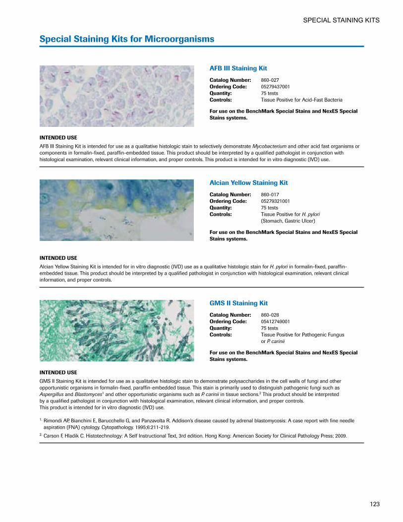
123
SPECIAL STAINING KITS
Special Staining Kits for Microorganisms
AFB III Staining Kit
Catalog Number: 860-027Ordering Code: 05279437001Quantity: 75 testsControls: Tissue Positive for Acid-Fast Bacteria
For use on the BenchMark Special Stains and NexES Special Stains systems.
INTENDED USE
AFB III Staining Kit is intended for use as a qualitative histologic stain to selectively demonstrate Mycobacterium and other acid fast organisms or components in formalin-fixed, paraffin-embedded tissue. This product should be interpreted by a qualified pathologist in conjunction with histological examination, relevant clinical information, and proper controls. This product is intended for in vitro diagnostic (IVD) use.
Alcian�Yellow�Staining�Kit
Catalog Number: 860-017Ordering Code: 05279321001Quantity: 75 testsControls: Tissue Positive for H. pylori
(Stomach, Gastric Ulcer)
For use on the BenchMark Special Stains and NexES Special Stains systems.
INTENDED USE
Alcian Yellow Staining Kit is intended for in vitro diagnostic (IVD) use as a qualitative histologic stain for H. pylori in formalin-fixed, paraffin- embedded tissue. This product should be interpreted by a qualified pathologist in conjunction with histological examination, relevant clinical information, and proper controls.
GMS II Staining Kit
Catalog Number: 860-028Ordering Code: 05412749001Quantity: 75 testsControls: Tissue Positive for Pathogenic Fungus
or P. carinii
For use on the BenchMark Special Stains and NexES Special Stains systems.
INTENDED USE
GMS II Staining Kit is intended for use as a qualitative histologic stain to demonstrate polysaccharides in the cell walls of fungi and other opportunistic organisms in formalin-fixed, paraffin-embedded tissue. This stain is primarily used to distinguish pathogenic fungi such as Aspergillus and Blastomyces1 and other opportunistic organisms such as P. carinii in tissue sections.2 This product should be interpreted by a qualified pathologist in conjunction with histological examination, relevant clinical information, and proper controls. This product is intended for in vitro diagnostic (IVD) use. 1. Rimondi AP, Bianchini E, Barucchello G, and Panzavolta R. Addison’s disease caused by adrenal blastomycosis: A case report with fine needle
aspiration (FNA) cytology. Cytopathology. 1995;6:211-219. 2. Carson F, Hladik C. Histotechnology: A Self Instructional Text, 3rd edition. Hong Kong: American Society for Clinical Pathology Press; 2009.

124
SPECIAL STAINING KITS
Gram Staining Kit
Catalog Number: 860-039Ordering Code: 06890105001Quantity: 75 tests Controls: See Package Insert
For use on the BenchMark Special Stains system.
INTENDED USE
Gram Staining Kit is intended for use as a qualitative histologic stain to demonstrate gram-negative and gram-positive bacteria in formalin-fixed, paraffin-embedded tissue. This product should be interpreted by a qualified pathologist in conjunction with histological examination, relevant clinical information, and proper controls. This product is intended for in vitro diagnostic (IVD) use.
Steiner Staining Kit
Catalog Number: 860-018Ordering Code: 05279330001Quantity: 40 testsControls: Tissue with H. pylori Organisms
and Gastric Ulcer
For use on the NexES Special Stains system.
INTENDED USE
Ventana Medical Systems’ (Ventana) Special Stains Steiner Staining Kit is intended for in vitro diagnostic (IVD) use as a qualitative histologic silver stain to aide in the identification of the causative organisms of diseases such as syphilis, some gastric ulcers (H. pylori), Lyme disease, Legionnaire’s disease, cat scratch fever, and others in formalin-fixed, paraffin-embedded tissue. This product should be interpreted by a qualified pathologist in conjunction with histological examination, relevant clinical information, and proper controls.
Steiner II Staining Kit
Catalog Number: 860-030Ordering Code: 06521894001Quantity: 40 testsControls: Tissue Containing H. pylori, Spirochetes, or
L. pneumophila
For use on the BenchMark Special Stains system.
INTENDED USE
Steiner II Staining Kit is intended for in vitro diagnostic (IVD) use. It is a qualitative histologic stain used to study specific argyrophilic microorganisms in formalin-fixed, paraffin-embedded tissue. This product should be interpreted by a qualified pathologist in conjunction with histological examination, relevant clinical information, and proper controls.
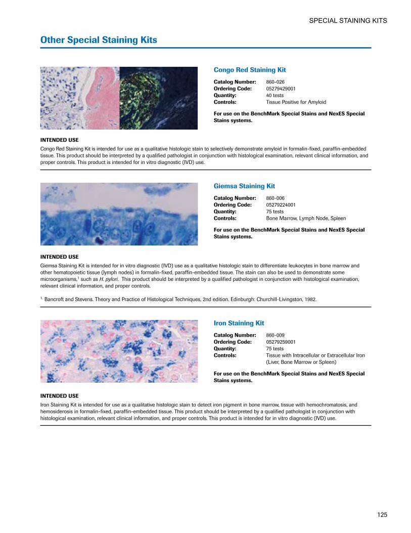
125
SPECIAL STAINING KITS
Other Special Staining Kits
Congo Red Staining Kit
Catalog Number: 860-026Ordering Code: 05279429001Quantity: 40 testsControls: Tissue Positive for Amyloid
For use on the BenchMark Special Stains and NexES Special Stains systems.
INTENDED USE
Congo Red Staining Kit is intended for use as a qualitative histologic stain to selectively demonstrate amyloid in formalin-fixed, paraffin-embedded tissue. This product should be interpreted by a qualified pathologist in conjunction with histological examination, relevant clinical information, and proper controls. This product is intended for in vitro diagnostic (IVD) use.
Giemsa Staining Kit
Catalog Number: 860-006Ordering Code: 05279224001Quantity: 75 testsControls: Bone Marrow, Lymph Node, Spleen
For use on the BenchMark Special Stains and NexES Special Stains systems.
INTENDED USE
Giemsa Staining Kit is intended for in vitro diagnostic (IVD) use as a qualitative histologic stain to differentiate leukocytes in bone marrow and other hematopoietic tissue (lymph nodes) in formalin-fixed, paraffin-embedded tissue. The stain can also be used to demonstrate some microorganisms,1 such as H. pylori. This product should be interpreted by a qualified pathologist in conjunction with histological examination, relevant clinical information, and proper controls. 1. Bancroft and Stevens. Theory and Practice of Histological Techniques, 2nd edition. Edinburgh: Churchill-Livingston, 1982.
Iron Staining Kit
Catalog Number: 860-009Ordering Code: 05279259001Quantity: 75 testsControls: Tissue with Intracellular or Extracellular Iron
(Liver, Bone Marrow or Spleen)
For use on the BenchMark Special Stains and NexES Special Stains systems.
INTENDED USE
Iron Staining Kit is intended for use as a qualitative histologic stain to detect iron pigment in bone marrow, tissue with hemochromatosis, and hemosiderosis in formalin-fixed, paraffin-embedded tissue. This product should be interpreted by a qualified pathologist in conjunction with histological examination, relevant clinical information, and proper controls. This product is intended for in vitro diagnostic (IVD) use.

126
SPECIAL STAINING KITS

VENTANA Instruments
H&E Staining . . . . . . . . . . . . . .128-129
IHC/ISH Automation . . . . . . . .130-133
Research Platforms . . . . . . . .134-135
Special Stains . . . . . . . . . . . . .136-137
Digital Pathology . . . . . . . . . . .138-144
Workflow Solutions . . . . . . . . .145-146
Leadership Knowledge and experience
underscored by the world’s largest installed base of IHC/ISH automated staining instruments

INSTRUMENTS
128
VENTANA HE 600 system Automated H&E staining
The VENTANA HE 600 system fully automates H&E staining, from tissue drying through coverslipping. Featuring individual slide staining and Lean design for intuitive operation, the system provides enhanced quality, workflow and safety that benefits both patients and pathology professionals.
Quality • Count on accurate, reproducible stains:
Consistent, high-quality stains provide confidence in patient diagnosis.
Workflow • Improve laboratory workflow: Lean workflow
reduces touchpoints and wait points for improved lab efficiency and turnaround time.
Safety • Increase lab safety: Elimination of xylene and
alcohol increases safety for technicians and the environment.
• Improve patient safety: Individual slide staining mitigates cross-contamination, providing greater patient safety. VENTANA HE 600 system ancillary products
Ordering Code
VENTANA HE 600 system Hematoxylin 07024282001
VENTANA HE 600 system Eosin 06544304001
VENTANA HE 600 system Differentiating Solution 06544339001
VENTANA HE 600 system Bluing 06544347001
VENTANA HE 600 system Organic Solution 07095163001
VENTANA HE 600 system Transfer Fluid 06544380001
VENTANA HE 600 system Wash 06544312001
VENTANA HE 600 system Cleaning Solution 07257538001
VENTANA HE 600 system Coverslip Activator 07534396001
VENTANA HE 600 system Coverslips 06711138001

INSTRUMENTS
129
Individual slide staining
Fresh reagents on every slide produce consistent, high-quality H&E stains for excellent clarity and enhanced visualization of micro-anatomic detail. Discrete staining for every patient slide protects against tissue cross-contamination.
Protocol flexibility
The system supports more than 400 different protocols with customization options for optimizing stain intensity to meet pathologists’ preferences.
Reagent management
All reagents are ready to use, with no mixing or dilution needed, so laboratory staff can focus on more value-added activities. The reagents are recyclable, poke yoke designed, color coded and individually numbered with radio frequency identification (RFID) technology for easy management and error prevention.
Remote monitoring and technical assistance
The VENTANA HE 600 system is equipped with CareGiver automated remote monitoring technology to help maximize uptime and minimize interruptions. This service includes remote software updates, system calibration and training, and provides system information to Roche service experts to expedite troubleshooting.
For ordering information, contact your Roche representative .
Protecting the integrity of Sue’s slide
H&E staining is the critical first step in
making a cancer diagnosis based on biopsy.
The VENTANA HE 600 system utilizes
individual slide staining, which mitigates
tissue specimen cross-contamination and
helps safeguard patients from the risk of
misdiagnosis. Improved quality in H&E
staining enhances pathologists’ diagnostic
confidence to help ensure the right diagnosis
for Sue and patients like her.
———————— ~ ————————

INSTRUMENTS
130
Throughput flexibility
IHC, ISH, SISH, Dual Stain and FITC slides can be processed simultaneously, improving turnaround times. The fully automated workflow enables processing of 90 slides in 8 hours, or 120 slides with an overnight run.
Remote monitoring and technical assistance
The BenchMark ULTRA system is equipped with CareGiver automated remote monitoring technology to help maximize uptime and minimize interruptions. This service includes remote software updates, system calibration and training, and provides system information to Roche service experts to expedite troubleshooting.
For ordering information, contact your Roche representative .
BenchMark ULTRA system True continuous access IHC/ISH slide staining
Offering unprecedented workflow efficiency, the VENTANA BenchMark ULTRA system enables your lab to process samples as they arrive and easily manage urgent requests. The fully integrated staining solution delivers medical value through continuous and random processing of patient samples.
• Maximize capacity: Individual slide drawers enable single piece flow to limit wasted slide positions.
• Optimize efficiency: Single piece workflow allows for protocol flexibility and continuous access to each slide without sacrificing stain quality.
• Improve processing time: Continuous flow decreases preparation time required for slide sorting and matching of reagent.
• Quickly respond to test requests: Ultimate Reagent Access (URA) allows immediate processing of urgent and late-arriving samples.
• Increase workflow flexibility: With 30 individual slide drawers and 35 reagent positions, the instrument enables you to add or remove slides and reagents at any time.

INSTRUMENTS
131
EZ Prep (10x) Catalog Number: 950-102 Ordering Code: 05279771001 Quantity: 2 L bottle Format: Concentrate
Ventana Medical Systems, Inc. (Ventana) EZ Prep Concentrate (10X) solution (EZ Prep) is used for paraffin removal from tissue samples during immunohistochemistry and in situ hybridization reactions, and to dilute 2X SSC during stringency washes during in situ hybridization reactions carried out on VENTANA automated slide stainers. This product is designed for use on BenchMark Series automated slide stainers. This reagent is intended for in vitro diagnostic (IVD) use.
Reaction Buffer (10x) Catalog Number: 950-300 Ordering Code: 05353955001 Quantity: 2 L bottle Format: Concentrate
Ventana Medical Systems, Inc. (Ventana) Reaction Buffer Concentrate (10X) is a Tris based buffer solution (pH 7.6 ± 0.2) used to rinse slides between staining steps and provide a stable aqueous environment for the immunohistochemistry (IHC) or in situ hybridization (ISH) reactions carried out on BenchMark and BenchMark XT automated slide staining systems. This reagent is intended for in vitro diagnostic (IVD) use.
ULTRA Cell Conditioning (ULTRA CC1) Catalog Number: 950-224 Ordering Code: 05424569001 Quantity: 2 L bottle Format: Ready to Use
ULTRA Cell Conditioning Solution (ULTRA CC1) from Ventana Medical Systems, Inc. (Ventana) is a pre diluted solution used as a pretreatment step in the processing of tissue samples for immuno- histochemistry (IHC) reactions carried out on the VENTANA BenchMark ULTRA instrument. This product is intended for in vitro diagnostic (IVD) use.
ULTRA Cell Conditioning (ULTRA CC2) Catalog Number: 950-223 Ordering Code: 05424542001 Quantity: 1 L bottle Format: Ready to Use
Ventana Medical Systems, Inc. (Ventana) ULTRA Cell Conditioning Solution (ULTRA CC2) is a prediluted solution used as a pretreatment step in the processing of tissue samples during immunohistochemistry (IHC) and in situ hybridization (ISH) on the VENTANA BenchMark ULTRA instrument. This product is intended for in vitro diagnostic (IVD) use.
ULTRA LCS (Predilute) Catalog Number: 650-210 Ordering Code: 05424534001 Quantity: 2 L bottle Format: Ready to Use
ULTRA LCS (Predilute) from Ventana Medical Systems, Inc. (Ventana), also known as ULTRA LCS or ULTRA Liquid Coverslip (High Temperature), is a prediluted coverslip solution used as a barrier between the aqueous reagents and the air. This barrier prevents evaporation, thereby providing a stable aqueous environment for the immuno- histochemistry or in situ hybridization reactions carried out on the VENTANA BenchMark ULTRA instrument. This product is designed for use on the VENTANA BenchMark ULTRA instrument. This reagent is not to be used on the VENTANA NexES IHC, NexES Special Stains or ES Automated Slide Stainers. This reagent is intended for in vitro diagnostic use.
SSC (10X) Catalog Number: 950-110 Ordering Code: 05353947001 Quantity: 2 L bottle Format: Concentrate
Ventana Medical Systems, Inc. (Ventana) 10X SSC, Sodium Chloride Sodium Citrate buffer solution is used for stringency washes and to rinse slides between staining steps and provide a stable aqueous environment for the in situ hybridization reactions carried out on a VENTANA automated slide stainer. This reagent is intended for in vitro diagnostic use.
BenchMark ULTRA bulk reagents

INSTRUMENTS
132
Throughput capabilities
The BenchMark XT system features a single drawer with 30 slide positions, each with individual temperature control, and a reagent carousel with 35 positions. The fully automated workflow enables processing of up to 60 slides in 8 hours, or 90 slides with an overnight run.
For ordering information, contact your Roche representative .
BenchMark XT system Automated IHC/ISH slide staining
The VENTANA BenchMark XT system offers the flexibility to expand your test menu, increase testing volume and improve turnaround time. The fully automated, random-access platform delivers consistent, high-quality results while improving workflow and efficiency.
• Enhance flexibility: Users can simultaneously process IHC, ISH, SISH, Dual Stain and FITC slides in the same run without limitations on protocol compatibility.
• Increase efficiency: The fully automated workflow decreases preparation time required for slide sorting.
• Improve turnaround time: Full automation increases slide throughput by 25% and eliminates labor by 80% compared to manual and semiautomated staining.

INSTRUMENTS
133
Cell Conditioning 1 (CC1) Catalog Number: 950-124 Ordering Code: 05279801001 Quantity: 2 L bottle Format: Ready to Use
Ventana Medical Systems, Inc. (Ventana) Cell Conditioning Solution (CC1) is a pre diluted solution used as a pretreatment step in the processing of tissue samples for immunohistochemistry (IHC) reactions carried out on VENTANA BenchMark and BenchMark XT automated slide stainers. This reagent is intended for in vitro diagnostic use.
Cell Conditioning 2 (CC2) Catalog Number: 950-123 Ordering Code: 05279798001 Quantity: 1 L bottle Format: Ready to Use
Ventana Medical Systems, Inc. (Ventana) Cell Conditioning Solution (CC2) is a prediluted solution used as a pretreatment step in the processing of tissue samples during immunohistochemistry (IHC) and in situ hybridization (ISH) on VENTANA BenchMark, BenchMark GX, and BenchMark XT instruments. This product is intended for in vitro diagnostic (IVD) use.
EZ Prep (10x) Catalog Number: 950-102 Ordering Code: 05279771001 Quantity: 2 L bottle Format: Concentrate
Ventana Medical Systems, Inc. (Ventana) EZ Prep Concentrate (10X) solution (EZ Prep) is used for paraffin removal from tissue samples during immunohistochemistry and in situ hybridization reactions, and to dilute 2X SSC during stringency washes during in situ hybridization reactions carried out on VENTANA automated slide stainers. This product is designed for use on BenchMark Series automated slide stainers. This reagent is intended for in vitro diagnostic (IVD) use.
LCS (predilute) Catalog Number: 650-010 Ordering Code: 05264839001 Quantity: 2 L bottle Format: Ready to Use
Ventana Medical Systems, Inc. (Ventana) LCS (Predilute), also known as LCS or Liquid Coverslip (High Temperature) is a prediluted coverslip solution used as a barrier between the aqueous reagents and the air. This barrier prevents evaporation, thereby providing a stable aqueous environment for the immunohistochemistry or in situ hybridization reactions carried out on VENTANA automated slide stainer. This product is designed for use on the BenchMark and BenchMark XT automated slide stainers. This reagent is not to be used on the VENTANA NexES IHC, NexES Special Stains or ES automated slide stainers. This reagent is intended for in vitro diagnostic use.
Reaction Buffer (10x) Catalog Number: 950-300 Ordering Code: 05353955001 Quantity: 2 L bottle Format: Concentrate
Ventana Medical Systems, Inc. (Ventana) Reaction Buffer Concentrate (10X) is a Tris based buffer solution (pH 7.6 ± 0.2) used to rinse slides between staining steps and provide a stable aqueous environment for the immunohistochemistry (IHC) or in situ hybridization (ISH) reactions carried out on BenchMark and BenchMark XT automated slide staining systems. This reagent is intended for in vitro diagnostic (IVD) use.
SSC (10X) Catalog Number: 950-110 Ordering Code: 05353947001 Quantity: 2 L bottle Format: Concentrate
Ventana Medical Systems, Inc. (Ventana) 10X SSC, Sodium Chloride Sodium Citrate buffer solution is used for stringency washes and to rinse slides between staining steps and provide a stable aqueous environment for the in situ hybridization reactions carried out on a VENTANA automated slide stainer. This reagent is intended for in vitro diagnostic use.
BenchMark XT bulk reagents

INSTRUMENTS
134
DISCOVERY ULTRA system The flexibility of manual, the freedom of automation
Offering our highest level of protocol flexibility for assay development, the VENTANA DISCOVERY ULTRA system enables your lab to fully automate a wide variety of IHC and ISH assays with the freedom to shape the platform to your project requirements.
• Save valuable development time: Fully automated baking through staining and ready-to-use reagents minimize manual steps.
• Improve workflow efficiency: True random access enables addition and removal of reagents to support multiple assay types and users.
• Achieve clear, sensitive detection: Innovative reagents, five color options and enhanced immuno- fluorescence capabilities enable reproducible, high-quality IHC and ISH stains.
• Accelerate assay development: Open software, protocol flexibility and reagent choice streamline optimization of automated high-complexity assays.
Throughput flexibility
With 30 independent reaction chambers, the DISCOVERY ULTRA system enables you to run both manual and fully automated experiments simultaneously with no synchronization errors. The system provides the ability to fully automate a broad range of IHC and ISH assays, including FISH, gene and protein IHC/ISH, mRNA ISH and multiplex assays (with any combination of IHC and ISH).
For ordering information, contact your Roche representative .
DISCOVERY products are for research use only . Not for use in diagnostic procedures .

INSTRUMENTS
135
DISCOVERY XT system Efficiency and flexibility for IHC/ISH research
DISCOVERY products are for research use only . Not for use in diagnostic procedures .
The VENTANA DISCOVERY XT system offers exceptional reproducibility and productivity for IHC and ISH slide preparation and processing for research. The flexible platform and sophisticated detection and reagent systems will help you accelerate the discovery of new targets and biomarkers.
• Increase efficiency: Fully automated baking through staining and ready-to-use reagents reduce manual steps for enhanced productivity.
• Improve workflow flexibility: Assay automation with your choice of reagents and detection chemistry gives you flexibility to meet your project needs.
• Rely on consistent quality: Ready-to-use reagents optimized for automation enhance slide-to-slide and run-to-run reproducibility.
Throughput capabilities
The DISCOVERY XT system enables fully automated processing of IHC, ISH and FISH assays—independently or simultaneously. The instrument provides individual temperature control for precise heating of up to 30 slides and has a capacity of up to 35 reagents.
For ordering information, contact your Roche representative .
Giving Robert hope for a breakthrough
The same innovation that goes into our VENTANA BenchMark IHC/ISH staining systems for clinical
use is found in the VENTANA DISCOVERY line of research-use-only instruments. The high-quality
data generated on DISCOVERY IHC/ISH research instruments help your lab uncover information
that can lead to better understanding of disease and, in turn, enable advances in treatment. Your next
discovery could be the one that helps patients like Robert have a brighter future.

INSTRUMENTS
136
BenchMark Special Stains system Automated baking through staining
Building on the industry-leading BenchMark system technology, the VENTANA BenchMark Special Stains instrument brings complete baking through staining to the histology laboratory for special stains, so your lab can consistently deliver exceptional quality.
Quality • Deliver reliable, high-quality stains:
Ready-to-use, quality-controlled reagents provide consistent staining quality.
Safety • Improve lab safety: Reduced exposure to harmful
chemicals decreases risk to technicians.
• Safeguard patients: Individual slide staining helps protect patient samples.
Workflow • Achieve superior workflow efficiency: Manual
processes and temperature dependencies are eliminated through automated deparaffinization and independent slide heating.
• Expand your lab’s capabilities: Reducing manual processes frees laboratory professionals for high- value activities.
• Improve efficiencies: Automated baking through staining, random batch access, and bulk and waste fluid sensing improve turnaround time and optimize workflow.
• Expand capacity: The BenchMark Special Stains system features 20 slide positions, each with independent temperature control, and a reagent carousel with 25 positions.
• Simplify operation: The intuitive VENTANA System Software interface, used with all BenchMark systems, simplifies operation and enables connection to a BenchMark ULTRA instrument.
Remote monitoring and technical assistance
The BenchMark Special Stains automated slide stainer is equipped with CareGiver automated remote monitoring technology to help maximize uptime and minimize interruptions. This service includes remote software updates, system calibration and training, and provides system information to Roche service experts to expedite troubleshooting.
For ordering information, contact your Roche representative .

INSTRUMENTS
137
Special Stains Wash Two Part Kit Catalog Number: 860-040 Ordering Code: 07383568001 Quantity: 1 Kit Ventana Medical Systems, Inc.’s (Ventana) Special Stains Wash Two Part Kit is used on the VENTANA BenchMark Special Stains auto-mated slide stainer and VENTANA NexES Special Stains automated slide stainer. When diluted according to instructions, Special Stains Wash Two Part Kit produces an optimized solution for use with the VENTANA special stains reagent kits. This solution provides a stable aqueous environment for special histological staining reactions.
BenchMark Special Stains Deparaffinization Solution (10X) Catalog Number: 860-036 Ordering Code: 06523102001 Quantity: 2 L bottle Format: ConcentrateBenchMark Special Stains Deparaffinization Solution (10X) is used for paraffin removal from tissue samples during special stains reactions carried out on the VENTANA BenchMark Special Stains automated slide stainer. This reagent is intended for in vitro diagnostic (IVD) use.
BenchMark Special Stains Wash Solution (10X) Catalog Number: 860-035 Ordering Code: 06523099001 Quantity: 2 L bottle Format: ConcentrateBenchMark Special Stains Wash Solution (10X) is used on the VENTANA BenchMark Special Stains automated slide stainer. When diluted according to instructions, BenchMark Special Stains Wash Solution (10X) is an optimized solution used for VENTANA special stains reagent kits used on the VENTANA BenchMark Special Stains automated slide stainer. This solution provides a stable aqueous environment for special histological staining reactions performed on the VENTANA BenchMark Special Stains automated slide stainer. This product is intended for in vitro diagnostic (IVD) use.
Special Stains Cleaning Kit Catalog Number: 860-016 Ordering Code: 05279313001 Quantity: 50 cleaning cyclesVENTANA Special Stains Cleaning Kit is intended for use during the automated cleaning procedure on the VENTANA BenchMark Special Stains automated slide stainer and the VENTANA NexES Special Stains automated slide stainer. This kit is intended for in vitro diagnostic use.
Special Stains Clean Plus Catalog Number: 860-037 Ordering Code: 06649327001 Quantity: 50 cleaning cycles
The Special Stains Cleaning Plus, used with the Special Stains Cleaning Kit (860-016), is used to provide a deep clean of the reagent aspiration and dispense mechanism of the BenchMark Special Stains system. *1 kit for 50 cleaning cycles. (Standard cleaning cycle)
BenchMark Special Stains Liquid Coverslip Catalog Number: 860-034 Ordering Code: 06523072001 Quantity: 2 L bottle Format: Ready to UseBenchMark Special Stains Liquid Coverslip is a prediluted coverslip solution used as a barrier between the aqueous reagents and the air. This barrier prevents evaporation, thereby providing a stable aqueous environment for the special stains reactions carried out on a VENTANA BenchMark Special Stains automated slide stainer.BenchMark Special Stains Liquid Coverslip may also be used as a deparaffinization agent in cases where microorganisms or cellular structures are not compatible with the BenchMark Special Stains Deparaffinization Solution. In these cases, BenchMark Special Stains Liquid Coverslip will be automatically applied by the BenchMark Special Stains automated slide stainer.This product is designed for use on the BenchMark Special Stains automated slide stainer. This reagent is not to be used on the VENTANA BenchMark, BenchMark GX, BenchMark XT or BenchMark ULTRA instruments or NexES Special Stains automated slide stainers. This reagent is intended for in vitro diagnostic (IVD) use.
Special Stains Rinse (SSR) Solution Catalog Number: 860-038 Ordering Code: 06890059001 Quantity: 2 L bottleSSR Solution is used on the VENTANA BenchMark Special Stains automated slide stainer. The SSR Solution is optimized to be used in conjunction with specific VENTANA special stains protocols on the VENTANA BenchMark Special Stains automated slide stainer. This product is intended for in vitro diagnostic (IVD) use.
BenchMark Special Stains bulk reagents and ancillary products
For use on the NexES Special Stains Instrument Instrument no longer available.
Special Stains Wash (10x) Catalog Number: 860-015 Ordering Code: 05279305001 Quantity: 2 L bottle
Liquid Coverslip (Low Temperature) Catalog Number: 250-009 Ordering Code: 02530981001 Quantity: 2 L bottle Format: Ready to Use

INSTRUMENTS
138
VENTANA Digital Pathology Solutions Transforming the practice of pathology to improve patient care
Our growing family of VENTANA innovations is changing the practice of pathology, empowering you with a comprehensive image and workflow solution that helps inform decision making, deliver medical value and improve patient outcomes.
• Increase workflow efficiency: When used together, our integrated staining platforms, assays, scanners, algorithms and workflow management solution optimize your process, from staining to reporting.
• Deliver enhanced medical value: Anywhere, anytime access and algorithm-based image analysis support efficient and informed treatment decisions.
• Improve patient care: Sharing digital images in real time, with instant access to expertise, eliminates the boundaries of time and distance for faster treatment decisions.
Bringing confidence to Sam’s diagnosis
VENTANA digital pathology solutions
are changing the practice of medicine to
benefit patients like Sam. Digital images
of a patient’s tissue slide can be shared
for remote consults and quick access to
second opinions. Tissue specimens undergo
precise assay scoring and image analysis
for consistent, objective interpretations.
VENTANA digital pathology solutions
offer Sam and patients like him peace of
mind by enabling pathologists to share
images and expertise to help improve
diagnostic and treatment decisions.
———————— ~ ————————

INSTRUMENTS
139
Virtuoso Image and Workflow Management Software Built for the clinical laboratory
VENTANA Virtuoso software combines rich function-ality with an easy-to-use interface to deliver a complete digital pathology solution, from image management to customized reporting.
• Enhance patient care: Timely, efficient access to slide images enables real-time collaboration, with faster turnaround for second opinions.
• Improve patient safety: Integration with VENTANA VANTAGE software provides efficient data entry and specimen tracking.
• Increase flexibility: Distributed pathology workflow enables remote viewing and case sharing, while role-based portals enhance ease of use.
• Expand your reach: Web-based software application simplifies access and makes it easy to add new users.
View, annotate and analyze slide images anytime, anywhere.

INSTRUMENTS
140
Companion Algorithm Image Analysis Software Clinical confidence with a comprehensive breast cancer diagnostic panel
VENTANA Companion Algorithm image analysis software is integrated with VENTANA Virtuoso image and workflow management software, VENTANA BenchMark IHC/ISH staining instruments and the VENTANA iScan Coreo slide scanner to provide optimized processing, from staining to reporting.
• Improve clinical confidence: Using the algorithms enables a pathologist to make consistent, objective interpretations, thus reducing both intra- and inter- observer variability in interpretation.
• Ease reimbursement processes: Using our algorithms can alleviate the burden of undergoing a payer-specific technical review, if required.
The most advanced, clinically validated pathology solutions
Roche was the first company in the industry to offer a comprehensive breast portfolio. Our full breast panel includes HER2 (4B5), ER (SP1), PR (1E2), Ki-67 (30-9) and p53 (DO-7) image analysis algorithms for use along with their accompanying VENTANA IHC assays.*
All IHC breast markers in the VENTANA portfolio have both image analysis and digital read applications available for use with the iScan Coreo slide scanner.There are two intended uses:
• Clinical use of the software algorithm to semi- quantify the biomarker
• Digital read to manually read and score the biomarker using a computer monitor
* Algorithms are for use only on the iScan Coreo scanner.

INSTRUMENTS
141
Roche offers VENTANA image analysis companion algorithms for the five key breast panel biomarkers
The VENTANA Companion Algorithm Estrogen Receptor (ER)
(SP1) image analysis software
The VENTANA Companion Algorithm HER2 (4B5) image
analysis software
The VENTANA Companion Algorithm Ki-67 (30-9) image
analysis software
The VENTANA Companion Algorithm p53 (DO-7) image
analysis software
The VENTANA Companion Algorithm Progesterone Receptor
(PR) (1E2) image analysis software
Companion Algorithm breast panel image analysis
Algorithms HER2 (4B5) image analysis software ER (SP1) image analysis software Ki-67 (30-9) image analysis software p53 (DO-7) image analysis software PR (1E2) image analysis software
Digital slide scanner
iScan Coreo slide scanner
Image manage-ment software
Virtuoso software
Regulatory status
FDA 510(k) cleared

INSTRUMENTS
142
iScan Coreo Slide Scanner Flexible brightfield scanning platform
The VENTANA iScan Coreo slide scanner is the right fit for small- to medium-volume laboratories, offering 160-slide capacity with a small footprint for placement flexibility. Paired with VENTANA Virtuoso software, it provides a complete digital pathology solution, from image acquisition through customized reporting.
• Increase confidence in results: The instrument has FDA clearance for both digital read and image analysis on the five key breast panel markers: ER, PR, HER2, Ki-67 and p53.
• Enhance flexibility: Scanning at four objectives (4x, 10x, 20x and 40x) and the unique web-based Live Mode application for remote viewing and collaboration, provide versatility.
• Improve efficiency: Walkaway scanning, digital image archiving and customized reporting increase operational efficiencies and productivity.
Remote monitoring and technical assistance
The iScan Coreo slide scanner is equipped with CareGiver automated remote monitoring technology to help maximize uptime and minimize interruptions. This service includes remote software updates, system calibration and training, and provides system information to Roche service experts to expedite troubleshooting.
For ordering information, contact your Roche representative .
iScan Coreo slide scanner . Pathology . Advanced .
• High-speed brightfield scanning—a 15 x 15 mm tissue area can be scanned in less than two minutes at 20x
• Automatic slide detection for walkaway scanning, with random access to the slide rack
• Automatic tissue region identification with option to manually override
• Volume scanning with the ability to capture multiple z-plane images
• Integrated with image viewing software that supports annotation and precise digital measurements
iScan Image Viewer software
• Windows application used to view local and remote iScan images instantly
• Precise digital tools for annotation, measurement and cell counting
• Simultaneous multiple image viewing
• Images can be viewed at magnifications ranging from 1x to 80x (80x achieved with digital zoom)
• Export areas of interest
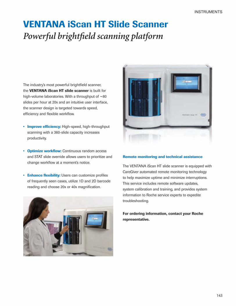
INSTRUMENTS
143
VENTANA iScan HT Slide Scanner Powerful brightfield scanning platform
The industry’s most powerful brightfield scanner, the VENTANA iScan HT slide scanner is built for high-volume laboratories. With a throughput of ~80 slides per hour at 20x and an intuitive user interface, the scanner design is targeted towards speed, efficiency and flexible workflow.
• Improve efficiency: High-speed, high-throughput scanning with a 360-slide capacity increases productivity.
• Optimize workflow: Continuous random access and STAT slide override allows users to prioritize and change workflow at a moment’s notice.
• Enhance flexibility: Users can customize profiles of frequently seen cases, utilize 1D and 2D barcode reading and choose 20x or 40x magnification.
Remote monitoring and technical assistance
The VENTANA iScan HT slide scanner is equipped with CareGiver automated remote monitoring technology to help maximize uptime and minimize interruptions. This service includes remote software updates, system calibration and training, and provides system information to Roche service experts to expedite troubleshooting.
For ordering information, contact your Roche representative .

INSTRUMENTS
144
VENTANA Vector Software Software for education and collaboration
VENTANA Vector software is Research Use Only (RUO) software for academic medical centers and pathology groups who wish to share digital slides with students or other pathologists for education and collaboration.
• Enhance the learning experience: Unlike the microscope, the software enables standardization of content (including rare cases) and provides flexible content and evaluation management tools, based on teaching or project objectives.
• Increase engagement: Images can be viewed by multiple participants at any time, and users can create quizzes for students and forums to foster group collaboration.
• Optimize efficiency: Anywhere, anytime access creates a collaborative learning environment for students as well as pathologists who are working remotely on projects.
Academic medical centers
Pathology groups
Access using tablets, mobile phones and desktop computers
VENTANA Vector software is for research use only. Not for use in diagnostic procedures .

INSTRUMENTS
145
VANTAGE Workflow Solution See your lab for the first time
The VENTANA VANTAGE workflow solution helps protect patients from the serious risks associated with sample misidentification. Finally, labs can enjoy complete chain of custody, Lean integrated processes, and full visibility into lab operations any time, from anywhere.
• Fully track lab operations: Our comprehensive solution for histology labs—hardware, software and workflow consulting—offers a commanding view of your complex operation from a single strategic perspective.
• Improve patient safety: Implemented in over 200 labs worldwide, the VANTAGE workflow solution maintains positive sample ID with barcode scanners at each workstation and single piece flow workflow.
• Increase efficiency: The VANTAGE workflow solution is designed using Lean Six Sigma principles and includes expert workflow consulting to help you obtain immediate and ongoing workflow benefits.
• Optimize workflow: Immediately locate any patient’s slide, at any time, from any last scanned location. See a comprehensive dashboard of lab performance at any time.
VANTAGE workflow solution Ordering Code
VANTAGE HP ProLiant DL380 Gen9 Server 07570520001VANTAGE HP L5015tm Touchscreen 07924682001VANTAGE HP EliteDesk 800 Mini PC 07926359001VANTAGE Scanner 07171650001VANTAGE Printer 06543863001VANTAGE Slide Barcode Label 05524997001VANTAGE Overlay Dispenser 05909490001VANTAGE Clear Overlay (3750/roll) 05525276001
Ensuring the identity of David’s specimen
As the single, safety-focused point of
connection in the anatomic pathology lab,
VANTAGE workflow solution helps improve
patient safety by ensuring complete chain
of custody throughout the histology
workflow. From sample accessioning to
results, VANTAGE barcode-driven workflow
management reduces errors and protects
patients from the serious risks associated
with sample misidentification. So David,
and the medical team that cares for him,
can be assured that high-confidence
results are used to determine his diagnosis
and treatment.
———————— ~ ————————

INSTRUMENTS
146
VENTANA Connect Middleware Solution Streamlining lab connectivity
VENTANA Connect middleware solution provides an efficient point of connection from Laboratory Information Systems (LIS) to VENTANA systems and software, automatically exchanging critical case information and eliminating test request re-entries that can introduce errors and slow you down.
• Optimize lab efficiency: Single, flexible interface connects multiple LIS platforms to current and future VENTANA instruments.
• Reduce errors: Important case information flows seamlessly to instruments and systems through a single, secure connection.
• Improve patient safety: VENTANA instruments and software accept LIS-originated case identifiers, increasing patient safety by using a single unique identifier at every point in the process.
VENTANA Connect middleware solution at a glance
• Supports uni- and bi-directional communications from LIS
• Eliminates the need for data re-entry through use of LIS-originated case identifiers, accepted by VENTANA instruments and software
• Uses a unique identifier everywhere in the process, saving time and reducing errors
• Manages highly configurable, easy mapping of LIS data to VENTANA instruments
• Provides integration to digital pathology systems and images
• Delivers enterprise-class software for maximum scalability and reliability

Customer Experience
Passion Fueled by the needs
of patients and their healthcare professionals

148
CUSTOMER EXPERIENCE
VENTANA Workflow Consulting Solutions Expert support to optimize workflow
Highly trained in Lean Six Sigma principles, our workflow consultants will work with you to help make your lab processes easier, faster, safer and more reliable. Working closely with your team, we’ll identify and address opportunities to optimize your workflow, maximize results and lower your costs.
• Optimize workflow: Standardized processes improve performance, throughput and turnaround times.
• Improve patient safety: Improved patient sample identification helps reduce errors and decrease risk.
• Increase efficiency: Simplifying workflow and eliminating non-value-added activities reduce costs and improve productivity.
• Enhance patient care: Streamlined processes help you deliver results faster, for earlier treatment decisions.
Optimize your lab’s performance from the moment you receive a sample to the time you deliver a patient’s results.

149
CUSTOMER EXPERIENCE
To help your lab achieve the greatest benefit from our products, Roche Commercial Education is committed to providing an engaging training experience with a range of resources focused on continuous learning and enhancing overall job proficiency.
Designed to prepare your lab for success, Roche Diagnostics University (RDU) is a one-stop online registration and learning platform supporting the right training, for the right role, at the right time.
• Experience interactive learning: Courses consist of an optimal mix of on-demand product and disease state e-Learning content, and Indianapolis-based classroom, hands-on experience.
• Learn from experts: Training is conducted by highly qualified histology instructors with professional certification, field or technical support expertise.
• Gain proficiency: Spanning the histology workflow process, RDU courses are designed to help you gain expertise with Roche instrumentation, including daily operation, maintenance and troubleshooting, through interactive participation.
• Easily access training documentation: Administrative dashboards provide easy access to training documentation, training history and program reports.
Getting started
Ask your Roche representative for instructions on creating a Roche Diagnostics University account and for additional training details.
Empowering continuous learning

150
CUSTOMER EXPERIENCE
Roche Support Network (RSN) Always on, always ready to help
At Roche, support is not an afterthought—it’s our promise and our passion. Backed by a variety of highly experienced, specialized teams, the Roche Support Network provides a host of solutions to assist you at every stage—from procurement to ongoing onsite and offsite support.
• Get personalized support: We work with you to build a foundation for success, assessing your needs and objectives to provide customized solutions based on your lab’s data and workflow, onsite observations and Lean Six Sigma techniques.
• Gain peace of mind: You can be confident knowing you’ll receive expert support, including project management and product training, to ensure peak performance and successful implementation with minimal disruption of lab operations.
• Maintain optimal lab performance: To ensure your success, we partner with you and provide ongoing support that includes remote monitoring, field service and technical applications expertise, consulting and a 24/7/365 call center.

151
CUSTOMER EXPERIENCE
CareGiver Remote Support The help you need, when you need it
Because we understand the critical nature of the work you do for patients, Roche offers CareGiver remote support—an innovative automated support solution for anatomic pathology labs. Through a secure Internet connection, the CareGiver system provides continuous monitoring and remote service of VENTANA instruments,* helping maximize performance to keep your lab running smoothly.
• Gain peace of mind: Remote instrument monitoring, troubleshooting and training with secure data communication help keep your lab operating at peak performance.
• Save time: System software upgrades are provided via secure Internet connection, and improved, automated data collection decreases management time requirements.
• Increase up time: Remote monitoring and support help to maintain optimum instrument performance and reduce downtime.
* CareGiver remote support is currently available for the VENTANA HE 600 system, VENTANA BenchMark ULTRA IHC/ISH system, VENTANA BenchMark Special Stains platform, VENTANA Virtuoso software, VENTANA iScan Coreo slide scanner, and the VENTANA iScan HT slide scanner.

152
CUSTOMER EXPERIENCE
Reimbursement Tough questions. One easy answer.
Reimbursement for the assays and services you provide can seem confusing. The Roche Diagnostics reimbursement team can help. Our team is a reliable and credible resource you can turn to for the latest information to address reimbursement questions and issues regarding Roche Diagnostics products.
• Get the answers you need: The Roche Diagnostics reimbursement team can help you address reimbursement questions about FDA-cleared or approved products marketed and sold in the U.S. by Roche Diagnostics Corporation and its affiliates.
• Save time: Our team monitors reimbursement trends and policies to keep track of the latest developments that might affect your business or have an impact on Roche Diagnostics products, so you can turn to us for answers and free up your time for more important activities.
• Count on 24/7 support: We’re available by phone or email around the clock, enabling you to contact our team for assistance at your convenience.
Reimbursement is complex. Contacting us is easy.
If you are a healthcare professional working in a hospital, laboratory, physician office or other professional setting, you can contact our team for assistance at your convenience 24 hours a day, 7 days a week.
Please provide your name, the name of your organization and your contact information, and a brief description of your inquiry, via one of the following:
Reimbursement Inquiry Line (toll-free voicemail and automated response system)
1-800-428-5074, ext. 13967
Reimbursement Inquiry Email [email protected]

Appendices
Courage Challenging the standard
of care to improve the quality of patients’ lives

APPENDICES
154
General Index AA-1-Antichymotrypsin (polyclonal), 18A-1-Antitrypsin (polyclonal), 18ACTH (polyclonal), 18Actin, Muscle Specific (HHF35), 18Actin, Smooth Muscle (1A4), 19ALK (D5F3), VENTANA CDx Assay, 7, 11, 19Alpha-Fetoprotein Rabbit Polyclonal, 20Amplification Kit, 111amplification products, 110–111Annexin A1 (MRQ-3), 21anti-Actin, Muscle (HUC1-1), 19anti-Androgen Receptor (SP107) Rabbit Monoclonal, 20anti-Arginase-1 (SP156) Rabbit Monoclonal, 21anti-bcl-2 (SP66) Rabbit Monoclonal, 23anti-BRAF V600E (VE1) Mouse Monoclonal, 24anti-Calcitonin (SP17), Rabbit Monoclonal, 28anti-CD7 (SP94) Rabbit Monoclonal, 31anti-CD22 (SP104) Rabbit Monoclonal, 35anti-CD30 (Ber-H2) Mouse Monoclonal, 36anti-CD43 (L60) Mouse Monoclonal, 37anti-CD44 (SP37) Rabbit Monoclonal, 38anti-CEA (TF 3H8-1), 44anti-Chromogranin A (LK2H10), 44anti-c-MYC (Y69) Rabbit Monoclonal, 25anti-Cytokeratin 5/6 (D5-16B4) Mouse Monoclonal, 47anti-Cytokeratin 10 (SP99) Rabbit Monoclonal, 49anti-Cytokeratin 14 (SP53) Rabbit Monoclonal, 49anti-Cytokeratin 17 (SP95) Rabbit Monoclonal, 50anti-Cytokeratin (CAM 5.2) Mouse Monoclonal, 47anti-EGFR E746-A750 del (SP111) Rabbit Monoclonal, 53anti-EGFR L858R (SP125) Rabbit Monoclonal, 53anti-Epithelial Related Antigen (MOC-31) Mouse Monoclonal, 54anti-ERG (EPR3864) Rabbit Monoclonal, 55anti-EZH2 (SP129) Rabbit Monoclonal, 55anti-Glypican 3 (GC33) Mouse Monoclonal, 64anti-INI-1 (MRQ-27) Mouse Monoclonal, 71anti-MLH1 (M1) Mouse Monoclonal, 78anti-MUC1 (H23) Mouse Monoclonal, 79anti-NSE (MRQ-55) Mouse Monoclonal, 85anti-p40 (BC28) Mouse Monoclonal, 7, 87anti-p53 (Bp53-11), 88anti-p57Kip2 (Kp10) Mouse Monoclonal, 88anti-Pan Keratin (AE1/AE3/PCK26), 89anti-S100P (16/f5) Mouse Monoclonal, 21anti-TFE3 (MRQ-37) Rabbit Monoclonal, 101anti-Thyroid Transcription Factor-1 (SP141) Rabbit Monoclonal, 102
BBCA-225 (Cu-18), 22bcl-6 (GI191E/A8), 23BenchMark Special Stains Deparaffinization Solution (10x), 137BenchMark Special Stains staining kits, 116–126BenchMark Special Stains system, 136 BenchMark Special Stains Deparaffinization
Solution (10X), 137 BenchMark Special Stains Liquid Coverslip, 137 BenchMark Special Stains Wash Solution
(10X), 137 Special Stains Clean Plus, 137 Special Stains Cleaning Kit, 137 Special Stains Rinse (SSR) Solution, 137 Special Stains Wash Two Part Kit, 137BenchMark Special Stains Wash Solution (10x), 137BenchMark ULTRA system, 130 EZ Prep (10x), 131 Reaction Buffer (10x), 131 SSC (10x), 131 ULTRA Cell Conditioning (ULTRA CC1), 131 ULTRA Cell Conditioning (ULTRA CC2), 131 ULTRA LCS (Predilute), 131
BenchMark XT system, 132 Cell Conditioning 1 (CC1) reagent, 133 Cell Conditioning 2 (CC2) reagent, 133 EZ Prep (10x) reagent, 133 LCS (predilute) reagent, 133 Reaction Buffer (10x) reagent, 133 SSC (10x) reagent, 133Beta-Catenin (14), 23BG8, Lewisy (F3), 24BOB.1 (SP92), 24
CC3d Rabbit polyclonal, 26C4d (polyclonal), 26C4d (SP91), 26CA 19-9 (121SLE), 27CA-125 (OC125), 27Cadherin 17 (SP183) Rabbit Monoclonal, 27Calcitonin (polyclonal), 28Caldesmon (E89), 28Calponin-1 (EP798Y), 29Calveolin-1 (SP43) Rabbit Monoclonal, 29Carcinoembryonic Antigen (CEA31), 43CareGiver remote support, 151CD1a (EP3622), 30CD2 (MRQ-11), 30CD13 (SP187) Rabbit Monoclonal, 32CD14 (EPR3653) Rabbit Monoclonal, 33CD16 (SP175) Rabbit Monoclonal, 33CD21 (2G9), 34CD21 (EP3093), 34CD25 (4C9), 35CD31 (JC70), 36CD33 (SP266) Rabbit Monoclonal, 37CD45 (LCA) (2B11 & PD7/26), 38CD45R (MB1), 39CD56 (MRQ-42), 40CD57 (NK-1), 40CD61 (2f2), 40CD63 (NKI/C3), 41CD71 (MRQ-48) Mouse Monoclonal, 41CD138/syndecan-1 (B-A38), 42CD163 (MRQ-26), 43CDX-2 (EPR2764Y), 43CEA (CEA31) Mouse Monoclonal, 43Cell Conditioning 1 (CC1) reagent (XT system), 133Cell Conditioning 2 (CC2) reagent (XT system), 133CINtec Histology Kit, 44CINtec p16 Histology, 45Collagen Type IV (CIV22), 45companion diagnostics, 10-11 VENTANA ALK (D5F3) CDx Assay, 11 VENTANA INFORM HER2 Dual ISH Assay, 11 VENTANA PATHWAY anti-HER2/neu (4B5)
Rabbit Monoclonal, 11 VENTANA PATHWAY c-Kit (9.7), 11complementary diagnostics, 12 VENTANA PD-L1 (SP142) Assay, 12 CONFIRM anti-ALK1 (ALK01), 20CONFIRM anti-bcl-2 (124) Mouse Monoclonal, 22CONFIRM anti-CD3 (2GV6) Rabbit Monoclonal, 30CONFIRM anti-CD4 (SP35) Rabbit Monoclonal, 31CONFIRM anti-CD5 (SP19) Rabbit Monoclonal, 31CONFIRM anti-CD8 (SP57) Rabbit Monoclonal, 32CONFIRM anti-CD15 (MMA) Mouse Monoclonal, 33CONFIRM anti-CD20 (L26), 34CONFIRM anti-CD23 (SP23) Rabbit Monoclonal, 35 CONFIRM anti-CD34 (QBEnd/10), 37CONFIRM anti-CD45, LCA (RP2/18), 38CONFIRM anti-CD45RO (UCHL-1), 39CONFIRM anti-CD56 (123C3) Mouse Monoclonal, 39 CONFIRM anti-CD68 (KP-1), 41CONFIRM anti-CD79a (SP18) Rabbit Monoclonal, 42 CONFIRM anti-CD99 (O13) Mouse Monoclonal, 42CONFIRM anti-Cytokeratin (AE1), 46CONFIRM anti-Cytokeratin 7 (SP52) Rabbit Monoclonal, 48CONFIRM anti-Cytokeratin 20 (SP33) Rabbit Monoclonal, 50
CONFIRM anti-Desmin (DE-R-11), 51CONFIRM anti-EGFR (5B7) Rabbit Monoclonal, 53CONFIRM anti-EMA (E29) Mouse Monoclonal, 54CONFIRM anti-Epidermal Growth Factor Receptor (3C6), 52CONFIRM anti-Estrogen Receptor (ER) (SP1) Rabbit Monoclonal, 55CONFIRM anti-IGF-1R (G11) Rabbit Monoclonal, 69CONFIRM anti-Kappa Rabbit Polyclonal, 71CONFIRM anti-Keratin (AE3), 72CONFIRM anti-Keratin (34βE12) Mouse Monoclonal, 72CONFIRM anti-Ki-67 (30-9) Rabbit Monoclonal, 73CONFIRM anti-Lambda Rabbit Polyclonal, 74CONFIRM anti-LMO2 (1A9-1) Mouse Monoclonal, 74CONFIRM anti-MART-1/melan A (A103) Mouse Monoclonal, 76CONFIRM anti-Melanosome (HMB45) Mouse Monoclonal, 77CONFIRM anti-MITF (C5/D5) Mouse Monoclonal, 78CONFIRM anti-MSH6 (44) Mouse Monoclonal, 79CONFIRM anti-p53 (DO-7), 88CONFIRM anti-PAX5 (SP34) Rabbit Monoclonal, 90CONFIRM anti-Progesterone Receptor (PR) (1E2) Rabbit Monoclonal, 94CONFIRM anti-Prostate Specific Antigen (PSA) Rabbit Polyclonal, 94CONFIRM anti-S100 (4C4.9), 96CONFIRM anti-S100 (polyclonal), 97CONFIRM anti-Synaptophysin (SP11) Rabbit Monoclonal, 100CONFIRM anti-Thymidine Phosphorylase (P-GF.44C) Mouse Monoclonal, 101CONFIRM anti-Thyroid Transcription Factor-1 (8G7G3/1) Mouse Monoclonal, 102CONFIRM anti-Topoisomerase IIa (JS5B4) Rabbit Monoclonal, 103CONFIRM anti-Total c-MET (SP44) Rabbit Monoclonal, 103CONFIRM anti-Tyrosinase (T311) Mouse Monoclonal, 105CONFIRM anti-Vimentin (V9), 106CONFIRM anti-Vimentin (Vim 3B4), 106CONFIRM Calretinin (SP65) Rabbit Monoclonal, 29CONFIRM Negative Control Rabbit Ig, 84Connect middleware, 146COX-2 (SP21), 45 Customer Service phone number, 158Cytokeratin (35betaH11), 46Cytokeratin 5 (SP27) Rabbit Monoclonal, 47Cytokeratin 5/14 (EP1601Y/LL002), 48Cytokeratin 8 & 18 (B22.1 & B23.1), 48Cytokeratin 14 (LL002), 49Cytokeratin 19 (A53-B/A2.26), 50
DDesmoglein 3 (5G11) Mouse Monoclonal, 7, 51 detection chemistries, 110 Amplification Kit, 111 ISH iVIEW Blue Plus Detection Kit, 113 iVIEW DAB Detection Kit, 112 OptiView Amplification Kit (50), 111 OptiView Amplification Kit (250), 111 OptiView DAB IHC Detection Kit, 112 ultraView Red ISH DIG Detection Kit, 114 ultraView SISH Detection Kit, 113 ultraView SISH DNP Detection Kit, 114 ultraView Universal Alkaline Phosphatase Red
Detection Kit, 112 ultraView Universal DAB Detection Kit, 113disclaimers and trademarks, 158 DISCOVERY ULTRA system, 134DISCOVERY XT system, 135disease state, IHC/ISH reagents, 16-17 DOG1 (SP31) Rabbit Monoclonal, 51
EE-Cadherin (EP700Y), 52Ep-CAM (Ber-EP4), 54EZ Prep reagent (ULTRA system), 131EZ Prep (10x) reagent (XT system), 133

APPENDICES
155
FFactor VIII-R Ag. (polyclonal), 56Factor XIIIa (AC-1A1), 56Factor XIIIa (EP3372), 56Fascin (55k-2), 57FITC anti-Albumin Primary Antibody, 57FITC anti-C1q Primary Antibody, 57FITC anti-C3 Primary Antibody, 58FITC anti-C4 Primary Antibody, 58FITC anti-Fibrinogen Primary Antibody, 58FITC anti-IgA Primary Antibody, 59FITC anti-IgG Primary Antibody, 59FITC anti-IgM Primary Antibody, 59FITC anti-Kappa Primary Antibody, 60FITC anti-Lambda Primary Antibody, 60FOXA1 (2F83) Mouse Monoclonal, 60FSH (polyclonal), 61
GGalectin-3 (9C4), 61Gastrin (polyclonal), 61GATA3 (L50-823) Mouse Monoclonal, 61GCDFP-15 (EP1582Y) Rabbit Monoclonal, 62GFAP (EP672Y), 62GH (Polyclonal), 62Glial Fibrillary Acidic Protein (GFAP) (EP672Y), 62Glucagon Rabbit Polyclonal, 63GLUT1 Rabbit Polyclonal, 63Glutamine Synthetase (GS-6) Mouse Monoclonal, 63Glycophorin A (GA-R2), 64Glypican-3 (1G12), 64Granzyme B (polyclonal), 65
HHBME-1 (HBME-1), 65hCG (polyclonal), 65H&E staining, 128–129Hemoglobin A (SP212) Rabbit Monoclonal, 66Hepatocyte Specific Antigen (0CH1E5), 66HGAL (MRQ-49) Mouse Monoclonal, 67HHV-8 (13B10), 67hPL (Human Placental Lactogen) (polyclonal), 68HPV mRNA ISH Probes, 8 Human Equilibrative Nucleoside Transporter 1 (SP120) Rabbit Monoclonal, 68Human Placental Lactogen (hPL) (polyclonal), 68
IIgA (polyclonal), 68IgD (polyclonal), 69IgG (polyclonal), 69IgG4 (MRQ-44) Mouse Monoclonal, 70IgM (polyclonal), 70IHC/ISH reagents breast, 16 colorectal, 16 dermatopathology, 16 gastrointestinal, 16 hematopathology, 17 lung, 17 prostate, 17image analysis software, 140–141image management software, 139Inhibin, alpha (R1), 70Insulin (polyclonal), 71iScan Coreo slide scanner, 142iScan HT slide scanner, 143ISH iVIEW Blue Plus Detection Kit, 113iVIEW DAB Detection Kit, 112
KKsp-cadherin (MRQ-33), 73
LLCS (predilute) reagent (XT system), 133LH (polyclonal), 74LMO2 (SP51), 75Lysozyme (polyclonal), 75
MMacrophage (HAM-56), 75Mammaglobin (31A5), 76Melanoma Associated Antigen (KBA.62) Mouse Monoclonal, 76Melanoma Associated Antigen (PNL2) Mouse Monoclonal, 77Melanoma Triple Cocktail (HMB45+A103+T311), 77mouse monoclonal antibodies, 15MSH2 (G219-1129) Mouse Monoclonal, 78MUC2 (MRQ-18), 79MUC5AC (MRQ-19), 80MUC6 (MRQ-20), 80MUM1 (MRQ-43), 81Myeloperoxidase (polyclonal), 81MyoD1 (EP212) Rabbit Monoclonal, 81Myogenin (F5D), 82Myoglobin (polyclonal), 82Myosin, Smooth Muscle (SMMS-1), 82
NNapsin A (polyclonal), 83Napsin A (MRQ-60) Mouse Monoclonal, 83Negative Control (Monoclonal), 83Nerve Growth Factor Receptor (NGFR) (MRQ-21), 84Neurofilament (2F11), 84Neutrophil Elastase (SP203) Rabbit Monoclonal, 85NexES Special Stains, 117, 137 NKX3.1 (EP356) Rabbit Monoclonal, 7, 85
OOct-2 (MRQ-2), 86Oct-4 (MRQ-10), 86 Olig2 (EP112) Rabbit Monoclonal, 8, 86OptiView Amplification Kit (50), 111OptiView Amplification Kit (250), 111OptiView DAB IHC detection, 110–111OptiView DAB IHC Detection Kit, 112
Pp21WAF1 (DCS-60.2), 87p27kip1 (SX53G8), 87Parathyroid Hormone (PTH) (MRQ-31) Mouse Monoclonal, 90PATHWAY Anti-c-KIT (9.7), 25PATHWAY anti-HER-2/neu (4B5) Rabbit Monoclonal, 67PAX-8 (MRQ-50), 90PD-1 (NAT105) Mouse Monoclonal, 91 PD-L1 (SP142) Assay, VENTANA, 6, 12, 91PD-L1 (SP263) Rabbit Monoclonal, VENTANA, 8, 91Perforin (MRQ-23), 92PGP 9.5 Rabbit Polyclonal, 92Phosphohistone-H3 (PHH3), 92PLAP (NB10), 93PMS2 (EPR3947) Rabbit Monoclonal, 93Podoplanin (D2-40) Mouse Monoclonal, 93Primary antibodies, 14–107Prolactin (polyclonal), 94PSA (ER-PR8), 95PSAP (PASE/4LJ), 95PTEN (SP218) Rabbit Monoclonal, VENTANA, 8, 95
Rrabbit monoclonal antibodies, 15Rabbit Monoclonal Negative Control Ig, 96rabbit polyclonal antibodies, 15RDU (Roche Diagnostics University), 149Reaction Buffer (10X) reagent (ULTRA), 131Reaction Buffer (10X) reagent (XT system), 133ready-to-use primary antibodies, 14–107 breast, 14 colorectal, 14 gastrointestinal, 14 lung, 14 mouse monoclonal antibodies, 15 rabbit monoclonal antibodies, 15 rabbit polyclonal antibodies, 15reimbursement, 152Renal Cell Carcinoma (RCC) (PN-15), 96Roche Diagnostics University (RDU), 149RSN (Roche Support Network), 150
SSALL4 (6E3) Mouse Monoclonal, 97slide scanners VENTANA iScan Coreo, 142 VENTANA iScan HT, 143Smoothelin (R4A) Mouse Monoclonal, 97software image analysis, 140–141 image management, 139 VENTANA Companion Algorithm image analysis
software, 140–141 VENTANA Connect middleware, 146 VENTANA Vector RUO software, 144 VENTANA Virtuoso software, 139 workflow management software, 139Somatostatin (polyclonal), 98SOX-2 (SP76), 98SOX-10 (SP267) Rabbit Monoclonal, 7, 98SOX-11 (MRQ-58) Mouse Monoclonal, 99Special Stains, 116 AFB III Staining Kit, 117, 123 Alcian Blue for PAS, 117, 118 Alcian Blue Staining Kit, 117, 118 Alcian Yellow Staining Kit, 117, 123 BenchMark Special Stains system, 136–137 Congo Red Staining Kit, 117, 125 connective tissue, 117, 120–122 Diastase Kit, 117, 118 Elastic Staining Kit, 117, 120 Giemsa Staining Kit, 117, 125 GMS II Staining Kit, 117, 123 Gram Staining Kit, 117, 124 Green for Trichrome, 117, 120 Iron Staining Kit, 117, 125 Jones H&E Staining Kit, 117, 120 Jones Light Green Staining Kit, 117, 121 Light Green for PAS, 117, 119 microorganisms, 117, 123–124 Mucicarmine Staining Kit, 117, 119 mucin, 117, 118–119 PAS Staining Kit, 117, 119 Platform Selection Guide, 117 Reticulum II Staining Kit, 117, 121 Steiner II Staining Kit, 117, 124 Steiner Staining Kit, 117, 124 Trichrome II Blue Staining Kit, 117, 121 Trichrome III Blue Staining Kit, 117, 122 Trichrome III Green Staining Kit, 117, 122 Trichrome Staining Kit, 117, 122Special Stains Clean Plus, 137Special Stains Cleaning Kit, 137Special Stains Liquid Coverslip, 137Special Stains Rinse (SSR) Solution, 137Special Stains Wash Two Part Kit, 137Spectrin (RBC2/3D5), 99SSC (10X) reagent (ULTRA), 131SSC (10X) reagent (XT system), 133Synaptophysin (MRQ-40) Rabbit Monoclonal, 99

APPENDICES
156
TTAG-72 (B72.3), 100T-bet (MRQ-46) Rabbit Monoclonal, 100TdT (polyclonal), 101Thyroglobulin (2H11+6E1), 102TRAcP (9C5) Mouse Monoclonal, 104 trademarks and disclaimers, 158Tryptase (G3), 104TSH (polyclonal), 104
UULTRA Cell Conditioning (ULTRA CC1) reagent, 131ULTRA Cell Conditioning (ULTRA CC2) reagent, 131ULTRA LCS (Predilute) reagent, 131ultraView SISH Detection Kit, 113ultraView SISH DNP Detection Kit, 114ultraView Universal Alkaline Phosphatase Red Detection Kit, 112ultraView Universal DAB Detection Kit, 113Uroplakin III (SP73) Rabbit Monoclonal, 105
VVANTAGE workflow solution, 145Vector RUO software, 144VENTANA ALK (D5F3) CDx Assay, 7, 11, 19VENTANA anti-CD10 (SP67) Rabbit Monoclonal, 32VENTANA anti-Cyclin D1 (SP4-R) Rabbit Monoclonal, 46VENTANA anti-E-cadherin (36) Mouse Monoclonal, 52VENTANA anti-Helicobacter pylori (SP48) Rabbit Monoclonal, 66VENTANA anti-p63 (4A4) Mouse Monoclonal, 89VENTANA anti-p120 catenin (98) Mouse Monoclonal, 89VENTANA Basal Cell Cocktail (34βE12+p63), 22VENTANA Companion Algorithm image analysis software, 140–141VENTANA companion diagnostics, 11 VENTANA ALK (D5F3) CDx Assay, 11 VENTANA INFORM HER2 Dual ISH Assay, 11 VENTANA PATHWAY anti-HER2/neu (4B5) Rabbit Monoclonal, 11 VENTANA PATHWAY c-Kit (9.7), 11VENTANA complementary diagnostics, 12 VENTANA PD-L1 (SP142) Assay, 12VENTANA Connect middleware, 146VENTANA Digital Pathology Solutions, 138VENTANA HE 600 system, 128–129VENTANA iScan Coreo slide scanner, 142VENTANA iScan HT slide scanner, 143VENTANA PD-L1 (SP142) Assay, 6, 12, 91VENTANA PD-L1 (SP263) Rabbit Monoclonal, 8, 91 VENTANA PTEN (SP218) Rabbit Monoclonal, 8, 95 VENTANA total solution, 5 VENTANA Vector RUO software, 144VENTANA Virtuoso software system, 139VENTANA workflow consulting, 148Villin (CWWB1), 105Virtuoso software system, 139
WWhat’s New, 6-7 Workflow consulting solutions, 148Workflow management software, 139WT1 (6F-H2), 106
ZZAP-70 (2F3.2), 107
Index of Primary Antibodies by Clone0CH1E5, Hepatocyte Specific Antigen, 661A4, Actin, Smooth Muscle, 191A9-1, CONFIRM anti-LMO2 Mouse Monoclonal, 741E2, CONFIRM anti-Progesterone Receptor (PR) Rabbit Monoclonal, 941G12, Glypican-3, 642B11, CD45 (LCA), 382f2, CD61, 402F3.2, ZAP-70, 1072F11, Neurofilament, 842F83, FOXA1 Mouse Monoclonal, 602G9, CD21, 342GV6, CONFIRM anti-CD3 Rabbit Monoclonal, 302H11+6E1, Thyroglobulin, 1023C6, CONFIRM anti-Epidermal Growth Factor Receptor, 524A4, VENTANA anti-p63 Mouse Monoclonal, 894B5, PATHWAY anti-HER-2/neu Rabbit Monoclonal, 674C4.9, CONFIRM anti-S100, 964C9, CD25, 355B7, CONFIRM anti-EGFR Rabbit Monoclonal, 535G11, Desmoglein 3 Mouse Monoclonal, 7, 516E3, SALL4 Mouse Monoclonal, 976F-H2, WT1, 1068G7G3/1, CONFIRM anti-Thyroid Transcription Factor-1 Mouse Monoclonal, 1029.7, PATHWAY Anti-c-KIT, 259C4, Galectin-3, 619C5, TRAcP Mouse Monoclonal, 10413B10, HHV-8, 6714, Beta-Catenin, 2316/f5, anti-S100P Mouse Monoclonal, 2130-9, CONFIRM anti-Ki-67 Rabbit Monoclonal, 7331A5, Mammaglobin, 7634βE12, CONFIRM anti-Keratin Mouse Monoclonal, 7234βE12+p63, VENTANA Basal Cell Cocktail, 2235betaH11, Cytokeratin, 4636, VENTANA anti-E-cadherin Mouse Monoclonal, 5244, CONFIRM anti-MSH6 Mouse Monoclonal, 7955k-2, Fascin, 5798, VENTANA anti-p120 catenin Mouse Monoclonal, 89121SLE, CA 19-9, 27123C3, CONFIRM anti-CD56 Mouse Monoclonal, 39124, CONFIRM anti-bcl-2 Mouse Monoclonal, 22
AA53-B/A2.26, Cytokeratin 19, 50A103, CONFIRM anti-MART-1/melan A Mouse Monoclonal, 76AC-1A1, Factor XIIIa, 56AE1, CONFIRM anti-Cytokeratin, 46AE1/AE3/PCK26, anti-Pan Keratin, 89AE3, CONFIRM anti-Keratin, 72ALK01, CONFIRM anti-ALK1, 20
BB22.1, Cytokeratin 8 & 18, 48B23.1, Cytokeratin 8 & 18, 48B72.3, TAG-72, 100B-A38, CD138/syndecan-1, 42BC28, anti-p40 Mouse Monoclonal, 7, 87Ber-EP4, Ep-CAM, 54Ber-H2, anti-CD30 Mouse Monoclonal, 36Bp-53-11, anti-p53, 88
CCam 5.2, anti-Cytokeratin Mouse Monoclonal, 47CEA31, Carcinoembryonic Antigen, 43CD5/D5, CONFIRM anti-MITF Mouse Monoclonal, 78CEA31, CEA Mouse Monoclonal, 43CIV22, Collagen Type IV, 45Cu-18, BCA-225, 22CWWB1, Villin, 105
DD2-40, Podoplanin Mouse Monoclonal, 93D5-16B4, anti-Cytokeratin 5/6 Mouse Monoclonal, 47D5F3, VENTANA ALK CDx Assay, 7, 11, 19DCS-60.2, p21WAF1, 87DE-R-11, CONFIRM anti-Desmin, 51DO-7, CONFIRM anti-p53, 88
EE6H4 CINtec Histology Kit, 44 CINtec p16 Histology, 45E29, CONFIRM anti-EMA Mouse Monoclonal, 54E89, Caldesmon, 28 EP112, Olig2 Rabbit Monoclonal, 8, 86EP212, MyoD1 Rabbit Monoclonal, 81 EP356, NKX3.1 Rabbit Monoclonal, 7, 85EP672Y, Glial Fibrillary Acidic Protein (GFAP), 62EP700Y, E-Cadherin, 52EP798Y, Calponin-1, 29EP1582Y, GCDFP-15 Rabbit Monoclonal, 62EP1601Y/LL002, Cytokeratin 5/14, 48EP3093, CD21, 34EP3372, Factor XIIIa, 56EP3622, CD1a, 30EPR2764Y, CDX-2, 43EPR3653, CD14 Rabbit Monoclonal, 33EPR3864, anti-ERG Rabbit Monoclonal, 55EPR3947, PMS2 Rabbit monoclonal, 93ER-PR8, PSA, 95
FF3, BG8, Lewisy, 24F5D, Myogenin, 82
GG3, Tryptase, 104G11, CONFIRM anti-IGF-1R Rabbit Monoclonal, 69G219-1129, MSH2 Mouse Monoclonal, 78GA-R2, Glycophorin A, 64GC33, anti-Glypican 3 Mouse Monoclonal, 64GI191E/A8, bcl-6, 23GS-6, Glutamine Synthetase Mouse Monoclonal, 63
HH23, anti-MUC1 Mouse Monoclonal, 79HAM-56, Macrophage, 75HBME-1, HBME-1, 65HHF35, Actin, Muscle Specific, 18HMB45, CONFIRM anti-Melanosome Mouse Monoclonal, 77HMB45+A103+T311, Melanoma Triple Cocktail, 77HUC-1, anti-Actin, Muscle, 19
JJC70, CD31, 36JS5B4, CONFIRM anti-Topoisomerase IIa Rabbit Monoclonal, 103
KKBA.62, Melanoma Associated Antigen Mouse Monoclonal, 76KP-1, CONFIRM anti-CD68, 41Kp10, anti-p57Kip2 Mouse Monoclonal, 88
LL26, CONFIRM anti-CD20, 34L50-823, GATA3 Mouse Monoclonal, 61L60, anti-CD43 Mouse Monoclonal, 37LK2H10, anti-Chromogranin A, 44LL002, Cytokeratin 14, 49
APPENDICES
157
MM1, anti-MLH1 Mouse Monoclonal, 78MB1, CD45R, 39MMA, CONFIRM anti-CD15 Mouse Monoclonal, 33MOC-31, anti-Epithelial Related Antigen Mouse Monoclonal, 54MOPC21, Negative Control, 83MRQ-2, Oct-2, 86MRQ-3, Annexin A1, 21MRQ-10, Oct-4, 86MRQ-11, CD2, 30MRQ-18, MUC2, 79MRQ-19, MUC5AC, 80MRQ-20, MUC6, 80MRQ-21, Nerve Growth Factor Receptor (NGFR), 84MRQ-23, Perforin, 92MRQ-26, CD163, 43MRQ-27, anti-INI-1 Mouse Monoclonal, 71MRQ-31, Parathyroid Hormone (PTH) Mouse Monoclonal, 90MRQ-33, Ksp-cadherin, 73MRQ-37, anti-TFE3 Rabbit Monoclonal, 101MRQ-40, Synaptophysin Rabbit Monoclonal, 99MRQ-42, CD56, 40MRQ-43, MUM1, 81MRQ-44, IgG4 Mouse Monoclonal, 70MRQ-46, T-bet Rabbit Monoclonal, 100MRQ-48, CD71 Mouse Monoclonal, 41MRQ-49, HGAL Mouse Monoclonal, 67MRQ-50, PAX-8, 90MRQ-55, anti-NSE Mouse Monoclonal, 85MRQ-58, SOX-11 Mouse Monoclonal, 99MRQ-60, Napsin A Mouse Monoclonal, 83
NNAT105, PD-1 Mouse Monoclonal, 91NB10, PLAP, 93NK-1, CD57, 40NKI/C3, CD63, 41
OO13, CONFIRM anti-CD99 Mouse Monoclonal, 42OC125, CA-125, 27
PPASE/4LJ, PSAP, 95PD7/26, CD45 (LCA), 38P-GF.44C, CONFIRM anti-Thymidine Phosphorylase Mouse Monoclonal, 101PHH3, Phosphohistone-H3, 92PN-15, Renal Cell Carcinoma (RCC), 96PNL2, Melanoma Associated Antigen Mouse Monoclonal, 77polyclonals A-1-Antitrypsin, 18 A-1-Antichymotrypsin, 18 ACTH, 18 Alpha-Fetoprotein Rabbit Polyclonal, 20 C3d Rabbit Polyclonal, 26 C4d, 26 Calcitonin, 28 CONFIRM anti-Kappa Rabbit Polyclonal, 71 CONFIRM anti-Lambda Rabbit Polyclonal, 74 CONFIRM anti-Prostate Specific Antigen (PSA)
Rabbit Polyclonal, 94 CONFIRM anti-S100, 97 Factor VIII-R Ag., 56 FITC anti-Albumin Primary Antibody, 57 FITC anti-C1q Primary Antibody, 57 FITC anti-C3 Primary Antibody, 58 FITC anti-C4 Primary Antibody, 58 FITC anti-Fibrinogen Primary Antibody, 58 FITC anti-IgA Primary Antibody, 59 FITC anti-IgG Primary Antibody, 59 FITC anti-IgM Primary Antibody, 59 FITC anti-Kappa Primary Antibody, 60 FITC anti-Lambda Primary Antibody, 60 FSH, 61
Gastrin, 61 GH, 62 Glucagon Rabbit Polyclonal, 63 GLUT1 Rabbit Polyclonal, 63 Granzyme B, 65 hCG, 65 Human Placental Lactogen (hPL), 68 IgA, 68 IgD, 69 IgG, 69 IgM, 70 Insulin, 71 LH, 74 Lysozyme, 75 Myeloperoxidase, 81 Myoglobin, 82 Napsin A, 83 PGP 9.5 Rabbit Polyclonal, 92 Prolactin, 94 Somatostatin, 98 TdT, 101 TSH, 104
QQBEnd/10, CONFIRM anti-CD34, 37
RR1, Inhibin, alpha, 70R4A, Smoothelin Mouse Monoclonal, 97RBC2/3D5, Spectrin, 99RP2/18, CONFIRM anti-CD45, LCA, 38
SSMMS-1, Myosin, Smooth Muscle, 82SP1, CONFIRM anti-Estrogen Receptor (ER) Rabbit Monoclonal, 55SP4-R, VENTANA anti-Cyclin D1 Rabbit Monoclonal, 46SP11, CONFIRM anti-Synaptophysin Rabbit Monoclonal, 100SP17, anti-Calcitonin Rabbit Monoclonal, 28SP18, CONFIRM anti-CD79a Rabbit Monoclonal, 42SP19, CONFIRM anti-CD5 Rabbit Monoclonal, 31SP21, COX-2, 45SP23, CONFIRM anti-CD23 Rabbit Monoclonal, 35SP27, Cytokeratin 5 Rabbit Monoclonal, 47SP31, DOG1 Rabbit Monoclonal, 51SP33, CONFIRM anti-Cytokeratin 20 Rabbit Monoclonal, 50SP34, CONFIRM anti-PAX5 Rabbit Monoclonal, 90SP35, CONFIRM anti-CD4 Rabbit Monoclonal, 31SP37, anti-CD44 Rabbit Monoclonal, 38SP43, Calveolin-1 Rabbit Monoclonal, 29SP44, CONFIRM anti-Total c-MET Rabbit Monoclonal, 103SP48, VENTANA anti-Helicobacter pylori Rabbit Monoclonal, 66SP51, LMO2, 75SP52, CONFIRM anti-Cytokeratin 7 Rabbit Monoclonal, 48SP53, anti-Cytokeratin 14 Rabbit Monoclonal, 49SP57, CONFIRM anti-CD8 Rabbit Monoclonal, 32SP65, CONFIRM Calretinin Rabbit Monoclonal, 29SP66, anti-bcl-2 Rabbit Monoclonal, 23SP67, VENTANA anti-CD10 Rabbit Monoclonal, 32SP73, Uroplakin III Rabbit Monoclonal, 105SP76, SOX-2, 98SP91, C4d, 26SP92, BOB.1, 24SP94, anti-CD7 Rabbit Monoclonal, 31SP95, anti-Cytokeratin 17 Rabbit Monoclonal, 50SP99, anti-Cytokeratin 10 Rabbit Monoclonal, 49SP104, anti-CD22 Rabbit Monoclonal, 35SP107, anti-Androgen Receptor Rabbit Monoclonal, 20SP111, anti-EGFR E746-A750 del Rabbit Monoclonal, 53SP120, Human Equilibrative Nucleoside Transporter 1 Rabbit Monoclonal, 68SP125, anti-EGFR L858R Rabbit Monoclonal, 53SP129, anti-EZH2 Rabbit Monoclonal, 55
SP141, anti-Thyroid Transcription Factor-1 Rabbit Monoclonal, 102 SP142, VENTANA PD-L1 Assay, 6, 12, 91SP156, anti-Arginase-1 Rabbit Monoclonal, 21SP175, CD16 Rabbit Monoclonal, 33SP183, Cadherin 17 Rabbit Monoclonal, 27SP187, CD13 Rabbit Monoclonal, 32SP203, Neutrophil Elastase Rabbit Monoclonal, 85SP212, Hemoglobin A Rabbit Monoclonal, 66SP218, VENTANA PTEN Rabbit Monoclonal, 8, 95 SP263, VENTANA PD-L1 Rabbit Monoclonal, 8, 91 SP266, CD33 Rabbit Monoclonal, 37SP267, SOX-10 Rabbit Monoclonal, 7, 98SX53G8, p27kip1, 87
TT311, CONFIRM anti-Tyrosinase Mouse Monoclonal, 105TF 3H8-1, anti-CEA, 44
UUCHL-1, CONFIRM anti-CD45RO, 39
VV9, CONFIRM anti-Vimentin, 106VE1, anti-BRAF V600E Mouse Monoclonal, 24Vim 3B4, CONFIRM anti-Vimentin, 106
YY69, anti-c-MYC Rabbit Monoclonal, 25

APPENDICES
158
Trademarks and Disclaimers
VENTANA, the VENTANA logo, BENCHMARK, CAREGIVER, CINTEC, COMPANION ALGORITHM, CONFIRM, DISCOVERY, HYBREADY, INFORM, ISCAN, ISCAN COREO, NEXES, OPTIVIEW, PATHEXCHANGE, PATHWAY, SYMPHONY, ULTRAVIEW, VANTAGE, VENTANA CONNECT, VENTANA HE, VENTANA ISCAN, VENTANA VECTOR, VENTANA VIEW, and VIRTUOSO are trademarks of Roche.
All other product names and trademarks are the property of their respective owners.
This document should not be considered a substitute for the most current version of the package insert for any particular referenced product. Always refer to the package insert, application sheets, bulletins, manuals and other labeling for a more complete and specific description of instructions, procedures, precautions, limitations and requirements for the applicable product, and the most up-to-date information. Roche Diagnostics hereby disclaims all liability for any laboratory’s approval, use, or revision of these procedures or the laboratory’s failure to comply with any laws, regulations, accreditation requirements, or international standards in regards to such approval, use, or revision.
Catalog Numbers and Ordering Codes
Catalog Numbers are legacy product numbers from Ventana Medical Systems, Inc. The 11-digit Ordering Codes are Roche material numbers.
During a limited transition period, both numbers are being provided for your convenience. Please use the 11-digit Ordering Codes for future transactions.
Customer Service: 1-800-227-2155 • Select Menu Option 1 for Technical Support • Select Menu Option 2 to place an order
For your records, enter your Roche Account (SAP) Number here:
For questions about your order or for more information on VENTANA products from Roche, please email: [email protected]


© 2017 Roche PP-US-09353-0317
Roche Diagnostics Corporation 9115 Hague Road Indianapolis, Indiana 46256
usdiagnostics.roche.com