Robotic spleen-preserving laparoscopic distal ... · Robotic spleen-preserving laparoscopic distal...
8
RESEARCH Open Access Robotic spleen-preserving laparoscopic distal pancreatectomy: a single-centered Chinese experience Yang Liu, Wen-Bin Ji, Hong-Guang Wang, Ying Luo, Xian-Qiang Wang, Shao-Cheng Lv and Jia-Hong Dong * Abstract Background: Spleen-preserving laparoscopic distal pancreatectomy is technically challenging. New surgical robotic systems are now available and show promising outcomes but were very recently implemented in China. Methods: Seven patients underwent laparoscopic distal pancreatectomy using the da Vinci Robotic System (RDP) for benign or borderline malignant pancreatic tumors. Spleen preservation rate, blood loss, and operative complications were assessed. Results: Mean age was 44.6 ± 13.7 years. Surgery was uneventful in all patients, without conversion to laparotomy. The surgical time (including anesthesia induction, robot docking, operation, and postoperative awaking time) was 460 ± 154 min, while the operation time was 368 ± 126 min. Blood losses were 200 ± 110 mL. The minor (Clavien I+II) complication rate was 14.3 %, and the major (Clavien III+IV) complication rate was 14.3 %, including hemorrhage and pancreatic leakage. The spleen preservation rate was 100 %. All complications were successfully managed and cured. Intraoperative laparoscopic ultrasound examination successfully identified the correct surgical resection margins. Mean postoperative hospitalization was 8.7 ± 6.6 days. No patient had to undergo a second pancreas surgery. Patients were followed up for a median of 6.8 months (range, 6 to 22 months). All patients survived and reported few discomforts. Conclusions: RDP is feasible and allows the preservation of the splenic vessels. Keywords: Robotic surgery, Laparoscopy, Distal pancreatectomy, Spleen preservation, Minimally invasive surgery Background Spleen-preserving laparoscopic distal pancreatectomy is currently accepted for the treatment of benign and bor- derline malignant pancreas tumors located distally. However, preserving splenic vessels during distal pan- createctomy is an important issue. Two procedures (Kimura’ s and Warshaw’ s) allow spleen preservation dur- ing distal pancreatectomy [1, 2]. In the Kimura proced- ure, the splenic vessels are preserved, ensuring excellent blood supply to the spleen [1]. In the Warshaw proced- ure, the short gastric and left gastroepiploic arteries and veins are preserved, but the splenic vessels are sacrificed [2]. During traditional laparoscopic distal pancreatec- tomy (LDP), inherent limits and shortcomings such as limited visibility, poor ergonomics, and limited dexterity may cause a hemorrhage from vessels’ branches, and a conversion to open surgery may occur. With recent advances in three-dimensional optics and computer-enhanced motion control, robotic-assisted surgery (RAS) achieved the potential to overcome some of the limits observed with LDP. Indeed, it allows per- forming complex pancreatic resections with improved ergonomics, visualization, precision, and dexterity dur- ing spleen-preserving LDP. Previous studies have shown that the use of RAS was as safe and efficient as open surgery [3, 4]. However, robotic distal pancreatectomy (RDP) was im- plemented only recently in China, and there is a lack of reports about this technique in Chinese patients. In this report, we present our initial RDP experience. * Correspondence: [email protected] Hepatobiliary Department, PLA General Hospital of China, Beijing 100853, China WORLD JOURNAL OF SURGICAL ONCOLOGY © 2015 Liu et al. Open Access This article is distributed under the terms of the Creative Commons Attribution 4.0 International License (http://creativecommons.org/licenses/by/4.0/), which permits unrestricted use, distribution, and reproduction in any medium, provided you give appropriate credit to the original author(s) and the source, provide a link to the Creative Commons license, and indicate if changes were made. The Creative Commons Public Domain Dedication waiver (http://creativecommons.org/publicdomain/zero/1.0/) applies to the data made available in this article, unless otherwise stated. Liu et al. World Journal of Surgical Oncology (2015) 13:275 DOI 10.1186/s12957-015-0671-x
-
Upload
vuongnguyet -
Category
Documents
-
view
234 -
download
3
Transcript of Robotic spleen-preserving laparoscopic distal ... · Robotic spleen-preserving laparoscopic distal...
WORLD JOURNAL OF SURGICAL ONCOLOGY
Liu et al. World Journal of Surgical Oncology (2015) 13:275 DOI 10.1186/s12957-015-0671-x
RESEARCH Open Access
Robotic spleen-preserving laparoscopicdistal pancreatectomy: a single-centeredChinese experience
Yang Liu, Wen-Bin Ji, Hong-Guang Wang, Ying Luo, Xian-Qiang Wang, Shao-Cheng Lv and Jia-Hong Dong*Abstract
Background: Spleen-preserving laparoscopic distal pancreatectomy is technically challenging. New surgical roboticsystems are now available and show promising outcomes but were very recently implemented in China.
Methods: Seven patients underwent laparoscopic distal pancreatectomy using the da Vinci Robotic System (RDP)for benign or borderline malignant pancreatic tumors. Spleen preservation rate, blood loss, and operativecomplications were assessed.
Results: Mean age was 44.6 ± 13.7 years. Surgery was uneventful in all patients, without conversion to laparotomy. Thesurgical time (including anesthesia induction, robot docking, operation, and postoperative awaking time) was 460 ±154 min, while the operation time was 368 ± 126 min. Blood losses were 200 ± 110 mL. The minor (Clavien I+II)complication rate was 14.3 %, and the major (Clavien III+IV) complication rate was 14.3 %, including hemorrhage andpancreatic leakage. The spleen preservation rate was 100 %. All complications were successfully managed and cured.Intraoperative laparoscopic ultrasound examination successfully identified the correct surgical resection margins. Meanpostoperative hospitalization was 8.7 ± 6.6 days. No patient had to undergo a second pancreas surgery. Patients werefollowed up for a median of 6.8 months (range, 6 to 22 months). All patients survived and reported few discomforts.
Conclusions: RDP is feasible and allows the preservation of the splenic vessels.
Keywords: Robotic surgery, Laparoscopy, Distal pancreatectomy, Spleen preservation, Minimally invasive surgery
BackgroundSpleen-preserving laparoscopic distal pancreatectomy iscurrently accepted for the treatment of benign and bor-derline malignant pancreas tumors located distally.However, preserving splenic vessels during distal pan-createctomy is an important issue. Two procedures(Kimura’s and Warshaw’s) allow spleen preservation dur-ing distal pancreatectomy [1, 2]. In the Kimura proced-ure, the splenic vessels are preserved, ensuring excellentblood supply to the spleen [1]. In the Warshaw proced-ure, the short gastric and left gastroepiploic arteries andveins are preserved, but the splenic vessels are sacrificed[2]. During traditional laparoscopic distal pancreatec-tomy (LDP), inherent limits and shortcomings such aslimited visibility, poor ergonomics, and limited dexterity
* Correspondence: [email protected] Department, PLA General Hospital of China, Beijing 100853,China
© 2015 Liu et al. Open Access This article isInternational License (http://creativecommoreproduction in any medium, provided youlink to the Creative Commons license, andDedication waiver (http://creativecommonsarticle, unless otherwise stated.
may cause a hemorrhage from vessels’ branches, and aconversion to open surgery may occur.With recent advances in three-dimensional optics and
computer-enhanced motion control, robotic-assistedsurgery (RAS) achieved the potential to overcome someof the limits observed with LDP. Indeed, it allows per-forming complex pancreatic resections with improvedergonomics, visualization, precision, and dexterity dur-ing spleen-preserving LDP. Previous studies have shownthat the use of RAS was as safe and efficient as opensurgery [3, 4].However, robotic distal pancreatectomy (RDP) was im-
plemented only recently in China, and there is a lack ofreports about this technique in Chinese patients. In thisreport, we present our initial RDP experience.
distributed under the terms of the Creative Commons Attribution 4.0ns.org/licenses/by/4.0/), which permits unrestricted use, distribution, andgive appropriate credit to the original author(s) and the source, provide aindicate if changes were made. The Creative Commons Public Domain.org/publicdomain/zero/1.0/) applies to the data made available in this
Liu et al. World Journal of Surgical Oncology (2015) 13:275 Page 2 of 8
MethodsBetween June 2009 and March 2012, 28 consecutive pa-tients diagnosed with benign or borderline masses in thedistal pancreas (by preoperative ultrasound and computertomography or magnetic resonance imaging) and evaluatedby the same group of surgeons at the People’s LiberationArmy General Hospital were approached for participationin the present study. After discussion about the pros andcons of RDP vs. LDP, patients chose the approach theywanted, resulting in seven patients choosing RDP Table 1.Patients who underwent LDP were included as controls forcomparing some features between RDP and LDP.Patients underwent robotic-assisted surgery using the
da Vinci S system (Intuitive, Sunnyvale, CA, USA).This study was approved by the Institutional Review
Board of the Chinese People’s Liberation Army GeneralHospital. All possible advantages and disadvantages ofRDP were clearly explained to the patients. Written in-formed consent was obtained from each patient.
Robotic-assisted distal pancreatectomyRDP was performed according to the Kimura procedure[1] by a single chief surgeon having about 30 years of ex-perience in pancreatectomy. The patient was positionedin a 30° reverse-Trendelenburg position with his armstucked at his sides and the legs in the low lithotomyposition. An intra-abdominal pressure was establishedat 14 mmHg using the Veress needle technique. Fiveoperating trocars were placed as shown in Fig. 1a: a12-mm camera port (C), two 8-mm da Vinci trocars(R2, R3), one 8-mm port (R1), and a 12-mm port(A1) for the assistant.The lesser sac was entered by dividing the gastrocolic
ligament with preservation of the gastroepiploic artery.The pancreas body was then exposed. In order to achieve abetter surgical exposure, R3 was used to pull up the stom-ach, which is a much more stable approach compared withlaparoscopy, reducing the need for assistance. After a care-ful exploration of the peritoneal cavity and viscera, intraop-erative laparoscopic ultrasound examination wasperformed using a diagnostic ultrasound machine (Aloka,Tokyo, Japan) with a UST-5410 variable angle high-frequency linear array probe at 4–13 MHz. The probe wasinserted into the abdominal cavity through port R1 to seekfor previously undetected lesions and to determine the ac-curate surgical resection margins (Fig. 1b). Marks weremade with an electrotome to define the margins.We isolated the upper and lower edges of the pancreas
on the right side of the tumor. The splenic artery andsplenic vein branches and tributaries were isolated usingan electric coagulation scalpel attached to one of the ro-botic arms. For vascular control, ligature was more oftenused, but transfixating sutures were also used if neces-sary (Fig. 2a). These two hemostatic methods are
difficult to perform using LDP but are easy when usingRDP [5]. By dividing these structures from the pancreasin a head-to-tail direction, the splenic artery and veinwere undamaged, and the blood supply system was com-pletely preserved (Fig. 2b) [1]. An intraoperative ultra-sound was performed again to identify the location ofthe lesion and its relative position with vessels and otherorgans and to confirm the negative margins. Splenic ar-tery and vein were carefully identified. The dissectionthen begun from the right side, with margins of 1 cmaround the tumor, using a surgical stapler (Echelon 60,Johnson & Johnson, U.S.A, Fig. 2c). The body and tail ofthe pancreas harboring the tumor were totally dissected.After transection using the Echelon 60 surgical stapler(Johnson & Johnson, USA), three rows of cross pins ineach incisional edge were used to tightly block the twotransverse pancreatic arteries. This was usually sufficientto control bleeding. If bleeding occurred, 4-0 Prolene su-ture was used (“8” suture method). Hemostasis was per-formed on the pancreatic stump by electrocautery ortransfixation (Fig. 2d). Due to the learning curve, aslightly improved method for splenic artery and veinidentification was used for the later cases: the pancreasbody was first transected without any vascular control,and then the splenic vessels were identified and dis-sected free from the pancreas towards the splenic hilum.Using this approach, splenic artery and vein were easilyidentified. After irrigation in the surgical bed, andchecking for bleeding and pancreatic leak, two drainswere systematically placed around the pancreasstump. The specimen was then extracted from the ab-domen using a plastic bag.
Data collection and follow-upPatients’ demographics, operative time, complications,and length of hospital stay were recorded. The surgicaltime was calculated as the time between anesthesia andpostoperative awaking time (including anesthesia induc-tion, robot docking, operation, and postoperative awak-ing time). The operative time was calculated as the timebetween skin incision and the last port skin closure. Theexploratory laparoscopy, the robotic set up and docking,and any associated required procedures were included inthe surgery time.Patients were followed up during visits to the out-
patient department or by telephone. Follow-up ended atthe last visit recorded in the patient’s medical chart. Thepatients were asked if they felt any discomforts and iftheir work and daily life were impaired in any manner.
Statistical analysisResults are presented as means ± standard deviation.SPSS 15.0 (SPSS Inc., Chicago, IL, USA) was used for
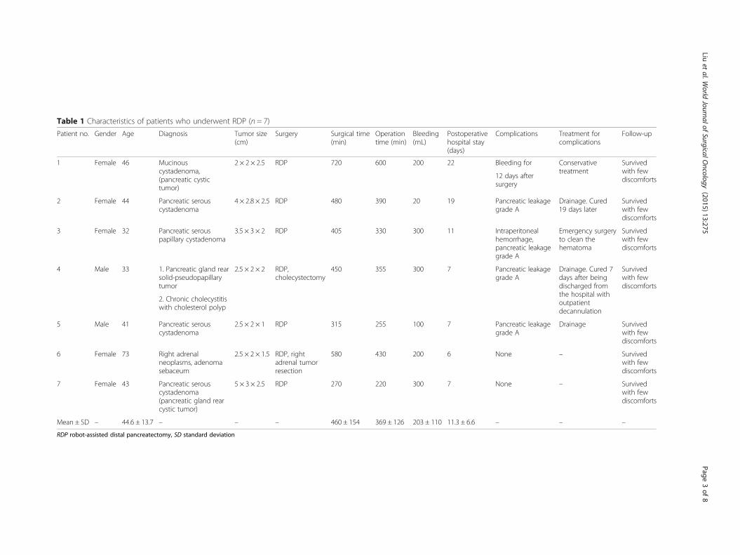
Table 1 Characteristics of patients who underwent RDP (n = 7)
Patient no. Gender Age Diagnosis Tumor size(cm)
Surgery Surgical time(min)
Operationtime (min)
Bleeding(mL)
Postoperativehospital stay(days)
Complications Treatment forcomplications
Follow-up
1 Female 46 Mucinouscystadenoma,(pancreatic cystictumor)
2 × 2 × 2.5 RDP 720 600 200 22 Bleeding for Conservativetreatment
Survivedwith fewdiscomforts12 days after
surgery
2 Female 44 Pancreatic serouscystadenoma
4 × 2.8 × 2.5 RDP 480 390 20 19 Pancreatic leakagegrade A
Drainage. Cured19 days later
Survivedwith fewdiscomforts
3 Female 32 Pancreatic serouspapillary cystadenoma
3.5 × 3 × 2 RDP 405 330 300 11 Intraperitonealhemorrhage,pancreatic leakagegrade A
Emergency surgeryto clean thehematoma
Survivedwith fewdiscomforts
4 Male 33 1. Pancreatic gland rearsolid-pseudopapillarytumor
2.5 × 2 × 2 RDP,cholecystectomy
450 355 300 7 Pancreatic leakagegrade A
Drainage. Cured 7days after beingdischarged fromthe hospital withoutpatientdecannulation
Survivedwith fewdiscomforts
2. Chronic cholecystitiswith cholesterol polyp
5 Male 41 Pancreatic serouscystadenoma
2.5 × 2 × 1 RDP 315 255 100 7 Pancreatic leakagegrade A
Drainage Survivedwith fewdiscomforts
6 Female 73 Right adrenalneoplasms, adenomasebaceum
2.5 × 2 × 1.5 RDP, rightadrenal tumorresection
580 430 200 6 None – Survivedwith fewdiscomforts
7 Female 43 Pancreatic serouscystadenoma(pancreatic gland rearcystic tumor)
5 × 3 × 2.5 RDP 270 220 300 7 None – Survivedwith fewdiscomforts
Mean ± SD – 44.6 ± 13.7 – – – 460 ± 154 369 ± 126 203 ± 110 11.3 ± 6.6 – – –
RDP robot-assisted distal pancreatectomy, SD standard deviation
Liuet
al.World
JournalofSurgicalO
ncology (2015) 13:275
Page3of
8
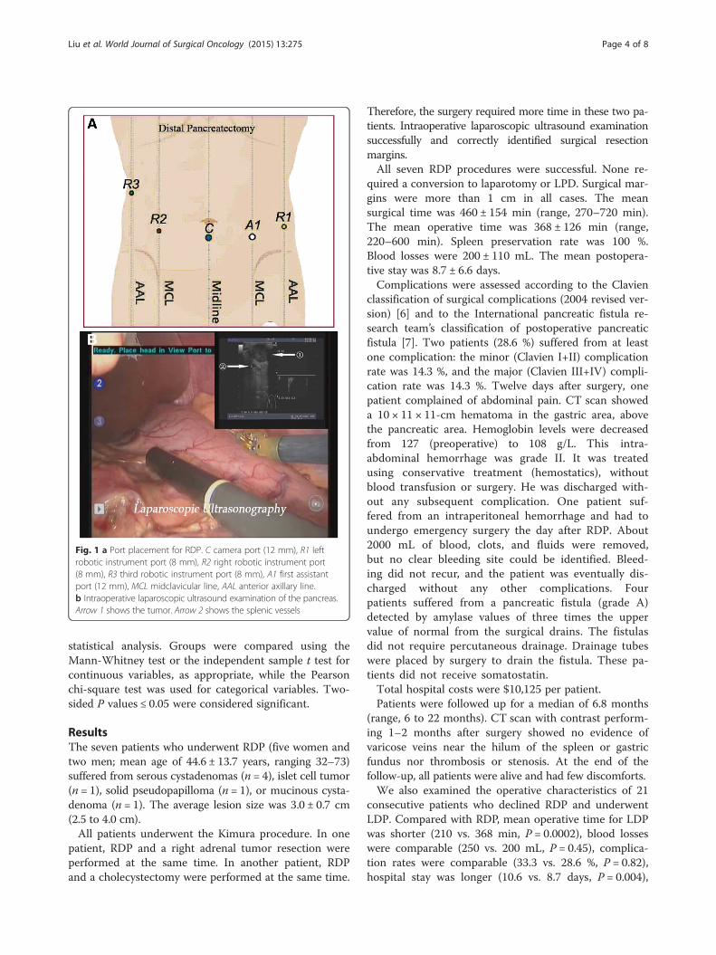
Fig. 1 a Port placement for RDP. C camera port (12 mm), R1 leftrobotic instrument port (8 mm), R2 right robotic instrument port(8 mm), R3 third robotic instrument port (8 mm), A1 first assistantport (12 mm), MCL midclavicular line, AAL anterior axillary line.b Intraoperative laparoscopic ultrasound examination of the pancreas.Arrow 1 shows the tumor. Arrow 2 shows the splenic vessels
Liu et al. World Journal of Surgical Oncology (2015) 13:275 Page 4 of 8
statistical analysis. Groups were compared using theMann-Whitney test or the independent sample t test forcontinuous variables, as appropriate, while the Pearsonchi-square test was used for categorical variables. Two-sided P values ≤ 0.05 were considered significant.
ResultsThe seven patients who underwent RDP (five women andtwo men; mean age of 44.6 ± 13.7 years, ranging 32–73)suffered from serous cystadenomas (n = 4), islet cell tumor(n = 1), solid pseudopapilloma (n = 1), or mucinous cysta-denoma (n = 1). The average lesion size was 3.0 ± 0.7 cm(2.5 to 4.0 cm).All patients underwent the Kimura procedure. In one
patient, RDP and a right adrenal tumor resection wereperformed at the same time. In another patient, RDPand a cholecystectomy were performed at the same time.
Therefore, the surgery required more time in these two pa-tients. Intraoperative laparoscopic ultrasound examinationsuccessfully and correctly identified surgical resectionmargins.All seven RDP procedures were successful. None re-
quired a conversion to laparotomy or LPD. Surgical mar-gins were more than 1 cm in all cases. The meansurgical time was 460 ± 154 min (range, 270–720 min).The mean operative time was 368 ± 126 min (range,220–600 min). Spleen preservation rate was 100 %.Blood losses were 200 ± 110 mL. The mean postopera-tive stay was 8.7 ± 6.6 days.Complications were assessed according to the Clavien
classification of surgical complications (2004 revised ver-sion) [6] and to the International pancreatic fistula re-search team’s classification of postoperative pancreaticfistula [7]. Two patients (28.6 %) suffered from at leastone complication: the minor (Clavien I+II) complicationrate was 14.3 %, and the major (Clavien III+IV) compli-cation rate was 14.3 %. Twelve days after surgery, onepatient complained of abdominal pain. CT scan showeda 10 × 11 × 11-cm hematoma in the gastric area, abovethe pancreatic area. Hemoglobin levels were decreasedfrom 127 (preoperative) to 108 g/L. This intra-abdominal hemorrhage was grade II. It was treatedusing conservative treatment (hemostatics), withoutblood transfusion or surgery. He was discharged with-out any subsequent complication. One patient suf-fered from an intraperitoneal hemorrhage and had toundergo emergency surgery the day after RDP. About2000 mL of blood, clots, and fluids were removed,but no clear bleeding site could be identified. Bleed-ing did not recur, and the patient was eventually dis-charged without any other complications. Fourpatients suffered from a pancreatic fistula (grade A)detected by amylase values of three times the uppervalue of normal from the surgical drains. The fistulasdid not require percutaneous drainage. Drainage tubeswere placed by surgery to drain the fistula. These pa-tients did not receive somatostatin.Total hospital costs were $10,125 per patient.Patients were followed up for a median of 6.8 months
(range, 6 to 22 months). CT scan with contrast perform-ing 1–2 months after surgery showed no evidence ofvaricose veins near the hilum of the spleen or gastricfundus nor thrombosis or stenosis. At the end of thefollow-up, all patients were alive and had few discomforts.We also examined the operative characteristics of 21
consecutive patients who declined RDP and underwentLDP. Compared with RDP, mean operative time for LDPwas shorter (210 vs. 368 min, P = 0.0002), blood losseswere comparable (250 vs. 200 mL, P = 0.45), complica-tion rates were comparable (33.3 vs. 28.6 %, P = 0.82),hospital stay was longer (10.6 vs. 8.7 days, P = 0.004),

Fig. 2 Robotic-assisted spleen-preserving laparoscopic distal pancreatectomy. a Ligation of the splenic vein (arrow), which passes through thepancreas. b The pancreas is totally free from the splenic artery and vein. All branches were treated by sonic shear (<2 mm) or ligature (≥2 mm).Splenic artery and vein were completely preserved. c Use of the surgical stapler (EC60) to perform pancreatic dissection (with ≥1-cm margin). Thetumor is indicated by an arrow. d Verification of the pancreatic section, hemorrhage, and pancreatic leak
Liu et al. World Journal of Surgical Oncology (2015) 13:275 Page 5 of 8
and mean hospital costs were lower ($6921 vs. $10,125,P = 0.0002) (Table 2).
DiscussionIn the present series of patients treated using RDP at asingle institution, surgery was uneventful in all patients,without conversion to laparotomy. The spleen preserva-tion rate was 100 %, the minor (Clavien I+II)
Table 2 Surgical outcomes, complications and hospital costsaccording to the use of a robot during distal pancreatectomy
RDP (n = 7) LDP (n = 21) P value
Mean operative time (min) 368 210 0.0002
Mean blood loss (mL) 200 250 0.451
Transfusion (no. ofpatients) (%)
0 0
Conversion (no. ofpatients) (%)
0 0
Reverse operation(no. of patients) (%)
0 1 (4.7)a
Mean postoperative stay (days) 8.7 10.6 0.004
Complications (no.of patients) (%)
2 (28.6) 7 (33.3) 0.815
Grade, Clavien classification II (1), III (1) I (5), II (1), III (1)
Hospital costs ($) 10,125 6921 0.0002
RDP robot-assisted distal pancreatectomy, LDP laparoscopicdistal pancreatectomyaA second operation was required in one patient of the LPD group (4.7 %)who had a postoperation hemorrhage
complication rate was 57.1 %, and the major (Clavien III+IV) complication rate was 14.3 %. No patient had toundergo a second pancreas surgery. All patients survivedand reported few discomforts. This study presents thefirst Chinese experience using RDP. Intraoperative lap-aroscopic ultrasound examination was performed to de-fine the surgical resection margins, which couldcontribute to better outcomes. These results are sup-ported by a previous study by Hwang et al [8]. However,in comparison, the present study showed a slightly shorteroperation (369 ± 126 vs. 399 ± 166 min), and blood lossesthat were nearly halved (203 ± 110 vs. 360 ± 360 mL).However, the hospital stay was longer (11 ± 7 vs. 7 ±2 days).Indeed, preserving the immunological functions of the
spleen can help to avoid the incidence of leukocytosis,thrombocytosis, overwhelming post-splenectomy sepsis,and low-grade immunodeficiency. A retrospective studyfrom the Memorial Sloan-Kettering Cancer Center in125 patients who underwent distal pancreatectomywith or without splenectomy showed that there werepostoperative complications in 49 % of patients whounderwent splenectomy vs. 39 % of patients who didnot [9]. In addition, perioperative infectious complica-tions and severe complications were more frequent inthe splenectomy group (28 vs. 9 % and 11 vs. 2, re-spectively). Thus, spleen preservation during distalpancreatectomy for benign or borderline malignanttumors is now an accepted practice.
Liu et al. World Journal of Surgical Oncology (2015) 13:275 Page 6 of 8
The Warshaw procedure is one of the two methodsfor preserving the spleen, but the spleen’s blood supplycannot be guaranteed [10]. In an attempt to alleviate themorbidity associated with splenectomy, we were inter-ested in preserving the whole splenic blood supply dur-ing distal pancreatectomy. The Kimura procedurecompletely preserves the splenic artery and vein, as wellas perfusion of the spleen, without noticeable changes inphysiological functions. During our follow-up period, wedid not observe any evidence of varicose veins near thehilum of the spleen or gastric fundus in RDP patients,which might be better than outcomes achieved using theWarshaw procedure since a previous series reportedperigastric varices in 25 % of patients during follow-upafter the Warshaw surgery [10]. However, this procedureis difficult to perform using traditional laparoscopy, andit is much easier to accomplish using a robotic laparos-copy approach [5].Since the technology allowing this approach is rela-
tively novel, RDP is believed to be an uncommon surgi-cal procedure. Since Olah et al. [11] first reported“robotic resection of a pancreatic neuroendocrinetumor” in 2003, Giulianotti et al. [12] reported five pan-creatic resections using RDP with an average surgicaltime of 270 min (range, 210–360 min). Among these fivecases, only two patients (one for an insulinoma and onefor a benign cystic lesion) underwent spleen-preservingpancreatic resection. Some other studies are also avail-able from different countries comparing RDP vs. LDPvs. open surgery, with or without splenic vessel preserva-tion [13–17]. A number of case reports and small seriesare also available.Robotic surgery offers the opportunity to combine the
advantages of both minimally invasive and open surgicalapproaches [4]. The patients promptly return to full ac-tivity and have a short hospital stay. For the surgeons,RDP has the advantage of requiring less laparoscopic ex-perience. Furthermore, the dissection of the splenic veinand artery and the creation of the retropancreatic tunnelwere more easily performed using the da Vinci systemcompared with LDP [18]. Tumors in the body and tail ofthe pancreas are commonly treated with minimally inva-sive surgical techniques, and the advantage of decreasedtissue trauma may prove to be beneficial for a patientwith a benign disease. Thus, a minimally invasive ap-proach should be advocated for this type of disease, par-ticularly because of the benefits it provides in terms ofpostoperative outcomes, such as improved respiratoryfunction and less operative stress.According to our experience, the key feature during
RDP is freeing the upper and lower edges of the pancreasfollowed by venous ligation or transfixion of the vesselsclose to the hilus of the spleen. Intra-operative venousbleeding can usually be managed with a combination of
direct electric coagulation or sutures. Theoretically, usinga robot can greatly increase the ability to deal with smalland delicate branches of splenic vessels because of the su-perior visualization and precision offered by the da Vincisurgical system. Nevertheless, postoperative hemorrhagemay occur, and hemorrhage is the most common and ser-ious complication of this surgery.We used the EC60 stapler for managing the pancre-
atic stump because this method is simple, quick, andknown to be a safe alternative to the standard sutureclosure technique [19]. We usually use the gold cart-ridge to cutoff the pancreas because the staple lengthis 3.8 mm and the closed height is then 1.8 mm,making the pancreatic stump wrinkled, reducing theincidence of pancreatic fistula.However, using a robot presents a number of disad-
vantages. The surgeons who control the robot in thesurgical console do not feel any touch sensation asso-ciated with physically touching the tissues, and theyhave to rely on visual feedback only [20]. It is com-mon to encounter a hemorrhage while grasping orpulling the pancreas during the division of smallsplenic vessels from the pancreas. Sutures or electro-coagulation using Biclamp are two good options forhemostasis, but these procedures are onerous interms of surgical time. However, compared withlaparoscopy, suture of the spleen is not so risky any-more [5]. The setup of the robotic arms necessitatesgood spatial planning and is time-consuming, espe-cially at the beginning of the learning curve [13].Pancreatic fistula is one of the complications thatmay be encountered during RDP, but it is usuallymanageable by drainage. For overweight patients, setupand pancreatic exposure may not be easy, and one or twosupplementary ports might be necessary. Another import-ant criticism is the higher cost of RDP compared withLDP and is one of the main obstacles for the use of RDPin usual clinical practice [5, 13].In the present study, 71 % (5/7) of the patients suf-
fered from complications. A previous study reported acomplication rate of 26 % after robotic pancreaticsurgery [4], but it was reported after performing ro-botic pancreatectomy in 134 patients (compared with7 in the present study), which may suggest an effectof the learning curve on the incidence of complica-tions. In addition, this previous study [4] included alltypes of pancreatectomy, while the present study fo-cused on RDP with preservation of the splenic ves-sels. A previous study in 246 patients who underwentlaparoscopic distal pancreatectomy showed a complicationrate of 31.3 % [21]. Previous studies also reported similarcomplication rates between RDP and LDP [17, 22]. Itmight be expected that with more experience, the compli-cation rate of RDP should decrease.
Liu et al. World Journal of Surgical Oncology (2015) 13:275 Page 7 of 8
RDP has been implemented in some rare centers inChina only a few years ago. With experience and im-provements of this technique, it is anticipated that thehospital stay, complication rate, and costs should be im-proved, making it more attractive than traditional ap-proaches. Nevertheless, in the context of the Chinesehealthcare system, the hospital stay was not overtly long.In addition, if the first two patients were excluded, themean hospital stay would be significantly shorter, andthe complication rates would be lower. Seemingly, inthis study and in contrast to LDP, RDP could improvesome operative characteristics (e.g., blood losses) but in-creased the mean operative time, complication rate, andmean hospital costs. However, since the characteristicsof the patients (e.g., age, disease type, and lesion size)were not comparable between the RDP and LDP groups,the treatment efficacy between the two procedures couldnot be compared directly.The present study has some limitations. Even if we
compared two groups of patients, the sample size wastoo small to draw any firm conclusion. Since the patientschose which surgical approach they wanted, based onsafety and costs, it is possible that the patients choosingthe robotic approach had a higher income than thosechoosing LDP, as well as a standard of living favoringgood outcomes. However, the present report is a caseseries presenting our initial experience in a newly imple-mented technology in China. We are currently conduct-ing a prospective trial in a larger number of patients. Inthe present study, postoperative stays were particularlylong. However, this issue lays in the Chinese healthcaresystem, which requires that all patients are completelysymptom-free upon discharge, which mean without anyabdominal cavity drainage tube. In addition, even milddiscomfort expressed by the patient will make the hos-pital unwilling to discharge him.
ConclusionsOur preliminary real-life study suggests that RDP is afeasible and lowly invasive option for the resection ofbenign or borderline malignant tumors of the pancreas.We used the Kimura procedure with RDP, and resultssuggest that it is beneficial for preserving the splenicvessels. However, postoperative complication rate andcosts were high, and hospital stay and surgical time werelong. Additional multiple-center randomized prospectivestudies with a larger number of patients are required toassess the efficacy of RDP.
AbbreviationsLDP: laparoscopic distal pancreatectomy; RAS: robotic-assisted surgery;RDP: robotic distal pancreatectomy.
Competing interestsThe authors declare that they have no competing interests.
Authors’ contributionsYLi and JHD have made substantial contributions to conception and design;YLi, WBJ, HGW, YLu, XQW, SCL, and JHD have made substantial contributionsto the acquisition of data or analysis and interpretation of data; YLi and JHDhave been involved in drafting the manuscript or revising it critically forimportant intellectual content. All authors read and approved the finalmanuscript.
Received: 3 June 2015 Accepted: 3 August 2015
References1. Kimura W, Inoue T, Futakawa N, Shinkai H, Han I, Muto T. Spleen-preserving
distal pancreatectomy with conservation of the splenic artery and vein.Surgery. 1996;120:885–90.
2. Warshaw AL. Conservation of the spleen with distal pancreatectomy.Arch Surg. 1988;123:550–3.
3. Palep JH. Robotic assisted minimally invasive surgery. J Minim Access Surg.2009;5:1–7.
4. Giulianotti PC, Sbrana F, Bianco FM, Elli EF, Shah G, Addeo P, et al.Robot-assisted laparoscopic pancreatic surgery: single-surgeon experience.Surg Endosc. 2010;24:1646–57.
5. Waters JA, Canal DF, Wiebke EA, Dumas RP, Beane JD, Aguilar-Saavedra JR,et al. Robotic distal pancreatectomy: cost effective? Surgery.2010;148:814–23.
6. Clavien PA, Barkun J, de Oliveira ML, Vauthey JN, Dindo D, Schulick RD, et al.The Clavien-Dindo classification of surgical complications: five-yearexperience. Ann Surg. 2009;250:187–96.
7. Bassi C, Dervenis C, Butturini G, Fingerhut A, Yeo C, Izbicki J, et al.Postoperative pancreatic fistula: an international study group (ISGPF)definition. Surgery. 2005;138:8–13.
8. Hwang HK, Chung YE, Kim KA, Kang CM, Lee WJ. Revisiting vascular patencyafter spleen-preserving laparoscopic distal pancreatectomy withconservation of splenic vessels. Surg Endosc. 2012;26:1765–71.
9. Shoup M, Brennan MF, McWhite K, Leung DH, Klimstra D, Conlon KC. Thevalue of splenic preservation with distal pancreatectomy. Arch Surg.2002;137:164–8.
10. Ferrone CR, Konstantinidis IT, Sahani DV, Wargo JA, Fernandez-del Castillo C,Warshaw AL. Twenty-three years of the Warshaw operation for distalpancreatectomy with preservation of the spleen. Ann Surg.2011;253:1136–9.
11. Olah A. Surgery of the pancreas. Magy Seb. 2009;62:258–64.12. Giulianotti PC, Coratti A, Angelini M, Sbrana F, Cecconi S, Balestracci T, et al.
Robotics in general surgery: personal experience in a large communityhospital. Arch Surg. 2003;138:777–84.
13. Kang CM, Chi HS, Hyeung WJ, Kim KS, Choi JS, Lee WJ, et al. The first koreanexperience of telemanipulative robot-assisted laparoscopic cholecystectomyusing the da vinci system. Yonsei Med J. 2007;48:540–5.
14. Kang CM, Kim DH, Lee WJ, Chi HS. Conventional laparoscopic androbot-assisted spleen-preserving pancreatectomy: does da Vinci haveclinical advantages? Surg Endosc. 2011;25:2004–9.
15. Kang CM, Kim DH, Lee WJ, Chi HS. Initial experiences using robot-assisted central pancreatectomy with pancreaticogastrostomy:a potential way to advanced laparoscopic pancreatectomy.Surg Endosc. 2011;25:1101–6.
16. Abood GJ, Can MF, Daouadi M, Huss HT, Steve JY, Ramalingam L, et al.Robotic-assisted minimally invasive central pancreatectomy: technique andoutcomes. J Gastrointest Surg. 2013;17:1002–8.
17. Daouadi M, Zureikat AH, Zenati MS, Choudry H, Tsung A, Bartlett DL, et al.Robot-assisted minimally invasive distal pancreatectomy is superior to thelaparoscopic technique. Ann Surg. 2013;257:128–32.
18. Ntourakis D, Marzano E, Lopez Penza PA, Bachellier P, Jaeck D, Pessaux P.Robotic distal splenopancreatectomy: bridging the gap between pancreaticand minimal access surgery. J Gastrointest Surg. 2010;14:1326–30.
19. Takeuchi K, Tsuzuki Y, Ando T, Sekihara M, Hara T, Kori T, et al.Distal pancreatectomy: is staple closure beneficial? ANZ J Surg.2003;73:922–5.
20. Kim DH, Kang CM, Lee WJ, Chi HS. The first experience of robot assistedspleen-preserving laparoscopic distal pancreatectomy in Korea. Yonsei MedJ. 2011;52:539–42.
Liu et al. World Journal of Surgical Oncology (2015) 13:275 Page 8 of 8
21. Zhou ZQ, Kim SC, Song KB, Park KM, Lee JH, Lee YJ. Laparoscopicspleen-preserving distal pancreatectomy: comparative study of spleenpreservation with splenic vessel resection and splenic vessel preservation.World J Surg. 2014;38(11):2973–9.
22. Lai EC, Tang CN. Current status of robot-assisted laparoscopicpancreaticoduodenectomy and distal pancreatectomy: a comprehensivereview. Asian J Endosc Surg. 2013;6:158–64.
Submit your next manuscript to BioMed Centraland take full advantage of:
• Convenient online submission
• Thorough peer review
• No space constraints or color figure charges
• Immediate publication on acceptance
• Inclusion in PubMed, CAS, Scopus and Google Scholar
• Research which is freely available for redistribution
Submit your manuscript at www.biomedcentral.com/submit


![Disclaimer - s-space.snu.ac.krs-space.snu.ac.kr/bitstream/10371/142374/1/000000150887.pdf · 1994, when Cuschieri [5] reported the first laparoscopic distal pancreatectomy and Gagner](https://static.fdocuments.net/doc/165x107/6050877ca02ba774652bd998/disclaimer-s-spacesnuackrs-spacesnuackrbitstream103711423741-1994.jpg)