Robotic repair of vesicovaginal fistula (VVF) · with guided wire (Bander Ureteral Diversion Stent...
19
© 2012 THE AUTHORS 1416 BJU INTERNATIONAL © 2 0 1 2 B J U I N T E R N A T I O N A L | 1 0 9 , 1 4 1 6 – 1 4 3 4 | doi:10.1111/j.1464-410X.2012.10148.x BJUI BJU INTERNATIONAL SOTELO ET AL. Surgery Illustrated – Surgical Atlas Robotic repair of vesicovaginal fistula (VVF) Rene Sotelo, Vassilis Moros*, Rafael Clavijo and Vassilis Poulakis* Department of Urology, La Floresta Medical Institute, Caracas, Venezuela; and * Department of Urology, Metropolitan Hospital, Athens, Greece INTRODUCTION For centuries, vesicovaginal fistula (VVF) has presented a challenge for surgeons and a significant social and hygienic problem for patients, especially when it recurs or leads to severe complications [1]. While worldwide, the predominant cause of VVF is obstructed labour due to poor obstetric care, in obstetrically developed countries this is usually an iatrogenic complication of gynaecological surgery, most commonly abdominal hysterectomy, occurring in one in every 1800 hysterectomies [2–4]. Generally, VVF occurs 1–6 weeks after gynaecological or obstetric surgery, while recurrent fistula can develop within the first 3 months after primary repair [2]. The proper timing for corrective surgery and the ideal type of procedure is currently controversial [5]. The early vs delayed repair of VVF has been debated. In select cases, early reconstruction is favourable [6]. Although a trial of conservative therapy including proper and undisturbed bladder drainage for several weeks, treatment with antibiotics when indicated and in some cases a fulguration attempt of a small fistulous tract should be tried, the probability of spontaneous closure of VVF is low (7–12.5%) [3,4]. When VVF is large or does not respond to conservative measures, surgical correction is indicated, with a success rate of 75–97% in cases of fistula resulting from surgical injury [3–5]. Various surgical techniques have been proposed for repair of VVF, depending on the cause, location and surgeon’s experience. As most VVFs result from difficult ILLUSTRATIONS by STEPHAN SPITZER, www.spitzer-illustration.com a Cystoscopy b VVF preparation c Omentum mobilization d Room Positioning Surgeon Assistant Anesthesia Scrub nurse a b c 12 mm Camera Port 8 mm Port (4th arm) 8 mm Robotic Ports 5 mm Ports (Assistant) hysterectomy, the initial repair is usually attempted transvaginally, mostly because this approach has less morbidity and it is more familiar to gynaecological surgeons [3]. However, the vaginal exposure has its limitations, especially when the VVF is high lying on the posterior bladder wall and the vagina severely scarred [5]. Although the morbidity of open abdominal repair is significant compared with that of the transvaginal approach, abdominal surgery is usually preferred in patients with a large (>3 cm) or supratrigonal
-
Upload
trinhhuong -
Category
Documents
-
view
217 -
download
0
Transcript of Robotic repair of vesicovaginal fistula (VVF) · with guided wire (Bander Ureteral Diversion Stent...
© 2 0 1 2 T H E A U T H O R S
1 4 1 6 B J U I N T E R N A T I O N A L © 2 0 1 2 B J U I N T E R N A T I O N A L | 1 0 9 , 1 4 1 6 – 1 4 3 4 | doi:10.1111/j.1464-410X.2012.10148.x
BJUIB J U I N T E R N A T I O N A L
S O T E L O E T A L .
Surgery Illustrated – Surgical Atlas Robotic repair of vesicovaginal fi stula (VVF) Rene Sotelo , Vassilis Moros * , Rafael Clavijo and Vassilis Poulakis * Department of Urology, La Floresta Medical Institute, Caracas, Venezuela ; and * Department of Urology, Metropolitan Hospital, Athens, Greece
INTRODUCTION
For centuries, vesicovaginal fi stula (VVF) has presented a challenge for surgeons and a signifi cant social and hygienic problem for patients, especially when it recurs or leads to severe complications [ 1 ] .
While worldwide, the predominant cause of VVF is obstructed labour due to poor obstetric care, in obstetrically developed countries this is usually an iatrogenic complication of gynaecological surgery, most commonly abdominal hysterectomy, occurring in one in every 1800 hysterectomies [ 2 – 4 ] . Generally, VVF occurs 1 – 6 weeks after gynaecological or obstetric surgery, while recurrent fi stula can develop within the fi rst 3 months after primary repair [ 2 ] .
The proper timing for corrective surgery and the ideal type of procedure is currently controversial [ 5 ] . The early vs delayed repair of VVF has been debated. In select cases, early reconstruction is favourable [ 6 ] . Although a trial of conservative therapy including proper and undisturbed bladder drainage for several weeks, treatment with antibiotics when indicated and in some cases a fulguration attempt of a small fi stulous tract should be tried, the probability of spontaneous closure of VVF is low (7 – 12.5%) [ 3,4 ] .
When VVF is large or does not respond to conservative measures, surgical correction is indicated, with a success rate of 75 – 97% in cases of fi stula resulting from surgical injury [ 3 – 5 ] . Various surgical techniques have been proposed for repair of VVF, depending on the cause, location and surgeon ’ s experience. As most VVFs result from diffi cult
ILLUSTRATIONS by STEPHAN SPITZER, www.spitzer-illustration.com
a Cystoscopy
b VVF preparation
c Omentum mobilization
d Room Positioning
Surgeon
Assistant
Anesthesia
Scrub nurse
a
b
c
12 mm Camera Port
8 mm Port (4th arm)
8 mm Robotic Ports
5 mm Ports(Assistant)
hysterectomy, the initial repair is usually attempted transvaginally, mostly because this approach has less morbidity and it is more familiar to gynaecological surgeons [ 3 ] . However, the vaginal exposure has its limitations, especially when the VVF is high lying on the posterior bladder wall and the vagina severely scarred [ 5 ] . Although the morbidity of open abdominal repair is signifi cant compared with that of the transvaginal approach, abdominal surgery is usually preferred in patients with a large ( > 3 cm) or supratrigonal
S U R G E R Y I L L U S T R A T E D
© 2 0 1 2 T H E A U T H O R S
B J U I N T E R N A T I O N A L © 2 0 1 2 B J U I N T E R N A T I O N A L 1 4 1 7
fi stula or a fi stula in close proximity to ureteric orifi ces and especially in patients with multiple complicated or recurrent VVF after transvaginal repair [ 7,8 ] .
To decrease the morbidity of the abdominal approach, a laparoscopic repair of VVF has been used, which is primarily associated with similar success rates, minimal surgical trauma and lesser morbidity, thus allowing more rapid convalescence [ 9 ] . Despite initial enthusiasm, laparoscopy has not gained in popularity, probably because laparoscopic VVF dissection and intracorporeal suturing are technically challenging. Robotic assistance in complex laparoscopic procedures has overcome the technical diffi culties of the laparoscopic approach, even in challenging cases of recurrent VVF [ 10 ] . Using the technological advantages of robotic technology (EndoWrist TM instruments with increased degrees of freedom leading to improved dexterity and absence of fatigue, three-dimensional [ 3-D ] vision with improved depth perception, motion scaling, tremor fi ltration, higher magnifi cation, and surgeon ’ s ergonomic position in a long-standing and time-consuming operation), it is possible to perform a repair of VVF laparoscopically, respecting the basic surgical principles of fi stula reconstruction [ 4,6,8 ] , namely:
1) clear and wide exposure of the fi stula track and surrounding tissue 2) circumferential excision of fi brous and scar tissue from the fi stula edges 3) saving both ureteric orifi ces 4) use of suitable suture material 5) individual tension-free suture repair of freshened fi stula edges in the vagina and the bladder 6) watertight, multiple-layer closure 7) non-overlapping suture lines 8) interposition of various well-vascularised tissues, such as omentum [ 11 ] , peritoneum, bladder fl ap taken from a site away from the fi stula or epiploic appendix taken from the sigmoid colon 9) continuous and adequate postoperative bladder drainage, especially early after repair
PLANNING AND PREPARATION
INDICATIONS AND CONTRAINDICATIONS
The indications for robotic repair of VVF are the same as those for open abdominal
surgical correction. Although most VVF in the developed and industrialized world are amenable to transvaginal repair, there are certain indications for an abdominal approach [ 3 – 5 ] , such as:
1) inadequate exposure related to a high or retracted fi stula in a narrow vagina 2) close proximity of the fi stulous tract to the ureter 3) associated pelvic pathology, e.g. an associated ureteric injury (e.g. ureterovaginal fi stula) or a complex fi stula involving another intra-abdominal organ 4) multiple fi stulae
Nevertheless, a previously failed transvaginal attempt at repair of VVF is not necessarily a contraindication for a new transvaginal approach as excellent results can be achieved in this setting [ 6 ] .
Absolute contraindications are massive haematoma in the pelvis and active UTI.
SPECIFIC PATIENT PREPARATION
Although the mechanical (with polyethylene glycol and a liquid diet of 4 – 5 L) and antibacterial bowel preparation the day before surgery is not absolutely necessary, we still routinely use it, especially in cases of complicated VVF. A prophylactic dose of heparin (low molecular weight adjusted to the body weight of the patient) is given the evening before surgery and every 24 h thereafter until ambulation. Before the operation begins, all patients are shaved from nipples to mid-thighs. As infection prophylaxis is essential for surgery outcome, antibiotics with amoxycillin/clavulanic acid and metronidazole orally are commenced 1 day before the operation and continued parenteral when the procedure begins. Metronidazole is continued for 48 h and amoxycillin/clavulanic acid until all drains and catheters are removed.
SPECIFIC EQUIPMENT AND MATERIALS REQUIRED FOR ROBOTIC REPAIR OF V VF
Robotic equipment (da Vinci, Intuitive Surgical, Mountain View, CA, USA):
• Three (for four-arm robotic system) or two (for conventional three-arm device) trocars (8 mm)
• 0 ° and 30 ° 3-D laparoscope • Hot Shears TM (monopolar curved scissors) • Maryland bipolar forceps • Two large needle drivers • Two ProGrasp TM forceps
Laparoscopic equipment:
• One trocar (12 mm) for the robotic telescope • Two trocars (5 mm) for the bedside assistant • 5-mm endoscopic long suction irrigator (45 cm) • 5-mm endoscopic scissors • 5-mm endoscopic locking grasper • 5-mm endoscopic needle driver
Energy sources/haemostatics:
• Monopolar/bipolar electrocautery • 5.5-mm Ethicon UltraCision harmonic scalpel coagulating shears, pistol grip (36 cm, 15-mm active blade – 5.5 mm diameter)
Suture materials:
• 3-0 poliglecaprone 25 monofi lament synthetic absorbable suture (Ethicon, Monocryl TM ) with RB-1 needle (half circle, 17 mm diameter) for bladder and vagina closure. Alternative 3-0 glycomer 631 monofi lament synthetic absorbable suture (Biosyn TM ) with CV-25 needle (half circle, 22 mm diameter). • 2-0 polyglycolic acid, braided suture with round-bodied needle (half circle, 30 mm diameter) using as fi xation/anchor suture for the interposition graft (omentum or peritoneal fl ap)
Other materials:
• Two long mono-J ureteric stents 7 or 6 F with guided wire (Bander Ureteral Diversion Stent Set, Cook Urological, Inc., Spencer, IN, USA) • Simple ureteric catheter 5 F for retrograde visualisation of ureters with contrast medium and for canalisation of VVF • Silicon Foley catheter 22 F • Cystourethroscope 22 F with a ureteric catheterisation bridge (30 ° telescope) • C-arm device for assessing the proper placement of ureteric catheters
S O T E L O E T A L .
© 2 0 1 2 T H E A U T H O R S
1 4 1 8 B J U I N T E R N A T I O N A L © 2 0 1 2 B J U I N T E R N A T I O N A L
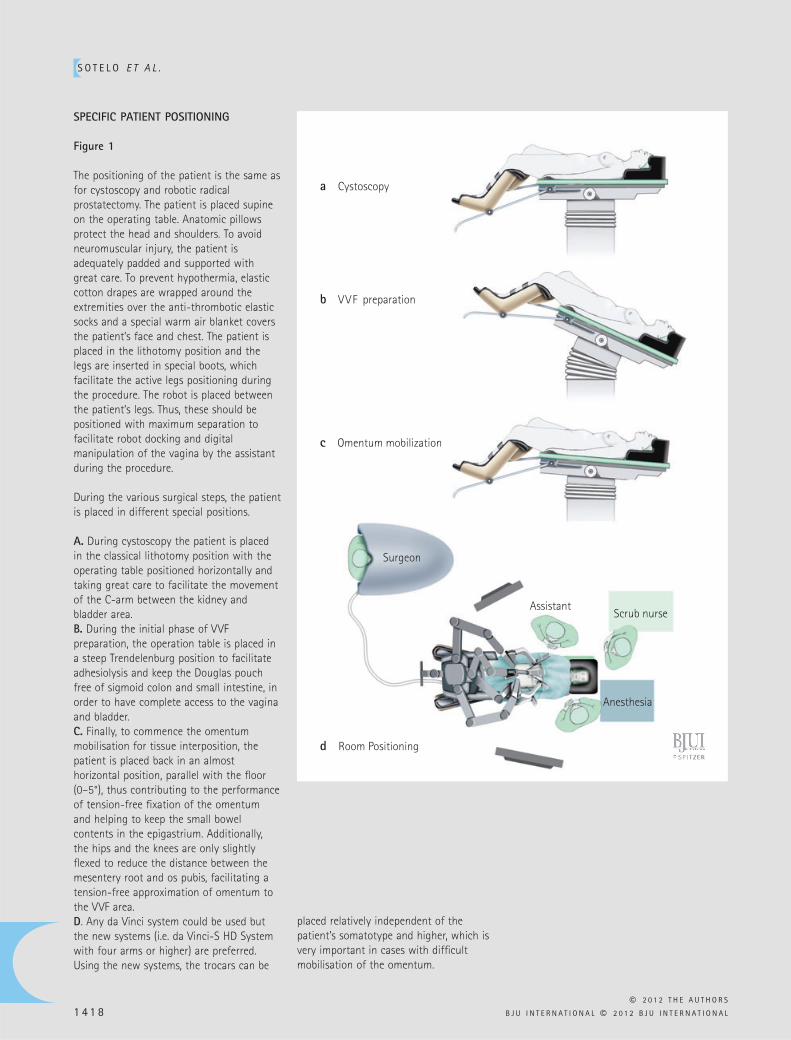
SPECIFIC PATIENT POSITIONING
Figure 1
The positioning of the patient is the same as for cystoscopy and robotic radical prostatectomy. The patient is placed supine on the operating table. Anatomic pillows protect the head and shoulders. To avoid neuromuscular injury, the patient is adequately padded and supported with great care. To prevent hypothermia, elastic cotton drapes are wrapped around the extremities over the anti-thrombotic elastic socks and a special warm air blanket covers the patient ’ s face and chest. The patient is placed in the lithotomy position and the legs are inserted in special boots, which facilitate the active legs positioning during the procedure. The robot is placed between the patient ’ s legs. Thus, these should be positioned with maximum separation to facilitate robot docking and digital manipulation of the vagina by the assistant during the procedure.
During the various surgical steps, the patient is placed in different special positions.
A. During cystoscopy the patient is placed in the classical lithotomy position with the operating table positioned horizontally and taking great care to facilitate the movement of the C-arm between the kidney and bladder area. B. During the initial phase of VVF preparation, the operation table is placed in a steep Trendelenburg position to facilitate adhesiolysis and keep the Douglas pouch free of sigmoid colon and small intestine, in order to have complete access to the vagina and bladder. C. Finally, to commence the omentum mobilisation for tissue interposition, the patient is placed back in an almost horizontal position, parallel with the fl oor (0 – 5 ° ), thus contributing to the performance of tension-free fi xation of the omentum and helping to keep the small bowel contents in the epigastrium. Additionally, the hips and the knees are only slightly fl exed to reduce the distance between the mesentery root and os pubis, facilitating a tension-free approximation of omentum to the VVF area. D . Any da Vinci system could be used but the new systems (i.e. da Vinci-S HD System with four arms or higher) are preferred. Using the new systems, the trocars can be
a Cystoscopy
b VVF preparation
c Omentum mobilization
d Room Positioning
Surgeon
Assistant
Anesthesia
Scrub nurse
placed relatively independent of the patient ’ s somatotype and higher, which is very important in cases with diffi cult mobilisation of the omentum.
S U R G E R Y I L L U S T R A T E D
© 2 0 1 2 T H E A U T H O R S
B J U I N T E R N A T I O N A L © 2 0 1 2 B J U I N T E R N A T I O N A L 1 4 1 9
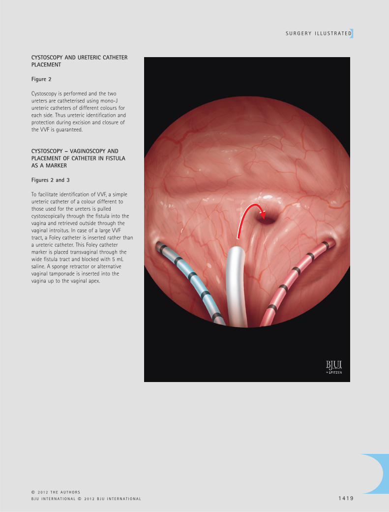
CYSTOSCOPY AND URETERIC CATHETER PLACEMENT
Figure 2
Cystoscopy is performed and the two ureters are catheterised using mono-J ureteric catheters of different colours for each side. Thus ureteric identifi cation and protection during excision and closure of the VVF is guaranteed.
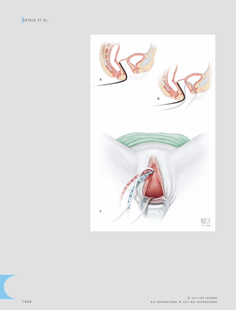
CYSTOSCOPY – VAGINOSCOPY AND PLACEMENT OF CATHETER IN FISTULA AS A MARKER
Figures 2 and 3
To facilitate identifi cation of VVF, a simple ureteric catheter of a colour different to those used for the ureters is pulled cystoscopically through the fi stula into the vagina and retrieved outside through the vaginal introitus. In case of a large VVF tract, a Foley catheter is inserted rather than a ureteric catheter. This Foley catheter marker is placed transvaginal through the wide fi stula tract and blocked with 5 mL saline. A sponge retractor or alternative vaginal tamponade is inserted into the vagina up to the vaginal apex.
S O T E L O E T A L .
© 2 0 1 2 T H E A U T H O R S
1 4 2 0 B J U I N T E R N A T I O N A L © 2 0 1 2 B J U I N T E R N A T I O N A L
a
b
c
S U R G E R Y I L L U S T R A T E D
© 2 0 1 2 T H E A U T H O R S
B J U I N T E R N A T I O N A L © 2 0 1 2 B J U I N T E R N A T I O N A L 1 4 2 1
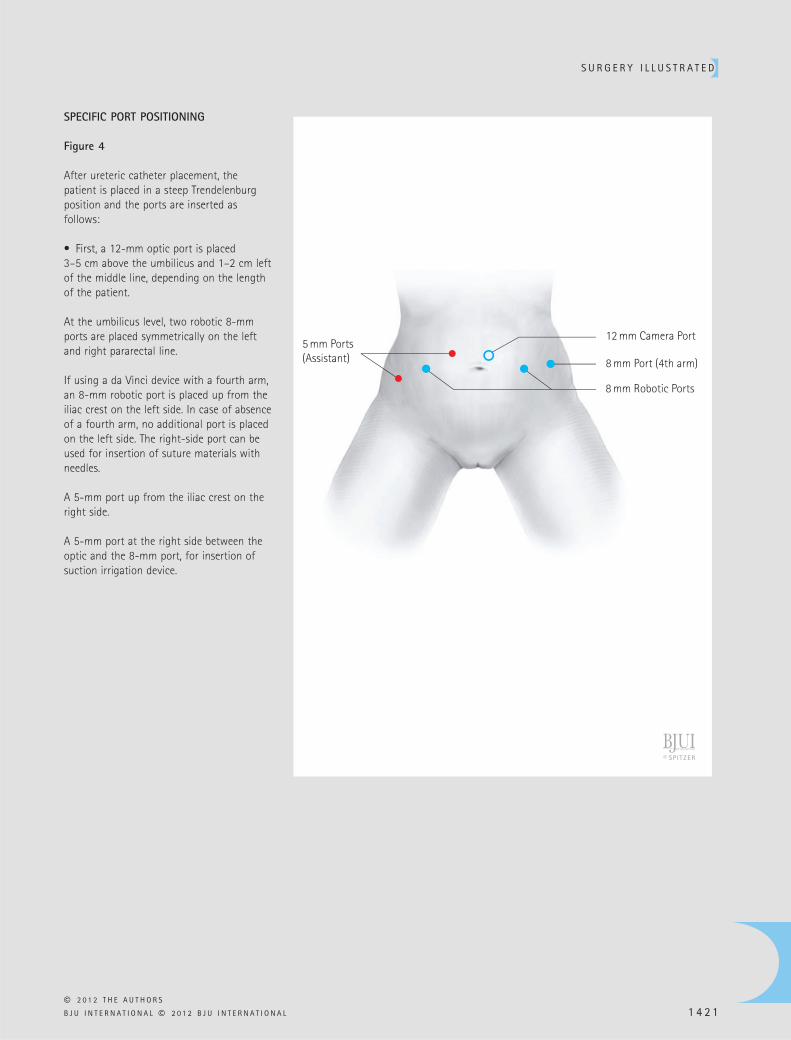
SPECIFIC PORT POSITIONING
Figure 4
After ureteric catheter placement, the patient is placed in a steep Trendelenburg position and the ports are inserted as follows:
• First, a 12-mm optic port is placed 3 – 5 cm above the umbilicus and 1 – 2 cm left of the middle line, depending on the length of the patient.
At the umbilicus level, two robotic 8-mm ports are placed symmetrically on the left and right pararectal line.
If using a da Vinci device with a fourth arm, an 8-mm robotic port is placed up from the iliac crest on the left side. In case of absence of a fourth arm, no additional port is placed on the left side. The right-side port can be used for insertion of suture materials with needles.
A 5-mm port up from the iliac crest on the right side.
A 5-mm port at the right side between the optic and the 8-mm port, for insertion of suction irrigation device.
12 mm Camera Port
8 mm Port (4th arm)
8 mm Robotic Ports
5 mm Ports(Assistant)
S O T E L O E T A L .
© 2 0 1 2 T H E A U T H O R S
1 4 2 2 B J U I N T E R N A T I O N A L © 2 0 1 2 B J U I N T E R N A T I O N A L
PROCEDURE
PERITONEOSCOPY AND ADHESIOLYSIS
Figure 5
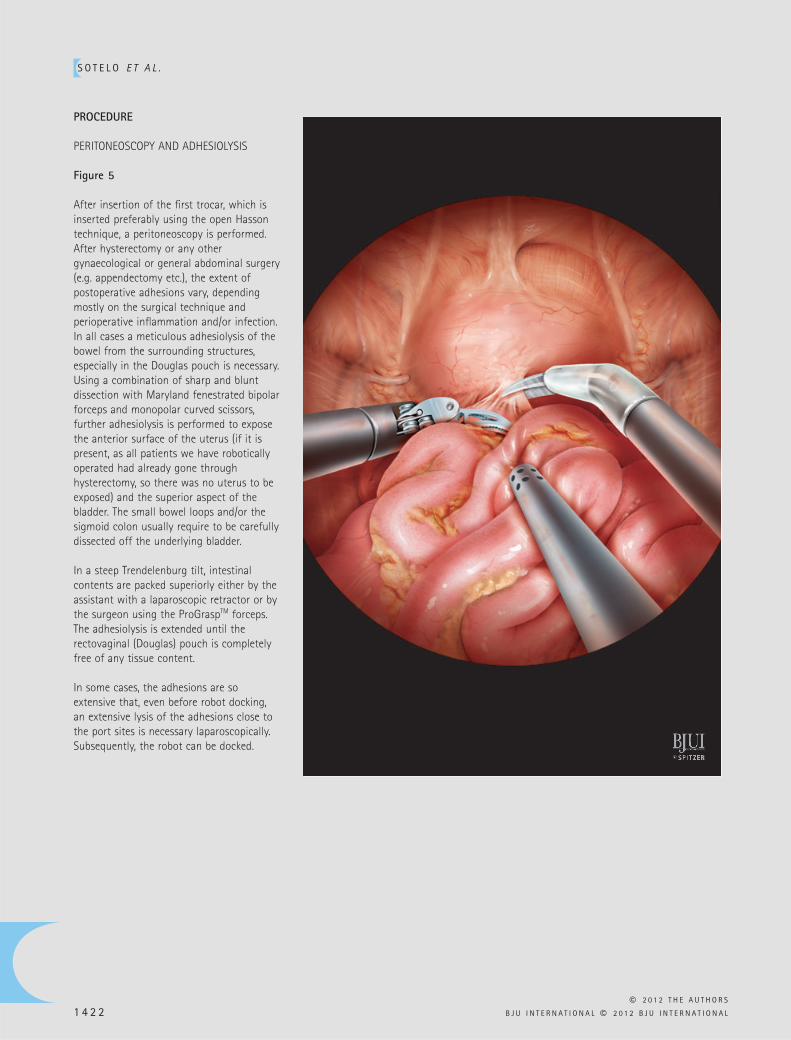
After insertion of the fi rst trocar, which is inserted preferably using the open Hasson technique, a peritoneoscopy is performed. After hysterectomy or any other gynaecological or general abdominal surgery (e.g. appendectomy etc.), the extent of postoperative adhesions vary, depending mostly on the surgical technique and perioperative infl ammation and/or infection. In all cases a meticulous adhesiolysis of the bowel from the surrounding structures, especially in the Douglas pouch is necessary. Using a combination of sharp and blunt dissection with Maryland fenestrated bipolar forceps and monopolar curved scissors, further adhesiolysis is performed to expose the anterior surface of the uterus (if it is present, as all patients we have robotically operated had already gone through hysterectomy, so there was no uterus to be exposed) and the superior aspect of the bladder. The small bowel loops and/or the sigmoid colon usually require to be carefully dissected off the underlying bladder.
In a steep Trendelenburg tilt, intestinal contents are packed superiorly either by the assistant with a laparoscopic retractor or by the surgeon using the ProGrasp TM forceps. The adhesiolysis is extended until the rectovaginal (Douglas) pouch is completely free of any tissue content.
In some cases, the adhesions are so extensive that, even before robot docking, an extensive lysis of the adhesions close to the port sites is necessary laparoscopically. Subsequently, the robot can be docked.
S U R G E R Y I L L U S T R A T E D
© 2 0 1 2 T H E A U T H O R S
B J U I N T E R N A T I O N A L © 2 0 1 2 B J U I N T E R N A T I O N A L 1 4 2 3
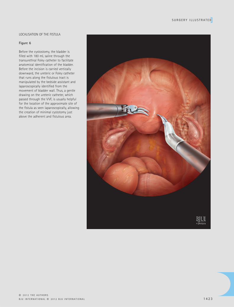
LOCALISATION OF THE FISTULA
Figure 6
Before the cystostomy, the bladder is fi lled with 180 mL saline through the transurethral Foley catheter to facilitate anatomical identifi cation of the bladder. Before the incision is carried vertically downward, the ureteric or Foley catheter that runs along the fi stulous tract is manipulated by the bedside assistant and laparoscopically identifi ed from the movement of bladder wall. Thus, a gentle drawing on the ureteric catheter, which passed through the VVF, is usually helpful for the location of the approximate site of the fi stula as seen laparoscopically, allowing the creation of minimal cystotomy just above the adherent and fi stulous area.
S O T E L O E T A L .
© 2 0 1 2 T H E A U T H O R S
1 4 2 4 B J U I N T E R N A T I O N A L © 2 0 1 2 B J U I N T E R N A T I O N A L
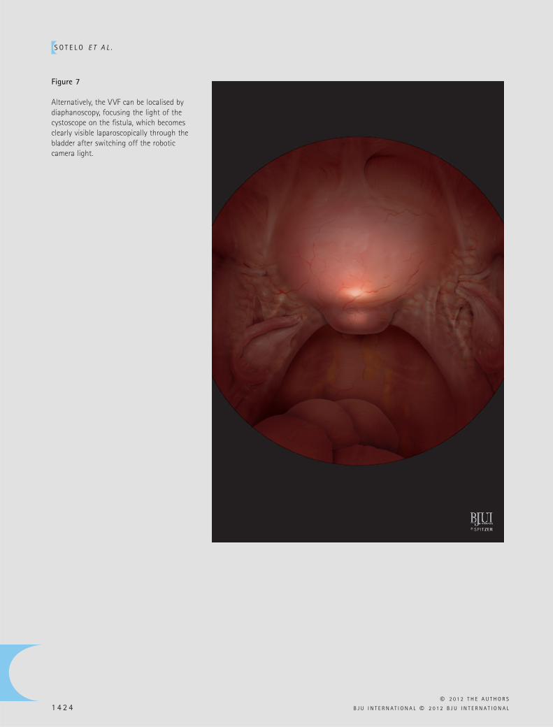
Figure 7
Alternatively, the VVF can be localised by diaphanoscopy, focusing the light of the cystoscope on the fi stula, which becomes clearly visible laparoscopically through the bladder after switching off the robotic camera light.
S U R G E R Y I L L U S T R A T E D
© 2 0 1 2 T H E A U T H O R S
B J U I N T E R N A T I O N A L © 2 0 1 2 B J U I N T E R N A T I O N A L 1 4 2 5
CYSTOTOMY
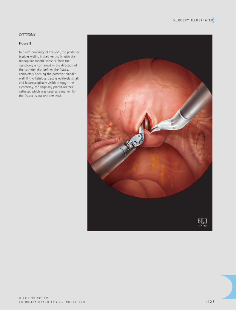
Figure 8
In direct proximity of the VVF, the posterior bladder wall is incised vertically with the monopolar robotic scissors. Then the cystotomy is continued in the direction of the catheter that defi nes the fi stula, completely opening the posterior bladder wall. If the fi stulous tract is relatively small and laparoscopically visible through the cystotomy, the vaginally placed ureteric catheter, which was used as a marker for the fi stula, is cut and removed.
S O T E L O E T A L .
© 2 0 1 2 T H E A U T H O R S
1 4 2 6 B J U I N T E R N A T I O N A L © 2 0 1 2 B J U I N T E R N A T I O N A L
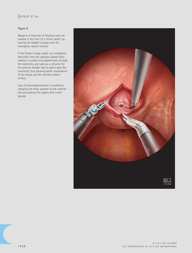
Figure 9
Margins of resection of fi stulous tract are marked in the form of a ‘ tennis racket ’ by scoring the bladder mucosa with the monopolar robotic scissors.
If the fi stula is large and/or not completely identifi ed, then the vaginally placed Foley catheter is pulled intra-abdominally through the cystotomy and used as a retractor for the anterior bladder wall to splint open the cystotomy thus allowing better visualisation of the fi stula and the stented ureteric orifi ces.
Loss of pneumoperitoneum is avoided by clamping the Foley catheter at the external site and packing the vagina with a wet sponge.
S U R G E R Y I L L U S T R A T E D
© 2 0 1 2 T H E A U T H O R S
B J U I N T E R N A T I O N A L © 2 0 1 2 B J U I N T E R N A T I O N A L 1 4 2 7
FISTULA AND NECROTIC TISSUE EXCISION
Figure 10
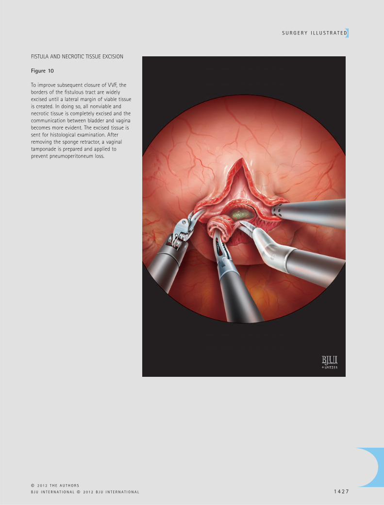
To improve subsequent closure of VVF, the borders of the fi stulous tract are widely excised until a lateral margin of viable tissue is created. In doing so, all nonviable and necrotic tissue is completely excised and the communication between bladder and vagina becomes more evident. The excised tissue is sent for histological examination. After removing the sponge retractor, a vaginal tamponade is prepared and applied to prevent pneumoperitoneum loss.
S O T E L O E T A L .
© 2 0 1 2 T H E A U T H O R S
1 4 2 8 B J U I N T E R N A T I O N A L © 2 0 1 2 B J U I N T E R N A T I O N A L
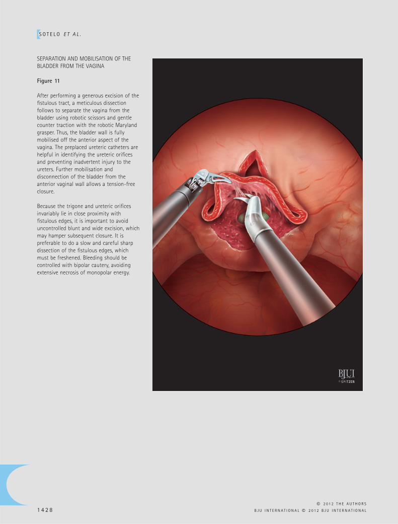
SEPARATION AND MOBILISATION OF THE BLADDER FROM THE VAGINA
Figure 11
After performing a generous excision of the fi stulous tract, a meticulous dissection follows to separate the vagina from the bladder using robotic scissors and gentle counter traction with the robotic Maryland grasper. Thus, the bladder wall is fully mobilised off the anterior aspect of the vagina. The preplaced ureteric catheters are helpful in identifying the ureteric orifi ces and preventing inadvertent injury to the ureters. Further mobilisation and disconnection of the bladder from the anterior vaginal wall allows a tension-free closure.
Because the trigone and ureteric orifi ces invariably lie in close proximity with fi stulous edges, it is important to avoid uncontrolled blunt and wide excision, which may hamper subsequent closure. It is preferable to do a slow and careful sharp dissection of the fi stulous edges, which must be freshened. Bleeding should be controlled with bipolar cautery, avoiding extensive necrosis of monopolar energy.
S U R G E R Y I L L U S T R A T E D
© 2 0 1 2 T H E A U T H O R S
B J U I N T E R N A T I O N A L © 2 0 1 2 B J U I N T E R N A T I O N A L 1 4 2 9
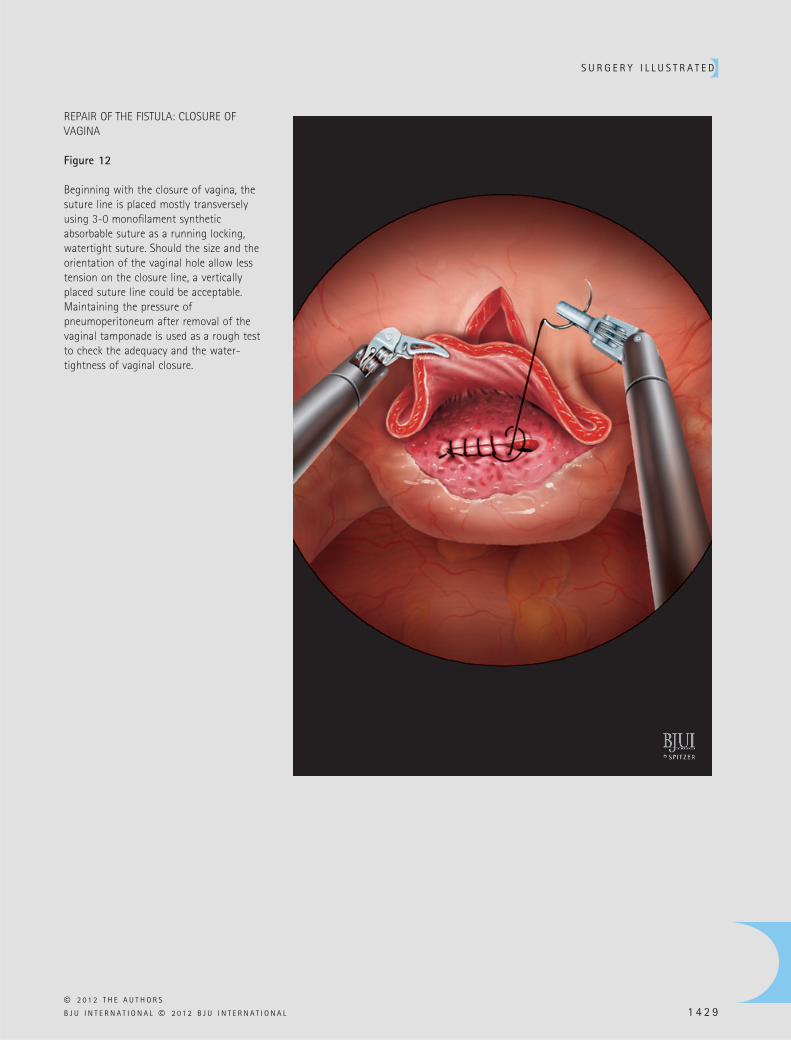
REPAIR OF THE FISTULA: CLOSURE OF VAGINA
Figure 12
Beginning with the closure of vagina, the suture line is placed mostly transversely using 3-0 monofi lament synthetic absorbable suture as a running locking, watertight suture. Should the size and the orientation of the vaginal hole allow less tension on the closure line, a vertically placed suture line could be acceptable. Maintaining the pressure of pneumoperitoneum after removal of the vaginal tamponade is used as a rough test to check the adequacy and the water-tightness of vaginal closure.
S O T E L O E T A L .
© 2 0 1 2 T H E A U T H O R S
1 4 3 0 B J U I N T E R N A T I O N A L © 2 0 1 2 B J U I N T E R N A T I O N A L
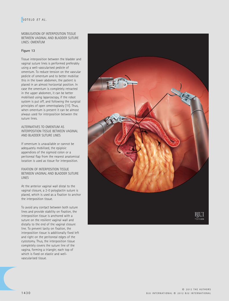
MOBILISATION OF INTERPOSITION TISSUE BETWEEN VAGINAL AND BLADDER SUTURE LINES: OMENTUM
Figure 13
Tissue interposition between the bladder and vaginal suture lines is performed preferably using a well-vascularised pedicle of omentum. To reduce tension on the vascular pedicle of omentum and to better mobilise this in the lower abdomen, the patient is placed in an almost horizontal position. In case the omentum is completely retracted in the upper abdomen, it can be better mobilised using laparoscopy, if the robot system is put off, and following the surgical principles of open omentoplasty [ 11 ] . Thus, when omentum is present it can be almost always used for interposition between the suture lines.
ALTERNATIVES TO OMENTUM AS INTERPOSITION TISSUE BETWEEN VAGINAL AND BLADDER SUTURE LINES
If omentum is unavailable or cannot be adequately mobilised, the epiploic appendices of the sigmoid colon or a peritoneal fl ap from the nearest anatomical location is used as tissue for interposition.
FIXATION OF INTERPOSITION TISSUE BETWEEN VAGINAL AND BLADDER SUTURE LINES
At the anterior vaginal wall distal to the vaginal closure, a 2-0 polyglactin suture is placed, which is used as a fi xation to anchor the interposition tissue.
To avoid any contact between both suture lines and provide stability on fi xation, the interposition tissue is anchored with a suture on the resilient vaginal wall and distally to the end of the vaginal closure line. To prevent laxity on fi xation, the interposition tissue is additionally fi xed left and right on the peritoneal edges of the cystotomy. Thus, the interposition tissue completely covers the suture line of the vagina, forming a triangle; each top of which is fi xed on elastic and well-vascularised tissue.
S U R G E R Y I L L U S T R A T E D
© 2 0 1 2 T H E A U T H O R S
B J U I N T E R N A T I O N A L © 2 0 1 2 B J U I N T E R N A T I O N A L 1 4 3 1
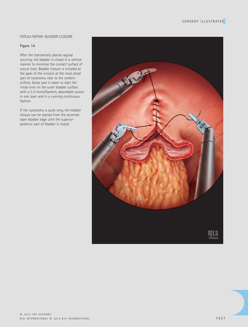
FISTULA REPAIR: BLADDER CLOSURE
Figure 14
After the transversely placed vaginal suturing, the bladder is closed in a vertical manner to minimise the contact surface of suture lines. Bladder closure is initiated at the apex of the incision at the most distal part of cystotomy near to the ureteric orifi ces. Great care is taken to start the initial knot on the outer bladder surface with a 3-0 monofi lament, absorbable suture in one layer and in a running continuous fashion.
If the cystotomy is quite long, the bladder closure can be started from the proximal open bladder edge until the superior-posterior part of bladder is closed.
S O T E L O E T A L .
© 2 0 1 2 T H E A U T H O R S
1 4 3 2 B J U I N T E R N A T I O N A L © 2 0 1 2 B J U I N T E R N A T I O N A L
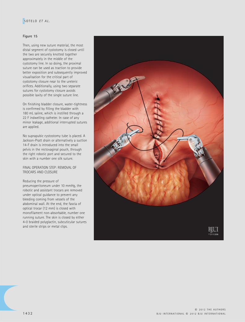
Figure 15
Then, using new suture material, the most distal segment of cystotomy is closed until the two are securely knotted together approximately in the middle of the cystotomy line. In so doing, the proximal suture can be used as traction to provide better exposition and subsequently improved visualisation for the critical part of cystotomy closure near to the ureteric orifi ces. Additionally, using two separate sutures for cystotomy closure avoids possible laxity of the single suture line.
On fi nishing bladder closure, water-tightness is confi rmed by fi lling the bladder with 180 mL saline, which is instilled through a 22 F indwelling catheter. In case of any minor leakage, additional interrupted sutures are applied.
No suprapubic cystostomy tube is placed. A Jackson-Pratt drain or alternatively a suction 14-F drain is introduced into the small pelvis in the rectovaginal pouch, through the right robotic port and secured to the skin with a number one silk suture.
FINAL OPERATION STEP: REMOVAL OF TROCARS AND CLOSURE
Reducing the pressure of pneumoperitoneum under 10 mmHg, the robotic and assistant trocars are removed under optical guidance to prevent any bleeding coming from vessels of the abdominal wall. At the end, the fascia of optical trocar (12 mm) is closed with monofi lament non-absorbable, number one running suture. The skin is closed by either 4-0 braided polyglactin, subcuticular sutures and sterile strips or metal clips.
S U R G E R Y I L L U S T R A T E D
© 2 0 1 2 T H E A U T H O R S
B J U I N T E R N A T I O N A L © 2 0 1 2 B J U I N T E R N A T I O N A L 1 4 3 3
POSTOPERATIVE CARE AND FOLLOW-UP
Postoperative care of robotic VVF repair is no different from that of the open procedure [ 8 ] and may include the following points:
1) The drain is removed on postoperative day (POD) 1 or 2 when drainage fl uid is < 50 mL in 24 h. The patient is discharged home with an indwelling urethral catheter and two ureteric catheters, which are exteriorized through the urethra and fi xed around the urethral catheter with a number one silk suture. During the initial experience, insertion of the ureteric catheters is benefi cial mostly because the bladder remains almost completely dry to facilitate the healing process. Thus, they remain in place for 1 week. Gaining more experience, this was deemed unnecessary and dispensed with in subsequent cases, removing the ureteric catheters on POD 1. 2) Normally the urethral catheter is removed on POD 14 or 21, depending on the quality of reconstruction and the surgeon ’ s preference. A retrograde cystogram may be performed before removal of the bladder catheter to confi rm fi stula closure but is not always mandatory. Patients are warned to avoid the use of tampons and refrain from sexual activity for at least 2 months postoperatively. 3) Appropriate prophylactic antibiotics are generally given until all tubes are removed. A urine culture is routinely performed after the removal of the transurethral catheter and 2 weeks after the discontinuation of antibiotic prophylaxis. Any UTI or bacteriuria must be treated. 4) Great care is taken to secure constant urine fl ow of the transurethral catheter and ureteric catheters (if present), by preventing clot obstruction and retention. Only when there is suspicion of catheter obstruction, is irrigation of these tubes necessary. 5) Early mobilisation and ambulation is encouraged using the principles of fast-track postoperative care: no nasogastric tube, gradual nutrition and mobilisation starting 8 h after the end of the procedure.
FROM SURGEON TO SURGEON
DIFFICULT CASES
By defi nition, robotic VVF repair is a challenging operation, as fi stula formation is
a complication arising from a previous gynaecological operation. Previous abdominal surgeries, especially in the small pelvis, may have created extensive peritoneal adhesions requiring diffi cult adhesiolysis before inserting the ports. A history of failed attempts of open/abdominal or vaginal repair of VVF, ruptured viscera or peritonitis deserves special caution. The operation becomes more diffi cult in markedly obese (body mass index > 40 kg/m 2 ) women and in those who have had radiation or complicated VVF. Special attention must be paid when accepting women with these factors during early robotic experience.
TIPS AND TRICKS: THINGS TO MAKE ROBOTIC SURGERY EASIER
One of the most important factors in making robotic repair of VVF easier is a consistent and effi cient robotic team performing the complete spectrum of robotic urological surgery. In some special conditions, e.g. in the narrow space of cystotomy, the movements of the robotic instruments can be restricted in the dissection and/or reconstruction of VVF near to the ureteric orifi ces. The experienced assistant can move the robotic arms medially, thereby changing the angle of the instruments.
Using appropriate lenses, 0 ° , 30 ° up and 30 ° down, in various steps of the procedure maximises the visualisation of the operative fi eld and prevents clashing between telescope and instruments in the narrow pelvic space. When severe adhesions are present, the use of a 5-mm EndoEYE TM lens with fl exible tip is also recommended to facilitate the adhesiolysis because it has different angles of view and can be used through any trocar.
Generally, the fi rst step for successful treatment outcome of a surgical disease is the correct diagnosis. The proper diagnosis of VVF is supported by clinical evaluation. Offi ce fl exible cystoscopy, vaginal examination, three-swab test, retrograde cystourethrography and excretory urography confi rm VVF and exclude concurrent ureteric injury or complicated fi stula [ 5 ] . Ureteric involvement or decreased bladder capacity may necessitate ureter new implantation in the bladder or augmentation cystoplasty, respectively.
If there is any doubt about the timing of VVF repair, it is preferable for surgical treatment to be performed at least 3 months after the initial diagnosis. Although the early repair of VVF could be an attractive option mainly because of the advantage of the immediate improvement in quality of life, the classical strategy is the delayed repair, undertaken after 3 – 6 months and allowing the tissue of any infl ammation to heal and re-vascularise. As the best chance of success is with the fi rst repair, the proper selection of patients for the earliest date for surgery is essential. This necessitates regular follow-up of the bladder and vagina with cystoscopy to exclude active infl ammation, such as devitalised tissues, oedema (confi rming a destructive process), cystitis or encrustation. When other bladder pathologies (suture, stone and diverticulum) exist, then surgery should be postponed until these problems are resolved.
Because all patients with VVF have undergone at least one previous gynaecological surgery, the access for creating pneumoperitoneum is performed either by open access or alternatively by inserting Veress in the left hypochondrium. In case of previous open surgical repair of VVF, extensive adhesion lyses may have to be performed before repairing the fi stula robotically. To facilitate a complete adhesiolysis for safe insertion of robotic trocars, it is sometimes advisable to insert an additional 5-mm port more cephalic and lateral outside the standard trocar positions for better bowel retraction or insertion of special instruments (i.e. ultrasonic shears, Ethicon UltraCision harmonic scalpel coagulating shears, 5.5 mm diameter) for effective and bloodless dissection.
As no thick laparoscopic instruments (e.g. 10-mm clip applier) are necessary, both assistant ports can be 5 mm in diameter to reduce the risk of postoperative bleeding and formation of port hernias. The needles are passed through the 8-mm robotic or 12-mm camera port.
Completely respecting the basic surgical principles of VVF reconstruction is the key factor for a successful closure. However, in our experience, the two most important surgical principles for a successful outcome are the interposition of well-vascularised tissues, preferably omentum, and adequate postoperative bladder drainage.
S O T E L O E T A L .
© 2 0 1 2 T H E A U T H O R S
1 4 3 4 B J U I N T E R N A T I O N A L © 2 0 1 2 B J U I N T E R N A T I O N A L
Interposition of omentum is absolutely the fi rst choice, especially in post-irradiation conditions. Only if the omentum is resected or unavailable because of extensive adhesions after previous surgeries in the upper abdomen, should another tissue fl ap be placed between the suture lines. To facilitate the mobilisation of omentum, the following tricks may be applied:
i. the patient is placed at horizontal or even reversed Trendelenburg position ii. using the 30 ° down lens and the help of the bedside assistant, who grasps the omentum with atraumatic laparoscopic grasper iii. when omentum is not long enough to reach the area of VVF in the small pelvis, a conventional laparoscopic omentoplasty detaching the great epiplon from the great gastric curvature with creation of omental fl ap is mandatory. The use of ultrasonic shears along with the assistance of a laparoscopic surgeon who is familiar with advanced laparoscopy in the upper abdomen (especially gastric sleeve or resection) is crucial in laparoscopic omentoplasty.
If it is impossible to interpose healthy tissue between the suture lines because of signifi cant intraperitoneal adhesions, which may preclude access to suitable tissue, we tend to use a Martius fl ap via an additional vaginal incision or alternatively an absorbable fi brin sealant patch (TachoSil TM patch, Baxter Healthcare Corporation Westlake Village, CA, USA). However, the use of a biological patch or sealant is questionable. In using this biological sealant in two of our patients, we had one case with fi stula recurrence and no longer recommend it.
In our experience, it is essential to keep the bladder completely dry to provide an optimal healing process. If inserting ureteric catheters, there is the option of removing these early without additional intervention (cystoscopy) or of keeping them for a longer
period if there is suspicion of inadequate VVF closure.
In challenging cases, especially if the fi stula is quite large and in close proximity to a ureteric orifi ce, it is diffi cult to perform closure of the cystotomy in the manner mentioned above. In such cases, a Z-form bladder wall reconstruction or a Y-V-plasty can be performed to provide a tension-free suture line. To prevent suture laxity, the running sutures are intermittently locked or knotted.
To relieve the tension on the sutures of cystotomy closure, especially in cases of recurrent VVF where the bladder has lost elasticity, the paravesical space can be opened bilaterally, leaving the bladder suspended from the umbilical ligaments.
There is an additional procedure that can be performed to gain better visualisation of the bladder for its dissection with the vagina that has not been described in this article: sutures are placed in the lateral aspects of the bladder with a straight needle, so that they can be exteriorised and counter traction can be made permanently, this manoeuvre is particularly suitable in complex VVFs. The watertight cystotomy closure is best confi rmed by fi lling the bladder with 180 mL saline with Methylene blue, so the specifi c location of any leakage can be clearly identifi ed. If it is planned to remove the ureteric catheters postoperatively (on POD 1 or thereafter), it is important to ensure that the catheters are easily mobilised and not attached with any sutures after bladder closure. After having gained more experience with robotic repair of VVF, we normally remove the ureteric catheters during the same procedure while the patient is still under anaesthesia.
REFERENCES
1 Miller EA , Webster GD . Current management of vesicovaginal fi stulae . Curr Opin Urol 2001 ; 11 : 417 – 21
2 Hilton P , Ward A . Epidemiological and surgical aspects of urogenital fi stulae: A review of 25 years ’ experience in southeast Nigeria . Int Urogynecol J Pelvic Floor Dysfunct 1998 ; 9 : 189 – 94
3 Thomas K , Williams G . Medicolegal aspects of vesicovaginal fi stulae . BJU Int 2000 ; 86 : 354 – 9
4 Hilton P. Vesico-vaginal fi stula: New perspectives . Curr Opin Obstet Gynecol 2001 ; 13 : 513 – 20
5 Romics I , Kelemen Z , Fazakas Z . The diagnosis and management of vesicovaginal fi stulae . BJU Int 2002 ; 89 : 764 – 6
6 Blaivas JG , Heritz DM , Romanzi LJ . Early versus late repair of vesicovaginal fi stulas: vaginal and abdominal approaches . J Urol 1995 ; 153 : 1110 – 3
7 Lee RA , Symmonds RE , Williams TJ . Current status of genitourinary fi stula . Obstet Gynecol 1998 ; 72 : 313 – 9
8 Chapple C , Turner-Warwick R . Surgery Illustrated – Surgical Atlas. Vesico-vaginal fi stula . BJU Int 2005 ; 95 : 193 – 214
9 Sotelo R , Mariano MB , Garcia-Segui A , et al . Laparoscopic repair of vesicovaginal fi stula . J Urol 2005 ; 173 : 1615 – 8
10 Hemal AK , Kolla SB , Wadhwa P . Robotic reconstruction for recurrent supratrigonal vesicovaginal fi stulas . J Urol 2008 ; 180 : 981 – 5
11 Paparel P , Caillot J-L , Perrin P , Ruffi on A . Surgery Illustrated – Focus on details. Surgical principles of omentoplasty in urology . BJU Int 2007 ; 99 : 1191 – 6
Correspondence: Vassilis Poulakis, Department of Urology, Metropolitan Hospital, Athens, Greece. e-mail: [email protected]
Abbreviations : VVF , vesicovaginal fi stula ; 3-D , three-dimensional ; POD , postoperative day.