RNASCOPE MANUAL SOLUTIONS BIOMARKER ANALYSIS BY … · RNASCOPE ® MANUAL SOLUTIONS BIOMARKER...
12
RNASCOPE ® MANUAL SOLUTIONS BIOMARKER ANALYSIS BY RNA IN SITU HYBRIDIZATION Get Quantitative Molecular Detection with Morphological Context in a Single Assay Advanced Cell Diagnostics
Transcript of RNASCOPE MANUAL SOLUTIONS BIOMARKER ANALYSIS BY … · RNASCOPE ® MANUAL SOLUTIONS BIOMARKER...
RNASCOPE® MANUAL SOLUTIONSBIOMARKER ANALYSIS BY RNA IN SITU HYBRIDIZATIONGet Quantitative Molecular Detection with Morphological Context in a Single Assay
Advanced Cell Diagnostics
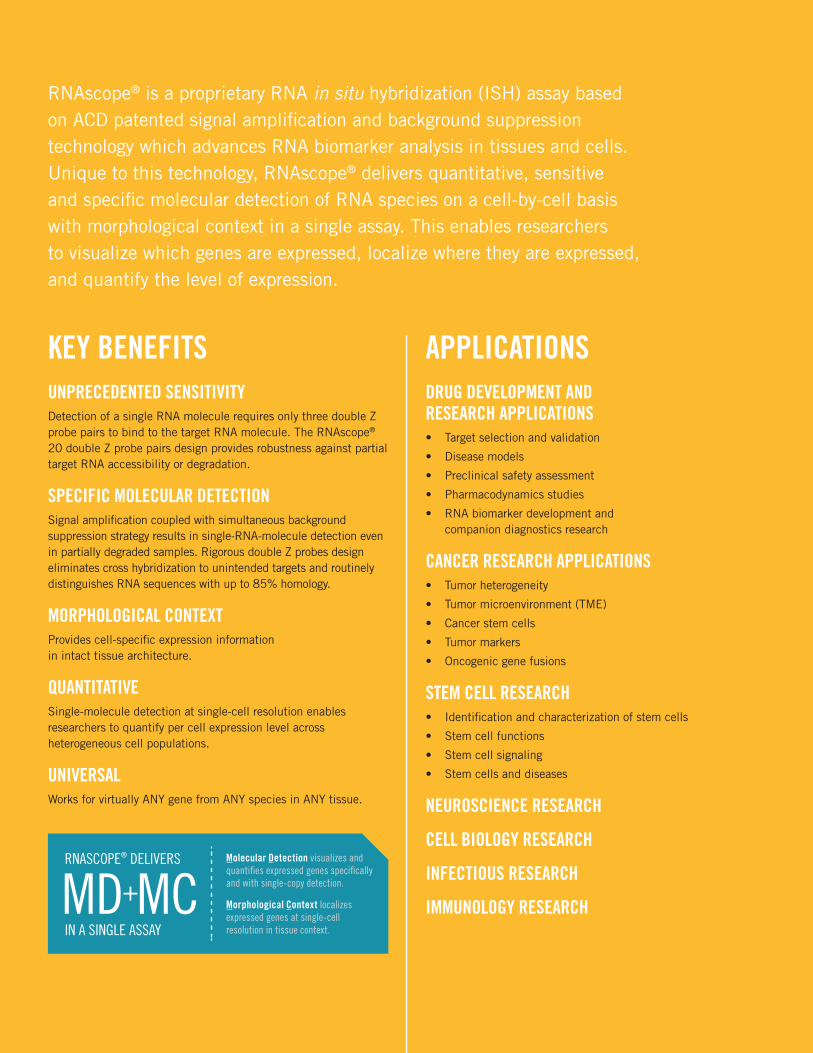
RNAscope® is a proprietary RNA in situ hybridization (ISH) assay based on ACD patented signal amplification and background suppression technology which advances RNA biomarker analysis in tissues and cells. Unique to this technology, RNAscope® delivers quantitative, sensitive and specific molecular detection of RNA species on a cell-by-cell basis with morphological context in a single assay. This enables researchers to visualize which genes are expressed, localize where they are expressed, and quantify the level of expression.
KEY BENEFITSUNPRECEDENTED SENSITIVITYDetection of a single RNA molecule requires only three double Z probe pairs to bind to the target RNA molecule. The RNAscope® 20 double Z probe pairs design provides robustness against partial target RNA accessibility or degradation.
SPECIFIC MOLECULAR DETECTIONSignal amplification coupled with simultaneous background suppression strategy results in single-RNA-molecule detection even in partially degraded samples. Rigorous double Z probes design eliminates cross hybridization to unintended targets and routinely distinguishes RNA sequences with up to 85% homology.
MORPHOLOGICAL CONTEXTProvides cell-specific expression information in intact tissue architecture.
QUANTITATIVESingle-molecule detection at single-cell resolution enables researchers to quantify per cell expression level across heterogeneous cell populations.
UNIVERSALWorks for virtually ANY gene from ANY species in ANY tissue.
APPLICATIONSDRUG DEVELOPMENT AND RESEARCH APPLICATIONS • Target selection and validation
• Disease models
• Preclinical safety assessment
• Pharmacodynamics studies
• RNA biomarker development and companion diagnostics research
CANCER RESEARCH APPLICATIONS• Tumor heterogeneity
• Tumor microenvironment (TME)
• Cancer stem cells
• Tumor markers
• Oncogenic gene fusions
STEM CELL RESEARCH • Identification and characterization of stem cells
• Stem cell functions
• Stem cell signaling
• Stem cells and diseases
NEUROSCIENCE RESEARCH
CELL BIOLOGY RESEARCH
INFECTIOUS RESEARCH
IMMUNOLOGY RESEARCHMorphological Context localizes expressed genes at single-cell resolution in tissue context.
Molecular Detection visualizes and quantifies expressed genes specifically and with single-copy detection.
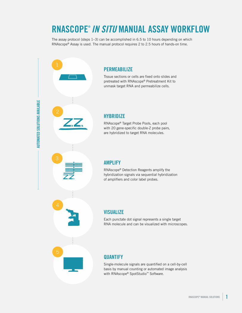
RNASCOPE® IN SITU MANUAL ASSAY WORKFLOW
2
4
5
1
3
PERMEABILIZETissue sections or cells are fixed onto slides and pretreated with RNAscope® Pretreatment Kit to unmask target RNA and permeabilize cells.
HYBRIDIZERNAscope® Target Probe Pools, each pool with 20 gene-specific double-Z probe pairs, are hybridized to target RNA molecules.
AMPLIFYRNAscope® Detection Reagents amplify the hybridization signals via sequential hybridization of amplifiers and color label probes.
VISUALIZEEach punctate dot signal represents a single target RNA molecule and can be visualized with microscopes.
QUANTIFYSingle-molecule signals are quantified on a cell-by-cell basis by manual counting or automated image analysis with RNAscope® SpotStudio™ Software.
The assay protocol (steps 1–3) can be accomplished in 6.5 to 10 hours depending on which RNAscope® Assay is used. The manual protocol requires 2 to 2.5 hours of hands-on time.
RNASCOPE® MANUAL SOLUTIONS | 1
AUTO
MAT
ED S
OLUT
IONS
AVA
ILAB
LE
PERMEABLIZE
SAMPLE PREPARATION AND PRETREATMENTIn order to perform the RNAscope® Assay, start with properly prepared and pretreated standard samples. Multiple sample types are compatible with RNAscope® Assays and include: formalin-fixed, paraffin-embedded (FFPE) (including archival) tissue, fresh-frozen (FF) tissue, fixed-frozen tissue, tissue microarray (TMA), and cell preparations. Sample preparation and pretreatment include the following steps:
• Fixation of cells if needed (fresh-frozen, cultured cells, PBMCs, etc.)
• Deparaffinization if needed (FFPE)
• Applying pretreatment reagents included in the RNAcope® Reagent Kit
Visit www.acdbio.com or contact [email protected] for more information.
HYBRIDIZE
HYBRIDIZATION OF DOUBLE Z TARGET PROBE PAIRS TO THE TARGET MOLECULE
RNASCOPE® TARGET PROBESUsing the proprietary ACD RNAscope® Probe Design pipeline, we design double-Z oligo probe pools that hybridize to your specific RNA target of interest. We can design probe pools for virtually ANY gene in ANY genome for interrogation in ANY tissue.
The probe pools consist of proprietary oligonucleotides designed for detecting specific targets. Every target probe pool also contains a tag that enables the associated target to be visualized in a specific “color channel” under the microscope.
Select from our growing catalog of over 4,000 in situ hybridization target probe pools for coding RNA and long noncoding RNA (lncRNA). Our RNA ISH probe pools span a variety of species including human, mouse, rat, dog, cow, zebrafish, rabbit, pig, chicken, monkeys, HPV, HIV, HCV, and many others.
See if your gene of interest has been designed at www.acdbio.com/probesearch
RNASCOPE® CUSTOM PROBES If ACD catalog probes are not available for your gene of interest, we can create new probes within two weeks using public or proprietary sequences. ACD probe design pipeline can also accommodate non-standard designs such as probe pools for detection of fusion genes, detection of biomarkers in xenografts, or any other non-standard application in any species. Standard and non-standard RNA ISH probe pools can be designed for use with any of our RNAscope® Reagent Kits, including singleplex, duplex, multiplex, manual, or automated assay configurations.
Interested in custom probes? Tell us your gene of interest and let’s get started: www.acdbio.com/customprobes
2 | ADVANCED CELL DIAGNOSTICS
“RNAscope has streamlined in situ RNA hybridization to the point where it can become a standard technique easily implemented in most labs, even by individuals with no experience with this often difficult technique. Moreover the ability to multiplex ISH to analyze simultaneously mRNA levels of up to three different genes allowed the study of changes in gene expression in specific cell populations in the CNS in response to different experimental conditions. Also this technique has been useful to analyze mRNA sub-cellular localization, which may prove important to understand disease mechanisms in neurodegenerative diseases.”
Miguel Sena-Esteves, PhD, Associate Professor, University of Massachusetts Medical School
RNASCOPE® CONTROL PROBES RNAscope® Control Probes: In addition to target probes, we also provide species-specific housekeeping gene positive control probes and DapB negative control probes. They are designed to work with RNAscope® Reagent Kits and the positive control probes span from high to very low levels of expression, providing appropriate experimental controls for RNA in situ hybridization and ensuring high confidence when working with varying or unknown levels of gene expression.
See our list of species-specific control probes at www.acdbio.com/controlprobes
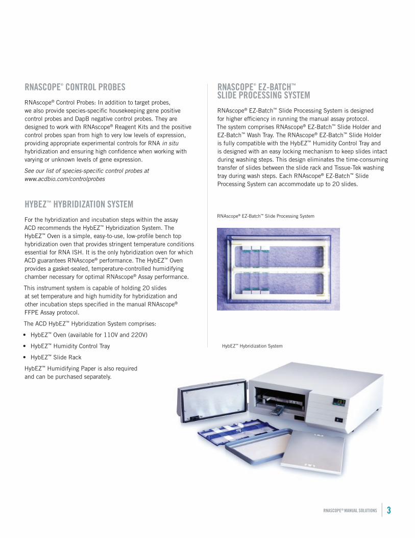
HYBEZ™ HYBRIDIZATION SYSTEMFor the hybridization and incubation steps within the assay ACD recommends the HybEZ™ Hybridization System. The HybEZ™ Oven is a simple, easy-to-use, low-profile bench top hybridization oven that provides stringent temperature conditions essential for RNA ISH. It is the only hybridization oven for which ACD guarantees RNAscope® performance. The HybEZ™ Oven provides a gasket-sealed, temperature-controlled humidifying chamber necessary for optimal RNAscope® Assay performance.
This instrument system is capable of holding 20 slides at set temperature and high humidity for hybridization and other incubation steps specified in the manual RNAscope® FFPE Assay protocol.
The ACD HybEZ™ Hybridization System comprises:
• HybEZ™ Oven (available for 110V and 220V)
• HybEZ™ Humidity Control Tray
• HybEZ™ Slide Rack
HybEZ™ Humidifying Paper is also required and can be purchased separately.
RNASCOPE® EZ-BATCH™ SLIDE PROCESSING SYSTEM
RNAscope® EZ-Batch™ Slide Processing System is designed for higher efficiency in running the manual assay protocol. The system comprises RNAscope® EZ-Batch™ Slide Holder and EZ-Batch™ Wash Tray. The RNAscope® EZ-Batch™ Slide Holder is fully compatible with the HybEZ™ Humidity Control Tray and is designed with an easy locking mechanism to keep slides intact during washing steps. This design eliminates the time-consuming transfer of slides between the slide rack and Tissue-Tek washing tray during wash steps. Each RNAscope® EZ-Batch™ Slide Processing System can accommodate up to 20 slides.
HybEZ™ Hybridization System
RNAscope® EZ-Batch™ Slide Processing System
RNASCOPE® MANUAL SOLUTIONS | 3
AMPLIFY
AMPLIFY HYBRIDIZATION SIGNALS VIA SEQUENTIAL HYBRIDIZATION OF AMPLIFIERS AND LABEL PROBES
RNASCOPE® DETECTION REAGENTS ACD RNAscope® Chromogenic and Multiplex Fluorescent Assays are robust, highly sensitive, and specific. They have been utilized successfully in over 100 peer-reviewed publications in the first 36 months. The following products are available for use with any target probes:
• RNAscope® 2.0 HD Reagent Kit-BROWN
• RNAscope® 2.0 HD Reagent Kit-RED
• RNAscope® 2-plex Chromogenic Reagent Kit
• RNAscope® Multiplex Fluorescent Assay
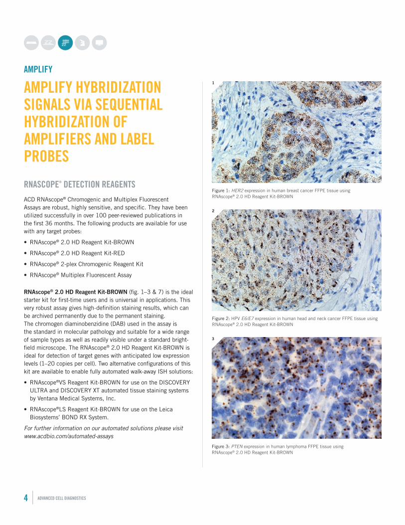
RNAscope® 2.0 HD Reagent Kit-BROWN (fig. 1–3 & 7) is the ideal starter kit for first-time users and is universal in applications. This very robust assay gives high-definition staining results, which can be archived permanently due to the permanent staining. The chromogen diaminobenzidine (DAB) used in the assay is the standard in molecular pathology and suitable for a wide range of sample types as well as readily visible under a standard bright-field microscope. The RNAscope® 2.0 HD Reagent Kit-BROWN is ideal for detection of target genes with anticipated low expression levels (1–20 copies per cell). Two alternative configurations of this kit are available to enable fully automated walk-away ISH solutions:
• RNAscope®VS Reagent Kit-BROWN for use on the DISCOVERY ULTRA and DISCOVERY XT automated tissue staining systems by Ventana Medical Systems, Inc.
• RNAscope®LS Reagent Kit-BROWN for use on the Leica Biosystems’ BOND RX System.
For further information on our automated solutions please visit www.acdbio.com/automated-assays
Figure 1: HER2 expression in human breast cancer FFPE tissue using RNAscope® 2.0 HD Reagent Kit-BROWN
1
4 | ADVANCED CELL DIAGNOSTICS
Figure 2: HPV E6/E7 expression in human head and neck cancer FFPE tissue using RNAscope® 2.0 HD Reagent Kit-BROWN
2
Figure 3: PTEN expression in human lymphoma FFPE tissue using RNAscope® 2.0 HD Reagent Kit-BROWN
3
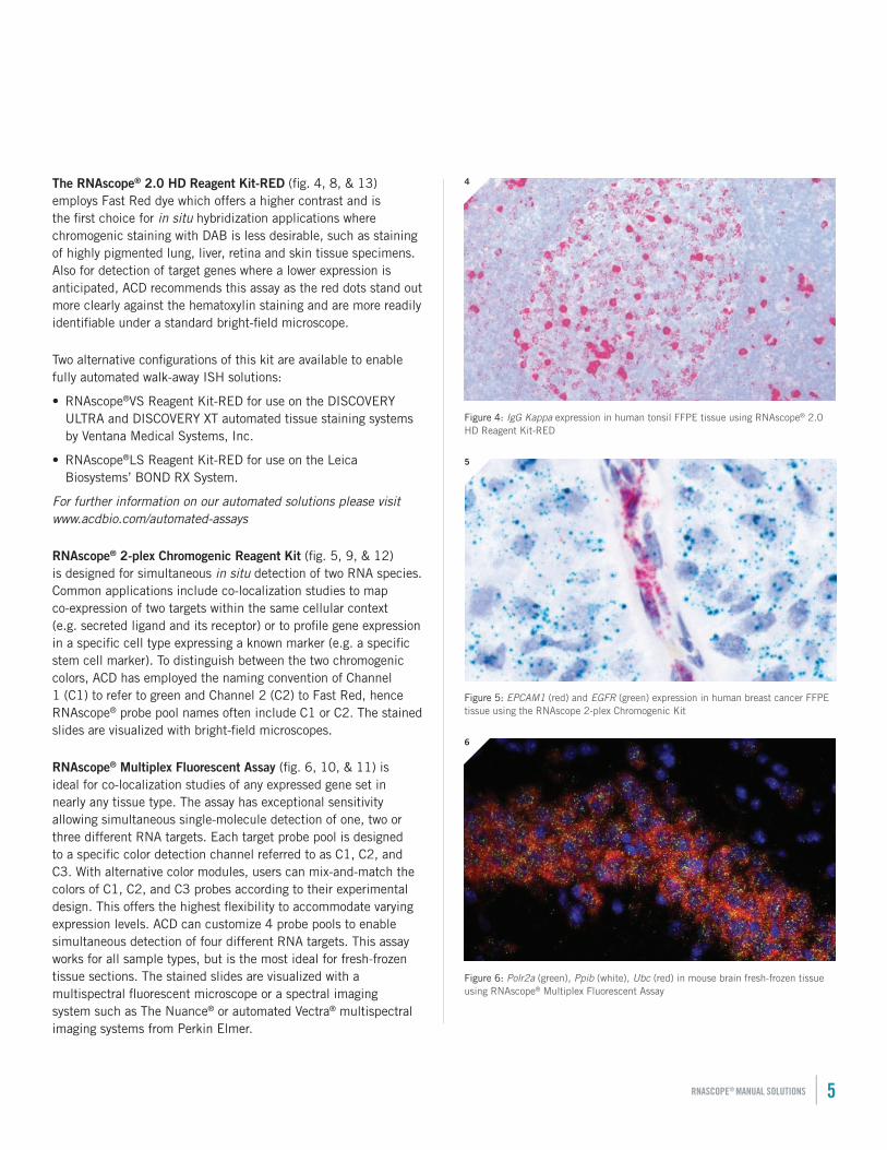
The RNAscope® 2.0 HD Reagent Kit-RED (fig. 4, 8, & 13) employs Fast Red dye which offers a higher contrast and is the first choice for in situ hybridization applications where chromogenic staining with DAB is less desirable, such as staining of highly pigmented lung, liver, retina and skin tissue specimens. Also for detection of target genes where a lower expression is anticipated, ACD recommends this assay as the red dots stand out more clearly against the hematoxylin staining and are more readily identifiable under a standard bright-field microscope.
Two alternative configurations of this kit are available to enable fully automated walk-away ISH solutions:
• RNAscope®VS Reagent Kit-RED for use on the DISCOVERY ULTRA and DISCOVERY XT automated tissue staining systems by Ventana Medical Systems, Inc.
• RNAscope®LS Reagent Kit-RED for use on the Leica Biosystems’ BOND RX System.
For further information on our automated solutions please visit www.acdbio.com/automated-assays
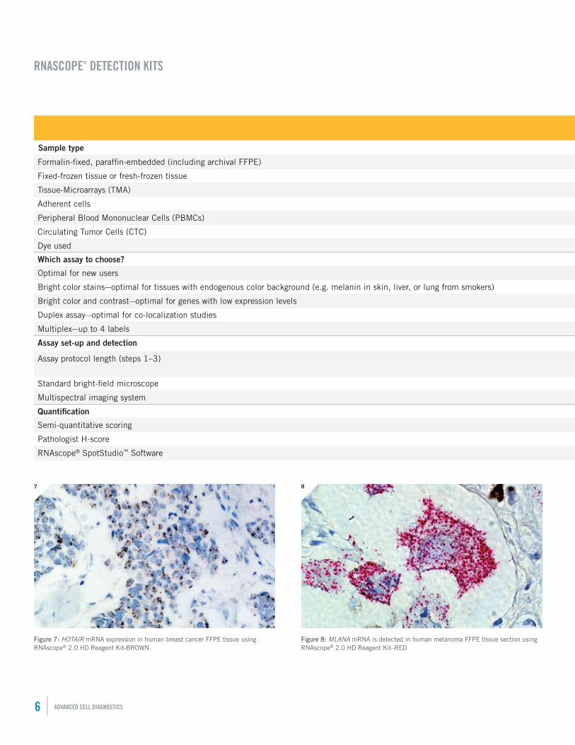
RNAscope® 2-plex Chromogenic Reagent Kit (fig. 5, 9, & 12) is designed for simultaneous in situ detection of two RNA species. Common applications include co-localization studies to map co-expression of two targets within the same cellular context (e.g. secreted ligand and its receptor) or to profile gene expression in a specific cell type expressing a known marker (e.g. a specific stem cell marker). To distinguish between the two chromogenic colors, ACD has employed the naming convention of Channel 1 (C1) to refer to green and Channel 2 (C2) to Fast Red, hence RNAscope® probe pool names often include C1 or C2. The stained slides are visualized with bright-field microscopes.
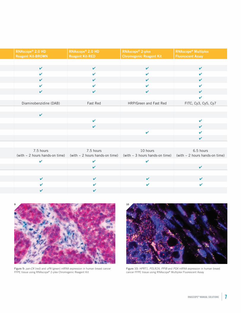
RNAscope® Multiplex Fluorescent Assay (fig. 6, 10, & 11) is ideal for co-localization studies of any expressed gene set in nearly any tissue type. The assay has exceptional sensitivity allowing simultaneous single-molecule detection of one, two or three different RNA targets. Each target probe pool is designed to a specific color detection channel referred to as C1, C2, and C3. With alternative color modules, users can mix-and-match the colors of C1, C2, and C3 probes according to their experimental design. This offers the highest flexibility to accommodate varying expression levels. ACD can customize 4 probe pools to enable simultaneous detection of four different RNA targets. This assay works for all sample types, but is the most ideal for fresh-frozen tissue sections. The stained slides are visualized with a multispectral fluorescent microscope or a spectral imaging system such as The Nuance® or automated Vectra® multispectral imaging systems from Perkin Elmer.
Figure 5: EPCAM1 (red) and EGFR (green) expression in human breast cancer FFPE tissue using the RNAscope 2-plex Chromogenic Kit
5
Figure 6: Polr2a (green), Ppib (white), Ubc (red) in mouse brain fresh-frozen tissue using RNAscope® Multiplex Fluorescent Assay
6
RNASCOPE® MANUAL SOLUTIONS | 5
Figure 4: IgG Kappa expression in human tonsil FFPE tissue using RNAscope® 2.0 HD Reagent Kit-RED
4
RNAscope® 2.0 HD Reagent Kit-BROWN
RNAscope® 2.0 HD Reagent Kit-RED
RNAscope® 2-plex Chromogenic Reagent Kit
RNAscope® Multiplex Fluorescent Assay
Sample type
Formalin-fixed, paraffin-embedded (including archival FFPE) ✔ ✔ ✔ ✔
Fixed-frozen tissue or fresh-frozen tissue ✔ ✔ ✔ ✔
Tissue-Microarrays (TMA) ✔ ✔ ✔ ✔
Adherent cells ✔ ✔ ✔ ✔
Peripheral Blood Mononuclear Cells (PBMCs) ✔ ✔ ✔ ✔
Circulating Tumor Cells (CTC) ✔
Dye used Diaminobenzidine (DAB) Fast Red HRP/Green and Fast Red FITC, Cy3, Cy5, Cy7
Which assay to choose?
Optimal for new users ✔
Bright color stains—optimal for tissues with endogenous color background (e.g. melanin in skin, liver, or lung from smokers) ✔ ✔
Bright color and contrast—optimal for genes with low expression levels ✔ ✔
Duplex assay—optimal for co-localization studies ✔ ✔
Multiplex—up to 4 labels ✔
Assay set-up and detection
Assay protocol length (steps 1–3) 7.5 hours (with ~ 2 hours hands-on time)
7.5 hours (with ~ 2 hours hands-on time)
10 hours (with ~ 3 hours hands-on time)
6.5 hours (with ~ 2 hours hands-on time)
Standard bright-field microscope ✔ ✔ ✔
Multispectral imaging system ✔ ✔
Quantification
Semi-quantitative scoring ✔ ✔ ✔ ✔
Pathologist H-score ✔ ✔ ✔ ✔
RNAscope® SpotStudio™ Software ✔ ✔
Figure 8: MLANA mRNA is detected in human melanoma FFPE tissue section using RNAscope® 2.0 HD Reagent Kit–RED
8
Figure 7: HOTAIR mRNA expression in human breast cancer FFPE tissue using RNAscope® 2.0 HD Reagent Kit-BROWN
7
6 | ADVANCED CELL DIAGNOSTICS
RNASCOPE® DETECTION KITS
RNAscope® 2.0 HD Reagent Kit-BROWN
RNAscope® 2.0 HD Reagent Kit-RED
RNAscope® 2-plex Chromogenic Reagent Kit
RNAscope® Multiplex Fluorescent Assay
Sample type
Formalin-fixed, paraffin-embedded (including archival FFPE) ✔ ✔ ✔ ✔
Fixed-frozen tissue or fresh-frozen tissue ✔ ✔ ✔ ✔
Tissue-Microarrays (TMA) ✔ ✔ ✔ ✔
Adherent cells ✔ ✔ ✔ ✔
Peripheral Blood Mononuclear Cells (PBMCs) ✔ ✔ ✔ ✔
Circulating Tumor Cells (CTC) ✔
Dye used Diaminobenzidine (DAB) Fast Red HRP/Green and Fast Red FITC, Cy3, Cy5, Cy7
Which assay to choose?
Optimal for new users ✔
Bright color stains—optimal for tissues with endogenous color background (e.g. melanin in skin, liver, or lung from smokers) ✔ ✔
Bright color and contrast—optimal for genes with low expression levels ✔ ✔
Duplex assay—optimal for co-localization studies ✔ ✔
Multiplex—up to 4 labels ✔
Assay set-up and detection
Assay protocol length (steps 1–3) 7.5 hours (with ~ 2 hours hands-on time)
7.5 hours (with ~ 2 hours hands-on time)
10 hours (with ~ 3 hours hands-on time)
6.5 hours (with ~ 2 hours hands-on time)
Standard bright-field microscope ✔ ✔ ✔
Multispectral imaging system ✔ ✔
Quantification
Semi-quantitative scoring ✔ ✔ ✔ ✔
Pathologist H-score ✔ ✔ ✔ ✔
RNAscope® SpotStudio™ Software ✔ ✔
Figure 9: pan-CK (red) and uPA (green) mRNA expression in human breast cancer FFPE tissue using RNAscope® 2-plex Chromogenic Reagent Kit
9
Figure 10: HPRT1, POLR2A, PPIB and PGK mRNA expression in human breast cancer FFPE tissue using RNAscope® Multiplex Fluorescent Assay
10
RNASCOPE® MANUAL SOLUTIONS | 7
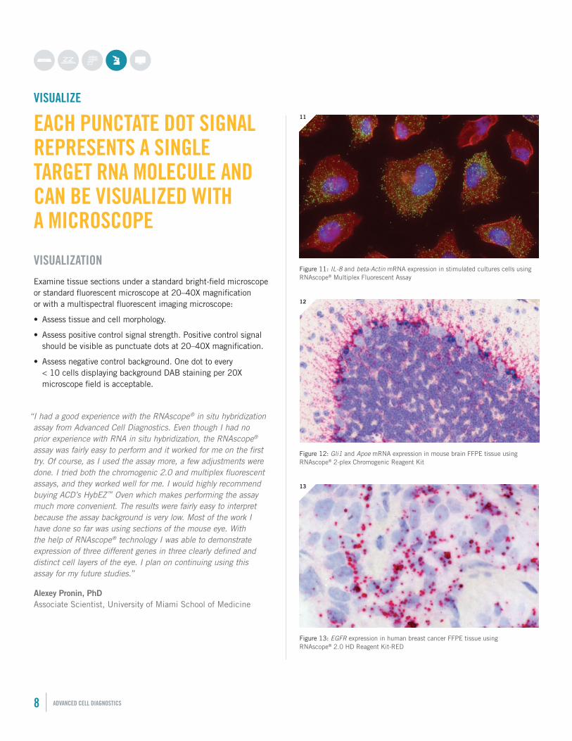
Figure 12: Gli1 and Apoe mRNA expression in mouse brain FFPE tissue using RNAscope® 2-plex Chromogenic Reagent Kit
12
Figure 13: EGFR expression in human breast cancer FFPE tissue using RNAscope® 2.0 HD Reagent Kit-RED
13
Figure 11: IL-8 and beta-Actin mRNA expression in stimulated cultures cells using RNAscope® Multiplex Fluorescent Assay
11
8 | ADVANCED CELL DIAGNOSTICS
VISUALIZE
EACH PUNCTATE DOT SIGNAL REPRESENTS A SINGLE TARGET RNA MOLECULE AND CAN BE VISUALIZED WITH A MICROSCOPE
VISUALIZATIONExamine tissue sections under a standard bright-field microscope or standard fluorescent microscope at 20–40X magnification or with a multispectral fluorescent imaging microscope:
• Assess tissue and cell morphology.
• Assess positive control signal strength. Positive control signal should be visible as punctuate dots at 20–40X magnification.
• Assess negative control background. One dot to every < 10 cells displaying background DAB staining per 20X microscope field is acceptable.
“I had a good experience with the RNAscope® in situ hybridization assay from Advanced Cell Diagnostics. Even though I had no prior experience with RNA in situ hybridization, the RNAscope® assay was fairly easy to perform and it worked for me on the first try. Of course, as I used the assay more, a few adjustments were done. I tried both the chromogenic 2.0 and multiplex fluorescent assays, and they worked well for me. I would highly recommend buying ACD’s HybEZ™ Oven which makes performing the assay much more convenient. The results were fairly easy to interpret because the assay background is very low. Most of the work I have done so far was using sections of the mouse eye. With the help of RNAscope® technology I was able to demonstrate expression of three different genes in three clearly defined and distinct cell layers of the eye. I plan on continuing using this assay for my future studies.”
Alexey Pronin, PhD Associate Scientist, University of Miami School of Medicine
QUANTIFY
QUANTIFY MANUALLY OR AUTOMATICALLY
QUANTIFICATIONThe single-molecule sensitivity and visualization of RNAscope® Assay Technology makes quantitative RNA in situ hybridization analysis a reality. You can evaluate target probe signals manually using the scoring guidelines we provide with our kits. To facilitate and improve quantitative scoring, Advanced Cell Diagnostics offers RNAscope® SpotStudio™ Software for RNAscope® brown and red assays.
This advanced image analysis solution brings objective and accurate quantification to RNA in situ hybridization. For the first time, gene expression can be measured quantitatively and interpreted by research pathologists within histopathological context.
RNAscope® SpotStudio™ Software is designed for research pathologists with no prior training in image analysis software. It is an intuitive automated solution that generates standardized and objective results in minutes. Results obtained with RNAscope® SpotStudio™ are comparable to manual annotations by experienced research pathologists. The software is compatible with bright-field image data captured using whole slide scanners and microscopes.
Users simply load an image taken from a whole slide scanner or microscopic camera, set the analysis parameters: cell identification—hematoxylin levels and nucleus diameter; spot identification—staining type (DAB or Fast Red), spot staining level and spot diameter; then draw a region of interest (ROI) (fig. 14A), and run the analysis (fig. 14B). Graphical results show each cell identified and outlined in the color that corresponds to the number of dots per cell. Additionally spreadsheet results are available; listing each cell individually identified and with the estimated dots/cell value (fig. 14C) enabling further statistical analysis.
C
B
A
RNASCOPE® MANUAL SOLUTIONS | 9
“RNAscope® SpotStudio™ Software is very easy to use. I especially like the ability to interact with the image in the result section—to connect the quantitative result to the image of the cell in question is invaluable!”
Olga Shebanova, Research Investigator, Novartis
Figure 14: RNAscope® SpotStudio™ Software: The brown punctate dots are expression of HOTAIR gene seen exclusively in cancer cells but not stroma cells of FFPE human breast cancer tissue using RNAscope® 2.0 HD Reagent Kit—BROWN. A: Region of interest selected for analysis. B. Graphical results of each cell identified C. Spreadsheet results showing number of dots per single cell.
Advanced Cell Diagnostics
3960 Point Eden Way, Hayward, CA 94545
1-510-576-8800 (Main) 1-877-576-3636 (Toll Free)
ORDERING INFORMATION
This document is provided for informational use only. Molecular Biology Applications (MBA), not intended for diagnosis. Refer to appropriate regulations. VENTANA, Discovery Ultra and Discovery XT are registered trademarks of Roche. LEICA and Leica BOND are registered trademarks of Leica Microsystems IR GmbH. RNAscope, HybEZ, EZ-Batch and SpotSudio are registered trademarks of Advanced Cell Diagnostics, Inc. in the United States or other countries. All rights reserved. ©2014 Advanced Cell Diagnostics, Inc.Doc#: 321245/081414/revA
1. Each RNAscope® Reagent Kit contains three sub-kits: a Pretreatment Kit, a Detection Kit, and Wash Buffer for use with 20 slides.
2. Each RNAscope® Reagent Kit contains three sub-kits: a Pretreatment Kit, a Detection Kit, and Wash Buffer for use with 60 slides.
3. Products needed from Ventana Medical Systems:
• 06634443001 (760‐225): mRNA Amplification, Pretreatment & DAB Kit—This kit contains prefilled mRNA DAB Detection reagents and the user fillable empty dispensers for all ACD reagents, except probes. (In some geographies customers may order DISCOVERY mRNA Pretreat Kit (06614345001), mRNA Probe Amplification Kit (06614337001), mRNA DAB Detection Kit (06614353001) as separately.)
• 07017944001 (760‐4840): Discovery Inhibitor—This product is required when using mRNA DAB and mRNA Red software version 3.0 and higher, all customers new to running mRNA should order this product.
• 05279828001, 05279836001, 05279844001 (960‐761 thru 960‐780, please reference www.ventana.com for a complete list of corresponding ordering code information): User fillable probe dispensers (you will need 1 for the negative control probe, 1 for the positive control probe and 1 for each of your target probes)
• 05266262001 (760‐105): Ribowash buffer.This reagent is recommended for all assays on DISCOVERY instruments, but is REQUIRED in the mRNA procedure
• 06883354001 (950‐210): 1xSSC Wash Buffer (to be diluted 1:10 in the “option” reagent container)
• Software to run the RNAscopeVS Assay—Accessible via the local Roche technical support specialist
• If you use your own hematoxylin and bluing reagents or the ones included in the ACD kit as complimentary, you will also need to order Counterstain 1 (771‐741) and Counterstain 2 (771‐742) user fillable dispensers from Ventana.
• If you have used the Ventana hematoxylin and blueing reagent for other applications, you can continue using those for the RNAscopeVS assay.
4. Products needed from Leica Biosystems:
• Cat No. Op309700 BOND Open Containers 30 mL
• Cat No. S21.2001 BOND Universal Convertiles 100 pack
• Cat No. AR9961 & AR9640 BOND Epitope Retrieval Solution 1 & 2 1L (RTU) each
• Cat No. S21.1971 BOND Mixing Stations 6 vials
• Cat No. AR9222 BOND Dewax Solution—1L (RTU)
• Cat No. AR9590 BOND Wash Solution 10X Concentrate
• Cat No. DS9800 BOND Polymer Refine Detection and Hematoxylin—1 kit
• Cat No. CS9100 BOND Aspirating Probe Cleaning System 1 system, 15 cleans
• Cat No. S21.1971 BOND Mixing Stations 6 vials
CAT # Product Name & Descriptions300055 RNAscope® Introductory Package—human
- Human-specific positive control (Hs-PPIB) and DapB negative control probes - FFPE Control Slide Pack—Human Hela Cell Pellet - RNAscope® 2.0 HD Reagent Kit-BROWN1 - ImmEdge™ Hydrophobic Barrier Pen
Target probes are not included.
300056 RNAscope® Introductory Package—mouse- Mouse-specific positive control (Mm-PPIB) and DapB negative control probes - FFPE Control Slide Pack—Mouse 3T3 Cell Pellet - RNAscope® 2.0 HD Reagent Kit-BROWN1 - ImmEdge™ Hydrophobic Barrier Pen
Target probes are not included.
310036 HybEZ™ Hybridization System (110V)
320700 HybEZ™ Hybridization System (220V)
310014 HybEZ™ Humidifying paper
310007 RNAscope® EZ-Batch™ Slide Processing System
320771 RNAscope® 2.0 HD Reagent Kit-BROWN1 Workflow requires: RNAscope® Target Probes, RNAscope® Control Probes, RNAscope® Control slides (optional)
320751 RNAscope® 2.0 HD Reagent Kit-RED1 Workflow requires: RNAscope® Target Probes, RNAscope® Control Probes, RNAscope® Control slides (optional)
310035 RNAscope® 2-plex Reagent Kit1 Workflow requires: RNAscope® 2-plex Target Probes, RNAscope® 2-plex Control Probes, RNAscope® Control slides (optional)
320850 RNAscope® Multiplex Fluorescent Reagent Kit1 Workflow requires: RNAscope® Target Probes (C1/C2/C3), RNAscope® Control Probes(C1/C2/C3) RNAscope® Control slides (optional)
320600 RNAscope®VS Reagent Kit-BROWN2 Workflow requires: RNAscope® VS Target Probes, RNAscope® VS Control Probes, RNAscope® Control slides (optional) and products from Ventana Medical Systems3
320610 RNAscope®VS Reagent Kit-RED2 Workflow requires: RNAscope® VS Target Probes, RNAscope® VS Control Probes, RNAscope® Control slides (optional) and products from Ventana Medical Systems3
321100 RNAscope®LS Reagent Kit-BROWN2 Workflow requires: RNAscope® LS Target Probes, RNAscope® LS Control Probes, RNAscope® Control slides (optional) products from Leica Biosystems4
300063 RNAscope® SpotStudio™ v1.0 Software (Trial Pack for analysis of 15 Regions of Interest)
300062 RNAscope® SpotStudio™ v1.0 Software (150 Regions of Interest)
300060 RNAscope® SpotStudio™ v1.0 Software (3000 Regions of Interest)
Get started today at www.acdbio.com/products