R.KAVITHA, M.PHARM, LECTURER, DEPARTMENT OF PHARMACEUTICS ... · LECTURER, DEPARTMENT OF...
28
R.KAVITHA, M.PHARM, LECTURER, DEPARTMENT OF PHARMACEUTICS, SRM COLLEGE OF PHARMACY, SRM UNIVERSITY, KATTANKULATHUR.
-
Upload
trinhkhanh -
Category
Documents
-
view
238 -
download
2
Transcript of R.KAVITHA, M.PHARM, LECTURER, DEPARTMENT OF PHARMACEUTICS ... · LECTURER, DEPARTMENT OF...

R.KAVITHA, M.PHARM,LECTURER, DEPARTMENT OF PHARMACEUTICS,SRM COLLEGE OF PHARMACY,SRM UNIVERSITY, KATTANKULATHUR.

MICROBIOLOGICAL ASSAY OF ANIBIOTICS The inhibition of growth under standardized conditions maybe utilized for demonstrating the therapeutic efficacy ofantibiotics.
Any subtle change in the antibiotic molecule which may notbe detected by chemical methods will be revealed by achange in the antimicrobial activity and hence microbiologicalassays are very useful for resolving doubts regardingpossible change in potency of antibiotics and theirpreparations.
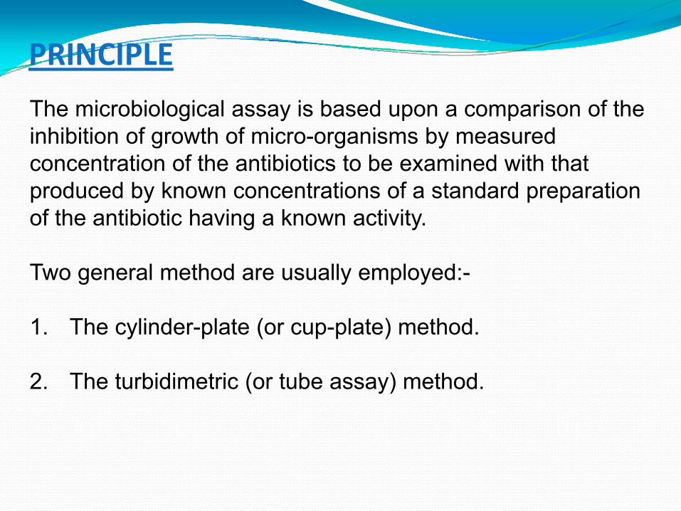
PRINCIPLE
The microbiological assay is based upon a comparison of the inhibition of growth of micro-organisms by measured concentration of the antibiotics to be examined with that produced by known concentrations of a standard preparation of the antibiotic having a known activity.
Two general method are usually employed:-
1. The cylinder-plate (or cup-plate) method.
2. The turbidimetric (or tube assay) method.

PREPARATION OF MEDIA:
The Media required for the preparation of test organism are made from the ingredients.
Minor modifications of the individual ingredients may be made, or reconstituted dehydrated media may be used provided the resulting media have equal or better growth-promoting properties and give a similar standard curve response.
Dissolve the ingredients in sufficient water to produce 1000 ml and add sufficient 1M Sodium hydroxide or 1M Hydrochloride acid, as required so that after sterilization the PH is b\w 6.5 to 7.5.

PREPARATION BUFFER SOLUTIONS• Buffer solutions are prepared by dissolving thefollowing quantities of dipotassium hydrogen phosphate andpotassium dihydrogen phosphate in sufficient water toproduce 1000 ml after adjustive the pH with 8 M phosphoricacid or 10M potassium hydroxide.
Buffer No.
DipotassiumHydrogen
Phospahate, K2HPO4 (g)
PotassiumDihydrogenPhosphate,KH2PO4 (g)
pH adjusted after sterilisation to :
1 2.0 8.0 6.0 + 0.1
2 16.73 0.532 8.0 + 0.1
3 ‐ 13.61 4.5 + 0.1
4 20.0 80.00 6.0 + 0.1
5 35.0 ‐ 10.5 + 0.1*
6 13.6 4.0 7.0 + 0.2
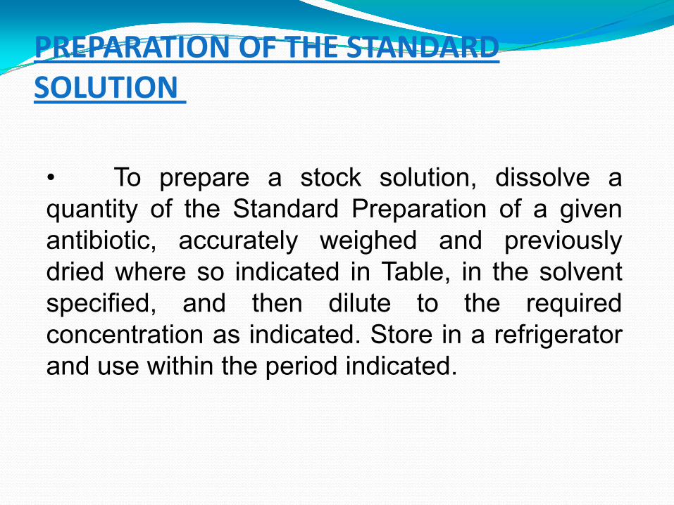
PREPARATION OF THE STANDARD SOLUTION
• To prepare a stock solution, dissolve aquantity of the Standard Preparation of a givenantibiotic, accurately weighed and previouslydried where so indicated in Table, in the solventspecified, and then dilute to the requiredconcentration as indicated. Store in a refrigeratorand use within the period indicated.
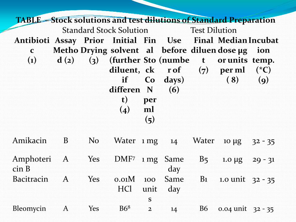
TABLE – Stock solutions and test dilutions of Standard PreparationStandard Stock Solution Test Dilution
Antibiotic(1)
AssayMethod (2)
Prior Drying
(3)
Initial solvent (furtherdiluent,
if differen
t)(4)
Final Stock CoNperml(5)
Use before(numbe
r of days) (6)
Final diluen
t(7)
Mediandose µgor unitsper ml( 8)
Incubation
temp. (*C)(9)
Amikacin B No Water 1 mg 14 Water 10 µg 32 ‐ 35
Amphotericin B
A Yes DMF7 1 mg Same day
B5 1.0 µg 29 ‐ 31
Bacitracin A Yes 0.01M HCl
100units
Same day
B1 1.0 unit 32 ‐ 35
Bleomycin A Yes B68 2 14 B6 0.04 unit 32 ‐ 35

PREPARETION TEST ORGANISMS• The test organism for each antibiotic is listed in Table,together with its identification number in the American TypeCulture Collection (ATCC) and the National Collection of TypeCultures (NCTC) or the National Collection of IndustrialBacteria (VCIB).
TABLE – Test Organisms for Microbiological Assay of AntibioticsAntibiotic Test Organism ATCC1 No. NCTC2 No.
(NCIB3 No.)Amikacin Staphylococcus
aureus29737 7447
Amphotericin B Saccharomycescerevisiae
9763 10716
Bacitracin Micrococcus luteus 10240 7743
Bleomycin Mycobacterium smegmatis
607 _
Carbenicillin Pseudomonas aeruginosa
25619 _
Doxycycline Staphylococcus aureus
29737 7447

CYLINDER‐PLATE OR CUP‐PLATE METHOD• Inoculate a previously liquified medium appropriate tothe assay, with the requsite quantity of suspension of themicro organism, add the suspension to the medium at atemperature between 40 and 50 and immediately pour theinoculated medium into the petri dishes or large rectangularplates to give a depth of 3 to 4 mm.
• Ensure that the layers of medium are uniform inthickness, by placing the dishes or plates on a level surface.
• Using the appropriate buffer solutions, preparesolutions of known concentrations of the antibiotic to beexamined
• Apply the solutions to the surface of the solid medium in sterile cylinders or in cavities prepared in the agar. The volume of solution added to each cylinder or cavity must be uniform and sufficient almost to fill the holes when these are used.
• Leave the dishes or plates standing for 1 to 4 hours at room temperature or at 4 , as appropriate, as a period of pre-incubation diffusion to minimise the effects of variation in time between the application of the different solutions. Incubate them for about 18 hours at the particular temperature.
• Accurately measure the diameters or areas of the circular inhibition zones and calculate the results.

L = 3a + 2b + c – e : H = 3e + 2d + c- a5 5
WhereL = the calculated zone diameter for the lowest concentration of the standard curve response line.
H = the calculated zone diameter for the highest concentration of the standard curve response line.
c = average zone diameter of 36 readings of the reference point standard solution.
a, b, d, e = corrected average values for the other standard solutions, lowest to highest concentrations, respectively.

TURBIDIMETRIC OR TUBE ASSAY METHOD
• The method has the advantage of a shorterincubation period for the growth of the test organism(usually 3 to 4 hours) but the presence of solvent residuesor other inhibitory substances affects this assay more thanthe cylinder plates assay.• Prepare five different concentrations of the standardsolution for preparing the standard curve by diluting thestock solution of the Standard Preparation of the antibiotic& increasing stepwise in the ration 4:5.• Select the median concentration & dilute the solutionof the substance being examined (unknown) to obtainapproximately this concentration.• Place 1 ml of each concentration of the standardsolution and of the sample solution in each of the tubes induplicate.
• To each tube add 9 ml. of nutrient medium, previouslyseeded with the appropriate test organism.
• At the same time prepare three control tubes, onecontaining the inoculated culture medium (culture control),another identical with it but treated immediately with 0.5 mlof dilute formaldehyde solution (blank) and a third containinguninoculated culture medium.
• Place all the tubes, randomly distributed or in arandomized block arrangement, in an incubator or water-bathand maintain them at the specified temperature, for 3 to 4hours. After incubation add 0.5 ml of dilute formaldehydesolution to each tube. Measure the growth of the testorganism by determining the absorbance at about 530nm ofeach of the solutions in the tubes against the blank.
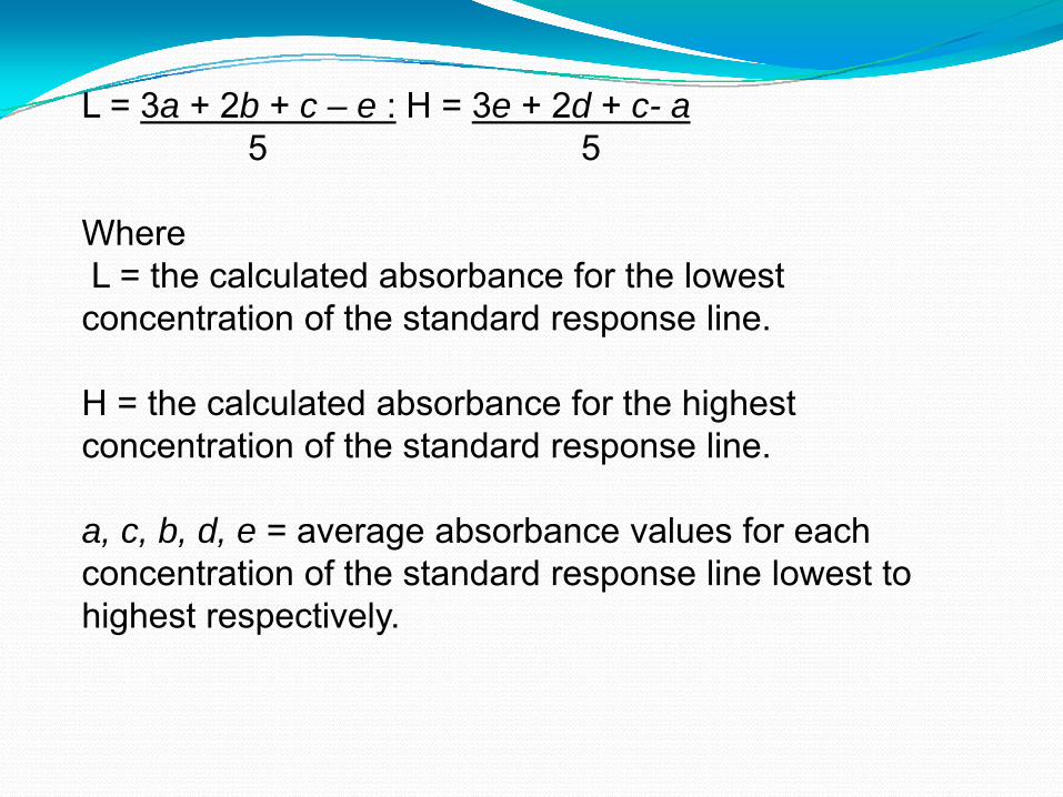
L = 3a + 2b + c – e : H = 3e + 2d + c- a5 5
WhereL = the calculated absorbance for the lowest concentration of the standard response line.
H = the calculated absorbance for the highest concentration of the standard response line.
a, c, b, d, e = average absorbance values for each concentration of the standard response line lowest to highest respectively.

DEFINITION:
Microbiological assay of vitamins is a type of biological assay performed with the aid of microorganisms.
Many therapeutic agents, which either inhibit the growth of microorganisms or are essential for the growth of them are standardized by microbial assay.
Principles of microbial assay were developed in 1920s.
MICROBIOLOGICAL ASSAY OF VITAMINS

MICROBIOLOGICAL ASSAY OF VITAMINS:PRINCIPLE:
Vitamins and amino acids are essential for the growth of microorganisms.
The basis of this assay is to measure the ability of test organism to utilize the substance being assayed under a proper nutritional condition.
The organisms require these growth factors (vitamins & amino acids) in micro or nano grams.

The response (growth of test organism) isproportional to the dose (amount of factor)added to medium.
Materials required for Microbial assay ofvitamins & amino acids:
A stock solution.A inoculum media.Assay medium.A standard curve.

MICROBIAL ASSAY OF VITAMIN B12:
About VitB12:
Also known as cyanocobalamin. Its a water solublevitamin.Structure is similar to that of heme where the iron isreplaced with cobalt as a centre of molecule.Its main sources are liver, eggs , milk, meat & fish.VitB12 deficiency causes Macrolytic anemia,pernicious anemia.
National Research Council, USA recommends adaily intake of about 5mg of vitB12

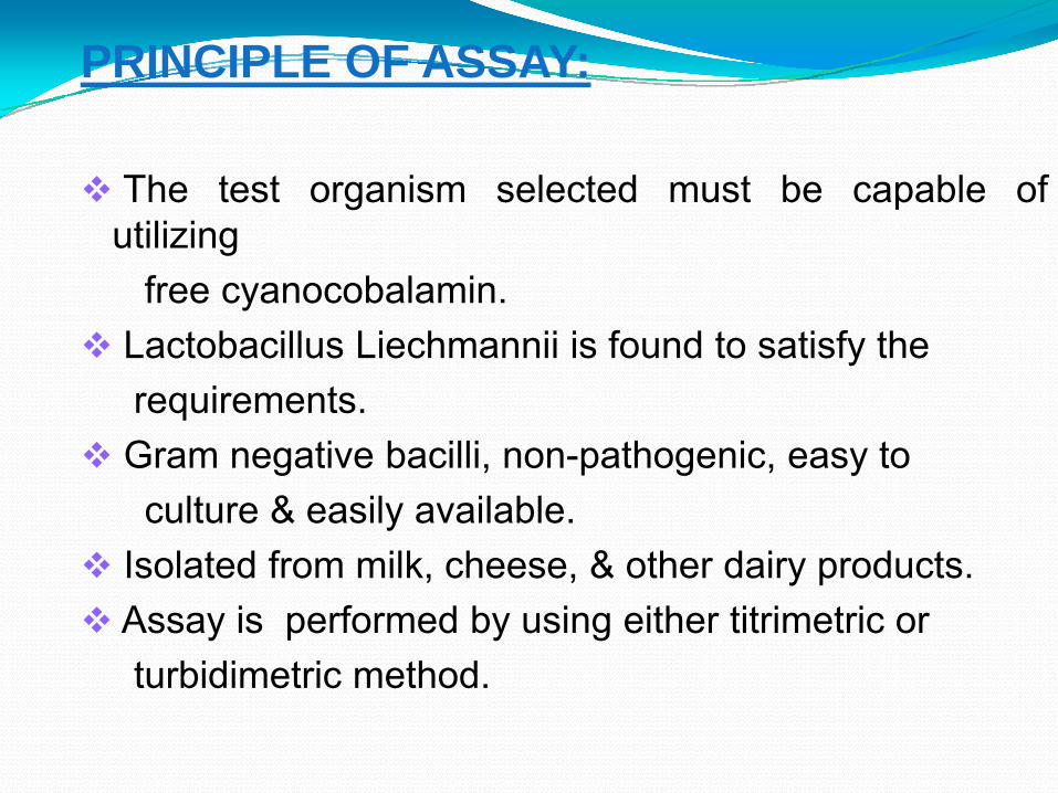
PRINCIPLE OF ASSAY:
The test organism selected must be capable ofutilizing
free cyanocobalamin.Lactobacillus Liechmannii is found to satisfy therequirements.Gram negative bacilli, non-pathogenic, easy toculture & easily available.
Isolated from milk, cheese, & other dairy products.Assay is performed by using either titrimetric orturbidimetric method.

PRECAUTIONS:
Great care must be taken to avoid contamination
All the glass wares must be free from detergents andother chemicals.
Glass wares must be heated to 2500C for atleast 1hrbefore use.
The whole experiment must be carried out underproper aseptic condition.
Reagents Required :
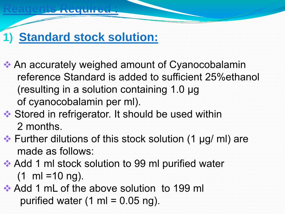
1) Standard stock solution:
An accurately weighed amount of Cyanocobalaminreference Standard is added to sufficient 25%ethanol(resulting in a solution containing 1.0 μgof cyanocobalamin per ml).Stored in refrigerator. It should be used within2 months.Further dilutions of this stock solution (1 μg/ ml) aremade as follows:
Add 1 ml stock solution to 99 ml purified water(1 ml =10 ng).
Add 1 mL of the above solution to 199 mlpurified water (1 ml = 0.05 ng).

2) Test solution to be assayed:-:
Accurate amount of Vitamin to be assayed is taken& dissolved in water, Dil HCl or NaOH is added toadjust pH at 6.0.
Make up to volume with water.
3) Preparation of Inoculum:-:
Transfer a loop full of Lactobacillus Liechmanniifrom a recent sub-culture into two tubes eachcontaining 10ml of sterile culture medium.
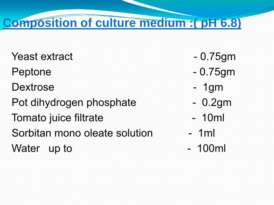
Composition of culture medium :( pH 6.8)
Yeast extract - 0.75gmPeptone - 0.75gmDextrose - 1gmPot dihydrogen phosphate - 0.2gmTomato juice filtrate - 10mlSorbitan mono oleate solution - 1mlWater up to - 100ml
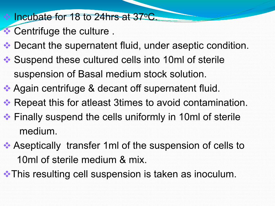
Incubate for 18 to 24hrs at 37oC.Centrifuge the culture .Decant the supernatent fluid, under aseptic condition.Suspend these cultured cells into 10ml of sterilesuspension of Basal medium stock solution.Again centrifuge & decant off supernatent fluid.Repeat this for atleast 3times to avoid contamination.Finally suspend the cells uniformly in 10ml of sterilemedium.
Aseptically transfer 1ml of the suspension of cells to10ml of sterile medium & mix.
This resulting cell suspension is taken as inoculum.
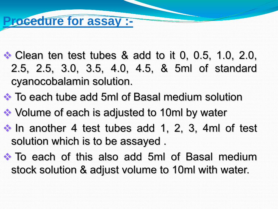
Procedure for assay :-
Clean ten test tubes & add to it 0, 0.5, 1.0, 2.0,2.5, 2.5, 3.0, 3.5, 4.0, 4.5, & 5ml of standardcyanocobalamin solution.To each tube add 5ml of Basal medium solutionVolume of each is adjusted to 10ml by waterIn another 4 test tubes add 1, 2, 3, 4ml of test
solution which is to be assayed .To each of this also add 5ml of Basal medium
stock solution & adjust volume to 10ml with water.

Sterilize all test tubes in autoclave at 121oC for15mins.Cool the test tubes at room temperature.Inoculate a drop of inoculum prepared of lactobacillusliechmannii.Incubate the test tubes for 64 to 72hrs at temperaturerange of 30 to 37oC.After incubation period titrate contents of each testtube with 0.05N NaOH using bromothymol blue asindicator until green colour.
Record all the titre readings clearly.
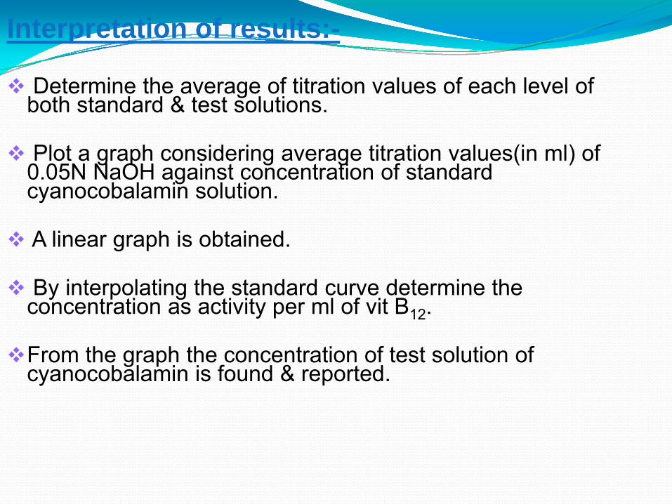
Interpretation of results:-
Determine the average of titration values of each level of both standard & test solutions.
Plot a graph considering average titration values(in ml) of 0.05N NaOH against concentration of standard cyanocobalamin solution.
A linear graph is obtained.
By interpolating the standard curve determine the concentration as activity per ml of vit B12.
From the graph the concentration of test solution of cyanocobalamin is found & reported.