Risk factors for and Strategies to Prevent Complications ...
80
ACTA UNIVERSITATIS UPSALIENSIS UPPSALA 2021 Digital Comprehensive Summaries of Uppsala Dissertations from the Faculty of Medicine 1752 Risk factors for and Strategies to Prevent Complications of Endoscopic Retrograde Cholangiopancreatography EVA-LENA SYRÉN ISSN 1651-6206 ISBN 978-91-513-1232-3 URN urn:nbn:se:uu:diva-445875
Transcript of Risk factors for and Strategies to Prevent Complications ...

ACTAUNIVERSITATIS
UPSALIENSISUPPSALA
2021
Digital Comprehensive Summaries of Uppsala Dissertationsfrom the Faculty of Medicine 1752
Risk factors for and Strategiesto Prevent Complicationsof Endoscopic RetrogradeCholangiopancreatography
EVA-LENA SYRÉN
ISSN 1651-6206ISBN 978-91-513-1232-3URN urn:nbn:se:uu:diva-445875

Dissertation presented at Uppsala University to be publicly examined in H:son-Holmdahlsalen, Ing 100, Akademiska Sjukhuset, Uppsala, Friday, 17 September 2021 at13:00 for the degree of Doctor of Philosophy (Faculty of Medicine). The examination will beconducted in Swedish. Faculty examiner: Riadh Sadik (Göteborgs Universitet).
AbstractSyrén, E.-L. 2021. Risk factors for and Strategies to Prevent Complications of EndoscopicRetrograde Cholangiopancreatography. Digital Comprehensive Summaries of UppsalaDissertations from the Faculty of Medicine 1752. 79 pp. Uppsala: Acta UniversitatisUpsaliensis. ISBN 978-91-513-1232-3.
Aim: The overall aim of this thesis was to study risk factors for and strategies to preventcomplications of Endoscopic Retrograde Cholangiopancreatography (ERCP).
Methods: Prospectively registered data from the Swedish National Quality Registerfor Gallstone Surgery and ERCP (GallRiks) 2006-2018 were retrospectively retrieved andreviewed. In Study I, ERCP procedures performed for common bile duct stones (CBDS),were analysed and cross-checked with the National Patient Register (NPR) in order to assessrisk factors for post-ERCP pancreatitis (PEP). In Study II, different techniques for CBDSclearance over time at different hospital levels and the effectiveness and safety of postoperativerendezvous ERCP compared to intraoperative rendezvous ERCP were studied. In Study III,the rate of postoperative cardiovascular events in CBDS-patients treated with ERCP only,cholecystectomy only, cholecystectomy followed by delayed ERCP, cholecystectomy togetherwith ERCP, or ERCP followed by delayed cholecystectomy were analysed. In Study IV,associations between ERCP success and complications, and endoscopist- and centre case-volumes regarding procedures for CBDS, and suspected or confirmed malignancy wereanalysed.
Results: Women, patients<65 years, patients with hyperlipidaemia, and those with a previoushistory of recent acute pancreatitis had a higher risk for PEP, while patients with diabetes had alower risk (all p<0.05). Intraoperative ERCP increased during the period of the study, whereaspreparation for postoperative ERCP decreased. CBDS management differed between differenthospital levels. Total rate of intra- and postoperative complications as well as intraoperativebleeding, postoperative bile leakage, and postoperative infection with abscess were higher inthe postoperative rendezvous ERCP group (all p<0.05). However, PEP, postoperative bleeding,cholangitis, percutaneous drainage, antibiotic treatment, ICU stay, readmission/reoperationwithin 30 days, and 30-day mortality did not differ between the groups. Nor did risk forcardiovascular complication or death within 30 days differ between patients treated for CBDSby cholecystectomy and/or ERCP. A high endoscopist case-volume was associated with highersuccessful cannulation rate and lower PEP rate (p<0.05). Centres with a high annual case-volume were associated with higher successful cannulation rates (p<0.05).
Conclusions: Age, sex, hyperlipidaemia, and previous history of recent acute pancreatitis allincreased the risk for PEP while diabetes reduced the risk. Techniques for management of CBDSdiscovered at cholecystectomy have changed over time and differ between hospitals levels.Though intraoperative rendezvous ERCP is the method of choice, postoperative rendezvousERCP is an acceptable alternative when adequate ERCP resources are lacking or limited.Primary ERCP as well as cholecystectomy for CBDS may be performed with acceptable safety.Higher endoscopist- and centre case-volumes lead to safer and more successful ERCP.
Keywords: ERCP, rendezvous ERCP, post-ERCP pancreatitis, choledocholithiasis,cardiovascular complications, case-volume
Eva-Lena Syrén, Department of Surgical Sciences, Upper Abdominal Surgery, Akademiskasjukhuset ing 70 1 tr, Uppsala University, SE-751 85 Uppsala, Sweden.
© Eva-Lena Syrén 2021
ISSN 1651-6206ISBN 978-91-513-1232-3URN urn:nbn:se:uu:diva-445875 (http://urn.kb.se/resolve?urn=urn:nbn:se:uu:diva-445875)

“To struggle and to understand. Never the last without the first. That is the law.”
George Herbert Leigh Mallory (18 June 1886 – 8 or 9 June 1924)
British teacher, explorer, and mountaineer
“ERCP is most dangerous for people who need it least”
Peter B. Cotton (born 1939),
British Gastroenterologist
To Johan


List of Papers
This thesis is based on the following papers, which are referred to in the text by their Roman numerals.
I Syrén E, Eriksson S, Enochsson L, Eklund A, Sandblom G.
Risk factors for pancreatitis following endoscopic retrograde cholangiopancreatography BJS Open. 2019 Apr 2;3(4):485-489. doi: 10.1002/bjs5.50162. eCollection 2019 Aug.BJS Open. 2019. PMID: 31406957
II Syrén E, Sandblom G, Eriksson S, Eklund A, Isaksson B, Enochsson L. Postoperative rendezvous endoscopic retrograde cholangiopancreaticography as an option in the management of choledocholithiasis. Surg Endosc. 2020 Nov;34(11):4883-4889. doi: 10.1007/s00464-019-07272-1. Epub 2019 Nov 25.Surg En-dosc. 2020. PMID: 31768727
III Syrén E, Enochsson L, Eriksson S, Eklund A, Isaksson B, Sand-blom G. Cardiovascular complications after common bile duct stone extrations. Surg Endosc. 2020 Jul 1. doi: 10.1007/s00464-020-07766-3. Online ahead of print.Surg Endosc. 2020. PMID: 32613302
IV Syrén E, Sandblom G, Enochsson L, Eklund A, Isaksson B, Österberg J, Eriksson S. Outcome of endoscopic retrograde chol-angiopancreatography related to case-volume. Submitted
Reprints were made with permission from the respective publishers.


Contents
Introduction ................................................................................................... 11
Background ................................................................................................... 13 Common bile duct stones, cholecystectomy and intraoperative cholangiography ....................................................................................... 13 Leaving common bile duct stones in situ ................................................. 14 Laparoscopic transcystic stone extraction and laparoscopic choledochotomy ....................................................................................... 15 Endoscopic retrograde cholangiopancreatography ................................... 15 Intraoperative rendezvous ERCP ............................................................. 17 Postoperative rendezvous ERCP .............................................................. 19 Complications of ERCP ........................................................................... 20 Post-ERCP pancreatitis ............................................................................ 20 Acute pancreatitis ..................................................................................... 21 Bleeding, cholangitis and perforation ...................................................... 22 Cardiovascular complications .................................................................. 23 Other ERCP complications ...................................................................... 23 Stent dysfunction ...................................................................................... 24 Prevention of ERCP complications .......................................................... 26 Antibiotic prophylaxis .............................................................................. 26 Prevention of post-ERCP pancreatitis ...................................................... 27 Non-steroidal anti-inflammatory drugs and post-ERCP pancreatitis ....... 28 Other pharmacological prevention of post-ERCP pancreatitis ................ 28 Outcome of ERCP related to case-volume ............................................... 29 GallRiks .................................................................................................... 29 National Patient Register .......................................................................... 30
Rationale behind this thesis .......................................................................... 31
Aims .............................................................................................................. 32 Paper I ...................................................................................................... 32 Paper II ..................................................................................................... 32 Paper III .................................................................................................... 32 Paper IV ................................................................................................... 32
Methods ........................................................................................................ 33 Paper I ...................................................................................................... 33
Paper II ..................................................................................................... 34 Paper III .................................................................................................... 36 Paper IV ................................................................................................... 37
Statistical Analyses ....................................................................................... 39 Paper I ...................................................................................................... 39 Paper II ..................................................................................................... 39 Paper III .................................................................................................... 39 Paper IV ................................................................................................... 40
Ethical Considerations .................................................................................. 41 Paper I ...................................................................................................... 41 Paper II ..................................................................................................... 41 Paper III .................................................................................................... 41 Paper IV ................................................................................................... 41
Results ........................................................................................................... 42 Paper I ...................................................................................................... 42 Paper II ..................................................................................................... 44 Paper III .................................................................................................... 48 Paper IV ................................................................................................... 51
Discussion ..................................................................................................... 55
Conclusions ................................................................................................... 60 Paper I ...................................................................................................... 60 Paper II ..................................................................................................... 60 Paper III .................................................................................................... 60 Paper IV ................................................................................................... 60
Proposals for future clinical research ............................................................ 61
Summary of the thesis in Swedish ................................................................ 62 Bakgrund .................................................................................................. 62 Delarbete I ................................................................................................ 63 Delarbete II ............................................................................................... 63 Delarbete III ............................................................................................. 64 Delarbete IV ............................................................................................. 64 Sammanfattning ....................................................................................... 65
Acknowledgements ....................................................................................... 66
References ..................................................................................................... 69

Abbreviations
ASA American Society of Anesthesiologists CBDS Common Bile Duct Stones CH County/Community Hospital CI Confidence Interval DASE Dilation-Assisted Stone Extraction EHL Electrohydraulic Lithotripsy ERCP Endoscopic Retrograde Cholangio- pancreatography EST Endoscopic Sphincterotomy ESWL Extracorporeal shock wave lithotripsy EUS Endoscopic Ultrasonography GallRiks The Swedish National Quality Register for Gallstone
Surgery and ERCP ICD International Classification of Diseases IOC Intraoperative Cholangiography LAC Laparoscopic Cholecystectomy LC Laparoscopic Choledochotomy LERV Laparo-endoscopic Rendezvous LTSE Laparoscopic Transcystic Stone Extraction MRCP Magnetic Resonance Cholangiopancreatography NPR National Patient Register NOAK Non-Vitamin K Oral Anticoagulants NSAID Non-Steroidal Anti-inflammatory Drugs OR Odds Ratio OCBDE Open Common Bile Duct Exploration PSC Primary Sclerosing Cholangitis PEP Post-ERCP Pancreatitis PTC Percutaneous Transhepatic Cholangiography PTBD Percutaneous Transhepatic Biliary Drainage PTE Pulmonary Thromboembolism SEMS Self-Expandable Metal Stents SOD Sphincter of Oddi Dysfunction TRH Tertiary Referral Hospital UCR Uppsala Clinical Research Centre


11
Introduction
Since 2007, when I became a specialist in General Surgery, my clinical work has focused on Endoscopic Retrograde Cholangiopancreatography (ERCP) and advanced endoscopy. Over the last decade there has been considerable technical progress in advanced endoscopy. Minimally invasive methods for imaging and treating patients with diseases of the upper gastrointestinal tract such as biliary stones and malignancy, have become methods of choice while some open surgical procedures are seldom performed today. In Sweden, in-traoperative rendezvous ERCP has become the predominating method for managing choledocholithiasis detected at cholecystectomy, and peroral chol-angiopancreatoscopy is now a natural part of the ERCP procedure.
Unfortunately, ERCP complications are still quite common and sometimes life-threatening despite technical progress and national and European treat-ment guidelines. In the Swedish National Quality Register for Gallstone Sur-gery and ERCP (GallRiks), which started 2005, the frequency of the most common surgical ERCP complication, Post-ERCP Pancreatitis (PEP), has re-mained constant over the years. In my research I have chosen to focus on risk factors for ERCP complications and how to avoid them.

12

13
Background
Common bile duct stones, cholecystectomy and intraoperative cholangiography The lifetime risk of developing gallstones is approximately 20%. Of those who have gallstones >20%, or about 2 – 3 % per year, develop symptoms or com-plications secondary to the stones. Risk factors for gallstones include female sex, age, pregnancy, physical inactivity, obesity and over-nutrition [1-4]. Common bile duct stone (CBDS) is relatively frequent with a prevalence of 10-20% in patients with gallstones. CBDSs are associated with serious condi-tions, such as obstructive jaundice, acute cholangitis, and acute pancreatitis [5]. Transabdominal ultrasound combined with adequate assessment of clini-cal symptoms and elevated liver function tests, is often used as a first-line diagnostic tool for CBDS. In cases with persistent clinical suspicion but insuf-ficient evidence of stones on ab-dominal ultrasonography, endo-scopic ultrasonography (EUS), or magnetic resonance cholangio-pancreatography (MRCP) are the methods of choice (sensitivity 97 % vs. 90 % and specificity 87 % vs. 92 % for EUS and MRCP, re-spectively) [6, 7].
Laparoscopic cholecystectomy (LAC) is the method of choice for treatment of gallstone disease worldwide. In Sweden alone, 13 000 cholecystectomies are per-formed each year, predominantly using the laparoscopic technique [8-10]. Intraoperative cholangi-ography (IOC) has been shown to be effective in visualising the anatomy of the biliary tree and detecting CBDS, found at 10-15% of operations [8-13].
Four strategies to manage CBDS are available: preoperative endoscopic retrograde cholangiopancreatography (ERCP) plus LAC; LAC plus laparo-scopic stone extraction; LAC plus intraoperative ERCP, also called rendez-vous; and, finally, LAC plus postoperative ERCP. The optimal method for
Figure 3. ERCP as treatment for CBDS.

14
managing CBDS as well as the timing of treatment is still the subject of debate, and treatment regimen decisions are largely based on local tra-ditions [14]. A meta-analysis comparing preoperative ERCP plus LAC, LAC plus LC, LAC plus intraoperative laparo-endoscopic rendez-vous (LERV), and LAC plus postoperative ERCP con-cluded that the combination of LAC and LERV had the lowest rate of complications and appeared to be the most successful [15]. One-stage procedures, if logistically possible, are preferable since they result in shorter hospital stay and a higher success rate [16, 17].
Leaving common bile duct stones in situ Even if the natural history of CBDSs is not fully understood there are data and guidelines advocating an active approach to clear the common bile duct [18, 19]. A GallRiks study in which 3969 patients with CBDSs on IOC were in-cluded concluded that if CBDSs are detected, they should be extracted to avoid late complications. Within 4 years follow-up, 25.3 % of patients with CBDSs in situ developed complications (pancreatitis, cholangitis, or obstruc-tion of the bile duct) vs. 12.7 % of patients who had undergone CBDS removal (odds ratio [OR] 0.44, 95 % CI 0.35 – 0.55). The likelihood of an unfavourable outcome increased with size of CBDS, but the complication rate for CBDS less than 4 mm was still 5.9 % vs. 8.9 % for larger CBDSs (OR 0.52, 95 % CI 0.34 – 0.79) [20]. However, previous studies have shown that many small stones pass into the duodenum spontaneously without serious complications. They may thus be left in situ, thereby sparing the patient a potentially unnec-essary and harmful intervention [21-23]. A conservative approach can there-fore be considered in fragile patients at high risk for complications of surgical or endoscopic CBDS extraction [19].
Figure 4. LC plus LERV at Akademiska Hospital, Uppsala.
15
Laparoscopic transcystic stone extraction and laparoscopic choledochotomy Established options to treat choledocholithiasis include Laparoscopic Transcystic Stone Extraction (LTSE) and Laparoscopic Choledochotomy (LC). Both techniques have some limitations and are technically challenging, but have been shown to be effective in the treatment of bile duct stones, with low morbidity compared to the traditional alternative of Open Common Bile Duct Exploration (OCBDE) the use of which has decreased in recent years [24-29]. Laparoscopic cholecystectomy plus LBCDE appears to reduce the risk for acute pancreatitis but may be associated with a higher risk for biliary leakage [15].
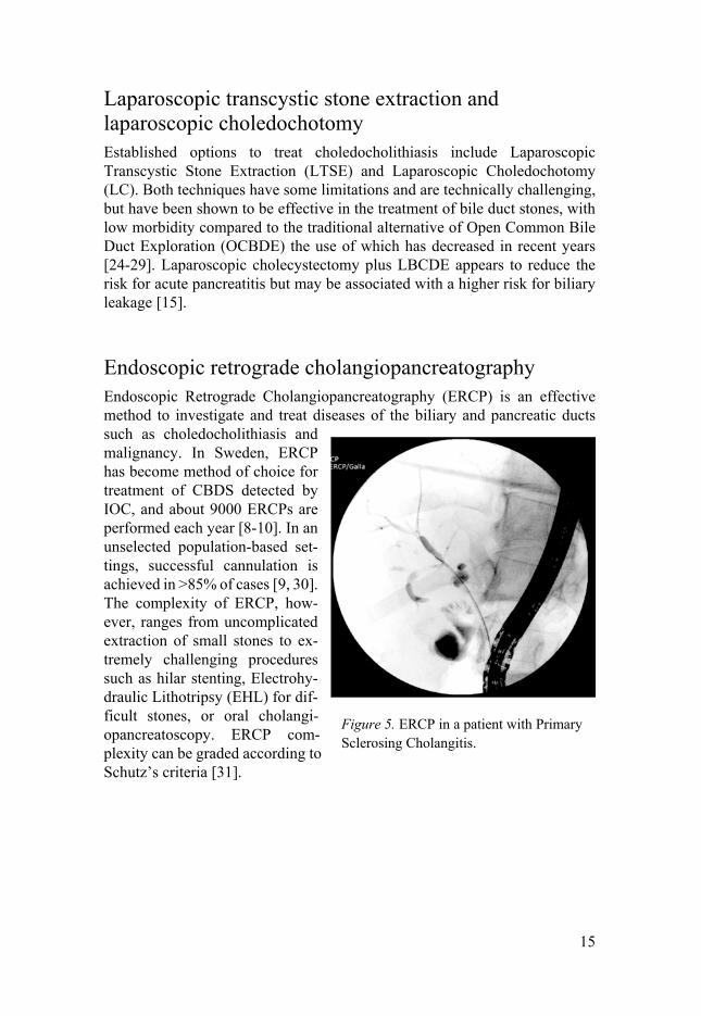
Endoscopic retrograde cholangiopancreatography Endoscopic Retrograde Cholangiopancreatography (ERCP) is an effective method to investigate and treat diseases of the biliary and pancreatic ducts such as choledocholithiasis and malignancy. In Sweden, ERCP has become method of choice for treatment of CBDS detected by IOC, and about 9000 ERCPs are performed each year [8-10]. In an unselected population-based set-tings, successful cannulation is achieved in >85% of cases [9, 30]. The complexity of ERCP, how-ever, ranges from uncomplicated extraction of small stones to ex-tremely challenging procedures such as hilar stenting, Electrohy-draulic Lithotripsy (EHL) for dif-ficult stones, or oral cholangi-opancreatoscopy. ERCP com-plexity can be graded according to Schutz’s criteria [31].
Figure 5. ERCP in a patient with Primary Sclerosing Cholangitis.
16
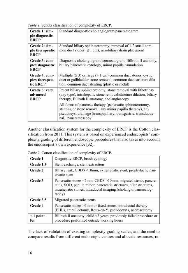
Table 1. Schutz classification of complexity of ERCP. Grade 1: sim-ple diagnostic ERCP
Standard diagnostic cholangiogram/pancreatogram
Grade 2: sim-ple therapeutic ERCP
Standard biliary sphincterotomy; removal of 1-2 small com-mon duct stones (≤ 1 cm); nasobiliary drain placement
Grade 3: com-plex diagnostic ERCP
Diagnostic cholangiogram/pancreatogram, Billroth II anatomy, biliary/pancreatic cytology, minor papilla cannulation
Grade 4: com-plex therapeu-tic ERCP
Multiple (≥ 3) or large (> 1 cm) common duct stones, cystic duct or gallbladder stone removal, common duct stricture dila-tion, common duct stenting (plastic or metal)
Grade 5: very advanced ERCP
Precut biliary sphincterotomy, stone removal with lithotripsy (any type), intrahepatic stone removal/stricture dilation, biliary therapy, Billroth II anatomy, cholangioscopy All forms of pancreas therapy (pancreatic sphincterotomy, stenting or stone removal, any minor papilla therapy), any pseudocyst drainage (transpapillary, transgastric, transduode-nal), pancreatoscopy
Another classification system for the complexity of ERCP is the Cotton clas-sification from 2011. This system is based on experienced endoscopists’ com-plexity grading of different endoscopic procedures that also takes into account the endoscopist’s own experience [32].
Table 2. Cotton classification of complexity of ERCP. Grade 1 Diagnostic ERCP, brush cytology Grade 1.5 Stent exchange, stent extraction Grade 2 Biliary leak, CBDS <10mm, extrahepatic stent, prophylactic pan-
creatic stent Grade 3 Pancreatic stones <5mm, CBDS >10mm, migrated stents, pancre-
atitis, SOD, papilla minor, pancreatic strictures, hilar strictures, intrahepatic stones, intraductal imaging (cholangio/pancreatog-raphy)
Grade 3.5 Migrated pancreatic stents Grade 4 Pancreatic stones >5mm or fixed stones, intraductal therapy
(EHL), ampullectomy, Roux-en-Y, pseudocysts, necrosectomy + 1 point for
Billroth II anatomy, child <3 years, previously failed procedure or procedure performed outside working hours
The lack of validation of existing complexity grading scales, and the need to compare results from different endoscopic centres and allocate resources, re-
17
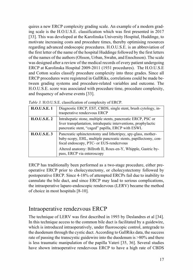
quires a new ERCP complexity grading scale. An example of a modern grad-ing scale is the H.O.U.S.E. classification which was first presented in 2017 [33]. This was developed at the Karolinska University Hospital, Huddinge, to motivate increasing costs and procedure times, thereby optimising resources regarding advanced endoscopic procedures. H.O.U.S.E. is an abbreviation of the first letter of the name of the hospital Huddinge followed by the first letters of the names of the authors (Olsson, Urban, Swahn, and Enochsson). The scale was designed after a review of the medical records of every patient undergoing ERCP at Karolinska Hospital 2009-2011 (1931 procedures). The H.O.U.S.E and Cotton scales classify procedure complexity into three grades. Since all ERCP procedures were registered in GallRiks, correlations could be made be-tween grading systems and procedure-related variables and outcome. The H.O.U.S.E. score was associated with procedure time, procedure complexity, and frequency of adverse events [33].
Table 3. H.O.U.S.E. classification of complexity of ERCP. H.O.U.S.E. 1 Diagnostic ERCP, EST, CBDS, single stent, brush cytology, in-
traoperative rendezvous ERCP H.O.U.S.E. 2 Intrahepatic stone, multiple stents, pancreatic ERCP, PSC or
liver transplantation, intrahepatic interventions, prophylactic pancreatic stent, “caged” papilla, ERCP with ESWL
H.O.U.S.E. 3 Pancreatic sphincterotomy and lithotripsy, spy-glass, mother-baby-scopy, EHL, multiple pancreatic stents, papillectomy, con-focal endoscopy, PTC- or EUS-rendezvous Altered anatomy: Billroth II, Roux-en-Y, Whipple, Gastric by-pass, ERCP via enteroscopy
ERCP has traditionally been performed as a two-stage procedure, either pre-operative ERCP prior to cholecystectomy, or cholecystectomy followed by postoperative ERCP. Since 4-18% of attempted ERCPs fail due to inability to cannulate the bile duct, and since ERCP may lead to serious complications, the intraoperative laparo-endoscopic rendezvous (LERV) became the method of choice in most hospitals [8-10].
Intraoperative rendezvous ERCP The technique of LERV was first described in 1993 by Deslandres et al [34]. In this technique access to the common bile duct is facilitated by a guidewire, which is introduced intraoperatively, under fluoroscopic control, antegrade to the duodenum through the cystic duct. According to GallRiks data, the success rate of passing the transcystic guidewire into the duodenum is >80% and there is less traumatic manipulation of the papilla Vateri [35, 36]. Several studies have shown intraoperative rendezvous ERCP to have a high rate of CBDS

18
clearance with few complications, particularly post-ERCP pancreatitis, compared to conventional ERCP [36-45].
Figure 6. Intraoperative rendezvous ERCP, drawing by Fredrik Swahn.
Figure 7. Intraoperative rendezvous ERCP.

19
Postoperative rendezvous ERCP In postoperative rendezvous ERCP, the antegrade transcystic guidewire is passed into the du-odenum and anchored to the cystic duct with clips. The other end of the guidewire is then passed through the abdominal wall and attached by tape to the skin, leaving the guidewire in situ. The cholecystectomy is then completed and the rendez-vous ERCP conducted at a later second session, usually within 1-2 days.
Intraoperative rendezvous ERCP is preferred since it is as-sociated with shorter hospital stay, reduced cost, and appears to have lower morbidity than postoperative rendezvous ERCP [14, 46-48]. Postoperative ren-dezvous ERCP is an alternative to intraoperative ERCP in situa-tions where adequate endoscopic resources are limited or lacking.
Figure 8. Postoperative rendezvous ERCP, drawing by Fredrik Swahn.
Figure 9. Patient prepared for postoperative rendezvous ERCP and subsequent post-operative rendezvous ERCP.
20
Complications of ERCP Of all the procedures that endoscopists perform on a regular basis, ERCP is that associated with the greatest risk, with a complication rate of 10-15%. Most complications are recognized during or shortly after ERCP, but some complications such as bleeding following sphincterotomy are delayed. The risk for adverse events in ERCP depends on patient risk factors, risk factors related to the technical procedure and experience of the endoscopist and team [8, 9, 43, 49, 50]. The indications and benefits of ERCP must balance potential harm to the patient.
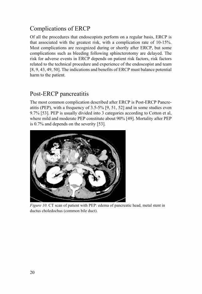
Post-ERCP pancreatitis The most common complication described after ERCP is Post-ERCP Pancre-atitis (PEP), with a frequency of 3.5-5% [9, 51, 52] and in some studies even 9.7% [53]. PEP is usually divided into 3 categories according to Cotton et al, where mild and moderate PEP constitute about 90% [49]. Mortality after PEP is 0.7% and depends on the severity [53].
Figure 10. CT scan of patient with PEP: edema of pancreatic head, metal stent in ductus choledochus (common bile duct).

21
Table 4. Cotton classification of severity of post-ERCP pancreatitis. Mild Pan-creatitis
Abdominal pain plus p-amylase elevated ≥3 times normal value re-quiring hospital admission or prolongation of planned admission up to 3 days.
Moderate Pancreati-tis
Requires hospitalisation for 4-10 days.
Severe Pancreati-tis
Requires hospitalisation for >10 days plus signs of local or sys-temic complications (necrosis, pseudocysts, multi-organ failure), or requires surgical or percutaneous intervention.
There are a number of PEP risk factors described in the literature, and the risk depends on both technical- and patient-related factors [49-51, 54-57]. Alt-hough PEP is widely accepted as the primary outcome measure following ERCP, the risk factors for PEP are like other adverse events such as bleeding, perforation, and other procedure-related complications. PEP may thus be con-sidered a surrogate endpoint for the safety and success of ERCP.
Table 5. Examples of risk factors for post-ERCP pancreatitis. Examples of patient-related risk factors for PEP Sphincter of Oddi Dysfunction (SOD), female gender, previous pancreatitis in-cluding PEP, younger age, no chronic pancreatitis, normal bilirubin level, non- dilated bile duct
Examples of procedure-related risk factors for PEP Cannulation>10 minutes, pancreatic guidewire passage, or contrast injection, Precut sphincterotomy, pancreatic sphincterotomy, biliary balloon-sphincter dila-tion, failure of CBDS clearance, intraductal ultrasound
Acute pancreatitis The most common causes of acute pancreatitis are biliary stone and alcohol abuse. Other conditions such as long-term haemodialysis or peritoneal dialy-sis, hepatic disease, hyperlipidaemia, hypercalcaemia, and diabetes, that are known to increase the risk for acute pancreatitis, may also be risk factors for PEP [58-68].

22
Bleeding, cholangitis and perforation Besides PEP, other well-known complications of ERCP are bleeding, cholan-gitis, and perforation.
Figure 11. Retroperitoneal perforation during ERCP with leakage of gas and contrast.
Clinically significant bleeding occurs in 1-3% of ERCPs. It may occur imme-diately but is often delayed up to 2 weeks [9, 49, 50]. Bleeding is graded as mild (no blood transfusion), moderate (up to 4 units of blood), or severe (>5 units of blood or surgical/angiographic intervention) [49]. Risk factors for bleeding include coagulopathy, anticoagulation therapy and in some studies precut sphincterotomy [49, 50]. Other studies, however, have not shown any increased risk for bleeding when early precut is performed for difficult biliary access [69, 70].
Cholangitis is reported in 0.5-5% of ERCPs, and bacteraemia in up to 27% of procedures [71, 72]. Biliary infection may be a result of failed complete drainage after the ERCP procedure. Other risk factors include hilar cholangi-ocarcinoma and primary sclerosing cholangitis (PSC) [9, 49]. Cholangitis is graded as mild (temperature >38 ºC for 24-48 hours), moderate (≥3 days in-hospital care or ERCP/PTC (Percutaneous Transhepatic Cholangiography) in-tervention), or severe (septic shock or need for surgical intervention) [49].
Perforation sometimes occurs when the guidewire or catheter penetrates the wall of the pancreatic or biliary ductal system. The exact frequency of perfo-ration is not known since they are seldom reported and adverse consequences for the patient are rare [73]. Duodenal perforation related to sphincterotomy occurs in <1% of ERCPs [9, 49, 50]. This perforation is retroperitoneal and can be managed conservatively with antibiotics. The risk for sphincterotomy-related perforation increases if the cut is large and extends beyond “1-2 o’clock” or in cases of repeated sphincterotomy, but does not seem to increase

23
in cases where early precut has been performed [69, 70]. Perfo-ration following pancreatic sphincterotomy either at the main or minor papilla is extremely rare [74]. Perforations are classified into mild (<3 days of hospital care and conservative treatment), moderate (4-10 days of hospital care) or severe (>10 days in hos-pital and need for surgical inter-vention or drainage) [49]. Routine CT investigations in asympto-matic patients after uncompli-cated sphincterotomy have shown retro- or periduodenal gas in up to 10% of cases [75].
The risk for perforation, bleeding or cholangitis does not differ between ERCP-patients with or without a periampullary diverticulum [76].
Cardiovascular complications Transient cardiac dysrhythmias and hypoxia are usually seen and managed during ERCP procedures, and only rarely do they result in clinical conse-quences or adverse events. Cardiovascular complications and pulmonary thromboembolism (PTE) occur in 0.5-1% of ERCPs and laparoscopic chole-cystectomies [9, 10, 50, 77-80]. The prevalence of CBDS increases with age. This complicates management since comorbidity and frailty increase the risk for intervention-related complications and death [81-83]. Cardiovascular dis-ease and biliary stone disease share risk factors such as obesity, hypertension, diabetes, dyslipidaemia and cigarette smoking [84-86]. There also appears to be an association between gallstone disease and cardiovascular disease [87]. Though early cholecystectomy appears to be safe in the elderly, there is a ten-dency to choose a minimally-invasive treatment method such as ERCP when it comes to elderly frail patients with high comorbidity [88].
Other ERCP complications Other complications related to ERCP include endoscopic perforation of the oesophagus, stomach, or duodenum. The risk for these perforations is usually low, about 1:1000, but increases in patients with altered anatomy such as after Billroth II gastrectomy or when stenosis is present in the upper gastrointestinal
Figure 12. Perforation of guide wire in bile duct with extravasation of contrast.

24
tract [73, 89]. There are also rare reports of penetration and perforation of the duode-num, small bowel or colon after migration of plastic stents from the bile duct [90, 91].
Other examples of complications are basket impaction during an attempt to re-move a large stone from the bile duct, chol-ecystitis caused by self-expandable metal stents (SEMS), contrast injection into the portal venous system, and in rare cases pseudomonas infection due to inadequate disinfection and cleaning of the duodeno-scope. Thirty-day mortality after ERCP is about 0.5% [9, 10, 50, 92, 93].
Endoscopic sphincterotomy (EST) was previously thought to be a risk factor for the development of cholangiocarcinoma due to reflux of gut bacteria into the biliary sys-tem, though recent studies have not seen an increase in cancer risk after EST [94, 95].
Stent dysfunction Endoscopic biliary stent placement is an effective treatment for pa-tients with benign or malignant bil-iary obstruction causing jaundice and/or cholangitis [96]. If the deci-sion is made to proceed with bili-ary drainage in patients with ma-lignant distal biliary obstruction and who are planned for curative resection, the endoscopic route is preferred to percutaneous transhepatic biliary drainage (PTBD) because of better patient survival and fewer peritoneal/liver recurrences in the endoscopic group [97-99]. Regarding palliative treatment of patients with hilar and extrahepatic malignant biliary obstruction, ERCP has a lower adverse event rate, shorter time in hospital, and lower cost
Figure 13. Late perforation of plastic stent through duodenal wall opposite to the major papilla.
Figure 14. Plastic stents in biliary duct.
25
compared to PTBD [100], as well as lower morbidity and mortality rates compared to surgical bypass [101, 102]. The diagnosis of stent dys-function is usually based on a combination of clinical crite-ria and a less than 20 % fall in serum bilirubin following stent insertion i.e., failed bili-ary drainage with development of cholangitis and jaundice. Sometimes, transabdominal ul-trasound is needed to confirm stent failure [101, 103].
Two types of stent are rou-tinely used in current practice; plastic stents and self-expand-
ing metal stents (SEMS). Several studies have shown that metal stents are as-sociated with significantly longer stent patency and lower re-intervention rate in the palliative management of malignant bile duct obstruction, compared to plastic stents [104-106]. This results in shorter hospital stay, reduction in fre-quency of complicating diseases due to stent dysfunction, and improvement in quality of life [107, 108]. Moreover, some studies have shown that metal stents are associated with longer survival compared to plastic stents [109, 110]. The median patency times of metal and plastic stents in patients with distal ma-lignant obstruction were found to be longer than 8 months and 4 to 6 months, respectively [109, 111]. The median pa-tency times of metal and plastic stents in patients with hilar cholangiocarcinoma were found to be 3 to 6 months and 1 to 2 months, respectively [111, 112]. SEMS is the method of choice in poten-tially curable patients with obstructive jaundice while waiting for surgery or in a neoadjuvant situation, because stent dysfunction and stent-related complica-tions are fewer compared to plastic stents [96, 113, 114]. Covered SEMS have a lower risk for tumour ingrowth but a higher risk for stent migration and tumour overgrowth compared to uncovered SEMS. However, meta-analyses show largely equivalent results regarding
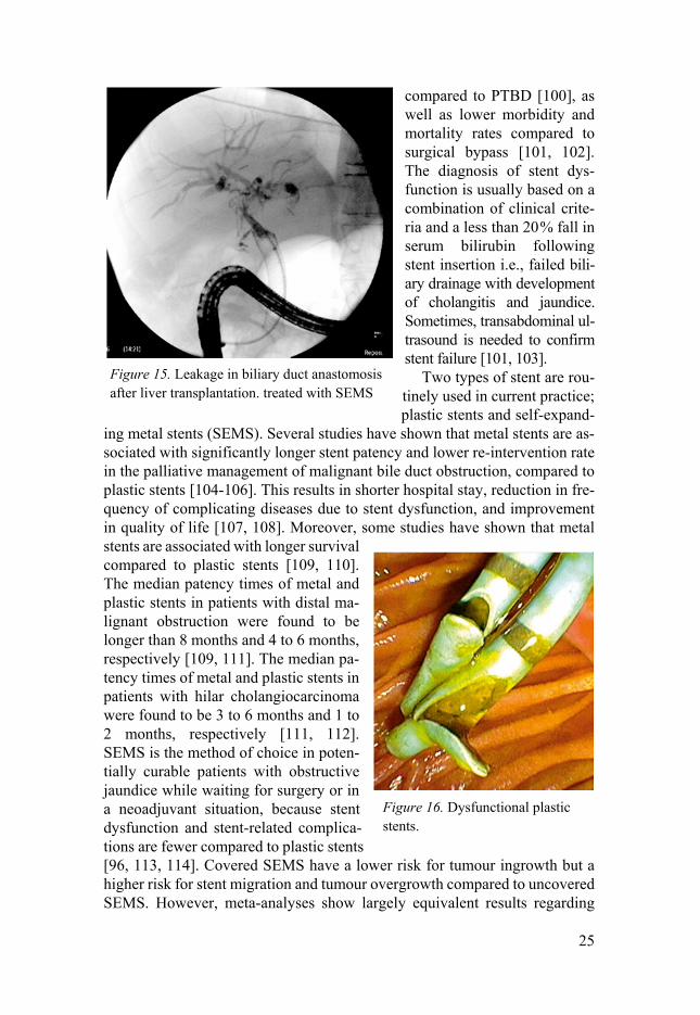
Figure 15. Leakage in biliary duct anastomosis after liver transplantation. treated with SEMS
Figure 16. Dysfunctional plastic stents.

26
proportions of patients with stent dysfunction, overall complications, and patient survival when comparing covered, partially covered, and un-covered SEMS [115-117]. There does not seem to be difference in risk for cholecystitis after insertion of covered vs. uncovered SEMS [117, 118].
Prevention of ERCP complications Anticoagulants such as warfarin or non-vitamin K oral anticoagulants (NOAKs), must be discontinued prior to ERCP, and any coagulopathy man-aged. Pure-cut diathermy has been shown to be associated with a higher risk for bleeding compared to blended diathermy [119]. Large balloon dilation in combination with sphincterotomy for treatment of large stones, DASE (Dila-tation Assisted Stone Extraction), is associated with less risk for bleeding compared to plain sphincterotomy [120].
Carbon dioxide via the duodenoscope has not been shown to reduce the complication rate after ERCP, but causes less post-ERCP abdominal pain and is therefore recommended as part of the procedure [51].
Antibiotic prophylaxis Evidence supporting prophylactic antibiotics to prevent infection after ERCP is limited, and meta-analyses are contradictory [121, 122]. The agents most commonly studied are cefotaxim and piperacillin. Even though the frequency of bacteraemia is less, antibiotic prophylaxis has not been shown to signifi-cantly prevent ERCP-induced cholangitis in unselected patients. The use of prophylactic antibiotics is recommended in patients where the risk for incom-plete drainage of the bile duct is high, for example PSC patients and patients with a hilar tumour or where drainage is unsuccessful [51]. Immunosup-pressed patients also seem to benefit from prophylaxis [123]. ERCP with con-comitant cholangioscopy is associated with bacteraemia in up to 13.9% of
Figure 17. Covered SEMS.
27
cases and infectious complications up to 9.7%. Thus antibiotic prophylaxis is recommended in this situation [124].
Prevention of post-ERCP pancreatitis Difficult cannulation (several guidewire passages and contrast injections into the pancreatic duct, repeated cannulation attempts, and long time taken to reach the bile duct) is associated with an increased rate of ERCP-related complications [51, 125-131]. Difficult cannulation has been defined by Halttunen et al as fulfilling at least one of the fol-lowing criteria: ≥5 cannulation attempts ≥5 minutes cannulation ≥2 passages of guidewire into the pancreatic duct. When using this definition, diffi-cult cannulation has been shown to increase the PEP rate fourfold probably because of oedema and trauma to the papilla [132]. A care-ful cannulation technique including use of a guidewire instead of the catheter and the use of contrast to identify the bile duct, has been shown to reduce the risk for PEP [133-135].
Rendezvous-ERCP as a way to manage CBDS found at cholecystec-tomy is another way to reduce the risk for PEP [36, 41].
Some studies have shown an in-creased risk for PEP when precut sphincterotomy is performed. How-ever, recent meta-analyses have con-cluded that there is a lower risk for PEP when precut is performed at an early stage in patients with difficult biliary access [136, 137].
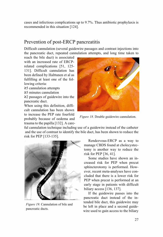
If the guidewire passes into the pancreatic duct instead of the in-tended bile duct, this guidewire may be left in place and a second guide-wire used to gain access to the biliary
Figure 18. Double guidewire cannulation.
Figure 19. Cannulation of bile and pancreatic ducts.

28
ducts in the so called “double guidewire cannulation technique” [138-140]. As an alternative to the pancreatic guidewire-assisted technique, a pancreatic sphincterotomy, possibly with the help of a pancreatic stent, could help when cannulation of the bile duct is difficult. The complication rates of these ma-noeuvres are comparable to precut sphincterotomy [141-143]. In order to re-duce the risk for PEP, it is recommended to leave a prophylactic pancreatic stent before completion of the ERCP [138, 144-146]. Several studies have shown the use of prophylactic pancreatic stents to reduce the risk for PEP, and guidelines recommend a 5 Fr diameter stent [51, 147].
Some studies indicate that the PEP rate is higher in cases when precut sphincterotomy is performed during ERCP compared to conventional sphinc-terotomy. This is probably a matter of timing since no increase in PEP rate has been shown if the precut is performed early on in the procedure [69, 148-151].
Non-steroidal anti-inflammatory drugs and post-ERCP pancreatitis Since non-steroidal anti-inflammatory drugs (NSAIDs) are potent inhibitors of phospholipase A, an enzyme thought to play an important role in the path-ogenesis of acute pancreatitis, studies have been conducted to assess the pos-sible role of NSAIDs as a protective measure against PEP. Randomised stud-ies and meta-analyses have shown that rectally administered indomethacin or diclofenac 100 mg as premedication before ERCP decreases the risk for PEP compared to placebo, particularly in high-risk patients [54, 152-160]. On the other hand, NSAIDs administered orally or intramuscularly have not been shown to be effective as protective against PEP [161, 162].
There are, however, prospective randomised controlled studies showing no difference in PEP rate between rectally administered diclofenac/indomethacin and placebo [153, 163-165]. A limitation of the meta-analyses published so far, regardless of conclusions, is that they include studies with relatively few subjects, different study designs, and varying proportions of high-risk pa-tients. As a consequence of this, guidelines vary widely, and in 2016-2017 only 25% of ERCP-patients in Sweden were given NSAID as PEP prophylaxis [9].
Other pharmacological prevention of post-ERCP pancreatitis Several agents have been studied regarding a possible protective effect against PEP, but results have been contradictory. Examples are: Protease inhibitors,

29
glyceryl trinitrate, octreotide, and somatostatin [51, 166-169]. Prophylactic antibiotics have not been shown to be effective against PEP [170].
Outcome of ERCP related to case-volume Lack of experience has been shown to be associated with poor outcome in major surgical procedures [171]. Likewise, extensive training and high ERCP case-volume have been shown to correlate with high success rates in terms of successful cannulation with fewer complications [30, 57, 172-176]. Experi-enced endoscopists have lower complication rates and higher success rates than their less experienced colleagues, which emphasises the importance of education and training. Technical failure of ERCP has been shown to increase the complication rate three-fold [177, 178]. Several studies have shown that high-volume ERCP centres have better results and lower complication rates than low-volume centres [172, 173, 179, 180], though there are data indicating that high quality ERCP may be performed in low-volume units [181-183]. It is important to select patients with correct indications since ERCP is most hazardous for patients who need it the least. Potential benefits of the procedure must exceed potential risks. Centralisation of complicated ERCPs to high-vol-ume centres with highly experienced endoscopists may well increase the safety and success of this procedure [184].
GallRiks The studies in this thesis are mainly based on data from GallRiks (The Swe-dish National Quality Register for Gallstone Surgery and ERCP). GallRiks was started in 2005 under the direction of the Swedish National Board of Health and Welfare and the Swedish Surgical Society, and has since been ad-ministered by the Uppsala Clinical Research Centre (UCR). GallRiks covers around 90% of all cholecystectomies and ERCPs performed in Sweden with almost all hospitals participating. Data coverage is assessed by cross-linkage with the Swedish Hospital Discharge Register, and GallRiks is also linked to the Cause of Death Register. External validatation of GallRiks is regularly performed through periodic audits at each hospital once every three years. Complete match between medical records and the GallRiks data-base has been shown in 97.3% of ERCPs when results from the first 25 audited hospitals were analysed [185]. The validation process and national coverage rate are published each year. Registration in GallRiks is managed online via an inter-net platform (www.ucr.uu.se/gallriks) and data are entered by the endoscopist at the time of the procedure. Records include patient- and procedure-related data with the possibility to describe more than 100 different variables, as well as multiple-choice questions. Intraoperative complications are registered, and
30
when all variables are filled in, the online form is closed. Collection of follow-up data including intra- and postoperative complications is managed locally at each hospital by a specific coordinator (often a nurse or sometimes a secretary) 30 days after ERCP [8, 186, 187].
National Patient Register The National Patient Register (NPR) collects data on healthcare of all patients admitted to hospital and in outpatient specialist care. It is maintained by the Swedish National Board of Health and Welfare. Though under-reporting of inpatient data is low, there has been a problem with outpatient care data, but this has improved greatly since 2001. A quality control of submitted data in the register is performed regularly, checking for quality and validity of per-sonal registration number, hospital, and main diagnosis, amongst other things. If submitted data are suspected of being erroneous or invalid, new data are requested from the care-providers [188-190].

31
Rationale behind this thesis
The overall aim of this thesis was to study and to gain a deeper understanding of the complications and risk factors associated with ERCP, and any protec-tive measures that may reduce these complications.

32
Aims
Paper I To assess whether clinical variables and comorbidities influence the risk for PEP.
Paper II The primary aim was to determine how various techniques for management of CBDS clearance in patients undergoing cholecystectomy have changed with time at tertiary referral hospitals (TRH) and county/community hospitals (CH). The secondary aim was to explore whether postoperative rendezvous ERCP is a safe, effective, and feasible alternative to intraoperative rendezvous ERCP in the management of CBDS.
Paper III To compare the rate of postoperative cardiovascular events in patients with CBDS treated with the following: ERCP only; cholecystectomy only; chole-cystectomy followed by delayed ERCP; cholecystectomy together with ERCP; or ERCP followed by delayed cholecystectomy.
Paper IV To analyse the association between ERCP success and complication rates, and endoscopist- and centre case-volumes.

33
Methods
Paper I
Figure 20. Flow diagram for the study. ERCP, endoscopic retrograde cholangio-pancreatography; CBD, common bile duct.

34
Data were retrieved from the Swedish Register for Cholecystectomy and ERCP (GallRiks) including all ERCP procedures performed 2006–2014 for common bile duct stones. A total of 15 800 procedures were identified and cross-checked. Univariable and multivariable logistic regression analyses were conducted with PEP as endpoint and the following covariables: age, gen-der, ASA grade, previous history of acute pancreatitis, diabetes, hyperlipidae-mia, hypercalcaemia, kidney disease, and liver cirrhosis.
Paper II Data were retrieved from the Swedish Register for Cholecystectomy and ERCP (GallRiks) 2006-2016. All cholecystectomies where CBDS were found at intraoperative cholangiography, and with complete 30-day follow-up (n=10386) were identified. Data concerning intraoperative and postoperative
Figure 21. Flowchart cholecystectomies in Sweden 2006–2016.

35
complications, readmission, and reoperation within 30 days were retrieved for patients where intraoperative ERCP (n=2290) or preparation for postoperative ERCP was performed (n=2283).
Figure 22. Flowchart common bile duct stones in Sweden 2006–2016.

36
Paper III Paper III was based on data from procedures for gallstone disease registered in the Swedish Register for Cholecystectomy and ERCP (GallRiks) 2006–2014. ERCP and cholecystectomy procedures performed for confirmed or sus-pected CBDS were included.
Figure 23. Flow chart. Confirmed or suspected CBDS as indication for treatment.
Patients with confirmed or suspected CBDS were divided into five treatment groups: ERCP only; cholecystectomy only; cholecystectomy followed by de-layed ERCP; cholecystectomy combined with ERCP; or ERCP followed by delayed cholecystectomy.
Postoperative events were registered by cross-matching GallRiks with the National Patient Register (NPR). A postoperative cardiovascular event was defined as an ICD-code in the discharge notes indicating myocardial infarct,

37
pulmonary embolism, or cerebrovascular incident within 30 days after sur-gery. In cases where a patient had undergone ERCP and cholecystectomy on separate occasions, the 30-day interval was timed from the first intervention.
Paper IV Data from GallRiks on all ERCPs 2009-2018 performed for common bile duct stone (n=17873) and malignancy (n=6152), with complete registration and 30-day follow-up, were collected and compiled. Procedures for any other indica-tion, procedures on patients having undergone previous ERCP since 2006, and rendezvous ERCPs were excluded from the analysis. Associations between
Figure 24. Flow chart showing study group assembly.

38
endoscopist ERCP case-volume as well as centre case-volume and successful cannulation rate, procedure time, intraoperative complication rate, and post-operative complication rate within 30 days (PEP, perforation, and intra- and postoperative bleeding) were analysed. Case-volumes were based on those during the year preceding the observations. When calculating cumulative vol-ume of ERCP procedures for endoscopists and centres, no ERCPs were ex-cluded.
39
Statistical Analyses
Paper I Univariable and multivariable logistic regression analyses with PEP as end-point were undertaken. In the multivariable analyses, adjustment was made for sex and age (at least 65 years versus less than 65 years) based on assump-tions of cause-effect relationships.
A subgroup analysis was conducted on patients with a previous history of pancreatitis. In this subgroup analysis the mean time that had elapsed since the previous episode of pancreatitis and ERCP was compared between patients who developed PEP following ERCP and those who did not suffer this com-plication, using Student’s t test.
Paper II Univariate and multivariate regression analyses were used as well as Pearson Chi Square Test and Student’s T-Test. The analyses were based on patients undergoing cholecystectomy with intraoperative ERCP, and patients under-going cholecystectomy as well as postoperative ERCP in two separate proce-dures. The complication rate was determined by extracting intraoperative and postoperative complications within 30 days after the cholecystectomy as well as the postoperative ERCP. In univariate and multivariate logistic regression analyses, the odds ratio for intra- and postoperative complications was deter-mined, adjusted for gender, age and ASA score. Statistical significance was defined as p<0.05.
Paper III To adjust for confounders, multivariate logistic regression analyses were per-formed, with cardiovascular event (myocardial infarct and/or pulmonary em-bolus and/or cerebrovascular incident), and death within 30 days as endpoints. The multivariate models were based on age (≥80 years vs <80 years), ASA class (III-V vs I-II), gender, treatment, and history of cardiovascular condition or event (myocardial infarct, heart failure, peripheral vascular disease, cere-brovascular incident, diabetes with secondary complication, or pulmonary
40
embolism). Patients who underwent cholecystectomy and ERCP during the same operation and those who underwent cholecystectomy and delayed ERCP were grouped together with the cholecystectomy group, whereas those who underwent ERCP and delayed cholecystectomy were grouped together with the ERCP group. This grouping was based on the intervention primarily in-tended to manage the CBDS. Poisson regression was used to calculate the 30-day age- and gender-adjusted standardised mortality ratio (SMR) based on the expected mortality rate extrapolated from the Swedish general population in 2007.
Paper IV Univariable and multivariable logistic regression analyses with successful cannulation, procedure time, intraoperative complication rate, and postopera-tive complication rate within 30 days (PEP, perforation, and intra- and post-operative bleeding) as endpoints were carried out with endoscopist- and centre case-volumes as exposure variables. In the multivariable logistic regression analyses, adjustments were made for gender, age, and year of ERCP. The ad-justments made in the multivariable analysis were based on assumptions of cause-effect relationships. Analyses were made with case-volumes on a log scale (n=0-4, 5-10, 11-20, 21-40, 41-80, 81-160 or 161-320 for endoscopist and n=0-20, 21-40, 41-80, 81-160, 161-320 or >320 for centre).

41
Ethical Considerations
Paper I The Regional Ethics Review Board in Stockholm approved the study the 18th March 2015 (reference number 2015/339-31/1).
Paper II The Regional Ethics Review Board in Uppsala approved the study the 2nd November 2016 (reference number: 2016/281/1).
Paper III The Regional Ethics Review Board in Stockholm approved the study the18th March 2015 (IRB-approval, reference number: 2015/339-31/1).
Paper IV The Regional Ethics Review Board in Stockholm approved the study the 17th June 2020 (IRB-approval, reference number: 2020-01450).

42
Results
Paper I Women (Odds Ratio [OR] 1.33, 95% confidence interval [CI] 1.14-1.55), pa-tients aged less than 65 years (OR 1.68, CI 1.45-1.94), patients with hyper-lipidaemia (OR 1.32, CI 1.02-1.70), and those with a previous history of acute pancreatitis (OR 5.44, CI 4.68- 6.31) faced a significantly higher risk for PEP. In a subgroup analysis of patients with a previous history of acute pancreatitis, the mean time from previous pancreatitis to ERCP was 4423 days in patients who developed PEP versus 6990 days in patients who did not (P =0.037). However, when the previous episode of pancreatitis had occurred more than 30 days before ERCP, this association was no longer significant (P =0.858). Patients with diabetes had a lower risk for PEP (OR 0.64, CI 0.48-0.85).

43
Table 6. Univariable and multivariable logistic analysis of risk factors for pancreati-tis after endoscopic retrograde cholangiopancreatography. Adjustments were made for sex and age (at least 65 years versus less than 65 years).
Incidence of post-ERCP pancreatitis
Univariable analysis
Multivariable analysis
Odds ratio p Odds ratio p Gender Men 250/6140 (4.1%) Women 515/9660 (5.3%) 1.33 (1.14-
1.55) <0.001
Age ≥65 years 349/9140 (3.8%) <65 years 416/6660 (6.2%) 1.68 (1.45-
1.94) <0.001
History of acute pancreatitis
363/2567 (14.1%) 5.26 (4.53-6.10)
<0.001 5.44 (4.68-6.31)
<0.001
Diabetes (all) 56/1947 (2.9%) 0.55 (0.42-0.72)
<0.001 0.64 (0.48-0.85)
0.002
Diabetes type 1 21/564 (3.6%) 0.72 (0.47-1.13)
0.724 0.84 (0.54-1.31)
0.437
Liver cirrhosis 12/185 (6.5%) 1.37 (0.76-2.47)
0.296 1.39 (0.77-2.51)
0.277
Hyperlipidaemia 72/1394 (5.2%) 1.08 (0.84-1.38)
0.556 1.32 (1.02-1.70)
0.036
Hypercalcaemia 2/58 (3.4%) 0.70 (0.17-2.88)
0.622 0.76 (0.18-3.11)
0.756
Kidney disease 27/579 (4.7%) 0.96 (0.65-1.42)
0.838 1.16 (0.78-1.72)
0.474

44
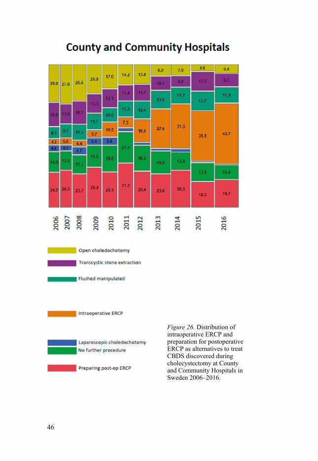
Paper II Intraoperative ERCP increased (7.5% 2006; 43.1% 2016) whereas preparation for postoperative ERCP decreased (21.2% 2006; 17.2% 2016) 2006-2016. CBDS management differed between TRHs and CHs. Complications were higher in the postoperative rendezvous ERCP group: (Odds Ratio [OR] 1.69, 95% confidence interval [CI] 1.16-2.45) for intraoperative complications and (OR 1.50, CI 1.29-1.75) for postoperative complications. The risks for in-traoperative bleeding (OR 2.46, CI 1.17-5.16), postoperative bile leakage (OR 1.89, CI 1.23-2.90), and postoperative infection with abscess (OR 1.55, CI 1.05-2.29) were higher in the postoperative group. Post-ERCP pancreatitis, postoperative bleeding, cholangitis, percutaneous drainage, antibiotic treat-ment, ICU stay, readmission/reoperation within 30 days, and 30-day mortality did not differ between groups.

45
Figure 25. Distribution of intra- operative ERCP and prepara-tion for postoperative ERCP as alternatives to treat CBDS discovered during cholecystec-tomy at Tertiary Referral Hos-pitals in Sweden 2006–2016.
46
Figure 26. Distribution of intraoperative ERCP and preparation for postoperative ERCP as alternatives to treat CBDS discovered during cholecystectomy at County and Community Hospitals in Sweden 2006–2016.

47
Table 7. Intra- and postoperative (within 30 days) complication rates n (%). Statistically significant values are given in bold text. Pearson Chi Square.
Intraop ERCP
(%)
Preparation postop
ERCP (%) P*
Intraoperative Overall 2.0 3.4 0.0031 Bleeding 0.4 1.1 0.0106
Postoperative
Overall 15.6 21.8 <0.0001 Bleeding 1.2 0.9 0.2501 Pancreatitis 4.7 4.4 0.6362 Cholangitis 0.6 0.9 0.2314 Bile leakage 1.4 2.7 0.0025 Infection with abscess 1.9 2.9 0.0197 Percutaneous drainage 2.2 3.0 0.0925 Antibiotic treatment 9.7 10.4 0.4697 ICU stay 0.3 0.1 0.3191 Readmission within 30 days 0.7 0.3 0.0498
Reop within 30 days 2.0 2.1 0.8232
Mortality 30 days 0.31 0.04 0.0341
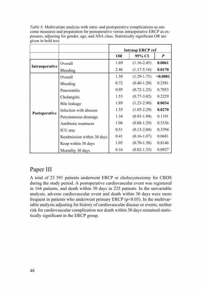
48
Table 8. Multivariate analysis with intra- and postoperative complications as out-come measures and preparation for postoperative versus intraoperative ERCP as ex-posures, adjusting for gender, age, and ASA class. Statistically significant OR are given in bold text.
Intraop ERCP ref OR 95% CI P
Intraoperative Overall 1.69 (1.16-2.45) 0.0061
Bleeding 2.46 (1.17-5.16) 0.0170
Postoperative
Overall 1.50 (1.29-1.75) <0.0001
Bleeding 0.72 (0.40-1.28) 0.2581
Pancreatitis 0.95 (0.72-1.25) 0.7053
Cholangitis 1.53 (0.77-3.02) 0.2229
Bile leakage 1.89 (1.23-2.90) 0.0034
Infection with abscess 1.55 (1.05-2.29) 0.0270
Percutaneous drainage 1.34 (0.93-1.94) 0.1191
Antibiotic treatment 1.06 (0.88-1.29) 0.5336
ICU stay 0.51 (0.13-2.04) 0.3394
Readmission within 30 days 0.41 (0.16-1.07) 0.0681
Reop within 30 days 1.05 (0.70-1.58) 0.8146
Mortality 30 days 0.16 (0.02-1.35) 0.0927
Paper III A total of 23 591 patients underwent ERCP or cholecystectomy for CBDS during the study period. A postoperative cardiovascular event was registered in 164 patients, and death within 30 days in 225 patients. In the univariable analysis, adverse cardiovascular event and death within 30 days were more frequent in patients who underwent primary ERCP (p<0.05). In the multivar-iable analysis adjusting for history of cardiovascular disease or events, neither risk for cardiovascular complication nor death within 30 days remained statis-tically significant in the ERCP group.

49

50
Table 10. Univariable and multivariable analyses of factors predicting cardiovascu-lar event and death within 30 days after surgical and/or endoscopic treatment for confirmed or suspected CBDS in the Swedish National Quality Register for Chole-cystectomy and Endoscopic Retrograde Cholangiopancreatography (GallRiks) 2006–2014.
Univariable Cardiovascular complication Death Odds ratio (95%
confidence inter-val)
p Odds ratio (95% con-fidence interval)
p
Age≥80 years (ref <80 years)
4.37 (3.20-5.60) <0.001 9.60 (7.20-12.79) <0.001
Men (ref women)
1.16 (0.85-1.59) 0.340 1.19 (0.91-1.55) 0.197
ASA I (ref) ASA II 3.83 (2.16-6.79) <0.001 6.42 (3.08-13.35) <0.001
ASA III 9.82 (5.51-17.52) <0.001 31.39 (15.32-64.31) <0.001
ASA IV 26.03 (11.44-59.22)
<0.001 150.02 (67.94-331.23)
<0.001
ASA V - - 343.38 (32.20-3662.14)
<0.001
History of car-diovascular disease or event*
10.20 (7.12-14.60) <0.001 6.25 (4.74-8.23) <0.001
ERCP (ref cholecystec-tomy)**
2.74 (1.95-3.84) <0.001 4.10 (3.00-5.62) <0.001
Multivariable
Cardiovascular complication Death
ERCP (ref cholecystec-tomy)*
1.12 (0.77-1.64) 0.548 1.38 (0.97-1.96) 0.071
* History of myocardial infarct, heart failure, peripheral vascular disease, cerebro-vascular incident, diabetes with secondary complication, or pulmonary embolism.
**In cases where ERCP as well as cholecystectomy were performed, the procedures were grouped according to the primary procedure. If cholecystectomy and ERCP were performed as one procedure, the procedure was included in the cholecystec-tomy group.

51
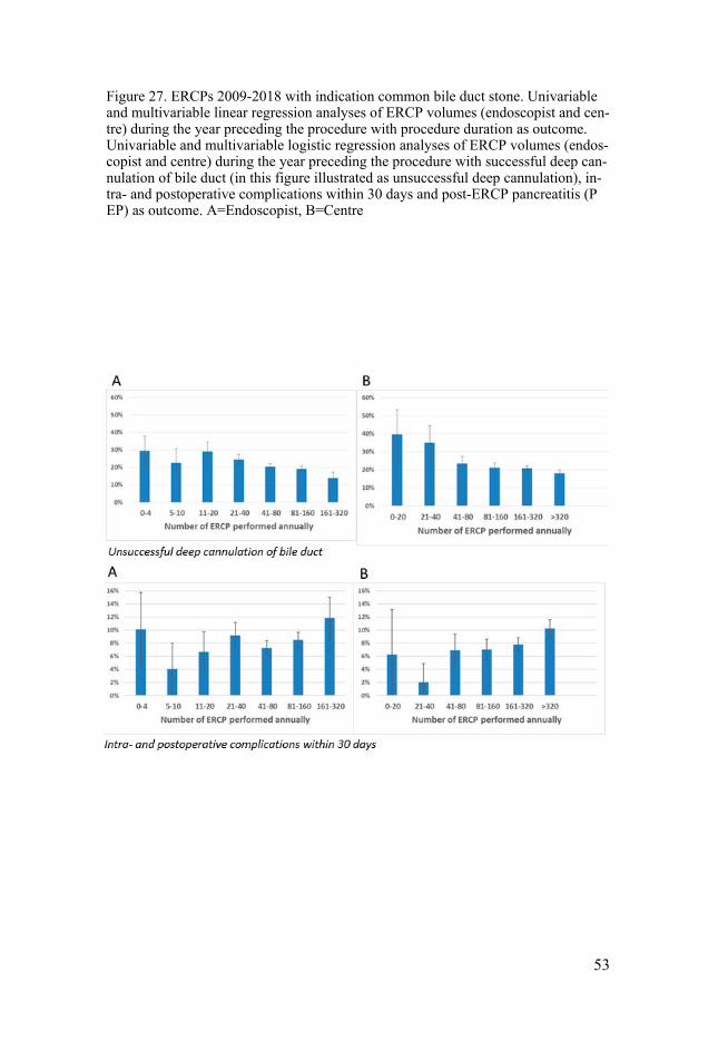
Paper IV In the multivariable analysis of the CBDS group adjusting for age, gender and year, a high endoscopist case-volume was associated with higher successful cannulation rate with lower complication and PEP rates and shorter procedure time (p<0.05). High annual case-volume centres were associated with high successful cannulation rate and shorter procedure time (p<0.05), but not lower complication and PEP rates.
When indication for ERCP was malignancy, a high endoscopist case-volume was associated with high successful cannulation rate and low PEP rates (p<0.05), but not shorter procedure time or lower complication rate. Centres with high case-volume were associated with high successful cannulation rate and low complica-tion and PEP rates (p<0.05), but not shorter procedure time.

52
53
Figure 27. ERCPs 2009-2018 with indication common bile duct stone. Univariable and multivariable linear regression analyses of ERCP volumes (endoscopist and cen-tre) during the year preceding the procedure with procedure duration as outcome. Univariable and multivariable logistic regression analyses of ERCP volumes (endos-copist and centre) during the year preceding the procedure with successful deep can-nulation of bile duct (in this figure illustrated as unsuccessful deep cannulation), in-tra- and postoperative complications within 30 days and post-ERCP pancreatitis (P EP) as outcome. A=Endoscopist, B=Centre

54
Figure 28. ERCPs 2009-2018 with indication malignancy. Univariable and multi-variable linear regression analyses of ERCP volumes (endoscopist and centre) during the year preceding the procedure with procedure duration as outcome. Uni-variable and multivariable logistic regression analyses of ERCP volumes (endos-copist and centre) during the year preceding the procedure with successful deep cannulation of bile duct (in this figure illustrated as unsuccessful deep cannula-tion), intra- and postoperative complications within 30 days and post-ERCP pan-creatitis (PEP) as outcome. A=Endoscopist, B=Centre

55
Discussion
The results of the studies in this thesis may serve to identify patients and situ-ations where there is an increased risk for ERCP-related complications. Even if procedure-related complications cannot be eliminated, awareness of poten-tial risk factors may help to optimise safety in situations where problems are foreseen. When hazards and risk factors are identified, the care of patients with gallstone disease can be organised to prevent them.
The prevalence of gallstone-related symptoms, including CBDS, in the population is 7-15%. Choledocholithiasis is the most common indication for ERCP, about 3 times more common than malignancy as indication, and pro-cedures for CBDS are performed at almost all hospitals in Sweden where gall-stone surgery is carried out [9, 185]. High age is a significant risk factor for prolonged hospital stay and for death after any procedure for gallstone re-moval [81, 82]. The comorbidity rate in elderly patients undergoing treatment for choledocholithiasis is high compared to younger patients, and there is a tendency to choose minimally-invasive treatment methods such as ERCP when it comes to older, frail patients with high comorbidity [83, 88]. ERCP performed for CBDS may be complicated, for example large impacted stones that require advanced methods such as electrohydraulic lithotripsy (EHL). The majority of ERCPs for CBDS, however, are uncomplicated and fall into H.O.U.S.E. category I [33], Cotton Grade II, or Schutz II [31, 32]. ERCP for the diagnosis and treatment of malignancy is often more complicated than ERCP for CBDS, especially if the malignancy is intrahepatic. These proce-dures are associated with greater risk and higher adverse event rates. ERCP for malignancy is graded at least H.O.U.S.E. II, Cotton III, or Schutz IV [31-33].
The reported complication rate of ERCP is 10-15%. The most common ad-verse event is post-ERCP pancreatitis (PEP), with a rate of 3.5-5% [9, 51, 52]. The risk for ERCP complications depends on both patient risk factors and technical risk factors related to the procedure and experience of the endosco-pist and team [8, 9, 43, 49, 50]. High endoscopist- and centre ERCP case-volumes have been shown to be correlated to high success rates in terms of successful cannulation and fewer adverse events [30, 57, 172-176].
In the case of CBDS found at IOC, the frequency of open choledochotomy, once considered the first-hand technique, has decreased in recent years, while at the same time, minimally invasive laparoscopic and laparo-endoscopic methods, mainly intraoperative rendezvous ERCP, have come to predominate

56
[9, 10, 14, 18, 20, 21, 24-29]. ERCP has traditionally been performed as a two-stage procedure, either as preoperative ERCP followed by laparoscopic chol-ecystectomy or laparoscopic cholecystectomy followed by postoperative ERCP. However, 4–18% of attempted ERCPs are interrupted due to inability to cannulate the bile duct. ERCP may also lead to serious complications such as PEP [9, 44, 51].
The technique of intraoperative rendezvous ERCP is straight-forward and suitable for almost all patients with CBDS. In this way cholecystectomy and management of CBDS are performed at the same time, thereby limiting an-aesthesia to one procedure with minimal hospital stay, healthcare resources, and costs [36-43]. Even if intraoperative rendezvous ERCP is recommended as method of choice, postoperative rendezvous ERCP is an alternative to in-traoperative ERCP in situations when ERCP resources are limited [14, 46-48]. As the lack of uniform logistic routines has made it impossible to conduct a prospective randomised controlled trial comparing the two methods, the best evidence regarding the safety and effectiveness of the two approaches has been derived from large population-based studies.
In Papers I and III we focused on risk factors for PEP and cardiovascular complications and death after surgical treatment for CBDS. In accordance with previous studies, we found that women, patients aged less than 65 years, and those with a previous history of acute pancreatitis had a significantly greater risk for developing PEP [49-51, 54, 55]. Since it is difficult to distin-guish a new episode of acute pancreatitis from an exacerbation of an ongoing process, we excluded patients with pancreatitis immediately before ERCP. This showed that if the previous episode of pancreatitis occurred more than 30 days before the ERCP, the time factor was not associated with risk for PEP. As shown in previous studies, hypertriglyceridaemia and hyperlipidaemia both increase the risk for PEP while liver cirrhosis is not a risk factor [58, 59, 62, 68].
Associated comorbidities such as obesity, alcohol abuse and use of medi-cations were not investigated in the present study since these data were not available in GallRiks.
Although previous reports give contrasting results with respect to hyper-calcaemia/kidney disease and risk for PEP [60, 61, 64], it should be noted that only 58 patients in the present cohort had hypercalcaemia and 579 had kidney disease. With no data on the degree of renal failure it is difficult to draw any conclusion regarding the association between hypercalcaemia/kidney disease and PEP.
Whereas previous studies have shown diabetes to be associated with acute pancreatitis [65, 67], we paradoxically found a lower risk for PEP in patients with diabetes. It has been observed that the risk for acute pancreatitis is de-pendent on the type of diabetes medication the patient receives [63]. The co-hort in the present study included diabetic patients on different kinds of dia-betic treatment, and the register lacked information on disease severity and

57
treatment. Thus, associations between type of diabetes treatment and PEP were not investigated.
The five CBDS treatment groups in Paper III were not predetermined, and the treatments used depended on several factors such as complexity and state of the biliary disease and preference of the surgeon responsible or local treat-ment guidelines [191]. We believe that the choice of ERCP in patients that are frail and have greater comorbidity explains why ERCP was significant in uni-variate analysis. In multivariable analysis, however, adjusting for history of cardiovascular disease or events, neither risk for cardiovascular complication nor death within 30 days remained statistically significant in the ERCP group.
No subsequent cholecystectomy was registered for any of the 8790 patients with ERCP as sole intervention. It is possible, however, that some of the pa-tients underwent cholecystectomy after completion of the study. Since chole-cystectomy at a later stage was unlikely to be performed to prevent CBDS, such cases are irrelevant in the present study.
Regarding Paper III, it is possible that procedure-related complications pre-disposed to cardiovascular complications. This must also be taken into ac-count when deciding on method of treatment for common bile duct stones. Even if most complications are included, it cannot be excluded that registra-tion of some adverse events might have been neglected in the analysis of pa-tients who underwent both ERCP and cholecystectomy on two separate occa-sions with a long interval between.
Tobacco use and obesity are major risk factors that must be taken into ac-count when assesing the risk for cardiovascular complications following a sur-gical or endoscopic intervention. Even if smoking and BMI are included in the ASA physical status, these risk factors per se were not routinely registered in GallRiks during the period of the study, and data on medications, including anticoagulation, were lacking [192]. Anaesthesia was not included as risk fac-tor in the present study, though this was explored in a recent study based on GallRiks data, showing more postoperative complications after ERCPs per-formed under deep sedation compared to those performed under general an-aesthesia [193].
The burden of cardiovascular disease differs between Sweden and other parts of the world. U.S. and Swedish data are more similar than when com-paring western countries with areas outside Western Europe and North Amer-ica [194].
In Paper II we looked at how the management of CBDS found at IOC has changed over time as well as differences in choice of treatment between ter-tiary referral hospitals and smaller community/county hospitals. We focused on the two most common treatment options for choledocholithiasis i.e., in-traoperative and postoperative rendezvous ERCP, and compared these meth-ods regarding intraoperative and postoperative complication rates as well as readmission, reoperation, and mortality.

58
During the period 2006-2016, ERCP gradually became the method of choice to manage CBDS at all hospitals in Sweden, and by 2016 was used in 60% of procedures. Though intraoperative rendezvous ERCP was the method of choice at most hospitals, it was mostly used in TRHs. On the other hand, in 2016, preparation for postoperative rendezvous ERCP was performed twice as often in CHs than in TRHs, probably due to lack of endoscopy resources for performing intraoperative ERCP in non-specialised centres.
Intraoperative complication rates as well as rates within 30 days after the procedure were assessed and compared between intraoperative ERCP and preparation for postoperative ERCP. Since intraoperative ERCP is carried out during cholecystectomy and postoperative ERCP is usually performed within 1 or 2 days after cholecystectomy, it cannot be excluded that some of the com-plications observed could have been the result of cholecystectomy rather than the ERCP.
Overall intra- and postoperative complication rates, as well as intraopera-tive bleeding, postoperative bile leakage and postoperative infection with ab-scess were higher with postoperative rendezvous ERCP compared to in-traoperative rendezvous ERCP. Manipulation of the guidewire while prepar-ing for postoperative ERCP could be one possible explanation for the higher rate of postoperative bile leakage and infection in this group. If the clips around the cystic duct anchoring the guide wire are applied too loosely, the risk for subsequent bile leakage is considerable.
The rate of the most common surgical complication, PEP, as well as post-operative bleeding, cholangitis, need for percutaneous drainage, antibiotic treatment, ICU stay, readmission/reoperation within 30 days, and 30-day mor-tality did not differ between intraoperative and postoperative ERCP. Prepara-tion for postoperative rendezvous ERCP by leaving a guidewire for definitive treatment of CBDS 1-2 days after cholecystectomy, is thus a feasible alterna-tive. The routine of leaving a guidewire through the abdominal wall and taped to the skin causes some discomfort for the patient, though most seem to toler-ate the guidewire quite well.
Based on the results of this study we believe that postoperative rendezvous ERCP is an acceptable alternative to intraoperative rendezvous ERCP when adequate ERCP resources are lacking or limited.
In Paper IV it was demonstrated that case-volume of the endoscopist has a great impact on ERCP outcome, especially when performed for CBDS. The pattern was more obscure for procedures performed for suspected malignancy. At the centre level, annual case-volume was also associated with safer out-come.
To obtain a more homogenous study population, we excluded all proce-dures where the indication was unclear, which to some extent limits the exter-nal validity of the study. Registration of incorrect indication and incomplete-

59
ness and low frequency of 30-day follow-up affect results and outcome. Re-garding complicated ERCP procedures, postoperative complication rates have been shown to be higher in units where follow-up is complete
[195]. GallRiks has not yet been linked to the Swedish National Patient Register (NPR), so some complications, particularly those occurring after 30 days, may have been neglected. However, it is more likely that most adverse events following ERCP occur in the immediate postoperative period.
Since perioperative complication rates, in particular PEP, are low, we chose to exclude rendezvous ERCPs [36, 41]. Endoscopists with the greatest expe-rience and centres with the highest volumes had the highest cannulation suc-cess rate, shortest procedure times, and lowest complication rates when the indication for ERCP was CBDS. Paradoxically, the outcome of ERCP per-formed for malignancy by more experienced endoscopists was poorer, with longer procedure times and higher complication rates. This was probably the result of selection bias since the most experienced high-volume endoscopist performs the most complex and time-consuming ERCP procedures with the greatest risk for adverse events. This results in residual confounding, which was not captured in the analyses of the present study. Furthermore, high-vol-ume endoscopists use more advanced ERCP techniques such as needle-knife sphincterotomy, and are more likely to persevere longer and spend greater ef-fort cannulating the bile duct before terminating the procedure [196].
Case-volume is an important issue in ERCP training, and it is important that the training of future advanced endoscopists is carried out at high-volume centres. The learning curve among trainees in advanced endoscopy varies sig-nificantly, but the success rate of trainees performing ERCP increases with experience [197, 198].

60
Conclusions
Paper I Age, female gender, hyperlipidaemia, and previous history of recent acute pancreatitis increase the risk for PEP. Diabetes, however, appeared to lower the risk for PEP.
Paper II The choice of technique for management of CBDS found at cholecystectomy has changed in recent years and differs between TRHs and CHs. Rendezvous ERCP is now the technique of choice and is performed at practically all hos-pitals in Sweden.
Rendezvous ERCP is a safe and effective method. Intraoperative rendez-vous ERCP is to be preferred, but postoperative rendezvous ERCP is a per-fectly acceptable alternative when adequate ERCP resources are lacking or limited.
Paper III Primary ERCP as well as cholecystectomy may be performed for CBDS with acceptable safety. More studies are required to provide reliable guidelines for the management of CBDS.
Paper IV The results of Study IV suggest that higher endoscopist- and centre case-vol-umes lead to safer and more effective ERCPs.

61
Proposals for future clinical research
• Prospective randomised studies are required to clarify the potential protective effect of NSAID regarding PEP. Since the results of pre-viously published prospective randomised controlled studies are contradictory, recommendations concerning PEP prophylaxis in Sweden differ and most ERCP patients are not given NSAID [9, 51, 153, 165]. We are therefore planning a nationwide multicentre study comparing the frequency and severity of PEP, according to Cotton’s criteria, between patients randomised to receive100 mg diclofenac administered rectally immediately prior to ERCP and those with no prophylaxis. Adverse event and mortality rates will also be analysed. The Regional Ethics Review Board in Uppsala approved the study the 31st May, 2017 (reference number: 2017/067). The Medical Products Agency approved the study the 20th August, 2018 (reference number: 5.1-2018-56693, EU-number: 2017-004250-42).
• A supplementary research study would be to retrieve GallRiks data regarding ERCP patients who received prophylactic rectally admin-istered NSAID and compare their intra- and postoperative compli-cation rates with patients who were not premedicated with NSAID.
• Another option would be to compare complication and success rates in patients undergoing ERCP who have been treated with early precut sphincterotomy with those who have been treated with reg-ular sphincterotomy.

62
Summary of the thesis in Swedish
Bakgrund Endoskopisk Retrograd Kolangiopankreatografi (ERCP) är en endoskopisk röntgenbaserad behandlingsmetod för att utreda och behandla sjukdomstill-stånd i gallvägar och i bukspottkörtel. Årligen utförs drygt 9000 ERCP i Sve-rige och den vanligaste indikationen är sten i djupa gallvägarna följt av tumör-orsakad gallvägsförträngning. Den totala komplikationsfrekvensen efter ERCP uppgår till 10-15%. Risken för komplikation beror både på patient- och procedurrelaterade faktorer liksom på endoskopistens och teamets erfarenhet och undersökningsvolym. Den vanligaste komplikationen utgörs av bukspott-körtelinflammation, post-ERCP pankreatit (PEP), vilken drabbar 3.5-5% av patienter. PEP definieras som buksmärtor>24 timmar samt s-amylas (bukspottkörtelenzym i blodet) >3 gånger normalvärdet tillika med förlängd vårdtid eller återinläggning på sjukhus. Inflammationen i bukspottkörteln blir i de allra flesta fall lätt till måttlig och läker inom några dagar men kan ibland bli allvarlig och kräva intensivvård eller, i sällsynta fall, till och med leda till döden. Exempel på riskfaktorer för PEP är långvarig manipulation av gall-gångsmynningen, ingjutning av röntgenkontrast i bukspottkörtelgången, ung ålder och kvinnligt kön.
Livstidsrisken att utveckla gallsten är >20% och av personer med gallsten drabbas 10-20% av stensjukdom i djupa gallvägarna med risk för allvarliga komplikationer som obstruktiv gulsot, gallvägsrelaterad infektion (kolangit) eller bukspottkörtelinflammation (pankreatit). Riskfaktorer för att utveckla gallsten inkluderar hög ålder, kvinnligt kön, graviditet, fysisk inaktivitet och fetma. Att operera bort gallblåsan med titthålsteknik (laparoskopisk kole-cystektomi) är förstahandsbehandling för gallsten runt om i världen och enbart i Sverige utförs 13 000 kolecystektomier årligen. Intraoperativ röntgen av gallvägarna (kolangiografi) har visats vara effektiv för att klargöra gallvägsa-natomin samt detektera sten i djupa gallvägarna, vilket sker i 10-15% av ko-lecystektomier. Den vanligaste metoden i vårt land att åtgärda sten i djupa gallvägarna som upptäcks under pågående galloperation är peroperativ ren-dezvous ERCP. Med denna teknik underlättas accessen till de djupa gallvä-garna med hjälp av en ledare (guidewire) som under röntgengenomlysning förs via ytliga gallgången till tolvfingertarmen. Peroperativ rendezvous ERCP har, jämfört med konventionell icke-rendezvous ERCP, visats ge hög grad av stenfrihet och minskad risk för komplikationer, framför allt PEP. I situationer
63
där tillgång till endoskopisk expertis saknas kan ett alternativ vara att förankra ledaren via ytliga gallgången till tolvfingertarmen, avsluta kolecystektomin och utföra ERCP i en andra seans, så kallad postoperativ rendezvous ERCP. Hos vissa äldre och sköra patienter där komplikationsrisken vid kirurgi be-döms alltför hög kan ERCP med stenextraktion utgöra den enda behandlingen vid sten i djupa gallvägarna.
Delarbetena i denna avhandling utgår från data från det svenska kvalitets-registret för gallstenskirurgi och ERCP (GallRiks). GallRiks startade 2005 och omfattar >90% av alla kolecystektomier och ERCP:er i Sverige. Patient- och procedurrelaterade data förs in på förhand och intra- och postoperativa kom-plikationer inom 30 dagar registreras lokalt av koordinator på varje sjuk-hus/enhet.
Delarbete I Syftet med detta arbete var att undersöka huruvida vissa förutbestämda para-metrar och sjukdomstillstånd påverkar risken att drabbas av post-ERCP pank-reatit. De parametrar/tillstånd som undersöktes var ålder, kön ASA klass (risk-klass I-IV utifrån tidigare sjuklighet och funktionsnivå), tidigare akut pankre-atit, diabetes, hyperlipidemi (höga blodfetter), hyperkalcemi (förhöjt kal-cium), njursjukdom och levercirrhos (skrumplever). 15 800 ERCP:er som utförts 2006-2014 på indikation sten i djupa gallvägarna analyserades och data samkördes med nationella patientregistret. Patienter <65 år, kvinnor, patienter med höga blodfetter och de som nyligen haft pankreatit hade en ökad risk för bukspottkörtelinflammation efter ERCP medan patienter med diabetes uppvi-sade en lägre risk.
Delarbete II I detta arbete studerades hur frekvensen av olika tekniker för att behandla sten i djupa gallvägarna som upptäcks under pågående galloperation har förändrats över tid på universitetssjukhus jämfört med övriga sjukhus. Vidare undersök-tes om postoperativ rendezvous ERCP utgör ett säkert och effektivt alternativ till peroperativ rendezvous ERCP. Under perioden 2006-2016 registrerades 10 386 kolecystektomier där man funnit sten i djupa gallvägarna vid perope-rativ röntgenundersökning. Av dessa utfördes peroperativ rendezvous ERCP i 2290 fall och förberedelse för postoperativ rendezvous ERCP skedde i 2283 fall.
Under den aktuella tidsperioden ökade andelen fall av peroperativ ERCP från 7.5% 2006 till 43.1% 2016 medan andelen fall av postoperativ ERCP minskade från 21.2% 2006 till 17.2% 2016. Metoder att hantera sten i djupa gallvägarna skilde sig åt mellan universitetssjukhus och övriga sjukhus. Den

64
totala komplikationsfrekvensen var högre för postoperativ ERCP jämfört med peroperativ ERCP. Vad gäller enskilda komplikationer var intraoperativ blöd-ning, postoperativt galläckage och postoperativ infektion med abscess (inkapslad varhärd) vanligare vid postoperativ ERCP. Dock sågs ingen skill-nad i komplikationsfrekvens mellan grupperna vad gäller bukspottkörtelin-flammation, postoperativ blödning, infektion från gallvägarna, behov av in-fektionsbehandling (antibiotika, dränageslang), behov av intensivvård, återin-läggning på sjukhus, reoperation eller mortalitet inom 30 dagar. Av dessa data drogs slutsatsen att postoperativ rendezvous ERCP kan utgöra andrahandsal-ternativ till peroperativ rendezvous ERCP i de fall ERCP resurser saknas eller är bristfälliga.
Delarbete III Syftet med denna studie var att jämföra frekvensen av kardiovaskulära kom-plikationer (hjärt- kärl komplikationer) och död inom 30 dagar (hjärtinfarkt, blodpropp i lungan eller hjärninfarkt/hjärnblödning) mellan patienter som be-handlats med olika metoder för sten i djupa gallvägarna. De metoder/grupper som studerades var: enbart ERCP; enbart kolecystektomi; kolecystektomi följt av ERCP; kolecystektomi tillsammans med ERCP; och ERCP följt av kole-cystektomi. Data från GallRiks samkördes med nationella patientregistret.
Under perioden 2006-2014 registrerades totalt 23 591 patienter som be-handlats för sten i djupa gallvägarna. Av dessa drabbades 164 patienter av kardiovaskulär komplikation inom 30 dagar och 225 patienter avled inom 30 dagar. Äldre och skörare patienter behandlades i större utsträckning med ERCP än med kolecystektomi. Efter att justering för tidigare känd kardiovas-kulär sjukdom utförts sågs ingen skillnad i kardiovaskulära komplikationer eller död mellan de olika behandlingsgrupperna. Således drogs slutsatsen att kirurgi och ERCP utgör likvärdiga behandlingsmetoder för sten i djupa gall-vägarna avseende risken för hjärt-kärl komplikationer. Äldre och skörare pa-tienter behandlas i större utsträckning med ERCP än kolecystektomi.
Delarbete IV I detta arbete undersöktes sambandet mellan ERCP-resultat och undersök-ningsvolym på undersökarnivå och enhetsnivå för förstagångs ERCP:
er som utförts på indikation sten i djupa gallvägarna respektive misstänkt eller känd malignitet (tumörsjukdom).
2009-2018 registrerades 17873 förstagångs ERCP:er som utförts på grund av sten i djupa gallvägarna och 6152 som utförts på indikation misstänkt eller känd malignitet, De parametrar som undersöktes och jämfördes mellan sko-pister och sjukhus med olika undersökningsvolym var: kanyleringsfrekvens

65
(andel fall där man lyckades få access till gallvägarna), undersökningstid, total intra- och postoperativ komplikationsfrekvens och frekvens bukspottkörtelin-flammation.
När indikationen för ERCP var sten i djupa gallvägarna var en hög ERCP-volym hos skopisten korrelerad till högre kanyleringsfrekvens, lägre frekvens av komplikationer inklusive bukspottkörtelinflammation och kortare under-sökningstid. En hög volym på enheten var korrelerad till högre kanylerings-frekvens och kortare undersökningstid men inte till lägre frekvens komplikat-ioner eller lägre frekvens bukspottkörtelinflammation.
När indikationen för ERCP var malignitet var hög undersökningsvolym hos skopisten kopplad till högre kanyleringsfrekvens och lägre frekvens bukspott-körtelinflammation men inte till kortare undersökningstid eller lägre total komplikationsfrekvens. Enheter med hög undersökningsvolym hade högre ka-nyleringsfrekvens och lägre frekvens av komplikationer och bukspottkörtelin-flammation men inte kortare undersökningstid.
Dessa resultat pekar mot att högre undersökningsvolym av ERCP på sko-pist- och enhetsnivå är korrelerad till säkrare och mer lyckosam ERCP.
Sammanfattning Ålder <65 år, kvinnligt kön, höga blodfetter och en nyligen genomgången bukspottkörtelinflammation ökar risken för bukspottkörtelinflammation efter ERCP medan diabetes minskar risken.
Tekniker för att behandla sten i djupa gallvägarna som upptäcks under på-gående galloperation har ändrats över tid och skiljer sig mellan universitets-sjukhus och övriga sjukhus.
Postoperativ rendezvous ERCP utgör ett alternativ till peroperativ rendez-vous ERCP för behandling av sten i djupa gallvägarna, som upptäcks under pågående galloperation, i de fall ERCP-resurser saknas eller är begränsade.
Kirurgi och ERCP utgör likvärdiga behandlingsmetoder för sten i djupa gallvägarna avseende risken för hjärt-kärl komplikationer.
Högre undersökningsvolym av ERCP på skopist- och enhetsnivå är korre-lerad till säkrare och mer lyckosam ERCP.

66
Acknowledgements
I would like to acknowledge and express my sincere gratitude to everyone who has supported me in my work of this thesis. A special thanks to the fol-lowing persons: My principal supervisor, Associate Professor Staffan Eriksson: For your fan-tastic support, encouragement, and wise counselling. As far as I know, you have never performed an ERCP in your entire life! Even so I could never have hoped for a more professional guide on my journey. My co-supervisor, Associate Professor Gabriel Sandblom: For excellent help with virtually everything at any time, especially with the statistical analyses. This thesis would not have been written without you. My co-supervisor Professor Lars Enochsson: For sharing your extensive sci-entific knowledge, and for your wise advice which has been of great value in the research process. Thanks to you I will always bear in mind the former world pole vault champion (stavhopp) Sergej Bubka when planning a new research project. My co-supervisor Arne Eklund PhD: For your excellent support, sharing re-search ideas and providing insightful comments on the manuscripts. I really appreciate your friendship and our happy moments both inside and outside the ERCP room. My co-supervisor Professor Bengt Isaksson: For your many constructive ideas during the research process and for great support and inspiration. Co-author, Johanna Österberg PhD: For your valuable, knowledgeable, and well-advised contribution to Study IV. Head of Department of Surgical Sciences, Uppsala University, professor Per Hellman, for kind assistance in research questions and contributing to create a great research climate.

67
Former Heads of Department of Surgery, Uppsala University Hospital, Kris-tiina Ahlgren and Peter Stålberg: For giving me the opportunity to combine research studies and clinical work in advanced endoscopy. Head of Department of Surgery and former Head of Hepato-Pancreatico-Bil-iary (HPB) Division, Uppsala University Hospital, Bengt Isaksson: For your great encouragement, positive energy, and ability to make things happen. Citation from our morning meeting autumn 2019 when Bengt was Head of the HPB-division: “Good morning, this day will be a bit tricky (knepig) to put together, but with some knockout pingpong (rundpingis)” everything will work out fine in the end.” Bengt, I am so happy to have been given the oppor-tunity to work with you and the colleagues at HBP! My fantastic fellow surgeons at the Division of Hepato-Pancreato-Biliary Sur-gery, Britt-Marie Karlson Christopher Månsson, Ann Langerth, Farima Brandt, Petter Früling, Laura Zarantonello, Nina Farrokhnia, Jon Unos-son, Selma Kabous, Hanna Sternby and Head of HPB-division, Jozef Ur-dzic: Thanks for the great discussions we have shared at work and all the nice lunches at “Heat” and “Kannan”. A special warm thanks to Britt-Marie for indispensable theoretical and practical support concerning ERCP and ad-vanced endoscopy, and for being a great colleague and friend. The excellent ERCP team at Uppsala University Hospital (doctors, nurses, and GallRiks- and ERCP-coordinators), Olof Persson, Mona Hansson, Sofia Österberg, Maarit Kokkonen, Jessica Lövgren, Annika Raiend Ekfeldt and her colleagues at the operation ward, Gudrun Berglund, Johanna Berlin, Rebecca Dautovic Bergström, Jaqueline Gradin, Mikael Ljung-dahl, Peter Moberger, Ann Langerth, Britt-Marie Karlson and Maria Söderström: For making every-day work a joy and for sharing passion for ERCP. Gustav Liedberg: Who first opened my eyes to advanced endoscopy when he introduced me to intraoperative ERCP at Kullbergska sjukhuset, Katrine-holm 2003, during my time as resident in general surgery. Urban Arnelo: For invaluable support and teaching during my ERCP fellow-ship at Karolinska University Hospital, Huddinge. Fredrik Swahn: For great inspiration, and for allowing me to use your pro-fessional drawings of rendezvous ERCP in my book. I certainly share your fascination for endoscopy, and your encouragement has led me to choose “the Right side”.

68
Martin Ljungman: For sharing with me your excellent photos of postopera-tive rendezvous ERCP and for great mentorship. I really appreciate your stable and calm guidance in the ERCP room in Västerås, especially on those occa-sions when I got lost in the duodenum or whenever a difficult papilla appeared. Head of Centre for Clinical Research (CKF), Västerås, Kent Nilsson and col-leagues, especially Maria Pettersson, Mariana Ehn and Maria Dell Uva Karlsson: for all your professional administrative support and constructive feed-back during this research project. Centre for Clinical Research (CKF) and the Västmanland Research Fund against Cancer: For financial support. Lucy Bai: For professional help with the front page illustration. All my fantastic friends, no one mentioned no one forgotten, for all joy and all nice moments we have spent together. My wonderful husband Johan: For outstanding support throughout this thesis as well as in all aspects of life over the years. You have the unique ability to create a calm and safe atmosphere around you, even in the most stressful sit-uations, and to make other people feel good. I will always love you. Aron and Joar, my beloved children: For always reminding me what is most important in life. My mother Monika: For great encouragement and for always being there for me. My brother Per-Olof: For many exciting moments and laughter together, and for sharing with me the “Syrén spirit”. Marie, my sister-in-law: For valuable friendship and pleasant company, merci beaucoup. My godchil-dren Theo and Tina: For lots of fun in Kvicksund, Sundbyberg, and Mariefred. Andrea and Martina, my children’s half-sisters: I am so happy to have had the privilege to get to know you.

69
References
1. Lammert, F., et al., Gallstones. Nat Rev Dis Primers, 2016. 2: p. 16024. 2. Shaffer, E.A., Gallstone disease: Epidemiology of gallbladder stone disease.
Best Pract Res Clin Gastroenterol, 2006. 20(6): p. 981-96. 3. Halldestam, I., et al., Development of symptoms and complications in individu-
als with asymptomatic gallstones. Br J Surg, 2004. 91(6): p. 734-8. 4. Ransohoff, D.F., et al., Prophylactic cholecystectomy or expectant management
for silent gallstones. A decision analysis to assess survival. Ann Intern Med, 1983. 99(2): p. 199-204.
5. Park, C.H., [The Management of Common Bile Duct Stones]. Korean J Gastro-enterol, 2018. 71(5): p. 260-263.
6. Giljaca, V., et al., Endoscopic ultrasound versus magnetic resonance cholangi-opancreatography for common bile duct stones. Cochrane Database Syst Rev, 2015(2): p. CD011549.
7. Meeralam, Y., K. Al-Shammari, and M. Yaghoobi, Diagnostic accuracy of EUS compared with MRCP in detecting choledocholithiasis: a meta-analysis of diag-nostic test accuracy in head-to-head studies. Gastrointest Endosc, 2017. 86(6): p. 986-993.
8. Persson, G., L. Enochsson, and G. Sandblom, Årsrapport för Svenskt Kvalitets-register för gallstenskirurgi. GallRiks website. http://www.ucr.uu.se/gallriks/. Published August 08. 2017. Accessed July 06. 2018. 2016.
9. GallRiks Annual Report; http://www.ucr.uu.se/gallriks/fou/arsrapporter. 2018. 10. Enochsson, L., et al., [GallRiks 10 years. Quality registry for gallstone surgery
have improved health care]. Lakartidningen, 2015. 112. 11. Tornqvist, B., et al., Selective intraoperative cholangiography and risk of bile
duct injury during cholecystectomy. Br J Surg, 2015. 102(8): p. 952-8. 12. Tornqvist, B., et al., Effect of intended intraoperative cholangiography and early
detection of bile duct injury on survival after cholecystectomy: population based cohort study. BMJ, 2012. 345: p. e6457.
13. Verma, S., et al., Intraoperative cholangiogram during laparoscopic cholecystec-tomy: A clinical trial in rural setting. Aust J Rural Health, 2016. 24(6): p. 415-421.
14. Rabago, L.R., et al., Two-stage treatment with preoperative endoscopic retro-grade cholangiopancreatography (ERCP) compared with single-stage treatment with intraoperative ERCP for patients with symptomatic cholelithiasis with pos-sible choledocholithiasis. Endoscopy, 2006. 38(8): p. 779-86.
15. Ricci, C., et al., Comparison of Efficacy and Safety of 4 Combinations of Lapa-roscopic and Intraoperative Techniques for Management of Gallstone Disease With Biliary Duct Calculi: A Systematic Review and Network Meta-analysis. JAMA Surg, 2018. 153(7): p. e181167.
16. Ding, G., W. Cai, and M. Qin, Single-stage vs. two-stage management for con-comitant gallstones and common bile duct stones: a prospective randomized trial with long-term follow-up. J Gastrointest Surg, 2014. 18(5): p. 947-51.

70
17. Singh, A.N. and R. Kilambi, Single-stage laparoscopic common bile duct explo-ration and cholecystectomy versus two-stage endoscopic stone extraction fol-lowed by laparoscopic cholecystectomy for patients with gallbladder stones with common bile duct stones: systematic review and meta-analysis of randomized trials with trial sequential analysis. Surg Endosc, 2018. 32(9): p. 3763-3776.
18. Steinarsdottir, T., E.B. Valsdottir, and P.H. Moller, [Choledocholithiasis after cholecystectomy at Landspitali--The National University Hospital of Iceland 2008-2011]. Laeknabladid, 2015. 101(6): p. 299-303.
19. Manes, G., et al., Endoscopic management of common bile duct stones: Euro-pean Society of Gastrointestinal Endoscopy (ESGE) guideline. Endoscopy, 2019. 51(5): p. 472-491.
20. Moller, M., et al., Natural course vs interventions to clear common bile duct stones: data from the Swedish Registry for Gallstone Surgery and Endoscopic Retrograde Cholangiopancreatography (GallRiks). JAMA Surg, 2014. 149(10): p. 1008-13.
21. Collins, C., et al., A prospective study of common bile duct calculi in patients undergoing laparoscopic cholecystectomy: natural history of choledocholithiasis revisited. Ann Surg, 2004. 239(1): p. 28-33.
22. Frossard, J.L., et al., Choledocholithiasis: a prospective study of spontaneous common bile duct stone migration. Gastrointest Endosc, 2000. 51(2): p. 175-9.
23. Murison, M.S., P.C. Gartell, and F.P. McGinn, Does selective peroperative chol-angiography result in missed common bile duct stones? J R Coll Surg Edinb, 1993. 38(4): p. 220-4.
24. Chen, D., A. Zhu, and Z. Zhang, Laparoscopic transcystic choledochotomy with primary suture for choledocholith. JSLS, 2015. 19(1): p. e2014 00057.
25. Grubnik, V.V., et al., Laparoscopic common bile duct exploration versus open surgery: comparative prospective randomized trial. Surg Endosc, 2012. 26(8): p. 2165-71.
26. Paganini, A.M., et al., Thirteen years' experience with laparoscopic transcystic common bile duct exploration for stones. Effectiveness and long-term results. Surg Endosc, 2007. 21(1): p. 34-40.
27. Stromberg, C., M. Nilsson, and C.E. Leijonmarck, Stone clearance and risk fac-tors for failure in laparoscopic transcystic exploration of the common bile duct. Surg Endosc, 2008. 22(5): p. 1194-9.
28. Thompson, M.H. and S.E. Tranter, All-comers policy for laparoscopic explora-tion of the common bile duct. Br J Surg, 2002. 89(12): p. 1608-12.
29. Zhang, W.J., et al., Treatment of gallbladder stone with common bile duct stones in the laparoscopic era. BMC Surg, 2015. 15: p. 7.
30. Baron, T.H., et al., Quality indicators for endoscopic retrograde cholangiopan-creatography. Am J Gastroenterol, 2006. 101(4): p. 892-7.
31. Schutz, S.M. and R.M. Abbott, Grading ERCPs by degree of difficulty: a new concept to produce more meaningful outcome data. Gastrointest Endosc, 2000. 51(5): p. 535-9.
32. Cotton, P.B., et al., Grading the complexity of endoscopic procedures: results of an ASGE working party. Gastrointest Endosc, 2011. 73(5): p. 868-74.
33. Olsson, G., et al., The H.O.U.S.E. classification: a novel endoscopic retrograde cholangiopancreatography (ERCP) complexity grading scale. BMC Gastroen-terol, 2017. 17(1): p. 38.
34. Deslandres, E., et al., Intraoperative endoscopic sphincterotomy for common bile duct stones during laparoscopic cholecystectomy. Gastrointest Endosc, 1993. 39(1): p. 54-8.

71
35. Tommasi, C., et al., Routine use of simultaneous laparoendoscopic approach in patients with confirmed gallbladder and bile duct stones: fit for laparoscopy fit for "rendezvous". World J Surg, 2013. 37(5): p. 999-1005.
36. Noel, R., et al., A 10-year study of rendezvous intraoperative endoscopic retro-grade cholangiography during cholecystectomy and the risk of post-ERCP pan-creatitis. Surg Endosc, 2013. 27(7): p. 2498-503.
37. Elgeidie, A., E. Atif, and G. Elebidy, Intraoperative ERCP for management of cholecystocholedocholithiasis. Surg Endosc, 2017. 31(2): p. 809-816.
38. Huang, L., et al., The Rendezvous Technique for Common Bile Duct Stones: A Meta-Analysis. Surg Laparosc Endosc Percutan Tech, 2015. 25(6): p. 462-70.
39. La Greca, G., et al., Simultaneous laparoendoscopic rendezvous for the treat-ment of cholecystocholedocholithiasis. Surg Endosc, 2009. 24(4): p. 769-80.
40. Lella, F., et al., Use of the laparoscopic-endoscopic approach, the so-called "ren-dezvous" technique, in cholecystocholedocholithiasis: a valid method in cases with patient-related risk factors for post-ERCP pancreatitis. Surg Endosc, 2006. 20(3): p. 419-23.
41. Swahn, F., et al., Rendezvous cannulation technique reduces post-ERCP pancre-atitis: a prospective nationwide study of 12,718 ERCP procedures. Am J Gastro-enterol, 2013. 108(4): p. 552-9.
42. Swahn, F., et al., Endoscopic retrograde cholangiopancreatography with rendez-vous cannulation reduces pancreatic injury. World J Gastroenterol, 2013. 19(36): p. 6026-34.
43. Tzovaras, G., et al., Laparoendoscopic rendezvous versus preoperative ERCP and laparoscopic cholecystectomy for the management of cholecysto-chole-docholithiasis: interim analysis of a controlled randomized trial. Ann Surg, 2012. 255(3): p. 435-9.
44. Enochsson, L., et al., Intraoperative endoscopic retrograde cholangiopancreatog-raphy (ERCP) to remove common bile duct stones during routine laparoscopic cholecystectomy does not prolong hospitalization: a 2-year experience. Surg En-dosc, 2004. 18(3): p. 367-71.
45. Tan, C., et al., Comparison of one stage laparoscopic cholecystectomy combined with intra-operative endoscopic sphincterotomy versus two-stage pre-operative endoscopic sphincterotomy followed by laparoscopic cholecystectomy for the management of pre-operatively diagnosed patients with common bile duct stones: a meta-analysis. Surg Endosc, 2018. 32(2): p. 770-778.
46. Garbarini, A., et al., Cost analysis of laparoendoscopic rendezvous versus pre-operative ERCP and laparoscopic cholecystectomy in the management of chol-ecystocholedocholithiasis. Surg Endosc, 2017. 31(8): p. 3291-3296.
47. Morino, M., et al., Preoperative endoscopic sphincterotomy versus laparoendo-scopic rendezvous in patients with gallbladder and bile duct stones. Ann Surg, 2006. 244(6): p. 889-93; discussion 893-6.
48. Noel, R., U. Arnelo, and F. Swahn, Intraoperative versus postoperative rendez-vous endoscopic retrograde cholangiopancreatography to treat common bile duct stones during cholecystectomy. Dig Endosc, 2018.
49. Cotton, P.B., et al., Endoscopic sphincterotomy complications and their man-agement: an attempt at consensus. Gastrointest Endosc, 1991. 37(3): p. 383-93.
50. Freeman, M.L., Post-ERCP pancreatitis: patient and technique-related risk fac-tors. JOP, 2002. 3(6): p. 169-76.
51. Dumonceau, J.M., et al., Prophylaxis of post-ERCP pancreatitis: European So-ciety of Gastrointestinal Endoscopy (ESGE) Guideline - updated June 2014. En-doscopy, 2014. 46(9): p. 799-815.

72
52. Committee, A.S.o.P., et al., Adverse events associated with ERCP. Gastrointest Endosc, 2017. 85(1): p. 32-47.
53. Kochar, B., et al., Incidence, severity, and mortality of post-ERCP pancreatitis: a systematic review by using randomized, controlled trials. Gastrointest Endosc, 2015. 81(1): p. 143-149 e9.
54. Chen, J.J., et al., Risk factors for post-ERCP pancreatitis: a systematic review of clinical trials with a large sample size in the past 10 years. Eur J Med Res, 2014. 19: p. 26.
55. Pezzilli, R., et al., Mechanisms involved in the onset of post-ERCP pancreatitis. JOP, 2002. 3(6): p. 162-8.
56. Johanson, J.F., et al., Quality assessment of ERCP. Endoscopic retrograde chol-angiopacreatography. Gastrointest Endosc, 2002. 56(2): p. 165-9.
57. Freeman, M.L., et al., Risk factors for post-ERCP pancreatitis: a prospective, multicenter study. Gastrointest Endosc, 2001. 54(4): p. 425-34.
58. Adler, D.G., et al., Efficacy and safety of therapeutic ERCP in patients with cir-rhosis: a large multicenter study. Gastrointest Endosc, 2016. 83(2): p. 353-9.
59. Albai, O., D. Roman, and M. Frandes, Hypertriglyceridemia, an important and independent risk factor for acute pancreatitis in patients with type 2 diabetes mellitus. Ther Clin Risk Manag, 2017. 13: p. 515-522.
60. Bai, H.X., et al., The association of primary hyperparathyroidism with pancrea-titis. J Clin Gastroenterol, 2012. 46(8): p. 656-61.
61. Chen, H.J., et al., Epidemiology and outcome of acute pancreatitis in end-stage renal disease dialysis patients: a 10-year national cohort study. Nephrol Dial Transplant, 2017. 32(10): p. 1731-1736.
62. Fortson, M.R., S.N. Freedman, and P.D. Webster, 3rd, Clinical assessment of hyperlipidemic pancreatitis. Am J Gastroenterol, 1995. 90(12): p. 2134-9.
63. Gonzalez-Perez, A., R.G. Schlienger, and L.A. Rodriguez, Acute pancreatitis in association with type 2 diabetes and antidiabetic drugs: a population-based co-hort study. Diabetes Care, 2010. 33(12): p. 2580-5.
64. Hou, S.W., et al., Increased risk of acute pancreatitis in patients with chronic hemodialysis: a 4-year follow-up study. PLoS One, 2013. 8(8): p. e71801.
65. Noel, R.A., et al., Increased risk of acute pancreatitis and biliary disease ob-served in patients with type 2 diabetes: a retrospective cohort study. Diabetes Care, 2009. 32(5): p. 834-8.
66. Shen, H.N., C.L. Lu, and C.Y. Li, Epidemiology of first-attack acute pancreatitis in Taiwan from 2000 through 2009: a nationwide population-based study. Pan-creas, 2012. 41(5): p. 696-702.
67. Shen, H.N., C.L. Lu, and C.Y. Li, Effect of diabetes on severity and hospital mortality in patients with acute pancreatitis: a national population-based study. Diabetes Care, 2012. 35(5): p. 1061-6.
68. Navaneethan, U., et al., Safety of ERCP in patients with liver cirrhosis: a national database study. Endosc Int Open, 2017. 5(4): p. E303-E314.
69. Mariani, A., et al., Early precut sphincterotomy for difficult biliary access to re-duce post-ERCP pancreatitis: a randomized trial. Endoscopy, 2016. 48(6): p. 530-5.
70. Tang, S.J., et al., Precut papillotomy versus persistence in difficult biliary can-nulation: a prospective randomized trial. Endoscopy, 2005. 37(1): p. 58-65.
71. Kullman, E., et al., Bacteremia following diagnostic and therapeutic ERCP. Gas-trointest Endosc, 1992. 38(4): p. 444-9.
72. Nelson, D.B., Infectious disease complications of GI endoscopy: part II, exoge-nous infections. Gastrointest Endosc, 2003. 57(6): p. 695-711.

73
73. Enns, R., et al., ERCP-related perforations: risk factors and management. En-doscopy, 2002. 34(4): p. 293-8.
74. Elton, E., et al., Endoscopic pancreatic sphincterotomy: indications, outcome, and a safe stentless technique. Gastrointest Endosc, 1998. 47(3): p. 240-9.
75. Genzlinger, J.L., et al., Significance of retroperitoneal air after endoscopic ret-rograde cholangiopancreatography with sphincterotomy. Am J Gastroenterol, 1999. 94(5): p. 1267-70.
76. Jayaraj, M., et al., Periampullary Diverticula and ERCP Outcomes: A Systematic Review and Meta-Analysis. Dig Dis Sci, 2019. 64(5): p. 1364-1376.
77. Andriulli, A., et al., Incidence rates of post-ERCP complications: a systematic survey of prospective studies. Am J Gastroenterol, 2007. 102(8): p. 1781-8.
78. Loureiro, E.R., et al., Laparoscopic cholecystectomy in 960 elderly patients. Rev Col Bras Cir, 2011. 38(3): p. 155-60.
79. Painter, N.P., P.A. Kumar, and H. Arora, Acute pulmonary embolism during an endoscopic retrograde cholangiopancreatography. Ann Card Anaesth, 2014. 17(2): p. 145-7.
80. Stein, P.D., F. Matta, and M.J. Sabra, Pulmonary embolism and deep venous thrombosis following laparoscopic cholecystectomy. Clin Appl Thromb He-most, 2014. 20(3): p. 233-7.
81. Cai, J.S., S. Qiang, and Y. Bao-Bing, Advances of recurrent risk factors and man-agement of choledocholithiasis. Scand J Gastroenterol, 2017. 52(1): p. 34-43.
82. Nassar, Y. and S. Richter, Management of complicated gallstones in the elderly: comparing surgical and non-surgical treatment options. Gastroenterol Rep (Oxf), 2019. 7(3): p. 205-211.
83. Scollay, J.M., et al., Mortality associated with the treatment of gallstone disease: a 10-year contemporary national experience. World J Surg, 2011. 35(3): p. 643-7.
84. Diehl, A.K., Cholelithiasis and the insulin resistance syndrome. Hepatology, 2000. 31(2): p. 528-30.
85. Portincasa, P., A. Moschetta, and G. Palasciano, Cholesterol gallstone disease. Lancet, 2006. 368(9531): p. 230-9.
86. Chavez-Tapia, N.C., et al., Association between cholecystectomy for gallstone disease and risk factors for cardiovascular disease. Ann Hepatol, 2012. 11(1): p. 85-9.
87. Shabanzadeh, D.M., et al., Screen-detected gallstone disease and cardiovascular disease. Eur J Epidemiol, 2017. 32(6): p. 501-510.
88. Loozen, C.S., et al., Early Cholecystectomy for Acute Cholecystitis in the El-derly Population: A Systematic Review and Meta-Analysis. Dig Surg, 2017. 34(5): p. 371-379.
89. Costamagna, G., ERCP and endoscopic sphincterotomy in Billroth II patients: a demanding technique for experts only? Ital J Gastroenterol Hepatol, 1998. 30(3): p. 306-9.
90. Gould, J., et al., Duodenal perforation as a delayed complication of placement of a biliary endoprosthesis. Radiology, 1988. 167(2): p. 467-9.
91. Ruffolo, T.A., et al., Biliary stent migration with colonic diverticular impaction. Gastrointest Endosc, 1992. 38(1): p. 81-3.
92. Katsinelos, P., et al., Pseudomonas aeruginosa liver abscesses after diagnostic endoscopic retrograde cholangiography in two patients with sphincter of Oddi dysfunction type 2. Surg Endosc, 2002. 16(11): p. 1638.
93. Blind, P.J., L. Oberg, and B. Hedberg, Hepatic portal vein gas following endo-scopic retrograde cholangiography with sphincterotomy. Case report. Eur J Surg, 1991. 157(4): p. 299-300.

74
94. Langerth, A., G. Sandblom, and B.M. Karlson, Long-term risk for acute pancre-atitis, cholangitis, and malignancy more than 15 years after endoscopic sphinc-terotomy: a population-based study. Endoscopy, 2015. 47(12): p. 1132-6.
95. Stromberg, C., et al., Endoscopic sphincterotomy and risk of cholangiocarci-noma: a population-based cohort study in Finland and Sweden. Endosc Int Open, 2016. 4(10): p. E1096-E1100.
96. Dumonceau, J.M., et al., Endoscopic biliary stenting: indications, choice of stents, and results: European Society of Gastrointestinal Endoscopy (ESGE) Clinical Guideline - Updated October 2017. Endoscopy, 2018. 50(9): p. 910-930.
97. Miura, F., et al., Prognostic impact of type of preoperative biliary drainage in patients with distal cholangiocarcinoma. Am J Surg, 2017. 214(2): p. 256-261.
98. Strom, T.J., et al., Comparative long-term outcomes of upfront resected pancre-atic cancer after preoperative biliary drainage. Surg Endosc, 2015. 29(11): p. 3273-81.
99. Uemura, K., et al., Impact of Preoperative Biliary Drainage on Long-Term Sur-vival in Resected Pancreatic Ductal Adenocarcinoma: A Multicenter Observa-tional Study. Ann Surg Oncol, 2015. 22 Suppl 3: p. S1238-46.
100. Inamdar, S., et al., Comparison of Adverse Events for Endoscopic vs Percutane-ous Biliary Drainage in the Treatment of Malignant Biliary Tract Obstruction in an Inpatient National Cohort. JAMA Oncol, 2016. 2(1): p. 112-7.
101. Davids, P.H., et al., Randomised trial of self-expanding metal stents versus pol-yethylene stents for distal malignant biliary obstruction. Lancet, 1992. 340(8834-8835): p. 1488-92.
102. Katsinelos, P., et al., Tannenbaum and metal stents in the palliative treatment of malignant distal bile duct obstruction: a comparative study of patency and cost effectiveness. Surg Endosc, 2006. 20(10): p. 1587-93.
103. Schmidt, A., et al., Wing-shaped plastic stents vs. self-expandable metal stents for palliative drainage of malignant distal biliary obstruction: a randomized mul-ticenter study. Endoscopy, 2015. 47(5): p. 430-6.
104. Almadi, M.A., A. Barkun, and M. Martel, Plastic vs. Self-Expandable Metal Stents for Palliation in Malignant Biliary Obstruction: A Series of Meta-Anal-yses. Am J Gastroenterol, 2017. 112(2): p. 260-273.
105. Bernon, M.M., et al., Distal malignant biliary obstruction: a prospective random-ised trial comparing plastic and uncovered self-expanding metal stents in the palliation of symptomatic jaundice. S Afr J Surg, 2018. 56(1): p. 30-34.
106. Mukai, T., et al., Metallic stents are more efficacious than plastic stents in unre-sectable malignant hilar biliary strictures: a randomized controlled trial. J Hepatobiliary Pancreat Sci, 2013. 20(2): p. 214-22.
107. Moss, A.C., et al., Do the benefits of metal stents justify the costs? A systematic review and meta-analysis of trials comparing endoscopic stents for malignant biliary obstruction. Eur J Gastroenterol Hepatol, 2007. 19(12): p. 1119-24.
108. Walter, D., et al., Higher quality of life after metal stent placement compared with plastic stent placement for malignant extrahepatic bile duct obstruction: a randomized controlled trial. Eur J Gastroenterol Hepatol, 2017. 29(2): p. 231-237.
109. Hong, W.D., et al., Metal versus plastic stents for malignant biliary obstruction: an update meta-analysis. Clin Res Hepatol Gastroenterol, 2013. 37(5): p. 496-500.
110. Zorron Pu, L., et al., Endoscopic stenting for inoperable malignant biliary ob-struction: A systematic review and meta-analysis. World J Gastroenterol, 2015. 21(47): p. 13374-85.

75
111. Raju, R.P., et al., Optimum palliation of inoperable hilar cholangiocarcinoma: comparative assessment of the efficacy of plastic and self-expanding metal stents. Dig Dis Sci, 2011. 56(5): p. 1557-64.
112. Sangchan, A., et al., Efficacy of metal and plastic stents in unresectable complex hilar cholangiocarcinoma: a randomized controlled trial. Gastrointest Endosc, 2012. 76(1): p. 93-9.
113. Gardner, T.B., et al., Cost-effectiveness and clinical efficacy of biliary stents in patients undergoing neoadjuvant therapy for pancreatic adenocarcinoma in a randomized controlled trial. Gastrointest Endosc, 2016. 84(3): p. 460-6.
114. Siddiqui, A.A., et al., Self-expanding metal stents (SEMS) for preoperative bil-iary decompression in patients with resectable and borderline-resectable pancre-atic cancer: outcomes in 241 patients. Dig Dis Sci, 2013. 58(6): p. 1744-50.
115. Almadi, M.A., A.N. Barkun, and M. Martel, No benefit of covered vs uncovered self-expandable metal stents in patients with malignant distal biliary obstruction: a meta-analysis. Clin Gastroenterol Hepatol, 2013. 11(1): p. 27-37 e1.
116. Chen, M.Y., et al., Covered Stents versus Uncovered Stents for Unresectable Malignant Biliary Strictures: A Meta-Analysis. Biomed Res Int, 2016. 2016: p. 6408067.
117. Li, J., et al., Covered versus Uncovered Self-Expandable Metal Stents for Man-aging Malignant Distal Biliary Obstruction: A Meta-Analysis. PLoS One, 2016. 11(2): p. e0149066.
118. Saleem, A., et al., Meta-analysis of randomized trials comparing the patency of covered and uncovered self-expandable metal stents for palliation of distal ma-lignant bile duct obstruction. Gastrointest Endosc, 2011. 74(2): p. 321-327 e1-3.
119. Verma, D., et al., Establishing a true assessment of endoscopic competence in ERCP during training and beyond: a single-operator learning curve for deep bil-iary cannulation in patients with native papillary anatomy. Gastrointest Endosc, 2007. 65(3): p. 394-400.
120. Li, G., et al., Dilation-assisted stone extraction: an alternative method for re-moval of common bile duct stones. Dig Dis Sci, 2014. 59(4): p. 857-64.
121. Bai, Y., et al., Prophylactic antibiotics cannot prevent endoscopic retrograde cholangiopancreatography-induced cholangitis: a meta-analysis. Pancreas, 2009. 38(2): p. 126-30.
122. Brand, M., D. Bizos, and P. O'Farrell, Jr., Antibiotic prophylaxis for patients undergoing elective endoscopic retrograde cholangiopancreatography. Cochrane Database Syst Rev, 2010(10): p. CD007345.
123. Cotton, P.B., et al., Infection after ERCP, and antibiotic prophylaxis: a sequential quality-improvement approach over 11 years. Gastrointest Endosc, 2008. 67(3): p. 471-5.
124. Othman, M.O., et al., A prospective study of the risk of bacteremia in directed cholangioscopic examination of the common bile duct. Gastrointest Endosc, 2016. 83(1): p. 151-7.
125. Kaffes, A.J., et al., Early institution of pre-cutting for difficult biliary cannula-tion: a prospective study comparing conventional vs. a modified technique. Gas-trointest Endosc, 2005. 62(5): p. 669-74.
126. Lee, T.H., et al., Can wire-guided cannulation prevent post-ERCP pancreatitis? A prospective randomized trial. Gastrointest Endosc, 2009. 69(3 Pt 1): p. 444-9.
127. Tenner, S., et al., American College of Gastroenterology guideline: management of acute pancreatitis. Am J Gastroenterol, 2013. 108(9): p. 1400-15; 1416.
128. Zhou, P.H., et al., Application of needle-knife in difficult biliary cannulation for endoscopic retrograde cholangiopancreatography. Hepatobiliary Pancreat Dis Int, 2006. 5(4): p. 590-4.

76
129. Testoni, P.A., S. Testoni, and A. Giussani, Difficult biliary cannulation during ERCP: how to facilitate biliary access and minimize the risk of post-ERCP pan-creatitis. Dig Liver Dis, 2011. 43(8): p. 596-603.
130. Elmunzer, B.J., Reducing the risk of post-endoscopic retrograde cholangiopan-creatography pancreatitis. Dig Endosc, 2017. 29(7): p. 749-757.
131. Pekgoz, M., Post-endoscopic retrograde cholangiopancreatography pancreatitis: A systematic review for prevention and treatment. World J Gastroenterol, 2019. 25(29): p. 4019-4042.
132. Halttunen, J., et al., Difficult cannulation as defined by a prospective study of the Scandinavian Association for Digestive Endoscopy (SADE) in 907 ERCPs. Scand J Gastroenterol, 2014. 49(6): p. 752-8.
133. Freeman, M.L. and N.M. Guda, ERCP cannulation: a review of reported tech-niques. Gastrointest Endosc, 2005. 61(1): p. 112-25.
134. Lella, F., et al., A simple way of avoiding post-ERCP pancreatitis. Gastrointest Endosc, 2004. 59(7): p. 830-4.
135. Tse, F., et al., Guidewire-assisted cannulation of the common bile duct for the prevention of post-endoscopic retrograde cholangiopancreatography (ERCP) pancreatitis. Cochrane Database Syst Rev, 2012. 12: p. CD009662.
136. Cennamo, V., et al., Can early precut implementation reduce endoscopic retro-grade cholangiopancreatography-related complication risk? Meta-analysis of randomized controlled trials. Endoscopy, 2010. 42(5): p. 381-8.
137. Tang, Z., et al., Early precut sphincterotomy does not increase the risk of adverse events for patients with difficult biliary access: A systematic review of random-ized clinical trials with meta-analysis and trial sequential analysis. Medicine (Baltimore), 2018. 97(36): p. e12213.
138. Testoni, P.A., et al., Papillary cannulation and sphincterotomy techniques at ERCP: European Society of Gastrointestinal Endoscopy (ESGE) Clinical Guide-line. Endoscopy, 2016. 48(7): p. 657-83.
139. Tse, F., et al., Pancreatic duct guidewire placement for biliary cannulation for the prevention of post-endoscopic retrograde cholangiopancreatography (ERCP) pancreatitis. Cochrane Database Syst Rev, 2016(5): p. CD010571.
140. Tse, F., et al., Double-guidewire technique in difficult biliary cannulation for the prevention of post-ERCP pancreatitis: a systematic review and meta-analysis. Endoscopy, 2017. 49(1): p. 15-26.
141. Cote, G.A., et al., Use of a pancreatic duct stent or guidewire facilitates bile duct access with low rates of precut sphincterotomy: a randomized clinical trial. Dig Dis Sci, 2012. 57(12): p. 3271-8.
142. Halttunen, J., et al., Pancreatic sphincterotomy versus needle knife precut in dif-ficult biliary cannulation. Surg Endosc, 2009. 23(4): p. 745-9.
143. Sugiyama, H., et al., Transpancreatic precut papillotomy versus double-guide-wire technique in difficult biliary cannulation: prospective randomized study. Endoscopy, 2018. 50(1): p. 33-39.
144. 144.Choudhary, A., et al., Pancreatic stents for prophylaxis against post-ERCP pancreatitis: a meta-analysis and systematic review. Gastrointest Endosc, 2011. 73(2): p. 275-82.
145. Mazaki, T., H. Masuda, and T. Takayama, Prophylactic pancreatic stent place-ment and post-ERCP pancreatitis: a systematic review and meta-analysis. En-doscopy, 2010. 42(10): p. 842-53.
146. Sugimoto, M., et al., Pancreatic stents for the prevention of post-endoscopic ret-rograde cholangiopancreatography pancreatitis should be inserted up to the pan-creatic body or tail. World J Gastroenterol, 2018. 24(22): p. 2392-2399.

77
147. Afghani, E., et al., 5-Fr vs. 3-Fr pancreatic stents for the prevention of post-ERCP pancreatitis in high-risk patients: a systematic review and network meta-analysis. Endoscopy, 2014. 46(7): p. 573-80.
148. Ding, X., F. Zhang, and Y. Wang, Risk factors for post-ERCP pancreatitis: A systematic review and meta-analysis. Surgeon, 2015. 13(4): p. 218-29.
149. Sundaralingam, P., P. Masson, and M.J. Bourke, Early Precut Sphincterotomy Does Not Increase Risk During Endoscopic Retrograde Cholangiopancreatog-raphy in Patients With Difficult Biliary Access: A Meta-analysis of Randomized Controlled Trials. Clin Gastroenterol Hepatol, 2015. 13(10): p. 1722-1729 e2.
150. Choudhary, A., et al., Effect of precut sphincterotomy on post-endoscopic retro-grade cholangiopancreatography pancreatitis: a systematic review and meta-analysis. World J Gastroenterol, 2014. 20(14): p. 4093-101.
151. Takano, Y., et al., Optimal timing for precutting in cases with difficult biliary cannulation. Endosc Int Open, 2018. 6(8): p. E1015-E1019.
152. Dai, H.F., X.W. Wang, and K. Zhao, Role of nonsteroidal anti-inflammatory drugs in the prevention of post-ERCP pancreatitis: a meta-analysis. Hepatobili-ary Pancreat Dis Int, 2009. 8(1): p. 11-6.
153. Dobronte, Z., et al., [Effects of rectal indomethacin in the prevention of post-ERCP acute pancreatitis]. Orv Hetil, 2012. 153(25): p. 990-6.
154. Elmunzer, B.J., et al., A randomized trial of rectal indomethacin to prevent post-ERCP pancreatitis. N Engl J Med, 2012. 366(15): p. 1414-22.
155. Li, X., L.P. Tao, and C.H. Wang, Effectiveness of nonsteroidal anti-in fl amma-tory drugs in prevention of post-ERCP pancreatitis: a meta-analysis. World J Gastroenterol, 2014. 20(34): p. 12322-9.
156. Murray, B., et al., Diclofenac reduces the incidence of acute pancreatitis after endoscopic retrograde cholangiopancreatography. Gastroenterology, 2003. 124(7): p. 1786-91.
157. Patai, A., et al., Indomethacin and diclofenac in the prevention of post-ERCP pancreatitis: a systematic review and meta-analysis of prospective controlled tri-als. Gastrointest Endosc, 2017. 85(6): p. 1144-1156 e1.
158. Puig, I., et al., How and when should NSAIDs be used for preventing post-ERCP pancreatitis? A systematic review and meta-analysis. PLoS One, 2014. 9(3): p. e92922.
159. Rustagi, T. and P.A. Jamidar, Endoscopic retrograde cholangiopancreatography (ERCP)-related adverse events: post-ERCP pancreatitis. Gastrointest Endosc Clin N Am, 2015. 25(1): p. 107-21.
160. Shi, N., et al., Rectal indomethacin for the prevention of post-ERCP pancreatitis: A meta-analysis of randomized controlled trials. Turk J Gastroenterol, 2015. 26(3): p. 236-40.
161. Cheon, Y.K., et al., Efficacy of diclofenac in the prevention of post-ERCP pan-creatitis in predominantly high-risk patients: a randomized double-blind pro-spective trial. Gastrointest Endosc, 2007. 66(6): p. 1126-32.
162. Senol, A., U. Saritas, and H. Demirkan, Efficacy of intramuscular diclofenac and fluid replacement in prevention of post-ERCP pancreatitis. World J Gastroen-terol, 2009. 15(32): p. 3999-4004.
163. Dobronte, Z., et al., Is rectal indomethacin effective in preventing of post-endo-scopic retrograde cholangiopancreatography pancreatitis? World J Gastroen-terol, 2014. 20(29): p. 10151-7.
164. Feng, Y., et al., Prophylactic rectal indomethacin may be ineffective for prevent-ing post-endoscopic retrograde cholangiopancreatography pancreatitis in gen-eral patients: A meta-analysis. Dig Endosc, 2017. 29(3): p. 272-280.

78
165. Levenick, J.M., et al., Rectal Indomethacin Does Not Prevent Post-ERCP Pancre-atitis in Consecutive Patients. Gastroenterology, 2016. 150(4): p. 911-7; quiz e19.
166. Ding, J., et al., Glyceryl trinitrate for prevention of post-ERCP pancreatitis and improve the rate of cannulation: a meta-analysis of prospective, randomized, controlled trials. PLoS One, 2013. 8(10): p. e75645.
167. Ohuchida, J., et al., Randomized controlled trial for efficacy of nafamostat me-silate in preventing post-endoscopic retrograde cholangiopancreatography pan-creatitis. Pancreas, 2015. 44(3): p. 415-21.
168. Wang, G., et al., Effect of somatostatin on prevention of post-endoscopic retro-grade cholangiopancreatography pancreatitis and hyperamylasemia: A system-atic review and meta-analysis. Pancreatology, 2018. 18(4): p. 370-378.
169. Zhang, Y., et al., Meta-analysis: octreotide prevents post-ERCP pancreatitis, but only at sufficient doses. Aliment Pharmacol Ther, 2009. 29(11): p. 1155-64.
170. Ishigaki, T., et al., Evaluation of antibiotic use to prevent post-endoscopic retro-grade cholangiopancreatography pancreatitis and cholangitis. Hepatogastroen-terology, 2015. 62(138): p. 417-24.
171. Birkmeyer, J.D., et al., Surgeon volume and operative mortality in the United States. N Engl J Med, 2003. 349(22): p. 2117-27.
172. Kapral, C., et al., Case volume and outcome of endoscopic retrograde cholangi-opancreatography: results of a nationwide Austrian benchmarking project. En-doscopy, 2008. 40(8): p. 625-30.
173. Lee, H.J., et al., Impact of Hospital Volume and the Experience of Endoscopist on Adverse Events Related to Endoscopic Retrograde Cholangiopancreatog-raphy: A Prospective Observational Study. Gut Liver, 2020.
174. Williams, E.J., et al., Are we meeting the standards set for endoscopy? Results of a large-scale prospective survey of endoscopic retrograde cholangio-pancre-atograph practice. Gut, 2007. 56(6): p. 821-9.
175. Cote, G.A., et al., Lower provider volume is associated with higher failure rates for endoscopic retrograde cholangiopancreatography. Med Care, 2013. 51(12): p. 1040-7.
176. Keswani, R.N., et al., Association Between Endoscopist and Center Endoscopic Retrograde Cholangiopancreatography Volume With Procedure Success and Adverse Outcomes: A Systematic Review and Meta-analysis. Clin Gastroenterol Hepatol, 2017. 15(12): p. 1866-1875 e3.
177. Freeman, M.L., Adverse outcomes of endoscopic retrograde cholangiopancrea-tography: avoidance and management. Gastrointest Endosc Clin N Am, 2003. 13(4): p. 775-98, xi.
178. Petersen, B.T., ERCP outcomes: defining the operators, experience, and envi-ronments. Gastrointest Endosc, 2002. 55(7): p. 953-8.
179. Swan, M.P., et al., Failed biliary cannulation: clinical and technical outcomes after tertiary referral endoscopic retrograde cholangiopancreatography. World J Gastroenterol, 2011. 17(45): p. 4993-8.
180. Varadarajulu, S., et al., Relationship among hospital ERCP volume, length of stay, and technical outcomes. Gastrointest Endosc, 2006. 64(3): p. 338-47.
181. Masci, E., et al., Prospective multicenter quality assessment of endotherapy of bil-iary stones: does center volume matter? Endoscopy, 2007. 39(12): p. 1076-81.
182. Riesco-Lopez, J.M., et al., Efficacy and safety of ERCP in a low-volume hospi-tal. Rev Esp Enferm Dig, 2013. 105(2): p. 68-73.
183. Testoni, P.A., et al., Risk factors for post-ERCP pancreatitis in high- and low-volume centers and among expert and non-expert operators: a prospective mul-ticenter study. Am J Gastroenterol, 2010. 105(8): p. 1753-61.
79
184. Cotton, P.B., ERCP is most dangerous for people who need it least. Gastrointest Endosc, 2001. 54(4): p. 535-6.
185. Enochsson, L., et al., Nationwide, population-based data from 11,074 ERCP pro-cedures from the Swedish Registry for Gallstone Surgery and ERCP. Gastroin-test Endosc, 2010. 72(6): p. 1175-84, 1184 e1-3.
186. Enochsson, L., et al., The Swedish Registry of Gallstone Surgery and Endo-scopic Retrograde Cholangiopancreatography (GallRiks): A nationwide registry for quality assurance of gallstone surgery. JAMA Surg, 2013. 148(5): p. 471-8.
187. Rystedt, J., A. Montgomery, and G. Persson, Completeness and correctness of cholecystectomy data in a national register--GallRiks. Scand J Surg, 2014. 103(4): p. 237-44.
188. Ludvigsson, J.F., et al., External review and validation of the Swedish national inpatient register. BMC Public Health, 2011. 11: p. 450.
189. Ludvigsson, J.F., et al., The Swedish personal identity number: possibilities and pitfalls in healthcare and medical research. Eur J Epidemiol, 2009. 24(11): p. 659-67.
190. Nilsson, A.C., et al., [Reliability of the hospital registry. The diagnostic data are better than their reputation]. Lakartidningen, 1994. 91(7): p. 598, 603-5.
191. Boerma, D. and M.P. Schwartz, Gallstone disease. Management of common bile-duct stones and associated gallbladder stones: Surgical aspects. Best Pract Res Clin Gastroenterol, 2006. 20(6): p. 1103-16.
192. Mayhew, D., V. Mendonca, and B.V.S. Murthy, A review of ASA physical sta-tus - historical perspectives and modern developments. Anaesthesia, 2019. 74(3): p. 373-379.
193. Hallerback, B. and L. Enochsson, A prospective nationwide study on the impact of the level of sedation on cannulation success and complications of endoscopic retro-grade cholangiopancreatography. Ann Gastroenterol, 2020. 33(3): p. 299-304.
194. Roth, G.A., et al., Global, Regional, and National Burden of Cardiovascular Dis-eases for 10 Causes, 1990 to 2015. J Am Coll Cardiol, 2017. 70(1): p. 1-25.
195. Enochsson, L., et al., Inversed relationship between completeness of follow-up and coverage of postoperative complications in gallstone surgery and ERCP: a potential source of bias in patient registers. BMJ Open, 2018. 8(1): p. e019551.
196. Cote, G.A., et al., Individual and practice differences among physicians who perform ERCP at varying frequency: a national survey. Gastrointest Endosc, 2011. 74(1): p. 65-73 e12.
197. Wani, S., et al., Variation in learning curves and competence for ERCP among advanced endoscopy trainees by using cumulative sum analysis. Gastrointest En-dosc, 2016. 83(4): p. 711-9 e11.
198. Zhang, R., et al., [Influencing factors for the performance of trainees after hands-on training of endoscopic retrograde cholangiopancreatography]. Zhonghua Yi Xue Za Zhi, 2015. 95(16): p. 1245-7.

Acta Universitatis UpsaliensisDigital Comprehensive Summaries of Uppsala Dissertationsfrom the Faculty of Medicine 1752
Editor: The Dean of the Faculty of Medicine
A doctoral dissertation from the Faculty of Medicine, UppsalaUniversity, is usually a summary of a number of papers. A fewcopies of the complete dissertation are kept at major Swedishresearch libraries, while the summary alone is distributedinternationally through the series Digital ComprehensiveSummaries of Uppsala Dissertations from the Faculty ofMedicine. (Prior to January, 2005, the series was publishedunder the title “Comprehensive Summaries of UppsalaDissertations from the Faculty of Medicine”.)
Distribution: publications.uu.seurn:nbn:se:uu:diva-445875
ACTAUNIVERSITATIS
UPSALIENSISUPPSALA
2021