RIBOSOME-MEMBRANE INTERACTION Nondestructive Disassembly ...
24
RIBOSOME-MEMBRANE INTERACTION Nondestructive Disassembly of Rat Liver Rough Microsomes into Ribosomal and Membranous Components M. R. ADELMAN, DAVID D. SABATINI, and GIJNTER BLOBEL From The Rockefeller University, New York 100~1. Dr Adelman's present address is the Department of Anatomy~ Duke University Medical Center, Durham, North Carolina ~7710. ABSTRACT In a medium of high ionic strength, rat liver rough microsomes can be nondestructively disassembled into ribosomes and stripped membranes if nascent polypeptides are discharged from the bound ribosomes by reaction with puromycin. At 750 mM KC1, 5 mM MgCl~, 50 m2~ Tris.HC1, pH 7 5, up to 85% of all bound ribosomes are released from the mem- branes after incubation at room temperature with 1 mM puromycin. The ribosomes are released as subunits which are active in peptide synthesis if programmed with polyuridylic acid. The ribosome-denuded, or stripped, rough microsomes (RM) can be recovered as intact, essentially unaltered membranous vesicles Judging from the incorporation of [SH]puromycin into hot acid-insoluble material and from the release of [SH]leucine-labeled nascent polypeptide chains from bound ribosomes, puromycin coupling occurs almost as well at low (25-100 rnM) as at high (500-1000 raM) KC1 concentrations. Since puromy- cin-dependent ribosome release only occurs at high ionic strength, it appears that ribosomes are bound to membranes via two types of interactions: a direct one between the mem- brane and the large ribosomal subunit (labile at high KC1 concentration) and an indirect one in which the nascent chain anchors the ribosome to the membrane (puromycin labile). The nascent chains of ribosomes specifically released by puromycin remain tightly asso- ciated with the stripped membranes. Some membrane-bound ribosomes (up to 40%) can be nondestructively released in high ionic strength media without puromycin; these appear to consist of a mixture of inactive ribosomes and ribosomes containing relatively short nascent chains. A fraction (NI5%) of the bound ribosomes can only be released from membranes by exposure of RM to ionic conditions which cause extensive unfolding of ribosomal subunits, the nature and significance of these ribosomes is not clear. INTRODUCTION Ever since it was recognized that tile rough endo- plasmic reticulum (RER) 1 is particularly well- 1 Abbrewations used m this paper: NaDOC, sodium de- oxycholate; poly A, polyadenylic acid; PLP, phos- pholipid; RER, rough endoplasmic reticulum, RM, rough microsomes; SDS, sodium dodecyl sulfate; developed in mammalian tissues with extensive secretory activity (1, 2), evidence has accumulated in support of the concept that free ribosomes syn- SM, smooth microsomes; STKM, 250 mM sucrose, 750 mM KC1, 5 mM MgC12, 50 mM Tris.HG1, pH 7.5; TCA, trichloroacetic acid. 206 THE JOURNAL OF CELL BIOLOGr • VOL~Z~S56, 1973 . pages ~06-~9 Downloaded from http://rupress.org/jcb/article-pdf/56/1/206/1386203/206.pdf by guest on 30 November 2021
Transcript of RIBOSOME-MEMBRANE INTERACTION Nondestructive Disassembly ...
RIBOSOME-MEMBRANE INTERACTION
Nondestructive Disassembly of Rat Liver Rough Microsomes
into Ribosomal and Membranous Components
M. R. A D E L M A N , D A V I D D. S A B A T I N I , and
G I J N T E R B L O B E L
From The Rockefeller University, New York 100~1. Dr Adelman's present address is the Department of Anatomy~ Duke University Medical Center, Durham, North Carolina ~7710.
A B S T R A C T
In a medium of high ionic strength, rat liver rough microsomes can be nondestructively disassembled into ribosomes and stripped membranes if nascent polypeptides are discharged from the bound ribosomes by reaction with puromycin. At 750 mM KC1, 5 mM MgCl~, 50 m2~ Tris.HC1, pH 7 5, up to 85% of all bound ribosomes are released from the mem- branes after incubation at room temperature with 1 mM puromycin. The ribosomes are released as subunits which are active in peptide synthesis if programmed with polyuridylic acid. The ribosome-denuded, or stripped, rough microsomes (RM) can be recovered as intact, essentially unaltered membranous vesicles Judging from the incorporation of [SH]puromycin into hot acid-insoluble material and from the release of [SH]leucine-labeled nascent polypeptide chains from bound ribosomes, puromycin coupling occurs almost as well at low (25-100 rnM) as at high (500-1000 raM) KC1 concentrations. Since puromy- cin-dependent ribosome release only occurs at high ionic strength, it appears that ribosomes are bound to membranes via two types of interactions: a direct one between the mem- brane and the large ribosomal subunit (labile at high KC1 concentration) and an indirect one in which the nascent chain anchors the ribosome to the membrane (puromycin labile). The nascent chains of ribosomes specifically released by puromycin remain tightly asso- ciated with the stripped membranes. Some membrane-bound ribosomes (up to 40%) can be nondestructively released in high ionic strength media without puromycin; these appear to consist of a mixture of inactive ribosomes and ribosomes containing relatively short nascent chains. A fraction (NI5%) of the bound ribosomes can only be released from membranes by exposure of RM to ionic conditions which cause extensive unfolding of ribosomal subunits, the nature and significance of these ribosomes is not clear.
I N T R O D U C T I O N
Ever since it was recognized that tile rough endo- plasmic reticulum (RER) 1 is particularly well-
1 Abbrewations used m this paper: NaDOC, sodium de- oxycholate; poly A, polyadenylic acid; PLP, phos- pholipid; RER, rough endoplasmic reticulum, RM, rough microsomes; SDS, sodium dodecyl sulfate;
developed in mammalian tissues with extensive secretory activity (1, 2), evidence has accumulated in support of the concept that free ribosomes syn-
SM, smooth microsomes; STKM, 250 mM sucrose, 750 mM KC1, 5 mM MgC12, 50 mM Tris.HG1, pH 7.5; TCA, trichloroacetic acid.
206 THE JOURNAL OF CELL BIOLOGr • VOL~Z~S 56, 1973 . pages ~ 0 6 - ~ 9
Dow
nloaded from http://rupress.org/jcb/article-pdf/56/1/206/1386203/206.pdf by guest on 30 N
ovember 2021

thesize proteins which remain in the cell sap, while m e m b r a n e - b o u n d ribosomes manufac tu re prod- ucts for export f rom the cell (3-6) R o u g h micro- somes (RM), which are isolated by cell f ract iona- t ion and represent the vesiculated remains of the R E R (1, 7-9), can be made to funct ion as minia- ture secreto W units in vitro; the ribosomes b o u n d to the outer surfaces of the t~M are active in amino acld incorpora t ion and, after react ion with the aminoacy l - tRNA analogue puromycin (10-13), release their nascent polypept ide chains vectorially to the microsomal vesicle proper (14, 15).
I t is known tha t ribosomes in teract wi th micro- somal membranes via the large (60S) subumt (16, 17) and tha t the nascent polypept ide chain, which grows wi th in a protected region in this subuni t (18, 19), enters into close relat ionship wi th the mem- b rane immedia te ly upon emerging from the r ibo- some (20) Nevertheless, b o t h in vivo and in vi t ro studies wi th puromycin (14, 21, 22) have failed to demonst ra te a role for the nascent chain in b ind ing the ribosomes to the membranes . Similarly, there is no indicat ion t ha t in r a t liver R M ribosome- m e m b r a n e in terac t ion is media ted by messenger R N A (21, also extensive unpubl i shed studies by T. Mor imoto and D. Sabat in i involving RNase di- gestions), a l though recent publ icat ions have sug- gested such a b ind ing mechan i sm exists in tissue cu l tme ceils (23, 24).
Detai led knowledge of the na tu re of r ibosome- m e m b r a n e in teract ion has been difficult to accu- mula te because of the extreme stability of the b ind- ing; it has not been possible nondestruct ively to disassemble the rough microsome into its compo- nent parts, v i z , r ibosome and membrane . Un t i l now, the only techniques avai lable for separat ion of these components were part ial ly destructive ones O n the one hand , chelat ing agents (16) and
concent ra ted salt solutions (25) release some or
most of the bound ribosomes, but produce a mix-
ture of in tac t membranes and damaged or dena-
tured ribosomes Detergents, on the other hand ,
release funct ional ribosomes (26) bu t only by ex-
tensively al ter ing or destroying m e m b r a n e struc-
ture. In this paper we describe a simple, nonde-
structive means of separat ing R M into r ibosomal
and m e m b r a n o u s components . O u r studies of the
mechan i sm of release provide some informat ion as
to the na tu re of r ibosome-membrane in terac t ion
A preI iminary repor t of this work has appeared
(27).
~ I A T E n I A L S A N D n ~ T H O D S
General
Puromyeln dihydrochlorlde and cycloheximide were obtained from ICN Nutrklonal Biochemicals Dlv. (International Chemical and Nuclear Corp , Clevdand, Ohio), [aH~puromycin (1.1 mC1/~mol) was obtained from New England Nuclear (Boston, Mass ) The sources of all other reagents, the general and analytical procedures used here, and a detailed description of the preparation of rough microsomes are presented elsewhere (28). RE{ were either used immediately for experiments or stored frozen (--20°C) as pellets for up to 1 mo before use. 1~5{ pel- lets were resuspended in the appropriate buffer, using a Vortex mixing apparatus (Lab-Line Instruments, Inc., Melrose Park, 111.), followed by gentle homoge- nization with a hand-operated, Teflon-pestle tissue grinder (Arthur H. Thomas Co, Philadelphia, Pa., sizes AA or A). Throughout this paper, use is made of the notation "S 250, K 750, Mg 5, T 50" to indicate, e.g., a solution containing 250 n~M sucrose, 750 toni KC1, 5 m M MgC12, and 50 m M Tris-HC1, pH 7.5 (at room temperature). In those cases where only the KC1 concentration is given, e.g, 750 mtNf KC1, i t is to be understood that the solution also con- tained 250 m M sucrose, 5 m M MgCI2, and 50 mM Tris.HC1, pH 7.5 (at room temperatm'e), unless otherwise specified. I t should be noted that S 250, K 25, Mg 5, T 50 is a commonly used cell fi-acnona- tion buffer often referred to as 0 25 M S T K M (19).
Centrifugation
General centrffugation techniques have been de- scribed elsewhere (28), including the use of the nota- tion "1 h-41K-SB283 (283,000)" to indicate centrlfu- gation for 1 h at 41,000 rpm in the SB283 rotor under which conditions g max is N283,000. The IEC (Inter- national Equipment Co., Needham Heights, l~lass.) rotor SB283 is roughly comparable to the Spinco SW41 (Spinco Div., Beckman Instruments, I nc , Palo Alto, Calif.). Linear sucrose density gradients ( ~ 1 2 ml, usually 15-40% in sucrose and always con- taining the same KC1, NIgCl.o and Tris.HC1 concen- trations as the applied samples) were formed by standard techniques (29) in either cellulose nitrate (Beckman Instruments, Inc., Fullerton, Calif.) or polyallomer tubes (IEC), and were centrifuged in the SB283 rotor which was allowed to coast to a stop Gradients were monitored and fractionated either using an ISCO model D gradient fraetionator and U V analyzer (with a 5-mm light path) (Instrumenta- tion Specialties Co , Lincoln, Neb.) or using a Buchler Auto Densi-Flow probe (Buchler Ins t rument D i v , Nuclear-Chicago Corp., Fort Lee, N J.), the effluent of which was led through an LKB Uvicord (3-ram
M. R. A~)~a~:% DArn) D. SXB~TINI, AnD GUNT~R BLOBEL Ribosome-Membrane Interaction 207
Dow
nloaded from http://rupress.org/jcb/article-pdf/56/1/206/1386203/206.pdf by guest on 30 N
ovember 2021

light path) (LKB Instruments, Inc., Rockville, 1V[d.). In each case, A~54nra was recorded The U V absorb- ance profiles are reproduced here such that in all figures the direction of sedimentation is from left to right. Fractions, usually ,~0.5 ml of the effluent, were collected and processed for liquid scintillation count- ing as follows.
(a) Fractions containing [3H]leueine- or [3H]puro- mycin-labcled polypeptides were mixed with an equal volume of ice-cold 20% trichloroacetic acid (TCA) and stored overnight at 0°C. An equal volume of H 2 0 was then added (to final TCA concentration of 5%) and the tubes were heated for 30 rain at 90°C. The tubes were cooled and the contents collected by filtra- tion onto Wha t m an 3 M M disks. 5% TCA was used to facilitate transfer to the disks which were then washed with 5% TCA and alcohol-ether (vol/vol), and air-dried
(b) Fractions containing ZH-labeled RNA were processed as in a, except that the incubation at 90°C was omitted and care was taken that all processing was done at or near 0°C. The dried filter paper disks were placed in glass vials containing ~-~10 ml toluene- Liquifluor and counted in a Beckman LS-250 with an efficiency between 5 and 10%.
Other
In vitro amino acid incorporations were carried out as previously described (28) Incorporation of [~H] puromycin into hot acid-lnsoluble material was as- sayed by the same filter paper disk method used in the amino acid incorporation studies. Counting effi- ciencies were 15-20°fv. The concentration of puromy- cin stock solutions was determined from U V absorb- ance measurements (30).
R E S U L T S
Since results f rom our labora tory had shown tha t puromycin t r ea tmen t in solutions of h igh ionic s tength led to disassembly of free polysomes into funct ional ly viable r ibosomal subunits (31), we examined, over a range of ionic conditions, the effect of puromycin on the stability of r ibosome- m e m b r a n e interact ion. O u r analyses clearly dem- onst ra ted tha t the combined action of puromycin and appropr ia te h igh KCI conditions led to effi- cient release of almost all the bound ribosomes from R M . Fig 1 serves as an overall display of this KCl -puromyc in release S p h e n o m e n o n
2 For convenience we define the following terms. (a) Salt-release. the release of ribosomes from R M
brought about by modifications of the I~2Cl and /o r MgC12 concentrations in the suspension medium. Modifications are defined relative to the common fractionation buffer S 250, K 25, Mg 5, T 50. Phrases
In this experiment, rough microsomes were sus- pended in S 250, K 25, M g 5, T 50 and b rough t by 1 : 1 di lut ion with appropr ia te solutions to vam- ous final KCI concentrat ions (in the presence of S 250, M g 5, T 50) with or wi thout 1 m M pnro- mycin After incubat ion (see legend to Fig 1), equal samples were analyzed by zone sedimenta- t ion on hnea r sucrose density gradients made up wi th the same final KC1, MgClz , and T r i s - H C l concent rauons as in the applied sample U n d e r these conditions of centrifugation, the r ibosome monomer sedimented slightly less than halfway down the gradient (M in Fig 1), and subunits, where present, were well-resolved from the mate- rial remain ing near the top of the gradient (The h igh U V absorbance at the top of each gradient m the lower row m Fig 1 is due to the pu romycm in the applied samples ) The absorpt ion near the bo t tom of these gradients ( indicated by cross- ha tch ing in Fig. 1, and by Mb in other figures) coincided with the presence of a b a n d of mem- branous mater ia l detectable, because of turbidity, by the naked eye. The sedimenta t ion profiles in the upper row of Fig. 1 show tha t in 25 m M KC1, all ribosomes sedimented into a pellet wi th the R M ; i .e , these R M were uncon tamina ted wi th "f ree" ribosomes However, when R M were exposed to KCI concentrat ions higher t han 25 mM, ribosomes were released from the membranes and the extent of release was a function of the salt concentraUon T r e a t m e n t of R M at 25 m M KC1 with puromycin did not lead to r ibosome release (compare upper and lower 25 m M KC1 profiles in Fig 1), which verified previous observations (14, 2I, 22) How- ever, a t h igher KC1 concentrat ions (compare upper and lower profiles), there was, in addi t ion to the salt release, ~ a p ronounced puromycin-de- penden t 2 mbosome release which, a t a given KC1 concentrat ion, was manifested as an increase in absorbance in the r ibosomal region of the gradients of samples t reated with puromycin, and most
such as "low KCI" or "high KCI" refer to S 250, Mg 5, T 50 plus various KCI concentrauous.
(b) KCl-puromycinrelease therelease ofrxbosomes from R M brought about by the combined action of puromycin and high levels of KC1.
(c) Puromycin-dependent or puromycin-specific release: the additional release of ribosomes under a particular set of ionic conditions due specifically to the action of puromycin. This is defined as b -- a and may range from very little release (e.g., at low KC1), to fairly extensive release (at high KC1).
208 T~E ~OURNAL OF CELL BIOLOGY • VOLU~E 56, 1973
Dow
nloaded from http://rupress.org/jcb/article-pdf/56/1/206/1386203/206.pdf by guest on 30 N
ovember 2021

750
i=
g, E o
2,0-
1.0-
O-
2.0-
E o
IO-
E.
M
,1
25mM KCI I00 250 500 I000
1
J J FIGURE 1 Release of ribosomes from rough microsomes by combined action of high KC1 and pm-omyein. Equal amounts of RM were incubated in S °-50, Mg 5, T 50 plus ~5, 100, ~50, 500, 750, or 1000 mM KC1 in the absence (upper panels) or presence (lower panels) of 0.79 X 10 -a M puromyem. Samples were in- cubated for 70 min at 0°C, 10 min at 37°C, and then 10 min at room temperatm'e Samples (0 45 ml, 0 37 mg RNA) were applied to 15-40% sucrose gradients eontaming the appropriate K, Mg, T buffer. Sedi- mentation was at ~0°C 1}{ h-40K-SB~83 ( ~ ~270,000). All profiles are presented with top of gradient at left, and the direction of sedimentation ~s from left to right. The small arrow (M) indicates the positron of the ribosomal monomer (80S). Gradients run at [KC1] >_ ~50 mM also show small (t0S) and large (60S) subunit peaks. The shading in the lower portions of these gradients is added to indicate that the UV absorption corresponded to a region in which membranes were wsible (turbidity) but no attempt is made to indicate the exact extent of the turbidity. In all subsequent figures, such membranous bands are designated with the symbol Mb.
strikingly by a shift of the m e m b r a n o u s compo- nents to a posit ion corresponding to a lower iso- pycnic density- This shift would be expected to occur upon release of h igh density r ibonucleopro- reins f rom less dense hpopro te in membranes
The m e m b r a n e bands in the gradients of Fig 1 were at or near thei r isopycnic positions, as shown by the fact t ha t they did not sediment fur ther dur- ing more prolonged centr i fugat ion I n fact, over- n igh t centr i fugat ion resulted in m e m b r a n e bands, located at a slightly lower density position (nearer the top of the gradient) t han after 1-2 h centrifu- gation. This change in density presumably re- flected the very slow phase of sal t - induced ribo- some release described below The sharpness of the m e m b r a n e peaks in Fig. 1 was in pa r t due to the
tendency of microsomes to aggregate unde r these ionic condit ions "vVith prepara t ions of R M which h ad been stored for several weeks at - 2 0 ° C , aggre- gat ion was more pronounced, the equ i l ib r ium position was reached after as little as 30 mi n of centrifugation, and frequently, mul t iple A254 l~m peaks were detected reflecting the presence of two or more adjacent m e m b r a n e bands Except wi th regard to this tendency to aggregate, we have noticed no major differences be tween fresh and stored R M , ne i ther have we found any differences between R M prepared by our new technique an d R M prepared by more convent ional procedures (19, 20)
Analysis on density gradients (data not in- cluded) of the superna tants of R M samples which,
~'~. R. ADELMAN, DAVID D. SA~ATINI, AND GI~NT]gR BLOB~L Ribosome-Membrane Interaction 209
Dow
nloaded from http://rupress.org/jcb/article-pdf/56/1/206/1386203/206.pdf by guest on 30 N
ovember 2021

50
co c3
G) 20
c o
o
<[
A 3 o c
mM KCI 750+ A ~ A
0 I 2 0
B 750 mM KCl
I 2
Time (h)
FIGtmE ~ Time-course of release of ribosomes from RM. Equal samples of RM were incubated in S ~50, Mg 5, T 50 at the indicated KC1 concentrations for various times in the absence (open symbols) or pres- ence (solid symbols) of 0.79 X 10 -a M puromyein. Samples were applied to 15--40% sucrose gradients in the appropriate K, Mg, T buffer and centrifuged. The absorbanee profiles were reproduced and the areas corresponding to ribosomal particles were cut out and weighed Absorbance released is presented in arbi- trary units. (A) Incubation at 3°C in e50 mM KCI (circles) or 750 mM KCt (triangles). Sedimentation at 3°C: 3 h-40K-SB~8$ (~t70,000). (B) Incubation was in 750 mM KCI for all samples at either room tem- perature (approximately o.~°C, circles) or at 87°C (triangles). Sedimentation at ~0°C: ¢ h-10K-SBe83 (e70,000). The times indicated are those for which samples were incubated before layering on the su- crose gradients. Since the "zero" time is uncea'tain, the data are arbitrarily extrapolated to zero release because such RM show little contamination with free ribosomes in S e50, I ( e5, Mg 5, T 50.
after incuba t ion (as in Fig. 1 legend), were centri- fuged at low speed (10 min-10K-SW39 [I 1,0001) to sediment microsomal membranes , revealed r ibo- somal profiles vir tual ly identical wi th those ob- ta ined when the total R1V[ were applied directly to the gradients W e therefore concluded tha t r ibo- some release occurred before density gradient cen- t r i fngat ion and was ne i ther due to pressure effects wi th in the gradients, nor to di lut ion dur ing sedi- menta t ion . A distinct tendency of the released ribosomes to exist as monomers a n d / o r h igher S forms ( ra ther t han as subunits) was observed ix gradients such as those in Fig. 1 when the analysis was carr ied out at a lower t empera ture (3°C) As has been discussed in greater detail elsewhere (31), m u c h less dissociation of inact ive ribosomes into
subunits occurs at lower tempera tures t han a t
20°C. In the following sections we present da ta to
fur ther define KCl -puromyc in release bo th quali-
tat ively and quant i ta t ively, and to elucidate the
mechan i sm of release in some detail as an approach
to unde r s t and ing the na tu re of r ibosome-mem-
b r a n e interact ion.
(a) Rate and Extent of Ribosome Release
A t different KC1 concentrat ions and tempera- tures of incubat ion, the ra te of r ibosome release was higher in the presence of puromycin (Fig 2); how- ever, in the presence or absence of puromycin, this ra te was an increasing function of bo th the ionic s t rength and the tempera ture The tempera ture dependence was more pronounced a t modera te (100-250 m M ) than at high (500-1000 m M ) KC1 concentrations. The extent of r tbosome release was de te rmined by chemical assay of the R N A a in m e m b r a n e and ribosome fractions separated by centr i fugat ion on discontinuous sucrose density gradients (Fig. 3) W i t h no added puromycin, as the KC1 concentra t ion was raised, increasing amounts of R N A were released from the mem- branes and recovered in the r ibosomal fraction unt i l a p la teau value of N 4 0 % release was reached
Since greater than 95% of all microsomal R_NA is ribosomal (32, 33), we assume that an estimate of per- cent RNA release is equivalent to an estimate of per- cent ribosome release.
2 1 0 ThE JOVRZ~AL OF C~LL BIOLOGY • VOLU~IE 56, 1973
Dow
nloaded from http://rupress.org/jcb/article-pdf/56/1/206/1386203/206.pdf by guest on 30 N
ovember 2021
o t
"~ +Puromycm % --Puromycln
<[
o
I I I k ~ I I I I
0 2;0 400 600 800 I000 200 400 600 800 I000 KCI (mM)
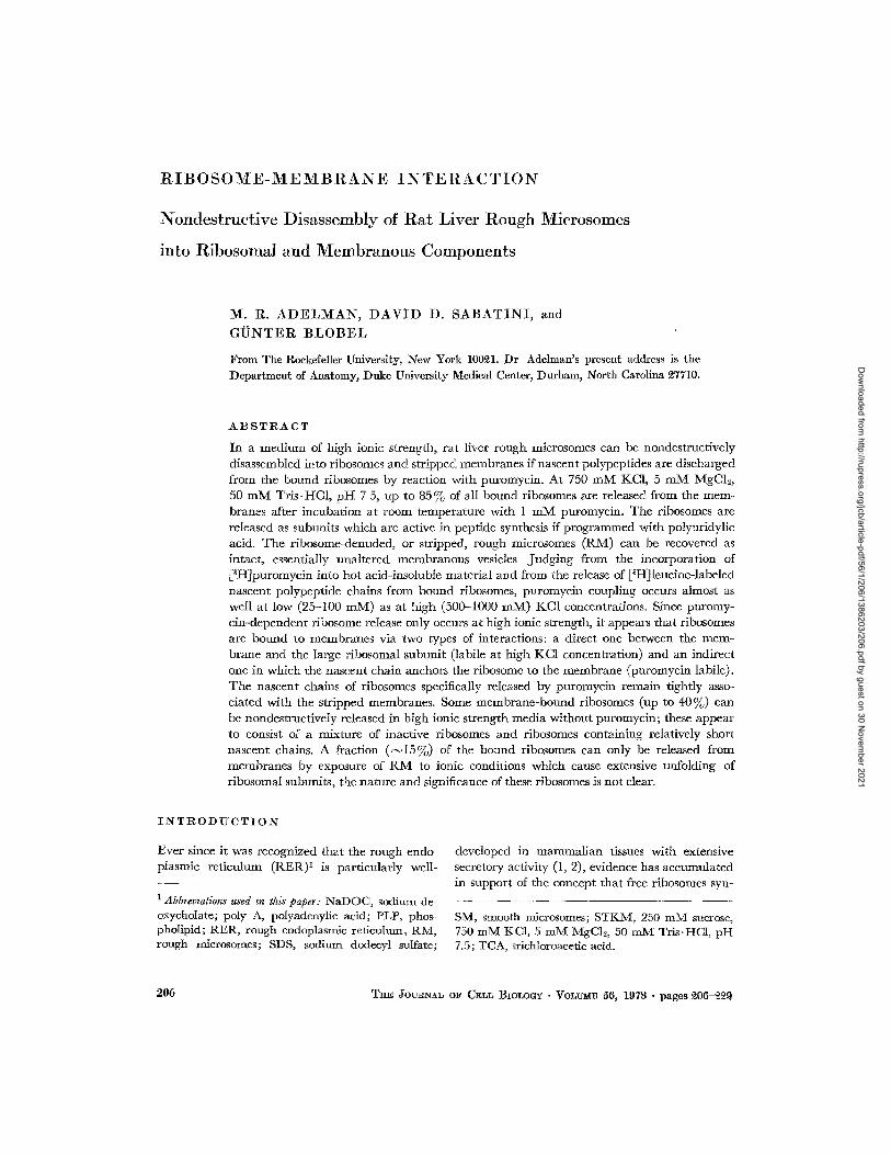
lq'mlJl~E 3 Separation of rough mierosomes into ribosomal and membranous components Rough micro- somes were incubated in S ~50, Mg 5, T 50 at various KC1 concentrations in the absence (open symbols) or presence (solid symbols) of 0.95 N 10 -~ 2vl puromycm. Samples (1 ml, eontaimng 0.5~6 mg RNA) were incubated an plastm centrifuge tubes for 60 rain at 3°C, 15 min at 37°C, and then chilled. Each sample was underlaid with 1 ml of 1.8 M sucrose containing the ~ppropriate K, :~Ig, T buffer. Sedimentation was at 3°C: ~4 h-39K-A3~1 (315,000). The membranes packed at the interface were removed, along with the dear sample zone and a minimal amount of the 1.8 ~{ sucrose layer The ribosomal pellets were re- suspended in the 1.8 M sucrose layer to which was added 1-~ ml of water. Membrane and ribosome frac- tions were theI1 analyzed for RNA content. Circles indicate ribosome RNA~ squares indicate membrane tlNA, triangles are the sum of the respective ribosome and membrane RNA contents The dashed line indicates the total input RN& or 100,% recovery level.
at --~500 m M KC1 At KCI concentrat ions greater t h a n 100 raM, pu romyc in t r ea tmen t resulted in addi t ional release of ribosomes unt i l a t >500 m M KCI a m a x i m u m of 75-80 % of all bound ribosomes was released. In o ther experiments, microsomal membranes were separated from released ribo- somes by differential centr i fugat ion and repeated washing Chemical analysis indicated tha t release of ribosomes by KCl -puromyc in reached a maxi- m u m of ~ 8 5 % when the incubat ions were carr ied out at room t empera tu re or 37°C as long as the samples were not subsequently cooled If samples were cooled before analysis, a small a m o u n t of " r e b i n d i n g " appeared to occur, since slightly more R N A (up to 25% of the total) was associated wi th the membranes
The effect of various KC1 and MgCI~ concen- trat ions on the extent of r ibosome release was evaluated on R M incuba ted for 2 h at room tem- pera ture in the presence of puromycin (Fig. 4) T h e release was strongly enhanced by elevated
KC1 concentra t ions at all levels of MgCI~., but , a t
a given KC1 concentra t ion, lower MgCI~ concen-
t rat ions only shghtly favored r ibosome release.
M a x i m u m release (Fig 4) was achieved at t m M MgC12 with KC1 in excess of 500 raM; chemical de terminat ions (data not included) showed tha t essentially 100% of the microsomal RATA was re- leased from the membranes unde r these conditions However, h igh ratios of K + : M g 2+ not only disso- ciate ribosomes, bu t also cause changes in the sedi- men ta t ion rate which have been a t t r ibu ted to unfolding of r ibosomal subunits (34) In par t icular , the 60S (large) r ibosomal subuni t undergoes a t ransi t ion to a slower-sedimenting species which is inact ive by vi r tue of having lost 5S R N A and several r ibosomal proteins (35). T h e arrows in Fig 4 indicate the approximate t ransi t ion regions (between 125 and 200 m M KC1 at 1 mNI Mg, be tween 750 and 1000 m M KC1 at 5 m M Mg) over which this subumt unfolding was detected in the sucrose gradients ~ i t must be reemphaslzed t h a t
~vVe cannot, of course, distinguish between actual conformational changes due to ionic conditions, per se, and pressure effects due to the high centrifugal fields (36), the data simply indicate that functional subunits are not recovered from gradients run under excessively high K+:Nlg 2+ ionic conditions.
1%[. R. ADEL~N, DAVID D S.&BATINI, AND G~NTER BLOBEL R~bosome-Membrane Interaction 211
Dow
nloaded from http://rupress.org/jcb/article-pdf/56/1/206/1386203/206.pdf by guest on 30 N
ovember 2021

3 0 O) t o
O)
-5
A:3
g d ~ <f
16 mM MgCI2 t
] / /
8
4 i
I I I I I I 0 200 400 600 800 I000
KC] (raM)
:FIotmE 4 Release of ribosomes from RM at various KC1 and MgC12 concentrations. Rough microsomes were suspended in a large volume ($5 ml; approxi- mately 0.£ mg RNA/ml) of S 250, T 50 and either 1 mM MgC12 (1), 5 mM MgC12 (O), or 10 mM MgC12 (A). After N10 min incubation on ice, the RM were centrifuged 15 min-80K-A~ll (~90,000). (These equilibration washes were shown to result in negligible release of ribosomes.) The RM were then resuspended in ~ $ ml of fresh buffer and brought by dilution to ilnal conditions of S 250, T 50, 0.79 X 10 -~ M pm'o- mycin, KC1 as indicated, and either 1, 5, or 10 m]~¢l 1VfgC12. Samples were incubated ~ h at room tempera- ture, and equal samples were applied to 15-40% sucrose gradients made in the appropriate K, Mg, T buffer. Sedimentation at g0°C: ~ h-40K-SB~83 (~70,000). Qtmntitation of absorbance released was as in ]Fig 2. The arrows next to the 1 mM Mg ( ~ - - ~ l ) and 5 mM Mg (O O) curves indicate the approxi- mate transition region for "unfolding" of the large ribosomal subunits (see text).
numerous experiments indicated that 80-85% of all R N A was released from R M incubated with puromycin in the presence of 750 m M KC1 and 5 m M MgCI~, conditions which did not lead to gross conformational changes in the subunits On the other hand, release of ribosomes in excess of 85% was only achieved under conditions which greatly altered the sedimentation of large ribosomal sub- units In fact, if the subunits are unfolded, corn-
plete ribosome release can be effected in the absence of puromycm For example, virtually all ribosomes can be stripped from R M by exposure to 1 0 mKC1, if MgCI~ is omitted (D. Borgese, un- published observations) Based on the above data, we adopted the following conditions for routine disassembly of rough mmrosomes which were chosen to maximize release of ribosomes and sepa- ration of ribosomal subunits, yet minimize dele- terious effects on either ribosomes or membranes. R M were incubated at a concentration of ~--1 mg R N A / m l with ~ 1 m M puromycin in S 250, K 750, Mg 5, T 50 The temperature was usually maintained at 3°C for 30-60 min and then raised to room temperature or 37°C for a time which depended on the particular experiment Since we have recently found that when R M are incubated at 37°C in S 250, K 25, Mg 5, T 50 subsequent treatment with KCl-puromycin leads to the release of partially aggregated large subunits, we prefer to complete the treatment by incubating the R M for 30-60 rain at room temperature. Separation was usually carried out by gradient centrifugation (as in Fig I) at 20°C, or, in those cases where only recovery of the stripped microsomes was desired, by differential centrifugation
(b ) Characterization of the Separated
Ribosomes and Membranes
To assay the functional capacity of the ribosomal subunits released from R M by KCl-puromycin t reatment (750 m M KC1, 1 m M puromycin) the separated subunits were recovered from the sucrose gradients, diluted with K25, Mg 5, T 50, and sedi- mented When recombined in an in vitro amino acid incorporation system (data not shown), these subunits were ~ - ~ as active as the original R M (per milligram RNA) in polyuridylie acid-depend- ent [14C]polyphenylalanine synthesis. While the 40S subunit assayed separately showed little ac- tivity, the samples of large subunit were N20 % as active as the recombined subunits, presumably be- cause of cross-contamination.
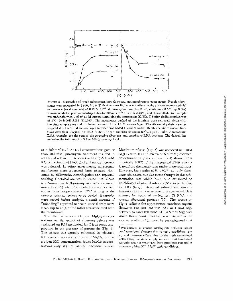
The appearance of the R M membranes re- covered after high KCl-puromycin incubation (stripped RM) and the extent of ribosome detach- ment were assessed by electron microscopy. Fig. 5 a presents a field of untreated B2v[ to be com- pared with a similar field of stripped membranes (Fig. 5 b). Most of the ribosome-denuded micro- somal membranes were recovered as intact vesicles,
212 T~E Joc~N~tL o~ CELL BIOLOGr • VOLtr~E 56, 1973
Dow
nloaded from http://rupress.org/jcb/article-pdf/56/1/206/1386203/206.pdf by guest on 30 N
ovember 2021

FIGVnE 5 Electron mlcrographs of R ~ I before and after removal of ribosomes with KC1 and puromycm. Both pellets were resuspended in S ~50 and processed for electron microscopy. Representat ive fields are shown a t a final magnification of Y( 5{),000.
:FIGVR~ 5 a Rough microsomes were suspended in an excess of S 250 and centrifuged a t 3°C. 30 m m - ~7K-No. 80 (~85,000).
M. R. ADELMAN, DAVID D. SA~ATI~I, AND G~dNTER BLOBEL R&osome-Membrane Interaction 213
Dow
nloaded from http://rupress.org/jcb/article-pdf/56/1/206/1386203/206.pdf by guest on 30 N
ovember 2021
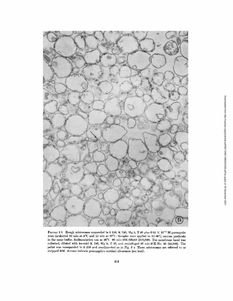
FmUR~ 5 b Rough microsomes suspended in S 250, K 750, Mg 5, T 50 plus 0 95 X 10 -~ M puromycin were incubated 60 rain a t 3°C and 15 mln a t 37°C. Samples were applied to 1 5 - 4 0 ~ sucrose gradients in the same buffer. Sedimentat ion was a t ~0°C 90 min-40K-SB~83 (~70,000). The membrane band was collected, diluted with ice-cold K 750, Mg 5, T 50, and centrifuged ~0 min-~TK-No. 30 (85,000). The pellet was resuspended in S 250 and resedimcnted as in Fig. 5 a These microsomes are referred to as str ipped R M . Arrows indicate presumpt ive residual ribosomes (see text).
214
Dow
nloaded from http://rupress.org/jcb/article-pdf/56/1/206/1386203/206.pdf by guest on 30 N
ovember 2021

TABLE l
Chemwal Analyszs-of Rough and Smooth 3fic~osomes before and after St~zppzng of Rzbosomes wzth KCl and Pu~omyczn
Sample Protein RNA PLP [~HJRNA RNA/Protein RNA 'PLP PLP/Protem
Rough mlcrosomes
Str ipped R?~I Smooth
mierosomes Str ipped SM
my~m[ mg/ml mg./ml @m ¢mg R~VA mg "my rag~rag mg %g 9.66 2.07 5 31 25,400 0 214 0 389 0 550
6.57 0 358 5 20 21,600 0 054 0.069 0 791 12.2 0 498 7 10 24,500 0.041 0.070 0 581
7 95 0 063 5.46 12,600 0.008 0 012 0 687
Both rough mlcrosomes and smooth microsomes were prepared at the same ttme from rats which had re- ceived an inject ion of [3H]orotic acid (200 #Ci, 2 5 mC1/mol) ~40 h before sacrifice. R1V~ and SNI pellets were resuspended m S 250, K 25, Mg 5, T 50. Samples of each were incuba ted for 2 h at room tempera tu re in the presence of (final concentrat ions) S 250, K 750,Mg 5, T 50, and 0 68 X 10- a M puromycin The sam- ples were di luted with an excess of room tempera ture K 750, Mg 5, T 50 and centr i fuged at room tempera- ture 15 min-30K-A211 (~90,000). The pellets were resuspended in cold buffer and recentr i fuged Each pellet was then resuspended in S 250, K 25, 1V~g 5, T 50 to the same volume as the original sample. Sam- ples of the un t rea ted R1V~ and S1V[, as well as the KCl-puromyeln- t rea ted mierosomes, were analyzed for protein, phosphol ipid phosphorus, RNA, and 3H cpm in the RN A hydrolyzate.
a l though the m e a n vesicle d iameter appeared to have been reduced slightly by the s tr ipping proce- dure Residual ribosomes, when detectable (arrow, Fig 5 b), most often appeared where two vesicles abu t ted on one another . T h e results of chemical analysis of typical str ipped R M which were, in this case, p repared from [3H]orotic acid-injected am- mals, are presented in Tab le I Inc luded for com- parison are da ta on the original R M , as well as on smooth microsomes before and after KCl-puro- mycin t r ea tmen t The phosphohpid (PLP) deter- minat ions indicate tha t the recovery of R M mem- branes after s t r ipping was near ly total, while tha t of smooth microsomes (SM) t reated for str ipping was somewhat lower (probably because of incom- plete sedimenta t ion of the membranes) T h e re- covered str ipped R M conta ined ~ of the init ial protein, and --o15% of the R N A While the un- t rea ted rough and smooth microsomes conta ined R N A labeled to a specific activity of N25,000 c p m / m g , the specific activity of the residual R N A in each str ipped m e m b r a n e fract ion was lower I t should be noted, however, t ha t the extremely small a m o u n t of R N A present in the str ipped SM was at the lower l imit of re l iablhty of the chemical assay, while the a m o u n t of R N A remain ing in the s t r ipped R M was more significant, an error of 5 - 1 0 % m the chemical assay could not be ruled out In other experiments also using R M conta in- ing t r i t ium-labeled R N A (same ba tch of R M used for the exper iment in Fig 3), wi th an init ial spe-
cific activity of 23,000 c p m / m g RNA, all residual m e m b r a n e s h ad specific activities of 22,000 -4- 2000 c p m / m g R N A except for those m e m b r a n e s recovered after pu romyc in t r ea tmen t at 400 m M KC1 or higher, which h ad lower values rang ing from 18,500 to 19,800 c p m / m g . T h e significance of this lower specific activity is no t clear, bu t it has been repor ted t ha t microsomes conta in polyaden- ylic acid (poly A) (37, 38) Computa t ions based on the publ ished da ta suggest t ha t a m i n i m u m of 0 1- 0 2 % of the microsomal R N A is poly A, an d since no estimate of poly A recovery was available, the poly A content of microsomes migh t be signifi- cant ly h igher Since we used [3H]orotic acid for R N A label ing and since orotic acid is not taken up into adenine (39), i t is not unreasonable to postu- late the existence in the residual R N A of unlabe led poly A amount ing to 1 - 2 % (15% residual, I 0 - 15% of this unlabeled) of the total microsomal R N A
(c) The Specific Nature of Puromycin-
Dependent Ribosome Release
T h e da ta presented up to now demonst ra te a pu romvc in -dependen t release of ribosomes which has been defined as the difference between the KCl -puromyc in release and the salt release. By prewashing R M to remove those ribosomes re- leased by salt alone, and then resuspending the R M in h igh KC1 buffer, it was possible to study
M. R. ADELMiN, DAVID D. SiBATINI, A.ND GI3NTER BLOBEI~ Ribosome-Membrane Interaction 215
Dow
nloaded from http://rupress.org/jcb/article-pdf/56/1/206/1386203/206.pdf by guest on 30 N
ovember 2021
15 ¸
E ~ 1 0
I f ) o J
,d
0 5
Control
Pu~omycln
Mb
L
+Puromycm
Mb
L
I s
Prewoshed
-Puromycm
Mb
+Puromycm
L
Mb
FIGVRE 6 Effect of prewashing on the extent and specificity of puromycin-dependent ribosome release. RM were suspended in S ft50, Mg 5, T 50 plus £5 mM JKCI (control) or 750 mM :KC1 (prewashed). Manip- ulations are described in the footnote to Table II. The mmrosomes were resuspended in S ~50, Ig: 750, Mg 5, T 50 in the absence or presence of 0.79 X 10 -3 M puromycim Incubation and sedimentation as in foot- note, Table II. S = small ribosomal subunit; L = large subunit; Mb = membrane band
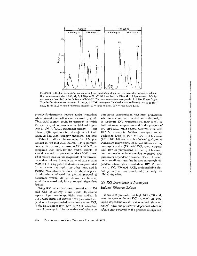
puromycin-dependent release under conditions where virtually no salt release occurred (Fig 6). Thus, R M samples could be prepared in which the specificity of puromycin action (defined in per- cent as 100 X [{KCl-puromycin release} - {salt release}]/[KCi-puromycin release]) at all ionic strengths had been strikingly enhanced The data m Table II indicate, for example, that RM pre- washed in 750 mM KC1 showed ~ 8 4 % puromy- cin-specific release (treatment at 750 mM KC1) as compared with 53% for the control sample It should be noted that prewashing the RM did some- what reduce the absolute magnitude of puromycin- dependent release. Reexamination of data such as those in Fig 2 suggested that salt release proceeded in two stages, one rapid, the other slow, and it seemed reasonable to conclude that the slow phase of salt release reflected the gradual removal of ribosomes which, during shorter incubations, would be released only in a puromycin-dependent fashion
Using R M which had been prewashed at 750 mlV[ KC1 (as for Fig 6 and Table II), several aspects of puromycin specificity were studied It was found (data not shown) that puromycin-de- pendent release proceeded more slowly at low KC1, in the cold, and at low (10-6-10 -4 M) concentra- tions of puromycin. The dependence of release on
puromycin concentration was most pronounced when incubations were carried out in the cold, or at moderate KC1 concentrations (250 mM), or both. At room temperature and in the presence of 750 mM KC1, rapid release occurred even with 10 -5 M puromycin. Neither puromycin amino- nucleoside (0 83 X 10 -6 M) nor cycloheximide (4.2 X 10 -3 M) was capable of releasing ribosomes from rough microsomes. Under conditions favoring puromycin action (750 mM KC1, room tempera- ture, 10 -6 M puromycin), neither cycloheximide nor puromycin aminonucleoside interfered with puromycin-dependent ribosome release. However, under conditions resulting in slow puromycin-de- pendent release (short incubation, 10 -~ M puro- mycin, 3°C, 750 mM KC1), cycloheximide (but not puromycin aminonucleoside) strongly in- hibited the effect
(d) KCl Dependence of Puromycin-
Induced Ribosome Release
When RM prewashed at high KCI (750 mM) were resuspended in low KCI (25 mM), no puro- mycin-dependent release was observed (data not
shown); thus, the puromycin-depeudent ribosome
release only occurred in the presence of high con-
216 THE JOURNAL OF CELL BIOLOGY " VOLUME 56, 1978
Dow
nloaded from http://rupress.org/jcb/article-pdf/56/1/206/1386203/206.pdf by guest on 30 N
ovember 2021
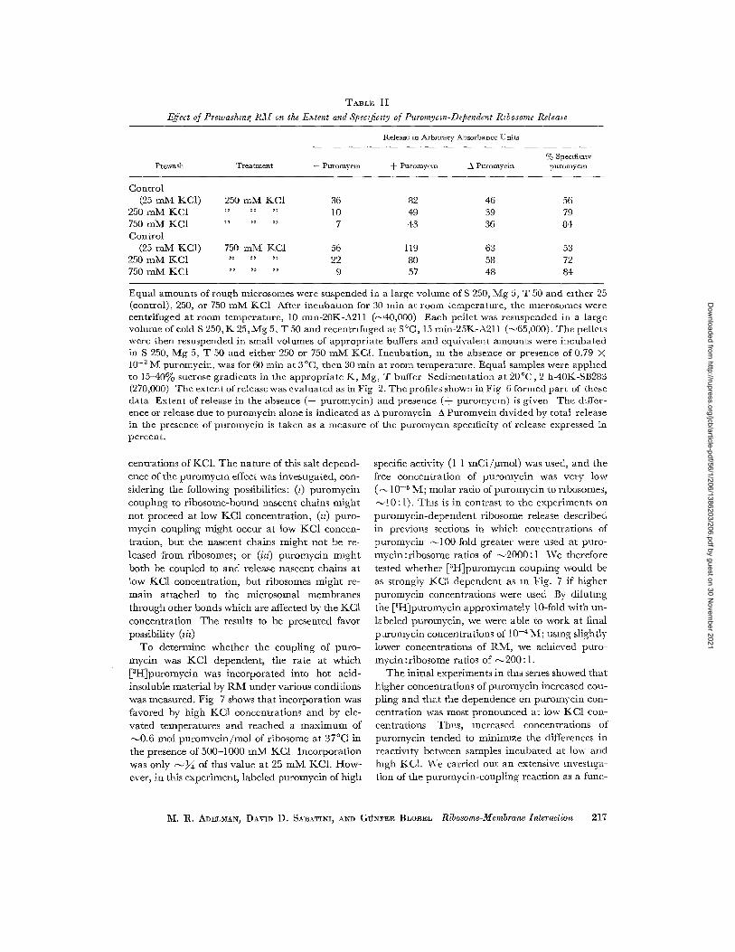
TABLE 11
Effect of Prewash~ng R~L r on the E:~tent and Speczfic~ty of Puromycm-Dependent R~bosome Release
Release an Arburary Absorbance Units
Prewas] ~, Treatment -- Puromycm -I- Puromyem ~ Puromycln % Specificity puromycm
Control (25 mM KC1) 250 mM KC1 36
250 mM KC1 " " " 10 750 mM KC1 . . . . . . 7 Control
(25 m M KC1) 750 m M KC1 56 250 m M KC1 " " " 22 750 m M KC1 . . . . . . 9
82 46 56 49 39 79 43 36 84
119 63 53 80 58 72 57 48 84
Equal amounts of rough microsomes were suspended in a large volmne of S 250, Mg 5, T 50 and e~ther 25 (control), 250, or 750 m M KC1 After i ncuba tmn for 30 min at room temperature , the mmrosomes were centr i fuged at room temperature , 10 mm-20K-A211 @~40,000) Each pellet was resuspended in a large volume of coId S 250, K 25, Mg 5, T 50 and recentmfuged at 3 °C, 15 min-25K-A211 (~65,000). The pellets were then resuspended in small volumes of appropr ia te buffers and equivalent amounts were incuba ted in S 250, Mg 5, T 50 and ei ther 250 or 750 m M KC1. Incubat ion , m the absence or presence of 0.79 >< 10 -~ IV[ puromycin, was for 60 rain at 3°C, then 30 min at room temperature . Equal samples were apphed to 15-40% sucrose gradients in the appropr ia te K, Mg, T buffer Sedimenta t ion at 20°C, 2 h-40K-SB283 (270,000) The extent of re leasewas evaluated as in Fig 2. The profiles shown in Fig 6 formed pa r t of these da ta Extent of release in the absence (-- puromycin) and presence (-~- puromycm) is given The differ- ence or release due to puromycin alone is indicated as A puromvcin A Puromvcln divided by total release in the presence of puromyein is taken as a measure of the puromycm specificity of release expressed in percent .
centrat ions of KC1. T he na tu re of this salt depend- ence of the puromycln effect was investigated, con- sidering the following possibilities: (0 puromyc in couphng to r ibosome-bound nascent chains m i g h t no t proceed at low KC1 concentra t ion, @) puro- mycin coupling migh t occur at low KC1 concen- trat ion, bu t the nascent chains migh t not be re- leased f rom ribosomes; or (iz0 purom:fcin migh t bo th be coupled to and release nascent chains at low KC1 concentrat ion, bu t ribosomes migh t re- ma in a t tached to the microsomal membranes t h rough other bonds which are affected by the KC1 concent ra t ion T he results to be presented favor possibility (~h)
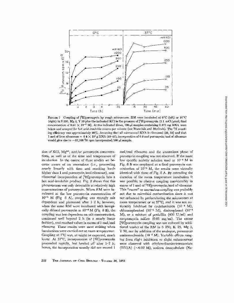
To de te rmine whether the coupl ing of puro- mycin was KC1 dependent , the ra te at which [aH]puromycin was incorpora ted into hot acid- insoluble mater ia l by R M under various condit ions was measured. Fig 7 shows t ha t incorpora t ion was favored by high KCI concentra t ions and by ele- va ted tempera tures and reached a m a x i m u m of ~ 0 . 6 mol p u r o m v c i n / m o l of r ibosome at 37°C in the presence of 500-1000 m M KC1 Incorpora t ion was only N ~ of this value at 25 m M KC1. How- ever, in this experiment, labeled puromycin of h igh
specific activity (1 1 mCi/ /~mol) was used, and the free concent ra t ion of pu romyc in was very low ( ~ 10 _5 M; molar ratio of puromyc in to mbosomes,
10:1). Thts is in contras t to the exper iments on pu romyc in -dependen t r ibosome release described in previous sections in which concentra t ions of puromycin ~ 100-fold greater were used at puro- myc in : r ibosome ratios of ~ 2 0 0 0 : 1 We therefore tested whe ther [3H]puromycm coupl ing would be as strongly KC1 dependen t as m Fig. 7 if h igher puromycin concentrat ions were used By di lut ing the [~H]puromycin approximate ly 10-fold wi th un- labeled puromycin, we were able to work at final puromycin concentrat ions of 10 -a M ; using slightly lower concentrat ions of R M , we achieved puro- mycin :r ibosome ratios of ~ 2 0 0 : 1.
T h e initial experiments in thts seines showed tha t higher concentrat ions of puromyc in increased cou- pling and t ha t the dependence on puromyc in con- cent ra t ion was most p ronounced at low KC1 con- centrat ions Thus, increased concentra t ions of puromycin tended to minimize the differences in reactivity between samples incuba ted at low and h igh KC1. We carr ied out an extensive mvesnga- t ion of the puromycin-coupl ing react ion as a func-
M. R. ADEL~AN, DAVID D. SAB-~TI~I, A~J) GffN~ER BLOBEL Ribosome-Membrane Interaction 217
Dow
nloaded from http://rupress.org/jcb/article-pdf/56/1/206/1386203/206.pdf by guest on 30 N
ovember 2021
2 0 -
~4 16- E
12-
o
T"
~ 4
o
0°C 57°C
mM KCI I000
I 2 3 4 22
T~me (h)
mM KCI 1000
5O0
25O .----.o
100 _ _ . - - - - - - o
, o
r I l I
15 50 45 60
Time (mm)
I~OURE 7 Coupling of [ztt]puromycin by rough mierosomes. RM were incubated at 0°C (left) or 37°C (right) in S 250, Mg 5, T 50 plus the indicated KC1 in the presence of [3It]puromycin (1.1 mCi//~mol; final concentration of 0.91 X 10 -5 M). At the indicated times, 100-/A samples containing 0.172 mg RNA were taken and assayed for hot acid-lnsoluble counts per minute (see Materials and Methods). The ztt count- ing efficiency was approximately 20%. Assuming that all microsomal RNA is ribosomal (32, 83) and that 1 tool of liver ribosomes = 2 4 X 10⁶ g RNA (40--42), incorporation of 0 5 mol puromycin/mol of ribosome would give rise to N17,500 ~H cpm incorporated/100/~l sample.
tion of KC1, M g 2+, a n d / o r puromycin concentra- tions, as well as of the time and temperature of incubation In the course of these studies we be- came aware of an anomalous (i .e, proceeding nearly linearly with time and reaching levels higher than 1 tool puromycin /mol ribosome), non- ribosomal incorporation of [~H]puromycin into a hot acid-insoluble product Fig. 8 shows that this phenomenon was only detectable at relatively high concentrations of puromycin. When t{IV[ were in- cubated at the low puromycin concentration of 10 -6 M (Fig 8 A), coupling was strongly salt dependent and plateaued after 1-2 h; however, when the same R M were incubated with isotopi- cally diluted puromyein at 10-* M (Fig 8 B), the coupling was less dependent on salt concentration, continued well beyond 2 h (in a nearly linear fashion), and reached values in excess of 1 mol /mol ribosome These results were most striking when incubations were carried out at room temperature. Coupling at 3°C was, as might be expected, much lower At 37°C, incorporation of [3HJpuromycin proceeded rapidly, but leveled off after 1-2 h; hence, the incorporation usually did not exceed 1
tool / tool ribosome and the anomalous phase of puromycin coupling was not observed. If the same low specific activity solution used at 10 -4 M in Fig. 8 B was employed at a final puromycin con- centration of 10 -5 M, the results were virtually identical with those of Fig. 8 A. By extending the duration of the room temperature incubation it was possible to observe coupling considerably in excess of 1 tool of [3H]puromycin/mol of ribosome This "excess" or anomalous coupling was probably not due to microbial contamination since it was not enhanced by preincubating the microsomes at room temperature or at 37°C, and it was not no- ticeably inhibited by cycloheximide (10 -4 M), chloramphenicol (10 -4 M), dinitrophenol (10 -4 M), or a mixture of penicillin (400 U / m l ) and streptomycin sulfate (0.05 mg/ml) . The excess [aI-~puromycin coupling was not reduced by addi- tional washes of the I{M in S 250, K 25, Mg 5, T 50, nor by addition of the analogue, puromycin aminonucleoside (10 -a M). Variable effects rang- ing from slight inhibition to slight enhancement were observed with ethylenediaminetetraacetatc (EDTA) (~0 .02 M), sodium deoxycholate (Na-
218 T~E JOURNAL OF CELL BIOLOGY " VOI~U~IE 56, 197~
Dow
nloaded from http://rupress.org/jcb/article-pdf/56/1/206/1386203/206.pdf by guest on 30 N
ovember 2021

25- ro b
E 20-
o
c 15-
> .
E o
n ~ i0-
T t~3
5-
Low puromycm concn
A
mM KCI 697 ~ ..._,.__.~o 248
...__.---o 23
I I I I I 2 3 4
f High p u r o m y c m conch
B mMo KC- ~
248 -25
o
-20 E Q,.
15 -
E o
Io a_
" l - t o
5
I 2 3 4
Thme (h)
FmUR~ 8 Coupling of [aFi]puromycin by rough microsomes. I~M were incubated at room temperatm'e (~-~°C), in S £80, Mg 4.5, T 45 plus the indicated XC1. Incubations were carried out at a pm'omycin: ribosome ratio of 16 mol/mol (left; 0.8~4 X 10 -5 M; 1.1 mCi/~mol) or at a ratio of 18~ mol/mol (right; 0 98~ X 10 -~ M; 0 097 mCi/~mol). At the indicated times, 100-~1 samples containing 0.108 mg Riga were taken for determination of counts per minute incorporated. In each panel, the counts per minute corre- sponding to incorporation of 1 mol puromycin/mol ribosome is indicated by the dashed line. :Note that the counts per minute scales differ by a factor of 10.
DOC) (0 5%), or Tri ton X-100 ( I -2%) . Both smooth mlcrosomes and stripped R M showed the excess coupling reacUon. By first reacting SM or R M briefly with 10 -3 M cold puromycin, sedi- menting the mierosomes, and then resuspending them in labeled puromycin, it was possible to label primarily the excess coupling component The in- corporated tritiated puromycin banded in sucrose density gradients coincident with the membranes Bound ribosomes isolated from detergent-solubi- lized R M in such a way (see legend to Fig. 9) as to minimize contamination of the isolated ribo- somes with solubilized membrane components showed no traces of excess puromycin coupling We concluded, therefore, that microsomal mem- branes contained some component, distinct from the attached ribosomes, which was capable of cou- pling puromycin to hot acid-insoluble material. The enzyme or enzymes involved required fairly high concentrations of puromycin and survived detergent solubilization, this phenomenon is being explored further
Using detergent-derived bound ribosomes, puro- myein coupling was studied without the compli- cations introduced by the anomalous coupling
reaction in R M . The results verified our initial conclusion that increased concentrations of puro- mycin and prolonged incubations (at room tem- perature) tended to minimize differences in reac- tivity between low and high KC1 incubations. The data in Fig 9 show that bound ribosomes coupled about the same amount of puromycin over a broad range of KC1 and MgC12 concentrations and that at very low KCI (less than 25 mM), extensive reaction occurred. Because it is reasonable to as- sume that microsomal ribosomes react with puro- myein essentially as well before as after detergent treatment, we concluded that membrane-bound ribosomes reacted as extensively with puromycin at low KC1 (100 raM) as at high KC1 (750 raM), even though such reaction only led to release of ribosomes at high ionic strength. Fur ther support for this conclusion was obtained from experiments in which we estimated the extent to which ribo- some release at high ionic strength was facilitated by previous incubation of the R M with puromycin at low ionic strenght R1V[ which were incubated with puromycin in 25 m M KC1 and washed (lower profiles in Fig 10; sample B) no longer reqmred the presence of puromycin to release ribosomes in
~¢[. !{. AOlgL~lg, DAVID D. SABATI~, AND Gt~NT~R BLOBEL Ribosome-Membrane Interaction 219
Dow
nloaded from http://rupress.org/jcb/article-pdf/56/1/206/1386203/206.pdf by guest on 30 N
ovember 2021

7 O
E CL (D
C (D
~" to o
0-
0
15
I I I I I 200 400 600 800 i000
KCI (raM)
FIeU2E 9 Coupling of [gH]puromycin by bound ribo- somes. RM were resuspended in S 250, K 35, Mg 5, T 50 and diluted by 10% with a solution of 5% DOC and 10% Triton X-100. The suspension ( ~ 5 ml/tube) was placed in plastic centrifuge tubes and underlaid with 2 ml of (9 parts S 1S50, K 35, Mg 5, T 50 + 1 part 5% DOC -- 10% Triton) and with 3 ml of S 1600, K 35, Mg 5, T 50. Sedimentation was at S°C: 13 h-58K- ASgl (~g00,000). The supernatant was discarded, the inside of the tubes was rinsed with a small amount of water, and the pellets were resuspended in appropriate buffer. These "bound" ribosomes were incubated for 3 h at room temperature in S ~30, T 45, the indicated KC1 concentrations, and either 0.91 (©), 4 5 (@), or 9.1 ( X ) mM N[gCI~. [gH]Puromycin (0.097 mCi / gmol) was present at a final concentration of 0.9~ X 10 -4 M; the puromycin/ribosome mole ratio was -o146. Duplicate 1O0-#l samples, containing 0.138 mg RNA were assayed for pm'omycin coupled to hot acid- insoluble material The dashed line at 1610 cpm indi- cates coupling of ~-~0 7 tool pm'omycin/mol ribosome.
750 m M KC1. R M pre incuba ted wi thout puro- mycin in 25 m M KC1 (upper profiles in Fig 10; sample A) behaved identically wi th a nonpre incu- ba ted control (sample C, profiles not shown), and requi red puromycin for release of ribosomes a t h igh ionic s t rength Quan t i t a t i on of the areas unde r the r ibosomal peaks in Fig. 10 indicated tha t 7 0 - 8 0 % of the ribosomes specifically released by puromyc in f rom sample A were released in the absence of added puromyc in from sample B T h e extent to which puromycin coupling occurs at low ionic s t rength was also inferred from the effect of p re incuba t ion wi th puromycin (nonlabeled) at low ionic s trength. Samples of A, B, and C (preincu-
10-
0 5 -
E 0
eJ <~
I 0
0 5 -
Premcubohon wdhout Puromycm
- Puro
L
Mb + Puro
L
Mb
Premcubatlon w~th Puromycm
- Puro
Mb
L.
+ Puro
Mb
L
FIGURE 10 Reaction of rough microsomes with pm'o- mycin at low KC1. RM were suspended in S 350, K 25, Mg 5, T 50 and separated into three equal samples, A, B, and C. A and B were incubated in the absence (A), and presence (B) of 0.86 X 10 -8 M puromycin for 60 min at 8°C and 60 rain at room temperatm'e All three samples ( C serving as a control for the room tempera- ture incubation) were diluted with cold S 250, K 25, Mg 5, T 50 and centrifuged at S°C: 15 min-27K-no. SO (85,000). The pellets were resuspended in S 250, K 25, Mg 5, T 50 and rewashed by zone sedimentation (at S°C) through 15-40% sucrose gradients made up in K 35, Mg 5, T 50; S0 min-40K-SB283 (270,000). The pellets were resnspended and incubated in S 350, K 750, Mg 5, T 50 in the absence and presence of 0.79 X 10 -8 M puromycin. Incubation was for 60 rain at S°C and 15 min at $7°C. Sedimentation analysis at 30°C: 2 h-40K-SB288 (~70,000). The upper panels show the results of analyzing sample A; virtually identical re- sults were obtained with the control sample C. The lower panels represent sample B (preincubation with puromycin).
2 2 0 THE JOURNAL OF C E L L B I O L O G Y • V O L U M E 56, 1 9 7 3
Dow
nloaded from http://rupress.org/jcb/article-pdf/56/1/206/1386203/206.pdf by guest on 30 N
ovember 2021

- Puromycln + Puromvcm
-Detergent +Detergent
A
15-
M
- Detergent + Detergent
C D
; 1
E I 0 -
I '
J
o 5 ]5 P 5 [5 5
Froct lon no
I I
I ~o
_o 6 ×
E
4
¢9
0 i5 P 5 15 P
FIeVR~ 11 Release of rtbosomal nascent chains by t r ea tment of rough microsomes with puromycin at low ](C1. The R • were prepared from rats which received an injection (portal veto) of [~H]leucine ~2 rain before excision of the liver. The specific act ivi ty of the isolated R M was ~ . ~ X 105 cpm/ rag R N A Equal amounts of R M were incubated in S ~50, K 100, ~J[g 5, T 50 in the absence (A and B) or presence (C and D) of 0.79 X 10 -~ M puromycin. Incubat ion was for 60 m m at 8°C and 15 m m at room temperature. Samples B and D were then diluted 8% with a solution of 5% DOC, ~20% Tri ton X-100 (A and C re- ceived water). Samples were applied to 15-40cY~o sucrose gradients made up m the K, Mg, T buffer Sedi- menta t ion was at ~20°C: 1 h-40K-SB~83 (~70,000). Frac tmns were collected and processed for scintillation counting; the pellet was also counted (P). A2~4, ; [sIt]leucine counts pet" mln, • . . . . . O.
15
I
U'3
e,I <[
0 5 -
-Puromycln
-Detergent + Detergent
B
Mb f
+L ,! /
5 15
M s i
5 15
Froct lon no
- Detergent
Mb
II II
I
II I I i I
f I I
I 15 P
+ Purornycln
+ Detergent
L
!s -4 ~Is
o 5 15 P
-t5
"lo T I
i,o
- 8 b
- E ~2L
FIGVRE lg Release of ribosomal nascent chains by t r ea tment of rough mlcrosomes with puromycm at high ]KC1. The experimental protocol was identical with tha t of Fig 11, with the following exceptions: (a) the concentration of R M was slightly lower t han m Fig. 11; (b) all solutions contained 750 m M KC1, rather t han 100 m M KC1; (c) sedimentat ion was for ~ h.
M. R. ADEr~AN, D~-VID D. SABATINI, AND GtnnT~R BLOBEL Ribosome-Membrane Interaction 221
Dow
nloaded from http://rupress.org/jcb/article-pdf/56/1/206/1386203/206.pdf by guest on 30 N
ovember 2021

bated and washed as in the legend to Fig. 10) were mixed with labeled puromycin at low KC1 and after 15 min at room temperature, the samples were diluted to final concentrations of S 250, K 750, Mg 5, T 50 and 0.90 X 10 -5 M [3H]puromy- cin (1 1 mCi/mmol, at this low puromycin concen- tration excess coupling was negligible). After incubation for 60 min at room temperature, assays of equal samples (100 ~ul) of A and C indicated incorporation of 10,519 and 10,767 cpm respec- tively, while the corresponding figure for B was 3028 cpm. These results and the data in Fig 10 showed that RM exposed to puromycin at low ionic strength reacted to at least 70 % of their max- imum coupling capability, yet the ribosomes were not released until the salt concentration was raised.
Having demonstrated that nascent chains in membrane-bound ribosomes do react extensively with puromycin at low KCI, we studied whether the peptidyl-puromycin molecules formed were re- leased from the ribosomes under such condmons. For these experiments we used RM prepared from animals injected 2 min before sacrifice with [~H~- leucine so as to label nascent polypeptide chains. The analyses (Figs. 11 and 12) were carried out using zone sedimentation in continuous sucrose density gradients since we had found that, by dif- ferential centrifugation, it was not possible to dis- tinguish ribosome-associated radioactivity from labeled material which simply cosedimented with ribosomes. In fact, as the data in Figs. i 1 and 12 show, if microsomal membranes are dissolved with detergents, the puromycin-released polypeptides which they contain have a striking tendency to aggregate and sediment heterogeneously through- out the gradient The analysis in Fig. 11 was carried out at 100 mM KC1, an ionic strength which somewhat reduced the chain aggregation (the aggregation is particularly troublesome in K 25, Mg 5, T 50), but was sufficiently low that no puromycm-dependent ribosome release occurred (cf Fig 1). At this low KCI concentration only a small amount of salt-released ribosomes containing some nascent polypeptide chains was present in the gradient (Fig 11 A), While the microsomes sedi- mented to the bottom carrying with them most of the ribosomes and [aH]leucine label. The use of detergent to dissolve the membranes and display all ribosomes within the gradient (Fig 11 B) re- vealed that only 50% of the radioactivity was actually associated with ribosomes (i e., in a radio- activity peak coincident with the absorbance pro- file in the ribosome region of the gradient). The
remainder, presumably in chains which had been completed and released from ribosomes during the 2-rain in vivo pulse, was found near the top of the gradient Addition of puromycin (Fig. 11 C) re- leased to the supernatant only the radioactivity in those nascent chains which were attached to the salt-released ribosomes (of. Fig. 11 A) and did not cause additional release of ribosomes from the microsomes. Detergent treatment of RM which had been incubated with puromycin (Fig 11 D) revealed a radmactivity distribution quite distinct from that seen in Fig 11 B A larger fraction of the radioactivity was found near the top; while the rest sedimented heterogeneously throughout the gradient, there was no sign of a peak of radioac- tivity coincident with the ribosomal absorbance peak, thus demonstrating that the peptidyl-puro- mycin molecules had been discharged from the ribosomes Summation of the radioactivity in frac- tions 4 through P in Fig. 11 D gave an estimate of labeled material which, by differential centrifuga- tlon, would have been sedimented into a pellet with the ribosomes This was found to be ~45 % of the corresponding total in Fig. 11 B. an estimate which is m good agreement with data (not shown) we have obtained using differential centrifugation
When a similar experiment as that in Fig 11 was carried out with RM at high ionic strength (Fig 12) (750 mM KC1), the addition of puromy- cin (Fig. 12 C) resulted in release of the bulk of the ribosomes as subunits, which were virtually devoid of associated nascent chains (cf Fig. 12 B). The stripped microsomal membranes banded isopyc- nically within the gradient and retained almost all of the radioactivity This radioactivity was released upon detergent dissolution of the stripped mem- branes (Fig. 12 D) and appeared both at the top of the gradient and sedimenting heterogeneously as in Fig. 11 D We therefore concluded that mem- brane-bound ribosomes not only reacted with puromycin at low KC1, but that this reaction re- sulted in release of nascent polypeptide chains from the ribosomes which was as efficient at low as at high KCt. We could not directly rule out the pos- sibility that the detergent treatment itself caused release of puromycin-coupled chains at 100 mlV[ KC1, but experiments with leucine-labeled free ribosomes not treated with detergents gave results (not shown) similar to those in Fig I 1, and there- fore rendered thts latter possibility unlikely
The experiments just described showed that when ribosomes were released from RM specifi- cally by puromycin, most of the labeled nascent
222 TI~E JO~'BNAL OF C:~LL BIoI,oGY • VoLuM~ 56, 1973
Dow
nloaded from http://rupress.org/jcb/article-pdf/56/1/206/1386203/206.pdf by guest on 30 N
ovember 2021

chains remained firmly associated with the re- sidual or stripped R M (Fig. 12 C), thus providing a demonstration of vectorial discharge (14). How- ever, because much of the radioactivity in Fig. 12 C represented completed chains not actually re- leased from ribosomes by puromycin, we used [~H]- puromycin to label specifically the nascent chains and demonstrate directly their vectorial discharge to the mmrosomes. As shown in Fig 13 B, incuba- tion of R M in high KC1 with [SH]puromycin re- sulted in release of ribosomes and the coupling of [3H]puromycin to hot acid-insoluble material which banded isopycnicalty with the stripped membranes. The small amount of radioactivity in the supernatant fractions probably represented puromycin coupled to chains from the salt-re- leased ribosomes (see Fig. 12 A), since when R M were prewashed at high KC1 and then treated and analyzed as in Fig 13 the amount of acid-insoluble [~H]puromgcin found in the supernatant fractions was reduced.
(e) The Nature of Salt-Released Ribosomes
To investigate if those ribosomes released by salt alone were functionally different from the bulk of the bound ribosome population, we compared the [aH]puromycin coupling ability in 750 m M KC1 of (0 total RM, (~i) salt-released ribosomes prepared from R M by preincubation at 750 r am KC1, and (tit) R M which had been depleted of sah-releasable mbosomes by washing in 750 m M KC1 The [~H]- puromycin radioactivity coupled was found to be 94,000, 48,000, and 119,000 c p m / m g of R N A for z, n, and iii respectively (puromycin 10 -~ M, 1.1 mCi /#mol) . Thus, the ribosomes released by salt alone were roughly 50 % as ' :active" as those which could onIy be released by incubation with puro- mycin Examinat ion of Figs 11 and t 2 also indi- cates that, after in vivo pulse labeling, salt-released ribosomes contained less radioactivity in nascent chains (per absorbance unit) than did those ribo- somes which remained attached to the R M and were released by detergent (compare Fig 11 B with Fig. 11 A, and Fig. 12 B with Fig 12 A) Moreover, to the extent that dissociation at high ionic strength into subunits in the absence of puro- mycin can be taken as an indication of reactivity (31), comparison of Figs 12 A and B also suggested that salt-released ribosomes were relatively inac- tive.
So as to determine whether the specific activity ([~H]leucine counts per minute per milligram
I
C4 <~:
15-
l o -
0 5 -
- Puromycm
A
ML
L
+ ['~H] P u romycm
B Mbl Rl II I/ II
II I I
L
i !15 L [ i 5 Fraction no,
-25
-2O t
O
-15 × E
o -IO E
o Q_
Emcee IS Distribution in sucrose gradient of [3H] puromycin incorporated by rough microsomes. Rough microsomes were incubated in S "250, K 730, )~Ig 5, T 50 in the absence (A) and presence (B) of 0.8~ X 10 -5 M [3H]puromycm (1 1 mQ//~mol; excess coupling was negligible under these conditions). Incubation was for 15 mm at S°C, then 80 min at 87°C. Samples were applied to 15-40% sucrose gradients in the 7g:, Mg, T buffer. Sedimentation at £0°C: ~2 h-40K-SB~8$ (~270,000). Fractions were collected from B and~ along with the pellet, were assayed for hot acid-insoluble [3Icl]puromyein counts per minute, • ..... • .
RNA) of salt-released ribosomes was a function of the KC1 concentration at whmh they had been re- leased, we analyzed pulse-labeled R M by sedi- mentat ion in continuous sucrose gradients (Fig 14). As expected, increasing amounts of ribosomal material were released at increasing levels of KC1 In addition, it was found that increasing amounts of ribosome-associated radioactivity (i e , coinci- dent with the ribosomal peaks) were released as a function of the KC1 concentration Evaluat ion of the data in several ways gave the same conclusion: the ribosomes released at low KCI had lower spe- cific activity than those released at high KC1 The exact magmtude of this effect depended on the means of computing specific activity, defined in terms of counts in a particular region of the gradi- ent divided by absorbance (in a rb i t raw units as
~I. R, ADELN-~N~ DAVID D. S~mATI~I, ~ ,~ GV~T~a BLOBEL Ribosome-Membrane Interaction 223
Dow
nloaded from http://rupress.org/jcb/article-pdf/56/1/206/1386203/206.pdf by guest on 30 N
ovember 2021

150 rnM KCI 250 400 600
e
/ I
,o41, o lfl
/L,, 0 '
0 l0
!
t
l,/ii /,/e i
20 0 I0 20
rl fl II ft
II M
L
i I-
0 I0 20 Fraction no
¢ fl L
II M
I II
- - q - - i i
0 I0 20
-750
500 ,r E
500 o. O
200 _3
100
~kGU~ 14 Distribution of [~I-I]leucine-labeled nascent polypeptides on ribosomes released from rough mierosomes at various I(CI eoneentl'ations. The RNI were prepared from rats which received an injec- tion (portal vein) of [3H]leucine 3 mm before excision of the liver; specific activity of the isolated RM was 0.59 X 10⁵ cpm/mg RNA The t tM were incubated in S 350, Mg 5, T 50 plus 150, 350, 400, or 600 mM I(CI. Incubation was for 90 min at 8°C, then 60 rain at room temperature. Equal samples were applied to 15-33~0 sucrose gradients made up in the appropriate K, NIg, T buffer. Sedimentation was at ~0°C: 31/~ h-~0K-SB~83 (370,0(10). Under these conditions, membranes sedimented into a pellet; they were not resuspended for analys~s. Fractions were colleeted and processed for seinti[latlon counting. Azs~ ~ - - ; [3It]leueine counts per minute, • ..... •
evaluated in Fig, 2) under the U V profile in that region The specific activities of " to ta l" ribosomes released ranged from 3.2 (at low KC1) to 5 2 (high KC1), while the specific activities of the "mono- mer" ribosomes ranged from 3 7 up to 9 9.
While these data indicated that ribosomes re- leased at low KC1 were less active than those re- leased at high KCI, they did not allow us to decide if the low specific activity of the former were pri- marily due to (I) the presence of a large percentage of inactive ribosomes (i e , bearing no nascent chains), or (ii) the presence of a fairly uniform ribosomal population carrying relauvely short nascent chains Since the extent to which inactive ribosomes are dissociated into subunits is a strong function of the salt concentration, possibility i seemed likely This was tested using salt-released ribosomes which were prepared by exposing pulse- labeled R M to various levels of KCI, removing the membranes by differential centrifugation, and sedimenting the ribosomes from the supernatants The ribosomal pellets were all resuspended at the same KC1 concentration (750 mM) and analyzed on sucrose gradients run at the same high ionic strength. The results (not shown) indicated that a
larger percentage of those ribosomes released from R M at low KC1 dissociated into subunits than was the case for those released at high KC1 Parallel gradients of samples treated with puromycin dem- onstrated that the monomers present in 750 m M KCI were active, i .e , dissociated upon puromycin treatment I t was found that the active monomers released at low KC1 had only slightly lower specific activity ([3H]leucine per A~4) than did those re- leased at high KC1. Thus, possibility i, above, was favored, but a small contribution of the nature of u could not be excluded.
(f) The Residual, or Unreleased, Ribosomes
As was discussed above, total release of ribo- somes from R M was only achieved under condi- tions leading to unfolding of ribosomal subumts Under tile high KCl-puromycin conditions which were considered to be nondestructive, a maximum of ~-~85 % of all membrane-bound ribosomes was released. We have investigated the nature of the residual ribosomes, i e , those resistant to release ~¢e subjected [3H]RNA-labeled RIV[ to zone sedi- mentation in sucrose gradients and found that the
2 2 4 THE JOURNAL OF CELL BIOLOGY • VOLU~E 56, 1973
Dow
nloaded from http://rupress.org/jcb/article-pdf/56/1/206/1386203/206.pdf by guest on 30 N
ovember 2021

residual labeled RNA banded isopycnically coin- cident with the membrane peak Using the residual radioactivity in this band as a measure of the extent of nonreleased ribosomes, we found that the release was not increased by (z) addition of an excess of unlabeled free ribosomes to the RM before KC1- puromycin treatment, as a test for nonspectfie trapping, (@ incubatmn of RM with puromycin at low salt and/or at moderate salt concentrations before bringing the KC1 concentration to 750 mM, as a test for rapid loss of activity of a special class of ribosomes due to exposure to high KC1; or @i) in- cubation of R M under conditions of amino acid incorporation with puromycin and KC1 added in various sequences and at various times during the incorporation, as a test for failure of some ribo- somes to react with puromycin because of a par- ticular carboxyterminal aminoacyl residue or be- cause of stoppage of some ribosomes with peptidyl- tRNA in the acceptor, rather than the donor site (43). Attempts to examine the specific activity ([~H]leucine counts per minute per milligram RNA) of the residual ribosomes, using pulse-ia- beled RM, gave inconclusive results because de- tergent treatment of the stripped R M released ag- grated ribosomes whose sedimentation profile was overlapped by the aggregated labeled material re- leased from the membranes.
In confirmation of published reports (32, 33), we found, using sodium dodecyl sulfate (SDS)- sucrose gradients, that essentially all RNA in the RlX{ was ribosomal SDS gradient analyses of the residual RNA were complicated because the ribo- somal RNAs had been cleaved into smaller frag- ments However, when equal amounts of RNA were analyzed, all RNA absorbance peaks in the profiles from stripped RM were also present in comparable amounts in total RM similarly treated. Gradient analyses of preparations containing ~H- labeled RNA demonstrated no differences in spe- cific activities of the various IKNA peaks between total RM-RNA and residual 1KM-RNA
D I S C U S S I O N
The data presented here demonstrate that it is pos- sible by treatment with puromycin at appropriate ionic conditions nondesu-uctivcly to disassemble rough microsomes into the component parts, v iz , r~bcsomes and membranes Incubation of RM with 1 mM puromycin in 0 25 M sucrose, 750 mM KC1, 5 mM MgCI~, 50 mM Tris.HC1, pH 7.5, rc- produclbIy results in release of N85 % of all bound ribosomes The ribosomal subunits released under
the above conditions may be recovered, and when recombined and programmed with polvuridylic acid, they are active in m vitro amino acid incorpo- ration We presume that the partial loss of activity" (Results, section b), can be avoided by lowering the K+:Mg -0+ concentration ratio in the media used for separating subumts, since 750/5 is close to the K+ /Mg -0+ value at which large subumts un- fold. As defined electron microscopically and by chemical assay, the microsomal membranes are not greatly altered by the separation procedure. Exposure of mlcrosomes to similar ionic conditions by other workers (44) has not been found to mark- edly reduce characteristic enzyme levels
Our investigations into the mechanism of the KCl-puromycin release phenomenon are best dis- cussed In terms of a schematic model of ribosome- membrane interaction in rough microsomes (Fig 15). It is clear that some membrane-bound ribo- somes are released from RM simply bv raising the KC1 concentration, while others are only released by the combined action of puromycm and high KCI The existence of a class of ribosomes which can only be released by puromycin strongly sug- gests that the nascent polypeptide chain plays a role in anchoring ribosomes to membranes The fact, however, that some active ribosomes bearing nascent chains can be released without addition of puromycin indicates that ribosome-membrane interaeuon is not always guaranteed by the pres- ence of a nascent chain Our studies with [~H]puro- mycm- and with [aH]leucine-labeled RM strongly suggest that bound ribosomes can react extensively, with pnromycin at low KC1, coupling the amino- acyl-tRNA analogue to hot acad-insoluble material and releasing their nascent chains The fact that such' mbosomes are released only when the KC1 concentration is subsequently elevated indicates that the nascent chain is not the only factor main- raining the ribosome-membrane interaction At low KC1, some other factor or factors are capable of maintaining ribosome-membrane binding, and only when this direct interaction is modified by the presence of high KC1 is it possible to identify the nascent chain role in binding.
We conclude, therefore, that ribosomes are at- tached to microsomal membranes via two types of interactions: a direct one, presumably involving interaction of the large ribosomal subunit with the membrane proper, and an indirect one in which the nascent chain anchors the ribosome to the membrane The releasing effect of high KC1 sug- gests that the direct interaction is an electrostatic
M. R. ADELMAN, DAVID D SABATINI, AND GINGER BLOB~L l:t~bosorne-Metnbrane Interaction 225
Dow
nloaded from http://rupress.org/jcb/article-pdf/56/1/206/1386203/206.pdf by guest on 30 N
ovember 2021

High KCI
" • High KCI
I Low KCI ~ + Puromycm I+Puromycm
I~OURE 15 Schematic model of ribosome-membrane interaction in rat liver rough mierosomes and of the mode of action of puromyein and/or high KC1.
one, but because salt-induced conformational changes in ribosomes or membranes are by no means unreasonable, other bonding types cannot be ruled out. It should be noted that in contrast to the unfolding of ehe large ribosomal subunit which is more sensitive to the K +: Mg ~+ ratio than to the absolute KC1 concentration (Results, section a, reference 35, and unpublished data), the direct ribosome-membrane interaction is more sensitive to the KC1 level than to the K+:Mg ~+ ratio. Thus, despite the fact that puromycin reaction (and therefore presumably chain release) is complete at low KCI and low Mg, ribosome release is very much less complete in K 100, Mg 1, than in K 500, Mg 5. The conclusion that divalent cations are relatively unimportant in the direct interaction was supported by experiments (not included) which showed that the addition of 5 mM Ca ~+ or 5 mM Mn ~+ to R M incubations in K 750, Mg 5 (1 mM puromycin) had no more than the small in- hibitory effect on ribosome release which might be predicted from comparison of the data (Fig 3) on release in 10 m M MgCI~ vs. 5 m2v[ MgC12. In this context, the fact that EDTA is not effective in re- leasing all membrane-bound ribosomes (16) is rele- vant as is our observation (data not included) that I mM [ethylenebis(oxyethylenenitrilo)]tetraaeetic acid (EGTA) did not increase the release observed when R1V[ were incubated in ~-,I mM puromycin, S 250, Mg 5, T 50 plus 100, 200, or 300 mM KC1.
Whatever the exact nature of the direct ribo- some membrane interaction, it is clear that the
bonding force(s) involved is (are) sufficiently weakened by elevated KC1 to create a situation in which ribosomes are only attached to the micro- somal membrane by their nascent chains Those ribosomes which are released by treatment with KC1 alone consist of a mixture of inactive ribo- somes (containing no nascent polypeptide chains and dissociating into subunits if the K + and Mg ~+ concentrations are appropriate) and of ribosomes which are active (as judged by the presence of labeled nascent chain, coupling of [~H~puromycin, and dissociation under appropriate ionic condi- tions only after puromycin reaction), but which have nascent chains of lower specific activity than the bulk of the tightly bound ribosomes It is tempt- ing to postulate that this lower specific activity re- flects the presence of relatively short chains. We would presume, therefore, that at fairly low KC1 (ca. 100-200 mM) most inactive ribosomes and those with very "short" chains are released As the KC1 concentration is raised (in the absence of puromycin), increasing amounts of ribosomes are released; the additional release probably involves some inactive ribosomes, but the major effect would appear to be released of active ribosomes with slightly "longer" chains. At fairly high KC1 (but under K +:Mg e+ conditions such that large subunit integrity is maintained), release plateaus at ca. 40% of the total bound ribosomes Another 40-50 % of the ribosomes can only be released by the action of puromycin; we presume that their nascent chains anchor the ribosomes tightly to the
226 ThE JOURNAL OF CELL BIOLOGY - Vonv~n 56, 1973
Dow
nloaded from http://rupress.org/jcb/article-pdf/56/1/206/1386203/206.pdf by guest on 30 N
ovember 2021

m e m b r a n e Whe the r the nascent chains pro t rude th rough the m e m b r a n e and serve as an anchor for steric reasons (folding of long polypeptides into par t ia l tertiary" structure), or because of in teract ion wi th m e m b r a n e enzymes (such as the dmulfide rea r ranging enzyme [45] or various glycosylating enzymes [46]), remains to be investigated The facts tha t release at h igh KC1 alone proceeds in two stages, one fairly rapid, the other r a the r slow, and tha t extensive prewashing of R M at h igh KC1 can release some of the ribosomes which, a t short t ime intervals, respond specifically to puromycin, sug- gest tha t the distinction between chains whmh are Ughtly a t tached to the m e m b r a n e and those which can be readily released is not an absolute one, there probably exists a gradat ion of cha in -mem- b rane affinities This distraction is a convenient first approximat ion, however, and it is quite clear tha t while acnve ribosomes str ipped from R M by KC1 alone respond to puromycin by discharging thelr chains into the su r roundmg medium, ribo- somes whmh can only be released by puromycin discharge thei r nascent chains vectorially, i .e , in such a way t ha t the nascent ehams r ema in t ightly associated with the str ipped R M T he na ture of the in teract ion between these polypept ide chains and the s tr ipped membranes is current ly unde r investi- gat ion
O u r data do not explain the fact tha t approxi- mate ly 15% of the m e m b r a n e - b o u n d ribosomes can only be released wi th detergents or by using KCI levels and K + : M g 2+ ratios which cause com- plete unfolding of r ibosomal subunits. ~hre have not ob ta ined satisfactory data as to whether these residual ribosomes are more or less active t han the bulk of the bound ribosomes I t mus t be empha- sized, however, t ha t the bulk of the residual R N A is ribosomal. Al though (after oronc acid labeling) the specific acma ty of the residual R N A may be ca 10-15% lower t han the total R N A (see Results, section b), we have no evidence for el ther a differ- ence in the specific activities of the r ibosomal RNAs or for the existence of any large, unlabeled, nonr ibosomal RNA. Wha teve r the na tu re of the resistant ribosomes, it is qui te ctear t ha t the proce- dures described here allow the nondestruct ive re- lease of most of the m e m b r a n e - b o u n d ribosomes, and tha t such separat ion is a necessary pre lude to studms of the r ibosome-membrane in teract ion site. We have already achieved par t ia l success in func- uona l reconstruct ion of the r i bosome-membrane in terac t ion and are now engaged in fur ther studies of the interact ion site
We are grateful to Dr. George E. Patade for helpful discussmns and to Miss Belinda Ulrich for general techmcal assmtance.
This work was supported by U S. Public Health Service Grant GI~I 16588 M R Adelman was the recipient of a National Science Foundation Post- doctoral Fellowship.
Recewed fot pubhcatwn t 0 July 1972, and m revised form 16 August 1972.
~ E F E ~ E N C E S
1. PAI_ADF., G E 1958 l~hcrosomes and Ribonu- cleoprotein Partmles. In Microsomal Parucles and Protein Synthesis. 1st Symposium of Bio- physical Society. R B Roberts, e&tor. Perga- mon Press, I nc , Elmsford, N.Y 36
9 BIRBECK, ~[. S C., and E H I~IERcER. 1961. Cytology of cells which synthesize protein. Nature (Lond. 1. 189:558
3. HICKS, S J., J ~ DRYSDALE, and H N l~iurqRo 1969. Preferential synthesis of ferritin and albu- min by different populations of hver polysomes. Saence (Wash. D.C.). 164:584.
4. REDMAN, C 1~ 1969 Biosynthesis of serum pro- teins and ferritin by free and attached ribo- somes ofrat l iver o r Bzol. Chem 24~t:4308
5. GavE, P., and R DENAMUR 1970. Preferennal synthesis of fl-lactoglobulin by the bound poty- mbosomes of the mammary gland. Bwchem. Bzo- phys. Res Commun ~1:266
6 TAKAGI, lXI., and K OGATA 1971 Isolation of serum albumin-synthesizing polysomes from ra thve r Biochem Bwphys Res. Commun -I2:125.
7. CLAUDE, A 1946. FracUonation of mammalian liver ceils by differenual eentrifugation I Problems, methods and preparation of extract J. Eap 3led. 84:51.
8. CLAUDE, A 1946 Fractionation of mammalian liver ceils by dafferennal centrffugatton. II. Experimental procedures and results. J Exp. 3led 84:61
9. SCHNEIn~R, W. C., and G. H HOGEBOOU~ 1951 Cytochemical studies of mammalian tissues the isolanon of cell components by differential centrifuganon A review Cancer Ref. 11:1.
10. YARMOLINSKY, ]~I B., and G. L. DE LA HABA. 1959 Inhibinon by puromycin of amino acid incorporation into protein Proc ;VaN Acad. Sci. U. S. A 45:1721.
11. ALLEN, D. W., and P. C. ZAMECNIK. 1962. The effect of puromyein on rabbit reticulocyte ribosomes. Bwch*m Bzgphys. Acta. 55:865.
12 NIORRIS, A. L , R AaLINGHAUS, S. FAVELUK~S, and R. SCHW~ET. 1963. Inhibition of hemo- globin synthesis of puromycin. Bwahemz~ttg. 2:1084.
M. t1,. ADELI~IAN, DAVID D. SA~A~rINI, AND GONTER BLOBEL Ribosorae-Merabrane Interaction 227
Dow
nloaded from http://rupress.org/jcb/article-pdf/56/1/206/1386203/206.pdf by guest on 30 N
ovember 2021

13. NATHANS, D. 1964. Pu romyc ln inhibi t ion of pro- tein synthesis incorpora t ion of pu romyc in into pept ide chains. Prec. Natl Acad. Sc~ U S. A. 51:585.
14 I~EDMAN, C. 2~J[., and D. D. SABATINI. 1966. Vec- torial d ischarge of pept ides released by puro- myc i n f rom a t tached ribosomes. Proe. Natl. Acad. Sa U. S A 56:608.
15 RlgDMAN, C. M. 1967. Studies on the transfer of incomple te polypept ide chains across ra t hver mmrosoma l m e m b r a n e s m vitro, or. Bwl. Chem. 242:761.
16. SABATINI, D. D., Y. TASHIRO, and G. E. PALAD~. 1966. O n the a t t a c h m e n t of r ibosomes to micro- somal membranes , o r. Mol Biol. 19:503.
17 FLORBNDO, N. T. 1969 Ribosome subs t ruc ture in in tac t mouse liver cells. J. Cell Bwl 41:335.
18. MALKIN, L. I., and A. RICH. 1967. Partial resist- ance of nascen t polypept ide chains to proteo- lytic digest ion due to r ibosomal shielding, d. l~'iol. Biol. 26:329
19. BLOBEL, G., a n d D. D. SABATINI. 1970 Con- trolled proteolysis of nascen t polypeptides m ra t hver cell fractions. I. Locat ion of the poly- pept ides wi th in ribosomes, or. Cell Biol. 45:130.
20. SABATINI, D. D., and G. BLOBEL. 1970. Con- trolled proteolysis of nascen t polypeptides in ra t liver cell fractions. II . Locat ion of the polypep- tides in rough microsomes J . Cell Bwl. 45:146
21. BLOBEL, G., and V. R. POTTER. 1967. Studies on free and m e m b r a n e - b o u n d ribosomes in rat liver II. In terac t ion of r ibosomes and m e m - branes. J. Mol. Bzol. 26:293.
22 BENNETT, J . , and T. HALLINAN. 1968. T h e at tach- m e n t of r ibosomes to the endoplasrmc re t i cu lum of liver. Life Scz. Part 1I Bwchem Gen A/iol. Bwl 7:553.
23. L ~ , S. Y , V. KRSMANOVIC, and G. BRAWERMAN. 1971. A t t a c h m e n t of r ibosomes to m e m b r a n e s du r ing polysome format ion in mouse sa rcoma 180 ceils J . Cell Bwl. 49:683.
24. ROSBASH, M , and S. P~NMAN 1971. M e m b r a n e - associated prote in synthesis of m a m m a l i a n cells. I. T h e two classes of membrane-assoc ia ted ribo- somes. J . ~Iol B~ol. 59:227.
25. SCOTT-BurDEN, T , and A. O. HAWTREY. 1969. Prepara t ion of ribosome-free m e m b r a n e s f rom ra t liver microsomes by means of l i th ium chlo- ride. Bwchem 3" 115:1063.
26. KIRSCH, J . F., P. SIBI~VlTZ, and G. E. PAEADE. 1960. A m i n o acid incorpora t ion in wtro by r ibonucleoprote in particles de tached f rom gu inea pig liver microsomes. J . Bwl. Chem. 235:1419.
27. ADI~LMAN, M R., G BLOBEL, and D D. SABATINI 1970. R i b o s o m e - m e m b r a n e interaction. J. Cell Biol. 47:3a (Abstr.)
28 ADELMAN, ~I. R., G. BLOBEL, and D D. SABATINL 1973. An improved cell f ract ionat ion procedure for the prepara t ion of ra t liver m e m b r a n e - b o u n d ribosomes, o r. Cell BwL 56:191.
29 BRITTEN, R J , and R. B ROBERTS 1960. High resolution densi ty gradient sedimenta t ion analysis Sczence (Wash. D.C.). 131:32.
30. WAILER, C. W., P. W. FRYTH, B. L. HUTCHINGS, and J. H. WILLIAMS. 1953. Achromycin T h e s t ructure of the antibiotic pu romyc in I ,7. Am. Chem. S0c. 75:2025.
31. BLOBEL, G., and D. D. SABATINI. 1971. Dissocia- t ion of m a m m a l i a n polyribosomes into sub- uni ts by pu romyc in Proc. Natl. Acad. Scz U.S.A. 68:390.
32 B~OB~L, G., and V R. POTTER. 1967. Studies on free and m e m b r a n e - b o u n d ribosomes in ra t liver. I. Dis t r ibut ion as related to total cellular R N A J. Mol. Bzol 26:279
33. WILSON, S. H., and R. V Quincey . 1969. Q u a n t i - tative de te rmina t ion of low molecular weight r lbonucleic acids in r a t liver microsomes, or. Biol Chem. 244:1092.
34 TASHIRO, Y , and P. SIBKEVlTZ. 1965 Ul t racen- trifugal studies on the dissociation of hepat ic
ribosomes. J. illol. Bzol. 11:149.
35 BLOBEL, G 1971. Isolation of a 5 S RNA-pro t e ln
complex f rom m a m m a l i a n r ibosomes Prov.
Natl. Acad. Svi U.S.A. 68:1881.
36 INFANTE, A A., and R. BAmRLEIN. 1971. Pres-
sure- induced dissociation of sed iment ing ribo-
somes effect on sed imenta t ion pat terns. Proc.
Natl. Acad. Scz. U.S.A 68:1780
37. HADJIVASSlLIOU, A., and G. BRAWERMAN. 1966
Polyadenyhc acid in the cy top lasm of ra t liver.
d Mol. Bzol. 20:1.
38 MATSUHISA, T. , K HIGASHI, S GOTOH, and Y.
SAXAMOTO. 1970 Properties of polyriboade-
nylic acid f rom ra t liver. Arch. Bzochem. Bzophys.
141:241.
39. DAVIDSON, J . N 1950 T h e Biochemistry of Nu-
cleic Acids. M e t h u e n and Co Ltd., London .
40. LOBNINO, U E. 1968. Molecular weights of ribo-
somal R N A in re l auon to evolution. J. Mol.
B~ol. 38:355.
41. McCoNKEY, E. H , and J W. HOPKINS 1969.
Molecular weights of some H e L a r ibosomal RNA's . J Mol. Bzol. 39:545.
42. FISHER, M. P., and C. W. DINGMA~ 1971. Role of
molecular conformat ion an de te rmin ing the electrophoretic properties of polynucleotides in
agarose-aerylamide composi te gels. Biochemistry.
10:1895.
43 LUCAs-LENARD, J , and F. LIPMANN. 1971. Prote in
biosynthesis. Annu. Rev. Biochem. 40:409.
228 TIlE JOURNAL OF CELL BIOLOGY • VOLU~IE 56, 1973
Dow
nloaded from http://rupress.org/jcb/article-pdf/56/1/206/1386203/206.pdf by guest on 30 N
ovember 2021

44. PHILLIPS, A H., R. R. WEIHING, V. C. h/[ANGANI - ELLO, and L. B. COHEN. 1967. The biosynthesis of protein and phosphohpid components of hepatic microsomal membranes I~ Protides of the Biological Fluids H Peeters, editor Else- 46. WINTERBURN, P. J., and C. F. PHELPS. 1972. The vler, Amsterdam 15:327.
45. WILLIAMS, D. J., D GURARI, and B R, RABIN.
1968. The effects of ribosomes on the activity of a membrane-bound enzyme catalyslng thiol- disulphide interchange. FEBS (Fed. Eur. Bw- chem. Soe )Lett. 2:133.
significance of glycosylated proteins. Nature (Lond.). 236:147.
M. R. ADELMAN, DAVl~ D. SABATINI, AND Gt~NTER BLOBEL R~bosome-Mernbrane Interaction 229
Dow
nloaded from http://rupress.org/jcb/article-pdf/56/1/206/1386203/206.pdf by guest on 30 N
ovember 2021