Rhythmia Brochure
12
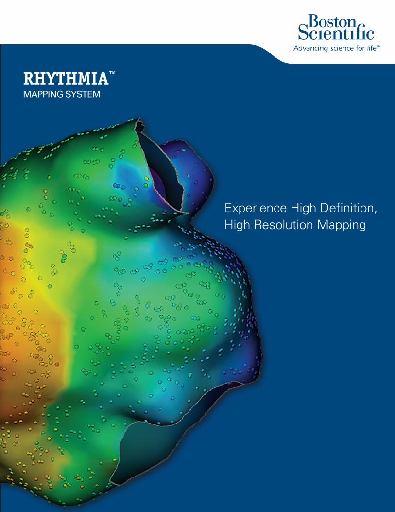
RHYTHMIA ™ MAPPING SYSTEM Experience High Definition, High Resolution Mapping
Transcript of Rhythmia Brochure
RHYTHMIA™ MAPPING SYSTEM
Experience High Definition,High Resolution Mapping
Rhythm Management300 Boston Scientific WayMarlborough, MA 01752-1234www.bostonscientific.com
Medical Professionals:1.800.CARDIAC (227.3422)
Customer Service: 1-888-272-1001
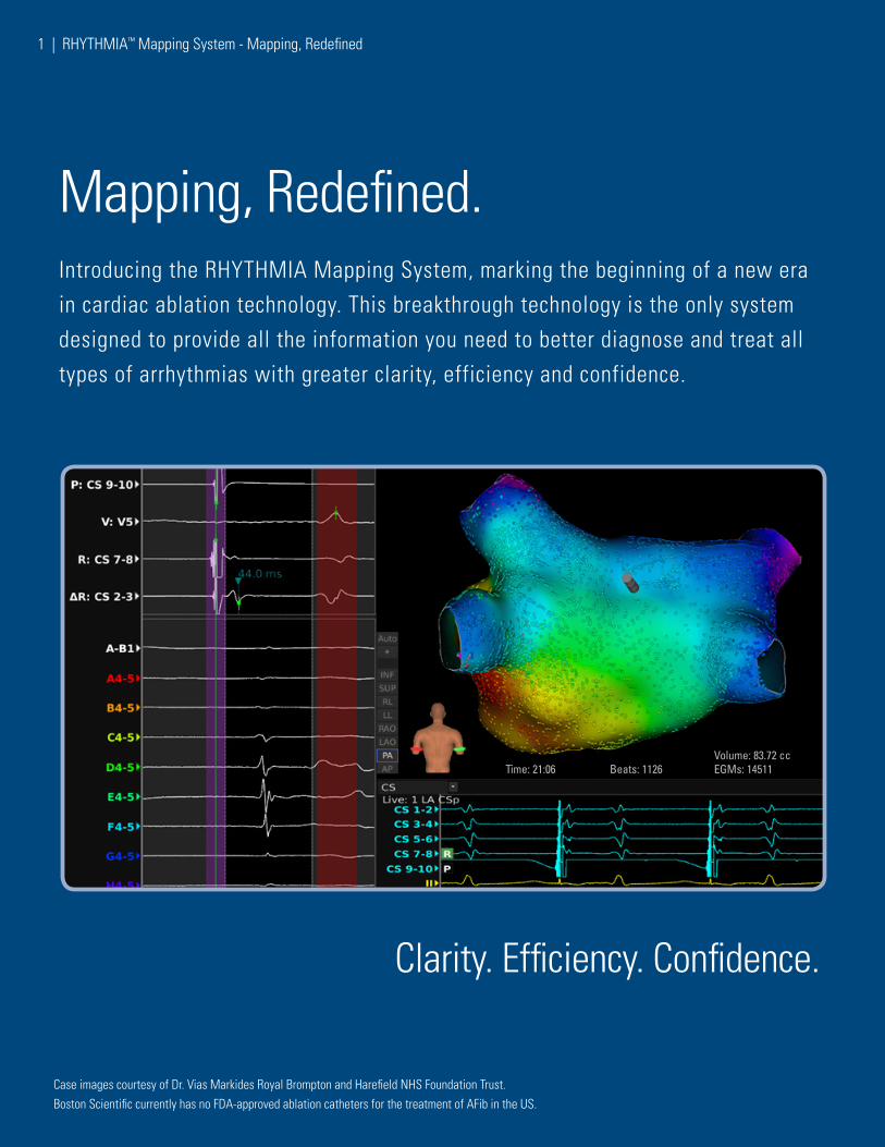
Introducing the RHYTHMIA Mapping System, marking the beginning of a new era in cardiac ablation technology. This breakthrough technology is the only system designed to provide all the information you need to better diagnose and treat all types of arrhythmias with greater clarity, efficiency and confidence.
Mapping, Redefined.
Clarity. Efficiency. Confidence.
Boston Scientific currently has no FDA-approved ablation catheters for the treatment of AFib in the US.Case images courtesy of Dr. Vias Markides Royal Brompton and Harefield NHS Foundation Trust.
1 | RHYTHMIA™ Mapping System - Mapping, Redefined
Volume: 83.72 ccTime: 21:06 Beats: 1126 EGMs: 14511
RHYTHMIA™ Mapping System - Mapping, Redefined | 2
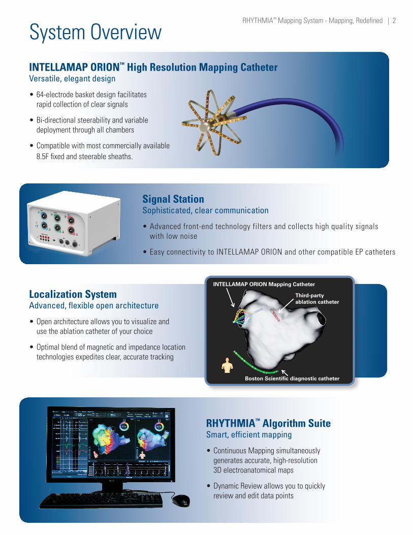
System OverviewINTELLAMAP ORION™ High Resolution Mapping Catheter Versatile, elegant design
•64-electrode basket design facilitates rapid collection of clear signals
•Bi-directional steerability and variable deployment through all chambers
•Compatible with most commercially available 8.5F fixed and steerable sheaths.
Signal Station Sophisticated, clear communication
•Advanced front-end technology filters and collects high quality signals with low noise
•Easy connectivity to INTELLAMAP ORION and other compatible EP catheters
Localization System Advanced, flexible open architecture
•Open architecture allows you to visualize and use the ablation catheter of your choice
•Optimal blend of magnetic and impedance location technologies expedites clear, accurate tracking
RHYTHMIA™ Algorithm Suite Smart, efficient mapping
•Continuous Mapping simultaneously generates accurate, high-resolution 3D electroanatomical maps
•Dynamic Review allows you to quickly review and edit data points
INTELLAMAP ORION Mapping Catheter
Third-partyablation catheter
Boston Scientific diagnostic catheter
1. Nakagawa H., Ikeda A., Sharma T., Lazzara R., Jackman W., Rapid high-resolution electroanatomical mapping: Evaluation of a new system in a canine atrial linear lesion model. Circulation Arrhythmia Electrophysiology. 2012 Apr;5(2):417-24.
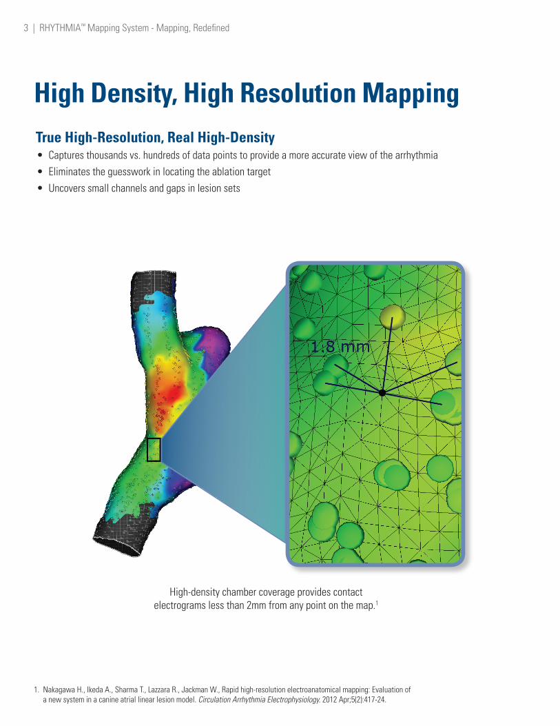
True High-Resolution, Real High-Density• Captures thousands vs. hundreds of data points to provide a more accurate view of the arrhythmia• Eliminates the guesswork in locating the ablation target• Uncovers small channels and gaps in lesion sets
High-density chamber coverage provides contact electrograms less than 2mm from any point on the map.1
High Density, High Resolution Mapping
3 | RHYTHMIA™ Mapping System - Mapping, Redefined
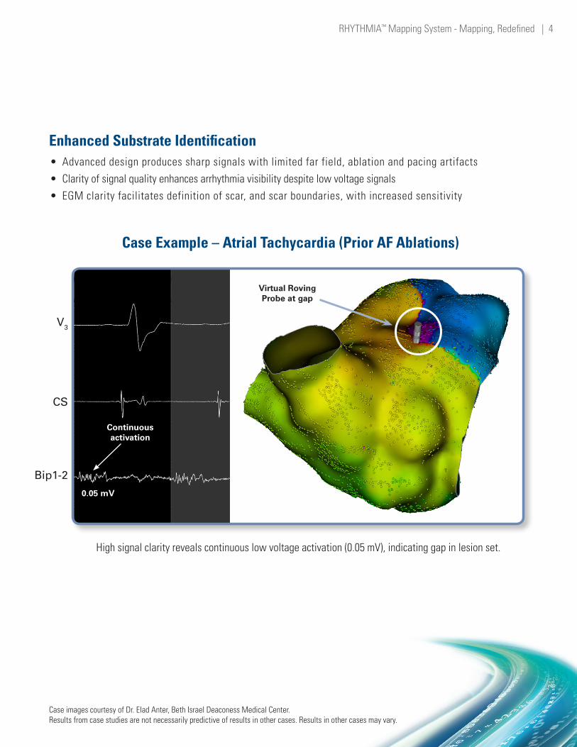
Enhanced Substrate Identification• Advanced design produces sharp signals with limited far field, ablation and pacing artifacts• Clarity of signal quality enhances arrhythmia visibility despite low voltage signals • EGM clarity facilitates definition of scar, and scar boundaries, with increased sensitivity
High signal clarity reveals continuous low voltage activation (0.05 mV), indicating gap in lesion set.
Case Example – Atrial Tachycardia (Prior AF Ablations)
Case images courtesy of Dr. Elad Anter, Beth Israel Deaconess Medical Center.Results from case studies are not necessarily predictive of results in other cases. Results in other cases may vary.
V3
CS
Bip1-2
Virtual Roving Probe at gap
Continuous activation
0.05 mV
RHYTHMIA™ Mapping System - Mapping, Redefined | 4

27,000+ EGM High-Resolution, 3D Electroanatomical Map Captured in 15 Minutes
2. Data on file.
Case images courtesy of Dr Tom McElderry, University of Alabama at Birmingham.Results from case studies are not necessarily predictive of results in other cases. Results in other cases may vary.
SpeedIdeal blend of speed and ease-of-use allows you to collect thousands of relevant data points within minutes.
Continuous Mapping• Continuous acquisition of points based on user-defined criteria creates maps in 1/3 of the time• Repeatable maps generated in minutes offer more predictability and less variability• 99.8% accuracy in automated annotation algorithm eliminates the need for manual beat acceptance2
5 | RHYTHMIA™ Mapping System - Mapping, Redefined
Volume: 91.00 ccTime: 15:45 Beats: 2433 EGMs: 27801

27,000+ EGM High-Resolution, 3D Electroanatomical Map Captured in 15 Minutes
Case images courtesy of Dr. P Boon Lim, Imperial College Health Trust.
Results from case studies are not necessarily predictive of results in other cases. Results in other cases may vary.
From the workstation PC, Virtual Roving Probe sweeps the left atrium to aid in the identification
and tagging of the target ablation site.
Case Example – Macroreentrant Atrial Tachycardia
Dynamic Review• Virtually review and edit high-density maps• Manually accept/reject beats or change annotations with full control
Tissue Targeting• Rapidly identify target ablation sites away from the patient table
RHYTHMIA™ Mapping System - Mapping, Redefined | 6
Virtual Roving Probe

Validation Mapping (vMap™)
vMap gives the ability to create quick high density, high resolution maps to verify lines of block, check for gaps, and confirm complete termination of arrhythmias.
Activation map enables identification of arrhythmia morphology.
Ablation delivered to create line of block. Successful termination of acute tachycardia.
Case images courtesy of Dr. Tom McElderry, University of Alabama at Birmingham Hospital.
Results from case studies are not necessarily predictive of results in other cases. Results in other cases may vary.
vMap – Lesion Validation Case Example – Left Atrial Flutter (Prior AF Ablation)
7 | RHYTHMIA™ Mapping System - Mapping, Redefined
Create Activation MapStep 1
Ablation DeliveryStep 2

Validation Mapping (vMap™)
Validation map is created displaying the area of ablation. Propagation and breakthrough are visible on the line of ablation. In just a few minutes, vMap provides the ability to assess complete block before ending the procedure.
vMap™
• Quickly create high-resolution validation maps multiple times during a procedure• Clearly assess gaps and verify lesion integrity• Potentially reveal concealed arrhythmias more efficiently
RHYTHMIA™ Mapping System - Mapping, Redefined | 8
vMap to Confirm Efficacy
165 ms165 ms 15 ms15 ms15 ms15 ms 135 ms135 ms
Step 3
At Boston Scientific, we are committed to delivering new ablation technologies that are built upon a foundation of technical innovation and clinical success. The introduction of the RHYTHMIA Mapping System marks a significant milestone along Boston Scientific’s journey to redefining electrophysiology.
9 | RHYTHMIA™ Mapping System - Mapping, Redefined
Redefining ElectrophysiologyRHYTHMIA™ MAPPING SYSTEM

To learn more about Boston Scientific’s breakthrough technologies visit www.bostonscientific.com
RHYTHMIA™ Mapping System - Mapping, Redefined | 10
Rhythm Management300 Boston Scientific WayMarlborough, MA 01752-1234www.bostonscientific.com
Medical Professionals:1.800.CARDIAC (227.3422)
Customer Service: 1-888-272-1001© 2015 by Boston Scientific Corporation or its affiliates. All rights reserved. EP-173906-AC DEC2015
RHYTHMIA™ Mapping System INDICATIONS FOR USE The Rhythmia™ Mapping System and accessories are indicated for catheter-based atrial and ventricular mapping. The mapping system allows real-time visualization of intracardiac catheters as well as display of cardiac maps in a number of different formats. The acquired patient signals, including body surface ECG and intracardiac electrograms, may also be recorded and displayed on the system’s display screen. CONTRAINDICATIONS There are no known contraindications. WARNINGS and PRECAUTIONS The use of the Rhythmia Mapping System in conjunction with radio frequency ablation and other medical devices, as a part of the diagnosis and treatment of cardiac arrhythmias, may pose a risk of adverse events, such as cardiac perforation and arrhythmias (new and/or exacerbation of existing arrhythmias) that may require additional intervention. Do not operate the Rhythmia Mapping System near flammable anesthetics. System operation near flammable anesthetics may cause an explosion that could cause injury or death to the patient or user. All devices that are connected to the Rhythmia Mapping System must meet IEC 60601-1 requirements and any other relevant safety standards. When connected to other devices, the combined systems’ configuration must meet the IEC 60601-1-1 safety standards. The use of the Rhythmia™ Mapping System with accessories and devices that do not comply with relevant standards may reduce the safety of the system, cause equipment damage or system malfunction, or harm to the patient or user. Only stimulators that are certified for IEC 60601 should be used with the Rhythmia Mapping System. Do not connect life-sustaining pacing through the Rhythmia Mapping System. The system is not intended to provide life-sustaining therapy and should not be used as such. In case of need for emergency pacing, or any failure of stimulator routing, directly connect the desired paced channel to the stimulator. The Rhythmia Mapping System is only designed to route the stimulation signal to the desired channel. To start or stop stimulation, always use the controls on the external stimulator. Use the Rhythmia Mapping System only with one of the following RF ablation generators: Maestro 3000™, Stockert™, or IBI™. Do not use the system with other RF ablation generators. Compatibility with other RF ablation generators has not been demonstrated. Do not apply RF energy larger than 150W to ablation catheters that are connected to the Maestro 3000 RF generator and the Rhythmia Mapping System. Do not apply RF energy larger than 70W to ablation catheters that are connected to the Stockert RF generator and the Rhythmia Mapping System. Do not apply RF energy larger than 50W to ablation catheters that are connected to the IBI RF generator and the Rhythmia Mapping System. To reduce the risk of electric shock or equipment damage, do not clean the Rhythmia Mapping System when it is plugged in, turned on, or connected to a patient. Cleaning the system while it is in use and connected to a power source may cause an electrical shock that could cause injury or death to the patient or user. To reduce the risk of electric shock, assure that any ECG cables and electrodes are not in contact with any other conductive parts, including ground. To reduce the risk of electric shock during defibrillation, assure that the exposed connector tips on the ECG output box are covered at all times with the protective, non-conductive material provided with the ECG output boxes. Do not use the ECG output box if the protective cover is damaged (see ECG Output Box). The system generates electrical impedance fields as part of its normal operation. Do not use other systems that also generate electrical impedance fields in the same procedure, as this may interfere with the system’s normal operation and reduce the quality of catheter localization, and signals. Magnetic Localization System Do not operate the Localization Generator within 200 mm of installed cardiac implantable electronic devices (CIEDs). Doing so may affect pacing, temporarily suspend tachycardia therapy delivery, or lead to patient discomfort. Signal Station To minimize the risk of electric shock, connect the Signal Station only to supply mains with a protective ground (earth) connection. Use only a functioning, properly tested supply main with protective ground (earth) to power the Rhythmia Mapping System. The use of a faulty, ungrounded supply main increases the risk of electrical shock and system malfunction. To minimize the risk of electric shock, prior to using the Rhythmia Mapping System, connect the equipotential socket (located on the Signal Station rear panel) to a common ground. This connection grounds the Rhythmia Mapping System and must remain connected at all times (see Signal Station Setup in the DFU). The Signal Station requires a dedicated, 24V DC power supply, which is provided by Boston Scientific with the Signal Station. To reduce the risk of Signal Station damage, use only the power supply provided by Boston Scientific for use with the Signal Station. To reduce the risk of Signal Station damage, do not connect or disconnect the Signal Station to its power supply while the Signal Station is turned on. To minimize potential exposure to water or liquid, prevent fluids from entering air vents. Do not place beverages or containers of water or liquid directly on or near the Signal Station or other system components. Do not block the air vent on the Signal Station during Signal Station use. Blocking the air vent during Signal Station use can cause the Signal Station to overheat, which may affect system operation. Use only a flat stable surface to hold the Signal Station and Signal Station-related accessories. Workstation To minimize potential exposure to water or liquid, do not place beverages or containers of water or liquid directly on or near the Workstation or other system components. Use only a flat stable surface to hold or transport the Workstation and Workstation-related accessories. To prevent loss of data, frequently back up the data by archiving cases no longer needed for immediate access. Cables Use only the ECG cables supplied by Rhythmia™ Medical for use with the Rhythmia Mapping System. ECG cables provided by Rhythmia Medical are designed and tested to protect the Signal Station from defibrillation energy. Using other ECG cables may cause serious damage to the system hardware. Prior to using the Rhythmia Mapping System, inspect all external connections and cable connectors. Make sure all connections are secure. Tighten any loose connections prior to using the system. Do not use excessive force when connecting or disconnecting cable connectors. Excessive force can damage the connectors, which may cause system malfunction. Do not kink or sharply bend cables. Kinks and sharp bends can damage the cables, which may cause system malfunction. To minimize the risk of damage, store unused system cables in a clean, dry, and secure location, consistent with storage guidelines (see Equipment Storage & Transporting in the DFU). Electrical Never use ungrounded electrical outlets to power any system components. Do not use extension cords or adapters for ungrounded outlets. Using ungrounded outlets, extension cords, or adapters may cause equipment damage, system failure or malfunction. Body Surface Electrodes Use care when attaching the body surface electrodes to lead connectors. To minimize the risk of electric shock, make sure that electrodes and lead connectors do not contact one another or contact ground. To prevent low quality signals from body surface electrodes, properly prepare the skin prior to attaching the electrodes. Do not use excessive gel as this may lead to shorts between different electrodes. Environmental Do not immerse any cable connectors in water or liquid. Immersion in water or liquid may damage connectors, which may cause system malfunction. Magnetic Localization System Manually disabling the Localization Generator disables all catheter visualization and localization capabilities, including impedance tracking. Do not place the Localization Unit (SCU) or Sensor Interface Unit (SIU) within 1m of the Localization Generator. Doing so may lead to inaccurate tracking. Do not place cables used with the Rhythmia Mapping System within 30mm of the Localization Generator cable. If these cables are within 30mm or less, particularly if they are parallel to each other, inaccurate tracking or “noisy” signals may occur. Do not coil the Localization Generator cable. Doing so can disturb the magnetic field of the Localization Generator, which may lead to inaccurate tracking. Do not use the Magnetic Localization System in the presence of other magnetic fields or large metal objects. Doing so may lead to inaccurate tracking. Localization Generator Manually disabling the Localization Generator disables all catheter visualization and localization capabilities, including impedance tracking. During the Procedure To reduce catheter configuration mistakes, when connecting catheters to the system, always verify the signals by reviewing the signal display and recording system to ensure correct configuration of catheter electrodes to displayed channels. To ensure correct clinical decisions, use fluoroscopy, ultrasound, pace mapping or other visualization techniques to verify mapping results and catheter position. Always compare the anatomical map to the patient’s expected anatomy. When a catheter localization error is encountered, use fluoroscopy or other visualization techniques to verify catheter location. Imported geometrical shells should only be used as a reference, for example to identify anatomical features in advance of mapping. Use other visualization tools, such as fluoroscopy or echocardiography to verify catheter location. During the mapping procedure, do not disconnect the Localization Unit from the Signal Station and/or the Localization Generator from the Localization Unit. Ensure caps are installed on Localization Unit SIU connection ports that are not in use.
INTELLAMAP ORION™ High Resolution Mapping Catheter INDICATIONS FOR USE The IntellaMap Orion High Resolution Mapping Catheter is indicated for electrophysiological mapping (recording or stimulating only) of the cardiac structures of the heart. CONTRAINDICATIONS The IntellaMap Orion Catheter should not be used in: Patients who are not candidates for transvascular catheter procedures. Patients with a hypercoagulable state or who cannot tolerate heparin anticoagulation therapy. Patients with prosthetic or stenotic valves, in the chamber where the prosthetic or stenotic valve reside. Patients with active systemic infection. Pediatric patients. Pregnant and/or nursing patients. Patients with any other condition where catheter manipulation may not be safe. The IntellaMap Orion Catheter should not be used for radio frequency (RF) ablation. The IntellaMap Orion Catheter should not be used inside an MRI machine. WARNINGS Keep the connector dry; wet connector pins may affect performance. Do not allow the handle or cabling to be immersed in fluid. Do not use the catheter to deliver ablation therapy. Do not expose the catheter to alcohol or other cleaning solvents. Do not operate the catheter against resistance. If resistance is felt during advancement, retraction, articulation, deployment or un-deployment, stop and evaluate device location under fluoroscopy. Do not advance or retract the catheter through a sheath when deployed or articulated. In order to reduce the risk of clot formation: Maintain an activated clotting time (ACT) of greater than 300 sec. at all times during use of the catheter, and continuously flush the electrode array with saline via the irrigation port at the proximal end. Do not use the catheter with equipment (such as stimulators or recording systems) that is not isolated. PRECAUTIONS To avoid cardiac damage, do not use excessive force when manipulating the catheter in vivo. Specifically, use caution when maneuvering while undeployed. Note that mapping and recording data do not require the use of force on the tissue. Always undeploy the catheter prior to removal from the patient. Use visualization (such as fluoroscopy) to verify undeployment. Always move the articulation control lever to its neutral position to straighten the catheter prior to removal from the patient. Only use guiding sheaths with curves that allow passage of the catheter without using excessive force. When used with a steerable guiding introducer sheath: Ensure under fluoroscopy that the guiding introducer sheath distal end is straight or, if necessary, only minimally curved prior to advancing or retracting the catheter through the sheath. Do not articulate the sheath while the catheter array is inside the articulating section. Do not deploy or articulate the catheter while the distal end is inside a sheath. Do not apply RF energy on an ablation catheter that is in direct contact with the electrodes on the IntellaMap Orion Catheter. To prevent entanglement, use care when using the catheter in the proximity of other catheters. When pacing, verify desired waveform is observed. Prior to insertion into vasculature, ensure removal of all air from the catheter lumen; use a pressured saline bag to flush saline through the catheter shaft and electrode array. POTENTIAL ADVERSE EVENTS Serious adverse events have been reported in the literature in relation to cardiac catheterization including: stroke, cardiac tamponade, perforation, myocardial infarction, pulmonary embolism, and death. Complications reported included also (in alphabetical order): air embolism, arrhythmia, AV fistula, hematomas, hemothorax, pneumothorax, pseudoaneurysm, thromboembolism, valvular damage, vascular bleeding, and vasovagal reactions.
CAUTION: Federal law (USA) restricts this device to sale by or on the order of a physician. Rx only. Prior to use, please see the complete “Directions for Use” for more information on Indications, Contrain-dications, Warnings, Precautions, Adverse Events, and Operator’s Instructions.
![RHYTHMIA HDx - Boston Scientific- US · 2 RHYTHMIA HDx™ G]G GWG4G5G0 v 1 Â i 1.2.2 2 ' ø ù ô(System Software) RhythmiaGKGcGVG4G5G0FÿF¸G}G G=GGGTG GEGuG VF÷ 8 ·FéG F¹FãFþGKGcGVG4G5G0FÿF¸](https://static.fdocuments.net/doc/165x107/5f02cbf97e708231d4060e5e/rhythmia-hdx-boston-scientific-us-2-rhythmia-hdxa-gg-gwg4g5g0-v-1-i-122.jpg)


![Ac Brochure 2009 Brochure]](https://static.fdocuments.net/doc/165x107/577d2f551a28ab4e1eb16a35/ac-brochure-2009-brochure.jpg)