Rho/Rho-associatedKinaseSignalRegulatesMyogenic ... · the Id3 gene; the Id3 gene product is a...
13
Rho/Rho-associated Kinase Signal Regulates Myogenic Differentiation via Myocardin-related Transcription Factor-A/Smad-dependent Transcription of the Id3 Gene * Received for publication, December 26, 2007, and in revised form, May 5, 2008 Published, JBC Papers in Press, May 12, 2008, DOI 10.1074/jbc.M710525200 Kazuhiro Iwasaki ‡§ , Ken’ichiro Hayashi ‡ , Tomoaki Fujioka § , and Kenji Sobue ‡¶1 From the ‡ Department of Neuroscience (D13), § Research Center for Child Mental Development, Osaka University Graduate School of Medicine, Yamadaoka 2-2, Suita, Osaka, 565-0871 and the ¶ Department of Urology, Iwate Medical University, Uchimaru 19-1, Morioka, Iwate 020-8505, Japan RhoA is known to be involved in myogenic differentiation, but whether it acts as a positive or negative regulator is controver- sial. To resolve this issue, we investigated the differentiation stage-specific roles of RhoA and its effector, Rho-associated kinase, using C2C12 myoblasts. We found that proliferating myoblasts show high levels of RhoA and serum-response factor activities and strong expression of the downstream target of RhoA, myocardin-related transcription factor-A (MRTF-A or MAL); these activities and expression are markedly lower in dif- ferentiating myocytes. We further demonstrated that, in prolif- erating myoblasts, an increase in MRTF-A, which forms a com- plex with Smad1/4, strikingly activates the expression level of the Id3 gene; the Id3 gene product is a potent inhibitor of myo- genic differentiation. Finally, we found that during differentia- tion, one of the forkhead transcription factors translocates into the nucleus and suppresses Id3 expression by preventing the association of the MRTF-A-Smad complex with the Id3 pro- moter, which leads to the enhancement of myogenic differenti- ation. We conclude that RhoA/Rho-associated kinase signaling plays positive and negative roles in myogenic differentiation, mediated by MRTF-A/Smad-dependent transcription of the Id3 gene in a differentiation stage-specific manner. Myogenic differentiation involves a sequence of processes as follows: withdrawal of myoblasts from the cell cycle, expression of myogenic differentiation markers in myocytes, and forma- tion of multinucleated myotubes by myocyte fusion. These pro- cesses are controlled by the myogenic regulatory factor (MRF) 2 family and belong to basic helix-loop-helix proteins, consisting of MyoD, Myf5, myogenin, and MRF4 (1–3). The MRFs inter- act with ubiquitous basic helix-loop-helix proteins such as E12/ E47 (E proteins) via their helix-loop-helix motifs, and the basic domains of the dimerized proteins activate transcription by binding to a conserved DNA sequence known as the E-box in the promoter regions of target genes. A family of inhibitors of DNA binding (Id) (Id1, Id2, Id3, and Id4), which has a helix- loop-helix domain but lacks the basic DNA-binding domain, counteracts the active MRF/E protein complexes by forming transcriptionally inactive complexes, consisting of either MRF/Id or E protein/Id (4 – 6). The Rho family GTPases are involved in a variety of cytoskel- eton-associated cellular events, such as reorganization of the actin cytoskeleton, microtubule dynamics, transcriptional reg- ulation, and cell differentiation, including myogenic differenti- ation (7). The RhoA-dependent activation of serum response factor (SRF) is required for the expression of MyoD and skeletal (SK) -actin. Thus, RhoA acts through SRF to positively regu- late myogenic differentiation (8 –11) and has therefore been referred to as a positive regulator of myogenic differentiation. However, recent studies have revealed that the expression of constitutively active RhoA results in failed myotube formation, and the inactivation of one of the downstream effectors of RhoA, Rho-associated kinase (ROCK), enhances myotube for- mation (12–15). Thus, apparently conflicting roles of RhoA sig- naling in myogenic differentiation have been reported. It is well documented that actin polymerization in response to RhoA signaling enhances SRF activity, as a consequence of the nuclear translocation of myocardin-related transcription factors (MRTF-A/B, also referred to as MAL1/2) (16). MRTF-A is reportedly involved in heart and smooth muscle gene expres- sion (17, 18). Consistent with these findings, the expression of dominant-negative forms of MRTF-A and MRTF-B (dn- MRTF-A and dn-MRTF-B), which inhibit SRF activation, gen- erates abnormally thin muscle fibers in vivo (19) and inhibits myogenic differentiation in vitro (20). The molecular mecha- nism underlying the contribution of the MRTFs to myogenic differentiation, however, remains unclear. One of the forkhead family transcription factors, FKHR (also named Foxo1), is required for myocyte fusion (15, 21, 22). Because ROCK directly phosphorylates FKHR in myocytes, resulting in the export of FKHR from the nucleus, the inactiva- tion of RhoA/ROCK signaling is prerequisite for the nuclear * This work was supported by Grant-in-aid for Scientific Research 15GS0312 (to K. S.) from the Ministry of Education, Science, Sports, and Culture of Japan. The costs of publication of this article were defrayed in part by the payment of page charges. This article must therefore be hereby marked “advertisement” in accordance with 18 U.S.C. Section 1734 solely to indi- cate this fact. 1 To whom correspondence should be addressed: Dept. of Neuroscience (D13), Osaka University Graduate School of Medicine, 2-2 Yamadaoka, Suita, Osaka, 565-0871, Japan. Tel.: 81-6-6879-3680; Fax: 81-6879-3689; E-mail: [email protected]. 2 The abbreviations used are: MRF, myogenic regulatory factor; ROCK, Rho- associated kinase; MRTF-A, myocardin-related transcription factor; Id, inhibitor of DNA binding; FKHR, forkhead in human rhabdomyosarcoma; SRF, serum response factor; MHC, myosin heavy chain; SBE, Smad binding element; GM, growth medium; DM, differentiation medium; ChIP, chroma- tin immunoprecipitation; RT, reverse transcription; GAPDH, glyceralde- hyde-3-phosphate dehydrogenase; HA, hemagglutinin; ca, constitutively active; BMP, bone morphogenetic protein. THE JOURNAL OF BIOLOGICAL CHEMISTRY VOL. 283, NO. 30, pp. 21230 –21241, July 25, 2008 © 2008 by The American Society for Biochemistry and Molecular Biology, Inc. Printed in the U.S.A. 21230 JOURNAL OF BIOLOGICAL CHEMISTRY VOLUME 283 • NUMBER 30 • JULY 25, 2008 by guest on October 7, 2018 http://www.jbc.org/ Downloaded from
Transcript of Rho/Rho-associatedKinaseSignalRegulatesMyogenic ... · the Id3 gene; the Id3 gene product is a...

Rho/Rho-associated Kinase Signal Regulates MyogenicDifferentiation via Myocardin-related TranscriptionFactor-A/Smad-dependent Transcription of the Id3 Gene*
Received for publication, December 26, 2007, and in revised form, May 5, 2008 Published, JBC Papers in Press, May 12, 2008, DOI 10.1074/jbc.M710525200
Kazuhiro Iwasaki‡§, Ken’ichiro Hayashi‡, Tomoaki Fujioka§, and Kenji Sobue‡¶1
From the ‡Department of Neuroscience (D13), §Research Center for Child Mental Development, Osaka University Graduate Schoolof Medicine, Yamadaoka 2-2, Suita, Osaka, 565-0871 and the ¶Department of Urology, Iwate Medical University, Uchimaru 19-1,Morioka, Iwate 020-8505, Japan
RhoA is known tobe involved inmyogenic differentiation, butwhether it acts as a positive or negative regulator is controver-sial. To resolve this issue, we investigated the differentiationstage-specific roles of RhoA and its effector, Rho-associatedkinase, using C2C12 myoblasts. We found that proliferatingmyoblasts show high levels of RhoA and serum-response factoractivities and strong expression of the downstream target ofRhoA, myocardin-related transcription factor-A (MRTF-A orMAL); these activities and expression aremarkedly lower in dif-ferentiating myocytes. We further demonstrated that, in prolif-erating myoblasts, an increase in MRTF-A, which forms a com-plex with Smad1/4, strikingly activates the expression level ofthe Id3 gene; the Id3 gene product is a potent inhibitor of myo-genic differentiation. Finally, we found that during differentia-tion, one of the forkhead transcription factors translocates intothe nucleus and suppresses Id3 expression by preventing theassociation of the MRTF-A-Smad complex with the Id3 pro-moter, which leads to the enhancement of myogenic differenti-ation. We conclude that RhoA/Rho-associated kinase signalingplays positive and negative roles in myogenic differentiation,mediated byMRTF-A/Smad-dependent transcription of the Id3gene in a differentiation stage-specific manner.
Myogenic differentiation involves a sequence of processes asfollows: withdrawal ofmyoblasts from the cell cycle, expressionof myogenic differentiation markers in myocytes, and forma-tion ofmultinucleatedmyotubes bymyocyte fusion. These pro-cesses are controlled by themyogenic regulatory factor (MRF)2
family and belong to basic helix-loop-helix proteins, consistingof MyoD, Myf5, myogenin, and MRF4 (1–3). The MRFs inter-act with ubiquitous basic helix-loop-helix proteins such as E12/E47 (E proteins) via their helix-loop-helix motifs, and the basicdomains of the dimerized proteins activate transcription bybinding to a conserved DNA sequence known as the E-box inthe promoter regions of target genes. A family of inhibitors ofDNA binding (Id) (Id1, Id2, Id3, and Id4), which has a helix-loop-helix domain but lacks the basic DNA-binding domain,counteracts the active MRF/E protein complexes by formingtranscriptionally inactive complexes, consisting of eitherMRF/Id or E protein/Id (4–6).The Rho family GTPases are involved in a variety of cytoskel-
eton-associated cellular events, such as reorganization of theactin cytoskeleton, microtubule dynamics, transcriptional reg-ulation, and cell differentiation, including myogenic differenti-ation (7). The RhoA-dependent activation of serum responsefactor (SRF) is required for the expression ofMyoD and skeletal(SK) �-actin. Thus, RhoA acts through SRF to positively regu-late myogenic differentiation (8–11) and has therefore beenreferred to as a positive regulator of myogenic differentiation.However, recent studies have revealed that the expression ofconstitutively active RhoA results in failed myotube formation,and the inactivation of one of the downstream effectors ofRhoA, Rho-associated kinase (ROCK), enhances myotube for-mation (12–15). Thus, apparently conflicting roles of RhoA sig-naling in myogenic differentiation have been reported.It is well documented that actin polymerization in response
to RhoA signaling enhances SRF activity, as a consequence ofthe nuclear translocation of myocardin-related transcriptionfactors (MRTF-A/B, also referred to asMAL1/2) (16).MRTF-Ais reportedly involved in heart and smoothmuscle gene expres-sion (17, 18). Consistent with these findings, the expression ofdominant-negative forms of MRTF-A and MRTF-B (dn-MRTF-A and dn-MRTF-B), which inhibit SRF activation, gen-erates abnormally thin muscle fibers in vivo (19) and inhibitsmyogenic differentiation in vitro (20). The molecular mecha-nism underlying the contribution of the MRTFs to myogenicdifferentiation, however, remains unclear.One of the forkhead family transcription factors, FKHR (also
named Foxo1), is required for myocyte fusion (15, 21, 22).Because ROCK directly phosphorylates FKHR in myocytes,resulting in the export of FKHR from the nucleus, the inactiva-tion of RhoA/ROCK signaling is prerequisite for the nuclear
* This work was supported by Grant-in-aid for Scientific Research 15GS0312(to K. S.) from the Ministry of Education, Science, Sports, and Culture ofJapan. The costs of publication of this article were defrayed in part by thepayment of page charges. This article must therefore be hereby marked“advertisement” in accordance with 18 U.S.C. Section 1734 solely to indi-cate this fact.
1 To whom correspondence should be addressed: Dept. of Neuroscience(D13), Osaka University Graduate School of Medicine, 2-2 Yamadaoka,Suita, Osaka, 565-0871, Japan. Tel.: 81-6-6879-3680; Fax: 81-6879-3689;E-mail: [email protected].
2 The abbreviations used are: MRF, myogenic regulatory factor; ROCK, Rho-associated kinase; MRTF-A, myocardin-related transcription factor; Id,inhibitor of DNA binding; FKHR, forkhead in human rhabdomyosarcoma;SRF, serum response factor; MHC, myosin heavy chain; SBE, Smad bindingelement; GM, growth medium; DM, differentiation medium; ChIP, chroma-tin immunoprecipitation; RT, reverse transcription; GAPDH, glyceralde-hyde-3-phosphate dehydrogenase; HA, hemagglutinin; ca, constitutivelyactive; BMP, bone morphogenetic protein.
THE JOURNAL OF BIOLOGICAL CHEMISTRY VOL. 283, NO. 30, pp. 21230 –21241, July 25, 2008© 2008 by The American Society for Biochemistry and Molecular Biology, Inc. Printed in the U.S.A.
21230 JOURNAL OF BIOLOGICAL CHEMISTRY VOLUME 283 • NUMBER 30 • JULY 25, 2008
by guest on October 7, 2018
http://ww
w.jbc.org/
Dow
nloaded from

localization of FKHR and myocyte fusion (15). In various othercell types, FKHR that is phosphorylated by Akt, but not byROCK, is also exported from the nucleus, leading to the inhibi-tion of FKHR-mediated transcription (23). However, the targetpartners of FKHR and its function in myogenic differentiationremain unknown, except for a Foxo1a-cyclic GMP-dependentkinase I interaction (22).Here, we investigated the role of RhoA/ROCK signaling dur-
ing myogenic differentiation using C2C12 cells and demon-strated that transcription of the Id3 gene is controlled byMRTF-A/Smad in an FKHR-dependent manner. In proliferat-ing myoblasts, where RhoA activity and MRTF-A expressionare relatively high, RhoA-triggered MRTF-A/Smad1/4 pre-dominantly activates transcription of the Id3 gene. Simulta-neously, FKHR, possibly phosphorylated by ROCK, is retainedin the cytoplasm. In contrast, in differentiating myocytes,where RhoA activity and MRTF-A expression are decreased,FKHR translocates into the nucleus, where it interacts withMRTF-A/Smad1/4, causing theMRTF-A-Smad1/4 complex todissociate from the Id3 promoter, and thereby suppressing theMRTF-A/Smad-dependent Id3 expression. Taken together,the results of this study uncover why there are conflicting viewsregarding the role of RhoA/ROCK signaling inmyogenic differ-entiation; differentiation stage-specific RhoA/ROCK signalingand activities of its downstream transcription factors,MRTF-Aand FKHR, regulate the Smad-dependent transcription of theId3 gene.
EXPERIMENTAL PROCEDURES
Reagents and Antibodies—Y-27632 was purchased fromCal-biochem. Tat-C3 was prepared according to a methoddescribed elsewhere (24). Commercially available antibodieswere as follows: anti-FLAG (M2), anti-�-sarcomeric actin(5C5), and anti-�-tubulin (DM1A) antibodies (Sigma); anti-he-magglutinin (HA) (3F10) antibody (Roche Applied Science);anti-MyoD, anti-myogenin, anti-SRF, anti-Smad1/5/8, andanti-Id3 antibodies (Santa Cruz Biotechnology); anti-myosinheavy chain (MHC) antibody (MF20) (Developmental StudiesHybridoma Bank); anti-FKHR antibody (Cell Signaling); horse-radish peroxidase-linked anti-rabbit or -mouse IgG (GEHealthcare); horseradish peroxidase-linked anti-rat IgG (Rock-land). Anti-MRTF-A and -MRTF-B antibodies were generatedin rabbits using recombinant proteins consisting of amino acids776–901 of human MRTF-A and amino acids 931–1061 ofhuman MRTF-B as the epitopes. IgGs were affinity-purifiedfrom the antisera raised againstMRTF-A andMRTF-B on theirrespective affinity columns and used in this study (25).Plasmid Constructs—The cDNAs of mouse RhoA, Smad1,
Smad4, MRTF-A, and FKHR were amplified by reverse tran-scriptase (RT)-PCR and inserted into a mammalian expressionplasmid, pCS2� or pcDNA3.1, with the indicated tags. Expres-sion plasmids for the constitutively active formof RhoA (RhoA-V14) and pseudo-phosphorylated Smad1 (26)were constructedby site-directed mutagenesis. The expression plasmid forN-terminally deleted MRTF-A, which acts as a constitutivelyactive form, was constructed as described previously (16). Thepromoter region of the mouse Id3 gene, spanning from �2000to �55, was isolated from the genome of C2C12 myoblasts
using PCRand inserted into the pGL3-Basic plasmid (Promega)(Id3 (�2000/�55)-Luc). Deletions and mutant derivativesfrom Id3 (�2000/�55)-Luc, as indicated, were constructedusing the PCR-mediated method. The mutations in the Smad-binding element (SBE) were introduced into pGL3-Id3-Luc asfollows: themutant SBEwas changed fromGTCTG toATGTAand from CAGAC to CGTGC. The tetracycline-regulatedinducible expression plasmids for HA-tagged RhoA-V14 andFLAG-tagged constitutively active MRTF-A (ca-MRTF-A)were constructed in the pTRE-tight expression plasmid (Clon-tech). The 3�CArG-Luc, referred to as 3D.ALuc (27), whichconsists of three copies of c-fos serum-responsive elementwithout the ternary complex factor binding element or thebasal promoter region of the Xenopus actin gene (28), was con-structed to assay the transcriptional activity of SRF. All theseconstructs were confirmed by sequencing.Cell Cultures and Transfection—C2C12 mouse myoblasts
were maintained in Dulbecco’s modified Eagle’s medium sup-plemented with 20% fetal calf serum (growth medium (GM)).To induce myogenic differentiation, the culture medium wasshifted to Dulbecco’s modified Eagle’s medium supplementedwith 2% horse serum (differentiation medium (DM)) at theindicated times. HEK293T cells were maintained in Dulbecco’smodified Eagle’s medium supplemented with 10% fetal calfserum. Transfection of the indicated expression plasmids intothese cells was performed using Lipofectamine 2000 (Invitro-gen). Stable transfectants of C2C12 cells carrying the tetracy-cline-regulated inducible expression system of HA-RhoA-V14and FLAG-ca-MRTF-A were isolated as follows. First, weselected C2C12 stable transfectants carrying the pTet-Off plas-mid by geneticin. Second, we introduced the expression plas-mid, pTER-HA-RhoA-V14 or pTRE-FLAG-ca-MRTF-A intostable C2C12-pTet-Off transfectants and isolated cells carryingboth the pTet-Off plasmid and the respective expression plas-mid (C2C12-HA-RhoA-V14 and C2C12-FLAG-ca-MRTF-A),by selection with hygromycin B.PromoterAssays—ProliferatingC2C12myoblasts cultured in
GM were co-transfected with the indicated plasmid and pSV-�-gal (Promega). Luciferase and �-galactosidase activities wereassayed 2 days after changing the medium from GM to DM asfollows. The cell extracts were prepared by passive lysis buffer(Promega) according to the manufacturer’s instructions andthen assayed for luciferase activity using the luciferase kit (Pro-mega). The promoter activity was expressed in relative unitsnormalized to the �-galactosidase activity in the cell extracts.These assays were done in triplicate and were performed inde-pendently at least three times.Immunocytochemistry—The cells were fixedwith 4% formal-
dehyde for 30 min, permeabilized, and blocked with 0.1% Tri-ton X-100 and 0.2% bovine serum albumin in phosphate-buff-ered saline, for 1 h at room temperature. The cells were thenincubated with the anti-MHC antibody for 2 h followed byAlexa Fluor 568 anti-mouse IgG (Molecular Probes), with orwithout Hoechst 33258, for 2 h at room temperature. Fluores-cence images were collected using a cooled charge-coupleddevice camera (Roper Scientific, Tucson, AZ), mounted on anOlympus IX-70 microscope with the appropriate filters, andMetaMorph software.
MRTF-A/Smad-dependent Transcription of the Id3 Gene
JULY 25, 2008 • VOLUME 283 • NUMBER 30 JOURNAL OF BIOLOGICAL CHEMISTRY 21231
by guest on October 7, 2018
http://ww
w.jbc.org/
Dow
nloaded from

GTP-boundRhoAPulldownAssay—RhoAactivitywas deter-mined by a GTP-bound RhoA pulldown assay using the Rhoactivation assay kit (Upstate Biotech), according to the manu-facturer’s protocol.Chromatin Immunoprecipitation (ChIP) Assay—ChIP assays
were carried out using the ChIP assay kit (Upstate Biotech),according to the manufacturer’s protocol with some modifica-tions. DNAs isolated from the input chromatin fragments andthose from chromatin fragments precipitated by the anti-MRTF-A antibody or control IgG were subjected to PCR usingprimers flanking the SBE motif in the mouse Id3 promoter(�531 to �316). These primer sequences were as follows: Id3sense primer, CTCTGGTCACAAGATAATTCC; Id3 anti-sense primer, GCGCCCAAGTTCTCTGAG.Gel-shift Assay—Aprobe containing the SBEmotif sequence
of the Id3 promoter was amplified by PCR and end-labeledwith[32P]dCTP by the Klenow fragment. The sequence of the sensestrand of this probe was AATTCCTGACGCCAGTGAGTCT-GGAGGTCAGACGAGCAGCAAATTGGGGA. The gel-shiftassay was carried out using whole-cell extracts from C2C12myoblasts (29).Semiquantitative RT-PCR—The total RNAs were extracted
from C2C12 cells cultured under the indicated conditions, andthe expression level of Id1, Id2, Id3, twist,myostatin, c-fos, andc-myc mRNAs was quantified by RT-PCR normalized to theexpression of glyceraldehyde-3-phosphate dehydrogenase(GAPDH) mRNA, as described previously (30). The specificprimer sets were as follows: Id1 sense primer, TGGACGAGC-AGCAGGTGAACG; Id1 antisense primer, GCACTGATCTC-GCCGTTCAGG; Id2 sense primer, AGCCTTCAGTCCGGT-GAGGTCC; Id2 antisense primer, TCAGATGCCTGCAAGG-ACACG; Id3 sense primer, TGCTACGAGGCGGTGTGCTG;Id3 antisense primer, AGTGAGCTCAGCTGTCTGGAT-CGG; twist sense primer, CCTCGGACAAGCTGAGCAA-GAT; twist antisense primer, CTAGTGGGACGCGGACAT-GGA; myostatin sense primer, ACGCTACCACGGAAACA-ATC;myostatin antisense primer, TGGTCCTGGGAAGGTT-ACAG; c-fos sense primer, GGATTTGACTGGAGGTCTGC;c-fos antisense primer, AAGTAGTGCAGCCCGGAGTA;c-myc sense primer, CAGCAGAGCGAGCTGCAGC; c-mycantisense primer, TCTTTGCGCGCAGCCTGGTA;HA-RhoA-V14 sense primer, TACCCATACGATGTTCCA-GAT; HA-RhoA-V14 antisense primer, CTAAACTATCAGG-GCTGTCG; GAPDH sense primer, TCTTCACCACCATGG-AGAAGG; GAPDH antisense primer, GAAGGCCAT-GCCAGTGAG.Quantitative Real Time RT-PCR—The expression of Id3
mRNA in C2C12 cells was analyzed by real time RT-PCR usingSYBR GreenER qPCR SuperMix (Invitrogen). The levels of Id3mRNA were normalized to GAPDH mRNA expression. Theprimers used in these analyses are as follows: Id3 sense primer,TCCTGGCACCTCCCGAAC; Id3 antisense primer, TAAGT-GAAGAGGGCTGGGTTAAG;GAPDHsense primer, CGTG-CCGCCTGGAGAAAC; GAPDH antisense primer, TGGGA-GTTGCTGTTGAAGTCG.Knockdown Using siRNA—Proliferating C2C12 cells were
transfected with siRNA using Lipofectamine RNAiMAX(Invitrogen) and were cultured in GM for 2 days. The siRNAs
against Id3 were purchased from Sigma, and their sequenceswere as follows: Id3 siRNA1, 5�-GCAUGGAUGAGCUUCGA-UCTT-3�; Id3 siRNA2, 5�-CUGGUCAGCAGCUGGGCA-ATT-3�. In control experiments, scrambled siRNA (Santa CruzBiotechnology) was used.Immunoprecipitation, Immunoblotting, and Cellular
Fractionation—The whole-cell extracts were prepared fromC2C12 and HEK293T cells transfected with the indicatedexpression plasmids by lysis in buffer containing 50 mM Tris-HCl, pH 7.5, 150mMNaCl, 0.1%Nonidet P-40, 5% glycerol, andprotease inhibitormixture tablets (RocheApplied Science), andsonication with a Digital Sonifier (Branson). The extracts thusobtainedwere incubatedwith the indicated antibodies for 3 h at4 °C, and the immune complexes were collected by incubationwith protein A- or protein G-Sepharose beads for 1 h at 4 °C.Proteins in the immunoprecipitates were detected by immuno-blotting using the indicated antibodies. The target proteinswere detected with a SuperSignal chemiluminescence detec-tion kit (Pierce). In immunoblotting analysis of the Id3 protein,we used CanGet Signal (TOYOBO) to detect chemifluorescentsignals. The cytosol and nuclear fractions were prepared asdescribed elsewhere (15).DNA Affinity Binding Assay—The proteins translated by in
vitro transcription/translation systems (Promega) were incu-batedwith a double-stranded biotinylatedDNAprobe (1mg) ingel-shift binding buffer (29) containing 5 mg of poly(dI-dC), 20mg of herring sperm DNA, and 0.5% Nonidet P-40 for 30 minon ice (31). Streptavidin M-280 Dynabeads (Dynal) were thenadded, and the mixture was further incubated with rotation for2 h at 4 °C. The DNA-bound proteins were analyzed by immu-noblotting with the indicated antibodies. The sequence of thesense strand of this probe was as follows: AATTCCTGA-CGCCAGTGAGTCTGGAGGTCAGACGAGCAGCAAATT-GGGGA.
RESULTS
RhoA/ROCK Signaling Orchestrates Myogenic Differentia-tion in a Stage-specific Manner—As described in the Introduc-tion, conflicting roles of RhoA signaling in myogenic differen-tiation have been reported. We hypothesized that RhoAsignaling may be differentially involved in myogenic differenti-ation in a stage-specific manner. To test our hypothesis, westudied the effects of RhoAonmyogenic phenotypes at both theproliferating and differentiating stages (Fig. 1A). First, we ana-lyzed the roles of RhoA and ROCK in myogenic differentiationusing the cell-permeable Rho-specific inhibitor, tat-C3, and theROCK inhibitor, Y-27632. As shown in Fig. 1B, treatment of theproliferating myoblasts with tat-C3 or Y-27632 suppressedthe formation of myotubes after myogenic differentiation wasinduced, as determined by monitoring the expression of MHCand myotube formation. In contrast, when these treatmentswere given after the cells had started differentiating (treatmentduring the last 2 days in DM), MHC-positive myotube forma-tionwasmarkedly enhanced. In accordancewith previous stud-ies (12, 15), treatment with Y-27632 during the entire differen-tiation period (all 4 days in DM) also enhanced myogenicdifferentiation (data not shown). We performed the additionalexperiments using Y-27632, because its effect on myotube for-
MRTF-A/Smad-dependent Transcription of the Id3 Gene
21232 JOURNAL OF BIOLOGICAL CHEMISTRY VOLUME 283 • NUMBER 30 • JULY 25, 2008
by guest on October 7, 2018
http://ww
w.jbc.org/
Dow
nloaded from
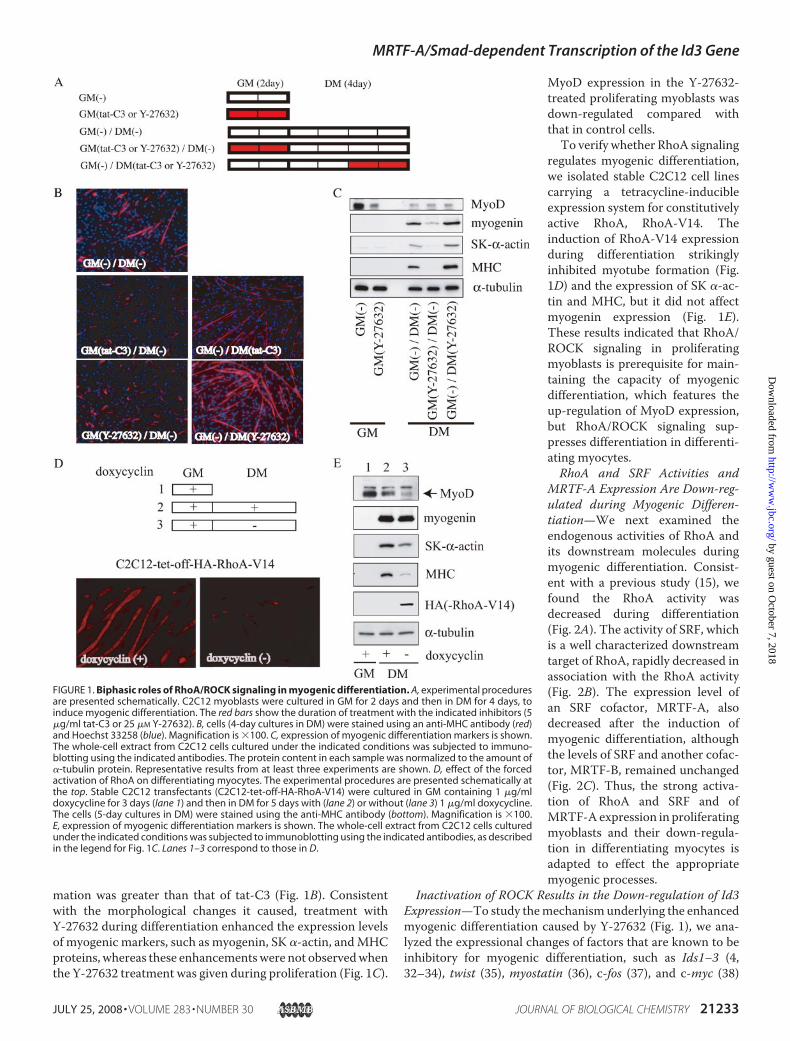
mation was greater than that of tat-C3 (Fig. 1B). Consistentwith the morphological changes it caused, treatment withY-27632 during differentiation enhanced the expression levelsof myogenic markers, such as myogenin, SK �-actin, andMHCproteins, whereas these enhancementswere not observedwhenthe Y-27632 treatment was given during proliferation (Fig. 1C).
MyoD expression in the Y-27632-treated proliferating myoblasts wasdown-regulated compared withthat in control cells.To verify whether RhoA signaling
regulates myogenic differentiation,we isolated stable C2C12 cell linescarrying a tetracycline-inducibleexpression system for constitutivelyactive RhoA, RhoA-V14. Theinduction of RhoA-V14 expressionduring differentiation strikinglyinhibited myotube formation (Fig.1D) and the expression of SK �-ac-tin and MHC, but it did not affectmyogenin expression (Fig. 1E).These results indicated that RhoA/ROCK signaling in proliferatingmyoblasts is prerequisite for main-taining the capacity of myogenicdifferentiation, which features theup-regulation of MyoD expression,but RhoA/ROCK signaling sup-presses differentiation in differenti-ating myocytes.RhoA and SRF Activities and
MRTF-A Expression Are Down-reg-ulated during Myogenic Differen-tiation—We next examined theendogenous activities of RhoA andits downstream molecules duringmyogenic differentiation. Consist-ent with a previous study (15), wefound the RhoA activity wasdecreased during differentiation(Fig. 2A). The activity of SRF, whichis a well characterized downstreamtarget of RhoA, rapidly decreased inassociation with the RhoA activity(Fig. 2B). The expression level ofan SRF cofactor, MRTF-A, alsodecreased after the induction ofmyogenic differentiation, althoughthe levels of SRF and another cofac-tor, MRTF-B, remained unchanged(Fig. 2C). Thus, the strong activa-tion of RhoA and SRF and ofMRTF-Aexpression in proliferatingmyoblasts and their down-regula-tion in differentiating myocytes isadapted to effect the appropriatemyogenic processes.
Inactivation of ROCK Results in the Down-regulation of Id3Expression—To study themechanism underlying the enhancedmyogenic differentiation caused by Y-27632 (Fig. 1), we ana-lyzed the expressional changes of factors that are known to beinhibitory for myogenic differentiation, such as Ids1–3 (4,32–34), twist (35), myostatin (36), c-fos (37), and c-myc (38)
FIGURE 1. Biphasic roles of RhoA/ROCK signaling in myogenic differentiation. A, experimental proceduresare presented schematically. C2C12 myoblasts were cultured in GM for 2 days and then in DM for 4 days, toinduce myogenic differentiation. The red bars show the duration of treatment with the indicated inhibitors (5�g/ml tat-C3 or 25 �M Y-27632). B, cells (4-day cultures in DM) were stained using an anti-MHC antibody (red)and Hoechst 33258 (blue). Magnification is �100. C, expression of myogenic differentiation markers is shown.The whole-cell extract from C2C12 cells cultured under the indicated conditions was subjected to immuno-blotting using the indicated antibodies. The protein content in each sample was normalized to the amount of�-tubulin protein. Representative results from at least three experiments are shown. D, effect of the forcedactivation of RhoA on differentiating myocytes. The experimental procedures are presented schematically atthe top. Stable C2C12 transfectants (C2C12-tet-off-HA-RhoA-V14) were cultured in GM containing 1 �g/mldoxycycline for 3 days (lane 1) and then in DM for 5 days with (lane 2) or without (lane 3) 1 �g/ml doxycycline.The cells (5-day cultures in DM) were stained using the anti-MHC antibody (bottom). Magnification is �100.E, expression of myogenic differentiation markers is shown. The whole-cell extract from C2C12 cells culturedunder the indicated conditions was subjected to immunoblotting using the indicated antibodies, as describedin the legend for Fig. 1C. Lanes 1–3 correspond to those in D.
MRTF-A/Smad-dependent Transcription of the Id3 Gene
JULY 25, 2008 • VOLUME 283 • NUMBER 30 JOURNAL OF BIOLOGICAL CHEMISTRY 21233
by guest on October 7, 2018
http://ww
w.jbc.org/
Dow
nloaded from

(Fig. 3A). In agreement with previous reports (4, 32–34), theexpression levels of Id1, Id2, and Id3mRNAs were significantlydown-regulated during myogenic differentiation. The Id3mRNA, but not the mRNA for twist, myostatin, c-fos, or c-myc,was further down-regulated in response to Y-27632 treatment.These results suggest that the Y-27632 treatment-inducedenhancement of myogenic differentiation might have been dueto the down-regulation of Id3 expression. To confirm the spe-cific inhibition of ROCK by Y-27632, siRNA-mediated ROCK1and ROCK2 knockdownwas carried out. The expression of Id3was decreased by knockdown of ROCK in proliferating myo-blasts (data not shown). Furthermore, we addressed a role ofRhoA, upstream effector of ROCK, in the transcription of Id3gene. The induction of RhoA-V14 increased the expression ofId3 in tetracycline-regulated C2C12 cells (Fig. 3B). Both analy-ses by semi-quantitative and real time RT-PCR revealed thatthe up-regulation of Id3 expression is modest in proliferatingmyoblasts but is marked in differentiating myocytes. Theseresults indicated that the transcription of Id3 gene is up-regu-
lated by RhoA/ROCK1 signaling but is not regulated by thepathway mediated through ROCK2.Forced Activation of MRTF-A in Differentiating Myocytes
Inhibits Myogenic Differentiation via the Induction of Id3Expression—Because the expression of MRTF-A was reducedin differentiating myocytes compared with its high levels inproliferating myoblasts (Fig. 2C), we focused on downstreamevents in the RhoA/MRTF-A pathway. To examine the effect ofMRTF-A on differentiatingmyocytes, we isolated stable C2C12cell lines carrying a tetracycline-inducible expression systemfor constitutively active MRTF-A (ca-MRTF-A). Using thesecell lines, we found that the induction of ca-MRTF-A after thestart of differentiation markedly suppressed myotube forma-tion (Fig. 4A) as well as the expression of myogenic differentia-tion markers, including myogenin, SK �-actin, and MHC (Fig.4B). We also analyzed the expression levels of factors that areinhibitory for myogenic differentiation. Id3 expression wassolely up-regulated at both the protein and mRNA levels inmyocytes expressing ca-MRTF-A during differentiation, but
FIGURE 2. Changes in RhoA activity and its downstream events during myogenic differentiation. A, time-dependent changes in RhoA activity measuredby a rhotekin pulldown assay are shown. Immunoblotting using anti-RhoA antibodies shows the amount of GTP-bound active RhoA protein associated withrhotekin (upper panel) and the total amount of RhoA protein (lower panel) in each whole-cell extract from C2C12 cells cultured under the indicated conditions(left). The relative RhoA activity, i.e. the image intensity of GTP-bound active RhoA protein normalized to that of total RhoA protein, is presented graphically(right). The intensity was measured using NIH Image software. Each value is expressed relative to the value for myoblasts cultured in GM, which was set as 1.0,and represents the mean � S.D. of the results from three independent experiments. B, change in transcriptional activity of SRF is shown. C2C12 myoblasts,cultured in GM, were transfected with 3�CArG-Luc and pSV-�-gal, and cell extracts prepared at the indicated periods were assayed for luciferase activity asdescribed under “Experimental Procedures.” The relative luciferase activities normalized to the �-galactosidase activity are shown. Each value represents themean � S.D. of the results from three independent experiments. C, expression levels of SRF and its co-factors, MRTF-A/B, are shown. The whole-cell extract fromC2C12 cells cultured under the indicated conditions was analyzed by immunoblotting using the indicated antibodies (left). The signal intensity is presentedgraphically (right). The gray, white, and black bars represent the signal intensity of MRTF-A, MRTF-B, and SRF, respectively. Each value is expressed relative to thevalue for myoblasts cultured in GM, which was set as 1.0, and represents the mean � S.D. of the results from three independent experiments. D means cultureday.
MRTF-A/Smad-dependent Transcription of the Id3 Gene
21234 JOURNAL OF BIOLOGICAL CHEMISTRY VOLUME 283 • NUMBER 30 • JULY 25, 2008
by guest on October 7, 2018
http://ww
w.jbc.org/
Dow
nloaded from

the other inhibitory factors examined did not show increasedexpression (Fig. 4, B and C). Taken together, these results sug-gest that MRTF-A-induced Id3 expression is critical for theRhoA/ROCK1 signaling-dependent inhibition of myogenicdifferentiation.To ask the degree of negative contribution of Id3 during
myogenic differentiation, we carried out siRNA-mediatedknockdown of Id3. It has been reported that confluent cultureof C2C12 myoblasts could differentiate into skeletal musclemarker-positivemyotubes even inGM (39).We then examinedthe effects of siRNA against Id3 on C2C12 cultures in GM. Theexpression of myogenin and MHC was markedly increased inproliferating myoblasts treated with siRNAs against Id3, Id3siRNA1, and Id3 siRNA2. Compared with the siRNA2, thesiRNA1 showed more potent effect on promotion of myogenicdifferentiation (Fig. 4D, left panel). Even under growing condi-tions, MHC-positive myotube was apparent in C2C12 culturestreated with Id3-siRNA1 (Fig. 4D, right panel); the populationof nuclei in MHC-positive myocytes was 27.58 � 4.21%. Bycontrast, in C2C12 cultures treated with control siRNA, suchpercentage was low (6.32 � 2.70%), and fused myocytes werehardly found. These results further support the above findingsthat Id3 would play a critical role in maintaining a myoblaststate.MRTF-A/Smad-mediatedTranscription of the Id3Gene—To
analyze the mechanism underlying the MRTF-A-induced Id3expression, we constructed several reporter plasmids carryingdifferent portions of the promoter region of the Id3 gene (Fig.5A) and examined the effects of MRTF-A on their promoteractivities in differentiating myocytes. The ca-MRTF-A mark-
edly enhanced the promoter activityof Id3 (�2000/�55)-Luc in a dose-dependent manner (Fig. 5B). Wefurther characterized the MRTF-A-responsive element using a series ofdeletion derivatives from Id3(�2000/�55)-Luc. The ca-MRTF-A caused equal transactivation ofthe promoter activities of Id3(�2000/�55)-Luc (5.9-fold activa-tion) and Id3 (�524/�55)-Luc (5.8-fold activation), but its effect on theactivity of Id3 (�279/�55)-Luc wasreduced to a negligible level (Fig.5C). We next performed ChIPassays to determine whetherMRTF-A is functionally involved inthe transcription of the Id3 gene. Asshown in Fig. 5D, endogenousMRTF-A in proliferating myoblastsphysically associated with the pro-moter region of the Id3 gene, indi-cating that MRTF-A plays a vitalrole in the regulation of Id3 tran-scription in vivo.The region of the Id3 promoter
spanning from �524 to �55 con-tains the SBE and its neighboring
sequences, which are conserved among humans, mice, andXenopus (40). A previous study using one of smooth musclemarker genes showed that transcription of the SM22� genewasenhanced by its association with myocardin/Smad3 in an SRF/CArG-box motif-independent manner (41). These findings ledus to speculate that activation of the Id3 promoter forMRTF-Amight be mediated through Smads. We therefore performedgel-shift assays to determine whether Smads interact with theSBE in the Id3 promoter region. The upper shifted band (Fig.5E, lane 1) was detected only when the SBE probe was incu-bated with whole-cell extracts from C2C12 myoblast; incuba-tion with the whole-cell extracts depleted of Smads did notform this band (Fig. 5E, lane 2). The intensity of this band wasdiminished by an excess amount of coldmutant SBE probe (Fig.5E, lane 3) but by cold SBE mutant one (lane 4). Furthermore,treatment with anti-Smad 1/5/8 antibodies (Fig. 5E, lane 5) butnot with control IgG (lane 6) attenuated the formation of thisband. These results strongly suggest that Smad1, -5, or -8,which are known regulators for Id3 expression under BMP sig-naling (42), interacts with the SBE in the Id3 promoter region.We then introduced mutations in the SBE within Id3 (�524/�55)-Luc to evaluate the contribution of the SBE to theMRTF-A-dependent activation of the Id3 promoter. Compared withthe wild-type construct, the mutations within the SBE mark-edly reduced the promoter activity (Fig. 5G). These results indi-cate that the SBE acts as a critical cis-element in the Id3 pro-moter in cooperation with MRTF-A.We next examined the interaction of MRTF-A with Smad,
becauseMRTF-A requires a DNA-binding transcription factorsuch as Smads to exert its activity. As shown in Fig. 5H, in
FIGURE 3. The expression of myogenic inhibitors in Y-27632-treated differentiating myocytes. A, semi-quantitative RT-PCR was performed using total RNA isolated from C2C12 cells cultured under the indicatedconditions (4 days in DM) to evaluate the mRNA levels for the indicated myogenic inhibitor genes. The cultureconditions are described in the legend for Fig. 1A. PCR cycle number and the size of the PCR product were asfollows: Id1, 28, 276 bp; Id2, 26, 350 bp; Id3, 26, 286 bp; twist, 34, 194 bp; myostatin, 38, 398 bp; c-fos, 32, 347 bp;c-myc, 32, 371 bp; GAPDH, 20, 398 bp. The mRNA level for the myogenic inhibitors was normalized to the levelof GAPDH mRNA. B, stable C2C12 transfectants (C2C12-tet-off-HA-RhoA-V14) were cultured in GM followed by4 days in DM with or without 1 �g/ml doxycycline. Semiquantitative RT-PCR (upper panel) and real RT-PCR(lower panel) were carried out using total RNAs extracted from the indicated culture condition. PCR cyclenumber of Id3 was 24 and 28.
MRTF-A/Smad-dependent Transcription of the Id3 Gene
JULY 25, 2008 • VOLUME 283 • NUMBER 30 JOURNAL OF BIOLOGICAL CHEMISTRY 21235
by guest on October 7, 2018
http://ww
w.jbc.org/
Dow
nloaded from

vitro-translated MRTF-A protein interacted directly withSmad1, which was used as representative of Smad1, -5, and -8,suggesting that Smad1 andMRTF-A might form a complex onthe Id3 promoter. To test this possibility, we performed DNAaffinity binding assays (31), in which the sequence of the Id3promoter containing the SBE was biotinylated and then incu-bated with in vitro-translated MRTF-A, pseudo-phosphoryla-ted-Smad1 (constitutively active Smad1: ca-Smad1) andSmad4. In contrast to the signal caused by the incubation ofMRTF-A and the biotinylated-SBE (which was nonspecific,because MRTF-A cannot bind to DNA), the signal intensity ofthe interaction of MRTF-A with the biotinylated SBE was sig-nificantly increased by the addition of ca-Smad1 and Smad4,indicating that MRTF-A interacts with the SBE via Smad1 andSmad4 (Fig. 5I).
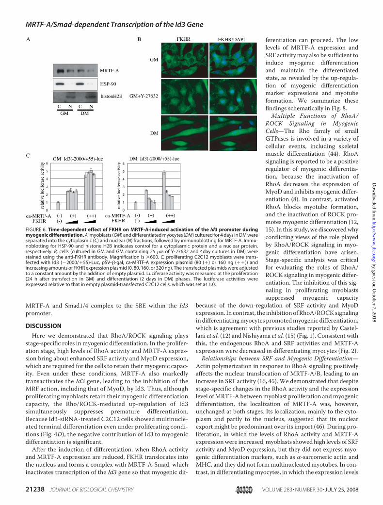
FKHR Relieves the MRTF-A/Smad-dependent Transcription ofthe Id3 Gene in DifferentiatingMyocytes—Because Id3 expressionwas down-regulated after the induc-tion ofmyogenic differentiation (Figs.3 and 4), the MRTF-A-dependenttransactivation of the Id3 gene mustbe repressed in differentiating myo-cytes. To elucidate this mechanism,we analyzed the localization ofMRTF-A at different differentiationstages and found that it wasmainly inthe cytoplasm and partly in thenucleus at both the proliferation(cytosol 72%, nucleus 28%) and dif-ferentiation stages (cytosol 75%,nucleus 25%) (Fig. 6A). No changesin its localization during the differ-entiation process suggest that someother mechanism regulates the Id3transcription.Bois and Grosveld (21) reported
that the forkhead transcription fac-tor FKHR is required for myotubeformation by myoblasts in primaryculture, inwhich FKHR translocatesfrom the cytoplasm to the nucleusin response to the induction ofmyo-genic differentiation. They alsodemonstrated that the phosphoryl-ation-dependent nuclear export ofFKHR is not mediated by phospha-tidylinositol 3-kinase/Akt signaling.Nishiyama et al. (15) showed thatthe inactivation of RhoA/ROCKsig-naling is prerequisite for the nuclearlocalization of FKHR inC2C12 cells,because ROCKdirectly phosphoryl-ates FKHR in vitro. Another reportshowed that a different forkheadtranscription factor, Foxo4, inter-acts with myocardin and represses
myocardin-dependent transcription in smooth muscle cells(43). We therefore examined the effect of FKHR on theMRTF-A-induced Id3 expression. First, we confirmed the localizationof FKHR. Consistent with previous reports (15, 21), FKHRtranslocated from the cytoplasm to the nucleus in response toinduction of myogenic differentiation and also accumulatedinto the nucleus in proliferating myoblasts treated withY-27632 (Fig. 6B).We next examined the effect of FKHR on theMRTF-A-induced activation of the Id3 promoter. FKHRshowed no apparent effect on MRTF-A-induced activation ofthe Id3 promoter in proliferating myoblasts but markedlyinhibited this activation in differentiating myocytes in a dose-dependent manner (Fig. 6C), indicating that FKHR serves as adifferentiating myocyte-specific repressor of the MRTF-A-de-pendent transcription of the Id3 gene.
FIGURE 4. Effect of forced ca-MRTF-A expression on myogenic differentiation. A, experimental proceduresare presented schematically at left. Stable C2C12 transfectants (C2C12-tet-off-FALG-ca-MRTF-A) were culturedunder the conditions described in the legend for Fig. 1D. Cells (5-day cultures in DM) were stained with ananti-MHC antibody (right). Magnification is �100. B and C, expression levels of myogenic differentiation mark-ers and myogenic inhibitors are shown. B, whole-cell extracts from C2C12 cells cultured under the indicatedconditions (5 days in DM) were subjected to immunoblotting using the indicated antibodies. C, total RNA fromC2C12 cells cultured under the indicated conditions (5 days in DM) was analyzed by RT-PCR as described in thelegend for Fig. 3. Lanes 1–3 (B and C) correspond to those in A. D, C2C12 cells were transfected with siRNAsagainst Id3 (Id3 siRNA1 and �2) and were cultured in GM for 2 days. The whole-cell extracts were subjected toimmunoblotting using the indicated antibodies. The cell morphology and expression of MHC were also ana-lyzed by staining with an anti-MHC antibody (red) and Hoechst 33258 (blue). Magnification is �200.
MRTF-A/Smad-dependent Transcription of the Id3 Gene
21236 JOURNAL OF BIOLOGICAL CHEMISTRY VOLUME 283 • NUMBER 30 • JULY 25, 2008
by guest on October 7, 2018
http://ww
w.jbc.org/
Dow
nloaded from

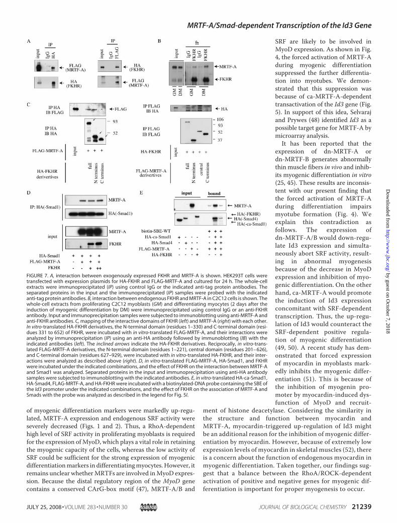
Immunoprecipitation followed by immunoblotting analysisrevealed that the exogenousMRTF-A and FKHRproteins asso-ciated with each other in HEK293T cells (Fig. 7A), and endog-enous MRTF-A potently interacted with FKHR in differentiat-ing myocytes and slightly in proliferating myoblasts (Fig. 7B).We further identified the mutual interacting domains ofMRTF-A and FKHRusing in vitro-translated proteins (Fig. 7C).We found that the N-terminal domain (residues 1–330) ofFKHR and the central domain (residues 201–626) of MRTF-A,including the basic, SAP, and leucine zipper domains, physi-cally interacted with each other.
To gain further insight into the inhibitory effect of FKHR onthe MRTF-A-dependent transactivation of the Id3 gene, weanalyzed whether FKHR influences the interactions amongMRTF-A, Smad1, and the SBE within the Id3 promoter. FKHRdid not affect the degree of MRTF-A binding to Smad1 in vitro(Fig. 7D). We next examined the effect of FKHR on the bindingof the complex consisting ofMRTF-A and Smad1/4 to the SBE.The binding of this complex to the Id3 promoter probe, whichincluded the SBE, was strongly inhibited by FKHR (Fig. 7E),indicating that FKHR represses the MRTF-A/Smad-inducedtransactivation of the Id3 gene by abrogating the binding of the
FIGURE 5. MRTF-A-dependent activation of the Id3 promoter. Transcriptional activity of the Id3 promoter in differentiating myocytes was measured asdescribed under “Experimental Procedures.” A, Id3 promoter constructs used in this analysis are illustrated. The effects of ca-MRTF-A co-expression on thepromoter activity of Id3 (2000/�55)-Luc (B), its derivatives (C), and Id3 (�524/�55)-Luc bearing a mutated SBE (G) are shown. The luciferase activities wereexpressed relative to that in empty plasmid-transfected C2C12 myocytes, which was set as 1.0 (B and G). C, luciferase activity of Id3 (�2000/�55) in cellsco-transfected with empty plasmid was set as 1.0. D, ChIP assay shows the interaction of endogenous MRTF-A with the promoter region of the Id3 gene inproliferating C2C12 myoblasts. The extracted chromatin fragments were immunoprecipitated (IP) with the indicated antibodies, and the precipitated genomicDNA was analyzed by PCR using primers for the Id3 promoter region containing the SBE. The size of the PCR product was 216 bp. PCR amplification was alsoperformed prior to immunoprecipitation for the input control. E, 32P-labeled SBE of the Id3 promoter was incubated with the whole-cell extracts of proliferatingC2C12 myoblasts (lanes 1 and 3– 6) or Smads-depleted whole-cell extracts prepared by pretreatment with anti-Smad1/5/8 antibody (lane 2) in the presence offollowing additives: 30-fold excess amounts of indicated cold competitors (lanes 3 and 4), an anti-Smad1/5/8 antibody (Ab) (lane 5) or control IgG (lane 6). Thereactants were subjected to 5% PAGE. Arrow indicates the SBE-Smad complex. mut, mutant; wt, wild type. F, depletion of Smad1/5/8 in whole-cell extracts ofproliferating C2C12 myoblasts was characterized by immunoblotting using anti-Smad1/5/8 antibody. The Smads in C2C12 whole-cell extracts were depletedby immunoprecipitation using anti-Smad1/5/8 antibody. Samples applied to immunoblotting were as follows: Smads-depleted C2C12 whole-cell extracts(lane 1), nondepleted one (lane 2), and immunoprecipitates by anti-Smad1/5/8 antibody (lane 3) or control IgG (lane 4). H, interaction between Smad1 andMRTF-A was analyzed using in vitro-translated proteins. HA-Smad1 and FLAG-MRTF-A proteins were co-incubated, and their interaction was analyzed byimmunoprecipitation followed by immunoblotting using the indicated antibodies. I, indicated tagged proteins were translated in vitro and then incubatedwith a biotinylated-DNA probe containing the SBE of the Id3 promoter. The proteins bound to the probe were collected with streptavidin-conjugated magneticbeads, and the SBE-interacting proteins were analyzed by immunoblotting using the indicated antibodies.
MRTF-A/Smad-dependent Transcription of the Id3 Gene
JULY 25, 2008 • VOLUME 283 • NUMBER 30 JOURNAL OF BIOLOGICAL CHEMISTRY 21237
by guest on October 7, 2018
http://ww
w.jbc.org/
Dow
nloaded from
MRTF-A and Smad1/4 complex to the SBE within the Id3promoter.
DISCUSSION
Here we demonstrated that RhoA/ROCK signaling playsstage-specific roles in myogenic differentiation. In the prolifer-ation stage, high levels of RhoA activity and MRTF-A expres-sion bring about enhanced SRF activity and MyoD expression,which are required for the cells to retain their myogenic capac-ity. Even under these conditions, MRTF-A also markedlytransactivates the Id3 gene, leading to the inhibition of theMRF action, including that of MyoD, by Id3. Thus, althoughproliferating myoblasts retain their myogenic differentiationcapacity, the Rho/ROCK-mediated up-regulation of Id3simultaneously suppresses premature differentiation.Because Id3-siRNA-treated C2C12 cells showed multinucle-ated terminal differentiation even under proliferating condi-tions (Fig. 4D), the negative contribution of Id3 to myogenicdifferentiation is significant.After the induction of differentiation, when RhoA activity
and MRTF-A expression are reduced, FKHR translocates intothe nucleus and forms a complex with MRTF-A-Smad, whichinactivates transcription of the Id3 gene so that myogenic dif-
ferentiation can proceed. The lowlevels of MRTF-A expression andSRF activitymay also be sufficient toinduce myogenic differentiationand maintain the differentiatedstate, as revealed by the up-regula-tion of myogenic differentiationmarker expressions and myotubeformation. We summarize thesefindings schematically in Fig. 8.Multiple Functions of RhoA/
ROCK Signaling in MyogenicCells—The Rho family of smallGTPases is involved in a variety ofcellular events, including skeletalmuscle differentiation (44). RhoAsignaling is reported to be a positiveregulator of myogenic differentia-tion, because the inactivation ofRhoA decreases the expression ofMyoD and inhibits myogenic differ-entiation (8). In contrast, activatedRhoA blocks myotube formation,and the inactivation of ROCK pro-motes myogenic differentiation (12,15). In this study,we discoveredwhyconflicting views of the role playedby RhoA/ROCK signaling in myo-genic differentiation have arisen.Stage-specific analysis was criticalfor evaluating the roles of RhoA/ROCK signaling in myogenic differ-entiation. The inhibition of this sig-naling in proliferating myoblastssuppressed myogenic capacity
because of the down-regulation of SRF activity and MyoDexpression. In contrast, the inhibition of RhoA/ROCKsignalingin differentiatingmyocytes promotedmyogenic differentiation,which is agreement with previous studies reported by Castel-lani et al. (12) andNishiyama et al. (15) (Fig. 1). Consistent withthis, the endogenous RhoA and SRF activities and MRTF-Aexpression were decreased in differentiating myocytes (Fig. 2).Relationships between SRF and Myogenic Differentiation—
Actin polymerization in response to RhoA signaling positivelyaffects the nuclear translocation of MRTF-A/B, leading to anincrease in SRF activity (16, 45). We demonstrated that despitestage-specific changes in the RhoA activity and the expressionlevel ofMRTF-A betweenmyoblast proliferation andmyogenicdifferentiation, the localization of MRTF-A was, however,unchanged at both stages. Its localization, mainly to the cyto-plasm and partly to the nucleus, suggested that its nuclearexport might be predominant over its import (46). During pro-liferation, in which the levels of RhoA activity and MRTF-Aexpressionwere increased,myoblasts showedhigh levels of SRFactivity and MyoD expression, but they did not express myo-genic differentiation markers, such as �-sarcomeric actin andMHC, and they did not formmultinucleatedmyotubes. In con-trast, in differentiatingmyocytes, in which the expression levels
FIGURE 6. Time-dependent effect of FKHR on MRTF-A-induced activation of the Id3 promoter duringmyogenic differentiation. A, myoblasts (GM) and differentiated myocytes (DM) cultured for 4 days in DM wereseparated into the cytoplasmic (C) and nuclear (N) fractions, followed by immunoblotting for MRTF-A. Immu-noblotting for HSP-90 and histone H2B indicates control for a cytoplasmic protein and a nuclear protein,respectively. B, cells (cultured in GM and GM containing 25 �M of Y-27632 and 4day cultures in DM) werestained using the anti-FKHR antibody. Magnification is �600. C, proliferating C2C12 myoblasts were trans-fected with Id3 (�2000/�55)-Luc, pSV-�-gal, ca-MRTF-A expression plasmid (80 (�) or 160 ng (��)) andincreasing amounts of FKHR expression plasmid (0, 80, 160, or 320 ng). The transfected plasmids were adjustedto a constant amount by the addition of empty plasmid. Luciferase activity was measured at the proliferation(24 h after transfection in GM) and differentiation (2 days in DM) phases. The luciferase activities wereexpressed relative to that in empty plasmid-transfected C2C12 cells, which was set as 1.0.
MRTF-A/Smad-dependent Transcription of the Id3 Gene
21238 JOURNAL OF BIOLOGICAL CHEMISTRY VOLUME 283 • NUMBER 30 • JULY 25, 2008
by guest on October 7, 2018
http://ww
w.jbc.org/
Dow
nloaded from
of myogenic differentiation markers were markedly up-regu-lated, MRTF-A expression and endogenous SRF activity wereseverely decreased (Figs. 1 and 2). Thus, a RhoA-dependenthigh level of SRF activity in proliferating myoblasts is requiredfor the expression ofMyoD, which plays a vital role in retainingthe myogenic capacity of the cells, whereas the low activity ofSRF could be sufficient for the strong expression of myogenicdifferentiationmarkers in differentiatingmyocytes.However, itremains unclear whetherMRTFs are involved inMyoD expres-sion. Because the distal regulatory region of the MyoD genecontains a conserved CArG-box motif (47), MRTF-A/B and
SRF are likely to be involved inMyoD expression. As shown in Fig.4, the forced activation of MRTF-Aduring myogenic differentiationsuppressed the further differentia-tion into myotubes. We demon-strated that this suppression wasbecause of ca-MRTF-A-dependenttransactivation of the Id3 gene (Fig.5). In support of this idea, Selvarajand Prywes (48) identified Id3 as apossible target gene for MRTF-A bymicroarray analysis.It has been reported that the
expression of dn-MRTF-A ordn-MRTF-B generates abnormallythin muscle fibers in vivo and inhib-its myogenic differentiation in vitro(25, 45). These results are inconsis-tent with our present finding thatthe forced activation of MRTF-Aduring differentiation impairsmyotube formation (Fig. 4). Weexplain this contradiction asfollows. The expression ofdn-MRTF-A/B would down-regu-late Id3 expression and simulta-neously abort SRF activity, result-ing in abnormal myogenesisbecause of the decrease in MyoDexpression and inhibition of myo-genic differentiation. On the otherhand, ca-MRTF-A would promotethe induction of Id3 expressionconcomitant with SRF-dependenttranscription. Thus, the up-regu-lation of Id3 would counteract theSRF-dependent positive regula-tion of myogenic differentiation(49, 50). A recent study has dem-onstrated that forced expressionof myocardin in myoblasts mark-edly inhibits the myogenic differ-entiation (51). This is because ofthe inhibition of myogenin pro-moter by myocardin-induced dys-function of MyoD and recruit-
ment of histone deacetylase. Considering the similarity inthe structure and function between myocardin andMRTF-A, myocardin-triggered up-regulation of Id3 mightbe an additional reason for the inhibition of myogenic differ-entiation by myocardin. However, because of extremely lowexpression levels of myocardin in skeletal muscles (52), thereis a concern about the function of endogenous myocardin inmyogenic differentiation. Taken together, our findings sug-gest that a balance between the RhoA/ROCK-dependentactivation of positive and negative genes for myogenic dif-ferentiation is important for proper myogenesis to occur.
FIGURE 7. A, interaction between exogenously expressed FKHR and MRTF-A is shown. HEK293T cells weretransfected with expression plasmids for HA-FKHR and FLAG-MRTF-A and cultured for 24 h. The whole-cellextracts were immunoprecipitated (IP) using control IgG or the indicated anti-tag protein antibodies. Theseparated proteins in the input and the immunoprecipitated (IP) samples were probed with the indicatedanti-tag protein antibodies. B, interaction between endogenous FKHR and MRTF-A in C2C12 cells is shown. Thewhole-cell extracts from proliferating C2C12 myoblasts (GM) and differentiating myocytes (2 days after theinduction of myogenic differentiation by DM) were immunoprecipitated using control IgG or an anti-FKHRantibody. Input and immunoprecipitation samples were subjected to immunoblotting using anti-MRTF-A andanti-FKHR antibodies. C, mapping of the interactive domains of FKHR (left) and MRTF-A (right) with each other.In vitro-translated HA-FKHR derivatives, the N-terminal domain (residues 1–330) and C-terminal domain (resi-dues 331 to 652) of FKHR, were incubated with in vitro-translated FLAG-MRTF-A, and their interactions wereanalyzed by immunoprecipitation (IP) using an anti-HA antibody followed by immunoblotting (IB) with theindicated antibodies (left). The inclined arrows indicate the HA-FKHR derivatives. Reciprocally, in vitro-trans-lated FLAG-MRTF-A derivatives, the N-terminal domain (residues 1–221), central domain (residues 201– 626),and C-terminal domain (residues 627–929), were incubated with in vitro-translated HA-FKHR, and their inter-actions were analyzed as described above (right). D, in vitro-translated FLAG-MRTF-A, HA-Smad1, and FKHRwere incubated under the indicated combinations, and the effect of FKHR on the interaction between MRTF-Aand Smad1 was analyzed. Separated proteins in the input and immunoprecipitation using anti-HA antibodysamples were subjected to immunoblotting with the indicated antibodies. E, in vitro-translated HA-ca-Smad1,HA-Smad4, FLAG-MRTF-A, and HA-FKHR were incubated with a biotinylated-DNA probe containing the SBE ofthe Id3 promoter under the indicated combinations, and the effect of FKHR on the association of MRTF-A andSmads with the probe was analyzed as described in the legend for Fig. 5I.
MRTF-A/Smad-dependent Transcription of the Id3 Gene
JULY 25, 2008 • VOLUME 283 • NUMBER 30 JOURNAL OF BIOLOGICAL CHEMISTRY 21239
by guest on October 7, 2018
http://ww
w.jbc.org/
Dow
nloaded from

FKHR Negatively Regulates MRTF-A/Smad-dependentTranscription of the Id3 Gene in DifferentiatingMyocytes—Thetranscriptional regulation of the Id3 gene has been partiallycharacterized. Wu and Lim (53) reported that Sp2 binding tothe GC-box-like motif, which is present within the promoterregion (�180/�1 bp) of the Id3 gene, positively regulates theId3 promoter in proliferating C2C12 myoblasts. von Bubnoffet al. (40) found, using the Xenopus animal cap system, that theId3 promoter contains a BMP-response element, whichincludes an SBE and a GC-rich element resembling an OAZ-binding site, and they demonstrated that these elements arenecessary for responsiveness to BMP. Smad7 is known to be anegative regulator of BMP (54) and transforming growth fac-tor-� (55) signaling and a positive regulator for myogenic dif-ferentiation by directly interacting with MyoD and enhance-ment of MyoD transcriptional activity (56). We examinedwhether Smad7 is involved in the regulation of Id3 transcrip-tion. As described by Kollias et al. (56), Smad7 expression wasincreased during C2C12 myogenic differentiation both inmRNA and protein levels. However, Id3 promoter activity wasnot affected by forced expression of Smad7, indicating thatSmad7 would not be involved in the Id3 transcriptional regula-tion (data not shown). In this way, the Id3 gene is transcription-ally regulated by diverse mechanisms, in a tissue- or stimulus-dependent manner. Here we demonstrated that MRTF-A actsas a positive regulator of Id3 gene transcription and, with Smad1/4, associates with the SBE in the Id3 promoter (Fig. 5).FKHR has been reported to be a regulator of myogenic dif-
ferentiation because it activates the transcription of targetgenes involved in cell fusion or extracellular matrix remodelingand promotes myoblast fusion (21). The interaction of FKHRwith cyclic GMP-dependent protein kinase I causes myoblastfusion by influencing the fusion rate (22). Despite these find-ings, conflicting results have been reported; normal myogenicdifferentiation is impaired by the forced expression of consti-tutively active or wild-type FKHR in skeletal muscle cells (57,58). FKHR also interacts with androgen receptor and sup-presses androgen-induced androgen receptor transcription inprostate cancer cells (59). Thus, this forkhead transcription fac-tor family member is involved in various transcriptional regu-lations. In this study, we identified a novel function of FKHR as
a repressor of the Id3 promoter.Wedemonstrated here that FKHR playsa positive role in myogenic differen-tiation through the inhibition of Id3expression. Foxo4 interacts withboth myocardin and SRF, and thisternary complex then aborts thetranscription ofmyocardin/SRF tar-get genes in smooth muscle cells(42). Using myocytes, we revealedthat FKHR inhibitsMRTF-A/Smad-dependent Id3 transcription byinteracting with MRTF-A/Smadsand thereby interrupting the bind-ing ofMRTF-A/Smads to the SBE inthe Id3 promoter. Further studyregarding the involvement of FKHR
in Id3-dependent developmental, physiological, and patholog-ical processes will be required to elucidate the diverse functionsof this forkhead transcription factor.
REFERENCES1. Molkentin, J. D., and Olson, E. N. (1996) Curr. Opin. Genet. Dev. 6,
445–4532. Olson, E. N., and Klein, W. H. (1994) Genes Dev. 8, 1–83. Sabourin, L. A., and Rudnicki, M. A. (2000) Clin. Genet. 57, 16–254. Benezra, R., Davis, R. L., Lockshon, D., Turner, D. L., and Weintraub, H.
(1990) Cell 61, 49–595. Jen, Y., Weintraub, H., and Benezra, R. (1992) Genes Dev. 6, 1466–14796. Ruzinova, M. B., and Benezra, R. (2003) Trends Cell Biol. 13, 410–4187. Etienne-Manneville, S., and Hall, A. (2002) Nature 420, 629–6358. Carnac, G., Primig, M., Kitzmann, M., Chafey, P., Tuil, D., Lamb, N., and
Fernandez, A. (1998)Mol. Biol. Cell 9, 1891–19029. Gauthier-Rouviere, C., Vandromme, M., Tuil, D., Lautredou, N., Morris,
M., Soulez, M., Kahn, A., Fernandez, A., and Lamb, N. (1996) Mol. Biol.Cell 7, 719–729
10. Takano, H., Komuro, I., Oka, T., Shiojima, I., Hiroi, Y., Mizuno, T., andYazaki, Y. (1998)Mol. Cell. Biol. 18, 1580–1589
11. Wei, L., Zhou, W., Croissant, J. D., Johansen, F. E., Prywes, R., Balasubra-manyam, A., and Schwartz, R. J. (1998) J. Biol. Chem. 273, 30287–30294
12. Castellani, L., Salvati, E., Alema, S., and Falcone, G. (2006) J. Biol. Chem.281, 15249–15257
13. Charrasse, S., Comunale, F., Grumbach, Y., Poulat, F., Blangy, A., andGauthier-Rouviere, C. (2006)Mol. Biol. Cell 17, 749–759
14. Meriane, M., Roux, P., Primig, M., Fort, P., and Gauthier-Rouviere, C.(2000)Mol. Biol. Cell 11, 2513–2528
15. Nishiyama, T., Kii, I., and Kudo, A. (2004) J. Biol. Chem. 279,47311–47319
16. Miralles, F., Posern, G., Zaromytidou, A. I., and Treisman, R. (2003) Cell113, 329–342
17. Cen, B., Selvaraj, A., and Prywes, R. (2004) J. Cell. Biochem. 93, 74–8218. Pipes, G. C., Creemers, E. E., and Olson, E. N. (2006) Genes Dev. 20,
1545–155619. Li, S., Czubryt, M. P., McAnally, J., Bassel-Duby, R., Richardson, J. A.,
Wiebel, F. F., Nordheim, A., and Olson, E. N. (2005) Proc. Natl. Acad. Sci.U. S. A. 102, 1082–1087
20. Selvaraj, A., and Prywes, R. (2003) J. Biol. Chem. 278, 41977–4198721. Bois, P. R., and Grosveld, G. C. (2003) EMBO J. 22, 1147–115722. Bois, P. R., Brochard, V. F., Salin-Cantegrel, A. V., Cleveland, J. L., and
Grosveld, G. C. (2005)Mol. Cell. Biol. 25, 7645–765623. Birkenkamp, K. U., and Coffer, P. J. (2003) Biochem. Soc. Trans. 31,
292–29724. Park, J., Kim, J. S., Jung, K. C., Lee, H. J., Kim, J. I., Kim, J., Lee, J. Y., Park,
J. B., and Choi, S. Y. (2003)Mol. Cells 16, 216–223
FIGURE 8. Model for the phase-dependent regulation of Id3 expression by MRTF-A in association withFKHR and RhoA/ROCK-signaling during myogenic differentiation. The detailed description is provided inthe text.
MRTF-A/Smad-dependent Transcription of the Id3 Gene
21240 JOURNAL OF BIOLOGICAL CHEMISTRY VOLUME 283 • NUMBER 30 • JULY 25, 2008
by guest on October 7, 2018
http://ww
w.jbc.org/
Dow
nloaded from

25. Morita, T., and Mayanagi, T., Sobue, K. (2007) J. Cell Biol. 3, 1027–104226. Qin, B. Y., Chacko, B.M., Lam, S. S., de Caestecker,M. P., Correia, J. J., and
Lin, K. (2001)Mol. Cell 8, 1303–131227. Copeland, J. W., and Treisman, R. (2002)Mol. Biol. Cell 13, 4088–409928. Mohun, T., Garrett, N., and Treisman, R. (1987) EMBO J. 6, 667–67329. Brennan, T. J., and Olson, E. N. (1990) Genes Dev. 4, 582–59530. Hayashi, K., Nakamura, S., Nishida, W., and Sobue, K. (2006) Mol. Cell.
Biol. 26, 9456–947031. Suzuki, T., Fujisawa, J. I., Toita, M., and Yoshida, M. (1993) Proc. Natl.
Acad. Sci. U. S. A. 15, 610–61432. Atherton,G.T., Travers,H., Deed, R., andNorton, J. D. (1996)CellGrowth
& Differ. 7, 1059–106633. Melnikova, I. N., and Christy, B. A. (1996) Cell Growth & Differ. 7,
1067–107934. Melnikova, I. N., Bounpheng, M., Schatteman, G. C., Gilliam, D., and
Christy, B. A. (1999) Exp. Cell Res. 247, 94–10435. Spicer, D. B., Rhee, J., Cheung,W. L., and Lassar, A. B. (1996) Science 272,
1476–148036. Langley, B., Thomas, M., Bishop, A., Sharma, M., Gilmour, S., and Kam-
badur, R. (2002) J. Biol. Chem. 277, 49831–4984037. Li, L., Chambard, J. C., Karin, M., and Olson, E. N. (1992) Genes Dev. 6,
676–68938. Miner, J. H., and Wold, B. J. (1991)Mol. Cell. Biol. 11, 2842–285139. Yoshiko, Y., Hirao, K., and Maeda, N. (2002) Am. J. Physiol. 283,
C1278–C128640. von Bubnoff, A., Peiffer, D. A., Blitz, I. L., Hayata, T., Ogata, S., Zeng, Q.,
Trunnell, M., and Cho, K. W. (2005) Dev. Biol. 281, 210–22641. Qiu, P., Ritchie, R. P., Fu, Z., Cao, D., Cumming, J., Miano, J. M., Wang,
D. Z., Li, H. J., and Li, L. (2005) Circ. Res. 97, 983–99142. Xu, L. (2006) Biochim. Biophys. Acta. 1759, 503–51343. Liu, Z. P., Wang, Z., Yanagisawa, H., and Olson, E. N. (2005) Dev. Cell 9,
261–27044. Travaglione, S., Messina, G., Fabbri, A., Falzano, L., Giammarioli, A. M.,
Grossi, M., Rufini, S., and Fiorentini, C. (2005) Cell Death Differ. 12,78–86
45. Kuwahara, K., Barrientos, T., Pipes, G. C., Li, S., and Olson, E. N. (2005)Mol. Cell. Biol. 25, 3173–3181
46. Vartiainen,M. K., Guettler, S., Larijani, B., andTreisman, R. (2007) Science22, 1749–1752
47. L’honore, A., Lamb, N. J., Vandromme, M., Turowski, P., Carnac, G., andFernandez, A. (2003)Mol. Biol. Cell 14, 2151–2162
48. Selvaraj, A., and Prywes, R. (2004) BMCMol. Biol. 5, 1349. French, B. A., Chow, K. L., Olson, E. N., and Schwartz, R. J. (1991) Mol.
Cell. Biol. 11, 2439–245050. Muscat, G. E., Emery, J., and Collie, E. S. (1992) Gene Expr. 2, 241–25751. Long, X., Creemers, E. E., Wang, D. Z., Olson, E. N., and Miano, J. M.
(2007) Proc. Natl. Acad. Sci. U. S. A. 104, 16570–1657552. Wang, D., Chang, P. S., Wang, Z., Sutherland, L., Richardson, J. A., Small,
E., Krieg, P. A., and Olson, E. N. (2001) Cell 105, 852–86253. Wu, J., and Lim, R. W. (2005) Biochim. Biophys. Acta 1731, 13–2254. Ishisaki, A., Yamato, K., Hashimoto, S., Nakao, A., Tamaki, K., Nonaka, K.,
ten Dijke, P., Sugino, H., and Nishihara, T. (1999) J. Biol. Chem. 274,13637–13642
55. Hayashi, H., Abdollah, S., Qiu, Y., Cai, J., Xu, Y. Y., Grinnell, B. W., Rich-ardson,M.A., Topper, J. N., Gimbrone,M.A., Jr.,Wrana, J. L., and Falb, D.(1997) Cell 27, 1165–1173
56. Kollias, H. D., Perry, R. L., Miyake, T., Aziz, A., and McDermott, J. C.(2006)Mol. Cell. Biol. 26, 6248–6260
57. Hribal, M. L., Nakae, J., Kitamura, T., Shutter, J. R., and Accili, D. (2003)J. Cell Biol. 162, 535–541
58. Kamei, Y., Miura, S., Suzuki, M., Kai, Y., Mizukami, J., Taniguchi, T.,Mochida, K., Hata, T.,Matsuda, J., Aburatani, H., Nishino, I., and Ezaki, O.(2004) J. Biol. Chem. 279, 41114–41123
59. Fan, W., Yanase, T., Morinaga, H., Okabe, T., Nomura, M., Daitoku, H.,Fukamizu, A., Kato, S., Takayanagi, R., and Nawata, H. (2007) J. Biol.Chem. 282, 7329–7338
MRTF-A/Smad-dependent Transcription of the Id3 Gene
JULY 25, 2008 • VOLUME 283 • NUMBER 30 JOURNAL OF BIOLOGICAL CHEMISTRY 21241
by guest on October 7, 2018
http://ww
w.jbc.org/
Dow
nloaded from
Kazuhiro Iwasaki, Ken'ichiro Hayashi, Tomoaki Fujioka and Kenji Sobue GeneId3
Myocardin-related Transcription Factor-A/Smad-dependent Transcription of the Rho/Rho-associated Kinase Signal Regulates Myogenic Differentiation via
doi: 10.1074/jbc.M710525200 originally published online May 12, 20082008, 283:21230-21241.J. Biol. Chem.
10.1074/jbc.M710525200Access the most updated version of this article at doi:
Alerts:
When a correction for this article is posted•
When this article is cited•
to choose from all of JBC's e-mail alertsClick here
http://www.jbc.org/content/283/30/21230.full.html#ref-list-1
This article cites 59 references, 34 of which can be accessed free at
by guest on October 7, 2018
http://ww
w.jbc.org/
Dow
nloaded from


![A Comparison of Efficiency and Robustness of ID3 and C4.5 ... · of the popular ones are ID3 [1] and C4.5 [2] by J.R Quinlan. II. ID3 VS. C4.5 ID3 algorithm selects the best attribute](https://static.fdocuments.net/doc/165x107/5f0f2afd7e708231d442d273/a-comparison-of-efficiency-and-robustness-of-id3-and-c45-of-the-popular-ones.jpg)