
Rheumatology pearls 9-19-2014
47
Rheumatology Pearls Primary Care Update 09/18/2014 Paul Sufka, M.D. HealthPartners Rheumatology
-
Upload
paul-sufka -
Category
Health & Medicine
-
view
1.062 -
download
0
Transcript of Rheumatology pearls 9-19-2014

Rheumatology Pearls Primary Care Update 09/18/2014
Paul Sufka, M.D.HealthPartners Rheumatology

No disclosures.
No off-label discussion.

Topics
•Common labs in Rheumatology
•Differentiating inflammatory from non-inflammatory arthritis
•Gout

Labs
•ANA
•RF
•CCP

ANA: Antinuclear Antibodies
• Autoantibodies that bind to contents of the cell nucleus
• Extremely common
• Low titer (up to 1:160) - up to 20% normal individuals
• Higher titers still found in up to 5% of normals
• Typically used to facilitate diagnosis of lupus and other connective tissue diseases.
• Negative ANA makes CTD highly unlikely, but positive doesn’t rule in.
Images: ACR Image Bank
Some causes of positive ANA
• Systemic autoimmune diseases
• SLE, Scleroderma, MCTD: nearly 100%
• RA: 45%
• Sjogren’s, polymyositis, dermatomyositis, etc.
• Organ specific autoimmune diseases
• Hashimotos, Graves’, autoimmune hepatitis, PBC
• Infections
• Viral: frequently seen w/ HCV, EBV, HIV, Parvovirus B19
• Malignancy
• Other: IBD, pulmonary fibrosis
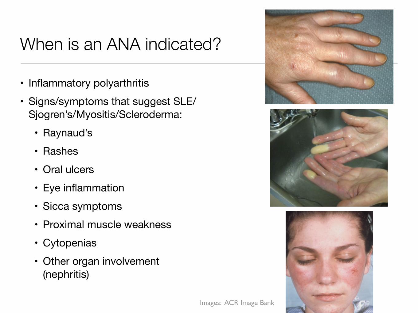
When is an ANA indicated?
• Inflammatory polyarthritis• Signs/symptoms that suggest SLE/
Sjogren’s/Myositis/Scleroderma:• Raynaud’s• Rashes• Oral ulcers• Eye inflammation• Sicca symptoms• Proximal muscle weakness• Cytopenias• Other organ involvement
(nephritis)
Images: ACR Image Bank
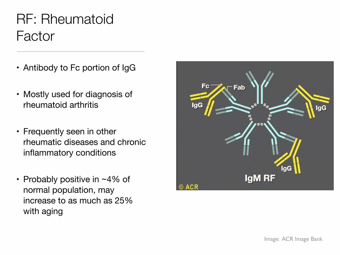
RF: Rheumatoid Factor
• Antibody to Fc portion of IgG
• Mostly used for diagnosis of rheumatoid arthritis
• Frequently seen in other rheumatic diseases and chronic inflammatory conditions
• Probably positive in ~4% of normal population, may increase to as much as 25% with aging
Image: ACR Image Bank

Causes of Rheumatoid Factor
• Rheumatic
• RA: 26-90%
• Sjogren’s: 75-90%
• Lupus: 15-35%
• Others: MCTD, cryoglobulinemia, myositis, vasculitis
• Non-rheumatic
• *** Chronic Hepatitis C: 26-76% ***
• Chronic Hep B, TB, endocarditis, syphilis
• Pulmonary dz: sarcoid, pulmonary fibrosis, silicosis, asbestosis
• Malignancy, PBC

When is a Rheumatoid Factor Indicated?
• When rheumatoid arthritis is suspected clinically
• In rheumatology, also helpful when looking for evidence of Sjogren’s or cryoglobulinemia

Cyclic Citrullinated Peptide (CCP) Antibodies
• Strongly associated with rheumatoid arthritis and development of more aggressive/erosive disease
• Can be helpful in differentiating RA from other forms of arthritis, such as hepatitis C related arthritis (since RF often positive in both RA and Hep C)
• Often present for many years (with RF) before the onset of clinical arthritis
Other notes on ANA and RF/CCP
•No use in monitoring ANA titer or RF/CCP - not clinically useful
•Unless new symptoms occur, no reason to recheck ANA

Arthritis history
• Differentiate inflammatory vs non-inflammatory pain
• What is the pattern?
• What joints are involved?
• Acute, subacute, chronic? Additive, migratory, episodic?
• Neck or back involved?
• Tendons, enthesitis, or dactylitis?
• Other clues: Complete ROS
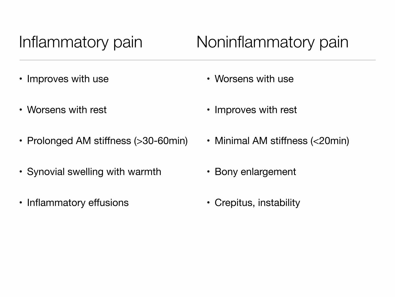
Inflammatory pain Noninflammatory pain
• Improves with use
• Worsens with rest
• Prolonged AM stiffness (>30-60min)
• Synovial swelling with warmth
• Inflammatory effusions
• Worsens with use
• Improves with rest
• Minimal AM stiffness (<20min)
• Bony enlargement
• Crepitus, instability

Recognizing joint inflammation
Remember the 5 cardinal signs of inflammation:1. Redness/erythema2. Swelling
• Look for loss of “dimples” around the joint & decreased skin lines over the joint
• Feel for the edges of the joint to feel “squishy” or less distinct• Feel small joint swelling/effusions by pushing with one finger & sensing
with the other3. Tenderness4. Heat
• Normal joint should be cooler than surrounding tissues5. Loss of function

Loss of dimples around joint
Image: ACR Image Bank

Recognizing joint inflammation
Fusiform swelling with decreased skin lines &
loss of dimples
Relatively normal joint for comparison
Image: ACR Image Bank

Recognizing joint inflammation
Swelling of entire MCP row showing loss of “peaks and valleys”
{ }Fusiform swelling with decreased skin lines &
loss of dimples
{ }
Image: ACR Image Bank

Rheumatoid arthritis
Image: ACR Image Bank

Osteoarthritis
Image: ACR Image Bank

Osteoarthritis
• Risk factors• Age (75% of pts >70yo)• Female• Hereditary• Mechanical stress (obesity,
misalignments, injury)• Neuromuscular dysfunction• Metabolic (crystals,
hemochromatosis)• Post-inflammatory
Image: ACR Image Bank

Distribution of primary osteoarthritis
• Hands: DIPs, PIPs, CMC (base of thumb)
• Cervical and lumbar spine
• Hips
• Knees
• 1st MTP
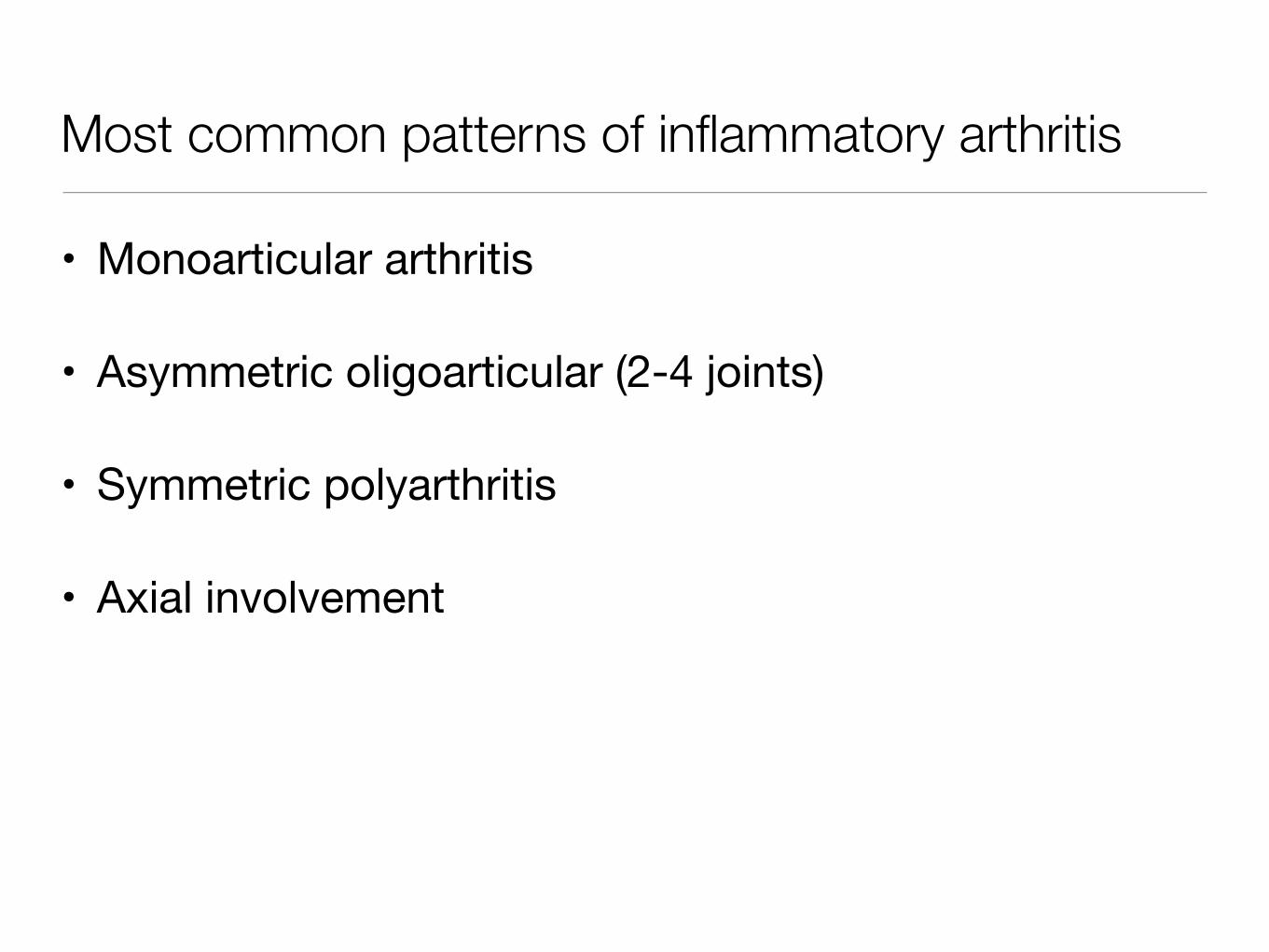
Most common patterns of inflammatory arthritis
• Monoarticular arthritis
• Asymmetric oligoarticular (2-4 joints)
• Symmetric polyarthritis
• Axial involvement

Most Helpful Temporal Patterns
• Abrupt (<24 hrs): trauma, crystals, infection
• Migratory (few days in each joint): disseminated gonococcal infection, acute rheumatic fever, early Lyme
• Episodic/intermittent: crystals (gout, pseudogout), Lyme
• Additive: most common pattern & least specific

Monoarthritis
• Most need to be aspirated
• Differential:
• INFECTION!!!
• Gout
• Pseudogout
• Fracture/hemarthrosis
• Lyme
• Osteoarthritis
• Rarely: RA, psoriatic, reactive present as monoarthritis
• Workup:
• Joint fluid: “the 3 C’s”
• ESR/CRP
• CBC, creatinine
• Uric acid
• +/- Lyme serologies/PCR
• Not indicated initially:
• ANA, RF, CCP

Synovial Fluid Analysis
“The 3 C’s” Non-inflammatory Inflammatory Septic
Cell count/diff <2000 2000 - ~50k >50k
Crystals None +/- +/-
Culture - - Positive
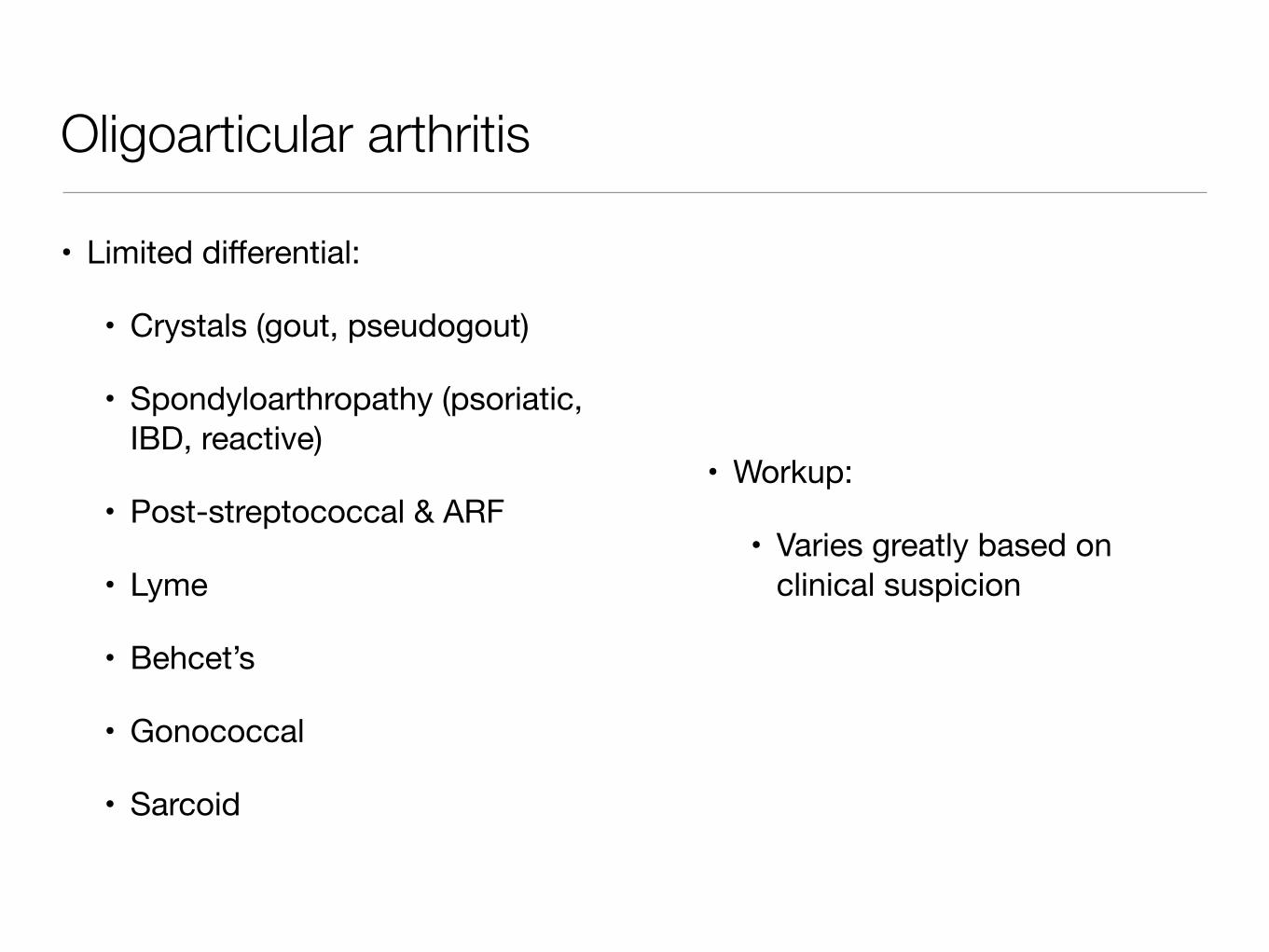
Oligoarticular arthritis
• Limited differential:
• Crystals (gout, pseudogout)
• Spondyloarthropathy (psoriatic, IBD, reactive)
• Post-streptococcal & ARF
• Lyme
• Behcet’s
• Gonococcal
• Sarcoid
• Workup:
• Varies greatly based on clinical suspicion

Symmetric polyarthritis
• ROS may provide helpful clues• A fair percentage is “seronegative”
so a good exam is the most important part
• Limited differential:• Rheumatoid arthritis• SLE/other CTD• Hepatitis B/C• Viral (parvovirus)• Psoriatic • Vasculitis• Sarcoid• Still’s disease
• Initial workup:• CBC, creatinine, LFTs, UA• ESR/CRP• RF, CCP• Hepatitis B/C tests• ANA• +/- Parvovirus serologies• Joint xrays (esp hand/feet)• +/- CXR
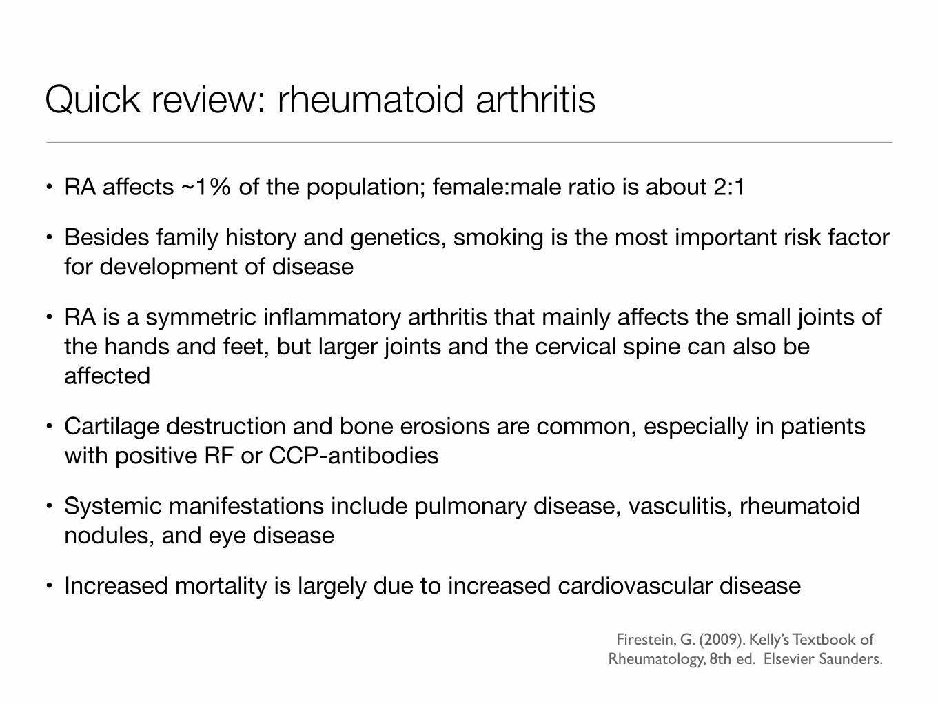
Quick review: rheumatoid arthritis
• RA affects ~1% of the population; female:male ratio is about 2:1
• Besides family history and genetics, smoking is the most important risk factor for development of disease
• RA is a symmetric inflammatory arthritis that mainly affects the small joints of the hands and feet, but larger joints and the cervical spine can also be affected
• Cartilage destruction and bone erosions are common, especially in patients with positive RF or CCP-antibodies
• Systemic manifestations include pulmonary disease, vasculitis, rheumatoid nodules, and eye disease
• Increased mortality is largely due to increased cardiovascular disease
Firestein, G. (2009). Kelly’s Textbook of Rheumatology, 8th ed. Elsevier Saunders.

ACR 2010 RA diagnostic criteria
New criteria are aimed at identifying newly presenting patients more quickly
A. Joint involvement1 large joint: 0 pts2-10 large joints: 1 pt1-3 small joints: 2 pts4-10 small joints: 3 pts>10 small joints: 5 pts
B. SerologyNegative RF/CCP: 0 ptsRF/CCP ≤ 3xNl: 2ptsRF/CCP >3xNl: 3 pts
C. Acute phase reactantsNormal ESR/CRP: 0 ptsElevated ESR/CRP: 1pt
D. Duration of symptoms<6 weeks: 0 pts≥6 weeks: 1 pt
6 points needed for diagnosis
Exception: rheumatoid nodules or erosions present
(also need to rule out other causes)Arthritis Rheum 2010; 62(9):2569
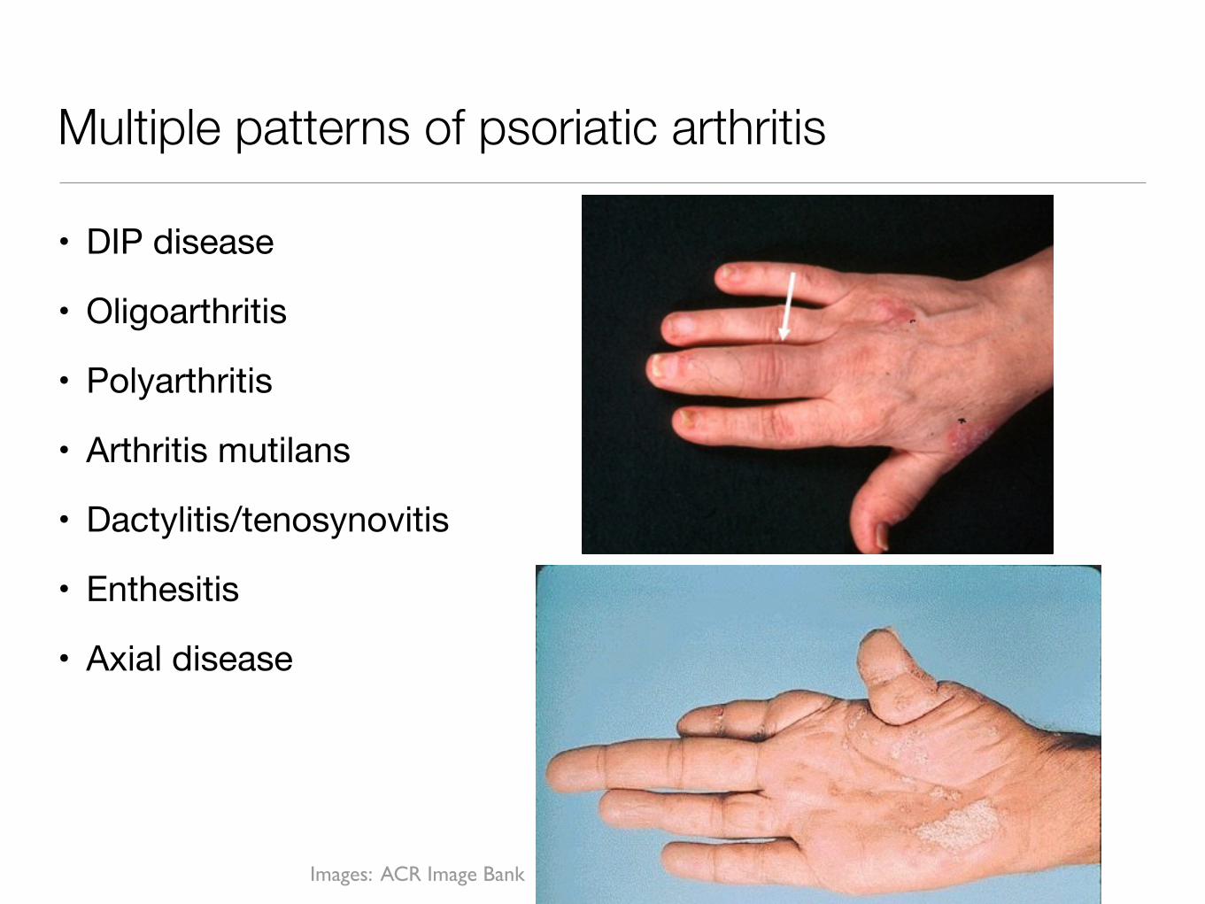
Multiple patterns of psoriatic arthritis
• DIP disease
• Oligoarthritis
• Polyarthritis
• Arthritis mutilans
• Dactylitis/tenosynovitis
• Enthesitis
• Axial disease
Images: ACR Image Bank

Dactylitis
Image: ACR Image Bank

DIP joint involvement
• If involved, think OA, psoriatic arthritis, gout
• If psoriatic arthritis, usually nail pitting is present
• DIPs spared in RA
Images: ACR Image Bank

Enthesitis
Image: ACR Image Bank

Axial joint involvement
• Axial joint involvement, usually starting from the bottom up
• Typical symptoms are inflammatory spine/buttock pain
• Exam findings: decreased spine mobility, SI joint tenderness
• Associated with HLA-B27
• Keep in mind HLA-B27 is positive in 8-10% of caucasians
• Confirmed radiographically

Modified Schober’s test
Images: ASAS Group
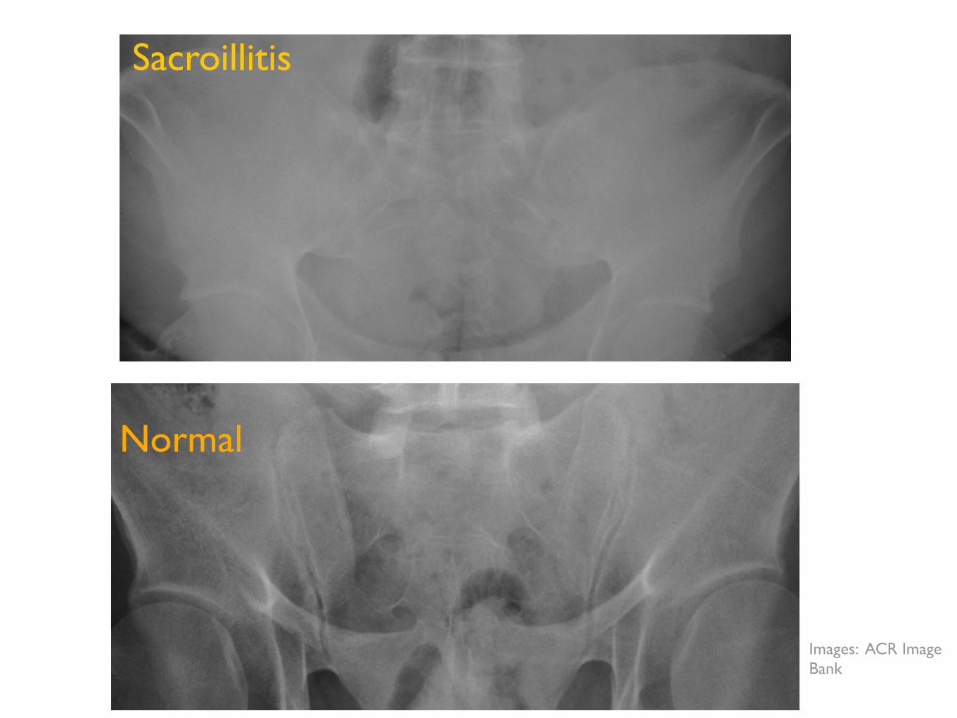
Normal
Sacroillitis
Images: ACR Image Bank

Image: ACR Image Bank

Gout
Images: ACR Image Bank

Gout Review
• Gout is caused by crystal deposition of monosodium urate secondary to hyperuricemia (uric acid >6.0mg/dL)
• Acute gout typically presents as rapid onset of severe inflammation in the affected joint.
• In most cases the 1st metatarsophalangeal (MTP) joint is the first joint involved (podagra)
• The natural history of gout is to worsen over time, with more frequent and diffuse joint flares, and development of tophi
• Strongly correlated with the metabolic syndrome and increased risk of cardiovascular disease

Gout Diagnosis
• Crystal proof of gout is preferred (although not always possible)
Arthritis Rheum 1998; 41(4):736Image: ACR Image Bank
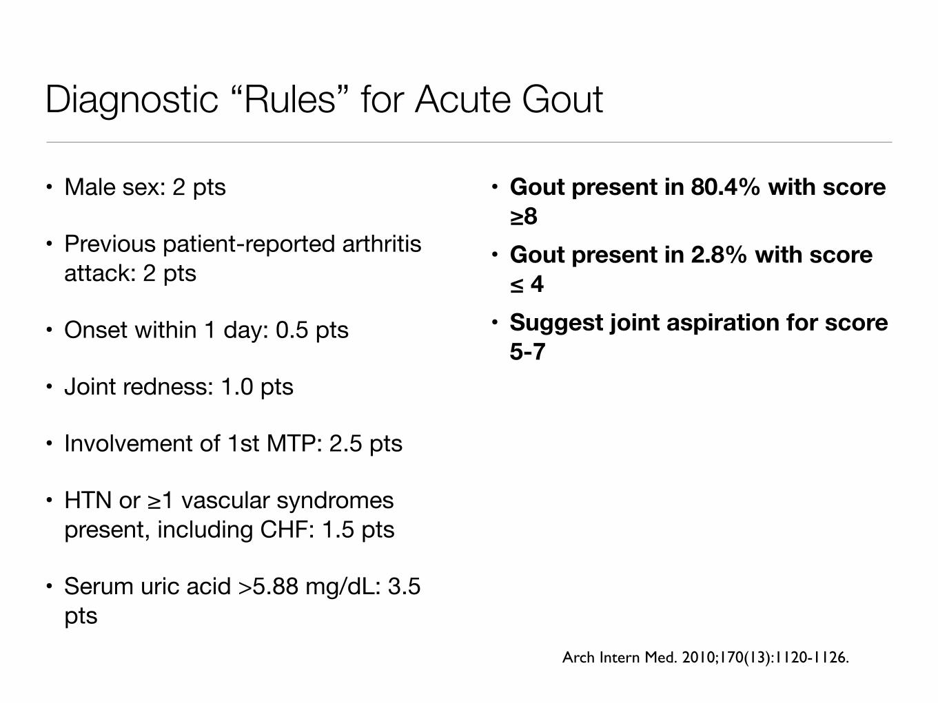
Diagnostic “Rules” for Acute Gout
• Male sex: 2 pts
• Previous patient-reported arthritis attack: 2 pts
• Onset within 1 day: 0.5 pts
• Joint redness: 1.0 pts
• Involvement of 1st MTP: 2.5 pts
• HTN or ≥1 vascular syndromes present, including CHF: 1.5 pts
• Serum uric acid >5.88 mg/dL: 3.5 pts
• Gout present in 80.4% with score ≥8
• Gout present in 2.8% with score ≤ 4
• Suggest joint aspiration for score 5-7
Arch Intern Med. 2010;170(13):1120-1126.
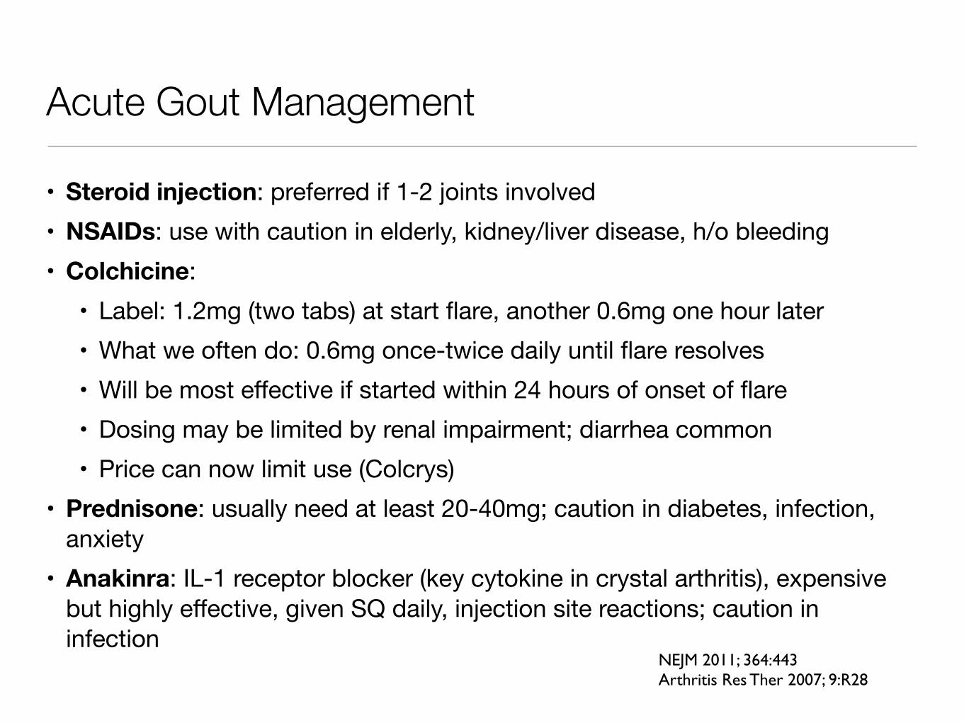
Acute Gout Management
• Steroid injection: preferred if 1-2 joints involved• NSAIDs: use with caution in elderly, kidney/liver disease, h/o bleeding• Colchicine:
• Label: 1.2mg (two tabs) at start flare, another 0.6mg one hour later• What we often do: 0.6mg once-twice daily until flare resolves• Will be most effective if started within 24 hours of onset of flare• Dosing may be limited by renal impairment; diarrhea common• Price can now limit use (Colcrys)
• Prednisone: usually need at least 20-40mg; caution in diabetes, infection, anxiety
• Anakinra: IL-1 receptor blocker (key cytokine in crystal arthritis), expensive but highly effective, given SQ daily, injection site reactions; caution in infection
NEJM 2011; 364:443Arthritis Res Ther 2007; 9:R28

Chronic Gout Management
The most important part of chronic gout therapy is lowering the uric acid ≤ 6.0
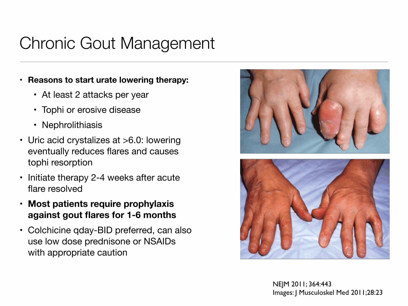
Chronic Gout Management
• Reasons to start urate lowering therapy:
• At least 2 attacks per year• Tophi or erosive disease• Nephrolithiasis
• Uric acid crystalizes at >6.0: lowering eventually reduces flares and causes tophi resorption
• Initiate therapy 2-4 weeks after acute flare resolved
• Most patients require prophylaxis against gout flares for 1-6 months
• Colchicine qday-BID preferred, can also use low dose prednisone or NSAIDs with appropriate caution
NEJM 2011; 364:443Images: J Musculoskel Med 2011;28:23

Urate Lowering Agents
• Allopurinol • Starting dose 50-100mg, increased q3-4 wks until serum urate ≤ 6.0• Most patients need 300mg/day, but can dose up to 800mg with normal
renal function• Use with caution in impaired renal function, but recent study suggests may
be safe to use with similar incidence of side effects• Rash in ~2%; allopurinol hypersensitivity (can be life threatening) in 0.1%
• Febuxostat (Uloric) • Second-line agent for when have contraindications (rash/hypersensitivity)
or inadequate response to allopurinol• Labeled as safe for GFR≥30 or with mild-moderate hepatic impairment
NEJM 2011; 364:443Arthritis Rheum 2011; 63(2):412

Final Pearls• Since ANA is positive in about 20% of normal patients, it
should only be checked when inflammatory arthritis or other autoimmune disease is suspected clinically.
• Be sure to check patients with a positive RF for hepatitis C.
• In chronic gout, the most important thing is to lower uric acid level below 6.0, but don’t forget to have a plan to prevent/treat flares that occur in this process.


![Rheumatology Visual Pearls for Internists [Read-Only] · World Wide Web (WWW 1.0) World Wide Web (WWW 2.0) Twitter, Second Life …. Learning management system Education delivered](https://static.fdocuments.net/doc/165x107/5f518b25297b621888184c2e/rheumatology-visual-pearls-for-internists-read-only-world-wide-web-www-10-world.jpg)


