Rheumatological diseases and cancer: the hidden variable ...radiological dose equivalence of 150...
4
Rheumatological diseases and cancer: the hidden variable of radiation exposure Eugenio Picano, 1 Richard Semelka, 2 James Ravenel, 3 Marco Matucci-Cerinic 4 Recently, several different meta-analyses have shown that rheumatological diseases —including systemic sclerosis (SSc), sys- temic lupus erythematosus, rheumatoid arthritis and psoriatic arthritis—are asso- ciated with an increased risk of cancer, in particular non-Hodgkin’s lymphoma, lung and liver cancer. 1–6 Several hypotheses have been advanced to explain this finding, including immune dysregulation, biological therapy and exposure to common environmental risk factors. 7 These data may suggest the inclusion of medical radiation as a potential, and veri- fiable, iatrogenic risk linking SSc and cancer. This malignant induction may be potentiated by the concomitant use of drugs, such as methotrexate and cyclo- phosphamide, or drugs such as paraceta- mol. In fact, these drugs are known to amplify the genotoxic effects of medical radiation. In addition, in some rheumatic diseases, the exposure to medical radi- ation is associated with an intrinsic higher vulnerability for DNA instability. Frauenfelder et al 8 present the pro- spective validation of a dedicated, 9-slice high resolution computed tomography (HRCT) protocol with reduced radiation dose for the detection of interstitial lung disease in SSc patients. This paper addresses clearly the fact that the dose reduction is becoming today a hot issue in clinics. Herein, we wish to draw the atten- tion of the rheumatological community to the hypothesis that, in rheumatological patients, the use of medical radiation may pose an additional risk for cancer devel- opment, possibly potentiated by the con- current use of antirheumatic drugs. MEDICAL RADIATION AS A POTENTIAL CARCINOGEN Medical radiation is suspected to contrib- ute to the development of cancer. According to the linear no-threshold theory, all radiation poses some risk of cancer induction, although the magnitude of this risk remains unclear, particularly at very low doses. The average background exposure of a US citizen approximates 3.0 milliSievert (mSv, corresponding to the radiological dose equivalence of 150 chest X-rays) per person per year, as estimated in 2006. 9 Doses of examinations commonly employed in rheumatology patients range from 2 to 8 mSv for a chest CT (100–400 chest X-rays) to 10–40 mSv for a myocar- dial perfusion scintigraphy (500–4000 chest X-rays). 10 The radiological risk is considered cumulative in nature; thus, each exam using radiation increases the risk. Furthermore, children are more sensi- tive to radiation than adults, and females are more sensitive to radiation risks than males. 11 Rheumatological patients, often young at disease onset and very frequently female, are therefore more prone to radi- ation related risks. The chronic nature of most rheumatological diseases also renders our patients more vulnerable to experien- cing the cumulative effects of multiple and repeated X-ray studies. Although imaging is a fundamental tool in the diagnosis and monitoring of disease evolution and response to therapy, 12 we wish to draw attention to the need of appropriate risk– benefit analysis for the type and frequency of studies. The available epidemiological evidence linking radiation exposure to increased cancer risk is well established for doses >10–50 mSv, which may be experienced by our patients during one admission, after one episode of care, or in some circum- stances a single examination. 12 A recent study of 180 000 people exposed to CT scan in the UK found an increasing risk of leukaemia and brain cancer with increasing radiation exposure, with cumulative ionis- ing radiation doses from three head CTs tripling the risk of brain tumours and 5–10 head CTs tripling the risk of leukaemia. 13 Among 680 000 Australians exposed to a CT scan when aged 0–19 years, cancer incidence was increased by 24% compared with the incidence in over 10 million unexposed people. 14 Epidemiological evi- dence is also emerging showing a link between increased radiation exposure and subsequent increased risk of cancer in adult populations of patients with ischae- mic heart disease. 15 In rheumatic patients, imaging of the chest (an anatomic region that includes the radiosensitve organs lung, female breast and bone marrow) is commonly per- formed. 16 The lungs may experience rela- tively high organ doses from right heart catheterisation for suspected pulmonary arterial hypertension, 17 and from chest CT employed for detection of lung involve- ment. 18 Gallium scintigraphy previously often used to assess pulmonary disease activity, 19 and cardiac stress scintigraphy with thallium or technetium-based tracers such as sestamibi to evaluate coronary and myocardial involvement. 20 The organ sites where imaging has been performed often correspond to where malignancy has developed. Cumulative radiation contri- butes to the increase in cancer. 21 Risk esti- mates are further complicated, and therefore more subject to being contested because cancer induction often occurs years or decades after exposure. VULNERABILITY OF RHEUMATOLOGICAL PATIENTS TO DAMAGING EFFECTS OF RADIATION: AGE, GENDER, GENES AND DRUGS The dose–risk curve in radiation risk is established from large epidemiological data banks, including >100 000 survivors of the atomic bomb, >400 000 nuclear power plant workers and >800 000 patients exposed to diagnostic medical radiation. The challenge ahead is to trans- late the generic population risk obtained from epidemiological data into a persona- lised risk. Several genetic and pharmaco- logical variables can affect the variability of damage observed for any given level of radiation. For instance, radiation-induced chromosomal damage is amplified by genetic polymorphisms or gene mutations (such as BRCA1 and BRCA2) of genes involved in DNA repair. 22 A similar situ- ation may occur in rheumatological patients in whom epidemiological, genetic and pharmacological factors may enhance vulnerability to a given dose of radiation, making the linear relationship linking risk to dose progressively steeper ( figure 1). First, rheumatological patients are much more often women (with a 4:1 ratio), who are 38% more vulnerable to radi- ation than men. 11 Second, the age at first 1 Institute of Clinical Physiology, CNR, Pisa, Italy; 2 Department of Radiology, University of North Carolina, Chapel Hill, North Carolina, USA; 3 Department of Radiology and Radiologic Sciences, Medical University of South Carolina, Charleston, South Carolina, USA; 4 Division of Rheumatology AOUC, Department of Experimental and Clinical Medicine, University of Florence, Florence, Italy Correspondence to Dr Marco Matucci-Cerinic, Division of Rheumatology AOUC, Department of Experimental and Clinical Medicine, University of Florence, Florence 50139, Italy; marco.matuccicerinic@unifi.it Picano E, et al. Ann Rheum Dis December 2014 Vol 73 No 12 2065 Editorial on June 15, 2020 by guest. Protected by copyright. http://ard.bmj.com/ Ann Rheum Dis: first published as 10.1136/annrheumdis-2014-206585 on 9 October 2014. Downloaded from
Transcript of Rheumatological diseases and cancer: the hidden variable ...radiological dose equivalence of 150...
Rheumatological diseases and cancer:the hidden variable of radiationexposureEugenio Picano,1 Richard Semelka,2 James Ravenel,3
Marco Matucci-Cerinic4
Recently, several different meta-analyseshave shown that rheumatological diseases—including systemic sclerosis (SSc), sys-temic lupus erythematosus, rheumatoidarthritis and psoriatic arthritis—are asso-ciated with an increased risk of cancer, inparticular non-Hodgkin’s lymphoma, lungand liver cancer.1–6 Several hypotheseshave been advanced to explain thisfinding, including immune dysregulation,biological therapy and exposure tocommon environmental risk factors.7
These data may suggest the inclusion ofmedical radiation as a potential, and veri-fiable, iatrogenic risk linking SSc andcancer. This malignant induction may bepotentiated by the concomitant use ofdrugs, such as methotrexate and cyclo-phosphamide, or drugs such as paraceta-mol. In fact, these drugs are known toamplify the genotoxic effects of medicalradiation. In addition, in some rheumaticdiseases, the exposure to medical radi-ation is associated with an intrinsic highervulnerability for DNA instability.
Frauenfelder et al8 present the pro-spective validation of a dedicated, 9-slicehigh resolution computed tomography(HRCT) protocol with reduced radiationdose for the detection of interstitial lungdisease in SSc patients. This paperaddresses clearly the fact that the dosereduction is becoming today a hot issue inclinics. Herein, we wish to draw the atten-tion of the rheumatological community tothe hypothesis that, in rheumatologicalpatients, the use of medical radiation maypose an additional risk for cancer devel-opment, possibly potentiated by the con-current use of antirheumatic drugs.
MEDICAL RADIATION AS APOTENTIAL CARCINOGENMedical radiation is suspected to contrib-ute to the development of cancer.According to the linear no-thresholdtheory, all radiation poses some risk ofcancer induction, although the magnitudeof this risk remains unclear, particularly atvery low doses. The average backgroundexposure of a US citizen approximates 3.0milliSievert (mSv, corresponding to theradiological dose equivalence of 150 chestX-rays) per person per year, as estimated in2006.9 Doses of examinations commonlyemployed in rheumatology patients rangefrom 2 to 8 mSv for a chest CT (100–400chest X-rays) to 10–40 mSv for a myocar-dial perfusion scintigraphy (500–4000chest X-rays).10 The radiological risk isconsidered cumulative in nature; thus,each exam using radiation increases therisk. Furthermore, children are more sensi-tive to radiation than adults, and femalesare more sensitive to radiation risks thanmales.11 Rheumatological patients, oftenyoung at disease onset and very frequentlyfemale, are therefore more prone to radi-ation related risks. The chronic nature ofmost rheumatological diseases also rendersour patients more vulnerable to experien-cing the cumulative effects of multiple andrepeated X-ray studies. Although imagingis a fundamental tool in the diagnosis andmonitoring of disease evolution andresponse to therapy,12 we wish to drawattention to the need of appropriate risk–benefit analysis for the type and frequencyof studies.The available epidemiological evidence
linking radiation exposure to increasedcancer risk is well established for doses>10–50 mSv, which may be experiencedby our patients during one admission, afterone episode of care, or in some circum-stances a single examination.12 A recentstudy of 180 000 people exposed to CTscan in the UK found an increasing risk ofleukaemia and brain cancer with increasingradiation exposure, with cumulative ionis-ing radiation doses from three head CTstripling the risk of brain tumours and 5–10head CTs tripling the risk of leukaemia.13
Among 680 000 Australians exposed to a
CT scan when aged 0–19 years, cancerincidence was increased by 24% comparedwith the incidence in over 10 millionunexposed people.14 Epidemiological evi-dence is also emerging showing a linkbetween increased radiation exposure andsubsequent increased risk of cancer inadult populations of patients with ischae-mic heart disease.15
In rheumatic patients, imaging of thechest (an anatomic region that includes theradiosensitve organs lung, female breastand bone marrow) is commonly per-formed.16 The lungs may experience rela-tively high organ doses from right heartcatheterisation for suspected pulmonaryarterial hypertension,17 and from chest CTemployed for detection of lung involve-ment.18 Gallium scintigraphy previouslyoften used to assess pulmonary diseaseactivity,19 and cardiac stress scintigraphywith thallium or technetium-based tracerssuch as sestamibi to evaluate coronary andmyocardial involvement.20 The organ siteswhere imaging has been performed oftencorrespond to where malignancy hasdeveloped. Cumulative radiation contri-butes to the increase in cancer.21 Risk esti-mates are further complicated, andtherefore more subject to being contestedbecause cancer induction often occursyears or decades after exposure.
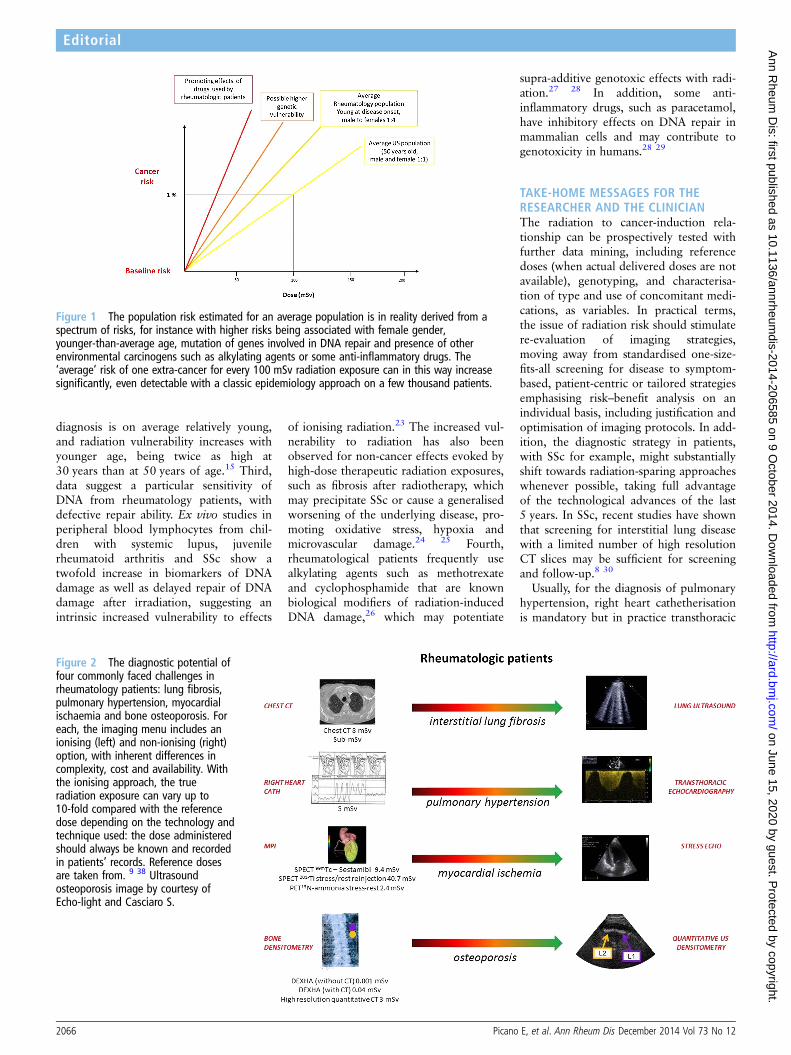
VULNERABILITY OFRHEUMATOLOGICAL PATIENTS TODAMAGING EFFECTS OF RADIATION:AGE, GENDER, GENES AND DRUGSThe dose–risk curve in radiation risk isestablished from large epidemiologicaldata banks, including >100 000 survivorsof the atomic bomb, >400 000 nuclearpower plant workers and >800 000patients exposed to diagnostic medicalradiation. The challenge ahead is to trans-late the generic population risk obtainedfrom epidemiological data into a persona-lised risk. Several genetic and pharmaco-logical variables can affect the variabilityof damage observed for any given level ofradiation. For instance, radiation-inducedchromosomal damage is amplified bygenetic polymorphisms or gene mutations(such as BRCA1 and BRCA2) of genesinvolved in DNA repair.22 A similar situ-ation may occur in rheumatologicalpatients in whom epidemiological, geneticand pharmacological factors may enhancevulnerability to a given dose of radiation,making the linear relationship linking riskto dose progressively steeper (figure 1).First, rheumatological patients are muchmore often women (with a 4:1 ratio),who are 38% more vulnerable to radi-ation than men.11 Second, the age at first
1Institute of Clinical Physiology, CNR, Pisa, Italy;2Department of Radiology, University of North Carolina,Chapel Hill, North Carolina, USA; 3Department ofRadiology and Radiologic Sciences, Medical Universityof South Carolina, Charleston, South Carolina, USA;4Division of Rheumatology AOUC, Department ofExperimental and Clinical Medicine, University ofFlorence, Florence, Italy
Correspondence to Dr Marco Matucci-Cerinic,Division of Rheumatology AOUC, Department ofExperimental and Clinical Medicine, University ofFlorence, Florence 50139, Italy;[email protected]
Picano E, et al. Ann Rheum Dis December 2014 Vol 73 No 12 2065
Editorial
on June 15, 2020 by guest. Protected by copyright.
http://ard.bmj.com
/A
nn Rheum
Dis: first published as 10.1136/annrheum
dis-2014-206585 on 9 October 2014. D
ownloaded from
diagnosis is on average relatively young,and radiation vulnerability increases withyounger age, being twice as high at30 years than at 50 years of age.15 Third,data suggest a particular sensitivity ofDNA from rheumatology patients, withdefective repair ability. Ex vivo studies inperipheral blood lymphocytes from chil-dren with systemic lupus, juvenilerheumatoid arthritis and SSc show atwofold increase in biomarkers of DNAdamage as well as delayed repair of DNAdamage after irradiation, suggesting anintrinsic increased vulnerability to effects
of ionising radiation.23 The increased vul-nerability to radiation has also beenobserved for non-cancer effects evoked byhigh-dose therapeutic radiation exposures,such as fibrosis after radiotherapy, whichmay precipitate SSc or cause a generalisedworsening of the underlying disease, pro-moting oxidative stress, hypoxia andmicrovascular damage.24 25 Fourth,rheumatological patients frequently usealkylating agents such as methotrexateand cyclophosphamide that are knownbiological modifiers of radiation-inducedDNA damage,26 which may potentiate
supra-additive genotoxic effects with radi-ation.27 28 In addition, some anti-inflammatory drugs, such as paracetamol,have inhibitory effects on DNA repair inmammalian cells and may contribute togenotoxicity in humans.28 29
TAKE-HOME MESSAGES FOR THERESEARCHER AND THE CLINICIANThe radiation to cancer-induction rela-tionship can be prospectively tested withfurther data mining, including referencedoses (when actual delivered doses are notavailable), genotyping, and characterisa-tion of type and use of concomitant medi-cations, as variables. In practical terms,the issue of radiation risk should stimulatere-evaluation of imaging strategies,moving away from standardised one-size-fits-all screening for disease to symptom-based, patient-centric or tailored strategiesemphasising risk–benefit analysis on anindividual basis, including justification andoptimisation of imaging protocols. In add-ition, the diagnostic strategy in patients,with SSc for example, might substantiallyshift towards radiation-sparing approacheswhenever possible, taking full advantageof the technological advances of the last5 years. In SSc, recent studies have shownthat screening for interstitial lung diseasewith a limited number of high resolutionCT slices may be sufficient for screeningand follow-up.8 30
Usually, for the diagnosis of pulmonaryhypertension, right heart cathetherisationis mandatory but in practice transthoracic
Figure 1 The population risk estimated for an average population is in reality derived from aspectrum of risks, for instance with higher risks being associated with female gender,younger-than-average age, mutation of genes involved in DNA repair and presence of otherenvironmental carcinogens such as alkylating agents or some anti-inflammatory drugs. The‘average’ risk of one extra-cancer for every 100 mSv radiation exposure can in this way increasesignificantly, even detectable with a classic epidemiology approach on a few thousand patients.
Figure 2 The diagnostic potential offour commonly faced challenges inrheumatology patients: lung fibrosis,pulmonary hypertension, myocardialischaemia and bone osteoporosis. Foreach, the imaging menu includes anionising (left) and non-ionising (right)option, with inherent differences incomplexity, cost and availability. Withthe ionising approach, the trueradiation exposure can vary up to10-fold compared with the referencedose depending on the technology andtechnique used: the dose administeredshould always be known and recordedin patients’ records. Reference dosesare taken from. 9 38 Ultrasoundosteoporosis image by courtesy ofEcho-light and Casciaro S.
2066 Picano E, et al. Ann Rheum Dis December 2014 Vol 73 No 12
Editorial
on June 15, 2020 by guest. Protected by copyright.
http://ard.bmj.com
/A
nn Rheum
Dis: first published as 10.1136/annrheum
dis-2014-206585 on 9 October 2014. D
ownloaded from
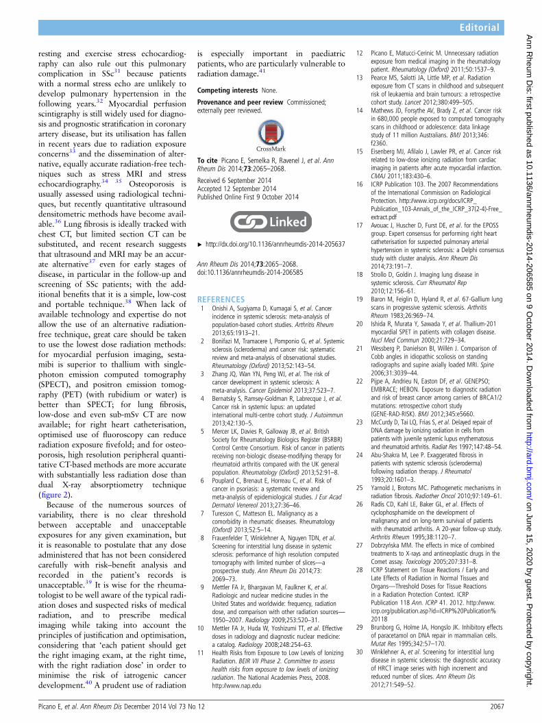
resting and exercise stress echocardiog-raphy can also rule out this pulmonarycomplication in SSc31 because patientswith a normal stress echo are unlikely todevelop pulmonary hypertension in thefollowing years.32 Myocardial perfusionscintigraphy is still widely used for diagno-sis and prognostic stratification in coronaryartery disease, but its utilisation has fallenin recent years due to radiation exposureconcerns33 and the dissemination of alter-native, equally accurate radiation-free tech-niques such as stress MRI and stressechocardiography.34 35 Osteoporosis isusually assessed using radiological techni-ques, but recently quantitative ultrasounddensitometric methods have become avail-able.36 Lung fibrosis is ideally tracked withchest CT, but limited section CT can besubstituted, and recent research suggeststhat ultrasound and MRI may be an accur-ate alternative37 even for early stages ofdisease, in particular in the follow-up andscreening of SSc patients; with the add-itional benefits that it is a simple, low-costand portable technique.38 When lack ofavailable technology and expertise do notallow the use of an alternative radiation-free technique, great care should be takento use the lowest dose radiation methods:for myocardial perfusion imaging, sesta-mibi is superior to thallium with single-photon emission computed tomography(SPECT), and positron emission tomog-raphy (PET) (with rubidium or water) isbetter than SPECT; for lung fibrosis,low-dose and even sub-mSv CT are nowavailable; for right heart catheterisation,optimised use of fluoroscopy can reduceradiation exposure fivefold; and for osteo-porosis, high resolution peripheral quanti-tative CT-based methods are more accuratewith substantially less radiation dose thandual X-ray absorptiometry technique(figure 2).
Because of the numerous sources ofvariability, there is no clear thresholdbetween acceptable and unacceptableexposures for any given examination, butit is reasonable to postulate that any doseadministered that has not been consideredcarefully with risk–benefit analysis andrecorded in the patient’s records isunacceptable.39 It is wise for the rheuma-tologist to be well aware of the typical radi-ation doses and suspected risks of medicalradiation, and to prescribe medicalimaging while taking into account theprinciples of justification and optimisation,considering that ‘each patient should getthe right imaging exam, at the right time,with the right radiation dose’ in order tominimise the risk of iatrogenic cancerdevelopment.40 A prudent use of radiation
is especially important in paediatricpatients, who are particularly vulnerable toradiation damage.41
Competing interests None.
Provenance and peer review Commissioned;externally peer reviewed.
To cite Picano E, Semelka R, Ravenel J, et al. AnnRheum Dis 2014;73:2065–2068.
Received 6 September 2014Accepted 12 September 2014Published Online First 9 October 2014
▸ http://dx.doi.org/10.1136/annrheumdis-2014-205637
Ann Rheum Dis 2014;73:2065–2068.doi:10.1136/annrheumdis-2014-206585
REFERENCES1 Onishi A, Sugiyama D, Kumagai S, et al. Cancer
incidence in systemic sclerosis: meta-analysis ofpopulation-based cohort studies. Arthritis Rheum2013;65:1913–21.
2 Bonifazi M, Tramacere I, Pomponio G, et al. Systemicsclerosis (scleroderma) and cancer risk: systematicreview and meta-analysis of observational studies.Rheumatology (Oxford) 2013;52:143–54.
3 Zhang JQ, Wan YN, Peng WJ, et al. The risk ofcancer development in systemic sclerosis: Ameta-analysis. Cancer Epidemiol 2013;37:523–7.
4 Bernatsky S, Ramsey-Goldman R, Labrecque J, et al.Cancer risk in systemic lupus: an updatedinternational multi-centre cohort study. J Autoimmun2013;42:130–5.
5 Mercer LK, Davies R, Galloway JB, et al. BritishSociety for Rheumatology Biologics Register (BSRBR)Control Centre Consortium. Risk of cancer in patientsreceiving non-biologic disease-modifying therapy forrheumatoid arthritis compared with the UK generalpopulation. Rheumatology (Oxford) 2013;52:91–8.
6 Pouplard C, Brenaut E, Horreau C, et al. Risk ofcancer in psoriasis: a systematic review andmeta-analysis of epidemiological studies. J Eur AcadDermatol Venereol 2013;27:36–46.
7 Turesson C, Matteson EL. Malignancy as acomorbidity in rheumatic diseases. Rheumatology(Oxford) 2013;52:5–14.
8 Frauenfelder T, Winklehner A, Nguyen TDN, et al.Screening for interstitial lung disease in systemicsclerosis: performance of high resolution computedtomography with limited number of slices—aprospective study. Ann Rheum Dis 2014;73:2069–73.
9 Mettler FA Jr, Bhargavan M, Faulkner K, et al.Radiologic and nuclear medicine studies in theUnited States and worldwide: frequency, radiationdose, and comparison with other radiation sources—1950–2007. Radiology 2009;253:520–31.
10 Mettler FA Jr, Huda W, Yoshizumi TT, et al. Effectivedoses in radiology and diagnostic nuclear medicine:a catalog. Radiology 2008;248:254–63.
11 Health Risks from Exposure to Low Levels of IonizingRadiation. BEIR VII Phase 2. Committee to assesshealth risks from exposure to low levels of ionizingradiation. The National Academies Press, 2008.http://www.nap.edu
12 Picano E, Matucci-Cerinic M. Unnecessary radiationexposure from medical imaging in the rheumatologypatient. Rheumatology (Oxford) 2011;50:1537–9.
13 Pearce MS, Salotti JA, Little MP, et al. Radiationexposure from CT scans in childhood and subsequentrisk of leukaemia and brain tumours: a retrospectivecohort study. Lancet 2012;380:499–505.
14 Mathews JD, Forsythe AV, Brady Z, et al. Cancer riskin 680,000 people exposed to computed tomographyscans in childhood or adolescence: data linkagestudy of 11 million Australians. BMJ 2013;346:f2360.
15 Eisenberg MJ, Afilalo J, Lawler PR, et al. Cancer riskrelated to low-dose ionizing radiation from cardiacimaging in patients after acute myocardial infarction.CMAJ 2011;183:430–6.
16 ICRP Publication 103. The 2007 Recommendationsof the International Commission on RadiologicalProtection. http://www.icrp.org/docs/ICRP_Publication_103-Annals_of_the_ICRP_37(2-4)-Free_extract.pdf
17 Avouac J, Huscher D, Furst DE, et al. for the EPOSSgroup. Expert consensus for performing right heartcatheterisation for suspected pulmonary arterialhypertension in systemic sclerosis: a Delphi consensusstudy with cluster analysis. Ann Rheum Dis2014;73:191–7.
18 Strollo D, Goldin J. Imaging lung disease insystemic sclerosis. Curr Rheumatol Rep2010;12:156–61.
19 Baron M, Feiglin D, Hyland R, et al. 67-Gallium lungscans in progressive systemic sclerosis. ArthritisRheum 1983;26:969–74.
20 Ishida R, Murata Y, Sawada Y, et al. Thallium-201myocardial SPET in patients with collagen disease.Nucl Med Commun 2000;21:729–34.
21 Wessberg P, Danielson BI, Willén J. Comparison ofCobb angles in idiopathic scoliosis on standingradiographs and supine axially loaded MRI. Spine2006;31:3039–44.
22 Pijpe A, Andrieu N, Easton DF, et al. GENEPSO;EMBRACE; HEBON. Exposure to diagnostic radiationand risk of breast cancer among carriers of BRCA1/2mutations: retrospective cohort study(GENE-RAD-RISK). BMJ 2012;345:e5660.
23 McCurdy D, Tai LQ, Frias S, et al. Delayed repair ofDNA damage by ionizing radiation in cells frompatients with juvenile systemic lupus erythematosusand rheumatoid arthritis. Radiat Res 1997;147:48–54.
24 Abu-Shakra M, Lee P. Exaggerated fibrosis inpatients with systemic sclerosis (scleroderma)following radiation therapy. J Rheumatol1993;20:1601–3.
25 Yarnold J, Brotons MC. Pathogenetic mechanisms inradiation fibrosis. Radiother Oncol 2010;97:149–61.
26 Radis CD, Kahl LE, Baker GL, et al. Effects ofcyclophosphamide on the development ofmalignancy and on long-term survival of patientswith rheumatoid arthritis. A 20-year follow-up study.Arthritis Rheum 1995;38:1120–7.
27 Dobrzyńska MM. The effects in mice of combinedtreatments to X-rays and antineoplastic drugs in theComet assay. Toxicology 2005;207:331–8.
28 ICRP Statement on Tissue Reactions / Early andLate Effects of Radiation in Normal Tissues andOrgans—Threshold Doses for Tissue Reactionsin a Radiation Protection Context. ICRPPublication 118 Ann. ICRP 41. 2012. http://www.icrp.org/publication.asp?id=ICRP%20Publication%20118
29 Brunborg G, Holme JA, Hongslo JK. Inhibitory effectsof paracetamol on DNA repair in mammalian cells.Mutat Res 1995;342:57–170.
30 Winklehner A, et al. Screening for interstitial lungdisease in systemic sclerosis: the diagnostic accuracyof HRCT image series with high increment andreduced number of slices. Ann Rheum Dis2012;71:549–52.
Picano E, et al. Ann Rheum Dis December 2014 Vol 73 No 12 2067
Editorial
on June 15, 2020 by guest. Protected by copyright.
http://ard.bmj.com
/A
nn Rheum
Dis: first published as 10.1136/annrheum
dis-2014-206585 on 9 October 2014. D
ownloaded from
31 Gargani L, Pignone A, Agoston G, et al.: Clinical andechocardiographic correlations of exercise-inducedpulmonary hypertension in systemic sclerosis: amulticenter study. Am Heart J 2013;165:200–7.
32 Picano E, Pellikka PA. Stress echo applicationsbeyond coronary artery disease. Eur Heart J2014;35:1033–40.
33 Smith-Bindman R, Miglioretti DL, Johnson E, et al.Use of diagnostic imaging studies and associatedradiation exposure for patients enrolled in largeintegrated health care systems, 1996–2010. JAMA2012;307:2400–9.
34 Picano E. Stress echocardiography: a historicalperspective. Am J Med 2003;114:126–30.
35 Sicari R, Nihoyannopoulos P, Evangelista A, et al.European Association of Echocardiography. Stress
Echocardiography Expert Consensus Statement—Executive Summary: European Association ofEchocardiography (EAE) (a registered branch of theESC). Eur Heart J 2009;30:278–89.
36 Link TM. Osteoporosis imaging: state of the art andadvanced imaging. Radiology 2012;263:3–17.
37 Gargani L, Doveri M, D’Errico L, et al. Ultrasoundlung comets in systemic sclerosis: a chest sonographyhallmark of pulmonary interstitial fibrosis.Rheumatology (Oxford) 2009;48:1382–7.
38 Barskova T, Gargani L, Guiducci S, et al. Lungultrasound for the screening of interstitial lungdisease in very early systemic sclerosis. Ann RheumDis 2013;72:390–5.
39 Picano E, Vañó E, Rehani MM, et al. The appropriateand justified use of medical radiation in
cardiovascular imaging. A position document of theESC associations of cardiovascular imaging,percutaneous cardiovascular interventionsand electrophysiology. Eur Heart J2014;35:665–72.
40 U.S. Food and Drug Administration. Initiative toReduce Unnecessary Radiation Exposure fromMedical Imaging. February 2010. http://www.fda.gov/downloads/Radiation-EmittingProducts/RadiationSafety/RadiationDoseReduction/UCM200087.pdf
41 Andreassi MG, Picano E. Reduction of radiation tochildren. Our responsibility to change. Editorialcomment. Circulation 2014;130:135–7.
2068 Picano E, et al. Ann Rheum Dis December 2014 Vol 73 No 12
Editorial
on June 15, 2020 by guest. Protected by copyright.
http://ard.bmj.com
/A
nn Rheum
Dis: first published as 10.1136/annrheum
dis-2014-206585 on 9 October 2014. D
ownloaded from