Rheumatoid Arthritis - buecher.de · disease activity in terms of inflammatory changes involving...
17
Definition ............................................................................................ " Epidemiology Affects 2% of the population · Peak incidence (diagnosis) in 4th and 5th deca- des · Women affected 3–4 times more often than men · Increased familial inci- dence · Up to 70% of patients have HLA antigen DR4. " Etiology, pathophysiology, pathogenesis Chronic inflammatory disease · Predilection for synovial membrane · With dis- ease progression, osseous destruction of affected joints · Etiology is complex and incompletely understood · Cellular immune reaction to an as yet unidenti- fied antigen · Synovium is the primary target organ of various immunological cascades, responding with inflammatory proliferative changes (pannus) · Sec- ondary destruction of the capsule–ligament complex, cartilage, and bone. Imaging Signs ............................................................................................ " Modality of choice Radiography · MRI. " Radiographic findings No radiologically identifiable skeletal changes in early disease · Often bilateral, symmetrical, and usually polyarticular pattern of involvement · Sites of predi- lection: phalangeal finger and toe joints as well as wrists · Findings divided into three groups based on Dihlmann: – Soft-tissue swelling · Joint effusion · Synovitis – Collateral phenomenon in rheumatoid arthritis: band-like areas of juxta-artic- ular osteoporosis – Direct signs: joint space widening (mainly due to joint effusion) · Symmetri- cal narrowing of joint space is an indirect sign of joint destruction (especially marked between carpal bones) · Erosions, sites of predilection: hand (MCP joints II–V, PIP, carpal bones, ulnar styloid process), foot (MTP joints II–V, IPJ) · Early visualization of disrupted subchondral bone end plate · Subchon- dral cysts · Ulnar deviation of the fingers · Buttonhole deformity and swan- neck deformity of the fingers · Ankylosis as end-stage finding. In cervical spine involvement, stepladder deformity, atlantoaxial dislocation, and pseudobasilar invagination. " MRI findings Symmetrical pattern of involvement is characteristic (see radiographic findings; DD: degenerative changes, psoriatic arthritis, gout) · Detection of synovitis on contrast-enhanced images enables early diagnosis, estimate of disease activity, and early medication, if possible before onset of bone destruction · Signal alter- ations analogous to those of bone marrow edema (especially on fat-saturated T2-weighted sequences) that have no corresponding findings in conventional ra- diographs represent pre-erosive changes, still potentially reversible · Tendon sheath inflammation seen as increased signal intensity on T2-weighted images · Direct imaging of cartilage destruction · Dynamic contrast-enhanced sequen- ces with rapid imaging time (less than 10 seconds per data set) appear to reflect ..... 93 Reiser et al. Direct Diagnosis in Radiology. Musculoskeletal Imaging (ISBN 9783131451613), © 2008 Georg Thieme Verlag KG Rheumatoid Arthritis 2 Inflammatory Diseases
Transcript of Rheumatoid Arthritis - buecher.de · disease activity in terms of inflammatory changes involving...

Definition. . . . . . . . . . . . . . . . . . . . . . . . . . . . . . . . . . . . . . . . . . . . . . . . . . . . . . . . . . . . . . . . . . . . . . . . . . . . . . . . . . . . . . . . . . . .
" EpidemiologyAffects 2% of the population · Peak incidence (diagnosis) in 4th and 5th deca-des · Women affected 3–4 times more often than men · Increased familial inci-dence · Up to 70% of patients have HLA antigen DR4.
" Etiology, pathophysiology, pathogenesisChronic inflammatory disease · Predilection for synovial membrane · With dis-ease progression, osseous destruction of affected joints · Etiology is complexand incompletely understood · Cellular immune reaction to an as yet unidenti-fied antigen · Synovium is the primary target organ of various immunologicalcascades, responding with inflammatory proliferative changes (pannus) · Sec-ondary destruction of the capsule–ligament complex, cartilage, and bone.
Imaging Signs. . . . . . . . . . . . . . . . . . . . . . . . . . . . . . . . . . . . . . . . . . . . . . . . . . . . . . . . . . . . . . . . . . . . . . . . . . . . . . . . . . . . . . . . . . . .
" Modality of choiceRadiography · MRI.
" Radiographic findingsNo radiologically identifiable skeletal changes in early disease · Often bilateral,symmetrical, and usually polyarticular pattern of involvement · Sites of predi-lection: phalangeal finger and toe joints as well as wrists · Findings divided intothree groups based on Dihlmann:– Soft-tissue swelling · Joint effusion · Synovitis– Collateral phenomenon in rheumatoid arthritis: band-like areas of juxta-artic-
ular osteoporosis– Direct signs: joint space widening (mainly due to joint effusion) · Symmetri-
cal narrowing of joint space is an indirect sign of joint destruction (especiallymarked between carpal bones) · Erosions, sites of predilection: hand (MCPjoints II–V, PIP, carpal bones, ulnar styloid process), foot (MTP joints II–V,IPJ) · Early visualization of disrupted subchondral bone end plate · Subchon-dral cysts · Ulnar deviation of the fingers · Buttonhole deformity and swan-neck deformity of the fingers · Ankylosis as end-stage finding.
In cervical spine involvement, stepladder deformity, atlantoaxial dislocation,and pseudobasilar invagination.
" MRI findingsSymmetrical pattern of involvement is characteristic (see radiographic findings;DD: degenerative changes, psoriatic arthritis, gout) · Detection of synovitis oncontrast-enhanced images enables early diagnosis, estimate of disease activity,and early medication, if possible before onset of bone destruction · Signal alter-ations analogous to those of bone marrow edema (especially on fat-saturatedT2-weighted sequences) that have no corresponding findings in conventional ra-diographs represent pre-erosive changes, still potentially reversible · Tendonsheath inflammation seen as increased signal intensity on T2-weighted images
· Direct imaging of cartilage destruction · Dynamic contrast-enhanced sequen-ces with rapid imaging time (less than 10 seconds per data set) appear to reflect
..... 93Reiser et al. Direct Diagnosis in Radiology. Musculoskeletal Imaging
(ISBN 9783131451613), © 2008 Georg Thieme Verlag KG
Rheumatoid Arthritis 2Inflam
matory
Diseases

.....94
Fig. 2.4 Long-standing rheumatoidarthritis. Radiographof both hands. Gen-eralized osteopenia.Symmetrical patternaffecting the carpus,MCP joints, and(slight) involvementof the PIP joints.Complete joint de-struction at somesites, but only jointnarrowing at others.Secondary degenera-tive changes. De-creased carpal heighton the left side andulnar translocationof the carpus.
Fig. 2.5 a, b Rheumatoid arthritis. MRI.a Fat-saturated T1-weighted SE sequence after administration of contrast material.
Synovial enhancement in the carpus as well as radiocarpal and distal radioulnar joint.Involvement of scapholunate ligament and osseous erosion of the scaphoid.
b Dynamic, contrast-enhanced MIP in another patient. Early arterial contrast enhance-ment around MCP joints II–IV and wrist joints, indicative of florid inflammation.
Reiser et al. Direct Diagnosis in Radiology. Musculoskeletal Imaging(ISBN 9783131451613), © 2008 Georg Thieme Verlag KG
Rheumatoid Arthritis2
Inflamm
atoryD
iseases

disease activity in terms of inflammatory changes involving the synovial mem-branes.
" Nuclear medicineThree-phase bone scan · Overview of pattern of involvement · May provide evi-dence of disease when radiographic findings are negative.
" Ultrasound findingsPannus (synovial proliferation, hyperechoic) · Joint effusion · Depiction of ten-don sheaths (potential for targeted injection of antiphlogistic drugs) · Bakercysts · Tendon ruptures · Power Doppler imaging: perfusion assessment (syno-vial hyperemia as indicator of disease activity).
Clinical Aspects. . . . . . . . . . . . . . . . . . . . . . . . . . . . . . . . . . . . . . . . . . . . . . . . . . . . . . . . . . . . . . . . . . . . . . . . . . . . . . . . . . . . . . . . . . . .
" Typical presentationNonspecific generalized symptoms · Painfully swollen joints with limitedmobility (Gaenslen sign: pain on pressure) · Rheumatoid nodules · In laterstages, severe malalignment, subluxation, and fibrous ankylosis · Periods ofexacerbation and remission are typical · Rarely, severe generalized signs of dis-ease, fever, or extra-articular involvement occur.
" Treatment options– Active and passive physical therapy (thermotherapy, cryotherapy, exercise
therapy, massage therapy, physiotherapy).– Medication (NSAIDs, glucocorticoids, disease-modifying drugs, and biologi-
cals, which are agents that interfere directly in the process of immunomodu-lation).
..... 95
Fig. 2.6 Pattern of involvementin rheumatoid arthritis.
Reiser et al. Direct Diagnosis in Radiology. Musculoskeletal Imaging(ISBN 9783131451613), © 2008 Georg Thieme Verlag KG
Rheumatoid Arthritis 2Inflam
matory
Diseases
– Radiation synovectomy, synovectomy.– Reconstructive surgery and prosthetic joint replacement.
" Course and prognosisUnfavorable prognosis: polyarticular involvement, high rheumatoid factor titer,high CRP levels, high ESR · In one-third of patients, joint changes lead to disabil-ity after a few years · Life expectancy may be decreased by complications (e.g.,secondary AA [reactive systemic] amyloidosis with nephrotic syndrome and pos-sible renal insufficiency).
" What does the clinician want to know?Stage and location · Treatment monitoring (does joint destruction cease or con-tinue to progress with treatment?).
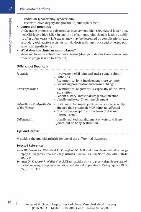
Differential Diagnosis. . . . . . . . . . . . . . . . . . . . . . . . . . . . . . . . . . . . . . . . . . . . . . . . . . . . . . . . . . . . . . . . . . . . . . . . . . . . . . . . . . . . . . . . . . . .
Psoriasis – Involvement of SI joint and entire spinal column– Enthesitis– Asymmetrical joint involvement more common– Coexisting proliferative and erosive changes
Reiter syndrome – Asymmetrical oligoarthritis, especially of the lowerextremities
– Patient history: intestinal/urogenital infection– Usually unilateral SI joint involvement
Polyarthrosis/polyarthritisof the fingers
– Distal interphalangeal joints usually more severelyaffected than proximal; MCP joints not affected
– No erosions except in erosive form of disease(“seagull sign”)
Collagenosis – Usually marked malalignment of wrist and fingerjoints, but no bony destruction
Tips and Pitfalls. . . . . . . . . . . . . . . . . . . . . . . . . . . . . . . . . . . . . . . . . . . . . . . . . . . . . . . . . . . . . . . . . . . . . . . . . . . . . . . . . . . . . . . . . . . .
Mistaking rheumatoid arthritis for one of the differential diagnoses.
Selected References
Keen HI, Brown AK, Wakefield RJ, Conaghan PG. MRI and musculoskeletal ultrasonog-raphy as diagnostic tools in early arthritis. Rheum Dis Clin North Am 2005; 31(4):699–714
Sommer OJ, Kladosek A, Weiler V, et al. Rheumatoid arthritis: a practical guide to state-of-the-art imaging, image interpretation, and clinical implications. Radiographics 2005;25(2): 381–398
.....96Reiser et al. Direct Diagnosis in Radiology. Musculoskeletal Imaging
(ISBN 9783131451613), © 2008 Georg Thieme Verlag KG
Rheumatoid Arthritis2
Inflamm
atoryD
iseases

Definition. . . . . . . . . . . . . . . . . . . . . . . . . . . . . . . . . . . . . . . . . . . . . . . . . . . . . . . . . . . . . . . . . . . . . . . . . . . . . . . . . . . . . . . . . . . .
" Epidemiology4–5% of all fractures · Predominantly occur in older patients.
" Etiology, pathophysiology, pathogenesisTrauma usually minimal · Fall onto an outstretched arm or direct blow to thelateral aspect of the humerus (often with osteoporosis) · In younger patients,more severe trauma is needed and displaced fractures or fracture-dislocationsare more common.
Imaging Signs. . . . . . . . . . . . . . . . . . . . . . . . . . . . . . . . . . . . . . . . . . . . . . . . . . . . . . . . . . . . . . . . . . . . . . . . . . . . . . . . . . . . . . . . . . . .
" Modality of choiceRadiography · CT.
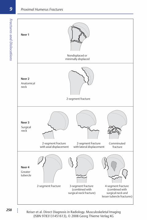
" Radiographic/CT findingsRadiograph of the shoulder joint in two planes (AP and transthoracic or Y-view)
· Perhaps axial view to evaluate lesser tubercle · Modified Neer classificationbased on number of fragments and malalignment of four main segments (hu-meral head epiphysis, humerus metaphysis/diaphysis (surgical neck), and lesserand greater tubercles) · Displacement is diagnosed on the basis of 1 cm or moredisplacement or 458 angulation.– Neer 1: nondisplaced fracture of one or more fragments.Displaced fractures:– Neer 2: fracture of the anatomical neck, two-fragment fracture, one displaced
fragment.– Neer 3: fracture of the surgical neck, two-fragment fracture, one displaced
fragment.– Neer 4: fracture of the greater tubercle, without or with additional fracture of
the surgical neck or lesser tubercle; two- to four-fragment fracture, up to threedisplaced fragments possible.
– Neer 5: fracture of the lesser tubercle, two- to four-fragment fracture.– Neer 6: fracture-dislocations.AO classification scheme provides an alternative system.
" CT findingsImaging of joint involvement, fragments, and displacement without superimpo-sitions.
Clinical Aspects. . . . . . . . . . . . . . . . . . . . . . . . . . . . . . . . . . . . . . . . . . . . . . . . . . . . . . . . . . . . . . . . . . . . . . . . . . . . . . . . . . . . . . . . . . . .
" Typical presentationPain · Swelling · Limited range of motion after fall onto arm. · Humeral headnecrosis, especially in fractures with three or four fragments and in fractures in-volving the anatomic neck (13–34%) · Post-traumatic shoulder stiffness ·Omarthritis · Rotator cuff tear · Nerve damage (axillary nerve) · Vascular inju-ries (axillary artery, 5% in displaced fractures).
.....256Reiser et al. Direct Diagnosis in Radiology. Musculoskeletal Imaging
(ISBN 9783131451613), © 2008 Georg Thieme Verlag KG
Proximal Humerus Fractures9
Fracturesand
Dislocations
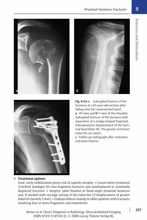
" Treatment optionsGoal: early mobilization given risk of capsule atrophy · Conservative treatment(Gilchrist bandage) for two-fragment fractures and nondisplaced or minimallydisplaced fractures · Surgery: plate fixation or fixed-angle proximal humerusnail, if needed with cerclage wiring of the tubercles (if displacement of greatertubercle exceeds 5 mm) · Endoprosthesis mainly in older patients with fracturesinvolving four or more fragments and omarthritis.
..... 257
Fig. 9.4 a–c Subcapital fracture of thehumerus in a 65-year-old woman afterfalling onto her outstretched hand.a AP view and b Y-view of the shoulder.Subcapital fracture of the humerus withseparation of a wedge-shaped fragment.Inferoposterior displacement of the hum-eral head (Neer III). The greater and lessertubercles are intact.c Follow-up radiograph after reductionand plate fixation.
Reiser et al. Direct Diagnosis in Radiology. Musculoskeletal Imaging(ISBN 9783131451613), © 2008 Georg Thieme Verlag KG
Proximal Humerus Fractures 9Fractures
andD
islocations
....258
Neer 2
Neer 1
Neer 3
Neer 4
Nondisplaced or minimally displaced
Anatomical neck
2-segment fracture
Surgical neck
2-segment fracture with axial displacement
2-segment fracture with lateral displacement
Comminuted fracture
Greatertubercle
2-segment fracture 3-segment fracture (combined with
surgical neck fracture)
4-segment fracture (combined with
surgical neck andlesser tubercle fractures)
Reiser et al. Direct Diagnosis in Radiology. Musculoskeletal Imaging(ISBN 9783131451613), © 2008 Georg Thieme Verlag KG
Proximal Humerus Fractures9
Fracturesand
Dislocations
.....324
A
accessory ossicle 323acetabular index, hip dysplasia 162acetabular roof angle,
hip dysplasia 162Achilles bursitis 240Achilles tendon rupture 239–241
complete 241incomplete 239, 240MRI findings 239, 240, 241
acromioclavicular dislocation252–255classification 252radiographic findings 252, 253, 254
acromion shapes 246, 248adolescent kyphosis 190–191, 191Albers–Schönberg disease 187, 188Albright syndrome 18algoneurodystrophy
see Sudeck atrophyAndersson lesion 109aneurysmal bone cyst 26–28
MRI findings 26, 27radiographic findings 26, 27, 28
ankle joint fractures 301–303associated injuries 301classification 301radiographic findings 301, 302, 303syndesmotic injury 301
ankylosing spondylitis 109–111bamboo spine 109, 110calcaneus 109MRI findings 109, 111peripheral 109radiographic findings 109, 110sacroiliac joint 109vertebral column 109
anterior disk herniation 124anterior iliac spine avulsion
fracture 242, 245
anteroinferior glenoid lesions 228anterolisthesis 134arteriovenous hemangioma 69arteriovenous malformation 69arthritis, gouty see goutarthritis mutilans 100avascular, ischemic, aseptic femoral
head necrosis in the adultsee femoral head necrosis
avulsion fracture 242–245acute 242chronic 242displaced 243nondisplaced 243radiographic findings 243, 244
B
bamboo spine 109, 110Bankart lesion 226, 260Barton fracture 270bayonet deformity 159, 160beak fracture 239Bekhterev disease see ankylosing
spondylitisbipartite scaphoid 277Blout disease see tibia varaBoehler angle, calcaneus fracture
304, 307bone contusion 312bone cysts
aneurysmal see aneurysmalbone cyst
juvenile see juvenile bone cystbone hemangioma 36–40
cranial 36CT findings 36, 38–39vertebral body 36, 38–39
bone infarct 213–215“cold-in-hot” spot 213“cold spot” 213“double-line sign” 213MRI findings 213, 214radiographic findings 213, 214
bone island (osteoma medullare)1, 2
Page numbers in italics refer toillustrations.
Reiser et al. Direct Diagnosis in Radiology. Musculoskeletal Imaging(ISBN 9783131451613), © 2008 Georg Thieme Verlag KG
Index

..... 325
bone lymphoma 61–64CT findings 61, 61, 62diffuse marrow infiltration 61focal lesions 61MRI findings 60, 62
bone marrow edema syndrome 199, 208Brodie abscess 90–92, 91brown tumors 156Buford complex 229
C
calcaneus fracture 304–307beak 307classification 304CT findings 304–305, 305, 306fracture lines 304, 307pathologic fractures 304peripheral 304radiographic findings 304, 305
calcaneus secundarius 307calcific tendinitis 234, 248capillary hemangioma 69cartilage flakes 225cartilage lesions 223–225
“arthrographic effect” 223MRI findings 223, 224
cartilaginous exostosis 12–14, 13cavernous hemangioma 69cement retraction 139Charcot joint see neuropathic
osteoarthropathyChauffeur fracture 270child abuse 186chisel fracture, radial head 263chondrocalcinosis 156chondrosarcoma 52–54, 53chronic recurrent multifocal
osteomyelitis (CRMO) 86chronic sclerosing osteomyelitis 319clavicle fracture 249–251
classification 249radiographic findings 249, 250
club foot 176–179angle measurement 178ossific nucleus 176
radiographic findings 176, 177treatment stages 176
Cobb angle, scoliosis 180, 181, 181collagenosis 96Colles fracture 270, 271compact bone island (osteoma
medullare) 1, 2compensatory hyperlordosis 192complex regional pain syndrome
116–118nuclear medicine 116radiographic findings 116, 117stages 116–118
condylar fracture, pediatric 267–269displaced 267lateral 267medial 267positive fat pad sign 267radiographic findings 267, 268
coxitis 196cruciate ligament tears 219–222
anterior 219, 220–221, 222contusions 219“deep notch” sign 219direct signs of rupture 219hyperextension injuries 219indirect signs of rupture 219MRI findings 220–221radiographic findings 219, 222
D
degenerative disease 119–146dens
fracture 284–286, 285persistent nonfusion 285
developmental disorders 159–192diabetic foot syndrome 144die-punch fracture 270diffuse idiopathic skeletal hyperostosis
(DISH) 141–143“dripping candle wax” appearance
141, 142radiographic findings 141, 142
diskitis 109dislocations 249–323
Reiser et al. Direct Diagnosis in Radiology. Musculoskeletal Imaging(ISBN 9783131451613), © 2008 Georg Thieme Verlag KG
Index

.....326
“double-density” sign, osteoidosteoma 5
dual-energy X-ray absorptiometry(DXA), osteoporosis 147
E
Edgren–Vaino sign 190elephant’s foot 321enchondroma 9–11
radiographic findings 9, 10“scalloping” 9
enchondromatosis 9endosteoma (osteoma medullare) 1, 2eosinophilic granuloma 192epiphyseal torsion angle 174Essex–Lopresti fracture 263Ewing sarcoma 55–57
MRI findings 55, 57radiographic findings 55, 56
extraosseous osteosarcoma 47, 48
F
failed back surgery syndrome129–130
fatigue fracture 315, 316femoral fracture, pertrochanteric
see pertrochanteric femoral fracturefemoral head necrosis 204–208
classification 204–205MRI findings 207non-traumatic 204post-traumatic 204radiographic findings 204–205, 206
femoral neck fracture 287–289classification 287complications 287radiographic findings 287, 288stress 289
fibrous dysplasia 18–22differential diagnosis 22monostotic 18MRI findings 18, 20–21polyostotic 18radiographic findings 18, 19, 20
fifth metatarsal baseepiphyseal plate 310fractures 308–310, 309
flatfoot deformity 179Forestier disease see diffuse idiopathic
skeletal hyperostosis (DISH)fractures 249–323
G
Gaenslen sign 95Gage’s sign 193Galeazzi fracture-dislocation 270gamekeeper’s (skier’s) thumb
281–283, 282Garré osteomyelitis 319giant cell tumor 29–31
CT findings 29, 30grading 29MRI findings 29radiographic findings 29, 30
giant cell tumor of tendon sheathssee pigmented villonodular synovitis
glenoid lesions, anteroinferior 228glenolabral articular disruption
(GLAD) 226gout 105–108
acute 105, 108chronic 108great toe 106, 107late-stage 105radiographic findings 105, 106soft-tissue 107“spike” 106tophi 105, 107
gouty arthritis see goutgranulation tissue, Brodie’s
abscess 90
H
hangman’s fracture 284hemangioma
arteriovenous 69bone see bone hemangiomacapillary 69
Reiser et al. Direct Diagnosis in Radiology. Musculoskeletal Imaging(ISBN 9783131451613), © 2008 Georg Thieme Verlag KG
Index

..... 327
cavernous 69soft-tissue see soft-tissue
hemangiomasynovial 69venous 69vertebral body 36, 38–39
Hilgenreiner line 162, 164Hill–Sach lesion 226, 260hip dysplasia 162–165
radiographic findings 162, 163radiographic lines 162, 164risk factors 162
Honda sign 315humerus fractures
distal intra-articular 268proximal see proximal humerus
fracturehumpback deformity 273Hutchinson fracture 270hyperparathyroidism 156–158
intracortical resorption 156MRI findings 156, 157primary 156radiographic findings 156, 157secondary 156subperiosteal resorption 156tertiary 156
hyperuricemiaprimary (familial) 105secondary 105
hypothyroidism 196
I
iliac spine avulsion fracture,anterior 242, 245
implant loosening 138–140cemented implant 138, 140cementless implant 138, 139radiographic findings
138, 139, 140inferior pubic ramus avulsion
fracture 242, 243inflammatory disease 86–118insufficiency fracture 315
sacral 315
intercondylar eminence avulsionfracture 242, 243
intervertebral disk extrusion 128intervertebral disk herniation
126–130annular tear 126, 127anterior 124MRI findings 126–129, 127myelogenic finding 129relationships 128sequestered disk 129
intervertebral disk protrusion126, 128, 129
intervertebral hernia 129intra-articular lesions 216–234ivory exostosis 1ivory osteoma 1
J
Jaffe–Lichtenstein disease see fibrousdysplasia
joint mouse 209Jones fractures 308, 310juvenile bone cyst 23–25
“fallen fragment” sign 23pseudotrabeculae 23radiographic findings 23, 24
juvenile kyphosis (Scheuermanndisease) 190–192, 191
juxtacortical osteoma 1
K
Kasabach–Merritt syndrome 69Kellgren–Lawrence (KL) grading
system, osteoarthritis 119, 120Kienböck disease see lunatomalaciaKlippel–Trénaunay–Weber
syndrome 69kyphoscoliosis 184kyphosis
adolescent 190–191, 191juvenile 190–192, 191
Reiser et al. Direct Diagnosis in Radiology. Musculoskeletal Imaging(ISBN 9783131451613), © 2008 Georg Thieme Verlag KG
Index

.....328
L
labral lesions 226–230classification 226, 228imaging signs 227, 227–229
lateral ankle ligament rupture235–238MRI findings 235–236, 236radiographic findings 235, 237
lateral ankle ligaments 237ligament injuries 235–248lipoma 77–78
MRI findings 77, 78variants 77
liposarcoma 73, 74Looser transformation zones,
osteomalacia 153lunate fractures 203lunatomalacia 201–203
MRI findings 201, 202radiographic findings 201, 202staging 201
lymphomabone see bone lymphomanon-Hodgkin 62, 63
M
Madelung deformity 159–161classification 159radiographic findings 159, 160Volar tilt 159
Maffucci syndrome 9, 69Maisonneuve fracture 301marble bone disease
see osteopetrosismarginal spondylitis 109mastocytosis 189meniscal lesions 216–218
classification 216clinical aspects 217–218MRI findings 216–217, 217radiographic findings 216signal abnormality 216, 218tear patterns 216, 218
metabolic disorders 147–158
metastases 44–46differential diagnosis 44–46osteoblastic 44osteolytic 44radiographic findings 44, 45
Milkman syndrome 153Monteggia fractures 266multiple cartilaginous exostoses 12multiple myeloma 58–60
clinical aspects 59–60MRI findings 58, 59radiographic findings 58, 59
Münchmeyer disease 82myositis ossificans 82–85
clinical aspects 84CT findings 82, 83radiographic findings 82, 83
myositis ossificans circumscripta 82neuropathic 82post-traumatic 82, 84
myositis ossificans localisatasee myositis ossificans circumscripta
myositis ossificans progressiva 82, 84
N
navicular fracture see scaphoid fractureneuropathic osteoarthropathy 144–146
early (active) stage 144late (reparative) chronic stage 144MRI findings 144, 145radiographic findings 144, 145
nidus 5non-Hodgkin’s lymphoma 62, 63nonossifying fibroma (NOF) 15–17, 16notochord, abnormal involution 192
O
occult fracture 311–314femoral neck 311MRI findings 311, 313, 314radiographic findings 312, 314
Ollier disease 9Osgood–Schlatter disease 242, 243os odontoideum 285
Reiser et al. Direct Diagnosis in Radiology. Musculoskeletal Imaging(ISBN 9783131451613), © 2008 Georg Thieme Verlag KG
Index

..... 329
os peroneum 310osteitis deformans see Paget diseaseosteitis fibrosa cystica
see hyperparathyroidismosteoarthritis 119–122
cartilage changes 119clinical aspects 120erosive 120etiology 119MRI findings 120, 121periarticular changes 119radiographic findings 119, 121“seagull” sign 119
osteochondritis dissecans 209–212MRI findings 209, 210, 211radiographic findings 209, 210staging 209
osteochondroma 12–14, 13osteochondrosis 123–125
associated conditions 123erosive 123Modic changes 125MRI findings 123–125, 124radiographic findings 123, 124
osteoclastoma see giant cell tumorosteogenesis imperfecta 184–186
diagnostic criteria 184radiographic findings 184, 185
osteoid osteoma 5–8CT findings 5, 6MRI findings 5, 7radiographic findings 5, 6
osteoma 1–4CT findings 1, 2ivory 1juxtacortical 1radiographic findings 1, 2
osteomalacia 153–155Looser transformation zones 153radiographic findings 153, 154
osteoma medullare 1, 2osteomyelitis 86–89
acute 86, 88chronic 86, 88classification 86differential diagnosis 89
MRI findings 87pathogens 86radiographic findings 86, 87, 88risk factors 86
osteonecrosis 193–215of the knee 197–200
MRI findings 197, 199radiographic findings 197, 198
osteopetrosis 187–189“bone-within-bone” appearance
187, 188infantile (malignant) 188inheritance 187precocious manifestations 187radiographic findings 187, 188“sandwich vertebral body”
appearance 187subtypes 187
osteopoikilosis 10osteoporosis 147–150
bone density findings 147femoral fracture 287, 290postmenopausal 147radiographic findings 147, 148secondary 147senile 147sonography 149stress fractures 315tibial plateau fracture 297
osteosarcoma 47–51CT findings 47–48differential diagnosis 50–51extraosseous 47, 48MRI findings 48–50, 49multicentric 47radiographic findings 47–48, 48variants 47
os vesalianum 310
P
Paget disease 41–43cranium 41radiographic findings 41, 42stages 41vertebral bodies 41
Reiser et al. Direct Diagnosis in Radiology. Musculoskeletal Imaging(ISBN 9783131451613), © 2008 Georg Thieme Verlag KG
Index
.....330
pannus 102paraprotein 58parosteal osteoma 1parosteal osteosarcoma 47, 48patella alta 293patella baja 293patella fracture 293–296
radiographic findings 294, 295patellar pole avulsion fracture 242, 243pathologic fracture 315periosteal osteosarcoma 47, 48peritendinitis 240Perkin–Ombrédanne line 162, 164Perthes disease 193–196
classification 193–194clinical aspects 195–196“head-at-risk” signs 193, 194radiographic findings 193–194, 194stages 193, 195
pertrochanteric femoral fracture290–292classification 290, 292displaced 290nondisplaced 290radiographic findings 290, 291reverse oblique fracture 290traumatic 290
pigmented villonodular synovitis79–81clinical aspects 79MRI findings 79, 80radiographic findings 79, 80
polyarthritis, fingers 96polyarthrosis, fingers 96postural scoliosis 182proximal humerus fracture 256–259
displaced 256Neer classification 256, 258–259radiographic findings 256, 257
pseudoarthrosis 321–323atrophic (nonreactive) 321hypertrophic (reactive) 321nonunion 321, 322radiographic findings 321, 322scaphoid fracture 273
pseudocapsules, soft-tissuesarcoma 73
pseudogout 108psoriasis 96psoriatic arthritis 97–101
“mouse ear” appearance 97pattern of involvement 99radiographic findings 97, 98, 99“sausage fingers” 97, 100“shining corner” appearance 97symptoms 100
psoriatic arthropathy 97–101
Q
quantitative CT (QCT),osteoporosis 147–149
R
rachitic rosary, rickets 151radial fracture, distal 270–272
classification 270radiographic findings 270, 271, 272
radial head fractures 263–266CT findings 263, 264nondisplaced 265positive fat pad sign 263, 264radiographic findings 263, 264, 265
reflex sympathetic dystrophysee Sudeck atrophy
Reichel syndrome see synovialchondromatosis
Reimer index, hip dysplasia 162Reiter syndrome 96retrolisthesis 134reverse Barton fracture 270reverse Hill–Sachs lesion 227, 260reverse Hutchinson fracture 270reverse pertrochanteric femoral
fracture 290rheumatoid arthritis 93–96
clinical aspects 95differential diagnosis 95MRI findings 93–95, 94pattern of involvement 93, 95
Reiser et al. Direct Diagnosis in Radiology. Musculoskeletal Imaging(ISBN 9783131451613), © 2008 Georg Thieme Verlag KG
Index

..... 331
radiographic findings 93, 94synovitis 93ultrasound findings 95
rheumatoid disease 122rickets 151–152, 152rotator cuff lesions 231–234
classifications 231clinical aspects 234degenerative tears 231, 233“magic angle” effect 231, 233MRI findings 231–233, 232, 233rupture 233traumatic tears 231
S
“salt and pepper skull,” multiplemyeloma 58
scaphoid, bipartite 277scaphoid fracture 273–277
classification 273instability signs 273MRI findings 273–277, 275–276radiographic findings 273, 274
Scheuermann disease (juvenilekyphosis) 190–192, 191
Schmorl modes 190scoliosis 180–183
Cobb angle 180, 181, 181idiopathic 180MRI findings 180postural 182radiographic findings 180, 181secondary 180skeletal maturity estimation
180, 183scurvy 152“seagull” sign, osteoarthritis 119Segond fracture 242, 243septic arthritis 102–104
“bare areas” 102causative agents 102clinical aspects 102MRI findings 102, 103pannus 102radiographic findings 102, 103
sessile osteochondroma 12shepherd’s crook deformity 18shin splints 320shoulder dislocation 226, 260–262
anterior 260associated injuries 260posterior 260radiographic findings 260, 261, 262types 260
shoulder impingement 246–248radiographic findings 246, 247
skier’s (gamekeeper’s) thumb281–283, 282
slipped capital femoral epiphysis(SCFE) 170–175acute 173, 174angle measurement 174chronic disease 170, 173, 174clinical aspects 173–174imminent 173MRI findings 170, 173radiographic findings 170, 171, 172remodeling changes 170
Smith fracture 270soft-tissue hemangioma 69–72
clinical aspects 71“cluster of grapes” appearance 69differential diagnosis 72MRI findings 69–71, 70–71
soft-tissue sarcoma 73–76clinical aspects 75histology 73MRI findings 73, 74radiographic findings 73, 74
spinal canal stenosis 131–133acute 133chronic 133“lost” subarachnoid space 131MRI findings 131, 132postmyelography CT findings
131, 132spinal claudication 133spondylarthritis 123spondylitis 112–115
ankylosing see ankylosing spondylitismarginal 109
Reiser et al. Direct Diagnosis in Radiology. Musculoskeletal Imaging(ISBN 9783131451613), © 2008 Georg Thieme Verlag KG
Index
.....332
spondylodiskitis 112–115abscess formation 114MRI findings 113, 114pathogens 112predisposing factors 112radiographic findings 112, 113spread 112, 114
spondylolisthesis 123see spondylolysis/spondylolisthesis
spondylolysis/spondylolisthesis134–137, 136bone marrow edema 134CT findings 134, 135grading 134radiographic findings 134, 135“Scotty dog with collar” 134secondary signs 134
spondylosis 123spontaneous aseptic osteonecrosis
of the knee see Ahlbäck diseasesquare vertebrae 109stalked osteochondroma 12Stener lesion 281stress fractures 315–320
calcaneus fracture 304cortical 315CT findings 315, 318femoral neck 289fifth metatarsal base fractures 308fracture lines 319malignant 315MRI findings 316, 317osteoporosis 315radiographic findings 315, 316,
317, 318sites of predilection 316–319
subacromial narrowing 246sublabral foramen 229sublabral recess 229subscapular nerve lesion 234superior labrum anterior to posterior
(SLAP) lesions 226, 227, 229supracondylar fracture 269surface osteosarcoma, high-grade
47, 48, 50
syndesmophytes 109synovial chondromatosis 32
MRI findings 32, 35radiographic findings 32, 33, 34
synovial endothelioma see pigmentedvillonodular synovitis
synovial hemangioma 69synovial sarcoma 73, 74synovitis, rheumatoid arthritis 93syringomyelia 144
T
talipes equinovarus see club foottelangiectatic osteosarcoma
47, 48, 50tendon injuries 235–248tibial pilon fracture 301tibial plateau fracture 297–300
classification 297, 299CT findings 297, 298radiographic findings 297, 298surgical treatment 300
tibial tuberosity avulsion fracture 242tibia vara 166–169
adolescent 166beaking 166clinical aspects 168infantile 166, 168juvenile 166, 168radiographic findings 166, 167stages 166, 168
Tillaux fracture 301tophi 105, 107Tossy injury see acromioclavicular
dislocationtransient osteoporosis 199, 208trimalleolar fracture 301triplane fracture 301triquetrum fracture 278–280
body 278CT findings 278, 279dorsal avulsion fracture 278radiographic findings 278, 279
tumors 1–85
Reiser et al. Direct Diagnosis in Radiology. Musculoskeletal Imaging(ISBN 9783131451613), © 2008 Georg Thieme Verlag KG
Index

..... 333
V
vacuum phenomenon 123, 124venous hemangioma 69vertebral body hemangioma
36, 38–39vertebral compression fracture,
neoplastic 65–68CT findings 65, 66MRI findings 65, 67
vertebral formation defects 182vertebral segmentation defects 182
vitamin C deficiency (scurvy) 152vitamin D deficiency
osteomalacia 153rickets 151
Volar tilt, Madelung deformity 159Volkmann triangle 301
W
World Health Organization (WHO)osteoporosis criteria 147
Reiser et al. Direct Diagnosis in Radiology. Musculoskeletal Imaging(ISBN 9783131451613), © 2008 Georg Thieme Verlag KG
Index