Rhesus Cytomegalovirus Contains Functional Homologues of US2
13
JOURNAL OF VIROLOGY, May 2005, p. 5786–5798 Vol. 79, No. 9 0022-538X/05/$08.000 doi:10.1128/JVI.79.9.5786–5798.2005 Copyright © 2005, American Society for Microbiology. All Rights Reserved. Rhesus Cytomegalovirus Contains Functional Homologues of US2, US3, US6, and US11 Nupur T. Pande, 1 Colin Powers, 1 Kwangseog Ahn, 2 and Klaus Fru ¨h 1 * Vaccine and Gene Therapy Institute, Oregon Health and Science University, Portland, Oregon, 1 and Seoul National University, Sillim-dong, Gwanak-gu, Seoul, Korea 2 Received 13 September 2004/Accepted 16 December 2004 Human cytomegalovirus (HCMV) is a paradigm for mechanisms subverting antigen presentation by major histocompatibility complex (MHC) molecules. Due to its limited host range, HCMV cannot be studied in animals. Thus, the in vivo importance of inhibiting antigen presentation for the establishment and mainte- nance of infection with HCMV is unknown. Rhesus cytomegalovirus (RhCMV) is an emerging animal model that shares many of the features of HCMV infection. The recent completion of the genomic sequence of RhCMV revealed a significant degree of homology to HCMV. Strikingly, RhCMV contains several genes with low homology to the HCMV US6 gene family of inhibitors of the MHC I antigen presentation pathway. Here, we examine whether the RhCMV US6 homologues (open reading frames Rh182, -184, -185, -186, -187, and -189) interfere with the MHC I antigen-processing pathway. We demonstrate that Rh182 and Rh189 function similarly to HCMV US2 and US11, respectively, mediating the proteasomal degradation of newly synthesized MHC I. The US3 homologue, Rh184, delayed MHC I maturation. Unlike US3, MHC I molecules eventually escaped retention by Rh184, so that steady-state surface levels of MHC I remained unchanged. Rh185 acted similarly to US6 and inhibited peptide transport by TAP and, consequently, peptide loading of MHC I molecules. Thus, despite relatively low sequence conservation, US6 family-related genes in RhCMV are functionally closely related to the conserved structural features of HCMV immunomodulators. The conserva- tion of these mechanisms implies their importance for immune evasion in vivo, a question that can now be addressed experimentally. Human cytomegalovirus (HCMV) is highly prevalent in the human population and establishes persistent infection of im- munocompetent hosts (47). This life-long infection occurs de- spite a significant CMV-specific cellular immune response, with up to 10% of the total T-cell population being CMV specific (22). Moreover, seropositive individuals can be rein- fected with a different strain of CMV even in the presence of preexisting immunity (8). Thus, the immune system is unable to eradicate CMV upon primary infection or to prevent rein- fection. Continuous immune surveillance is required to keep the viral infection in an asymptomatic state, since CMV disease is mostly observed during immunodeficiency, particularly in cell-mediated immunity, related to either immunologic imma- turity, pharmacologic immunosuppression (transplantation) (53), or the progressive immunodeficiency of human immuno- deficiency virus infection (52). Thus, a balance is established between immunological control of the viral infection and im- mune evasion by the virus. Immunomodulatory mechanisms encoded by CMV are thought to be central to maintaining this balance. It is conceiv- able that a large portion of the CMV genome, containing 250 open reading frames (ORFs), is dedicated to manipulating various aspects of the host defense. One of the major obstacles to elucidating these mechanisms is the fact that HCMV can infect only humans. This extreme host restriction of -herpes- viruses resulted in their coevolution with the host organisms. The most closely related nonhuman CMV is the chimpanzee cytomegalovirus (16). However, chimpanzees are not readily available for experimentation. Rodents are ideal for experi- mentation, but the genomic sequences of murine cytomegalo- viruses show that the vast majority of the HCMV immuno- modulatory genes are not conserved (49). Therefore, there is a need to establish CMV infection models in animals closely related to humans. Such an alternative animal model is rhesus macaque infection by rhesus cytomegalovirus (RhCMV) (3). RhCMV and HCMV have similar epidemiologies and patterns of infection in immunocompetent and immunodeficient hosts (43, 54, 58). In addition, the immune response to RhCMV is similar to that to HCMV, with a high percentage of T cells being CMV specific (5, 35–37, 48). The recently completed sequence of the RhCMV genome revealed that, in addition to genes with no obvious homology in HCMV, RhCMV encodes homologues of most of the known immunomodulators of HCMV (26). This list includes homologues of the viral inhib- itor of caspase activation, UL36; the viral mitochondrial inhib- itor of apoptosis, UL37; the interleukin-10 homologue, UL111; the Fc receptors, UL117/UL118; the viral CXC chemokine homologue, UL147; and the tumor necrosis factor receptor homologue, UL144 (26). The RhCMV genomic region Rh182 to -189 contains six genes with homology to the genes US2 to US11 in the unique short (US) region of HCMV. This region in HCMV encodes a group of eight glycoproteins that were originally grouped into two families, US2 and US6 (11), and later into three families, US2, US3, and US11 (9). Primary structure alignments, as well as their functional relationship, suggests that all of the US2, US3, and US6 family genes arose by gene duplication and thus * Corresponding author. Mailing address: Vaccine and Gene Ther- apy Institute, Oregon Health and Science University, 505 NW 185th Ave., Beaverton, OR 97006. Phone: (503) 418-2735. Fax: (503) 418- 2701. E-mail: [email protected]. 5786 Downloaded from https://journals.asm.org/journal/jvi on 13 January 2022 by 218.154.10.184.
Transcript of Rhesus Cytomegalovirus Contains Functional Homologues of US2

JOURNAL OF VIROLOGY, May 2005, p. 5786–5798 Vol. 79, No. 90022-538X/05/$08.00�0 doi:10.1128/JVI.79.9.5786–5798.2005Copyright © 2005, American Society for Microbiology. All Rights Reserved.
Rhesus Cytomegalovirus Contains Functional Homologues of US2,US3, US6, and US11
Nupur T. Pande,1 Colin Powers,1 Kwangseog Ahn,2 and Klaus Fruh1*Vaccine and Gene Therapy Institute, Oregon Health and Science University, Portland, Oregon,1 and
Seoul National University, Sillim-dong, Gwanak-gu, Seoul, Korea2
Received 13 September 2004/Accepted 16 December 2004
Human cytomegalovirus (HCMV) is a paradigm for mechanisms subverting antigen presentation by majorhistocompatibility complex (MHC) molecules. Due to its limited host range, HCMV cannot be studied inanimals. Thus, the in vivo importance of inhibiting antigen presentation for the establishment and mainte-nance of infection with HCMV is unknown. Rhesus cytomegalovirus (RhCMV) is an emerging animal modelthat shares many of the features of HCMV infection. The recent completion of the genomic sequence of RhCMVrevealed a significant degree of homology to HCMV. Strikingly, RhCMV contains several genes with lowhomology to the HCMV US6 gene family of inhibitors of the MHC I antigen presentation pathway. Here, weexamine whether the RhCMV US6 homologues (open reading frames Rh182, -184, -185, -186, -187, and -189)interfere with the MHC I antigen-processing pathway. We demonstrate that Rh182 and Rh189 functionsimilarly to HCMV US2 and US11, respectively, mediating the proteasomal degradation of newly synthesizedMHC I. The US3 homologue, Rh184, delayed MHC I maturation. Unlike US3, MHC I molecules eventuallyescaped retention by Rh184, so that steady-state surface levels of MHC I remained unchanged. Rh185 actedsimilarly to US6 and inhibited peptide transport by TAP and, consequently, peptide loading of MHC Imolecules. Thus, despite relatively low sequence conservation, US6 family-related genes in RhCMV arefunctionally closely related to the conserved structural features of HCMV immunomodulators. The conserva-tion of these mechanisms implies their importance for immune evasion in vivo, a question that can now beaddressed experimentally.
Human cytomegalovirus (HCMV) is highly prevalent in thehuman population and establishes persistent infection of im-munocompetent hosts (47). This life-long infection occurs de-spite a significant CMV-specific cellular immune response,with up to 10% of the total T-cell population being CMVspecific (22). Moreover, seropositive individuals can be rein-fected with a different strain of CMV even in the presence ofpreexisting immunity (8). Thus, the immune system is unableto eradicate CMV upon primary infection or to prevent rein-fection. Continuous immune surveillance is required to keepthe viral infection in an asymptomatic state, since CMV diseaseis mostly observed during immunodeficiency, particularly incell-mediated immunity, related to either immunologic imma-turity, pharmacologic immunosuppression (transplantation)(53), or the progressive immunodeficiency of human immuno-deficiency virus infection (52). Thus, a balance is establishedbetween immunological control of the viral infection and im-mune evasion by the virus.
Immunomodulatory mechanisms encoded by CMV arethought to be central to maintaining this balance. It is conceiv-able that a large portion of the CMV genome, containing �250open reading frames (ORFs), is dedicated to manipulatingvarious aspects of the host defense. One of the major obstaclesto elucidating these mechanisms is the fact that HCMV caninfect only humans. This extreme host restriction of �-herpes-viruses resulted in their coevolution with the host organisms.
The most closely related nonhuman CMV is the chimpanzeecytomegalovirus (16). However, chimpanzees are not readilyavailable for experimentation. Rodents are ideal for experi-mentation, but the genomic sequences of murine cytomegalo-viruses show that the vast majority of the HCMV immuno-modulatory genes are not conserved (49). Therefore, there is aneed to establish CMV infection models in animals closelyrelated to humans. Such an alternative animal model is rhesusmacaque infection by rhesus cytomegalovirus (RhCMV) (3).RhCMV and HCMV have similar epidemiologies and patternsof infection in immunocompetent and immunodeficient hosts(43, 54, 58). In addition, the immune response to RhCMV issimilar to that to HCMV, with a high percentage of T cellsbeing CMV specific (5, 35–37, 48). The recently completedsequence of the RhCMV genome revealed that, in addition togenes with no obvious homology in HCMV, RhCMV encodeshomologues of most of the known immunomodulators ofHCMV (26). This list includes homologues of the viral inhib-itor of caspase activation, UL36; the viral mitochondrial inhib-itor of apoptosis, UL37; the interleukin-10 homologue, UL111;the Fc receptors, UL117/UL118; the viral CXC chemokinehomologue, UL147; and the tumor necrosis factor receptorhomologue, UL144 (26).
The RhCMV genomic region Rh182 to -189 contains sixgenes with homology to the genes US2 to US11 in the uniqueshort (US) region of HCMV. This region in HCMV encodes agroup of eight glycoproteins that were originally grouped intotwo families, US2 and US6 (11), and later into three families,US2, US3, and US11 (9). Primary structure alignments, as wellas their functional relationship, suggests that all of the US2,US3, and US6 family genes arose by gene duplication and thus
* Corresponding author. Mailing address: Vaccine and Gene Ther-apy Institute, Oregon Health and Science University, 505 NW 185thAve., Beaverton, OR 97006. Phone: (503) 418-2735. Fax: (503) 418-2701. E-mail: [email protected].
5786
Dow
nloa
ded
from
http
s://j
ourn
als.
asm
.org
/jour
nal/j
vi o
n 13
Jan
uary
202
2 by
218
.154
.10.
184.

their products represent a single family of proteins (1, 20),which we will refer to as the US6 family. Six members of thisfamily have been shown to interfere with the major histocom-patibility complex class I (MHC I) antigen-processing pathway(42), and deletion of this region restores MHC I expression inHCMV-infected cells (33). Since MHC I molecules presentvirus-derived peptides to cytotoxic T cells, it is assumed thatthese proteins play an important role in viral immune evasionin vivo. Each of the US6 family proteins interferes at a distinctstep during the assembly and intracellular transport of MHC Iheterotrimers consisting of heavy chain (HC), �2 microglobu-lin (�2m), and antigen-derived peptides. Immediately uponheavy-chain synthesis, US2 and US11 extract newly synthesizedMHC I molecules from the lumen of the endoplasmic reticu-lum (ER) and send them for degradation by the proteasome(59, 60). US11 achieves this by simultaneously interacting withMHC I molecules and the cellular quality control protein der-lin 1, which extracts misfolded proteins from the ER (41, 61).US2 seems to act by a different mechanism, since it differs fromUS11 in several respects, e.g., it does not require the presenceof a cytosolic tail in its target molecules and does not interactwith derlin 1 (4, 41, 61). US6 inhibits peptide translocationacross the lumen of the ER membrane by the peptide trans-porter TAP, preventing the association of antigen-derived pep-tides with nascent MHC I molecules (2, 28, 30, 40). US3 retainsMHC I molecules in the ER (1, 25, 34, 39) by binding to andinterfering with tapasin, an MHC-specific chaperone that con-trols peptide loading (46). US10 has been shown to delay MHCI exit (18). At the cell surface, US8 binds to MHC I moleculesthat are endocytosed (55). However, both US8 and US10 seemto be relatively inefficient in their activities, since they do notaffect the steady-state levels of MHC I at the cell surface (2) orantigen presentation to T cells (32). The remaining two US6family members, US7 and US9, have no known function (32).
In contrast to the extensive studies performed on the mo-lecular functions of the HCMV proteins in vitro, their roles inthe establishment and maintenance of virus infection in vivoare unknown. Provided that the RhCMV homologues are alsofunctionally related to the HCMV US6 family, RhCMV couldrepresent a model to evaluate the in vivo function of thisprotein family. In this report, we examine whether the RhCMVhomologues interfere with the assembly and maturation ofMHC I. We demonstrate that RhCMV contains functionalhomologues for US2, US3, US6, and US11. In the presence ofRhCMV Rh182 and Rh189, nascent MHC I molecules werefound to be degraded in a proteasome-dependent manner intissue culture, suggesting that Rh182 and Rh189 are homo-logues of HCMV US2 and US11. The RhCMV ORF Rh185represents a homologue of HCMV US6, since Rh185 inhibitedpeptide transport by TAP and, consequently, peptide loadingof MHC I molecules. The US3 homologue, Rh185, was lessefficient than HCMV US3 in retaining MHC I molecules in theER but rather delayed MHC I exit, similar to the HCMV geneUS10. No significant effect on MHC I assembly, transport, orsurface expression was observed for the remaining US6 family-related molecules, Rh186 and Rh187. Given the low sequencesimilarity, the functional conservation of these molecularmechanisms is remarkable, indicating an important role forthem in natural infection by primate CMVs. Our data furthersuggest that the RhCMV model can be applied as a homolo-
gous model to study the functions of the modulators of antigenpresentation of HCMV.
MATERIALS AND METHODS
Virus, cells, and antibodies. RhCMV strain 68-1 was obtained from ScottWong (26). HeLa-Tet-Off cells were obtained from Clontech (Palo Alto, Calif.),and telomerized rhesus fibroblasts (TRFs) and U373-MG (human glioblastoma)and 293 cells were obtained from Jay Nelson. All cells were maintained inDulbecco’s modified Eagle’s medium supplemented with 10% fetal calf serumand 1� Pen-Strep-Glutamine (Invitrogen) unless otherwise noted. MHC I mol-ecules were detected with the following antibodies: the monoclonal antibodyW6/32, which is specific for MHC I heterodimers (10) (American Type CultureCollection); the monoclonal antibody HC-10, which recognizes free heavy chain(obtained from Hidde Ploegh); and antiserum K455, which recognizes both freeand assembled human HLA and �2m (obtained from Per Peterson). Anti-hemagglutinin (HA) antibody was purchased from Sigma, and anti-protein-disulfide isomerase (PDI) antibody was from Stressgen. Conjugated secondaryantibodies were purchased from Molecular Probes.
Plasmids. RhCMV ORFs Rh182, -184, -185, -186, -187, and -189 were ampli-fied by PCR from viral genomic DNA of RhCMV strain 68-1 (obtained fromScott Wong) using synthetic primers that spanned the start and stop codons ofthe respective ORFs and appropriate restriction sites (BglII or EcoRI at the 5�end and Asp718 at the 3� end). The amplified PCR products were inserted intovector pUHD10.1 (24) or pCDNA3.1 (Invitrogen) for expression in mammaliancells. HA-tagged versions of Rh182 to -189 were constructed using 3�-terminalPCR primers encoding the HA epitope in frame with the ORF and inserted intopCDNA3.1. Recombinant adenovirus (rAd) was generated using a plasmid-based recombinant system (27) with the following modifications. The promoterin the shuttle vector was replaced by a tetracycline-regulatable promoter (tet-offsystem) derived from plasmid pUHG10-3 (24) inserted as an AatII-XbaI frag-ment. In addition, a new synthetic polylinker (XhoI, SalI, ClaI, HindIII, EcoRV,BglII, SpeI, NotI, EagI, and XbaI) was introduced upstream of the XbaI restric-tion site. The resulting vector was named pShuttle-tetDXN.
ORFs Rh182, Rh185, Rh186, Rh187, and Rh189 were inserted into the BglIIand XbaI sites of pShuttle-tetDXN. The integrity of each of the clones wasconfirmed by sequence analysis. Linearized pShuttle-tetDXN constructs, alongwith the plasmid carrying the adenovirus backbone, pAdEasy-1, were coelectro-porated into electrocompetent Escherichia coli BJ8153 (a gift from MikeO’Conner, University of Minnesota) as described previously (27). RecombinantpAdEasy clones were selected on kanamycin (50 �g/ml) at 37°C overnight. Theclones thus selected were analyzed for genomic integrity and recombination byrestriction digestion.
Recombinant adenovirus. For heterologous expression of RhCMV US6 ho-mologues, we used the recombinant adenovirus system developed by He et al.(27). The replication-deficient adenovirus constructs lack the E1 and E3 regionsand thus do not interfere with MHC I expression. Recombinant adenovirusesexpressing Rh182, Rh185, Rh186, Rh187, and Rh189 were reconstituted in 293cells as described previously (27). Briefly, 5 nmol of adenovirus plasmid DNAwas digested with PacI (New England Biochemicals) and purified by phenol-chloroform extraction and ethanol precipitation. This vector was transfected into293 cells using Effectene reagent (QIAGEN), and the infection was allowed toproceed until �70% of the cells showed a cytopathic effect. Virus was harvestedfrom the cells by repeated freeze-thaw cycles. The virus stocks were amplified fora minimum of three rounds. The resulting virus was purified either over a 30%sucrose cushion (32) or by cesium chloride gradient centrifugation (27). Recom-binant viral genomes were verified by restriction analysis and by PCR. Virustiters were determined on 293 cells by limiting dilution.
Flow cytometry and immunofluorescence. MHC I surface expression was de-termined by flow cytometry using antibody W6/32. Transfected HeLa cells orTRFs were analyzed 42 h posttransfection, RhCMV-infected rhesus fibroblastswere analyzed up to 96 h postinfection, and recombinant adenovirus-transducedcells were analyzed 36 h posttransduction. Approximately 106 trypsinized cellswere collected and washed by centrifugation prior to incubation with W6/32(1:100 dilution in phosphate-buffered saline [PBS]–1% fetal calf serum) for 30min on ice. After being washed three times, bound antibodies were detected withgoat-anti-mouse phycoerythrin-conjugated secondary antibody (1:500; Dako).The stained cells were analyzed on a FACScalibur flow cytometer (BD Bio-sciences).
For immunofluorescence analysis, HeLa cells were transfected with HA-tagged ORFs. The cells were washed with PBS 40 h posttransfection, fixed with2% paraformaldehyde for 20 min at room temperature, quenched with NH4Clfor 10 min, and permeabilized with 0.2% Triton X-100 for 5 min at room
VOL. 79, 2005 RhCMV IMMUNE EVASION 5787
Dow
nloa
ded
from
http
s://j
ourn
als.
asm
.org
/jour
nal/j
vi o
n 13
Jan
uary
202
2 by
218
.154
.10.
184.

temperature. Nonspecific binding sites were blocked with 3% bovine serumalbumin and 0.5% fish gelatin in PBS for 1 h at room temperature, followed byincubation with primary and secondary antibodies. Coverslips were mounted onslides and covered with Vectashield H-1200 plus DAPI (4�,6�-diamidino-2-phe-nylindole) (Vector Laboratories, Burlingame, Calif.). Fluorescence was visual-ized using an Axiovert-2 light microscope (Zeiss, Thornwood, N.Y.). All pictureswere taken in monochrome, contrast enhanced, and false colored using Openlabsoftware (Improvision, Lexington, Mass.).
Metabolic labeling and immunoprecipitation. Cells were grown to 80 to 90%confluency in 60- or 100-mm-diameter tissue culture dishes (�6 � 105 to 6 � 106
cells) and transfected as described above or infected with recombinant adeno-virus at multiplicities of infection (MOI) as indicated for each experiment. Thecells were incubated in serum-free and methionine-free medium for 1 h andmetabolically labeled with [35S]cysteine-[35S]methionine (Amersham), either 60or 100 �Ci/dish, for the indicated times for each experiment. Proteasome inhib-itors, either ZL3VS (15 to 50 �M) (kindly provided by Hidde Ploegh) or MG132(10 �M) (Peptide International), were included in the starvation medium whereindicated. After being labeled, the cells were washed three times with PBS andlysed immediately in PBS containing 1% Triton X-100 or 1% digitonin andprotease inhibitors (Roche, Indianapolis, Ind.). The cell lysate was preclearedwith 20 �l of protein A-G agarose beads (Santa Cruz Biotechnology) for 1 h orovernight. The molecules of interest were immunoprecipitated by incubationwith appropriate antibodies either for 1 h or overnight, and the immune com-plexes were captured by incubation for 1 h with 30 �l of protein A-G beads. Theprecipitated proteins were washed five times with either 0.1% NP-40 or 0.25%digitonin. All samples were boiled in Laemmli buffer, resolved on a 12% acryl-amide gel, dried, and visualized by exposing them to X-ray film (Kodak BioMaxMR).
Peptide transport assay. HeLa cells or TRFs either mock transfected ortransfected with Rh186 or HCMV US6 were collected 24 h posttransfection. Thecells were permeabilized by adding �1 IU of activated Streptolysine O (MurexDiagnostics, Dartford, United Kingdom) in transport buffer (5 mM HEPES, pH7.3, 30 mM KCl, 10 mM NaCl, 1 mM CaCl2, 2 mM EGTA, 2 mM MgCl2) andincubating them at 37°C for 10 to 20 min. The efficiency of permeabilization wasassessed to be �90% by stain exclusion with Trypan blue dye (Sigma). About 5� 106 permeabilized cells from each set were incubated at 37°C for 10 min with5 �l of fluorescein-labeled peptide with the sequence CVNKTERAY(�200pM/�l) (a generous gift from Emmanuel Wiertz) in the presence of ATP (finalconcentration, 1 mM) or EDTA (final concentration, 1 mM). The transportreaction was terminated by adding ice-cold CI buffer (50 mM Tris-HCl, pH 8.0,10 mM EDTA, 500 mM NaCl, 2 mM MgCl2, 1% Triton X-100). The cells werelysed for 30 min at 4°C, and the nuclear debris was spun out by centrifugation at11,000 � g at 4°C. The supernatant was incubated with 100 �l of concanavalin ASepharose beads (Sigma) preequilibrated with the CI lysis buffer either for 2 h orovernight in the dark. The beads were washed twice with CI buffer, followed bytwo washes with wash buffer (50 mM Tris-HCl, pH 8.0, 500 mM NaCl). Theglycosylated peptide was eluted in 500 �l of elution buffer (50 mM Tris-HCl, pH8.0, 10 mM EDTA, and 500 mM mannopyranosidase) by incubation with shakingfor 1 h at room temperature. The recovered peptide was quantified by measuringfluorescence with 485-nm excitation and 530-nm emission filters (Bio-Rad).
RESULTS
RhCMV homologues of the HCMV US6 family. Hansen etal. reported that the genes Rh182, Rh184, Rh185, Rh186,Rh187, and Rh189 display homology to individual members ofthe US6 family of HCMV (26). All six ORFs encode predictedtype I transmembrane-spanning proteins with a signal se-quence, transmembrane domain, and C-terminal cytosolic do-main. The relative locations of these genes in the genomes ofRhCMV and HCMV are shown in Fig. 1A. RhCMV lacksinternal repeats and thus does not have a unique short region.However, the location and orientation of ORFs Rh181 to -189is similar to that of the region US1 to US11 in the HCMVgenome. ClustalW alignment reveals that the central regions ofall six RhCMV polypeptides share several conserved residueswith HCMV US6 family members (Fig. 1B). Most importantly,all of the proteins share conserved cysteine residues thatbracket an immunoglobulin (Ig)-like domain, similar to the
structure of HCMV US2 (20). These signature sequences sup-port the notion that all six RhCMV ORFs belong to the US6family. To determine the relationship between the six US6-likeglycoproteins encoded in region Rh181 to -189 and the eightUS6 family members of HCMV (Fig. 1A), we aligned each ofthe HCMV ORFs with each of the RhCMV ORFs using theGAP analysis algorithm (14, 29). Identity scores were �30% inall cases, and similarity scores were �43% (Fig. 1C). Theseoverall low scores rendered differences between individualalignments difficult to interpret and, in several instances, Rh-CMV ORFs showed the highest identity with one HCMVprotein but the highest similarity with another. For instance,Rh182 displayed the highest similarity with HCMV US2 butthe highest identity with HCMV US11. Rh184 demonstratedthe highest similarity with US3 but higher identity with HCMVUS9, US10, and US11. However, taking genome location intoaccount, these alignments indicated that each RhCMV ORFshowed either the highest percent identity or percent similaritywith the US6 family member located in a similar position in theHCMV genome (Fig. 1C). The combination of location andsequence similarity suggests that Rh182, Rh184, Rh186,Rh187, and Rh189 are homologues of US2, US3, US6, US8,US10, and US11, respectively, and that the genome of Rh-CMV does not contain homologues for US7 and US9. Thisinterpretation implies that all six ORFs of HCMV that displayinterference with MHC I assembly or transport (see the intro-duction) might be conserved in the RhCMV genome. To ex-amine whether these functions are indeed conserved in Rh-CMV, or if the proteins have acquired different functions, wecharacterized the functions of all six RhCMV ORFs with re-spect to the assembly and transport of MHC I.
RhCMV Rh182 to Rh189 are glycoproteins that are locatedin the endoplasmic reticulum. The RhCMV US6 family ofgenes encodes predicted type I transmembrane glycoproteinswith a predicted amino-terminal signal sequence and a car-boxy-terminal transmembrane domain that is followed by ashort cytoplasmic domain. Each of the RhCMV ORFs alsoencodes at least one N-linked glycosylation site in its putativeextracellular domain, with the exception of Rh184, which lacksa glycosylation site. The only ORF predicted to have more thanone glycosylation site is Rh187, which encodes four such sites.The predicted molecular masses of the nonglycosylated Rh182to -189 proteins (including the signal sequences) are as follows:Rh182, 23.1 kDa; Rh184, 20.6 kDa; Rh185, 18.9 kDa; Rh186,28.2 kDa; Rh187, 25.5 kDa; and Rh189, 26.9 kDa.
To determine the subcellular localizations of the US6 fam-ily-related ORFs of RhCMV, we generated epitope-taggedversions of all six of the ORFs and transfected the resultingconstructs into HeLa cells. Immunofluorescence staining re-vealed a perinuclear staining pattern in all cases, consistentwith localization in subcellular membranes of the exocyticpathway (Fig. 2). To examine whether this staining patterncorresponds to localization in the ER, we costained the cellswith antibodies specific for PDI, an ER-resident protein. In allcases, we observed partial to complete costaining, indicatingthat these proteins are located, at least partially, in the ER.
Since the high-mannose-type N-linked glycosylation of ER-resident glycoproteins is sensitive to digestion with endoglyco-sidase H (EndoH), we further examined whether the RhCMVproteins were sensitive to EndoH. HeLa cells were transfected
5788 PANDE ET AL. J. VIROL.
Dow
nloa
ded
from
http
s://j
ourn
als.
asm
.org
/jour
nal/j
vi o
n 13
Jan
uary
202
2 by
218
.154
.10.
184.

with the HA-tagged constructs and metabolically labeled for 20min, followed by a 60-min chase. Upon immunoprecipitationwith anti-HA, the precipitates were treated with EndoH priorto electrophoretic separation. Rh182, Rh186, and Rh189 re-mained completely EndoH sensitive during the chase, suggest-ing that these proteins remain in the ER. The apparent mo-lecular masses of Rh182 and Rh189 were also consistent withthe predicted mass, considering that removal of a signal se-quence reduces the mass by �2 kDa whereas the HA tag adds1.1 kDa (Fig. 3A). However, the apparent mass of 24 kDa ofthe Rh186 protein was considerably lower than the predictedmass of 28.2 kDa for unknown reasons. The only protein thatdisplayed any EndoH resistance was Rh187. At the beginningof the chase, this protein demonstrated a large shift in molec-ular mass upon EndoH treatment, consistent with predicted
multiple glycosylation sites (Fig. 3A). During the chase, a high-molecular-mass species appeared that was only partially re-duced in size by EndoH treatment (Fig. 3A), consistent with ahighly glycosylated protein that reaches the Golgi apparatusbut with only a fraction of oligosaccharides processed. Thisfinding is reminiscent of the murine CMV glycoprotein M4,which has three glycosylation sites, two of which remain En-doH sensitive after reaching the surface (38). These data sug-gest that Rh187 is partially located in or recycles through apost-ER compartment. However, a large proportion of theRh187 molecules seem to reside in the ER, as suggested bothby immunofluorescence staining and by the fact that a signif-icant portion of the molecules remained EndoH sensitive after1 h of chase. The Rh185 protein occurred in two species duringboth the pulse and chase periods (Fig. 3B). The higher-mass
FIG. 1. The US6 family of HCMV and RhCMV. (A) Schematic representation of HCMV and RhCMV genomes depicting the relativelocations and orientations of US6 family members in the genome of HCMV and their homologues in RhCMV. US6 family genes are shown indark gray. (B) Multiple sequence alignment (ClustalW) of the predicted Ig folds of all US6 family members of HCMV and RhCMV. Identicalresidues are shown as white on black background. Similar residues are shown as white on gray background. Conserved residues are representedas light gray on gray background, and weakly similar residues are dark gray on a light gray background. The N-linked glycosylation sites are inboldface letters and are underlined. (C) Pairwise sequence alignment between US6 family ORFs of HCMV and RhCMV. The percent similarityand identity were calculated by GAP alignment (PAM120 amino acid substitution matrix) with a gap penalty of 14 and an extension penalty of 2.
VOL. 79, 2005 RhCMV IMMUNE EVASION 5789
Dow
nloa
ded
from
http
s://j
ourn
als.
asm
.org
/jour
nal/j
vi o
n 13
Jan
uary
202
2 by
218
.154
.10.
184.

form represents an EndoH-sensitive protein that comigrateswith the lower-mass form upon EndoH treatment, suggestingthat Rh185 is only partially glycosylated. As expected, themobility of Rh184 did not change upon EndoH treatment,
since the protein does not contain a glycosylation site. Thus,the subcellular localization of Rh184 could be deduced onlyfrom its colocalization with PDI in immunofluorescence assays,which suggests that it is located in the ER (Fig. 2). Rh184 also
FIG. 2. Subcellular localization of RhCMV US6 family proteins. HA-tagged versions of RhCMV US6 family proteins were expressed in HeLacells after transient transfection. The transfectants were costained with anti-HA antibody (green; right column) and with anti-PDI antibody (red;left column), which is located in the ER. The middle column shows the merger of the red and green channels.
5790 PANDE ET AL. J. VIROL.
Dow
nloa
ded
from
http
s://j
ourn
als.
asm
.org
/jour
nal/j
vi o
n 13
Jan
uary
202
2 by
218
.154
.10.
184.

seemed to have a relatively short half-life, as indicated by thefact that less protein was immunoprecipitated at the end of thechase than at the beginning (Fig. 3B). After 2 h of chase, theprotein could no longer be detected (data not shown). Thisshort half-life could be related to the lack of glycosylation.Taken together, these data suggest that all US6-related pro-teins of RhCMV are located in the ER, with a subpopulationof Rh187 additionally reaching a post-ER compartment.
Reduction of MHC I surface levels by Rh182, Rh185, andRh189. To determine whether MHC I steady-state levels arereduced in RhCMV-infected cells, surface and total levels ofMHC I were examined in TRFs infected with RhCMV. Fibro-blasts were mock infected or infected with RhCMV (MOI, 2)for 24, 48, or 96 h prior to flow cytometry with antibody W6/32.As shown in Fig. 4A, MHC I surface levels were reduced at24 h and even further reduced at the later time points. Asignificant reduction of MHC I expression was also observedwhen total MHC I levels were measured in immunoblottingusing a polyclonal MHC I-specific antiserum. These data areconsistent with a mechanism by which RhCMV interferes withnewly expressed MHC I molecules but does not remove sur-face-expressed MHC I molecules synthesized prior to infec-tion.
To examine whether the RhCMV homologues interfere withthe MHC I pathway, we initially analyzed cell surface expres-sion of MHC I molecules by flow cytometry of cells transientlytransfected with the RhCMV ORFs or transduced with recom-binant adenovirus. Interference with assembly or transport isexpected to reduce MHC I steady-state levels. Two differentcell types were analyzed: rhesus fibroblasts, life extended bytransfection with telomerase (TRF cells), and human HeLa
FIG. 3. Glycosylation of RhCMV US6 family proteins. HeLa cellswere transiently transfected with HA-tagged versions of the US6-related genes of RhCMV and labeled with [35S]cysteine-[35S]methi-onine for 30 min, followed by a 60-min chase where indicated. US6family proteins were immunoprecipitated from 1% NP-40 lysates withmonoclonal anti-HA antibody (Sigma). The immunoprecipitates wereeither left untreated () or treated (�) with EndoH prior to sodiumdodecyl sulfate-polyacrylamide gel electrophoresis and autoradiogra-phy.
FIG. 4. MHC I expression in RhCMV-infected cells and in thepresence of individual RhCMV US6 family proteins. (A) Flow cytom-etry of RhCMV-infected fibroblasts. TRFs were infected with RhCMV(MOI, 2) for the indicated times (hpi, hours postinfection), and surfacelevels were monitored with W6/32 antibody. (B) Immunoblot of MHCI and calnexin using cell lysates from TRFs infected with RhCMV(MOI, 2) for the indicated times. MHC I was detected with antiserumK455. Anti-calnexin antiserum was obtained from Stressgen. (C) Rh-CMV US6 family genes, either wild type or HA tagged, were cotrans-fected with a GFP expression vector into either HeLa cells or TRFs.MHC I surface expression was monitored by flow cytometry using themonoclonal antibody W6/32 at 42 h posttransfection. Transfectionefficiencies, determined by GFP fluorescence, were �80 and 30% forHeLa cells and TRFs, respectively. The bars in the graph represent theratio of the mean W6/32 fluorescence (MF) of GFP-positive cells tothat of GFP-negative cells. The numbers (2, 4, 5, 6, 7, and 9) on the xaxis represent Rh182, Rh184, Rh185, Rh186, Rh187, and Rh189, re-spectively, and V is the vector control. (D) MHC I surface expressionupon transduction with adenovirus constructs. HeLa cells were in-fected with recombinant adenovirus (MOI, 25) for 36 h. As a control,cells were infected with tet-transactivator-expressing adenovirus(rtetA) (shaded). The cells were either stained with W6/32 (solid line)or unstained (broken line). Note the reduction of surface levels in cellstransduced with rAd/Rh182, rAd/185, or rAd/Rh189.
VOL. 79, 2005 RhCMV IMMUNE EVASION 5791
Dow
nloa
ded
from
http
s://j
ourn
als.
asm
.org
/jour
nal/j
vi o
n 13
Jan
uary
202
2 by
218
.154
.10.
184.

cells. In addition to the native proteins, we also tested theHA-tagged versions. For transient-transfection experiments,cells were cotransfected with a plasmid encoding green fluo-rescent protein (GFP) at a ratio of 8:1 to distinguish betweentransiently transfected cells and nontransfected cells. The re-sults are graphically depicted in Fig. 4C as the ratio betweenthe mean W6/32 fluorescences of GFP-negative and GFP-positive cells. The efficiencies of transfection were 77 to 81%for HeLa cells and 30 to 32% for TRFs, as indicated by GFPfluorescence. As shown in Fig. 4C, in both TRFs and HeLacells, transfection of Rh182, Rh185, and Rh189 resulted in asignificant reduction of MHC I cell surface expression, sug-gesting that these three molecules inhibit expression of humanand nonhuman primate MHC I. Interestingly, the HA-taggedversion of Rh182 was nonfunctional despite excellent expres-sion in transient transfection (Fig. 2 and 3). Also, the HA-tagged version of Rh189 seemed to be less active. Proteinsencoded by ORFs Rh184, Rh186, and Rh187 had no effect onthe surface expression of MHC I molecules. These proteinsalso did not interfere with MHC I steady-state levels whentransfected in combination (data not shown). Reduction ofMHC I surface levels was also observed when HeLa cells weretransduced with recombinant adenovirus expressing Rh182,Rh185, and Rh189, whereas Rh186 and Rh187 had no signif-icant effect (Fig. 4D). Rh184 has not yet been examined in anadenoviral expression system. Similar results were obtainedwith rhesus fibroblasts (data not shown). Thus, both transienttransfection and adenovirus expression suggest that Rh182,Rh185, and Rh189 interfere with assembly or transport of
human and rhesus MHC I molecules and that they are theRhCMV homologues of US2, US6, and US11.
Rh184 transiently retains MHC I molecules. At least two ofthe HCMV US6 family molecules, US8 and US10, bind toMHC I but do not affect MHC I surface levels (2, 18, 32, 55).Examining the steady-state abundance of MHC I molecules atthe cell surface might therefore fail to detect transient effectsof US6-related proteins on newly synthesized MHC I mole-cules. To examine whether Rh184, Rh186, and Rh187 affectthe maturation of newly synthesized MHC I molecules, weexamined the acquisition of EndoH resistance by MHC I mol-ecules in transiently transfected HeLa cells. No difference wasobserved between cells transfected with Rh186 or Rh187 andcells transfected with vector only (Fig. 5B). Thus, Rh187 doesnot seem to delay maturation of MHC I molecules, as observedfor US10, its closest HCMV homologue (18). In contrast, asubstantial portion of MHC I molecules were still in the En-doH-sensitive state in Rh184-transfected cells after a 30-minchase. At that time, all of the MHC I molecules in control cellshad acquired EndoH resistance (Fig. 5A). Since these resultswere obtained in transiently transfected cells, it is likely that asubstantially higher proportion of MHC I molecules, if not theentire population of MHC molecules, was retained in the ERin Rh184-transfected cells. This observation was reminiscent ofHCMV US3, which retains MHC I in the ER of transfectedcells (1, 34). Rh184 has the highest percentage of amino acidresidues that are either identical or similar to US3 (Fig. 1B).However, unlike Rh184, MHC I surface expression is signifi-cantly reduced in cells transiently or stably transfected with
FIG. 5. Rh184 delays intracellular transport of MHC I molecules. HeLa cells were transiently transfected with the constructs shown andpulse-chase labeled as indicated. MHC I heterodimers were immunoprecipitated with W6/32, and the maturation state of N-linked glycosylationwas determined by digestion with EndoH. (A) Retention of MHC I molecules in an EndoH-sensitive state by Rh184 and Rh185. (B) Rh186 andRh187 do not interfere with acquisition of EndoH resistance by MHC I. (C) Pulse-labeled Rh184-transfected HeLa cells chased for a longer time.MHC I molecules become EndoH resistant after 6 h of chase. The asterisks indicate a nonspecific protein band. Er, EndoH resistant; Es, EndoHsensitive. The two Es bands in panel C indicate incomplete digestion by EndoH.
5792 PANDE ET AL. J. VIROL.
Dow
nloa
ded
from
http
s://j
ourn
als.
asm
.org
/jour
nal/j
vi o
n 13
Jan
uary
202
2 by
218
.154
.10.
184.
US3 (1, 2). Therefore, we examined the fate of MHC I overlonger chase periods. As shown in Fig. 5C, the EndoH-sensi-tive population of MHC I disappeared during 3- and 6-h chaseperiods in Rh184-transfected cells. In contrast, HCMV US3retains MHC I molecules in an EndoH-sensitive state for pro-longed periods (1). While the EndoH-resistant pool was di-minished at the end of the chase, this was also observed in themock-infected cells, presumably as a result of natural turnover.We conclude that Rh184 is unable to prevent the exit of MHCI molecules from the ER, as observed for HCMV US3, butdelays MHC I maturation, as described for HCMV US10 (18).
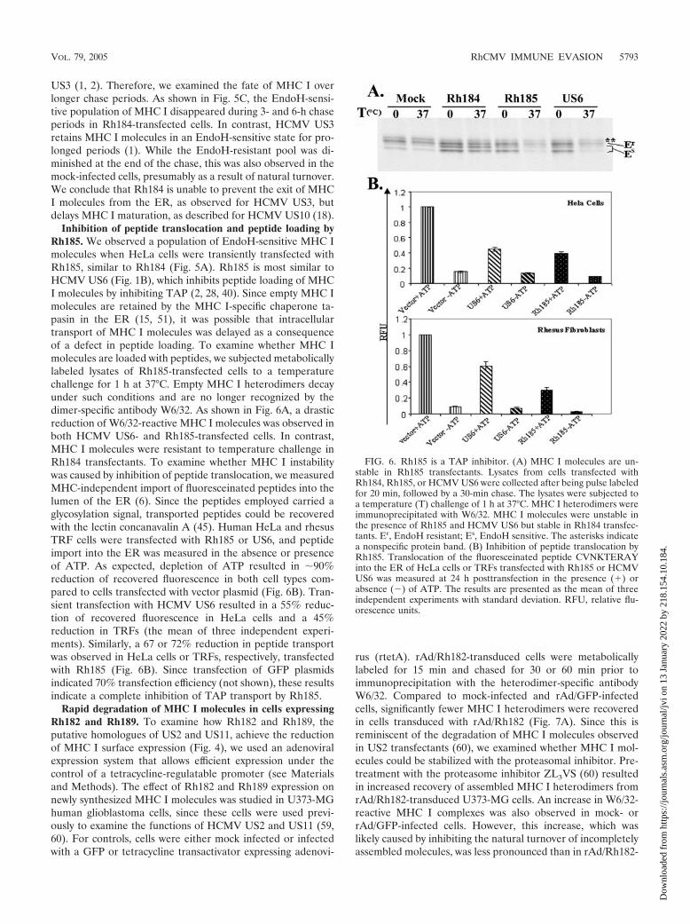
Inhibition of peptide translocation and peptide loading byRh185. We observed a population of EndoH-sensitive MHC Imolecules when HeLa cells were transiently transfected withRh185, similar to Rh184 (Fig. 5A). Rh185 is most similar toHCMV US6 (Fig. 1B), which inhibits peptide loading of MHCI molecules by inhibiting TAP (2, 28, 40). Since empty MHC Imolecules are retained by the MHC I-specific chaperone ta-pasin in the ER (15, 51), it was possible that intracellulartransport of MHC I molecules was delayed as a consequenceof a defect in peptide loading. To examine whether MHC Imolecules are loaded with peptides, we subjected metabolicallylabeled lysates of Rh185-transfected cells to a temperaturechallenge for 1 h at 37°C. Empty MHC I heterodimers decayunder such conditions and are no longer recognized by thedimer-specific antibody W6/32. As shown in Fig. 6A, a drasticreduction of W6/32-reactive MHC I molecules was observed inboth HCMV US6- and Rh185-transfected cells. In contrast,MHC I molecules were resistant to temperature challenge inRh184 transfectants. To examine whether MHC I instabilitywas caused by inhibition of peptide translocation, we measuredMHC-independent import of fluoresceinated peptides into thelumen of the ER (6). Since the peptides employed carried aglycosylation signal, transported peptides could be recoveredwith the lectin concanavalin A (45). Human HeLa and rhesusTRF cells were transfected with Rh185 or US6, and peptideimport into the ER was measured in the absence or presenceof ATP. As expected, depletion of ATP resulted in �90%reduction of recovered fluorescence in both cell types com-pared to cells transfected with vector plasmid (Fig. 6B). Tran-sient transfection with HCMV US6 resulted in a 55% reduc-tion of recovered fluorescence in HeLa cells and a 45%reduction in TRFs (the mean of three independent experi-ments). Similarly, a 67 or 72% reduction in peptide transportwas observed in HeLa cells or TRFs, respectively, transfectedwith Rh185 (Fig. 6B). Since transfection of GFP plasmidsindicated 70% transfection efficiency (not shown), these resultsindicate a complete inhibition of TAP transport by Rh185.
Rapid degradation of MHC I molecules in cells expressingRh182 and Rh189. To examine how Rh182 and Rh189, theputative homologues of US2 and US11, achieve the reductionof MHC I surface expression (Fig. 4), we used an adenoviralexpression system that allows efficient expression under thecontrol of a tetracycline-regulatable promoter (see Materialsand Methods). The effect of Rh182 and Rh189 expression onnewly synthesized MHC I molecules was studied in U373-MGhuman glioblastoma cells, since these cells were used previ-ously to examine the functions of HCMV US2 and US11 (59,60). For controls, cells were either mock infected or infectedwith a GFP or tetracycline transactivator expressing adenovi-
rus (rtetA). rAd/Rh182-transduced cells were metabolicallylabeled for 15 min and chased for 30 or 60 min prior toimmunoprecipitation with the heterodimer-specific antibodyW6/32. Compared to mock-infected and rAd/GFP-infectedcells, significantly fewer MHC I heterodimers were recoveredin cells transduced with rAd/Rh182 (Fig. 7A). Since this isreminiscent of the degradation of MHC I molecules observedin US2 transfectants (60), we examined whether MHC I mol-ecules could be stabilized with the proteasomal inhibitor. Pre-treatment with the proteasome inhibitor ZL3VS (60) resultedin increased recovery of assembled MHC I heterodimers fromrAd/Rh182-transduced U373-MG cells. An increase in W6/32-reactive MHC I complexes was also observed in mock- orrAd/GFP-infected cells. However, this increase, which waslikely caused by inhibiting the natural turnover of incompletelyassembled molecules, was less pronounced than in rAd/Rh182-
FIG. 6. Rh185 is a TAP inhibitor. (A) MHC I molecules are un-stable in Rh185 transfectants. Lysates from cells transfected withRh184, Rh185, or HCMV US6 were collected after being pulse labeledfor 20 min, followed by a 30-min chase. The lysates were subjected toa temperature (T) challenge of 1 h at 37°C. MHC I heterodimers wereimmunoprecipitated with W6/32. MHC I molecules were unstable inthe presence of Rh185 and HCMV US6 but stable in Rh184 transfec-tants. Er, EndoH resistant; Es, EndoH sensitive. The asterisks indicatea nonspecific protein band. (B) Inhibition of peptide translocation byRh185. Translocation of the fluoresceinated peptide CVNKTERAYinto the ER of HeLa cells or TRFs transfected with Rh185 or HCMVUS6 was measured at 24 h posttransfection in the presence (�) orabsence () of ATP. The results are presented as the mean of threeindependent experiments with standard deviation. RFU, relative flu-orescence units.
VOL. 79, 2005 RhCMV IMMUNE EVASION 5793
Dow
nloa
ded
from
http
s://j
ourn
als.
asm
.org
/jour
nal/j
vi o
n 13
Jan
uary
202
2 by
218
.154
.10.
184.

infected cells. To examine whether proteasome inhibitors sta-bilized a degradation intermediate that is not recoverable withthe heterodimer-specific W6/32 antibody, we used the antibodyHC-10, which specifically recognizes free MHC I heavy chains(50). HC-10-reactive heavy chains were immunoprecipitatedfrom lysates that had been precleared with W6/32. In theabsence of proteasome inhibitors, HC-10-reactive MHC I mol-ecules of control transfectants decreased during the chase due
to heterodimer formation. In Rh182-transduced cells, how-ever, very little HC-10-reactive MHC I was observed duringthe pulse and almost no free heavy chain was present duringthe chase, consistent with efficient degradation mediated byRh182. In the presence of proteasome inhibitors, we observeda stabilization of EndoH-sensitive free heavy chains in thecontrol cells. As discussed above, this is most likely due to theinhibition of natural protein turnover. However, a much more
FIG. 7. Degradation of nascent MHC I by Rh182 and Rh189. (A) Degradation of MHC I by Rh182. U373 cells were transduced withrAd/Rh182 (MOI, 100) plus rtetA (MOI, 20) or rAd/GFP (MOI, 120)for 18 h prior to metabolic labeling with [35S]Met for 15 min and chasingfor the times indicated either in the absence () or the presence (�) of 25 �M proteasomal inhibitor ZL3VS (7). Newly assembled MHC Iheterodimers were immunoprecipitated (IP) with the antibody W6/32. Remaining free heavy chains were precipitated with the monoclonalantibody HC-10 as shown. The maturation of MHC-I was assessed by EndoH digestion where indicated prior to electrophoretic separation. Thegel containing rAd/Rh182 samples was exposed for twice as long as the mock and rAd/GFP samples. Note the appearance of a deglycosylatedHC-reactive species in the presence of proteasome inhibitor (-CHO). The asterisks indicate a nonspecific band that comigrates with EndoH-resistant (Er) MHC I in some instances. EndoH sensitivity is labeled as in Fig. 5. (B) Degradation of MHC I molecules by Rh189. U373MG cellswere transduced with rAd/US11 and rAd/Rh189 (each at an MOI of 100), together with rtetA (MOI, 20). Control cells were either mock treatedor infected with rtetA (MOI, 120). Labeling conditions were as for panel A, except that the cells were pretreated for 4 h with (�) 10 �M MG132prior to starvation and pulse-chase labeling. Both free and assembled MHC I molecules were immunoprecipitated with the polyclonal antiserumK455. The cells were treated with EndoH as indicated, except for lane 8 of the mock control, which was mistakenly not EndoH treated.
5794 PANDE ET AL. J. VIROL.
Dow
nloa
ded
from
http
s://j
ourn
als.
asm
.org
/jour
nal/j
vi o
n 13
Jan
uary
202
2 by
218
.154
.10.
184.
pronounced stabilization was observed in rAd/Rh182-infectedcells. Importantly, a low-molecular-weight species of MHC Ithat comigrated with EndoH-sensitive heavy chains was stabi-lized during the chase period. This species might correspond toa deglycosylated intermediate of MHC I degradation describedpreviously for HCMV US2- and US11-transfected cells (59,60). Similar results were obtained with rhesus TRFs (data notshown). The increased stabilization of free heavy chains com-pared to that of assembled heterodimers could indicate thatRh182 predominantly attacks MHC I molecules that have notyet assembled with �2m. Alternatively, MHC I heterodimersare disassembled during the degradation process mediated byRh182. The occurrence of a deglycosylated intermediate alsoindicates that Rh182 reverse translocates the heavy chains intothe cytosol, since the N-glycanase activity required for degra-dation resides in the cytosol (31). Taken together, these dataindicate that, similar to HCMV US2, Rh182 destabilizes MHCI heavy chains for proteasomal destruction in the cytosol.
In a similar series of experiments, we compared the fates ofMHC I molecules in U373-MG cells transduced with rAd/Rh189 or rAd/US11 (kindly provided by D. Johnson). The cellswere metabolically labeled as described above, but MHC Imolecules were immunoprecipitated with the antiserum K455,which recognizes both free and assembled MHC I heavy andlight chains. As expected, the recovery of MHC I was reducedin U373-MG cells transduced with rAd/US11 compared to themock-transduced or control adenovirus-transduced cells, par-ticularly during the chase period (Fig. 7B). Heavy chains re-mained EndoH sensitive in US11-expressing cells prior to deg-radation, consistent with ER degradation of newly synthesizedMHC I molecules (59), whereas they acquired EndoH resis-tance in mock-infected and control adenovirus-infected cells.Similar to US11, there was an overall reduction of MHC Irecovery from rAd/Rh189-infected cells, particularly duringthe chase period, consistent with degradation of MHC I. Inaddition, MHC I also remained increasingly EndoH sensitivein Rh89-expressing cells, suggesting that Rh189 retained MHCI molecules prior to mediating their degradation. Importantly,increased recovery of MHC I molecules was observed in thepresence of the proteasomal inhibitor MG132 in both rAd/US11- and rAd/Rh189-infected cells. Unlike Rh182, however,we did not observe deglycosylated HC degradation intermedi-ates in the presence of proteasome inhibitors in cells trans-duced with either HCMV US11 or Rh189. Instead, proteaso-mal inhibition stabilized the EndoH-sensitive form of HCs. Asdiscussed above, proteasome inhibition also prevented the nat-ural turnover of MHC I in control cells, resulting in an in-creased EndoH-sensitive population during the chase. Thus, aportion of the EndoH-sensitive HC population recovered fromUS11- and Rh189-expressing cells in the presence of protea-some inhibitors could be a consequence of inhibiting normalMHC turnover. However, the difference between the totalamounts of HC recovered in the presence of proteasome in-hibition and in its absence was much more pronounced inRh189- and US11-infected cells than in control cells. The in-creased HC recovery thus suggests that the US11- and Rh189-mediated HC degradation was prevented or decreased by pro-teasome inhibitors. Therefore, we conclude that Rh189 actssimilarly to US11 by mediating the proteasomal degradation ofMHC I molecules.
DISCUSSION
The �-herpesviruses are thought to have emerged prior tomammalian radiation, and therefore, viral evolution has ac-companied mammalian speciation, so that CMV phylogenygenerally parallels species phylogeny (44). Thus, the differ-ences between the primary structure sequences in US6 familymembers of human and rhesus CMVs have accumulated overa period of 14 million years that separates humans from OldWorld nonhuman primates (23). During this time, consider-able divergence has occurred in the corresponding genes of thetwo virus species, resulting in low primary structure homology(Fig. 1). In part, this divergence is probably a coadaptation tothe significant divergence that occurred in the rhesus MHCcompared to the human MHC (17). In addition, mutationsmight have occurred as a result of random drift. Nevertheless,our data suggest that the functions of the major viral modula-tors of MHC I antigen presentation identified in HCMV areconserved in RhCMV. For US2, US3, US6, and US11, weobserved that each of the respective positional and sequencehomologues interferes with MHC I assembly or transport in amanner similar to that of its HCMV homologue. Differencesbetween HCMV and RhCMV were observed for Rh184, whichdid not reduce the steady-state levels of MHC I as efficiently asits HCMV counterpart. Interestingly, all RhCMV ORFs re-tained the ability to interfere with the maturation of humanMHC I molecules, suggesting that they recognize conservedfeatures within MHC molecules or within essential moleculesof the peptide-loading machinery, such as TAP and tapasin.We conclude that Rh182, Rh184, Rh185, and Rh189 are or-thologous to US2, US3, US6, and US11, respectively. There-fore, we suggest that these genes be referred to as RhUS2,RhUS3, RhUS6, and RhUS11. The relationship of Rh186 andRh187 to the remaining US6 family members of HCMV hasnot been firmly established. US8 was shown to bind to MHC Iand to be partially located in endosomes (55). However,Rh186, which showed the closest homology to US8, seemed tobe an ER-resident glycoprotein. Rh187 became partially En-doH resistant but did not seem to be located in the endosomalcompartment. Neither Rh186 nor Rh187 interfered with MHCI maturation.
The observed functional conservation despite considerablesequence divergence might help to delineate functionally im-portant residues in the orthologous sequences. Alignmentsbetween each pair of orthologues are shown in Fig. 8. ForHCMV US2, it was previously shown that the lumenal domainis sufficient to bind to MHC I (12, 21). However, the cytosolicdomain and the transmembrane domain were shown to beabsolutely necessary for US2 to mediate MHC I degradation(12, 19). A function of the cytosolic tail is also supported forRh182, since carboxy-terminal tagging rendered the moleculenonfunctional (Fig. 4). Interestingly, replacement of the US3carboxy-terminal domain with that of US2 transfers the abilityto mediate MHC I degradation to US3, suggesting that thelumenal domain mediates binding whereas the carboxy-termi-nal tail mediates degradation (13). Therefore, it is expectedthat very few residues in the carboxy terminus (including themembrane-proximal domain on the lumenal side and thetransmembrane domain, as well as the cytosolic tail) of Rh182are conserved compared to HCMV US2 (Fig. 8A). This dif-
VOL. 79, 2005 RhCMV IMMUNE EVASION 5795
Dow
nloa
ded
from
http
s://j
ourn
als.
asm
.org
/jour
nal/j
vi o
n 13
Jan
uary
202
2 by
218
.154
.10.
184.

ference renders it less likely that this region of US2 interactswith a highly conserved cellular protein, as observed for US11(see below), and is consistent with different mechanisms ofaction in US2 and US11. In contrast, the lumenal domain ofUS2 contains several conserved sequence motifs, includingresidues that were found to be in direct contact with MHC I, aswell as residues that were defined as “core” residues, i.e.,�90% buried in US2 (20). Secondary-structure predictionsfurther suggest that the Ig-like fold of six beta-sheets is alsoconserved between HCMV US2 and RhCMV Rh182. Notably,in HCMV US2, the glycosylation site is located between beta-sheets B and C, which places it well outside the MHC I inter-action domain (20), whereas the glycosylation site is locatedbetween beta-sheets F and G in Rh182, thus overlapping withresidues that were previously mapped to interact with MHC I(Fig. 8A). The consequences of this different placement of theglycosylation site for the interaction of Rh182 with MHC Iremain to be investigated.
The alignments of US3 and US6 to their RhCMV ortho-logues revealed a surprisingly low homology in the Ig-likedomains of these molecules, suggesting that the overall struc-ture rather than sequence motifs is responsible for their func-tion. Interestingly, sequence conservation occurred in regionsof the molecules that are not expected to be important for theirfunction as MHC I modulators. For instance, sequences adja-
cent to and within the signal peptide of Rh184 are conserved(Fig. 8B). Since this sequence is cleaved upon translocation,this conserved sequence is not expected to play a role in theinteraction with MHC I and tapasin. Potentially, the signalpeptide itself could be involved in other immunomodulatorymechanisms, as described for the signal peptide of UL40 (56,57). Also unexpected is the conservation of the carboxy-termi-nal amino acids in Rh185. Seven of the eight amino acids inHCMV US6 are also found in Rh185 (Fig. 8C). However,previous data suggested that the carboxy-terminal tail of US6is dispensable for MHC I downregulation (2). It is possible thatthis sequence motif optimizes US6 function, which would havebeen missed in the previous study, which relied on overexpres-sion and was not performed in the context of viral infection.Given the high conservation of this amino acid stretch, it canbe assumed that the region plays an important, but yet to bedefined, role in the TAP-inhibitory function of US6, or US6could perform an as-yet-unknown function that involves itscytosolic tail.
The highest sequence conservation among the orthologueswas observed for US11. Pairwise alignment clearly indicatesmultiple sequence motifs that are conserved within the lume-nal domain, as well as the transmembrane domain. Among theconserved intramembrane residues is a central glutamine thatis essential for the interaction of US11 with the cellular protein
FIG. 8. Sequence comparisons of orthologous immune modulator pairs of RhCMV and HCMV. Sequences of Rh182 (RhUS2), Rh184(RhUS3), Rh185 (RhUS6), and Rh189 (RhUS11) were aligned with their corresponding orthologue of HCMV using GAP alignment (PAM120)with a gap penalty of 14 and an extension penalty of 2. Signal peptides and transmembrane regions are boxed, identical sequences are white onblack, and conservative changes are white on gray. Glycosylation sites are boldface and underlined. The asterisks indicate HCMV US2 residuesthat interact with MHC I (20). The black arrows indicate experimentally confirmed beta-sheets in US2 (A, B, C, C�, D, E, F, and G) (20). Thegray arrows indicate predicted beta-sheets.
5796 PANDE ET AL. J. VIROL.
Dow
nloa
ded
from
http
s://j
ourn
als.
asm
.org
/jour
nal/j
vi o
n 13
Jan
uary
202
2 by
218
.154
.10.
184.

derlin 1 (41, 61) (Fig. 8C). This cellular protein is conserved inyeast and belongs to an ancient mechanism that disposes ofmisfolded proteins. The high conservation of the cellularmechanism used by US11 might explain its high sequenceconservation. Moreover, the comparably close sequence rela-tionship renders it highly likely that RhUS11 functions simi-larly to HCMV US11, despite our inability to observe a deg-radation intermediate.
Our study suggests that interference with MHC I assemblyand transport by multiple viral proteins is highly conservedbetween RhCMV and HCMV. The functional conservationdespite structural divergence further suggests important struc-tural characteristics of individual members of the US6 familythat can be tested experimentally in the future. Furthermore,the capability of studying RhCMV infections in nonhumanprimates will further enable us to evaluate the importance ofindividual MHC I modulatory mechanisms for viral immuneevasion in vivo.
ACKNOWLEDGMENTS
We are grateful to Hidde Ploegh, Per Peterson, Emmanuel Wiertz,Bert Vogelstein, Mike O’Conner, David Johnson, Nag Hedge, JayNelson, and Scott Wong for providing reagents used in this study. Wealso thank David Johnson for critical reading of the manuscript.
This work was supported by a grant-in-aid from the American HeartAssociation (0255977Z) and Oregon National Primate Center GrantRR00163 (to K.F.) and a grant from the Korean Ministry of Healthand Welfare, 03-PJ1-PG3-21200, 0003 (to K.A.).
REFERENCES
1. Ahn, K., A. Angulo, P. Ghazal, P. A. Peterson, Y. Yang, and K. Fruh. 1996.Human cytomegalovirus inhibits antigen presentation by a sequential mul-tistep process. Proc. Natl. Acad. Sci. USA 93:10990–10995.
2. Ahn, K., A. Gruhler, B. Galocha, T. R. Jones, E. J. Wiertz, H. L. Ploegh, P. A.Peterson, Y. Yang, and K. Fruh. 1997. The ER-luminal domain of theHCMV glycoprotein US6 inhibits peptide translocation by TAP. Immunity6:613–621.
3. Asher, D. M., C. J. Gibbs, Jr., D. J. Lang, D. C. Gajdusek, and R. M.Chanock. 1974. Persistent shedding of cytomegalovirus in the urine ofhealthy Rhesus monkeys. Proc. Soc. Exp. Biol. Med. 145:794–801.
4. Barel, M. T., M. Ressing, N. Pizzato, D. van Leeuwen, P. Le Bouteiller, F.Lenfant, and E. J. Wiertz. 2003. Human cytomegalovirus-encoded US2 dif-ferentially affects surface expression of MHC class I locus products andtargets membrane-bound, but not soluble HLA-G1 for degradation. J. Im-munol. 171:6757–6765.
5. Bitmansour, A. D., S. L. Waldrop, C. J. Pitcher, E. Khatamzas, F. Kern,V. C. Maino, and L. J. Picker. 2001. Clonotypic structure of the humanCD4� memory T cell response to cytomegalovirus. J. Immunol. 167:1151–1163.
6. Blevitt, J. M., K. Fruh, C. Glass, M. R. Jackson, P. A. Peterson, and S.Huang. 1999. A fluorescence-based high throughput screen for the trans-porter associated with antigen processing. J. Biomol. Screen. 4:87–91.
7. Bogyo, M., M. Gaczynska, and H. L. Ploegh. 1997. Proteasome inhibitors andantigen presentation. Biopolymers 43:269–280.
8. Boppana, S. B., L. B. Rivera, K. B. Fowler, M. Mach, and W. J. Britt. 2001.Intrauterine transmission of cytomegalovirus to infants of women with pre-conceptional immunity. N. Engl. J. Med. 344:1366–1371.
9. Britt, W. J., and C. A. Alford. 1996. Cytomegalovirus, p. 2493–2523. In B. N.Fields, D. M. Knipe, and P. M. Howley (ed.), Fields virology, 3rd ed., vol. 2.Lippincott-Raven Publishers, Philadelphia, Pa.
10. Brodsky, F. M., P. Parham, C. J. Barnstable, M. J. Crumpton, and W. F.Bodmer. 1979. Monoclonal antibodies for analysis of the HLA system. Im-munol. Rev. 47:3.
11. Chee, M. S., A. T. Bankier, S. Beck, R. Bohni, C. M. Browne, R. Cerny, T.Horsnell, C. A. Hutchison III, T. Kouzarides, J. A. Martignetti, E. Preddie,S. C. Satchwell, P. Tomlinson, K. M. Weston, and B. G. Barrell. 1990.Analysis of the protein-coding content of the sequence of human cytomeg-alovirus strain AD169, p. 125–171. In J. K. McDougall (ed.), Cytomegalovi-ruses. Springer-Verlag, Berlin, Germany.
12. Chevalier, M. S., G. M. Daniels, and D. C. Johnson. 2002. Binding of humancytomegalovirus US2 to major histocompatibility complex class I and IIproteins is not sufficient for their degradation. J. Virol. 76:8265–8275.
13. Chevalier, M. S., and D. C. Johnson. 2003. Human cytomegalovirus US3
chimeras containing US2 cytosolic residues acquire major histocompatibilityclass I and II protein degradation properties. J. Virol. 77:4731–4738.
14. Christensen, A. C., and S. Henikoff. 1992. Fact and fiction in alignment.Nature 358:271.
15. Cresswell, P., N. Bangia, T. Dick, and G. Diedrich. 1999. The nature of theMHC class I peptide loading complex. Immunol. Rev. 172:21–28.
16. Davison, A. J., A. Dolan, P. Akter, C. Addison, D. J. Dargan, D. J. Alcendor,D. J. McGeoch, and G. S. Hayward. 2003. The human cytomegalovirusgenome revisited: comparison with the chimpanzee cytomegalovirus ge-nome. J. Gen. Virol. 84:17–28.
17. Daza-Vamenta, R., G. Glusman, L. Rowen, B. Guthrie, and D. E. Geraghty.2004. Genetic divergence of the rhesus macaque major histocompatibilitycomplex. Genome Res. 14:1501–1515.
18. Furman, M. H., N. Dey, D. Tortorella, and H. L. Ploegh. 2002. The humancytomegalovirus US10 gene product delays trafficking of major histocompat-ibility complex class I molecules. J. Virol. 76:11753–11756.
19. Furman, M. H., H. L. Ploegh, and D. Tortorella. 2002. Membrane-specific,host-derived factors are required for US2- and US11-mediated degradationof major histocompatibility complex class I molecules. J. Biol. Chem. 277:3258–3267.
20. Gewurz, B. E., R. Gaudet, D. Tortorella, E. W. Wang, H. L. Ploegh, and D. C.Wiley. 2001. Antigen presentation subverted: structure of the human cyto-megalovirus protein US2 bound to the class I molecule HLA-A2. Proc. Natl.Acad. Sci. USA 98:6794–6799.
21. Gewurz, B. E., E. W. Wang, D. Tortorella, D. J. Schust, and H. L. Ploegh.2001. Human cytomegalovirus US2 endoplasmic reticulum-lumenal domaindictates association with major histocompatibility complex class I in a locus-specific manner. J. Virol. 75:5197–5204.
22. Gillespie, G. M., M. R. Wills, V. Appay, C. O’Callaghan, M. Murphy, N.Smith, P. Sissons, S. Rowland-Jones, J. I. Bell, and P. A. Moss. 2000.Functional heterogeneity and high frequencies of cytomegalovirus-specificCD8� T lymphocytes in healthy seropositive donors. J. Virol. 74:8140–8150.
23. Goodman, M., C. A. Porter, J. Czelusniak, S. L. Page, H. Schneider, J.Shoshani, G. Gunnell, and C. P. Groves. 1998. Toward a phylogenetic clas-sification of primates based on DNA evidence complemented by fossil evi-dence. Mol. Phylogenet. Evol. 9:585–598.
24. Gossen, M., and H. Bujard. 1992. Tight control of gene expression in mam-malian cells by tetracycline-responsive promoters. Proc. Natl. Acad. Sci.USA 89:5547–5551.
25. Gruhler, A., P. A. Peterson, and K. Fruh. 2000. Human cytomegalovirusimmediate early glycoprotein US3 retains MHC class I molecules by tran-sient association. Traffic 1:318–325.
26. Hansen, S. G., L. I. Strelow, D. C. Franchi, D. G. Anders, and S. W. Wong.2003. Analysis of the complete DNA sequence of rhesus cytomegalovirus.J. Virol. 77:6620–6636.
27. He, T. C., S. Zhou, L. T. da Costa, J. Yu, K. W. Kinzler, and B. Vogelstein.1998. A simplified system for generating recombinant adenoviruses. Proc.Natl. Acad. Sci. USA 95:2509–2514.
28. Hengel, H., J. O. Koopmann, T. Flohr, W. Muranyi, E. Goulmy, G. J.Hammerling, U. H. Koszinowski, and F. Momburg. 1997. A viral ER-resi-dent glycoprotein inactivates the MHC-encoded peptide transporter. Immu-nity 6:623–632.
29. Henikoff, S., and J. G. Henikoff. 1992. Amino acid substitution matrices fromprotein blocks. Proc. Natl. Acad. Sci. USA 89:10915–10919.
30. Hewitt, E. W., S. S. Gupta, and P. J. Lehner. 2001. The human cytomega-lovirus gene product US6 inhibits ATP binding by TAP. EMBO J. 20:387–396.
31. Hirsch, C., D. Blom, and H. L. Ploegh. 2003. A role for N-glycanase in thecytosolic turnover of glycoproteins. EMBO J. 22:1036–1046.
32. Huber, M. T., R. Tomazin, T. Wisner, J. Boname, and D. C. Johnson. 2002.Human cytomegalovirus US7, US8, US9, and US10 are cytoplasmic glyco-proteins, not found at cell surfaces, and US9 does not mediate cell-to-cellspread. J. Virol. 76:5748–5758.
33. Jones, T. R., L. K. Hanson, L. Sun, J. S. Slater, R. M. Stenberg, and A. E.Campbell. 1995. Multiple independent loci within the human cytomegalovi-rus unique short region down-regulate expression of major histocompatibil-ity complex class I heavy chains. J. Virol. 69:4830–4841.
34. Jones, T. R., E. J. Wiertz, L. Sun, K. N. Fish, J. A. Nelson, and H. L. Ploegh.1996. Human cytomegalovirus US3 impairs transport and maturation ofmajor histocompatibility complex class I heavy chains. Proc. Natl. Acad. Sci.USA 93:11327–11333.
35. Kaur, A., M. D. Daniel, D. Hempel, D. Lee-Parritz, M. S. Hirsch, and R. P.Johnson. 1996. Cytotoxic-T-lymphocyte responses to cytomegalovirus in nor-mal and simian immunodeficiency virus-infected rhesus macaques. J. Virol.70:7725–7733.
36. Kaur, A., C. L. Hale, B. Noren, N. Kassis, M. A. Simon, and R. P. Johnson.2002. Decreased frequency of cytomegalovirus (CMV)-specific CD4� T lym-phocytes in simian immunodeficiency virus-infected rhesus macaques: in-verse relationship with CMV viremia. J. Virol. 76:3646–3658.
37. Kern, F., T. Bunde, N. Faulhaber, F. Kiecker, E. Khatamzas, I. M. Rudaw-ski, A. Pruss, J. W. Gratama, R. Volkmer-Engert, R. Ewert, P. Reinke, H. D.Volk, and L. J. Picker. 2002. Cytomegalovirus (CMV) phosphoprotein 65
VOL. 79, 2005 RhCMV IMMUNE EVASION 5797
Dow
nloa
ded
from
http
s://j
ourn
als.
asm
.org
/jour
nal/j
vi o
n 13
Jan
uary
202
2 by
218
.154
.10.
184.
makes a large contribution to shaping the T cell repertoire in CMV-exposedindividuals. J. Infect. Dis. 185:1709–1716.
38. Kleijnen, M. F., J. B. Huppa, P. Lucin, S. Mukherjee, H. Farrell, A. E.Campbell, U. H. Koszinowski, A. B. Hill, and H. L. Ploegh. 1997. A mousecytomegalovirus glycoprotein, gp34, forms a complex with folded class IMHC molecules in the ER which is not retained but is transported to the cellsurface. EMBO J. 16:685–694.
39. Lee, S., J. Yoon, B. Park, Y. Jun, M. Jin, H. C. Sung, I. H. Kim, S. Kang, E. J.Choi, B. Y. Ahn, and K. Ahn. 2000. Structural and functional dissection ofhuman cytomegalovirus US3 in binding major histocompatibility complexclass I molecules. J. Virol. 74:11262–11269.
40. Lehner, P. J., J. T. Karttunen, G. W. Wilkinson, and P. Cresswell. 1997. Thehuman cytomegalovirus US6 glycoprotein inhibits transporter associatedwith antigen processing-dependent peptide translocation. Proc. Natl. Acad.Sci. USA 94:6904–6909.
41. Lilley, B. N., and H. L. Ploegh. 2004. A membrane protein required fordislocation of misfolded proteins from the ER. Nature 429:834–840.
42. Loenen, W. A., C. A. Bruggeman, and E. J. Wiertz. 2001. Immune evasion byhuman cytomegalovirus: lessons in immunology and cell biology. Semin.Immunol. 13:41–49.
43. London, W. T., A. J. Martinez, S. A. Houff, W. C. Wallen, B. L. Curfman,R. G. Traub, and J. L. Sever. 1986. Experimental congenital disease withsimian cytomegalovirus in rhesus monkeys. Teratology 33:323–331.
44. McGeoch, D. J., S. Cook, A. Dolan, F. E. Jamieson, and E. A. Telford. 1995.Molecular phylogeny and evolutionary timescale for the family of mamma-lian herpesviruses. J. Mol. Biol. 247:443–458.
45. Neefjes, J. J., F. Momburg, and G. J. Hammerling. 1993. Selective andATP-dependent translocation of peptides by the MHC-encoded transporter.Science 261:769–771.
46. Park, B., Y. Kim, J. Shin, S. Lee, K. Cho, K. Fruh, and K. Ahn. 2004. Humancytomegalovirus inhibits tapasin-dependent peptide loading and optimiza-tion of the MHC class I peptide cargo for immune evasion. Immunity20:71–85.
47. Pass, R. F. 2001. Cytomegalovirus, p. 2675–2705. In P. M. Howley, D. M.Knipe, D. E. Griffin, R. A. Lamb, M. A. Martin, B. Roizman, and S. E. Straus(ed.), Fields virology, 4th ed. Lippincott Williams & Wilkins, Philadelphia,Pa.
48. Pitcher, C. J., S. I. Hagen, J. M. Walker, R. Lum, B. L. Mitchell, V. C. Maino,M. K. Axthelm, and L. J. Picker. 2002. Development and homeostasis of Tcell memory in rhesus macaque. J. Immunol. 168:29–43.
49. Rawlinson, W. D., H. E. Farrell, and B. G. Barrell. 1996. Analysis of thecomplete DNA sequence of murine cytomegalovirus. J. Virol. 70:8833–8849.
50. Rein, R. S., G. H. Seemann, J. J. Neefjes, F. M. Hochstenbach, N. J. Stam,and H. L. Ploegh. 1987. Association with beta 2-microglobulin controls theexpression of transfected human class I genes. J. Immunol. 138:1178–1183.
51. Schoenhals, G. J., R. M. Krishna, A. G. Grandea III, T. Spies, P. A. Peterson,Y. Yang, and K. Fruh. 1999. Retention of empty MHC class I molecules byTapasin is essential to reconstitute antigen presentation in invertebrate cells.EMBO J. 18:743–753.
52. Sepkowitz, K. A. 2002. Opportunistic infections in patients with and patientswithout Acquired Immunodeficiency Syndrome. Clin. Infect. Dis. 34:1098–1107.
53. Soderberg-Naucler, C., and V. C. Emery. 2001. Viral infections and theirimpact on chronic renal allograft dysfunction. Transplantation 71:SS24–SS30.
54. Tarantal, A. F., M. S. Salamat, W. J. Britt, P. A. Luciw, A. G. Hendrickx, andP. A. Barry. 1998. Neuropathogenesis induced by rhesus cytomegalovirus infetal rhesus monkeys (Macaca mulatta). J. Infect. Dis. 177:446–450.
55. Tirabassi, R. S., and H. L. Ploegh. 2002. The human cytomegalovirus US8glycoprotein binds to major histocompatibility complex class I products.J. Virol. 76:6832–6835.
56. Tomasec, P., V. M. Braud, C. Rickards, M. B. Powell, B. P. McSharry, S.Gadola, V. Cerundolo, L. K. Borysiewicz, A. J. McMichael, and G. W.Wilkinson. 2000. Surface expression of HLA-E, an inhibitor of natural killercells, enhanced by human cytomegalovirus gpUL40. Science 287:1031.
57. Ulbrecht, M., S. Martinozzi, M. Grzeschik, H. Hengel, J. W. Ellwart, M. Pla,and E. H. Weiss. 2000. Cutting edge: the human cytomegalovirus UL40 geneproduct contains a ligand for HLA-E and prevents NK cell-mediated lysis.J. Immunol. 164:5019–5022.
58. Vogel, P., B. J. Weigler, H. Kerr, A. G. Hendrickx, and P. A. Barry. 1994.Seroepidemiologic studies of cytomegalovirus infection in a breeding popu-lation of rhesus macaques. Lab. Anim. Sci. 44:25–30.
59. Wiertz, E. J., T. R. Jones, L. Sun, M. Bogyo, H. J. Geuze, and H. L. Ploegh.1996. The human cytomegalovirus US11 gene product dislocates MHC classI heavy chains from the endoplasmic reticulum to the cytosol. Cell 84:769–779.
60. Wiertz, E. J., D. Tortorella, M. Bogyo, J. Yu, W. Mothes, T. R. Jones, T. A.Rapoport, and H. L. Ploegh. 1996. Sec61-mediated transfer of a membraneprotein from the endoplasmic reticulum to the proteasome for destruction.Nature 384:432–438.
61. Ye, Y., Y. Shibata, C. Yun, D. Ron, and T. A. Rapoport. 2004. A membraneprotein complex mediates retro-translocation from the ER lumen into thecytosol. Nature 429:841–847.
5798 PANDE ET AL. J. VIROL.
Dow
nloa
ded
from
http
s://j
ourn
als.
asm
.org
/jour
nal/j
vi o
n 13
Jan
uary
202
2 by
218
.154
.10.
184.

![Mandalas All Around Us2[1]](https://static.fdocuments.net/doc/165x107/55542be2b4c905987e8b4ff6/mandalas-all-around-us21.jpg)