Revision of the genus Spermophora Hentz in Southeast Asia and on the Pacific Islands, with
54
Revision of the genus Spermophora Hentz in Southeast Asia and on the Pacific Islands, with descriptions of three new genera (Araneae: Pholcidae) B.A. Huber Huber, B.A. Revision of the genus Spermophora Hentz in Southeast Asia and on the Pacific Islands, with descriptions of three new genera (Araneae: Pholcidae). Zool. Med. Leiden 79-2 (4), 22.vii.2005: 61-114, figs 1-172.— ISSN 0024-0672. Bernhard A. Huber. Alexander Koenig Zoological Research Museum, Adenauerallee 160, 53113 Bonn, Germany (e-mail: [email protected]). Key words: Araneae; Pholcidae; Spermophora; revision; taxonomy; Southeast Asia; Pacific. The main aim of the present paper is to delimit ‘true’ Spermophora, i.e. the group of species most closely related to the type species S. senoculata (Dugès). Apart from the type species, only three previously described species are included in this core group (S. estebani Simon, S. paluma Huber, S. yao Huber), together with nine newly described species: S. kerinci, S. tumbang, S. dumoga, S. maros, S. deelemanae, S. palau, S. kaindi, S. luzonica, and S. sumbawa. Except for the Holarctic and anthropophilic type species, all species have limited distributions in Southeast Asia, northeastern Australia, and the Pacific Islands, where they inhabit the leaf litter layer of tropical forests as well as caves. A tight correlation is documented in Spermophora between the male cheliceral apophyses (distance between the tips) and the pockets on the female external genitalia. In addition, three new Southeast Asian genera are described that appear similar to Spermophora but do not share the synapomorphies of the genus: Aetana n. gen. with three new species from the Philippines (A. omayan), Borneo (A. kinabalu), and Fiji (A. fiji); Savarna n. gen., with one new species from southern Thailand (S. thaleban) and two species from Sumatra and Malaysia trans- ferred from Spermophora [Savarna tessellata (Simon) n. comb., and S. baso (Roewer) n. comb.]; and Khorata n. gen., with four new species from Thailand (K. bangkok, K. schwendingeri) and Laos (K. khammouan, K. jaegeri). Introduction The genus Spermophora comprises mostly tiny, leaf-litter dwelling tropical spiders and presents one of the most intricate problems in pholcid taxonomy. Originally created for a species that was unusual for its small size and only six eyes (Hentz 1841), the ge- nus was soon considered to be ‘inhomogeneous’ (“n’est pas très homogène”, Simon 1893), and has since become a waste basket of many different taxa worldwide. Recent revisions have somewhat alleviated the situation, mainly by transferring a large number of species to other genera. For example, it is clear now that except for the synanthropic S. senoculata (and “S.” maculata which is incertae sedis), Spermophora is not present in the New World (Huber 2000). All New World species have been transferred to Metagonia and Anopsicus. Furthermore, all Mediterranean and Canary Island species have been transferred to Spermophorides (Wunderlich 1992; Senglet 2001). In Africa and Madagas- car, the situation is more difficult. Three recent revisions (Huber 2003a, b, c) have trans- ferred several species to Paramicromerys and three newly described genera (Zatavua, Buitinga, Quamtana), but several previously described and new species were tentatively kept in or assigned to Spermophora. This was explicitly considered tentative because it was justified by similarity rather than by synapomorphies. The present paper is prima-
Transcript of Revision of the genus Spermophora Hentz in Southeast Asia and on the Pacific Islands, with

Revision of the genus Spermophora Hentz in Southeast Asia and on the Pacific Islands, with descriptions of three new genera
(Araneae: Pholcidae)
B.A. Huber
Huber, B.A. Revision of the genus Spermophora Hentz in Southeast Asia and on the Pacific Islands, with descriptions of three new genera (Araneae: Pholcidae).Zool. Med. Leiden 79-2 (4), 22.vii.2005: 61-114, figs 1-172.— ISSN 0024-0672.Bernhard A. Huber. Alexander Koenig Zoological Research Museum, Adenauerallee 160, 53113 Bonn, Germany (e-mail: [email protected]).
Key words: Araneae; Pholcidae; Spermophora; revision; taxonomy; Southeast Asia; Pacific.The main aim of the present paper is to delimit ‘true’ Spermophora, i.e. the group of species most closely related to the type species S. senoculata (Dugès). Apart from the type species, only three previously described species are included in this core group (S. estebani Simon, S. paluma Huber, S. yao Huber), together with nine newly described species: S. kerinci, S. tumbang, S. dumoga, S. maros, S. deelemanae, S. palau, S. kaindi, S. luzonica, and S. sumbawa. Except for the Holarctic and anthropophilic type species, all species have limited distributions in Southeast Asia, northeastern Australia, and the Pacific Islands, where they inhabit the leaf litter layer of tropical forests as well as caves. A tight correlation is documented in Spermophora between the male cheliceral apophyses (distance between the tips) and the pockets on the female external genitalia. In addition, three new Southeast Asian genera are described that appear similar to Spermophora but do not share the synapomorphies of the genus: Aetana n. gen. with three new species from the Philippines (A. omayan), Borneo (A. kinabalu), and Fiji (A. fiji); Savarna n. gen., with one new species from southern Thailand (S. thaleban) and two species from Sumatra and Malaysia trans-ferred from Spermophora [Savarna tessellata (Simon) n. comb., and S. baso (Roewer) n. comb.]; and Khorata n. gen., with four new species from Thailand (K. bangkok, K. schwendingeri) and Laos (K. khammouan, K. jaegeri).
Introduction
The genus Spermophora comprises mostly tiny, leaf-litter dwelling tropical spiders and presents one of the most intricate problems in pholcid taxonomy. Originally created for a species that was unusual for its small size and only six eyes (Hentz 1841), the ge-nus was soon considered to be ‘inhomogeneous’ (“n’est pas très homogène”, Simon 1893), and has since become a waste basket of many different taxa worldwide. Recent revisions have somewhat alleviated the situation, mainly by transferring a large number of species to other genera. For example, it is clear now that except for the synanthropic S. senoculata (and “S.” maculata which is incertae sedis), Spermophora is not present in the New World (Huber 2000). All New World species have been transferred to Metagonia and Anopsicus. Furthermore, all Mediterranean and Canary Island species have been transferred to Spermophorides (Wunderlich 1992; Senglet 2001). In Africa and Madagas-car, the situation is more difficult. Three recent revisions (Huber 2003a, b, c) have trans-ferred several species to Paramicromerys and three newly described genera (Zatavua, Buitinga, Quamtana), but several previously described and new species were tentatively kept in or assigned to Spermophora. This was explicitly considered tentative because it was justified by similarity rather than by synapomorphies. The present paper is prima-

62 Huber. Revision of Spermophora. Zool. Med. Leiden 79 (2005)
rily an attempt to delimit ‘true’ Spermophora. It describes a number of East Asian and Pacific Island species that share putative synapomorphies with the type species S. sen-oculata. Together with only three further previously described species this taxon is here considered the core group of Spermophora. African representatives are kept in the genus for want of a better hypothesis, but future studies will probably remove most or all African species. A number of Asian species currently assigned to Spermophora are not treated herein because they will be removed to Belisana Thorell in a forthcoming revi-sion (Huber in press). A secondary aim of this paper is the description of three new East Asian genera that are similar to Spermophora but do not share the synapomorphies of Spermophora. Each of them is characterized by its own distinctive synapomorphies, justifying the creation of new genera.
Materials and Methods
Pholcids resembling Spermophora and potential relatives (Belisana, Spermophorides, Paramicromerys) were borrowed from more than 40 institutions and individuals world-wide, and the list below covers only those with material actually used in the present paper.
BPBM Bernice P. Bishop Museum, Honolulu, HawaiiCAS California Academy of Sciences, San FranciscoIRSB Institut royal des Sciences naturelles de Belgique, BrusselMHNG Muséum d’Histoire Naturelle, GenèveMNHN Muséum National d’Histoire Naturelle, ParisQMB Queensland Museum, BrisbaneRMNH National Museum of Natural History Naturalis, LeidenSMF Senckenbergmuseum Frankfurt
Methods and terminology are as in Huber (2000). Measurements are in mm (+/- 0.02 mm if two decimals are given) unless otherwise noted. Eye measurements are +/- 5 µm. Drawings were done with a camera lucida on a Nikon Labophot-2 compound micro-scope. Photos were made with a Nikon Coolpix 950 digital camera (1600x1200 pixels) mounted on a Nikon SMZ-U dissecting microscope. For SEM photos, specimens were cleaned ultrasonically, dried in HMDS (Brown 1993), and photographed with a Hitachi S-2460 scanning electron microscope. Material of new species was divided into holo-type and other material, the latter being expressly excluded from the type series.
Genus Spermophora Hentz, 1841
Spermophora Hentz, 1841: 116. Simon 1893: 471. Huber 2001: 124-126; 2003a: 304; 2003b: 593-594; 2003c: 515-516.
Notes.— The genus Spermophora currently (including the present paper) contains 44 nominal species (Appendix 1). Below I will treat only those species that appear most closely related to the type species S. senoculata, i.e. the core group of the genus. Only three previously described species apart from the type species fall into this group, two

Huber. Revision of Spermophora. Zool. Med. Leiden 79 (2005) 63
of which were described recently (S. yao, S. paluma: Huber 2001) and need not be in-cluded, and one of which is treated below (S. estebani Simon). The type species has been redescribed several times, including ultrastructure and genital mechanics (Huber 2000, 2002; Senglet 2001), and will therefore not be included. A number of Malagasy and mainland African species are provisionally kept in Spermophora (Appendix 1, see also Monophyly below). All of them have been included in recent revisions (Huber 2003a, b, c), and will not be treated herein. Finally, eight East Asian species will be transferred to Belisana Thorell in a upcoming revision (Huber in press), and six further species are incertae sedis (Appendix 1). Some species of the latter group were discussed recently (Huber 2000, 2001). Diagnosis.— Representatives of the core group are small (1.1-1.5 mm total body length), six-eyed spiders with relatively long legs. They share with some other repre-sentatives of the genus posterior pockets (paired or unpaired) on the female abdomen, and/or a ventral flap on the procursus. They are distinguished from these congeners by a serrated apophysis on the bulb (missing only in S. sumbawa, S. paluma). Description.— Total length usually 1.1-1.5 mm. Carapace without dorsal indenta-tion. Six eyes in two triads on barely elevated ocular area. Triads usually wide apart (distance PME-PME ~150-350 µm; figs 1-11), in S. sumbawa closer together (90 µm; fig. 12); distance PME-ALE small (~10-20 µm). Male sternum and clypeus unmodified. Male chelicerae with proximal rounded projections laterally and pair of apophyses dis-tally; distal apophyses usually without modified hairs, only S. senoculata with modified hairs (figs 24-25 in Huber 2000), chelicerae never with stridulatory ridges. Male palpal coxa unmodified, trochanter with short retrolateral apophysis (usually in rather ventral position), procursus complex, usually with ventral serrated flap (f in figs 22, 27; present but not serrated in S. estebani, S. luzonica, and S. sumbawa), bulb with tubular membra-nous embolus and sclerotized projection consisting of serrated and hooked elements (e.g., fig. 27). Tarsal organ capsulate (e.g., figs 33, 66). Legs long (usually ~4-5 x body length, 8.2 in the troglomorphic S. maros; tibia 1 L/d: 22-36, 55 in S. maros); leg formula 1423. Legs without spines, usually without curved hairs (present in S. luzonica: meta-tarsi 2), usually without vertical hairs (present on tibiae in S. kaindi and S. sumbawa); retrolateral trichobothrium of tibia 1 at ~10-15%; prolateral trichobothrium missing on tibia 1, present on others; tarsus 1 with about 10-15 pseudosegments. Abdomen globu-lar, without markings or with more or less distinct dark spots. Male gonopore with four epiandrous spigots (figs 35, 65, 96, 97), ALS with widened, pointed, and several cylin-drically shaped spigots (figs 34, 36, 42, 64, 67, 77, 99, 100). Sexual dimorphism very slight, females with slightly shorter legs, unmodified chelicerae. Epigynum usually a simple plate with pair of pockets (e.g., figs 25, 30, 38, 41) corresponding to male cheliceral apophyses (fig. 168), only in type species and S. sumbawa with a knob-shaped median structure (figs 9, 11 in Huber 2002; figs 101, 102). Behind epigynum with paired (type species) or unpaired pocket (e.g., figs 25, 30, 38, 41), presumably providing an anchor for the hook of the male bulbal apophysis (cf. Huber 2002 on S. senoculata). Monophyly.— Four representatives of the core group of Spermophora were included in previous cladistic analyses (S. senoculata, S. yao, S. kerinci [as S. sp. 1], S. sumbawa [as S. sp. 2]; Huber 2003a, b, c). The monophyly of this taxon (node 11 in Huber 2003c: appendix 2) was supported by pocket(s) behind the epigynum. Paired and unpaired

64 Huber. Revision of Spermophora. Zool. Med. Leiden 79 (2005)
pockets were scored as homologous, based on the assumption (Huber 2002) that the paired pockets of S. senoculata resulted from a split of an unpaired socket due to the position far back on the abdomen where the spinnerets precluded a median position. A further potential synapomorphy might be the serrated apophysis on the genital bulb. Cladistic analysis suggested this to be a synapomorphy of a species group within the core group (i.e. excepting S. sumbawa), but alternatively, the serration may have been lost in S. sumbawa (and in S. paluma, which was not included in the analysis). The closest relatives of this core group might be some South African species (repre-sented by S. peninsulae and S. schoemanae in the cladistic analysis of Huber 2003c), with which they share a ventral flap on the procursus. However, this flap is never serrated in South African taxa, and the homology is not clear. Beyond that, all other Spermophora species are either assigned tentatively to the genus or have not yet been removed (see Appendix 1). Cladistic analysis has not sup-ported sister relationships of any of these species or species groups with the core group above or with the core group plus South African Spermophora. However, striking simi-larities do exist in some cases, as for example posterior pockets on the female abdomen in some East African and Malagasy taxa (Huber 2003a, b), and ventral flaps on the pro-cursus in the East African S. berlandi and S. minotaura (Huber 2003b). The two Comoran species (S. lambilloni, S. jocquei) share a previously unnoticed synapomorphy with the Malagasy genus Paramicromerys (a strongly modified male palpal trochanter with pro-lateral and retrolateral apophyses), and might better be included in a slightly expanded concept of Paramicromerys. Generic Relationships.— The position of Spermophora in the pholcines sensu Hu-ber (2000) is strongly supported by the lateral cheliceral apophyses (Huber 2003b). Within this clade, however, relationships continue to be poorly resolved, and need further analysis. A hinged process on the procursus distinguishes Spermophora from Pholcus and related taxa, and may unite it with genera such as Buitinga, Paramicro-merys, Micromerys, Spermophorides (all Old World) and Metagonia (New World) (Huber 2003c). However, the hinged process is very distinct in some species but barely recog-nizable in others, so the homology and scoring is not unambiguous. Finally, a dorsal (rather than prolateral) attachment of the bulb to the cymbium seems to unite Spermo-phora with Metagonia, but this position sometimes results as an artifact of preservation in other taxa too. Natural History.— Most or all species except S. senoculata seem to have been col-lected from leaf litter in primary (rarely secondary) tropical forests. The type species S. senoculata is synanthropic in many parts of the world, but seems to have invaded native habitats in the USA and southern Europe (Kaston 1948; Brignoli 1971; Senglet 2001). Some data on the reproductive biology of S. senoculata have been published recently (Senglet 2001; Huber 2002). Distribution.— The core group as delimited here is geographically restricted to Southeast Asia, northern Australia, and some Pacific Islands (fig. 169). The type species makes an exception, being synanthropic and Holarctic. Interestingly, Antoine Senglet (pers. comm., 2003) has collected specimens in Iran that are extremely similar to S. senocu-lata yet appear to be a different species. If this interpretation proves correct, then we may expect the closest relatives of S. senoculata to occur in a very poorly collected area somewhere between Turkey and India rather than in Southeast Asia.

Huber. Revision of Spermophora. Zool. Med. Leiden 79 (2005) 65
Spermophora kerinci spec. nov.(figs 1, 2, 14, 22-26, 32-38, 47)
Spermophora sp. 1: Huber 2003a, b, c.
Type material.— Male holotype from Kerinci National Park (~101°18’E, 2°00’S), Sumatra, Indonesia; 800 m a.s.l., leaf litter near river, 21-30.vii.1988 (S. Djojosudharmo), in RMNH.Non-Type Material.— Indonesia: Sumatra: Kerinci N. P.: 2~15, together with holotype (RMNH); Bali: disturbed rainforest above Negara (114°37’E, 8°22’S), 21-22.vi.1997 (C. L. Deeleman-Reinhold), 12 (RMNH).
Diagnosis.— Distinguished from other representatives of the core group by the shape of the distal elements of the procursus, by the shape of the serrated apophysis on the bulb, by the palpal femur apophysis (indistinct), and by the distances between male cheliceral apophyses and epigynal pockets (figs 22-26). Description.— Male (holotype). Total length 1.12 (1.26 with clypeus), carapace width 0.58. Leg 1: 5.84 (1.44 + 0.24 + 1.56 + 1.88 + 0.72), tibia 2: 0.96, tibia 3: 0.80, tibia 4: 1.28; tibia 1 L/d: 26. Habitus as in figures 1 and 2. Carapace ochre-yellow with black margins and dark bands behind eye triads (fig. 1); sternum ochre yellow with small dark speckles. Legs pale ochre-yellow, without dark rings. Abdomen grey with large slightly darker spots, ventrally without pattern. Ocular area slightly elevated, thoracic furrow absent; distance PME-PME 180 µm; diameter PME 50 µm; distance PME-ALE ~15 µm. Clypeus unmodified. Sternum wider than long (0.40/0.36). Chelicerae as in fig. 24, with pair of long apophyses, tips 365 µm apart, without modified hairs. Palps as in figures 22 and 23; trochanter with simple retrolateral apophysis, femur with small pro-jection ventrally, procursus with serrated ventral flap and distinctive distal elements (figs 22, 23, 47); bulb with distinctively shaped serrated apophysis, hooked sclerite, and membranous embolus. Palpal tarsal organ capsulate (fig. 33). Retrolateral tricho-bothrium of tibia 1 at 10%; legs without spines, curved hairs, and vertical hairs; tarsus 1 with ~10 pseudosegments, only distally ~6 quite distinct. Gonopore with four epian-drous spigots (fig. 35). ALS with several spigots in addition to basic set of two (fig. 34). Variation. Tibia 1 in other males: 1.36, 1.44, 1.48. Female. In general very similar to male. Frontal view as in fig. 32. Tibia 1 in 16 fe-males: 1.18-1.30 (mean: 1.24). Palpal tarsal organ as in fig. 37. Epigynum wide (figs 14, 25, 38), with pair of lateral pockets 335 µm apart, with median posterior pocket. Dorsal view as in fig. 26. ALS as in male (fig. 36). Etymology.—The specific name is a noun in apposition, taken from the type local-ity. Distribution.—Known from Sumatra and Bali (fig. 169).
Spermophora tumbang spec. nov.(figs 3, 4, 15, 27-31, 39-42, 48)
Type material.— Male holotype from Tumbang Tabai (2º02’S, 113º35’E), Kalimantan Tengah (Central Kalimantan), Indonesia; primary marshy forest, 2-13.ix.1985 (S. Djojosudharmo), in RMNH.Non-Type Material.— Indonesia: Kalimantan Tengah: Tumbang Tabai: 6, some juveniles, together with holotype (RMNH).

66 Huber. Revision of Spermophora. Zool. Med. Leiden 79 (2005)
Diagnosis.— Distinguished from other representatives of the core group by the shape of the sclerite dorsally on the procursus (figs 28, 48), and by the distances be-tween male cheliceral apophyses and epigynal pockets (fig. 168). Description.— Male (holotype). Total length 1.08 (1.22 with clypeus), carapace width 0.52. Leg 1: 5.90 (1.47 + 0.20 + 1.53 + 2.00 + 0.70), tibia 2: 1.03, tibia 3: 0.83, tibia 4: 1.40; tibia 1 L/d: 25. Habitus as in figures 3 and 4. Carapace ochre-yellow with black margins and dark bands behind eye triads (fig. 3); sternum ochre yellow with small dark speckles. Legs pale ochre-yellow, without dark rings. Abdomen grey with large dark marks, ventrally without pattern. Ocular area slightly elevated, thoracic furrow absent; distance PME-PME 205 µm; diameter PME 45 µm; distance PME-ALE ~15 µm. Clypeus unmodified. Sternum wider than long (0.43/0.35). Chelicerae as in fig. 29, with pair of long apophyses, tips 265 µm apart, without modified hairs. Palps as in figures 27 and 28; trochanter with simple apophysis, femur without apophysis ventrally, procur-sus with serrated ventral flap and distinctive distal elements (figs 27, 28, 48); bulb with simple serrated apophysis, hooked sclerite, and membranous embolus (fig. 27). Retro-lateral trichobothrium of tibia 1 at 10%; legs without spines, curved hairs, and vertical hairs; tarsus 1 with ~15 pseudosegments, distally ~6 quite distinct. Female. In general very similar to male, but patterns on carapace more distinct in some, pattern on abdomen very indistinct in some. Tibia 1 in 6 females: 1.23-1.40 (mean: 1.31). Epigynum wide (figs 15, 30, 41), with pair of lateral pockets 230 µm apart, with median posterior pocket. Dorsal view as in fig. 31. Palpal tarsal organ as in fig. 39. ALS with several spigots in addition to basic set of two (fig. 42). Tarsus 4 with modified (serrated) hairs ventrally (fig. 40). Etymology.— The specific name is a noun in apposition, taken from the type lo-cality. Distribution.— Known from type locality only (fig. 169).
Spermophora dumoga spec. nov.(figs 5, 6, 16, 43-46, 49)
Type material.— Male holotype from watershed protection near Doloduo (0º31’N, 123º55’E), Dumoga, North Sulawesi, Indonesia; primary forest litter, 27-30.vii.1982 (C. L. & P. R. Deeleman), in RMNH.Non-Type Material.— Indonesia: Sulawesi: near Doloduo: 4 1 juv., together with holotype (RMNH); Dumoga, 1985 (R. Bosmans), 1 (IRSB).
Diagnosis.— Distinguished from other representatives of the core group by the prominent bifid sclerite dorsodistally on the procursus (figs 44, 49), and by the distances between male cheliceral apophyses and epigynal pockets (wider than in any species except S. estebani; fig. 168). Description.— Male (holotype). Total length 1.08 (1.26 with clypeus), carapace width 0.54. Leg 1: 6.04 (1.57 + 0.20 + 1.57 + 2.00 + 0.70), tibia 2: 1.00, tibia 3: 0.80, tibia 4: 1.40; tibia 1 L/d: 27. Habitus as in figures 5 and 6. Carapace ochre-yellow with black margins and dark bands behind eye triads (fig. 5); sternum ochre yellow with small dark spec-kles. Legs pale ochre-yellow, without dark rings. Abdomen grey with large darker marks, ventrally without pattern. Ocular area slightly elevated, thoracic furrow absent; distance PME-PME 215 µm; diameter PME 45 µm; distance PME-ALE ~15 µm. Clypeus unmodified. Sternum wider than long (0.40/0.36). Chelicerae similar to S. kerinci (cf.

Huber. Revision of Spermophora. Zool. Med. Leiden 79 (2005) 67
fig. 24), but distal apophyses wider apart (tips 460 µm apart). Palps as in figures 43 and 44; trochanter with simple apophysis, femur without apophysis ventrally, procursus with serrated ventral flap and distinctive distal elements (figs 44, 49); bulb with simple serrated apophysis, hooked sclerite, and membranous embolus (fig. 44). Retrolateral trichobothrium of tibia 1 at 11%; legs without spines, curved hairs, and vertical hairs; tarsus 1 with >10 pseudosegments, distally ~7 quite distinct. Variation. Distance between tips of apophyses in other male: 400 µm. Female. In general very similar to male. Tibia 1 in 4 females: 1.30-1.40. Epigynum wide (figs 16, 45), with pair of lateral pockets 435 µm apart, with median posterior pocket. Dorsal view as in fig. 46. ALS with at least three spigots in addition to basal set of two. Etymology.— The specific name is a noun in apposition, taken from the type lo-cality. Distribution.— Known only from Dumoga area, North Sulawesi (fig. 169).
Spermophora maros spec. nov.(figs 7, 17, 50-53)
Type material.— Male holotype from “Gua Tanette, Kappang”, Maros (~119°45’E, 5°00’S), Sulawesi, Indonesia; 18.vii.1989 (P. Leclerc), in RMNH.Non-Type Material.— Indonesia: Sulawesi: Maros, “Gua Tanette, Kappang”: 12 some juvs., together with holotype (RMNH); Maros, “Budo 145”, 1986 (L. Deharveng), 1 (RMNH).
Diagnosis.—Easily distinguished from other known representatives of the core group by the troglomorphic habitus: reduced eyes without pigment, long legs, pale coloration (fig. 7); also by the distal elements of the procursus (figs 50, 51). Otherwise very similar in genital shape to S. dumoga. Description.— Male (holotype). Total length 1.18 (1.40 with clypeus), carapace width 0.60. Leg 1: 11.60 (3.07 + 0.23 + 3.17 + 4.10 + 1.03), tibia 2: 2.17, tibia 3: 1.60, tibia 4: 2.40; tibia 1 L/d: 55. Habitus as in other representatives of the core group, but entire animal pale whitish (fig. 7). Ocular area not elevated, lenses still present but without pigment; thoracic furrow absent; distance PME-PME 150 µm; diameter PME ~25 µm. Clypeus unmodified. Sternum wider than long (0.44/0.40). Chelicerae similar to S. tum-bang (cf. fig. 29), but apophyses wider apart (310 µm). Palps in general as in S. dumoga (cf. figs 43, 44), bulb and procursus as in figures 50 and 51; procursus with serrated ven-tral flap and distinctive distal elements; bulb with simple serrated apophysis, hooked sclerite, and membranous embolus. Retrolateral trichobothrium of tibia 1 not seen; legs apparently without spines, curved hairs, and vertical hairs (many hairs missing); tarsus 1 with ~12 pseudosegments, distally ~7 quite distinct. Variation. Tibia 1 in other male: 2.63. Distance between tips of apophyses in other male from type locality: 330 µm. Female. In general very similar to male. Tibia 1 in 2 females: 2.73, 2.90. Epigynum moderately wide (fig. 17), with pair of lateral pockets 295 µm apart, with median pos-terior pocket (fig. 52). Dorsal view as in fig. 53. ALS with ~5 spigots in addition to basal set of two. Etymology.— The specific name is a noun in apposition, taken from the type lo-cality.

68 Huber. Revision of Spermophora. Zool. Med. Leiden 79 (2005)
Distribution.— Known from southern Sulawesi only (fig. 169).
Spermophora deelemanae spec. nov.(figs 8, 54-56)
Type material.—Male holotype from Ambon (=Amboina, 128°12’E, 3°43’S), Indonesia; Eri, 10 km SW of Ambon Town, gorge near sea, leaf litter, 2.ii.1995 (C. L. Deeleman), in RMNH.
Diagnosis.— Distinguished from other representatives of the core group by the strong distal sclerite on the procursus (fig. 55); also by the distance between the male cheliceral apophyses (fig. 56). Description.— Male (holotype). Total length 1.24 (1.34 with clypeus), carapace width 0.58. Leg 1: 7.36 (1.93 + 0.23 + 2.03 + 2.40 + 0.77), tibia 2: 1.33, tibia 3: 0.97, tibia 4: 1.63; tibia 1 L/d: 35. Habitus as in figure 8. Carapace ochre-yellow with narrow dark margins and wide dark bands behind eye triads; sternum ochre yellow with small dark speckles. Legs pale ochre-yellow, without dark rings. Abdomen grey with large darker marks, ventrally without pattern. Ocular area slightly elevated, thoracic furrow absent; distance PME-PME 230 µm; diameter PME 55 µm; distance PME-ALE ~15 µm. Clypeus unmodified. Sternum wider than long (0.44/0.34). Chelicerae as in fig. 56, with distinc-tive pair of apophyses, tips 225 µm apart, without modified hairs. Palps as in figures 54 and 55; trochanter with simple apophysis, femur without apophysis ventrally, procur-sus with serrated ventral flap and distinctive distal elements; bulb with slightly curved serrated apophysis, hooked sclerite, and membranous embolus. Retrolateral tricho-bothrium of tibia 1 at 15%; legs without spines, curved hairs, and vertical hairs (many hairs missing); tarsus 1 with >10 pseudosegments, distally ~7 quite distinct. Female. Unknown. Etymology.— Named for Christa Deeleman-Reinhold, who provided the single most important collection on which the present study is based. Distribution.— Known from type locality only (fig. 169).
Spermophora palau spec. nov.(figs 9, 18, 57-68)
Type material.— Male holotype from East of Ngetpang (134°30.8’E, 7°29.1’N), Babelthuap Island, Palau; 65 m a.s.l., Berlese funnel, 10.xii.1952 (J. L. Gressitt), in BPBM.Non-Type Material.— Micronesia: Pohnpei State: Ponape Island: Kolonia, coffee leaf molt, Berlese funnel, 7.i.1953 (J. L. Gressitt), 11 1 juv. (BPBM); same but not in coffee leaf malt, 1 (BPBM); same locality, rotten stump, 17.i.1953 (J. L. Gressitt), 4 (BPBM); same locality, wet compost, 7.i.1953 (J. L. Gressitt), 1 (BPBM); East of Kolonia, ~200 ft a.s.l., in pile of coconut husks, 5.vi.1973 (J. A. Beatty, J. W. Berry) 3 3 juvs. (BPBM); Ponape, wet forest litter, 8.vi.1973 (J. W. Berry, J. A. Beatty), 1 (BPBM); Ponape, SW of Sekere School, shaken from bushes on bank, 10.vi.1973 (J. W. Berry, J. A. Beatty), 1 (BPBM); Etscheit Property, near Kolonia, 7.vi.1973 (J. W. Berry, J. A. Beatty), 1 (BPBM); Sokehs Island (=Deke Sokehs, 158°13,5’E, 6°59.6’N): breadfruit and banana litter, 9.vi.1973 (J. W. Berry, J. A. Beatty), 11 1 juv. (BPBM); same data but in pile of coconut husks, 13 2 juvs. (BPBM). Chuuk State: Truk, Moen Island (151°51.2’E, 7°26.5’N), Mt. Teroken, 28.xii.1952 (J. L. Gressitt?), 1 (BPBM); Moen Island: forest litter, breadfruit and other trees, 12.vi.1973 (J. W. Berry, J. A. Beatty), 11 1 juv. (BPBM); same data but coco-nut litter, 5 (BPBM); sama data but tree shaking, mixed forest, hill above quarry, 2 (BPBM). Tol Island (151°37.1’E, 7°21’N): Mt. Unibot, 1.i.1953 (J. L. Gressitt), 1 (BPBM).
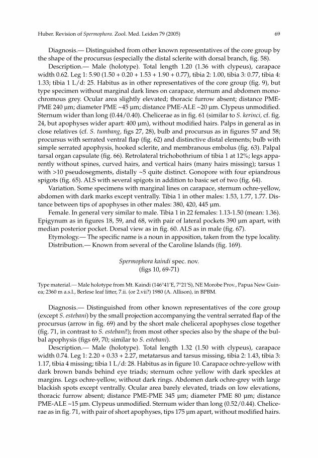
Huber. Revision of Spermophora. Zool. Med. Leiden 79 (2005) 69
Diagnosis.— Distinguished from other known representatives of the core group by the shape of the procursus (especially the distal sclerite with dorsal branch, fig. 58). Description.— Male (holotype). Total length 1.20 (1.36 with clypeus), carapace width 0.62. Leg 1: 5.90 (1.50 + 0.20 + 1.53 + 1.90 + 0.77), tibia 2: 1.00, tibia 3: 0.77, tibia 4: 1.33; tibia 1 L/d: 25. Habitus as in other representatives of the core group (fig. 9), but type specimen without marginal dark lines on carapace, sternum and abdomen mono-chromous grey. Ocular area slightly elevated; thoracic furrow absent; distance PME-PME 240 µm; diameter PME ~45 µm; distance PME-ALE ~20 µm. Clypeus unmodified. Sternum wider than long (0.44/0.40). Chelicerae as in fig. 61 (similar to S. kerinci, cf. fig. 24, but apophyses wider apart: 400 µm), without modified hairs. Palps in general as in close relatives (cf. S. tumbang, figs 27, 28), bulb and procursus as in figures 57 and 58; procursus with serrated ventral flap (fig. 62) and distinctive distal elements; bulb with simple serrated apophysis, hooked sclerite, and membranous embolus (fig. 63). Palpal tarsal organ capsulate (fig. 66). Retrolateral trichobothrium of tibia 1 at 12%; legs appa-rently without spines, curved hairs, and vertical hairs (many hairs missing); tarsus 1 with >10 pseudosegments, distally ~5 quite distinct. Gonopore with four epiandrous spigots (fig. 65). ALS with several spigots in addition to basic set of two (fig. 64). Variation. Some specimens with marginal lines on carapace, sternum ochre-yellow, abdomen with dark marks except ventrally. Tibia 1 in other males: 1.53, 1.77, 1.77. Dis-tance between tips of apophyses in other males: 380, 420, 445 µm. Female. In general very similar to male. Tibia 1 in 22 females: 1.13-1.50 (mean: 1.36). Epigynum as in figures 18, 59, and 68, with pair of lateral pockets 390 µm apart, with median posterior pocket. Dorsal view as in fig. 60. ALS as in male (fig. 67). Etymology.— The specific name is a noun in apposition, taken from the type locality. Distribution.— Known from several of the Caroline Islands (fig. 169).
Spermophora kaindi spec. nov.(figs 10, 69-71)
Type material.— Male holotype from Mt. Kaindi (146°41’E, 7°21’S), NE Morobe Prov., Papua New Guin-ea; 2360 m a.s.l., Berlese leaf litter, 7.ii. (or 2.vii?) 1980 (A. Allison), in BPBM.
Diagnosis.— Distinguished from other known representatives of the core group (except S. estebani) by the small projection accompanying the ventral serrated flap of the procursus (arrow in fig. 69) and by the short male cheliceral apophyses close together (fig. 71, in contrast to S. estebani!); from most other species also by the shape of the bul-bal apophysis (figs 69, 70; similar to S. estebani). Description.— Male (holotype). Total length 1.32 (1.50 with clypeus), carapace width 0.74. Leg 1: 2.20 + 0.33 + 2.27, metatarsus and tarsus missing, tibia 2: 1.43, tibia 3: 1.17, tibia 4 missing; tibia 1 L/d: 28. Habitus as in figure 10. Carapace ochre-yellow with dark brown bands behind eye triads; sternum ochre yellow with dark speckles at margins. Legs ochre-yellow, without dark rings. Abdomen dark ochre-grey with large blackish spots except ventrally. Ocular area barely elevated, triads on low elevations, thoracic furrow absent; distance PME-PME 345 µm; diameter PME 80 µm; distance PME-ALE ~15 µm. Clypeus unmodified. Sternum wider than long (0.52/0.44). Chelice-rae as in fig. 71, with pair of short apophyses, tips 175 µm apart, without modified hairs.

70 Huber. Revision of Spermophora. Zool. Med. Leiden 79 (2005)
Palps in general as in S. deelemanae (cf. figs 54, 55), trochanter with small apophysis, femur without apophysis ventrally, procursus with small process accompanying serrated ven-tral process (fig. 69) and distinctive distal elements; bulb with large serrated apophysis ending in hooked sclerite, and membranous embolus. Retrolateral trichobothrium of tibia 1 at 9 %; legs without spines and curved hairs, with several vertical hairs ventrally on tibiae (especially tibia 1); tarsus 2 with ~8 pseudosegments, difficult to see. Female. Unknown Etymology.— The specific name is a noun in apposition, taken from the type locality. Distribution.— Known from type locality only (fig. 169).
Spermophora estebani Simon, 1892(figs 11, 19, 20, 72-80)
Spermophora estebani Simon, 1892: 42.
Type material.— Two female syntypes from Cueva de Talbac ~5-6 km N Antipolo (~14º39’N, 121º11’E), Luzon, Philippines; spring 1890 (E. Simon), in MNHN (original label: “13285 S. Estebani ES, Cueva de Antipolo!”), examined.Non-Type Material.— Philippines: Luzon: Quezon N. P. (14º01’N, 121º51’E), 13-14.x.1979 (P.R. Deele-man), 15 2 juvs. (RMNH).
Notes.— Females within this species group are not easily distinguished, and the material below is assigned with some hesitation to Spermophora estebani Simon. Prosoma size and shape are quite identical, the epigynum is similar, and the two localities are just about 80 km apart. However, the lateral pockets are not visible on the epigyna of the syntypes and I preferred not to clear the type material in NaOH. More collecting at and around the type locality is necessary. The epigyna of the two syntypes appear slightly different, but this is probably a result from one epigynum being spread open. Diagnosis.— Distinguished from most representatives of the core group (except S. luzonica and S. sumbawa) by the absence of a serrated process on the procursus (fig. 73); from most species also by the shape of the bulbal apophysis (figs 72, 73; similar to S. kaindi) and by the widely spread male cheliceral apophyses (figs 74, 168). Description.— Male (Quezon N. P.). Carapace width 0.60, length 0.48 (0.60 with clypeus), abdomen missing. Leg 1: 7.10 (1.77 + 0.20 + 1.90 + 2.50 + 0.73), tibia 2: 1.20, tibia 3: 0.83, tibia 4: 1.50; tibia 1 L/d: 36. Prosoma as in figure 11. Carapace pale ochre-yellow with brown lateral margins and brown bands behind ocular triads; sternum and legs whitish. Ocular area barely elevated, triads on low elevations, thoracic furrow absent; distance PME-PME 205 µm; diameter PME 55 µm; distance PME-ALE ~10 µm. Clypeus unmodified. Sternum wider than long (0.40/0.36). Chelicerae as in fig. 74, with pair of long apophyses, tips 505 µm apart, without modified hairs. Palps as in figures 72 and 73; trochanter with small apophysis, femur without apophysis ventrally, procursus with distinctive distal elements, with ventral flap not serrated; bulb with large serrated apophysis ending in hooked sclerite, and membranous embolus (fig. 73). Retrolateral trichobothrium of tibia 1 not seen; legs without spines, curved hairs, and vertical hairs (many hairs missing); pseudosegments barely visible. Female. In general very similar to male, but pattern on abdomen very indistinct in

Huber. Revision of Spermophora. Zool. Med. Leiden 79 (2005) 71
some. Tibia 1 in 2 syntypes: 1.87, 1.97, in 4 females from Quezon N. P.: 1.60-1.73; cara-pace width in syntypes: 0.60, 0.61; in 4 other females: 0.56-0.61. Tarsal claws and palpal tarsal organ as in figures 79 and 80. Epigynum as in figs 19, 20, 75, and 78, with pair of lateral pockets 420 µm apart, with median posterior pocket. Dorsal view as in fig. 76. ALS with several spigots in addition to basic set of two (fig. 77). Distribution.— Known from two localities on Luzon (fig. 169).
Spermophora luzonica spec. nov.(figs 81-85)
Type material.— Male holotype from Cueva Santa, Quezon National Park (~14°00’N, 121°50’E), Luzon, Philippines; 12.iv.1977 (P. Strinati, V. Aellen), in MHNG.Non-Type Material.— Philippines: Luzon: Quezon National Park: 1, together with holotype (MHNG).
Diagnosis.— Distinguished from close relatives by the small cheliceral apophyses (fig. 83), the shape of the bulbal apophysis (fig. 82), and by the shape of the procursus with transparent projection on the ventral flap (fig. 82). Description.— Male (holotype). Total length 1.10 (1.20 with clypeus), carapace width 0.47. Leg 1: 5.72 (1.47 + 0.20 + 1.48 + 1.90 + 0.67), tibia 2: 0.97, tibia 3: 0.73, tibia 4: 1.17; tibia 1 L/d: 28. Habitus as in S. kerinci (cf. figs 1, 2). Carapace ochre-yellow with pair of large brown marks, margins also darker, sternum ochre yellow with dark speckles. Legs ochre-yellow, without dark rings. Abdomen monochromous grey. Ocular area not elevated, thoracic furrow absent; distance PME-PME ~150 µm (slightly dama-ged); diameter PME 55 µm; distance PME-ALE ~20 µm. Clypeus unmodified. Sternum about as wide as long (0.36/0.34). Chelicerae as in fig. 83, with pair of short apophyses, tips 45 µm apart, without modified hairs. Palps as in figures 81 and 82; trochanter with small retrolateral apophysis, procursus with distinctive transparent projection on ven-tral flap, distinctive distal elements; bulb with characteristically pointed apophysis and membranous embolus. Retrolateral trichobothrium of tibia 1 at 10%; legs without spi-nes and vertical hairs, with curved hairs apparently on metatarsi 2 only (many hairs missing); tarsus 1 with ~10 pseudosegments, difficult to see. Female. In general similar to male but sternum and abdomen darker; tibia 1: 1.23. Epigynum as in fig. 84, large light brown plate with pair of pockets 40 µm apart, with posterior median pocket. Dorsal view as in fig. 85. Etymology.— The species name is an adjective derived from the type locality. Distribution.— Known from type locality only (fig. 169).
Spermophora sumbawa spec. nov.(figs 12, 13, 21, 86-103)
Spermophora sp. 2: Huber 2003a, b, c.
Type material.— Male holotype from “Samokat”, 20 km from Sumbawa Besar (~116°46’E, 8°30’S), Sum-bawa, Lesser Sunda Islands, Indonesia; 480 m a.s.l., secondary forest leaf litter, 1-3.i.1990 (S. Djojosud-harmo), in RMNH.Non-Type Material.— Indonesia: Sumbawa: 17~65, together with holotype (RMNH); Lombok (~116°18’E, 8°42’S): Kute, 100 m a.s.l., secondary forest leaf litter, 8-18.i.1990 (S. Djojosudharmo), 11 (RMNH).
72 Huber. Revision of Spermophora. Zool. Med. Leiden 79 (2005)
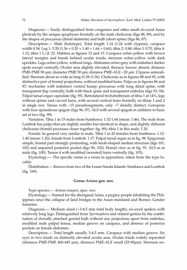
Diagnosis.— Easily distinguished from congeners and other small six-eyed Asian pholcids by the unique apophyses frontally on the male chelicerae (figs 88, 89), and by the shapes of procursus (distal elements) and bulb (short spine) (figs 86, 87). Description.— Male (holotype). Total length 1.14 (1.24 with clypeus), carapace width 0.54. Leg 1: 5.20 (1.36 + 0.20 + 1.40 + 1.64 + 0.60), tibia 2: 0.88, tibia 3: 0.70, tibia 4: 1.12; tibia 1 L/d: 22. Habitus as figures 12 and 13. Carapace ochre-yellow with brown lateral margins and bands behind ocular triads, sternum ochre-yellow with dark speckles. Legs ochre-yellow, without rings. Abdomen ochre-grey with indistinct darker spots except ventrally. Ocular area slightly elevated, thoracic furrow absent; distance PME-PME 90 µm; diameter PME 50 µm; distance PME-ALE ~20 µm. Clypeus unmodi-fied. Sternum about as wide as long (0.38/0.36). Chelicerae as in figures 88 and 92, with distinctive pair of frontal projections, without modified hairs. Palps as in figures 86 and 87; trochanter with indistinct ventral hump; procursus with long distal spine, with transparent flap ventrally; bulb with black spine and transparent embolus (figs 93, 94). Palpal tarsal organ capsulate (fig. 95). Retrolateral trichobothrium of tibia 1 at 14%; legs without spines and curved hairs, with several vertical hairs frontally on tibiae 1 and 2 in single row. Tarsus with ~15 pseudosegments, only ~7 distally distinct. Gonopore with four epiandrous spigots (figs 96, 97). ALS with several spigots in addition to basic set of two (fig. 99). Variation. Tibia 1 in 15 males from Sumbawa: 1.32-1.64 (mean: 1.46). The male from Lombok has palps that are slightly smaller but identical in shape, and slightly different chelicerae (frontal processes closer together: fig. 89); tibia 1 in this male: 1.20. Female. In general very similar to male. Tibia 1 in 20 females from Sumbawa: 1.12-1.40 (mean: 1.20), female from Lombok: 1.17. Palpal tarsal organ as in fig. 98. Epigynum simple, frontal part strongly protruding, with knob-shaped median structure (figs 101, 102) and unpaired posterior pocket (figs 90, 102). Dorsal view as in fig. 91. ALS as in male (fig. 100). Tarsus 4 with modified (serrated) hairs ventrally (fig. 103). Etymology.— The specific name is a noun in apposition, taken from the type lo-cality. Distribution.— Known from two of the Lesser Sunda Islands: Sumbawa and Lombok (fig. 169).
Genus Aetana gen. nov.
Type species.— Aetana omayan, spec. nov. Etymology.— Named for the aboriginal Aetas, a pygmy people inhabiting the Phil-ippines since the collapse of land bridges to the Asian mainland and Borneo. Gender feminine. Diagnosis.— Medium sized (~3-4.5 mm total body length), six-eyed spiders with relatively long legs. Distinguished from Spermophora and related genera by the combi-nation of dorsally attached genital bulb without any projections apart from embolus, modified male palpal femur, median groove on carapace, and absence of posterior pockets on female abdomen. Description.— Total length usually 3-4.5 mm. Carapace with median groove. Six eyes in two triads on distinctly elevated ocular area. Ocular triads widely separated (distance PME-PME 400-445 µm), distance PME-ALE small (25-90µm). Sternum un-
Huber. Revision of Spermophora. Zool. Med. Leiden 79 (2005) 73
modified. Male clypeus with or without modifications. Male chelicerae with proximal rounded projections laterally and pair of apophyses distally, distal apophyses without modified hairs, chelicerae never with stridulatory ridges. Male palpal coxa unmodified, trochanter with retrolateral apophysis (somewhat ventrally) and sometimes further projections, femur with distinctive projections prolaterally and ventrally (figs 110, 115, 121), procursus complex, with ventral flap and apparently hinged distal structures, bulb with tubular membranous embolus as single projection (figs 110, 115, 122). Tarsal organ capsulate. Legs long (about 9-10 x body length; tibia 1 L/d: ~50-90); leg formula apparently 1243 (only in A. fiji specimens with both legs 2 and 4 present). Legs without spines, vertical hairs in usual low density, curved hairs present in A. omayan, possibly also in other species (most hairs missing in most specimens); retrolateral trichobothrium of tibia 1 at 2-4%; prolateral trichobothrium missing on tibia 1, present on others; tarsus 1 with >15 pseudosegments, very indistinct. Abdomen oval to elongated (figs 104-109), with more or less distinct dark spots dorsally. Male gonopore not examined, ALS in A. fiji with only two spigots (widened and pointed spigots). Sexual dimorphism very slight, females with slightly shorter legs, unmodified clypeus and chelicerae. Epigynum usually a simple plate (figs 105, 107) with pair of pockets (figs 113, 119) corresponding to male cheliceral apophyses. Monophyly and Relationships.— None of the species described below has been included in previous cladistic analyses. A closer relationship with Spermophora is sug-gested by the combination of lateral cheliceral apophyses, a hinged process on the procursus, and a dorsal (rather than prolateral) attachment of the bulb to the cymbium. The modifications on the male palpal femur are unique within pholcines and probably constitute a synapomorphy of the genus. A further synapomorphy might be the ab-sence of sclerites on the genital bulb. Natural History.— The collection data of A. fiji suggest that this species builds webs in shrubs and trees. Body size and leg length also indicate that the three species described below are not litter-dwelling. Distribution.— Southeast Asia and Oceania (fig. 170). The distribution map sug-gests that further species might occur in other Southeast Asian Islands, especially on Sulawesi and Papua New Guinea. Composition.— The genus includes only the three species described below. A male specimen representing a fourth species has been collected recently on Fiji [S. Crews, pers. comm.: Taveuni Isl., Cakadrove Prov., Devo Peak Radio Tower, malaise in rain forest, 3.i.2003 (H. Irwin, E. Schlinger, M. Tokota’a), 179°58’E, 16°51’S, 1200m a.s.l.].
Aetana omayan spec. nov.(figs 104, 105, 110-114)
Type material.— Male holotype from Baguio, Crystal Cave (~16°25’N, 120°36’E), Luzon, Philippines; 13.iv.1977 (P. Strinati, V. Aellen), in MHNG.Non-Type Material.— Philippines: Luzon: 2, together with holotype (MHNG).
Diagnosis.— Easily distinguished form A. fiji by the male clypeal modifications (fig. 112), the bipartite ventral flap on the procursus (fig. 111), the prolateral apophysis on the male palpal femur (fig. 110), and the shape of the epigynum (fig. 113). From A.

74 Huber. Revision of Spermophora. Zool. Med. Leiden 79 (2005)
kinabalu by the much longer and more widely spaced cheliceral apophyses and the modified clypeus (fig. 112); also by the shapes of male palpal femur, bulb, and procursus (figs 110, 111). Description.— Male (holotype). Total length 4.25 (4.5 with clypeus), carapace width 1.8. Leg 1: 42.9 (10.1 + 0.6 + 10.0 + 18.5 + 3.7), tibia 2 missing, tibia 3: 4.9, tibia 4: 6.7; tibia 1 L/d: 56. Habitus as in figure 104. Carapace dark ochre-yellow with black median and lateral bands, clypeus and ocular area also darker; sternum whitish, only frontally light brown. Legs ochre-yellow, with darker rings on femora (subdistally), patellae, and ti-biae (subdistally). Abdomen grey with many black spots except ventrally. Ocular area elevated, each triad on additional elevation; thoracic furrow distinct but very shallow; distance PME-PME 400 µm; diameter PME 195 µm; distance PME-ALE ~90 µm; AME absent. Clypeus with pair of distinctive apophyses (fig. 112). Sternum wider than long (1.2/0.95). Chelicerae as in fig. 112, with pair of long apophyses, tips 865 µm apart, without modified hairs. Palps as in figures 110 and 111; trochanter with retrolateral, ventral and prolateral apophyses, femur with large prolateral projection, indistinct ven-tral projections, and retrolateral cone-shaped apophysis; procursus very complex, with distinctive bipartite ventral flap, apparently with hinged process, with strong spine distally accompanied by membranous flap; bulb with single projection (presumably the embolus). Retrolateral trichobothrium of tibia 1 at 2%; legs apparently without spines and vertical hairs, with curved hairs on metatarsi 3 only, but most hairs missing. Tarsus with >15 pseudosegments, very indistinct. Female. In general very similar to male, but clypeus unmodified, eyes not on eleva-tions. Tibia 1: 7.7. Epigynum simple brown plate with pair of whitish areas (figs 105, 113), with pair of pockets 760 µm apart. Intriguingly, the pockets do not open to the outside! Dorsal view as in fig. 114. Etymology.— The specific name is a noun in apposition, derived from the Omayan, invisible little people in Philippine folklore. Distribution.— Known from type locality only (fig. 170).
Aetana fiji spec. nov.(figs 106, 107, 115-120)
Type material.— Male holotype from Sawani (178°28’E, 18°01’S), near Suva, Viti Levu Island, Fiji; from epiphytes, 19.vii.1956 (R. R. Forster), in BPBM.Non-Type Material.— Fiji: Viti Levu: Sawani: 13, together with holotype (BPBM); Monasavu Water-shed (17º45’S, 178º04’E), 1100 m a.s.l., vegetation beating, 29-30.xi.2002 (D. Gruner), 1 (BPBM); Suva (178°25’E, 18°08’S), “in wettest bush”, 9.ix.1958 (Marples), 1 1juv. (BPBM); Colo-I-Suva, park near Nausori, in web on vegetation, wet forest, 6.v.1987 (J. A. Beatty), 1 (BPBM); 3 km E Monasavu Dam (178°03’E, 17°46’S), 1000 m a.s.l., 26.vii.1987 (G. Monteith, D. Cook), “pyrethrum, trees and logs”, 1 (QMB S50343); Mt. Victoria (=Tomanivi) (178°01’E, 17°37’S), 1100-1340 m a.s.l., 25.vii.1987 (G. Monteith, D. Cook), 1 (QMB S50345). Taveuni: Des Vocux Peak (179°58’W, 16°51’S), 900 m a.s.l., 16.vii.1987 (G. Monteith, D. Cook), “pyrethrum, tree trunks”, 1 3 juveniles (QMB S50349). Vanua Levu: Mt. Delaikoro (179°45’E, 16°33’S), 700 m a.s.l., 21.vii.1987 (G. Monteith, D. Cook), 1 1 juvenile (QMB S 50350); same locality, “pyrethrum, logs and trees”, 1 4 juveniles (QMB S50348). Kandavu: Mt. Korogatule (177°18’E, 17°41’S), 300 m a.s.l., near Matasawalevu, 4.vii.1987 (G. B. Monteith), 1 (QMB S50353).
Diagnosis.— Easily distinguished from A. kinabalu by the much longer and more widely spaced cheliceral apophyses and the modified clypeus (fig. 118); also by the

Huber. Revision of Spermophora. Zool. Med. Leiden 79 (2005) 75
shapes of male palpal femur, bulb, and procursus (figs 115, 116). From A. omayan by the male clypeal modifications (fig. 118), the simple ventral flap on the procursus (fig. 116), the apophyses on the male palpal femur (fig. 115), and the shape of the epigy-num (fig. 119). Description.— Male (holotype). Total length ~3.2 (it is unclear which of the detached abdomens belongs to the type), carapace width 1.3. Leg 1: 9.3 + 0.5 + 9.2, metatarsus and tarsus missing, tibia 2: 5.6, tibia 3 missing, tibia 4: 5.2; tibia 1 L/d: 86. Habitus as in fe-male (cf. fig. 106). Carapace ochre-yellow with dark median and lateral bands, clypeus and ocular area partly darkened; sternum ochre yellow. Legs ochre-yellow, with indis-tinct rings on femora (subdistally), patellae, and tibiae (subdistally). Abdomen grey with indistinct spots. Ocular area distinctly elevated, each triad on additional elevation; thoracic furrow distinct frontally; distance PME-PME 400 µm; diameter PME 105 µm; distance PME-ALE ~25 µm; AME absent. Clypeus with two pairs of black cones (between eyes and rim and at rim), and pair of long horns at rim (fig. 118). Sternum wider than long (0.86/0.64). Chelicerae as in figures 117 and 118, with pair of long apo-physes, tips 1020 µm apart, without modified hairs. Palps as in figures 115 and 116; trochanter with simple apophysis, femur with several modifications: large prolateral projection, pointed ventral apophysis, and retrolateral cone; procursus apparently with two successive hinges, with strong spine distally accompanied by membranous flap; bulb with single projection (presumably the embolus). Retrolateral trichobothrium of tibia 1 at 4%; legs apparently without spines, curved hairs, and vertical hairs (most hairs missing). Variation. Leg 1 in male from Suva: 42.0 (9.7 + 0.5 + 9.8 + 18.0 + 4.0); tarsus 1 with >20 pseudosegments, distally ~15 fairly distinct. Tibia 1 in male from 3 km E Monasavu Dam: 10.3. Female. In general very similar to male, but clypeus unmodified, eyes not on eleva-tions. Tibia 1 in 3 females (Viti Levu): 5.8, 6.0, 6.8. Epigynum simple plate with pair of lateral pockets 905 µm apart, without posterior pocket (figs 107, 119). Dorsal view as in fig. 120. ALS with only 2 spigots. The QMB has some female specimens from other Fiji islands (see above) with slightly different epigyna (larger and more sculptured). They are tentatively assigned to this species. Etymology.— The specific name is a noun in apposition, taken from the type locality. Distribution.— Known from the Fiji Islands only (fig. 170).
Aetana kinabalu spec. nov.(figs 108, 109, 121-123)
Type material.— Male holotype from Kinabalu National Park (headquarters) (~116°39’E, 6°09’N), Sabah, Borneo, Malaysia; 1550 m a.s.l., 3.v.1991 (C. L. Deeleman-Reinhold), in RMNH.Non-Type Material.— Malaysia: Sabah: “Kinabalu” (~6º00’N, 116º30’E), date unknown (C. L. & P. R. Deeleman), 1 (RMNH).
Diagnosis.— Easily distinguished from the two known congeners by the much shorter cheliceral apophyses (fig. 123) and the unmodified clypeus; also by the shapes of male palpal femur, bulb, and procursus (figs 121, 122). Description.-- Male (holotype). Total length 3.0 (3.2 with clypeus), carapace width 1.1. Leg 1: 8.7 + 0.4 + 8.6 + 15.3, tarsus missing, tibiae 2 and 4 missing, tibia 3: 3.3; tibia

76 Huber. Revision of Spermophora. Zool. Med. Leiden 79 (2005)
1 L/d: 81. Habitus as in figures 108 and 109. Carapace ochre-yellow with dark median and lateral margins, ocular area also darkened, clypeus ochre-yellow; sternum pale ochre-grey medially, light brown laterally. Legs light brown, without rings. Abdomen damaged. Ocular area barely elevated but each triad on stalk-like elevation; thoracic furrow very shallow frontally, absent posteriorly; distance PME-PME 445 µm; diameter PME 100 µm; distance PME-ALE ~25 µm; AME absent. Clypeus unmodified. Sternum wider than long (0.68/0.52). Chelicerae as in fig. 123, with pair of simple apophyses, tips 455 µm apart, without modified hairs. Palps as in figures 121 and 122; trochanter with simple apophysis, femur with several modifications: prolateral cone and complex ventral apophysis; procursus very complex, apparently with two successive hinges, with strong spine distally; bulb with small cone and single membranous projection (presumably the embolus). Retrolateral trichobothrium of tibia 1 at 3%; almost all hairs on legs missing. Variation. Other male with distinct black spots on abdomen (figs 108, 109). Distance between tips of cheliceral apophyses: 460 µm. Female. Unknown. Etymology.— The specific name is a noun in apposition, taken from the type locality. Distribution.— Known from Kinabalu area only (fig. 170).
Genus Savarna gen. nov.
Type species.— Savarna thaleban, spec. nov. Etymology.— Named for Savarnadvipa, the Land of Gold in early writings from India. This mystical, fantastically wealthy kingdom was said to lie in a far away and unknown land, and legend holds that it was on an odyssey in search of Savarnadvipa that the first Indians were lured to the Malay Peninsula. Gender feminine. Diagnosis.— Medium sized (~2-3 mm total body length), six-eyed spiders with relatively long legs. Distinguished from Spermophora and related genera by the long male palpal trochanter apophysis uniquely attached to the femur (figs 132, 136, 139), the laterally projecting proximal cheliceral apophyses (figs 133, 137, 140), the absence of distal cheliceral apophyses, and the absence of pockets on the epigynum. Description.— Total length usually 2-3 mm. Carapace with distinct median groove. Six eyes in two triads on elevated ocular area. Ocular triads distinctly separated (distance PME-PME 240-255 µm), distance PME-ALE small (20-30 µm). Sternum unmodified. Clypeus with or without modifications. Male chelicerae with distinctive proximal round-ed projections pointing laterally (figs 133, 137, 140), without distal apophyses, without stridulatory ridges. Male palpal coxa unmodified, trochanter with distinctive retrolateral apophysis that appears attached to the femur (figs 132, 136, 139), procursus medium com-plex, apparently without hinged process, bulb with sclerotized structure that appears to contain the sperm duct. Tarsal organ apparently capsulate. Legs long (~9 x body length; tibia 1 L/d: ~40-60); leg formula 1(2=4)3. Legs without spines and curved hairs, vertical hairs in high density in S. thaleban (all legs missing in S. tessellata); retrolateral trichoboth-rium of tibia 1 at ~7-8%; prolateral trichobothrium missing on tibia 1, present on others; tarsus 1 with >20 pseudosegments. Abdomen globular to oval, pointed at spinnerets, with or without dark spots dorsally (figs 124-130). Male gonopore not examined, ALS in S. thaleban with only two spigots (widened and pointed spigots).

Huber. Revision of Spermophora. Zool. Med. Leiden 79 (2005) 77
Sexual dimorphism very slight, females with slightly shorter legs, unmodified clypeus and chelicerae. Epigynum a simple plate without pockets (figs 126, 134). Monophyly and Relationships.— None of the species described below has been in-cluded in previous cladistic analyses. The modifications of the male palpal trochanter and the male chelicerae are unique within pholcines and probably constitute a synapo-morphy of the genus. The genus is probably a representative of pholcines, but its af-finities within that clade remain obscure. Natural History.— Nothing is known about the natural history of the three species described below, except that S. baso was collected in a cave. Body size and leg length also indicate that these species are not litter-dwelling. Distribution.— Known from three localities in southern Thailand, Malaysia, and Sumatra (fig. 171). Composition.— The genus includes only the three species described below.
Savarna thaleban spec. nov.(figs 124-126, 131-134)
Type material.— Male holotype from Thale Ban National Park (6º42.5’N, 100º10’E), Satun Prov., Thai-land; 270 m a.s.l., 15-18.x.2003 (ATOL Expedition 2003), in MACN.Non-Type Material.— Thailand: Satun Province: Thale Ban National Park: 1 2 juveniles, together with holotype (MACN); same collection data, 7 4 juveniles, in MACN.
Diagnosis.— Easily distinguished from known congeners by the shapes of procur-sus and bulbal apophysis (figs 131, 132), and by the unmodified male clypeus. Description.— Male (holotype). Total length 2.23 (2.42 with clypeus), carapace width 0.93. Leg 1: 22.2 (5.3 + 0.4 + 5.4 + 8.6 + 2.5), tibia 2: 3.1, tibia 3: 2.3, tibia 4: 3.1; tibia 1 L/d: 61. Habitus as in figures 124 and 125. Carapace ochre-grey with wide black lateral bands, ocular area and clypeus also mostly black, sternum black. Legs pale ochre-grey, with darker rings on femora (subdistally) and tibiae (proximally and subdistally). Ab-domen grey with three pairs of black spots dorsally, further large black marks laterally and posteriorly, three interconnected black spots ventrally. Ocular area elevated, triads on additional elevations; thoracic furrow distinct; distance PME-PME 255 µm; diameter PME 115 µm; distance PME-ALE ~30 µm; AME absent. Clypeus unmodified. Sternum wider than long (0.66/0.50). Chelicerae as in fig. 133, with distinctive lateral apophyses, frontally without apophyses, with black pattern. Palps as in figures 131 and 132; coxa unmodified, trochanter with rounded retrolateral projection and long distally hooked projection lying against femur, femur with distal rounded projection, procursus stron-gly bent dorsally, very complex distally, apparently without hinged process, bulb with roughly square-shaped, sclerotized single projection. Retrolateral trichobothrium of tibia 1 at 7%; legs without spines and curved hairs, with many short vertical hairs on all tibiae; tarsus 1 with >20 pseudosegments, only distally fairly distinct. Female. In general similar to male, but chelicerae and palps monochromous black, few vertical hairs on legs. Tibia 1 in 7 females: 4.5-5.0 (mean: 4.69). Epigynum a promi-nent oval plate without pockets (fig. 126), dorsal view as in fig. 134. ALS and PMS with two spigots each. Etymology.— The specific name is a noun in apposition, taken from the type locality. Distribution.— Known from type locality only (fig. 171).

78 Huber. Revision of Spermophora. Zool. Med. Leiden 79 (2005)
Savarna baso (Roewer, 1963) comb. nov(figs 127, 128, 135-137)
Spermophora baso Roewer, 1963: 229, figs 17-18.
Type material.— Male holotype from Sumatra, cave near Baso (100°28’E, 0°17’S), x.1913 (E. Jacobson), in SMF (RII/13853/122; left palp mounted on slide), together with a female prosoma, examined.
Note.— Roewer (1963) described only the male and did not mention further speci-mens, but the vial also contains a prosoma that is probably from an adult female. Diagnosis.— Distinguished from S. tessellata by the shapes of procursus and bulbal apophyses, by the paired clypeal projections (fig. 127), and by the much thicker palpal femur (figs 135, 136); from S. thaleban by the shapes of procursus, bulbal apophysis, and the modified clypeus. Description.— Male (holotype). Total length 2.65 (2.90 with clypeus), carapace width 1.20. Leg 1: 25.1 (6.5 + 0.5 + 6.1 + 9.6 + 2.4), tibia 2: 3.8, tibia 3: 2.8, tibia 4: 3.9; tibia 1 L/d: 43. Habitus as in figures 127 and 128. Carapace and legs ochre-yellow, sternum ochre-yellow to light brown, abdomen pale ochre-grey. Ocular area slightly elevated, distinctly separated from carapace, thoracic furrow distinct and deep; distance PME-PME 240 µm; diameter PME 105 µm; distance PME-ALE ~20 µm; AME absent. Clypeus with pair of unsclerotized rounded frontal projections (fig. 127). Sternum wider than long (0.80/0.67). Chelicerae as in fig. 137, with only one pair of projections proximally. Palps as in figures 135 and 136; trochanter with long distally hooked projection lying against femur, procursus complex but apparently without hinged process, bulb with proximal sclerite and single but complex process (‘embolar division’). Retrolateral trichobothrium of tibia 1 at 8%; legs without spines, vertical hairs, and curved hairs; tarsus 1 with >30 pseudosegments, distally quite distinct. Female. In general similar to male, but without clypeal apophyses. Carapace width 1.10, tibia 1: 5.9. Abdomen missing. Distribution.— Known from type locality only (fig. 171).
Savarna tessellata (Simon, 1901) comb. nov.(figs 129, 130, 138-140)
Spermophora tessellata Simon, 1901: 50.
Type material.— Female holotype from Malaysia, “Jalor: Biserat” (coordinates unknown), not examined (could not be found in the MNHN). See Note below.Material examined.— Malaysia, no further data, 11 and 1 juvenile, with Simon’s handwritten label “12185 Sp. tessellata E. S. Pen. Malayana (C. M)”, in MNHN.
Note.— Simon (1901) described only the female, and did not mention further specimens. It is not clear whether the material above (identified by Simon himself) is actually conspecific or not. Further collecting in Malaysia may reveal that Simon’s iden-tification was wrong and that the specimens treated here are a different species. Diagnosis.— Distinguished from S. baso by the shapes of procursus and bulbal apophyses, by the unpaired clypeal projection (fig. 129), and by the much thinner
Huber. Revision of Spermophora. Zool. Med. Leiden 79 (2005) 79
palpal tibia (figs 138, 139); from S. thaleban by the shapes of procursus, bulbal apophy-sis, and the modified clypeus. Description.— Male. Total length 2.75 (3.05 with clypeus), carapace width 1.27. All legs missing except right femur 3. Habitus as in figures 129 and 130. Entire specimen grey (probably artificial), only sternum darker. Ocular area slightly elevated, distinctly separated from carapace, thoracic furrow distinct and deep; distance PME-PME 240 µm; diameter PME 115 µm; distance PME-ALE ~25 µm; AME absent. Clypeus with unpaired median projection, 180 µm long, ~60 µm wide. Sternum wider than long (0.80/0.67). Chelicerae as in fig. 140, with only one pair of projections proximally. Palps as in figures 138 and 139; trochanter with long distally hooked projection lying against femur, tibia extremely long and slender, procursus large, complex distally, bulb with proximal sclerite and V-shaped distal projection. Female. In general similar to male but clypeus unmodified; carapace width: 1.15, tibia 3: 2.7, tibia 4: 3.6. Epigynum simple large plate with pair of elevations frontally, strongly protruding and opened (probably artificial and therefore not illustrated). Simon’s (1901) original description seems not in good agreement with the specimen examined (sternum colour, eye pattern). Distribution.— Known from type locality only (fig. 171).
Genus Khorata gen. nov.
Type species.— Khorata khammouan, spec. nov. Etymology.— Named for the smiths on Thailand’s Khorat Plateau, who by 3000 B.C. figured out that the strongest bronze alloy is made by mixing one part of tin with nine parts of copper, and were so doing better work than their Mesopotamian counter-parts, who made bronze by mixing copper and arsenic, with brittle and sometimes hazardous results. Gender feminine. Diagnosis.— Medium sized (~2-3 mm total body length), six-eyed spiders with relatively long legs. Distinguished from Spermophora and related genera by the sclero-tized ledges laterally on the male chelicerae (figs 154, 160, 163), the sclerotized proximal cheliceral apophyses provided with cuticular ridges or scales, the apophysis retrolater-ally on the male palpal femur (figs 152, 158, 162), and the relatively small male palpal tibia. Also by the combination of prolaterally attached genital bulb without any projec-tions apart from the embolus, shallow median groove on carapace, and absence of pos-terior pockets on the female abdomen. Description.— Total length about 2-3 mm. Carapace with shallow but distinct me-dian groove. Six eyes in two triads on elevated ocular area. Ocular triads relatively close together (distance PME-PME 115-215 µm), distance PME-ALE small (20-35 µm). Ster-num and clypeus unmodified. Male chelicerae with distinctive proximal projections that are sclerotized and provided with cuticular ridges or scales, with lateral sclerotized ledges; distal apophyses without modified hairs; chelicerae without stridulatory ridges. Male palpal coxa unmodified, trochanter with small retrolateral apophysis (somewhat ventrally) and sometimes further small apophyses, procursus medium complex, appar-ently without hinged process, bulb with membranous embolus, only in K. jaegeri with additional process. Tarsal organ apparently capsulate. Legs long (~8-11 x body length; tibia 1 L/d: ~45-70); leg formula 1243 or 1(2=4)3. Legs usually without spines (present

in K. schwendingeri), without curved hairs; vertical hairs in high density on distal seg-ments (tibiae or metatarsi); retrolateral trichobothrium of tibia 1 at 7-10%; prolateral trichobothrium missing on tibia 1, present on others; tarsus 1 with >20 pseudosegments. Abdomen globular to oval (figs 141-150), usually with dark spots dorsally (missing in the troglomorphic K. jaegeri). Male gonopore not examined, ALS with only two spigots (widened and pointed spigots). Sexual dimorphism very slight, females with slightly shorter legs, unmodified chelicerae. Epigynum a simple plate without pockets (figs 155, 167). Monophyly and Relationships.— None of the species described below has been included in previous cladistic analyses. The sclerotized and modified male lateral cheliceral apophyses as well as the lateral ledges on the male chelicerae are unique within pholcines and probably constitute synapomorphies of the genus. Further possi-ble synapomorphies are the retrolateral apophysis on the male palpal femur and the small size of the palpal tibia. The genus is probably a representative of pholcines, but its affinities within that clade remain obscure. Natural History.— Nothing is known about the natural history of the four species described below, except that two species were collected in and near limestone caves and the third on limestone. Distribution.— Known from Thailand and Laos (fig. 172). Composition.— The genus includes only the four species described below.
Khorata khammouan spec. nov.(figs 141-143, 151-156)
Type material.— Male holotype from 4.6 km WNW Ban Tathot (17º38’N, 105º06’E), Khammouan Prov-ince, Laos; at entrance to Tham Deua limestone cave, ~200 m a.s.l., 21.ii.2003 (P. Jäger), in SMF.Non-Type Material.— Laos: Khammouan Province: 4.6 km WNW Ban Tathot: 1, together with holotype (SMF); same collection data, 1 1 juvenile, in SMF; Thakek area, Ban Tham (17º26’N, 104º52’E), lime-stone cave and foot caves, ~180 m a.s.l., 27.ii.2003 (P. Jäger), 2 (only one abdomen), 2 juveniles, in SMF; 9.5 km NE Thakek (17°27’N, 104°52.5’E), 160 m a.s.l., foot cave and surroundings, by hand, 28.x.2004 (P. Jäger, V. Vedel), 57 6 juvs. in SMF.
Diagnosis.— Distinguished from known congeners by the male chelicerae (distal apophyses closer together than in K. jaegeri, wider apart than in K. schwendingeri and K. bangkok), and by the shape of the procursus (distal elements). From K. jaegeri also by the dark pattern and by the bulb without retrolateral projection. Description.— Male (holotype). Total length 2.45 (2.60 with clypeus), carapace width 1.10. Leg 1: 27.7 (6.9 + 0.5 + 6.7 + 10.6 + 3.0), tibia 2: 4.1, tibia 3: 2.9, tibia 4: 3.6; tibia 1 L/d: 60. Habitus as in figures 141 and 142. Carapace ochre with black margins and dark Y mark behind ocular area; sternum black. Legs ochre, with slightly darker rings on femora (subdistally) and tibiae (proximally and subdistally). Abdomen grey with large black spots, also ventrally. Ocular area slightly elevated and separated from carapace, thoracic furrow shallow but distinct; distance PME-PME 115 µm; diameter PME 125 µm; distance PME-ALE ~35 µm; AME absent. Clypeus unmodified. Sternum slightly wider than long (0.67/0.63). Chelicerae as in figures 153 and 154, with hooked frontal apophyses distally (distance between tips: 230 µm), strong proximal apophyses provided with scales, with lateral ledges, without modified hairs. Palps as in figures 151
80 Huber. Revision of Spermophora. Zool. Med. Leiden 79 (2005)

Huber. Revision of Spermophora. Zool. Med. Leiden 79 (2005) 81
and 152; trochanter with retrolateral apophysis and small ventral projection, femur with retrolateral apophysis, patella very large, procursus relatively simple except dis-tally, bulb very simple, with embolus and rounded hump retrolaterally. Retrolateral trichobothrium of tibia 1 at 8%; legs without spines, with short vertical hairs dorsally on all metatarsi except metatarsus 3, without curved hairs; tarsus 1 apparently with >20 pseudosegments, only distally a few fairly distinct. Variation. Tibia 1 in other male from type locality: 7.0. Distance between tips of dis-tal cheliceral apophyses: 195 µm, 235 µm. Tibia 1 in males from NE Thakek: 6.5, 7.1, 7.3. Female. In general similar to male. Tibia 1 in 8 females: 5.3-7.3 (mean 6.35). Epigy-num black, with two large rounded protrusions (figs 143, 155), apparently without pockets. Dorsal view as in fig. 156. ALS with only two spigots each. Etymology.— The specific name is a noun in apposition, taken from the type locality. Distribution.— Known from two localities in Laos (fig. 172).
Khorata jaegeri spec. nov.(figs 144, 145, 157-160)
Type material.—Male holotype from 4.6 km WNW Ban Tathot (17º38’N, 105º06’E), Khammouan Prov-ince, Laos; at entrance to Tham Deua limestone cave, ~200 m a.s.l., 21.ii.2003 (P. Jäger), in SMF.Non-Type Material.—Laos: Khammouan Province: 4.6 km WNW Ban Tathot, entrance to Tham Deua limestone cave: 2, together with holotype (SMF).
Diagnosis.— Easily distinguished from all known congeners by the male chelicerae (distal and proximal apophyses very long: figs 159, 160), by the shape of the procursus (distal elements: figs 157, 158), and by the pointed projection on the bulb (fig. 158). Description.— Male (holotype). Total length 3.1 (3.3 with clypeus), carapace width 1.30. Leg 1: 32.0 (7.9 + 0.5 + 8.1 + 12.2 + 3.3), tibia 2: 5.6, tibia 3: 4.4, tibia 4: 5.3; tibia 1 L/d: 68. Habitus as in figures 144 and 145. Carapace ochre-yellow with narrow dark median line; sternum and legs ochre-yellow; legs without dark rings. Abdomen mono-chromous greenish-grey. Ocular area barely elevated but distinctly separated from ca-rapace, thoracic furrow present but very shallow; distance PME-PME 215 µm; diameter PME 60 µm; distance PME-ALE ~20 µm; AME absent. Clypeus unmodified. Sternum wider than long (0.80/0.66). Chelicerae as in figures 159 and 160, with long frontal apo-physes hooked distally (distance between tips: 0.96), strong proximal apophyses provi-ded with scales, with lateral ledges, without modified hairs. Palps as in figures 157 and 158; trochanter with retrolateral apophysis, femur with small retrolateral apophysis, patella long, procursus relatively simple except distally, bulb very simple, with embo-lus and pointed projection retrolaterally. Retrolateral trichobothrium of tibia 1 at 7%; legs without spines, vertical hairs, and curved hairs; tarsus 1 apparently with >30 pseu-dosegments, distally fairly distinct. Variation. Tibia 1 in other male: 8.6. Distance between tips of distal cheliceral apo-physes in other males: 0.90, 0.94. Female. Unknown. Etymology.— Named for Peter Jäger from the Senckenberg Museum Frankfurt who collected this and many further interesting species. Distribution.— Known from type locality only (fig. 172).

Khorata schwendingeri spec. nov.(figs 146, 147, 161-165)
Type material.—Male holotype from Doi Tung (20°19.5’N, 99°49.9’E), Mae Sai District, Chiang Rai Province, Thailand; on limestone, 1160 m a.s.l., 17.xii.1992 (P. Schwendinger), in MHNG.
Diagnosis.— Easily distinguished from K. khammouan and K. jaegeri by the male chelicerae (distal apophyses close together), and by the shape of the procursus (distal elements). From K. bangkok only by the shape of the procursus (fig. 165). Description.— Male (holotype). Total length 2.05 (2.15 with clypeus), carapace width 1.00. Leg 1: 17.3 (4.4 + 0.4 + 4.3 + 6.3 + 1.9), tibia 2: 2.6, tibia 3: 1.9, tibia 4: 2.6; tibia 1 L/d: 48. Habitus as in figures 146 and 147. Carapace ochre with black Y mark behind ocular area and indistinct brown marks laterally; sternum dark brown. Legs ochre, with slightly darker rings on femora (subdistally) and tibiae (proximally and subdistally). Abdomen grey with large black spots, also ventrally. Ocular area elevated and dis-tinctly separated from carapace, thoracic furrow distinct, medium deep; distance PME-PME 115 µm; diameter PME 125 µm; distance PME-ALE ~25 µm; AME absent. Clypeus unmodified. Sternum about as wide as long (0.63/0.60). Chelicerae as in figures 163 and 164, with pair of long frontal apophyses, sclerotized proximal apophyses and lateral ledges, without modified hairs. Palps as in figures 161 and 162; trochanter with retrola-teral apophysis and small ventral and dorsal projections, femur with retrolateral apo-physis, procursus relatively simple except distally, bulb very simple, no projection apart from embolus. Retrolateral trichobothrium of tibia 1 at 10%; legs with spines ventrally on femora 1 (about 12) and 2 (about 5), with short vertical hairs on metatarsi 1 (mostly dorsally and laterally), without curved hairs; tarsus 1 apparently with >20 pseudoseg-ments, only distally a few barely visible. Female. Unknown. Etymology.— Named for Peter Schwendinger from the Muséum d’Histoire Na-turelle, Genève, who collected this and many further interesting species. Distribution.— Known from type locality only (fig. 172).
Khorata bangkok spec. nov.(figs 148-150, 166, 167)
Type material.— Male holotype from Phra Khanong (13°42’N, 100°36’E), Bangkok, Thailand; 26-31.iii.1990 (V. & B. Roth), in CAS.Non-Type Material.— Thailand: Bangkok: 2, together with holotype (CAS). Laos: Luang Nam Tha Prov.: Luang Nam Tha, between Ban Tavan 1 (580 m a.s.l.) and Ban Tavan2 (660 m a.s.l.) (~20°58.7’N, 101°28,8’E), valley with stream, disturbed primary forest, 9.i.2004 (P. Jäger, V. Vedel), 11 3 prosomata (SMF); 5 km N Luang Nam Tha (21°01.2’N, 101°24.6’E), 600 m a.s.l., secondary forest, by hand, 7.xi.2004 (P. Jäger, V. Vedel), 1 (SMF).
Diagnosis.— Very similar to K. schwendingeri, distinguished by the shape of the pro-cursus (compare figs 165 and 166) and the median stripe on the prosoma (fig. 148); from K. khammouan and K. jaegeri by the male chelicerae (distal apophyses close together), and by the shape of the procursus (distal elements). Description.— Male (holotype). Total length 2.25 (2.30 with clypeus), carapace
82 Huber. Revision of Spermophora. Zool. Med. Leiden 79 (2005)

Huber. Revision of Spermophora. Zool. Med. Leiden 79 (2005) 83
width 1.00. Leg 1 missing, tibia 2: 3.5, tibia 3: 2.55, tibia 4: 3.05. Habitus as in figures 148 and 149. Carapace ochre-yellow with dark median stripe including ocular area, clypeus only slightly darker; sternum black. Legs ochre-yellow, with darker rings on femora (subdistally) and tibiae (proximally and subdistally). Abdomen grey with distinct black pattern (figs 148, 149), ventrally longitudinal black band similar to female (cf. fig. 150). Ocular area elevated and distinctly separated from carapace, thoracic furrow distinct, medium deep; distance PME-PME 125 µm; diameter PME 135 µm; distance PME-ALE ~20 µm; AME absent. Clypeus unmodified. Sternum wider than long (0.6/0.5). Chelice-rae very similar to K. schwendingeri (cf. figs 163, 164), only slightly larger and frontal curved apophyses slightly shorter. Palps also very similar to K. schwendingeri (cf. figs 161, 162), only procursus slightly different, especially in prolateral view (fig. 166), bulb identical. Most hairs missing on legs. Variation. Tibia 1 in male from Laos: 5.3. In the males from Laos, a sclerite on the procursus (arrow in fig. 166) is slightly smaller and more angular. Female. In general similar to male; females from type locality: tibia 1: 4.6, tibia 2: 2.95 (missing in second specimen); tibia 1 in female from Luang Nam Tha: 5.1. Epigynum simple brown plate (fig. 150), only slightly sculptured (pair of large indentations near posterior border), dorsal view as in fig. 167. ALS and PMS with only 2 spigots each. Etymology.— The specific name is a noun in apposition, taken from the type locality. Distribution.— Known from Thailand and Laos (fig. 172).
Acknowledgements
This work was made possible by numerous people kindly sending loans, namely Joseph Beatty (USA), Christa Deeleman-Reinhold (The Netherlands), Tino Gonsalves (BPBM), Charles Griswold (CAS), Peter Jäger (SMF), Martin Ramirez (MACN), Peter Schwendinger (MHNG), Robert Raven (QMB), and Christine Rollard (MNHN). During the initial stages of the work, Günther Pass provided working space at the University of Vienna, and the Austrian Science Foundation provided financial support (FWF Project 14957).
References
Brignoli, P.M., 1971. Note sui Pholcidae d’Italia (Araneae).— Fragm. Entomol. 7(2): 79-101.Brown, B.V., 1993. A further chemical alternative to critical-point-drying for preparing small (or large)
flies.— Fly Times 11: 10.Hentz, N.M., 1841. Description of an American spider, constituting a new sub-genus of the tribe Inae-
quitelae of Latreille.— Sillimans J. Sci. Arts 41: 115-117.Huber, B.A., 2000. New World pholcid spiders (Araneae: Pholcidae): A revision at generic level.— Bull.
Amer. Mus. Nat. Hist. 254: 1-348. Huber, B.A., 2001. The pholcids of Australia (Araneae; Pholcidae): Taxonomy, biogeography, and rela-
tionships.— Bull. Amer. Mus. Nat. Hist. 260: 1-144.Huber, B.A., 2002. Functional morphology of the genitalia in the spider Spermophora senoculata (Pholci-
dae, Araneae).— Zool. Anz. 241: 105-116. Huber, B.A., 2003a. Cladistic analysis of Malagasy pholcid spiders reveals generic level endemism: Re-
vision of Zatavua n. gen. and Paramicromerys Millot (Pholcidae, Araneae).— Zool. J. Linn. Soc. 137: 261-318.

84 Huber. Revision of Spermophora. Zool. Med. Leiden 79 (2005)
Huber, B.A., 2003b. High species diversity in one of the dominant groups of spiders in East African montane forests (Araneae: Pholcidae: Buitinga n. gen., Spermophora Hentz).— Zool. J. Linn. Soc. 137: 555-619.
Huber, B.A., 2003c. Southern African pholcid spiders: Revision and cladistic analysis of Quamtana n. gen. and Spermophora Hentz (Araneae: Pholcidae), with notes on male-female covariation.— Zool. J. Linn. Soc. 139: 477-527.
Huber, B.A. in press. High species diversity, male-female coevolution, and metaphyly in Southeast Asian pholcid spiders: the case of Belisana Thorell, 1898 (Araneae, Pholcidae). Zoologica.
Kaston, B.J., 1948. Spiders of Connecticut. 1-874.— Hartford.Roewer, C.F., 1963. Über einige neue Arachniden (Opiliones und Araneae) der orientalischen und aus-
tralischen Region.— Senck. biol. 44(3): 223-230. Senglet, A., 2001. Copulatory mechanisms in Hoplopholcus, Stygopholcus (revalidated), Pholcus, Spermo-
phora and Spermophorides (Araneae, Pholcidae), with additional faunistic and taxonomic data.— Mitt. Schweiz. Entomol. Ges. 74: 43-67.
Simon, E., 1892. Étude sur les arthropodes cavernicoles de l’île de Luzon.— Ann. Soc. Entomol. France 61: 35-52; pl. 2.
Simon, E., 1893. Histoire Naturelle des Araignées. 2e edit, 1(2): 256-488. Paris.Simon, E., 1901. On the Arachnida collected during the “Skeat Expedition” to the Malay Peninsula,
1899-1900.— Proc. Zool. Soc. London 1901(2): 45-84.Wunderlich, J., 1992. Die Spinnen-Fauna der Makaronesischen Inseln. Taxonomie, Ökologie, Biogeogra-
phie und Evolution.— Beitr. Araneol. 1: 1-619.
Received: 16.xi.2004Accepted: 30.v.2005Edited: E.J. van Nieukerken

Huber. Revision of Spermophora. Zool. Med. Leiden 79 (2005) 85
Appendix 1.
List of species currently included in Spermophora. Only those of the core group are treated herein.
Spermophora (core group) S. deelemanae spec. nov. S. dumoga spec. nov. S. estebani Simon, 1892 S. kaindi spec. nov. S. kerinci spec. nov. S. luzonica spec. nov. S. maros spec. nov. S. palau spec. nov. S. paluma Huber, 2001 S. senoculata (Dugès, 1836) S. sumbawa spec. nov. S. tumbang spec. nov. S. yao Huber, 2001
Provisionally kept in Spermophora Madagascar S. ranomafana Huber, 2003 S. vyvato Huber, 2003 East Africa S. usambara Huber, 2003 S. masisiwe Huber, 2003 S. minotaura Berland, 1920 S. morogoro Huber, 2003 S. sangarawe Huber, 2003 S. berlandi Fage, 1936 West & Central Africa S. tonkoui Huber, 2003 S. kivu Huber, 2003
Comoros S. jocquei Huber, 2003 S. lambilloni Huber, 2003 South Africa S. gordimerae Huber, 2003 S. peninsulae Lawrence, 1964 S. schoemanae Huber, 2003 S. pembai Huber, 2003 S. suurbraak Huber, 2003
Currently in Spermophora, but will be transferred to Belisana (Huber in
press) S. akebona Komatsu, 1961 S. amabilis Paik, 1978 S. anhuiensis Xu & Wang, 1984 S. forcipata Tu, 1994 S. junkoae Irie 1997 S. limpida Simon, 1909 S. rollofoliolata Wang, 1983 S. yadongensis Hu, 1985
Incertae sedis S. dubia Kulczynski, 1911 S. elongata Yin & Wang, 1981 S. faveauxi Lawrence, 1967 S. maculata Keyserling, 1891 S. miser Bristowe, 1952 S. thorelli Roewer, 1942

86 Huber. Revision of Spermophora. Zool. Med. Leiden 79 (2005)
Figs 1-13. Spermophora spp., male habitus. 1-2, S. kerinci spec. nov.; 3-4, S. tumbang spec. nov.; 5-6, S. dumoga spec. nov.; 7, S. maros spec. nov.; 8, S. deelemanae spec. nov.; 9, S. palau spec. nov.; 10, S. kaindi spec. nov.; 11, S. estebani Simon; 12-13, S. sumbawa spec. nov.

Huber. Revision of Spermophora. Zool. Med. Leiden 79 (2005) 87
Figs 14-21. Spermophora spp., female abdomens, ventral views. 14, S. kerinci spec. nov.; 15, S. tumbang spec. nov.; 16, S. dumoga spec. nov.; 17, S. maros spec. nov.; 18, S. palau spec. nov.; 19-20, S. estebani Simon, syntype (19) and female from Quezon Nat. Park (20); 21, S. sumbawa spec. nov.

88 Huber. Revision of Spermophora. Zool. Med. Leiden 79 (2005)
Figs 22-26. Spermophora kerinci spec. nov. 22-23, left male palp in prolateral (22) and retrolateral (23) views; 24, male chelicerae, frontal view; 25-26, cleared epigynum in ventral (25) and dorsal (26) views; arrow points to frontal epigynal pocket. b=genital bulb, e=embolus, f=flap on procursus, p=posterior pocket, pr=procursus. Scale lines: 0.3 (22, 23), 0.2 (24-26).

Huber. Revision of Spermophora. Zool. Med. Leiden 79 (2005) 89
Figs 27-31. Spermophora tumbang spec. nov. 27-28, left male palp in prolateral (27) and retrolateral (28) views (asterisk marks distinctive sclerite); 29, male chelicerae, frontal view; 30-31, cleared epigynum in ventral (30) and dorsal (31) views, arrow points to frontal epigynal pocket. b=genital bulb, e=embolus, f=flap on procursus, p=posterior pocket. Scale lines: 0.3 (27, 28), 0.2 (29-31).

90 Huber. Revision of Spermophora. Zool. Med. Leiden 79 (2005)
Figs 32-42. Spermophora kerinci spec. nov. (32-38) and S. tumbang spec. nov. (39-42). 32, female prosoma, frontal view; 33, male palpal tarsal organ; 34, male ALS; 35, male gonopore with epiandrous spigots; 36, female ALS; 37, tip of female pedipalp with tarsal organ; 38, epigynum with posterior pocket (p) and frontal pocket (arrow); 39, female palpal tarsal organ; 40, modified ventral hairs (arrows) on female tarsus 4; 41, epigynum with posterior pocket (p) and frontal pocket (arrow); 42, female ALS. Scale lines: 10 µm (33, 34, 36, 37, 39, 42), 20 µm (35, 40), 200 µm (38, 41).

Huber. Revision of Spermophora. Zool. Med. Leiden 79 (2005) 91
Figs 43-46. Spermophora dumoga spec. nov. 43-44, left male palp in prolateral (43) and retrolateral (44) views; 45-46, cleared epigynum in ventral (45) and dorsal (46) views. Scale lines: 0.2 (43-46).

92 Huber. Revision of Spermophora. Zool. Med. Leiden 79 (2005)
Figs 47-49. Left procursi in retrolateral views, Spermophora kerinci spec. nov. (47), S. tumbang spec. nov. (48), and S. dumoga spec. nov. (49). Scale line: 0.2 (47-49).

Huber. Revision of Spermophora. Zool. Med. Leiden 79 (2005) 93
Figs 50-56. Spermophora maros spec. nov. (50-53) and S. deelemanae spec. nov. (54-56). 50-51, left male procursus and bulb in prolateral (50) and retrolateral (51) views; 52-53, cleared epigynum in ventral (52) and dorsal (53) views; 54-55. Left male palp in prolateral (54) and retrolateral (55) views; 56, male chelicerae, frontal view. Scale lines: 0.2 (50-53, 56), 0.3 (54-55).

94 Huber. Revision of Spermophora. Zool. Med. Leiden 79 (2005)
Figs 57-60. Spermophora palau spec. nov. 57-58, left male procursus and bulb in prolateral (57) and retrolateral (58) views; 59-60, cleared epigynum in ventral (59) and dorsal (60) views. Scale lines: 0.2 (57, 58), 0.3 (59, 60).
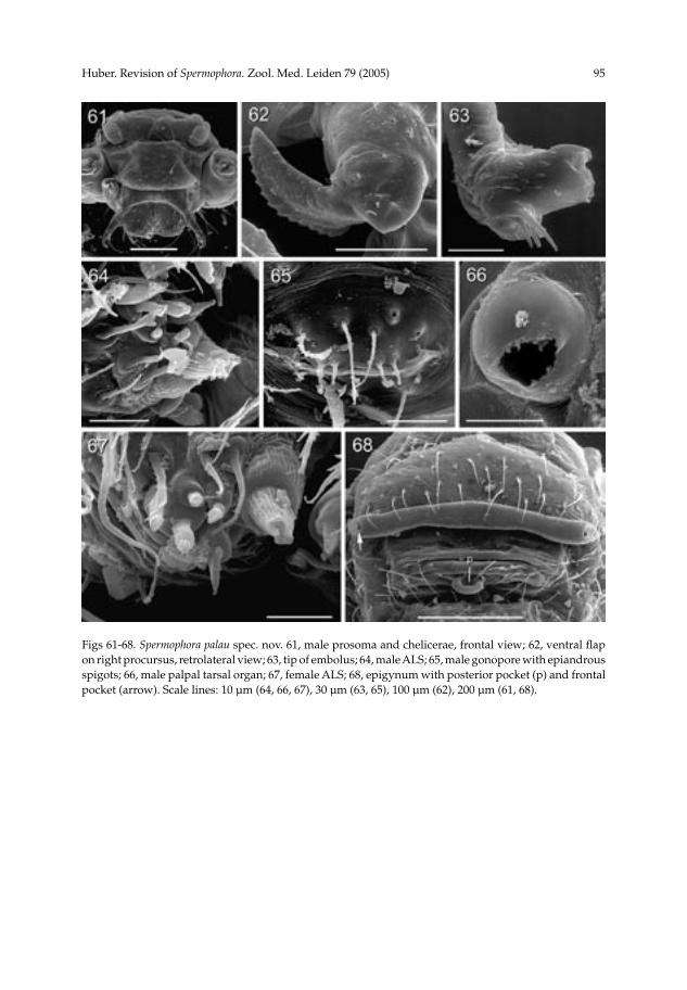
Huber. Revision of Spermophora. Zool. Med. Leiden 79 (2005) 95
Figs 61-68. Spermophora palau spec. nov. 61, male prosoma and chelicerae, frontal view; 62, ventral flap on right procursus, retrolateral view; 63, tip of embolus; 64, male ALS; 65, male gonopore with epiandrous spigots; 66, male palpal tarsal organ; 67, female ALS; 68, epigynum with posterior pocket (p) and frontal pocket (arrow). Scale lines: 10 µm (64, 66, 67), 30 µm (63, 65), 100 µm (62), 200 µm (61, 68).

96 Huber. Revision of Spermophora. Zool. Med. Leiden 79 (2005)
Figs 69-71. Spermophora kaindi spec. nov. 69-70, left male procursus and bulb in prolateral (69) and retrolateral (70) views; 71, male chelicerae, frontal view. Scale lines: 0.3 (69-71).

Huber. Revision of Spermophora. Zool. Med. Leiden 79 (2005) 97
Figs 72-76. Spermophora estebani Simon. 72-73, left male palp in prolateral (72) and retrolateral (73) views; 74, male chelicerae, frontal view; 75-76, cleared epigynum in ventral (75) and dorsal (76) views. Scale lines: 0.2 (74-76), 0.5 (72, 73).
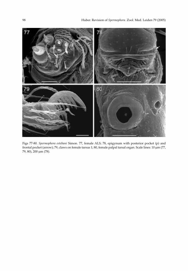
98 Huber. Revision of Spermophora. Zool. Med. Leiden 79 (2005)
Figs 77-80. Spermophora estebani Simon. 77, female ALS; 78, epigynum with posterior pocket (p) and frontal pocket (arrow); 79, claws on female tarsus 1; 80, female palpal tarsal organ. Scale lines: 10 µm (77, 79, 80), 200 µm (78).

Huber. Revision of Spermophora. Zool. Med. Leiden 79 (2005) 99
Figs 81-85. Spermophora luzonica spec. nov. 81-82, left male palp in prolateral (81) and retrolateral (82) views; 83, male chelicerae, frontal view; 84-85, cleared epigynum in ventral (84) and dorsal (85) views. Scale lines: 0.3 (81, 82), 0.2 (83-85).

100 Huber. Revision of Spermophora. Zool. Med. Leiden 79 (2005)
Figs 86-91. Spermophora sumbawa spec. nov. 86-87, left male palp in prolateral (86) and retrolateral (87) views; 88-89, male chelicerae, frontal view, Sumbawa (88) and Lombok (89); 90-91, cleared epigynum in ventral (90) and dorsal (91) views. Scale lines: 0.2 (86-91).

Huber. Revision of Spermophora. Zool. Med. Leiden 79 (2005) 101
Figs 92-103. Spermophora sumbawa spec. nov. 92, male chelicerae, frontal view; 93, genital bulb with apophysis and embolus (e); 94, apophysis and embolus (e) on genital bulb; 95, male palpal tarsal organ; 96, 97, male gonopores with epiandrous spigots; 98, tip of female pedipalp with tarsal organ; 99, male ALS and PMS; 100, female ALS and PMS; 101, epigynum, ventral view; 102, epigynum, slightly posterior view, with posterior pocket (p); 103, modified ventral hairs on female tarsus 4 (arrows). Scale lines: 10 µm (95, 97-100), 30 µm (94, 96, 103), 50 µm (92), 100 µm (93, 101, 102).

102 Huber. Revision of Spermophora. Zool. Med. Leiden 79 (2005)
Figs 104-109. Aetana n. gen., spp. 104-105, A. omayan spec. nov., male habitus, dorsal view, and female abdomen, ventral view; 106-107, A. fiji spec. nov., female habitus, dorsal view, and female abdomen, ventral view; 108-109, A. kinabalu spec. nov., male habitus, dorsal and lateral views.
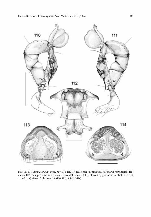
Huber. Revision of Spermophora. Zool. Med. Leiden 79 (2005) 103
Figs 110-114. Aetana omayan spec. nov. 110-111, left male palp in prolateral (110) and retrolateral (111) views; 112, male prosoma and chelicerae, frontal view; 113-114, cleared epigynum in ventral (113) and dorsal (114) views. Scale lines: 1.0 (110, 111), 0.5 (112-114).

104 Huber. Revision of Spermophora. Zool. Med. Leiden 79 (2005)
Figs 115-120. Aetana fiji spec. nov. 115-116, left male palp in prolateral (115) and retrolateral (116) views; 117-118, male prosoma and chelicerae, dorsal (117) and frontal (118) views; 119-120, cleared epigynum in ventral (119) and dorsal (120) views. Scale lines: 0.5 (115-120).

Huber. Revision of Spermophora. Zool. Med. Leiden 79 (2005) 105
Figs 121-123. Aetana kinabalu spec. nov. 121-122, left male palp in prolateral (121) and retrolateral (122) views; 123, male chelicerae, frontal view. Scale lines: 0.5 (121, 122), 0.3 (123).

106 Huber. Revision of Spermophora. Zool. Med. Leiden 79 (2005)
Figs 124-130. Savarna n. gen., spp. 124-126, S. thaleban spec. nov., male habitus, dorsal and lateral views, and female abdomen, ventral view; 127-128, S. baso (Roewer) n. comb., male habitus, dorsal and lateral views; 129-130, S. tessellata (Simon) n. comb., male habitus, dorsal and lateral views.

Huber. Revision of Spermophora. Zool. Med. Leiden 79 (2005) 107
Figs 131-134. Savarna thaleban spec. nov. 131-132, left male palp in prolateral (131) and retrolateral (132) views; 133, male chelicerae, frontal view; 134, cleared epigynum, dorsal view. Scale lines: 0.5 (131, 132), 0.2 (133), 0.3 (134).

108 Huber. Revision of Spermophora. Zool. Med. Leiden 79 (2005)
Figs 135-140. S. baso (Roewer) n. comb. (135-137), and S. tessellata (Simon) n. comb. (138-140). 135-136, right male palp in retrolateral (135) and prolateral (136) views; 137, male chelicerae, frontal view; 138-139, left male palp in prolateral (138) and retrolateral (139) views; 140, male chelicerae, frontal view. Scale lines: 0.5 (135, 136, 138, 139), 0.2 (137, 140).

Huber. Revision of Spermophora. Zool. Med. Leiden 79 (2005) 109
Figs 141-150. Khorata n. gen., spp. 141-143, K. khammouan spec. nov., male habitus, dorsal and lateral views, and female abdomen, ventral view; 144-145, K. jaegeri spec. nov., male habitus, dorsal and lateral views; 146-147, K. schwendingeri spec. nov., male habitus, dorsal and lateral views; 148-150, K. bangkok spec. nov., male habitus, dorsal and lateral views, and female abdomen, ventral view.

110 Huber. Revision of Spermophora. Zool. Med. Leiden 79 (2005)
Figs 151-156. Khorata khammouan spec. nov. 151-152, left male palp in prolateral (151) and retrolateral (152) views; 153-154, male chelicerae, frontal and lateral views; 155-156, cleared epigynum, ventral (155) and dorsal (156) views. Scale lines: 0.5 (151, 152), 0.3 (153, 154), 0.4 (155, 156).

Huber. Revision of Spermophora. Zool. Med. Leiden 79 (2005) 111
Figs 157-160. Khorata jaegeri spec. nov. 157-158, left male palp in prolateral (157) and retrolateral (158) views; 159-160, male chelicerae, frontal and lateral views. Scale lines: 0.5 (157-160).

112 Huber. Revision of Spermophora. Zool. Med. Leiden 79 (2005)
Figs 161-167. Khorata schwendingeri spec. nov. (161-165) and K. bangkok spec. nov. (166, 167). 161-162, left male palp in prolateral (161) and retrolateral (162) views; 163-164, male chelicerae, lateral and frontal views; 165-166, left procursi, prolateral views (at same magnification) (arrow points to sclerite that differs slightly in males from Laos); 167, cleared epigynum, dorsal view. Scale lines: 0.5 (161, 162), 0.3 (163-167).

Huber. Revision of Spermophora. Zool. Med. Leiden 79 (2005) 113
Fig. 168. Scatter diagram showing the tight correlation between the distance between the tips of the male cheliceral apophyses and the distance between the pockets on the female epigynum in the core group of Spermophora. The line marks perfect isometry.
Fig. 169. Known distribution of the core group of Spermophora. Data on Australian species from Huber (2001). Note that the cosmopolitan S. senoculata is not included. For further details, see text.

114 Huber. Revision of Spermophora. Zool. Med. Leiden 79 (2005)
Fig. 170. Known distribution of Aetana n. gen.
Fig. 172. Known distribution of Khorata n. gen.Fig. 171. Known distribution of Savarna n. gen.