


Revised Manuscript: M4:00292 Angiopoietin-3 is tethered on the cell
45
Revised Manuscript: M4:00292 Angiopoietin-3 is tethered on the cell surface via heparan sulfate proteoglycans Yin Xu, Yao-juan Liu and Qin Yu * Department of Pathobiology School of Veterinary Medicine University of Pennsylvania Philadelphia, PA 19104 Running Title: Angiopoietin-3 binds to the cell surface via HSPGs Key Words: Angiopoietin, Heparan Sulfate Proteoglycan, Angiogenesis, Endothelial cell To whom correspondence should be addressed: Qin Yu, Ph. D. 372E (old vet) 3800 Spruce Street Department of Pathobiology School of Veterinary Medicine University of Pennsylvania Philadelphia, PA 19104 Tel: 215-898-2967 Fax: 215-898-0719 Angiopoietin-3 binds to the cell surface via HSPGs 1 JBC Papers in Press. Published on July 27, 2004 as Manuscript M400292200 Copyright 2004 by The American Society for Biochemistry and Molecular Biology, Inc. by guest on November 15, 2018 http://www.jbc.org/ Downloaded from
Transcript of Revised Manuscript: M4:00292 Angiopoietin-3 is tethered on the cell
Revised Manuscript: M4:00292
Angiopoietin-3 is tethered on the cell surface via heparan sulfate proteoglycans
Yin Xu, Yao-juan Liu and Qin Yu*
Department of Pathobiology
School of Veterinary Medicine
University of Pennsylvania
Philadelphia, PA 19104
Running Title: Angiopoietin-3 binds to the cell surface via HSPGs
Key Words: Angiopoietin, Heparan Sulfate Proteoglycan, Angiogenesis, Endothelial cell
To whom correspondence should be addressed:
Qin Yu, Ph. D.372E (old vet)
3800 Spruce StreetDepartment of Pathobiology
School of Veterinary MedicineUniversity of Pennsylvania
Philadelphia, PA 19104Tel: 215-898-2967Fax: 215-898-0719
Angiopoietin-3 binds to the cell surface via HSPGs
1
JBC Papers in Press. Published on July 27, 2004 as Manuscript M400292200
Copyright 2004 by The American Society for Biochemistry and Molecular Biology, Inc.
by guest on Novem
ber 15, 2018http://w
ww
.jbc.org/D
ownloaded from
E-mail: [email protected]
SUMMARY
Angiopoietins are a family of factors that play important roles in angiogenesis, and their receptor, Tie-
2 receptor tyrosine kinase, is expressed primarily by endothelial cells. Three angiopoietins have been
identified so far, angiopoietin-1 (Ang-1), angiopietin-2 (Ang-2), and angiopoietin-3 (Ang-3). It has
been established that Ang-1 and Tie-2 play essential roles in embryonic angiogenesis. We have
demonstrated recently that unlike Ang-2, Ang-1 binds to the extracellular matrix (ECM), which
regulates the availability and activity of Ang-1 (1). However, the role and biochemical characteristics
of Ang-3 are unknown. In our current study, we demonstrated that unlike Ang-1 and Ang-2, Ang-3
is tethered on cell surface via heparan sulfate proteoglycans (HSPGs), especially perlecan. The cell-
surface bound Ang-3 is capable of binding to its receptor, Tie-2; suggesting HSPGs concentrate Ang-
3 on the cell surface and present Ang-3 to its receptor to elicit specific local reaction. Mutagenesis
experiment revealed that the coiled-coil domain of Ang-3 is responsible for its binding to the cell
surface. In addition, we demonstrated that the cell-surface bound Ang-3 but not soluble Ang-3
induces retraction and loss of integrity of endothelial monolayer, indicating the binding of Ang-3 to
the cell surface via HSPGs is require for this bioactivity of Ang-3.
Angiopoietin-3 binds to the cell surface via HSPGs
2
by guest on Novem
ber 15, 2018http://w
ww
.jbc.org/D
ownloaded from
Introduction
Angiogenesis plays important roles in embryonic organogenesis, postnatal tissue repair, female
reproductive function, arthritis, diabetes, and tumor growth and metastasis (2, 3, 4, and 5). Numerous
molecules, including pro- and anti-angiogenic factors and their receptors, proteases, adhesion
receptors, and the ECM components, are involved in angiogenesis (6, 7, and 8).
Angiopoietins are ligands of Tie-2 (tyrosine kinase with immunoglobulin and epidermal growth
factor homology domains-2) receptor kinase, which is primarily expressed by endothelial cells and
their precursors (9, 10, 11, and 12). Three Tie-2 ligands have been identified so far (13, 14, and 15),
angiopoietin-1 (Ang-1), angiopietin-2 (Ang-2), and angiopoietin-3 (Ang-3) in mouse or its ortholog
in human, angiopoietin-4 (Ang-4). Angiopoietins all contain a signal peptide, an amino terminal
coiled-coil domain, a linker peptide region, and a carboxyl terminal fibrinogen homology domain
(FHD). Studied have shown that the coiled-coil region is responsible for dimerization/mulimerization
of angiopoietins, while the FHD binds to Tie-2 receptor (14, 15, and 16).
It has been shown that angiopoietin-Tie-2 pathway plays an essential role during the late stages
Angiopoietin-3 binds to the cell surface via HSPGs
3
by guest on Novem
ber 15, 2018http://w
ww
.jbc.org/D
ownloaded from
of vascular development. Knock out of Ang-1 or Tie-2, or overexpression of Ang-2 is embryonic
lethal with similar vascular defects. These mice display normal vascular growth factor (VEGF)-
dependent early vascular development, however, there are profound defects in remodeling,
organization, and stabilization of the primitive vasculature in these mice (12, 14, 17, and 18). Studies
have shown that at least under some circumstances, Ang–2 and Ang-3 are naturally occurring
antagonists of Tie-2 receptor (14, 15). Ang-2 and Ang-3 are believed to compete with Ang-1 for the
binding of Tie-2 and block Tie-2 phosphorylation induced by Ang-1 (14, 15, 19, and 20). However,
how Ang-3 affects endothelial cell behavior and how bioactivity of Ang-3 is regulated have not been
established.
We have demonstrated previously that Ang-2 is primarily secreted and Ang-1 is incorporated
into the ECM and the ECM binding of Ang-1 regulates its availability and activity (1). In our current
study, we demonstrated that unlike Ang-1 and Ang-2, Ang-3 is tethered on cell surface via heparan
sulfate proteoglycans (HSPGs), especially perlecan, through the coiled-coil domain of Ang-3.
Perlecan is a HSPG that is present in the basement membrane and on cell surface (21). Perlecan binds
to the cell surface through integrin (22, 23, 24), and plays important roles in vascularogenesis,
angiogenesis, and tumorigenesis (21, 31). In addition, we have shown that the cell-surface bound
Ang-3 is capable of binding to Tie-2-Fc fusion protein. More importantly, the cell-surface bound
Ang-3 but not soluble Ang-3 induces retraction and loss of integrity of the endothelial monolayer,
indicating that the cell surface binding of Ang-3 is required for its function.
We have shown very recently that overexpression of Ang-3 inhibits pulmonary metastasis of
Angiopoietin-3 binds to the cell surface via HSPGs
4
by guest on Novem
ber 15, 2018http://w
ww
.jbc.org/D
ownloaded from

Lewis lung carcinoma and TA3 mammary carcinoma cells by blocking tumor angiogenesis and
promoting apoptosis of tumor cells (Xu et al., In Press)1. Furthermore, we have demonstrated that
tethering Ang-3 on the cell surface is required for effective inhibition of Ang-3 on tumor angiogenesis
and pulmonary metastasis (Xu et al., In Press)1. These results further confirmed that binding of Ang-3
to the cell surface via HSPGs enhances/facilitates the bioactivity of Ang-3.
The results reported in our previous (1) and current study demonstrated that angiopoietins not
only play different roles in regulating endothelial cell function, but also their activities are
differentially regulated by the ECM and HSPGs, which likely provides the basis for modulating
activities of angiopoietins in vivo therefore regulating angiogenesis in physiologic and pathologic
situations.
Experimental Procedures
Cells and Reagents
Lewis lung carcinoma (LLC) cell, A10 embryonic aorta smooth muscle (A10) cell, bovine pulmonary
artery endothelial cell (CAPE), C2C12 myoblast, and Cos-7 cell were obtained from the Cell Center
Services Facility of University of Pennsylvania. LLC transfectants were maintained in the condition as
described previously (1, 25). Anti-v5 epitope (Invitrogen), anti-Tie2 and anti-Ang-1, -2, and –3, and
anti-glypican-1 (Santa Cruz), anti-heparan sulfate (CalBiochem), anti-perlecan (NeoMarkers), and
anti-syndecan-1 (BD Bioscience) antibodies, heparinase I and III (Sigma), Str. hyaluronidase (ICN),
and Tie-2-Fc (R&D Systems) were used in the experiments.
Angiopoietin-3 binds to the cell surface via HSPGs
5
by guest on Novem
ber 15, 2018http://w
ww
.jbc.org/D
ownloaded from

Reverse Transcriptase-polymerase Chain Reaction (RT-PCR), Mutagenesis, and Expression
Constructions
RT-PCR was performed as described (25). Briefly, full-length Ang-1, -2, and -3 cDNAs were
amplified using mouse placenta cDNAs as templates together with Pfu DNA polymerase (Stratagene)
and the pairs of primers corresponding to the 5’ and 3’ end of 24 nucleotides of the coding sequence of
the each molecule derived from the GenBank under the accession numbers U83509, AF004326, and
AF113707, respectively. The stop codons were omitted from these reverse primers to fuse Ang-1, -2
and -3 to the C-terminal v5 epitope tag, which presents in the expression vector (pEF6/V5-His
TOPO, Invitrogen). The resulting PCR fragments were inserted into pEF6/V5-His TOPO vectors.
Authenticity and correct orientation of Ang-1, -2 and -3 inserts were verified by DNA sequencing.
The various deletion fragments of Ang-3 were generated as detailed in the figure using the full-
length Ang-3 in pEF6/v5-His vector as a template and the ExSite PCR-based site-directed
mutagenesis kit (Stratagene). The accuracy of the deletions was confirmed by DNA sequencing.
Transient and Stable Transfection
Cos-7 cells and LLC cells were transfected using Lipofectamine (Invitrogen) with the expression
constructs containing cDNA inserts encoding mouse Ang-1, Ang-2, Ang-3, or the deletional
constructs of Ang-3. The transient transfected Cos-7 cells were used 72 hours after the transfection.
The stably transfected LLC cells were selected for blasticidin resistance. The expression level of v5-
epitope tagged Ang-1, Ang-2, full-length and deletion fragments of Ang-3 in Cos-7 and LLC
Angiopoietin-3 binds to the cell surface via HSPGs
6
by guest on Novem
ber 15, 2018http://w
ww
.jbc.org/D
ownloaded from

transfectants was determined by Western blotting with anti-v5 antibody as described previously (1).
Protein Sample Preparation and Western Blot Analysis
To determine the distribution pattern of angiopoietins, Western blot was performed using the proteins
derived from the cell culture supernatants of LLC transfectants or transiently transfected Cos-7 cells,
lysates of the EDTA-lifted cells, and the ECM materials deposited on the cell culture dishes. The
transfected cells were either treated with trypsin for 10min at room temperature and washed with
10%FBS DMEM, or released from the culture dishes by phosphate-buffered saline solution (PBS)
containing 5mM EDTA and washed. The trypsin or EDTA-released cells were washed and lysed in 2
x SDS Laemmli sample buffer. The ECM materials remained on the cell culture dishes after removal
the cells with EDTA solution were washed and extracted with 2 x SDS sample buffer.
To determine the binding between the cell surface heparan sulfate (HS) and Ang-3, the EDTA-
lifted LLCAng-3 cells were treated with heparinase I (5units/ml) plus heparinase III (0.5unit/ml) or
Str. hyaluronidase (5unit/ml, as a control for heparinase) at 370C for two hours, washed, lysed in 2 x
SDS sample buffer and analyzed by Western blotting with anti-v5 antibody to determine whether
heparinases have dissociated Ang-3 protein from LLCAng-3 cell surface.
To determine whether glycosylphosphotidylinositol (GPI)-anchored glypicans bind to Ang-3,
LLCAng-3 cells were treated with or without 1uint/ml of phosphatidyl-inositol-specific
phospholipase C (PI-PLC) at 370C for two hours. These LLCAng-3 cells were washed and lysed with
RIPA buffer (50mMTris, pH7.4, 150mMNaCl, 1% Triton X-100, 1mMEDTA, 1µg/ml pepstain A,
Angiopoietin-3 binds to the cell surface via HSPGs
7
by guest on Novem
ber 15, 2018http://w
ww
.jbc.org/D
ownloaded from

1mMPMSF). 30µl (50µg) of lysed proteins were digested with or without heparinase I and III (1
unit/ml) at 370 for 6 hours (26), and the reactions were terminated by adding 5µl of 8xSDS sample
buffer and analyzed by Western blot with anti-glypican-1 (Santa Cruz) or anti-v5 antibody.
Immunoprecipitation was performed using the protein G bead bound anti-gnypican-1 antibody and
the supernatants of the PI-PLC digestion reactions. The immunoprecipitated proteins were digested
with or without heparinases as described above and analyzed by Western blot for the presence of
glypican-1 and Ang-3 protein.
Production and Purification of Angiopoietins
Five liters of cell culture supernatants derived from LLC transfectants expressing mouse or human
Ang-1, Ang-2, or Ang-3 protein were collected and purified through Ni+-Probond affinity columns
(Invitrogen) and the affinity columns conjugated with anti-v5 antibodies (Sigma). Purity of the
purified proteins was determined by silver staining of SDS-10% PAGE gel loaded with the purified
proteins under reducing conditions. The result showed that v5-epitope tagged Ang-1, Ang-2, and
Ang-3 reached more than 90% purity after two rounds of affinity purification, which was
demonstrated by the presence of a single silver stained band with the appropriate molecular weight.
Concentration of the purified proteins was determined by Bio-Rad Bradford protein assay using a
serial diluted bovine serum albumin (BSA, 2mg/ml) as a standard.
Solid Phase and Cell-based Ligand Binding Assay
Angiopoietin-3 binds to the cell surface via HSPGs
8
by guest on Novem
ber 15, 2018http://w
ww
.jbc.org/D
ownloaded from

96-well Elisa plates were coated overnight at 4oC with heparin, chondroitin sulfate, or hyaluronic acid
(HA, 1mg/ml, Sigma), or BSA (1mg/ml, Sigma) in triplicate. The coated plates were washed and
blocked with 0.5% BSA. Purified Ang-1v5 or Ang-3v5 proteins (500ng/ml) were applied to the
coated plates and incubated for overnight at 4oC. After extensive washing with phosphate buffer
containing 300mM NaCl, the 96-well bound Ang-1v5 or Ang-3v5 proteins were detected and
measured.
The cell-based ligand binding assays were performed by incubating purified Ang-3v5 (1µg) or
Ang-1v5 (1µg, as a control of Ang-3v5) with the EDTA-lifted 1x10⁶ of C2C12, A10, Cos-7, and
LLC cells; or by incubating purified Tie-2-Fc (2µg, Ρ&D System) or CD-8-Fc (2µg, as a control of
Tie-2-Fc) with the EDTA-lifted LLCAng-1, LLCAng-2, or LLCAng-3 cells at 40C for 2 hours. The
cells were then washed with PBS and lysed in 2x SDS sample buffer. The cell surface bound v5-
tagged angiopoietins or Tie-2-Fc fusion proteins were detected by Western blotting with anti-v5 mAb
or anti-human IgG antibody, respectively.
Heparin Affinity Column
To determine the binding profile and relative affinity of Ang-3 to heparin, 20µg of purified Ang-3v5
were applied into a heparin affinity column (heparin conjugated agarose, Sigma). Ang-3 protein was
eluted from the column using non-continuous gradient of NaCl (0.15, 0.3, 0.6, and 1.2M). Three
fractions (2ml/each) were collected for each NaCl concentration, and 20µl sample from each fraction
was analyzed by Western blotting with anti-v5 mAb.
Angiopoietin-3 binds to the cell surface via HSPGs
9
by guest on Novem
ber 15, 2018http://w
ww
.jbc.org/D
ownloaded from

Endothelial and Tumor Cell Co-culture Assay
Subconfluent LLCAng-3, LLCAng-2, and LLCAng-1 cells were labeled with CellTracker Green
CMFDA fluorescein (Molecular Probes) as described (27) and lifted with the EDTA solution, which
was known to preserve Ang-3 protein on the cell surface. These EDTA-lifted cells or soluble
angiopoietin (200ng/ml) were applied to the monolayers of bovine pulmonary artery endothelial cells
(CAPE, ATCC) in triplicate for four hours at 37 0C. The cells were then fixed and observed under
microscope and photographed and the endothelial retraction lesions were counted in ten randomly
selected microscopic (40x) fields.
Immuocytochemistry
LLC transfectants expressing Ang-1, Ang-2 or Ang-3, or transfected with the empty expression
vector were cultured in 35mm dishes until confluence. Some cells were lifted with the EDTA solution
as described (1), and the cell-free ECM and the confluence LLC transfectants were fixed with
methanol at –20oC for 15 minutes. The fixed cells and ECM were washed and blocked, and antibodies
against v5 epitope, Ang-1, Ang-2, Ang-3, perlecan were used to detected Ang-1, -2, or -3, or
perlecan.
To investigate the relative localization of Ang-3 protein and heparan sulfate (HS) on LLCAng-3
cell surface, immunocolocalization experiment was performed using anti-v5 antibody and TRITC-
conjugated secondary antibody to detect Ang-3v5 protein and anti-heparan sulfate (Calbiochem)
antibody and FITC-conjugated secondary antibody to detect HS, respectively. To determine the
Angiopoietin-3 binds to the cell surface via HSPGs
10
by guest on Novem
ber 15, 2018http://w
ww
.jbc.org/D
ownloaded from
relative localization of Ang-3 and perlecan, glypican, or syndecan and determine whether heparan
sulfate (HS) side chains on proteoglycans are required for the binding of Ang-3 to cell surface,
LLCAng-3 cells were cultured in the absence or presence of 100mM of sodium chlorate and fixed.
Immunocolocalization experiments were performed using anti-v5 antibody with TRITC-conjugated
secondary antibody to detect Ang-3v5, and anti-perlecan (NeoMarkers) or anti-glypican-1 antibody
with FITC-conjugated secondary antibody to detect perlecan or glypican-1, respectively; or using
FITC-conjugated anti-v5 antibody to detect Ang-3v5 protein and PE-conjugated anti-syndecan-1
(BD Bioscience) antibody to detect syndecan-1. The inhibitory effect of sodium chlorate on synthesis
of HS in LLCAng-3 cells was revealed by anti-HS antibody (CalBiochem).
To determine whether the cell surface-bound Ang-3 binds to Tie-2-Fc fusion protein, the
methanol fixed LLCAng-3 cells were washed, blocked, and incubated with Tie-2-Fc. The cell-
surface bound Tie-2-Fc fusion protein was revealed by TRITC-conjugated anti-human Fc antibody,
and Ang-3v5 protein in the same cells was detected with anti-v5 mAb with FITC-conjugated anti-
mouse secondary antibody.
Results
Unlike Ang-1 and Ang-2, Ang-3 is retained on the surface of various cells
It was noted previously that unlike producing Ang-2, it is difficult to produce and purify Ang-1
and Ang-3 proteins (14, 15). We have demonstrated recently that unlike Ang-2, which is primarily
Angiopoietin-3 binds to the cell surface via HSPGs
11
by guest on Novem
ber 15, 2018http://w
ww
.jbc.org/D
ownloaded from

secreted, Ang-1 is secreted and incorporated into the ECM and the binding of Ang-1 to the ECM
regulates availability and activity of Ang-1 (1). To determine whether Ang-3 binds to the ECM as
well, we investigated the distribution pattern of Ang-3 protein in the established LLC transfectants
expressing the C-terminal v5 epitope tagged Ang-3 (Ang-3v5). The v5 epitope is a 14 amino acid
epitope derived from P and V proteins of the paramyxovirus, SV5 (28).
The proteins used in the Western blotting were derived from the culture supernatants, the ECM
materials deposited on the cell culture dishes, and the EDTA-lifted LLC transfectants expressing v5-
tagged Ang-1 (LLCAng-1), Ang-2 (LLCAng-2), or Ang-3 (LLCAng-3). The results showed that
Ang-2 is largely secreted, Ang-1 is incorporated into the ECM, and a large fraction of Ang-3 is
associated with the transfected cells (Fig 1). The similar result was obtained in several other cell lines
tested including Cos-7, 293, and TA3 mammary carcinoma cells (data not shown). Like all the
proteins containing the coiled-coil domain, angiopoietins are aggregated to form dimers and oligomers
through their coiled-coil region and treatment of the proteins with β↑mercaptoethanol releases these
aggregates to monomers (13, 14, 15, 16, and data not shown). Angiopoietins are glycosylated, which
causes the multiple banding patterns in Western blot (13, 14, and 15). The ECM-bound Ang-1 is
aggregated to form oligomers, while the cell-associated Ang-3 exists in monomeric, dimeric, and
some oligomeric forms (Fig 1C, and Fig 2).
To determine whether endogenous Ang-3 behaves in the same way as the transfected ones, we
performed Western blot analysis of the proteins derived from C2C12 myoblasts and A10 embryonic
aortic smooth muscle cells, both of which express endogenous Ang-3 as assessed by RT-PCR (Xu et
Angiopoietin-3 binds to the cell surface via HSPGs
12
by guest on Novem
ber 15, 2018http://w
ww
.jbc.org/D
ownloaded from
al., In Press)1. Proteins derived from lysates of these cells were analyzed by Western blotting with
anti-Ang-3 antibody (Santa Cruz), and the results showed that Ang-3 protein is associated to A10 and
C2C12 cells as well (data not shown).
To determine whether the association of Ang-3 with LLC cells merely reflects the presence of
Ang-3 protein in intracellular secretory pathway of the cells or represents binding of Ang-3 to the cell
surface, we performed the following experiments. First, LLCAng-3 cells were treated with trypsin
(0.5mg/ml) or EDTA solution for 10 min at room temperature (RT), lifted, washed, lysed, and
analyzed by Western blotting with anti-v5 mAb. The result showed that the treatment of trypsin, but
not EDTA, dislodges Ang-3 protein from the cells suggesting that Ang-3v5 is localized on the cell
surface where it is accessible to trypsin (Fig 2A, lane 3-4). To confirm this, we performed Western
blot analysis of the proteins derived the cell membrane and soluble cytosolic fractions of LLCAng-3
cells, and found that Ang-3v5 is indeed present in the cell membrane fraction (Fig 2B, lanes 1-2).
Finally, a cell based binding assay was performed by incubating the EDTA-lifted C2C12, A10,
Cos-7, and LLC cells (1x10⁶) with purified Ang-3v5 or Ang-1v5 (2µg ) at 40C for 2 hours. The cells
were washed with cold PBS, lysed, and analyzed by Western blotting with anti-v5 mAb. The result
showed that Ang-3v5 but not Ang-1v5 binds to all the cells tested (Fig 2C).
The cell-surface bound Ang-3 is capable of binding to Tie-2-Fc protein
RT-PCR results indicated that these non-endothelial cells that displayed the binding affinity to
Ang-3 protein (Fig 2C) do not express Tie-2 receptor (data not shown), suggesting that Ang-3 binds
Angiopoietin-3 binds to the cell surface via HSPGs
13
by guest on Novem
ber 15, 2018http://w
ww
.jbc.org/D
ownloaded from
to these cell surface via binding partner(s) other than Tie-2 receptor. To determine whether the cell
surface-bound Ang-3 is capable of binding to Tie-2-Fc fusion protein, 1x10⁶ of the EDTA-lifted
LLC transfectants (LLCAng-1, -2, and -3) were incubated with 2µg of Tie-2-Fc or CD8-Fc (as a
negative control) at 40C for 2 hours. After extensive washing, the cells were lysed and Western
blotting was performed using anti-human IgG antibody to detect cell surface bound Tie-2-Fc or
CD8-Fc. The result showed that Tie-2-Fc but not CD8-Fc binds to LLCAng-3 cells but not to
LLCAng-1 and LLCAng-2 cells (Fig 3A).
To confirm this result, confluent LLCAng-3 cells were fixed, blocked, and the binding assay was
performed by applying 2µg/ml of Tie-2-Fc or CD8-Fc proteins (with or without prior incubation with
20µg/ml soluble Ang-3 protein) to these fixed cells. The cell surface bound Tie-2-Fc was revealed
by FITC-conjugated anti-human Fc antibody. The result showed that Tie-2-Fc but not CD8-Fc binds
to the surface of LLCAng-3 cells in a pattern that is similar to that of cell surface tethered Ang-3
protein, and the binding is blocked by the prior incubation of T-e-2-Fc with excess amount of soluble
Ang-3 protein (Fig 3B). To demonstrate the co-localization of the cell-surface tethered Ang-3 and
Tie-2-Fc fusion protein, the distribution of Ang-3 and Tie-2-Fc was revealed by anti-v5 antibody
with the FITC-conjugated secondary antibody, and TRITC conjugated anti-human Fc antibody,
respectively. The result showed that Tie-2-Fc is colocalized with the cell surface tethered Ang-3 (Fig
3C-c). Together, these results suggest that tethering Ang-3 on the cell surface likely provides a
mechanism to localize, concentrate and present Ang-3 to its receptor rather than to sequester Ang-3,
and that the domains of Ang-3 that bind to Tie-2 and the putative cell surface binding partner(s) are
Angiopoietin-3 binds to the cell surface via HSPGs
14
by guest on Novem
ber 15, 2018http://w
ww
.jbc.org/D
ownloaded from
different.
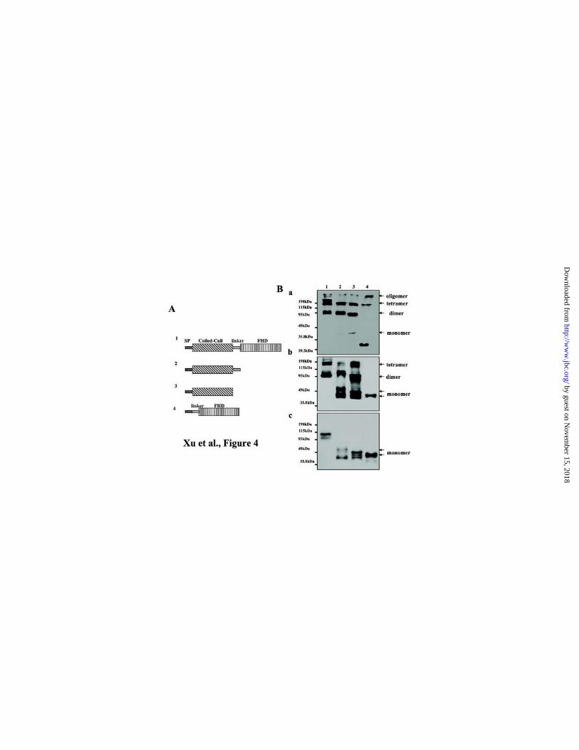
The coiled-coil domain of Ang-3 mediates the cell surface binding of Ang-3.
To determine which domain of Ang-3 mediating the binding of Ang-3 to the cell surface, we
made several Ang-3 deletion constructs, which contain the coiled-coil (C-C) domain plus the linker
peptide region, the C-C domain, or the linker peptide region plus the FHD (Fig 4A). The cDNA
fragment encoding the signal peptide (SP) of Ang-3 was constructed to the N-terminal of all the
deletion fragments, so that they can be expressed and secreted properly (1). In addition, v5-epitope
was tagged at the C-termini of these fragments for easy identification. The expression constructs
containing full-length Ang-3 and various Ang-3 fragments were used to transfect Cos-7 cells in
triplicate.
72 hours after the transfection, one set of the transfected Cos-7 cells were treated with trypsin
(0.5mg/ml) at RT for 10 min, which was known to release the cell-surface bound Ang-3 (Fig 2A).
Equal amount of the proteins derived from culture supernatants (Fig 4B-a) or cell lysates (Fig 4B-b
and c) of the transfected Cos-7 cells which were treated with (B-c) or without (B-b) trypsin, were
analyzed by Western blotting with anti-v5 mAb. The results showed that like full-length Ang-3, the
coiled-coil fragment of Ang-3 binds to the cells and the binding is sensitive to the trypsin treatment.
On the contrary, the FHD fragment of Ang-3 is either secreted or reside inside of the transfected cells
and is insensitive to the trypsin treatment (Fig 4B-b, c, lane 4). This result suggests that the coiled-coil
domain of Ang-3 mediates the binding of Ang-3 to the cell surface. This conclusion was further
Angiopoietin-3 binds to the cell surface via HSPGs
15
by guest on Novem
ber 15, 2018http://w
ww
.jbc.org/D
ownloaded from

supported by the immunocytochemistry analysis of these transfected Cos-7 cells. The results showed
that the coiled-coil fragment of Ang-3 displayed a cell surface distribution pattern that is similar to
that of full-length Ang-3, while the FHD fragment is indeed localized in the intracellular compartment
(data not shown).
Perlecan is a major HSPG that mediates the cell surface binding of Ang-3.
Our RT-PCR results indicated that the cells that displayed binding affinity to Ang-3 (Fig 2C)
do not express Tie-2 (data not shown), suggesting that Ang-3 binds to the cell surface via protein(s)
other than Tie-2. To determine which cell surface protein(s) bind(s) to Ang-3, we first investigated
localization of Ang-3 in LLCAng-3 cells by immunocytochemistry. We found that the localization of
Ang-3 resembles that of heparan sulfate (HS) glycosaminoglycans (GAGs) observed previously (not
shown). To determine whether Ang-3 is tethered on the cell surface via HS, we performed solid phase
binding assay to assess binding affinity of purified Ang-3v5 protein to heparin, an analog of HS;
chondroitin sulfate (CS) and hyaluronan (HA), both of which are negative charged GAGs and used as
controls of heparin. Purified Ang-1v5 was used as a control of Ang-3v5 in the assay. The result
showed that Ang-3v5 but not Ang-1v5 binds to heparin but not to CS and HA (Fig 5A), suggesting
that Ang-3 likely binds to the cell surface via HSPGs.
To determine binding profile and relative affinity of Ang-3 to heparin, we applied purified Ang-
3 to a heparin affinity column and Ang-3 was eluted using non-continuous gradient of NaCl (0.15,
0.3, 0.6, and 1.2M). The result showed that Ang-3 protein was eluted at two different salt
Angiopoietin-3 binds to the cell surface via HSPGs
16
by guest on Novem
ber 15, 2018http://w
ww
.jbc.org/D
ownloaded from

concentrations, 0.3M and 0.6M NaCl, which implies the presence two subsets of Ang-3 proteins that
bind to heparin with different affinities (low and high, Fig 5B).
To determine whether Ang-3 colocalizes with HS on the cell surface, we performed immuno-
colocalization experiment using anti-v5 mAb and TRITC-conjugated secondary antibody to detect
Ang-3v5 and anti-heparan sulfate antibody and FITC-conjugated secondary antibody to detect HS,
respectively. Our results showed that Ang-3v5 is colocalized with HS on the surface of LLCAng-3
cells (Fig 5C-a-c). It is well established that HS can exist as side chains of protein core structures in
the forms of HSPGs, which present in the ECM and on cell surface (29, 30, 31). To confirm that Ang-
3 binds to the cell surface via HSPG, the EDTA-lifted LLCAng-3 cells were treated with heparinase
or hyaluronidase (as a control of heparinase) at 370C for two hours, washed, lysed, and analyzed by
Western blotting with anti-v5 mAb. The result showed that the treatment of LLCAng-3 cells with
heparinase but not hyaluronidase releases Ang-3v5 from the cell surface (Fig 5D); suggesting that
Ang-3 is bound to the cell surface through HSPG.
The major cell surface HSPGs are syndecans, glypicans, CD44 variants containing v3 exon
(CD44v3, 32, 33), and perlecan (21, 30, 31). Our RT-PCR results demonstrated that LLC cells express
syndecan-1, -2, and -4, glypican-1, -2, -3, and -6, and perlecan and LLC cells express little CD44
variant isoforms containing v3 exon (data not shown). Thus, CD44 is not likely involved in the binding
of Ang-3 to LLC cell surface. To investigate whether syndecans bind to Ang-3, immunoprecipitation
was performed by incubating Ang-3v5 with the protein-A beads that were bound with syndecan-1-
Fc, syndecan-2-Fc, syndecan-4-Fc fusion proteins, or human IgG (as a control of syn-Fc). The
Angiopoietin-3 binds to the cell surface via HSPGs
17
by guest on Novem
ber 15, 2018http://w
ww
.jbc.org/D
ownloaded from

results showed that syndecan-1-Fc, to a less extent syndecan-4-Fc and syndecan-2-Fc but not
human-IgG, bind to Ang-3v5 (data not shown). This result indicates that syndecans binds to Ang-3 in
vitro.
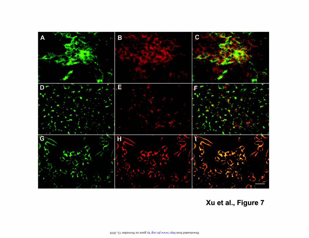
To determine whether syndecan, perlecan, and/or glypican are responsible for tethering Ang-3
on surface of the cells, we performed immunocolocalization of Ang-3v5 and perlecan, syndecan-1, or
glypican-1. The results showed that Ang-3v5 is colocalized with perlecan in both LLC and TA3 cells
(Fig 6 and 7G, H, I), however, Ang-3 does not displayed significant colocalization with syndecan-1
(Fig 7 A, B, C) or glypican-1 (Fig 7D, E, F). To determine whether HS side chains on perlecan are
required for the binding of Ang-3 to the cell surface, LLCAng-3 cells were cultured in the absence or
presence of 100mM of sodium chlorate. The inhibitory effect of sodium chlorate on synthesis of HS
was demonstrated by the absence of HS on the chlorate treated LLCAng-3 cells using anti-HS
antibody (Fig 6H). Immunocolocalization experiment showed that perlecan core protein lacking of HS
side chains is incapable of tethering Ang-3 to LLC cells. Only Ang-3 protein detected is located
inside of the chlorate treated LLCAng-3 cells (Fig 6F). Together, these data suggest that perlecan
tethers Ang-3 to the cell surface through HS side chains.
Western blot result demonstrated that some Ang-3 is deposited into the ECM (Fig 1). To
investigate whether the ECM-bound Ang-3 is colocalized with perlecan, confluent Ang-3 cells were
lifted with the EDTA solution, the cell-free ECM on the cell culture dishes were fixed and
immunocytochemistry was performed to detect the ECM-bound Ang-3 or perlecan. The result showed
that the EDAT lifted LLC cells and the pericellular localized perlecan, however, there are patches of
Angiopoietin-3 binds to the cell surface via HSPGs
18
by guest on Novem
ber 15, 2018http://w
ww
.jbc.org/D
ownloaded from

perlecan left in the cell-free ECM (Fig 6I) and Ang-3 was found to be colocalized with these perlecan
patches in the ECM (Fig 6 J, K).
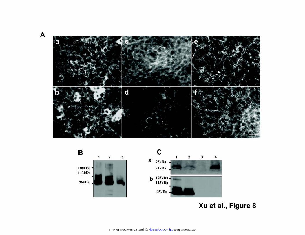
To further confirm that syndecan and glypican are not the major HSPGs that are responsible for
the cell surface binding of Ang-3, we performed the following biochemical experiments. We treated
LLCAng-3 cells without or with PI-PLC which releases GPI-anchored proteins including glypicans
from cell surface. The cells were washed and lysed. The protein lysates were digested with or without
heparinases and analyzed for the presence of glypican-1 or Ang-3, respectively by Western blotting.
The reaction supernatants were immunoprecipitated to recover cleaved glypican-1 and potential
glypican-1 bound Ang-3. The results showed that the PI-PLC treatment did release a large fraction of
glypican-1 from cell surface; however, it has little effect on the cell surface binding of Ang-3. In
addition, the released glypican-1 does not form complex with Ang-3 protein in the reaction
supernatant, suggesting that glypicans are not the major cell surface HSPGs that bind to Ang-3 in LLC
cells (Fig 8C).
To confirm that syndecan-1 play no major role in tethering Ang-3 on LLC cells, we treated
LLCAng-3 cells without or with 10µg/ml trypsin on ice for 15min which was known to be sufficient
to cleave syndecans from epithelial cell surface (34), or with 100µg/ml trypsin in RT for 15min. The
cells were then washed and lysed for Western blotting analysis. The mild trypsin treatment (10µg/ml)
did not lift LLCAng-3 cells from the culture dishes, which allowed us to perform
immunocytochemistry to determine whether the treatment released syndecan-1 from the cell surface.
Our results showed that there is a significant reduction of syndecan-1 but not perlecan-1 level on
Angiopoietin-3 binds to the cell surface via HSPGs
19
by guest on Novem
ber 15, 2018http://w
ww
.jbc.org/D
ownloaded from

surface of these LLCAng-3 cells which were treated with 10µg/ml trypsin (Fig 8A, B); however, there
is a little change of the level of the cell surface bound Ang-3 (Fig 8A, B); suggesting that syndecan-1
does not play a major role in tethering Ang-3 on surface of LLC cells. Together, these results (Fig 6-
8) indicate that at least in LLC and TA3 cells, perlecan is the major HSPG that tethers Ang-3 to the
cell surface.
The cell surface bound Ang-3 induces retraction and loss of integrity of the endothelial monolayer.
It is well established that the interaction between endothelial-endothelial cell and endothelial-
peri-endothelial cells are important for maintaining the vascular integrity and formation of functional
blood vessels. Ang-1 has been shown to play an important role in maintaining integrity of blood
vessels (35, 36, 37, 38, 39, 40). To assess function of the cell surface tethered Ang-3, we performed
tumor and endothelial cell co-culture assay by seeding green fluorescein labeled LLCAng-3,
LLCAng-2, and LLCAng-1 cells onto the monolayers of bovine pulmonary artery endothelial
(CAPE) cells. As controls, purified soluble angiopoietins (200ng/ml) alone were applied to the
monolayers. The cells were cultured for four hours at 37 0C, fixed and observed under microscope.
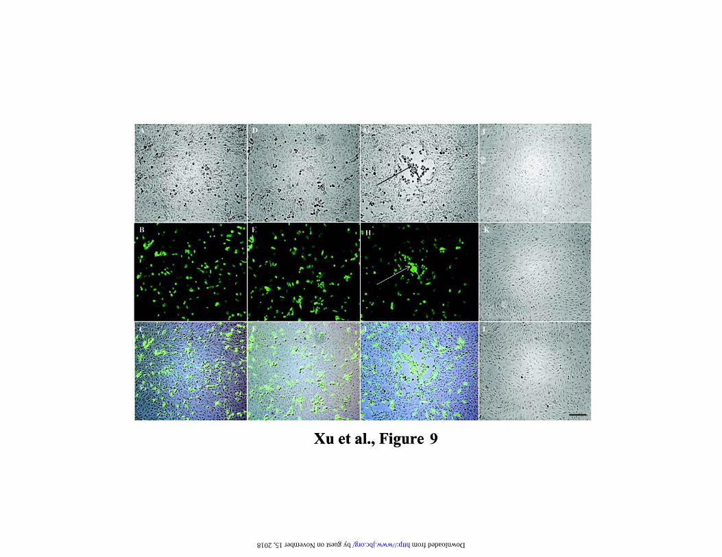
The results demonstrated that the cell surface bound Ang-3, but not soluble Ang-3, LLCAng-1 or
LLCAng-2 cells, induces retraction and loss of integrity of the CAPE monolayers (Fig 9).
The extent of endothelial monolayer retraction was determined by counting the retraction lesions
in ten randomly selected 40x microscopic fields. There are 17 retraction lesions/microscopic field that
were induced by LLCAng-3 cells, while only 0.2, 0.33, or 0 retraction lesions/microscopic field were
Angiopoietin-3 binds to the cell surface via HSPGs
20
by guest on Novem
ber 15, 2018http://w
ww
.jbc.org/D
ownloaded from
induced by LLCAng-1, LLCAng-2 cells, or by soluble Ang-3 alone. Thus, we have demonstrated
that inducing endothelial monolayer retraction is a specific function of the cell surface tethered Ang-3.
This result suggests that binding of Ang-3 to the cell surface via HSPG regulates Ang-3 activity.
Angiopoietin-3 binds to the cell surface via HSPGs
21
by guest on Novem
ber 15, 2018http://w
ww
.jbc.org/D
ownloaded from
Discussion
Ang-3 is tethered on the cell surface via HSPGs, especially perlecan, and the cell surface tethered
Ang-3 binds to Tie-2-Fc fusion protein
In our current study, we demonstrated that unlike Ang-1 and Ang-2, Ang-3 binds to the cell
surface via the interaction between its coiled-coil domain and HSPGs. The cell surface binding of
Ang-3 is saturable, the access amount of Ang-3 is present in soluble form and secreted (Fig 1 and data
not shown), suggesting the local concentration of HSPGs can be used to establish Ang-3 gradient,
which may be important during morphogenesis of the blood vessels. At least in LLC and TA3 cells,
perlecan is a major HSPG that tethers Ang-3 to the cell surface, which does not exclude the possibility
that other HSPGs may mediate cell surface binding of Ang-3 in other cell types.
HSPGs often serve as binding proteins for growth factors and play important roles in modulating
functions of these factors (29, 30, 31 and 41). In the current study, we have shown that the cell surface
bound Ang-3 is capable of binding to Tie-2-Fc fusion protein (Fig 3). This result suggests that
instead of sequestering Ang-3 from its receptor, tethering of Ang-3 on the cell surface such as that of
peri-vascular cells localizes and presents Ang-3 to Tie-2 receptor on adjacent endothelial cells to
elicit specific local reaction. Furthermore, binding of Ang-3 to the cell surface via HSPGs and sharing
the common binding partners with other HSPG-binding growth factors such as bFGF and VEGF
imply that Ang-3 may cross-talk with these factors, and vice versa.
It has been established that the coiled-coil region of angiopoietins is responsible for
dimerization/ mulimerization of the proteins, whereas the FHD binds to Tie-2 receptor (14, 15, and
Angiopoietin-3 binds to cell surface via HSPGs
22
by guest on Novem
ber 15, 2018http://w
ww
.jbc.org/D
ownloaded from
16). We have shown that soluble Ang-3 protein tends to form oligomers; whereas the cell surface
bound Ang-3 tends to form monomer, dimer, and some oligomers (Figs 1, 2, 4). Because the HSPG
binding domain of Ang-3 is in the coiled-coil region, binding of Ang-3 to HSPG is likely responsible
for the altered aggregation pattern. Thus, tethering Ang-3 on the cell surface by HSPGs likely
concentrates Ang-3 protein in the particular aggregated forms, which may generate the distinct local
signal that is different from the one derived from soluble Ang-3 protein.
The function of Ang-3 is regulated by its binding to cell surface
It is well established that the interaction between endothelial-endothelial cells and endothelial-
peri-endothelial cells are important for maintaining vascular health and integrity. Ang-1 has been
shown to enhance the interactions between endothelial-endothelial and endothelial-peri-endothelial
mural cells (18, 35, 38, 39, 40, 42). However, previous work has shed little light on the function of
Ang-3. In our present study, we have shown that the cell-surface bound Ang-3, but not soluble Ang-
3, induces retraction and loss of integrity of the endothelial monolayers (Fig 9). This result implied that
the cell-surface bound Ang-3 plays a distinct important role in regulating endothelial cell behavior,
and mechanisms that regulate the cell-surface binding of Ang-3 and release Ang-3 from the cell
surface (include shedding HSPGs from the cell surface by matrix metalloproteinases, 30, 43) likely
regulate the function of Ang-3.
Very recently, we have shown that overexpression of Ang-3 inhibits pulmonary metastasis of
LLC and TA3 mammary carcinoma cells by inhibiting tumor angiogenesis and promoting apoptosis of
Angiopoietin-3 binds to cell surface via HSPGs
23
by guest on Novem
ber 15, 2018http://w
ww
.jbc.org/D
ownloaded from

the tumor cells, and that the cell surface binding of Ang-3 is required for its effective inhibition on
tumor angiogenesis and metastasis. Furthermore, we have demonstrated that Ang-3 inhibits
endothelial cell proliferation and survival and blocks Ang-1- and VEGF-induced activation of
extracellular signal-regulated kinase 1/2 (Erk1/2) and Akt kinase, which likely underlie the Ang-3-
mediated inhibition on tumor angiogenesis and metastasis (Xu et al., In Press)1. Together with the
results obtained in the current study, these results suggest that Ang-1 and Ang-3 play opposite roles in
maintaining endothelial layer integrity and in inducing endothelial cell sprouting/retraction,
proliferation, and survival. Ang-1 and Ang-3 likely represent two important factors that are produced
by peri-endothelial cells and play antagonistic roles in maintaining health and integrity of blood
vessels in adult tissues. Furthermore, these data imply that the balanced activity of Ang-1 and Ang-3
is important for angiogenesis during embryogenesis and tissue repairing; and the imbalanced up-
regulation of Ang-3 activity and/or down-regulation of Ang-1 activity may contribute to blood vessel
regression and other vascular diseases such as atherosclerosis and restenosis.
HSPGs may enhance/regulate Ang-3 bioactivity by the following mechanisms. We found that
Ang-3 protein is cleaved (data not shown). Thus, tethering Ang-3 to the cell surface via HSPGs likely
protects Ang-3 from proteolytic cleavage, therefore extends its half-life. In addition, binding of Ang-
3 to the cell surface via HSPGs concentrates Ang-3 protein in a small area in the certain configuration
to evoke specific local reaction. Finally, Ang-3 and HSPG complexes may generate signals that are
different from the ones derived from soluble Ang-3 and HSPGs separately.
Angiopoietin-3 binds to cell surface via HSPGs
24
by guest on Novem
ber 15, 2018http://w
ww
.jbc.org/D
ownloaded from

Acknowledgments:
This work was supported by the funds from University of Pennsylvania, School of Veterinary
Medicine and NIH (5RO1HL074117). Authors are grateful to Dr. Renato Iozzo for his excellent
suggestions on the work related to perlecan. Authors thank the generous support of Dr. Ivan
Stamenkovic and Dr. Wilfried Weber and acknowledge the generous support to the School from the
Commonwealth of Pennsylvania.
References
1. Xu, Y., and Yu, Q. (2001) J. Biol. Chem. 276, 34990-34998
2. Folkman, J. (1971). N. Engl. J. Med. 285, 1182-1186
3. Folkman, J. (1995) Nat. Med. 1, 27-31
4. Hanahan, D., and Folkman, J. (1996) Cell 86, 353-364
5. Hanahan, D. (1997) Science 277, 48-50
6. Ingber, D. E., and Folkman, J. (1989) Cell 58, 803-805
7. Folkman, J., and D’Amore, P. (1996) Cell 87, 1153-1155
8. Risau, W. (1997) Nature 386, 671-674
9. Sato, T. N., Quin, Y., Kozak, C. A., and Audus, K. L. (1993) Proc. Natl. Acad. Sci. USA 90, 9355-
9358
10. Schnurch, H., and Risau, W. (1993) Development 119, 957-968
11. Dumont, D. J., Gradwohl, G., Fong, G. H., Puri, M. C., Gertsenstein, M., Auerbach, A., and Breit
Angiopoietin-3 binds to cell surface via HSPGs
25
by guest on Novem
ber 15, 2018http://w
ww
.jbc.org/D
ownloaded from
man, M. L. (1994) Genes Dev. 8, 1897-1909
12. Dumont, D. J., Gradwohl, G. J., Fong, G. H., Auerbach, R., and Breitman, M. L. (1993) Onco
gene 8, 1293-1301
13. Davis, S., Aldrich, T. H., Jones, P. F., Acheson, A., Compton, D. L., Jain, V., Ryan, T. E., Brun
o, J., Radziejewski, C., Maisonpierre, P. C., and Yancopoulos, G. D. (1996) Cell 87, 1161
-1169
14. Maisonpierre, P. C., Suri, C., Jones, P. F., Bartunkova, S., Wiegand, S. J., Radziejewski, C., Com
pton, D., McClain, J., Aldrich, T. H., Papadopoulos, N., Daly, T. J., Davis, S., Sato, T. N.,
and Yancopoulos, G, D. (1997) Science 277, 55-60
15. Valenzuela, D. M., Griffiths, J. A., Rojas, J., Aldrich, T. H., Jones, P. F., Zhou, H., McClain,
J., Copeland, N. G., Gilbert, D. J., Jenkins, N. A., Huang, T., Papadopoulos, N., Maisonpierre,
P. C., Davis, S., and Yancopoulos, G. D. (1999) Proc. Natl. Acad. Sci. USA 96, 1904-1909
16. Procopio, W. N., Pelavin, P. I., Lee, W. M. F., and Yeilding, N. M. (1999) J. Biol. Chem. 274,
30196-30201
17. Sato, T. N., Tozawa, Y., Deutsch,U., Wolburg-Burcholz, K., Fujiwara, Y., Gendron-Maguire, M.,
Gridley, T., Wolburg, H., Risau, W., and Qin, Y. (1995) Nature 376, 70-74
18. Suri, C., Jones, P. F., Patan, S., Bartunkova, S., Maisonpierre, P. C., Davis, S., Sato, T. N., and
Yancopoulos, G. D. (1996) Cell 87, 1171-1180
19. Holash, J., Maisonpierre, D., Compton, D., Boland, P., Alexander, C. R., Zagzag, D., Alexander,
C.R., Zagzag, D., Yancopoulos, G. D., and Wiegland, S. J. (1999) Science 284, 1994-
Angiopoietin-3 binds to cell surface via HSPGs
26
by guest on Novem
ber 15, 2018http://w
ww
.jbc.org/D
ownloaded from

1998
20. Yancopoulos, G. D., Davis, S., Gale, N. W., Rudge, J. S., Wiegand, S. J., Holash, J. (2000) Natur
e 407, 242-248
21. Iozzo, R. V. (2001) J. Clin. Invest. 108, 165-16722. Battaglia, C., Aumailley, M., Mann, K., Mayer, U., and Timpl, R. (1993) Eur J Cell Biol. 61, 92-
99
23.Chakravarti, S., Horchar, T., Jefferson, B., Laurie, G.W., and Hassell, J.R. (1995) J. Biol. Chem.270, 404-409
24. Brown, J.C., Sasaki, T., Gohring, W., Yamada, Y., Timpl, R. (1997) Eur J Biochem. 250, 39-46
25. Yu, Q., and Stamenkovic, I. (2001) Am. J. Pathol. 158, 563-570
26. Kleeff, J., Ishiwata, T., Kumbasar, A., Friess, H., Buchler, M.W., Lander, A.D., and Korc, M.
(1998) J Clin Invest. 102, 1662-73.
27. Yu, Q., Toole, B. P., and Stamenkovic, I. (1997) J. Exp. Med. 186, 1985-1996
28. Southern, J. A., Young, D. F., Heaney, F., Gaumgartner, W., and Randall, R. E. (1991) J. Gen.
Virol. 72, 1551-1557
29. Sanderson, R. D. (2001) Cell Dev. Biol. 12, 89-98
30. Bernfield, M., Gotte, M., Park, P. W., Reizes, O., Fitzgerald, M. L., Lincecum, J., and Zako,
M. (1999) Annu. Rev. Biochem. 68, 729-777
31. Iozzo, R. V., and San Antonio, J. D. (2001) J. Clin. Invest. 108, 349-355
32. Bennett, K. L, Jackson, D. G., Simon, J. C., Tanczos, E., Peach, R., Modrell, B., Stamenkovic
, I.,Plowman, G., and Aruffo, A. (1995) J. Cell Biol. 128, 687-698.
Angiopoietin-3 binds to cell surface via HSPGs
27
by guest on Novem
ber 15, 2018http://w
ww
.jbc.org/D
ownloaded from

33. Jackson, D. G., Bell, J. I., Dickinson, R., Timans, J., Shields, J., and Whittle, N. (1995) J. Cell Biol.
128, 673-68534. Rapraeger, A., and Bernfield, M. (1985) J. Biol. Chem. 260:4103-4109.
35. Koblizek, T. I., Weiss, C., Yancopoulos, G. D., Deutsch, U., and Risau, W. (1998) Curr. Biol. 8,
529-532
36. Kim, I., Moon, S.-O., Koh, K. N., Kim, H., Uhm, C.-S., Kwak, H. J., Kim, N.-G., and Koh, G. Y.
(1999) J. Biol. Chem. 274, 26523-26528
37. Kim, I., Kim, H. G., So, J.-N., Kim, J. H., Kwak, H. J., and Koh, G. Y. (2000) Circ. Res. 86,
24-29
38. Hayes, A. J., Huang, W.-Q., Mallah, J., Yang, D., Lippman, M. E. and Li, L.-Y. (1999)
Microvasc. Res. 58, 224-237
39. Thurston, G., Suri, C., Smith, K., McClain, J., Sato, T. N., Yancopoulos, G. D., and McDonald,
D. M. (1999) Science 286, 2511-2514
40. Thurston, G., Rudge, J. S., Ioffe, E., Zhou, H., Ross, L., Croll, S. D, Glazer, N., Holash, J.,
McDonald, D. M., and Yancopoulos, G. D. (2000) Nat. Med. 6, 460-463
41. Rapraeger, A. C., Krufka, A., and Olwin, B. B. (1991) Science 252, 1705-1708
42. Gamble, J.R., Drew, J., Trezise, L., Underwood, A., Parsons, M., Kasminkas, L., Rudge, J.,
Yancopoulos, G., Vadas, M.A. (2000) Circ Res.87, 603-7
43. Kato, M., Wang, H., Kainulainen, V., Fitzgerald, M. L., Ledbetter, S., Ornitz, D. M., and Bernf
ield, M. (1998) Nature Med. 4, 691-697
Angiopoietin-3 binds to cell surface via HSPGs
28
by guest on Novem
ber 15, 2018http://w
ww
.jbc.org/D
ownloaded from
44. Yu, Q., and Stamenkovic, I. (1999) Genes & Dev. 13, 35-48
Figure Legends
Figure 1. Ang–3 protein is associated with LLC cells. Western blotting was performed using anti-v5
antibody to determine the distribution patterns of the v5-epitope tagged Ang-1 (Ang-1v5), Ang-2
(Ang-2v5), and Ang-3 (Ang-3v5) in the cell culture supernatants (A), the ECM materials (B), and the
EDTA-lifted LLC transfectants (C) that express Ang-1 (lanes 1-2), Ang-2 (lanes 3-4), or Ang-3
(lanes 5-6). The Western blot analyses were performed under non-reducing conditions. KDa stands for
kilodalton. Arrows indicate the monomer, dimmer, and oligomer of Ang-3.
Figure 2. Ang-3 binds to the surface of various cells. A. The binding of Ang-3v5 to LLC cells is
sensitive to the trypsin treatment (10min in RT with 500µg/ml trypsin). Two independent clonal
LLCAng-3 transfectants were treated and lifted with trypsin (lanes 3 and 4) or EDTA solution (lanes 1
and 2), and these cells were washed and lysed with 2 x SDS sample buffer and analyzed by Western
blotting with anti-v5 mAb. B. Ang-3v5 protein is present in the cell membrane fraction. LLCAng3
transfectants were fractionated into the crude cell membrane (lanes 1 and 2) and soluble cytosolic
fractions (lanes 3 and 4) as described (44). 30 µg of proteins from each fraction were analyzed by
Western blotting with anti-v5 mAb. C. The cell based binding assay was performed by incubating the
purified Ang-3v5 (1µg, lanes 1-4) or Ang-1v5 (1µg, lanes 5-8) with 1x10⁶ of C2C12 (lanes 1 and 5),
A10, (lanes 2 and 6), Cos-7 (lanes 3 and 7), and LLC (lanes 4 and 8) cells at 40C for 2 hours. After
Angiopoietin-3 binds to cell surface via HSPGs
29
by guest on Novem
ber 15, 2018http://w
ww
.jbc.org/D
ownloaded from

washing with PBS, the cells were lysed in 2x SDS sample buffer. The cell-bound v5-tagged
angiopoietins were detected by Western blotting with anti-v5 mAb. Arrows indicate the monomer,
dimer, and oligomer of Ang-3.
Figure 3. The cell-surface bound Ang-3 is capable of binding to Tie-2-Fc protein. A. Tie-2-Fc binds
to LLCAng-3 cells. The binding assay was performed by incubating 2 µg of Tie-2-Fc (lanes 2, 4, and
6) or CD8-Fc (lanes 3, 5, and 7) fusion protein with 1x10⁶ of the EDTA-lifted LLCAng-1 (lanes 2-
3), LLCAng-2 (lanes 4-5), and LLCAng-3 (lanes 6-7) cells at 40C for 2 hours. The cell surface
bound Tie-2-Fc proteins were visualized by Western blotting with HRP-conjugated anti-human IgG
antibody. 100ng of purified Tie-2-Fc fusion proteins were loaded in lane 1. B. The binding pattern of
Tie-2-Fc to the cell-surface bound Ang-3 was reveled by applying 2µg/ml of Tie-2-Fc (B-b) or
CD8-Fc (B-c) to the fixed LLCAng-3 cells with (B-d) or without (B-b) prior incubation with
20µg/ml purified soluble Ang-3. The cell surface bound Tie-2-Fc or CD8-Fc was revealed by FITC-
conjugated anti-human Fc antibody. The distribution of Ang-3v5 was revealed by anti-v5 antibody
(B-a). C. Tie-2-Fc binds to LLC cell tethered Ang-3v5. The fixed LLCAng-3 cells were reacted
with Tie-2-Fc with TRIC conjugated anti-human Fc antibody (C-b) and anti-v5 mAb with FITC-
conjugated secondary antibody (C-a). Panels a and b were merged to show the colocalization of Tie-
2-Fc and Ang-3 v5 in yellow color (C-c). Bar: 30µm.
Figure 4. The coiled-coil domain of Ang-3 mediates its binding to the cell surface. A. Several
Angiopoietin-3 binds to cell surface via HSPGs
30
by guest on Novem
ber 15, 2018http://w
ww
.jbc.org/D
ownloaded from

deletional constructs of Ang-3 were made, which contain the full-length of Ang-3 (1), the coiled-coil
domain plus the linker peptide region (2), the coiled-coil domain (3), and the linker peptide region
plus FHD of Ang-3 (4). These expression constructs were used to transfect Cos-7 cells. 72 hours after
the transfection, the cells were treated with (B-c) or without (B-b) trypsin, washed, and lysed. 50µg of
proteins derived from the cell culture supernatants (B-a) and the cell lysates (B-b, c) were analyzed by
Western blotting with anti-v5 antibody. Arrows indicate the monomer, dimer, tetramer, and oligomer
of the coiled-coiled fragments of Ang-3.
Figure 5. Ang-3 binds to and colocalizes with HS. A. Ang-3 specifically binds to heparin in a solid
phase binding assay. Heparin, chondroitin sulfate (CS), hyaluronan (HA), and BSA (1mg/ml) were
coated onto 96-well Elisa plates in triplicate. Ang-3v5 or Ang-1v5 (500ng/ml) was incubated with
the plates at 40 for overnight. After washing, the Elisa plate-bound Ang-3v5 or Ang-1v5 was
detected and measured. B. Binding of Ang-3 to a heparin affinity column. 20µg of Ang-3v5 was
applied into a heparin column. The flow through (FT) was collected (lane 2), and the column was
washed with non-continuous gradient of NaCl from 0.15M (lanes 3-5), 0.3M (lanes 6-8), 0.6M (lane
9-11), to 1.2M (lanes 12-14), and collected for Western blot analysis using anti-v5 mAb. 100ng of
Ang-3v5 was loaded into lane 1. C. Ang-3 colocalizes with HS on LLCAng-3 cell surface. Confluent
LLCAng-3 (C, a-c, and f), LLCAng-1 (C-d), and LLCAng-2 (C-e) cells were fixed and the
localization of Ang-3v5 and HS were detected with anti-v5 mAb and TRITC-conjugated secondary
antibody (C-a, red fluorescence) or anti-HS antibody and FITC-conjugated secondary antibody (C-b,
Angiopoietin-3 binds to cell surface via HSPGs
31
by guest on Novem
ber 15, 2018http://w
ww
.jbc.org/D
ownloaded from
green fluorescence), respectively. Panels C-a and -b were merged to show the co-localization of
Ang-3v5 and HS in yellow color (C-c). Ang-1v5 (C-d) and Ang-2v5 (C-e) were detected by anti-
v5 mAb. In panel C-f, only secondary antibody was used. Bar; 32 µm. D. Binding of Ang-3 to the cell
surface is sensitive to heparinase treatment. 1x10⁶ of LLCAng-3 cells were incubated with 50mM
Tris-HCl buffer alone (150mM NaCl, 0.1%BSA, and 3.5mM CaCl2, lane 1), or containing heparinase
I (5units/ml) plus heparinase III (0.5unit/ml, lane 2), or Str. hyaluronidase (lane 3, 5 units/ml) at 370C
for 2 hours, washed, and lysed. The remaining cell-bound Ang-3v5 was detected by Western blotting
with anti-v5 mAb.
Figure 6. Ang-3 is colocalized with perlecan on surface of LLCAng-3 cells and HS side chains on
perlecan are required for the colocalization. LLCAng-3 cells were cultured in the presence (E-H) or
absence (A-D, I-L) of 100mM sodium chlorate. The confluence cells (A-H, L) or the cell-free ECM
(I-K) were fixed. The localization of perlecan and Ang-3v5 on surface of LLCAng-3 cells (A-H) or
in the ECM materials (I-K) deposited by LLCAng-3 cells were detected with anti-perlecan antibody
and FITC-conjugated secondary antibody (A, E, I; green fluorescence) and anti-v5 mAb and TRITC-
conjugated secondary antibody (B, F, J; red fluorescence), respectively. Panels A and B, E and F, and I
and J were merged to show the colocalization of Ang-3v5 and perlecan HSPG on LLCAng-3 cell
surface (C) in the ECM (K) in yellow color and non-co-localization of Ang-3v3 and perlecan core
protein on LLCAng-3 cell surface (G). HS synthesized by LLCAng-3 cells was revealed by anti-HS
antibody (D, H). In panel L, only secondary antibody was used. Bar; 35 µm.
Angiopoietin-3 binds to cell surface via HSPGs
32
by guest on Novem
ber 15, 2018http://w
ww
.jbc.org/D
ownloaded from
Figure 7. Ang-3 is colocalized with perlecan but not with syndecan-1 or glypican-1. LLCAng-3 (A-
F) and TA3Ang-3 (G-I) cells were fixed and the relative localization of Ang-3v5 and syndecan-1
were detected with FITC-conjugated anti-v5 mAb (A, green fluorescence) or TRITC-conjugated
anti-syndecan-1 antibody (B, red fluorescence), respectively. Panels A and B were merged to show
that there is little colocalization between Ang-3v5 and syndecan-1 (C). The relative localization of
glypican-1 (D, green fluorescence) and Ang-3v5 (E, red fluorescence) were revealed by anti-
glypican-1 antibody with FITC-conjugated secondary antibody and anti-v5 mAb with TRITC-
conjugated secondary antibody, respectively. Panels D and E were merged to show that there is little
colocalization between Ang-3v5 and glypican-1 (F). The relative localization of Ang-3v5 (G, green
fluorescence) and perlecan (H, red fluorescence) in the fixed TA3Ang-3 cells were revealed by anti-
v5 mAb with FITC-conjugated secondary antibody and anti-perlecan antibody with TRITC-
conjugated secondary antibody, respectively. Panels G and H were merged to show that Ang-3v5 and
perlecan are colocalized on surface of these cells (I). Bar; 35 µm.
Figure 8. Syndecan-1 and glypican-1 do not play major roles in tethering Ang-3 on LLC cell surface.
LLCAng-3 cells were treated without (A-a, c, e; B-lane 1) or with (A-b, d, f; B-lane 2) 10µg/ml
trypsin on ice for 15min, or with 100µg/ml trypsin in RT for 15min (B-lane 3). These cells were either
lysed and analyzed by Western blotting with anti-v5 antibody (B) to detected the cell-bound Ang-3v5
or fixed and analyzed by immunocytochemistry (A) to detect the cell-bound Ang-3v5, syndecan-1 or
perlecan using anti-v5 (A-a, b), -syndecan-1 (A-c, d), or perlecan (A-e, f) antibody. Bar; 35 µm. C.
Angiopoietin-3 binds to cell surface via HSPGs
33
by guest on Novem
ber 15, 2018http://w
ww
.jbc.org/D
ownloaded from
LLCAng-3 cells were treated without (C-a and b, lane 1) or with 1uint/ml of phosphatidyl-inositol-
specific phospholipase C (PI-PLC, C-a and b, lane 2) at 370C for two hours washed, and lysed. The
lysed proteins were analyzed directly for the cell-surface tethered Ang-3 (C-b), or digested with
heparinase I and III (1 unit/ml) at 370 for 6 hours and analyzed by Western blot with anti-glypican-1
antibody to detect cell surface bound glypican-1 (C-a). Immunoprecipitation was performed using the
protein G bead bound anti-gnypican-1 antibody and the supernatants derived from the PBS treated
(lane 3) or PI-PLC digestion (lane 4) LLCAng-3 cells. The immunoprecipitated proteins were
analyzed by Western blot for the presence of glypican-1 (C-a) or Ang-3 protein (C-b).
Figure 9. The cell surface bound Ang-3 induces retraction and loss of integrity of the endothelial
monolayers. 5x10⁵ of Green fluorescein labeled LLCAng-3 (G, H, and I), LLCAng-1 (A, B, and C),
or LLCAng-2 (D, E, and F) cells in 2%FBS medium, 2%FBS medium alone (J), or containing soluble
Ang-1 (K) or Ang-3 (L, 200ng/ml) were applied to the monolayers of CAPE (bovine pulmonary
artery endothelial) cells for four hours at 37 0C. The cells were fixed and observed under light (A, D,
G, J, K, and L) or fluorescence microscope (B, E, and H). The images under the same microscopic
fields of light or fluorescence were overlaid on top of each other to show the exact location of the
green fluorescein labeled tumor cells and the unlabeled CAPE cells (C, F, I). The results showed that
the cell surface bound Ang-3, but not the soluble Ang-3, LLCAng-1 or LLCAng-2 cells, induces
retraction and loss of integrity of CAPE monolayers. Bar: 150µm.
Angiopoietin-3 binds to cell surface via HSPGs
34
by guest on Novem
ber 15, 2018http://w
ww
.jbc.org/D
ownloaded from

Footnotes
1 Xu, Y., Yao-juan Liu., and Yu, Q. Angiopoietin-3 Inhibits Pulmonary Metastasis by
Inhibiting Tumor Angiogenesis. In Press. Cancer Res.
2. The abbreviation used are: Ang-1, angiopoietin-1; Ang-2, angiopoietin-2; Ang-3, angiopoietin-3;
HS: heparan sulfate; HSPG; heparan sulfate proteoglycan; ECM, extracellular matrix; HA, hyaluronan;
CS: chondroitin sulfate; GAG, glycosaminoglycan; kDa, kilodalton; PBS, phosphate-buffed saline
solution; mAb, monoclonal antibody; LLC, Lewis lung carcinoma; FHD, fibrinogen homology
domain; PAGE, polyacrymide gel electrophoresis; BSA, bovine serum albumin; PCR, polymerase
chain reaction; SFM: serum free medium; VEGF, vascular endothelial growth factor
Angiopoietin-3 binds to cell surface via HSPGs
35
by guest on Novem
ber 15, 2018http://w
ww
.jbc.org/D
ownloaded from

Yin Xu, Yao-juan Liu and Qin YuAngiopoietin-3 is tethered on the cell surface via heparan sulfate proteoglycans
published online July 27, 2004J. Biol. Chem.
10.1074/jbc.M400292200Access the most updated version of this article at doi:
Alerts:
When a correction for this article is posted•
When this article is cited•
to choose from all of JBC's e-mail alertsClick here
by guest on Novem
ber 15, 2018http://w
ww
.jbc.org/D
ownloaded from