ReviewArticle Epigenetics and Signaling Pathways in...
13
Review Article Epigenetics and Signaling Pathways in Glaucoma Angela C. Gauthier and Ji Liu Department of Ophthalmology and Visual Science, Yale School of Medicine, New Haven, CT 06510, USA Correspondence should be addressed to Ji Liu; [email protected] Received 14 October 2016; Revised 28 November 2016; Accepted 13 December 2016; Published 22 January 2017 Academic Editor: Dongsheng Yan Copyright © 2017 Angela C. Gauthier and Ji Liu. is is an open access article distributed under the Creative Commons Attribution License, which permits unrestricted use, distribution, and reproduction in any medium, provided the original work is properly cited. Glaucoma is the most common cause of irreversible blindness worldwide. is neurodegenerative disease becomes more prevalent with aging, but predisposing genetic and environmental factors also contribute to increased risk. Emerging evidence now suggests that epigenetics may also be involved, which provides potential new therapeutic targets. ese three factors work through several pathways, including TGF-, MAP kinase, Rho kinase, BDNF, JNK, PI-3/Akt, PTEN, Bcl-2, Caspase, and Calcium-Calpain signaling. Together, these pathways result in the upregulation of proapoptotic gene expression, the downregulation of neuroprotective and prosurvival factors, and the generation of fibrosis at the trabecular meshwork, which may block aqueous humor drainage. Novel therapeutic agents targeting these pathway members have shown preliminary success in animal models and even human trials, demonstrating that they may eventually be used to preserve retinal neurons and vision. 1. Introduction Glaucoma is a group of eye diseases characterized by retinal ganglion cell (RGC) degeneration and optic nerve neuroreti- nal rim loss. It affects approximately 3.5 percent of the world population aged 40 to 80, and it is most prevalent in those of African descent [1]. e condition is oſten, although not always, associated with increased intraocular pressure (IOP), which can lead to mechanical impairment, ischemia, oxidative stress, and inflammation of the optic nerve [2]. Patients may be asymptomatic or experience a gradual and subtle loss of peripheral or central vision before more severe visual function loss is noticed. Treatment generally consists of lowering IOP through medications, laser therapy, or surgery, although novel approaches promoting neuroprotection are now incipient [2]. Genetics and environmental influences play key roles in glaucoma development [3]. Studies have found that approxi- mately 16–20% of the risk of primary open angle glaucoma (POAG) is attributable to genetic factors, and first- and second-degree relatives of affected patients are both at risk [4, 5]. e process is governed by a complex inheritance pattern with evidence of gene-gene interaction [6, 7]. Mutations in a variety of genes associated with early-onset glaucoma, including MYOC, CYP1BI, FOXC1, PITX2, PAX6, and OPTN, typically disrupt normal development of the trabecular out- flow pathway [7]. Environmental factors that raise IOP, such as high wind instruments, coffee, certain yoga positions, tight neckties, and liſting weights, also seem to contribute to glaucoma [8]. Systemic diseases, such as hypertension or hypotension, hyperlipidemia, diabetes, obstructive sleep apnea, and thyroid disease, are sometimes considered risk factors for glaucoma, but this is controversial [9–16]. Exercise, antioxidants, and a diet rich in omega-6 and omega-3 fat seem to lower IOP and thus decrease risk [8]. Emerging research now implicates epigenetic regulation as an important causal factor for glaucoma. Epigenetics, together with genetics and environmental factors, influences the signaling pathways that are ultimately responsible for dis- ease progression. A better understanding of the mechanisms of glaucoma development is necessary to produce targeted treatment, which may hopefully preserve or even restore vision. 2. Epigenetics in Glaucoma 2.1. Histone and DNA Modification. Epigenetics is the study of heritable nonencoded genetic changes that turn genes on or off. Examples include activating changes such as histone acetylation and DNA demethylation, repressive changes like Hindawi BioMed Research International Volume 2017, Article ID 5712341, 12 pages https://doi.org/10.1155/2017/5712341
Transcript of ReviewArticle Epigenetics and Signaling Pathways in...

Review ArticleEpigenetics and Signaling Pathways in Glaucoma
Angela C. Gauthier and Ji Liu
Department of Ophthalmology and Visual Science, Yale School of Medicine, New Haven, CT 06510, USA
Correspondence should be addressed to Ji Liu; [email protected]
Received 14 October 2016; Revised 28 November 2016; Accepted 13 December 2016; Published 22 January 2017
Academic Editor: Dongsheng Yan
Copyright © 2017 Angela C. Gauthier and Ji Liu.This is an open access article distributed under theCreativeCommonsAttributionLicense, which permits unrestricted use, distribution, and reproduction in anymedium, provided the originalwork is properly cited.
Glaucoma is the most common cause of irreversible blindness worldwide.This neurodegenerative disease becomes more prevalentwith aging, but predisposing genetic and environmental factors also contribute to increased risk. Emerging evidence now suggeststhat epigenetics may also be involved, which provides potential new therapeutic targets. These three factors work through severalpathways, includingTGF-𝛽,MAPkinase, Rho kinase, BDNF, JNK, PI-3/Akt, PTEN,Bcl-2, Caspase, andCalcium-Calpain signaling.Together, these pathways result in the upregulation of proapoptotic gene expression, the downregulation of neuroprotective andprosurvival factors, and the generation of fibrosis at the trabecular meshwork, which may block aqueous humor drainage. Noveltherapeutic agents targeting these pathway members have shown preliminary success in animal models and even human trials,demonstrating that they may eventually be used to preserve retinal neurons and vision.
1. Introduction
Glaucoma is a group of eye diseases characterized by retinalganglion cell (RGC) degeneration and optic nerve neuroreti-nal rim loss. It affects approximately 3.5 percent of theworld population aged 40 to 80, and it is most prevalent inthose of African descent [1]. The condition is often, althoughnot always, associated with increased intraocular pressure(IOP), which can lead to mechanical impairment, ischemia,oxidative stress, and inflammation of the optic nerve [2].Patients may be asymptomatic or experience a gradual andsubtle loss of peripheral or central vision before more severevisual function loss is noticed. Treatment generally consists oflowering IOP through medications, laser therapy, or surgery,although novel approaches promoting neuroprotection arenow incipient [2].
Genetics and environmental influences play key roles inglaucoma development [3]. Studies have found that approxi-mately 16–20% of the risk of primary open angle glaucoma(POAG) is attributable to genetic factors, and first- andsecond-degree relatives of affected patients are both at risk [4,5]. The process is governed by a complex inheritance patternwith evidence of gene-gene interaction [6, 7]. Mutationsin a variety of genes associated with early-onset glaucoma,includingMYOC, CYP1BI, FOXC1, PITX2, PAX6, andOPTN,
typically disrupt normal development of the trabecular out-flow pathway [7]. Environmental factors that raise IOP, suchas high wind instruments, coffee, certain yoga positions,tight neckties, and lifting weights, also seem to contributeto glaucoma [8]. Systemic diseases, such as hypertensionor hypotension, hyperlipidemia, diabetes, obstructive sleepapnea, and thyroid disease, are sometimes considered riskfactors for glaucoma, but this is controversial [9–16]. Exercise,antioxidants, and a diet rich in omega-6 and omega-3 fat seemto lower IOP and thus decrease risk [8].
Emerging research now implicates epigenetic regulationas an important causal factor for glaucoma. Epigenetics,together with genetics and environmental factors, influencesthe signaling pathways that are ultimately responsible for dis-ease progression. A better understanding of the mechanismsof glaucoma development is necessary to produce targetedtreatment, which may hopefully preserve or even restorevision.
2. Epigenetics in Glaucoma
2.1. Histone and DNA Modification. Epigenetics is the studyof heritable nonencoded genetic changes that turn genes onor off. Examples include activating changes such as histoneacetylation and DNA demethylation, repressive changes like
HindawiBioMed Research InternationalVolume 2017, Article ID 5712341, 12 pageshttps://doi.org/10.1155/2017/5712341
2 BioMed Research International
histone deacetylation and DNA methylation, and modifica-tions induced by noncoding RNAs, such as MicroRNA andlong noncoding RNA (lncRNA). Epigenetic modificationscan modulate gene expression and/or alter cellular signal-ing pathways, which may affect individual susceptibility tovarious diseases. For example, epigenetic changes have beenassociated with the development of fibrosis in pulmonaryfibrosis and liver disease [17, 18].
Some evidence suggests that the glaucomatous eye isa hypoxic environment [19]. Hypoxia has been shown toinduce epigenetic changes in prostate cells, and this effectmay extend to other cell types [20]. Hypoxia causes Hypoxia-Inducible Factor 1-𝛼 (HIF1-𝛼) to travel from the cytoplasmto the nucleus, so it can recruit the histone acetyltransferaseCBP/p300 coactivator to regulate gene expression [21]. Thepromotor for HIF1-𝛼 has a HIF Response Element (HRE)that is methylated when oxygen levels are low [21]. The DNAand chromatin modifications allow HIFs to more easily bindto HREs, stimulating epithelial-to-mesenchymal transition[22, 23]. Epithelial cells change into extracellular matrix-secreting myofibroblasts, which leads to fibrosis. Hypoxiamay also cause trabecularmeshwork fibrosis in glaucomatouseyes, and this blocks the outflow of aqueous humor, leadingto an increased IOP.
Epigenetics regulates retinal development, so distur-bances in this regulationmay lead to ophthalmologic diseaseslike glaucoma, optic neuritis, and hereditary RGC degener-ation [24]. Histone lysine methyltransferases promote RGCsurvival by methylating lysines on histones 3 and 4 of theRGC developmental genes, Ath5 and 𝛽3-nAChR, therebyincreasing transcriptional activity [24]. In addition, acuteoptic nerve injury has been shown to increase nuclear histonedeacetylase 3 activity in dying RGCs [25, 26]. This leads towidespread gene silencing in the apoptotic cells. Interestingly,drugs that inhibit histone deacetylases, such as trichostatinA, induce RGC differentiation and neuritogenesis [27]. Thissuggests that histone deacetylation may be involved in thepathogenesis of glaucomatous optic neuropathy. Comple-mentarily, increased histone acetylation in the retina wasfound to be neuroprotective in a mouse model of nor-motension glaucoma [28]. Mice that fasted every other daywere found to have increased retinal histone acetylation,which was accompanied by decreased retinal degeneration,increased visual function, and upregulation of Brain DerivedNeurotrophic Factor (BDNF) and catalase [28].
Epigenetic forces that may contribute to glaucoma alsomanifest in lamina cribrosa cells. A study conducted byMcDonnell et al. comparing lamina cribrosa cells from glau-comatous human eyes with those from normal eyes foundthat glaucomatous eyes had significantly increased globalDNA methylation [29]. Genes involved in extracellularmatrix production, such as 𝛼-1 type I collagen and 𝛼-smooth muscle actin, were upregulated. However, they alsofound that glaucomatous eyes had more unmethylated DNAin the transforming growth factor-𝛽 (TGF-𝛽) 1 promotorregion, causing increased transcription of TGF-𝛽 [29]. Theyhypothesized that the generally increased methylation pri-marily applied to genes besides TGF-𝛽, turning them off.This may allow other genes such as TGF-𝛽 that promote
fibrosis to become uninhibited. A similar finding was seenin a mouse model of renal fibrosis. Mice with renal fibrosishad hypermethylation of the RASAL1 promotor in activatedfibroblasts [30]. This permitted more Ras expression, whichled to fibroblast proliferation [30].
Epigenetic changes associated with glaucoma may befound in cells beyond the eye as well. A prospective casecontrol study found that patients with POAG have higherlevels of DNA methylation in peripheral mononuclear cellsthan healthy controls [31]. The significance of this findingis still under investigation, but it is clear that glaucoma isassociated with epigenetic changes that may be responsiblefor disease progression.
2.2. MicroRNA. Noncoding RNA, such as MicroRNA, mayalso play a role in glaucoma [32]. MicroRNA is a short pieceof RNA that can bind toMessenger RNA, preventing its trans-lation into protein. Glaucoma modulates MicroRNA expres-sion, whichmay serve as a way to communicate damage fromthe anterior eye to the posterior eye. For example, trabecularmeshwork cells injured by oxidative stress in glaucomatouseyes release MicroRNA-21, MicroRNA-450, MicroRNA-107,and MicroRNA-149 into the aqueous humor [33]. TheseMicroRNAs travel via the uveoscleral pathway to the peripap-illary retina, which may affect the optic nerve [33]. However,other MicroRNAs are downregulated in glaucoma. Ratswith increased IOP due to a hypertonic saline eye injectionhad decreased expression of MicroRNA-181c, MicroRNA-497, MicroRNA-204, Let-7a, MicroRNA-29b, MicroRNA-16, MicroRNA-106b, and MicroRNA-25 in their retinas[34]. Human trabecular cells subjected to oxidative stressalso show decreased levels of MicroRNA-483-3p [35]. Amicroarray analysis study found that glaucoma patients had11 significantly upregulated and 18 significantly downreg-ulated MicroRNAs in their aqueous humor compared tocontrols [36].This alludes to the fact that differentMicroRNAfamiliesmay be protective or damaging in the pathogenesis ofglaucoma.
MicroRNA families may protect against glaucoma byreducing fibrosis of the trabecular meshwork. When Micro-RNA-483-3p was added to stressed human trabecular mesh-work cells, it decreased extracellular matrix production,which lowers fibrosis [35]. MicroRNA-483-3p turns offSmad4, an important player in TGF-𝛽 pathway-inducedfibrosis. In addition, increased expression of MicroRNA-29ain human trabecular meshwork cells decreased the extra-cellular matrix proteins SPARC, collagen I, collagen IV, andfibronectin [37]. Overexpression of the related MicroRNAbsuppressed laminin and fibronectin, achieving a similaroutcome [37].
Other MicroRNA families may contribute to glaucoma;blocking these targets may be protective. Mutations inthe transcription factor FOXC1 can cause Axenfeld-Riegersyndrome, a disorder of abnormal eye and tooth develop-ment that frequently involves glaucoma [38]. MicroRNA-204 decreased the expression of FOXC1 as well as its targetgenes:CLOCK, PLEKSHG5, ITG𝛽1, andMEIS2, indicating itsinvolvement in the disease [38]. In addition, the inhibitionof MicroRNA-100 via viral vector prevented apoptosis in

BioMed Research International 3
rat ganglion cells subjected to H2O2oxidative stress [39].
Blocking this MicroRNA also increased neurite growth andstimulated the prosurvival Akt/ERK pathway. The role ofMicroRNAs in glaucoma development is still incomplete,but future studies may clarify their involvement and furtherinvestigate the pathways by which they act.
2.3. Long Noncoding RNA. LncRNAs are RNA transcriptsover 200 nucleotides long that typically do not encompassopen reading frames of more than 100 amino acids [40].Theyare similar to messenger RNAs in that they are capped andpolyadenylated with several exons, but they are also shorterand expressed at decreased levels [40]. Approximately 85%of lncRNAs reside in the nucleus, and the rest are in thecytoplasm [41].
LncRNAs play many roles in cellular maintenance, lin-eage commitment, and differentiation, and evidence suggeststhat they are heavily involved in neuronal diversification[40, 41]. They influence gene expression by altering proteinsafter they have been translated and binding to miRNAs,blocking their ability to affect mRNA [41]. Because lncRNAsare so ubiquitous, mutations in the genes that code for themmay lead to a diverse array of diseases. For example, thelncRNA called ANRIL (antisense noncoding RNA in theINK4 locus) is a tumor suppressor which is transcribedin the antisense direction of Cyclin Dependent KinaseInhibitor 2B (CDKN2B) [41]. Variants in ANRIL have beenlinked to gliomas, leukemia, melanoma, basal cell carcinoma,breast cancer, ovarian cancer, and pancreatic cancer [42].In addition, they are associated with several eye conditions,including glaucoma, proliferative vitreoretinopathy, diabeticretinopathy, corneal vascularization, and ocular tumors [41].A retrospective observational case series analyzing severalANRIL single-nucleotide polymorphisms (SNPs) associatedwith glaucoma found that SNP rs3217992 was linked toan increased cup-to-disc ratio at lower IOPs, indicating apossible connection with normal tension glaucoma [43].Another ANRIL SNP variant, rs4977756, has been nameda susceptibility locus for POAG according to a genome-wide association study [44]. Mutations in CDKN2B havealso been associated with glaucoma, and one such variant(rs1063192) was related to higher levels of ANRIL expression[45]. Identification of more ANRIL SNPs may eventuallyallow physicians to screen patients at risk for glaucoma forthese alleles, leading to earlier diagnosis and treatment.
3. Signaling Pathways in Glaucoma
3.1. TGF-𝛽. TGF-𝛽 is a cytokine involved in many signalingcascades that cause differentiation, proliferation, chemotaxis,or fibrosis.There are three isoforms of TGF-𝛽 (TGF-𝛽1, TGF-𝛽2, and TGF-𝛽3), but TGF-𝛽2 has the most relevance tothe eye [46]. In healthy eyes, TGF-𝛽2 helps mediate cornealhealing and scar formation and preserves immune privilegein the anterior segment [46]. However, in glaucomatous eyes,increased TGF-𝛽2 activity causes fibrosis by increasing theproduction and deposition of extracellular matrix proteinsin trabecular meshwork cells, thereby blocking the outflowof aqueous humor. Patients with POAG have significantly
increased levels of TGF-𝛽2 in the aqueous humor com-pared to people with other types of glaucoma and healthycontrols [47, 48]. In fact, treatment of human trabecularmeshwork cells with TGF-𝛽2 upregulates Plasminogen Acti-vator Inhibitor 1 (PAI-1) gene expression and secretion offibronectin and PAI-1, which are involved in extracellu-lar matrix production [49]. TGF-𝛽2-induced extracellularmatrix deposition reduces outflow facility of aqueous humorby 27% in cultured human anterior segments, providingfurther evidence for its role in the pathogenesis of glaucoma[50].
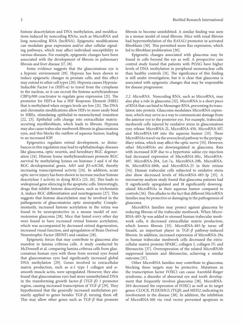
TGF-𝛽 increases extracellular matrix production andremodeling through the canonical Smad pathway as well asnoncanonical Mitogen-Activated Protein (MAP) kinase andRho-GTPase/Rho kinase pathways, which will be discussedin the following sections (Figure 1). In the Smad pathway,TGF-𝛽 binds to TGF-𝛽 receptors I and II, causing TGF𝛽 receptor II to phosphorylate TGF-𝛽 receptor I [51]. Theactivated TGF-𝛽 receptor I then phosphorylates Smad2and Smad3, which interact with Smad4 to form a SmadComplex. The Smad Complex migrates into the cell nucleus,where it activates the transcription of genes that eventuallylead to extracellular matrix production. When TGF-𝛽2 isoverexpressed in mouse eyes, it causes increased IOP andfibronectin expression in wild-type but not Smad3 knockoutmice, demonstrating the importance of the Smad signalingproteins in glaucoma [52].
3.2. MAP Kinase. TGF-𝛽 activates the MAP kinase pathwayby first binding to TGF-𝛽 receptor II, causing autophos-phorylation of tyrosine residues (Figure 1). This recruits SrcHomology Domain 2 Containing Protein (Shc) and GrowthFactor Receptor Binding Protein 2 (Grb2) to bind to the TGF-𝛽 receptor II [46]. Further binding the Shc-Grb2 complexis Son of Sevenless (SOS), a guanine nucleotide exchangefactor (GEF) that activates the GTPases Ras or Rac1. Rasactivates Raf, which triggers MAP ERK kinase (MEK) 1 andsubsequently extracellular signal-regulated kinase (ERK) 1/2activation [53]. ERK 1/2 can increase PAI-1 expression inhuman trabecular meshwork cells, which increases extra-cellular matrix production [54]. The GTPase Rac1 leads tothe activation of p38 MAP kinase pathway, which inducesexpression of the proinflammatory cytokine Interleukin 6and Secreted Protein Acidic and Rich in Cysteine (SPARC)in trabecular meshwork cells [55, 56]. SPARC binds toproteins in the extracellular matrix and regulates growthfactor efficacy and matrix metalloproteinase expression [57].
The p38 MAP kinase pathway can also be activatedwhen TGF-𝛽 binds to TGF-𝛽 receptor I and II, triggeringpolyubiquitination of TRAF6 at Lys63 [58]. TRAF6 is an E3ubiquitin ligase, which is physically associated with the TGF-𝛽 receptors. The polyubiquitination chains hang down andattach to TGF-𝛽 activated kinase (TAK1), activating it. TAK1then phosphorylates MAPK kinase 3/6, which then activatesthe p38 MAPK pathway [59].
3.3. Rho Kinase. TheRho family is composed of the Rho, Rac,and Cdc42 subfamilies, which are involved in cell migration,adhesion, proliferation, and actin cytoskeletal organization

4 BioMed Research International
RhoA Rac1Ras
ShcPP P
Smad3Smad2
PP
Smad4
Smad3Smad4
Smad2
Extracellular matrix production
Grb2 SOS
GTP
Raf
MEK1
GTP
P
P
ERK1/2
P
PAI-1Extracellular
matrix production
PAK
MKK 3/6
p38
P
P
CLAN organization
RhoGEF
P
Src
GTP
Rho kinase
MLCP
Stress fiber organization
Cell contractionECM organization
Extracellular matrix production
IL-6SPARC
𝛼-SMA
TGF-𝛽
TGF-𝛽 receptor I TGF-𝛽 receptor II
Figure 1: TGF-𝛽 signaling pathways. TGF-𝛽 increases extracellularmatrix production through the Rho-GTPase/Rho kinase, Smad, andMAPkinase pathways. TGF-𝛽 binds to TGF-𝛽 receptors I and II, triggering autophosphorylation.This activates RhoGEF, which attaches a GTP toRhoA. RhoA activates Rho kinase, which leads to the phosphorylation ofmyosin light chain (MLC).This leads to stress fiber organization, cellcontraction, extracellular matrix organization, and the expression of genes for 𝛼-smoothmuscle actin and extracellular matrix production. Inthe Smad pathway, TGF-𝛽 binding triggers TGF-𝛽 receptor I to phosphorylate Smad2 and Smad3, which form a Smad Complex with Smad4.The complex travels to the nucleus, where it helps transcribe genes for extracellular matrix production. TGF-𝛽 activates the MAP kinasepathway by causing autophosphorylation of the tyrosine residues on TGF-𝛽 receptor II. This recruits Shc, Grb2, and SOS. SOS activates theGTPases Ras or Rac1. Ras activates Raf, which triggers MEK1 and subsequently ERK 1/2 activation. ERK 1/2 can increase PAI-1 expression inhuman trabecular meshwork cells, which increases extracellular matrix production. The GTPase Rac1 activates p21-activated kinase (PAK),which activates MAP kinase kinase (MKK) 3/6, which activates p38. This induces expression of Interleukin 6 and SPARC.
(Figure 1) [46]. The Rac subfamily has been associatedwith the development of cross-linked actin network (CLAN)formation,which is seen in trabecularmeshwork cells of glau-comatous eyes [60]. Although it is currently unknown howexactly CLANsmay cause glaucoma, it has been hypothesizedthat CLANs can decrease the elasticity of cells, impairingaqueous humor outflow [61]. In addition, trabecular mesh-work cells that express a constitutively active form of RhoA,
a Rho-GTPase, were found to express increased levels offibronectin, tenascin C, laminin, 𝛼-smooth muscle actin,matrix assembly, actin stress fibers, and myosin light-chainphosphorylation, which are associated with the extracellularmatrix [62]. These cells were noted to exhibit increasedcontractile morphology. Rho kinase inhibitors decreasedfibronectin and 𝛼-smooth muscle actin [62]. This suggeststhat trabecular meshwork rigidity and extracellular matrix
BioMed Research International 5
production mediated by the Rho pathway may be involvedin decreasing aqueous humor outflow, raising IOP.
Whereas Rac activation and subsequent CLAN formationis triggered by the association of Shc, Grb2, and SOS asdescribed above, studies in human choriocarcinoma cellshave found that TGF-𝛽 uses Src-mediated phosphorylationto activate Vav2, a Rho-specific GEF [46, 63]. This pathwayeventually leads to the formation of the actin stress fibers thatincrease cell rigidity [62]. The Rho kinase pathway can alsobe activated by a variety of factors such as Thromboxane A2,Angiotensin II, Thrombin, Wnt, Endothelin-1, extracellularmatrix, and stretch [64]. These factors activate RhoGEF,which activates RhoA, which triggers Rho kinase. Rho kinaseinitiates many pathways to lead to cell contraction, extracel-lular matrix organization, 𝛼-smoothmuscle actin expression,and so forth [64].
The Rho kinase pathway has been shown to be apromising target for therapeutics [65]. Rho kinase inhibitorsreduce cell rigidity, increasing outflow [66]. Honjo et al.showed that the Rho-associated protein kinase (ROCK)inhibitor Y-27632 increased the outflow of aqueous humorand decreased IOP by 30% after 3 hours in rabbit eyes [67].Many ROCK inhibitors like netarsudil, RKI-983/SNJ-1656,AR-13324 (Rhopressa�), AR-12286 (Aerie), and AMA0076(Amakem) are now being tested in clinical trials [66, 68].Recently, the ROCK inhibitor Ripasudil was approved forthe treatment of glaucoma and ocular hypertension inJapan, and it is now being studied for the treatment ofdiabetic retinopathy [69]. Continued development of ROCKinhibitors will increase the pharmaceutical options availableto treat glaucoma and may someday be among the first-linetherapies.
3.4. BDNF and Other Neurotropic Factors. BDNF is a proteinproduced by the brain and retina among other organsthat supports the growth, differentiation, and survival ofneurons. BDNF is especially important for RGC survival[70]. Normally, BDNF and other neurotrophic factors aretransported from the brain to the RGCs [71]. However, inglaucoma, the increased IOP blocks axonal transport at theoptic nerve head, decreasing neurotrophic levels in the RGCs[72]. The loss of BDNF in these cells contributes to celldeath and thus glaucoma through JNK activation and c-Junphosphorylation, which eventually leads to caspase activation[73, 74]. RGCs try to prevent this outcome by upregulatingBDNF production if the optic nerve gets injured. A studydone in rats found that after optic nerves were crushed,RGCs expressed elevated BDNF which peaked at 48 hoursbut declined to baseline levels after two weeks [75].This effectis neuroprotective, but only temporarily [76]. Nevertheless,preliminary studies in animals show increased RGC survivaland even some regeneration after intravitreal neurotrophicfactor injection, indicating that it has the potential to even-tually become a therapeutic option [77, 78].
3.5. c-Jun N-Terminal Kinases (JNKs). The proapoptotic JNKpathway is initiated by cellular stress, such as ultraviolet radi-ation, heat shock, and the withdrawal of neurotrophic factors[79]. The aversive stimulus triggers JNK phosphorylation,
causing JNK to bind to the N-terminal region of c-Jun [71].This action phosphorylates c-Jun, a transcription factor forgenes that promote apoptosis [80]. It is elevated in the RGCsof rats with induced glaucoma, peaking at one week after therise in IOP [81]. Optic nerve transection and crush injuryalso increases c-Jun expression [81–83]. Likewise, humanswith glaucoma tend to have elevated levels of phosphorylatedJNK in nonglial retina cells [84]. Interestingly, several studieshave found that c-Jun activation may also promote survivaland regeneration of RGCs, showing that c-Jun may be moreversatile than originally thought [85]. Nevertheless, drugsthat inhibit JNK tend to guard against RGC loss, providingyet another potential target for neuroprotection [86, 87].
3.6. Phosphoinositide-3 Kinase (PI-3 Kinase)/Akt Pathway.The PI-3 kinase/Akt pathway promotes survival and neu-roprotection in neurons [88]. Growth factors bind to themembrane-bound tyrosine receptor kinase, which activatesPI-3 kinase. PI-3 kinase phosphorylates phosphatidylinositol(4,5)-bisphosphate (PIP
2) to phosphatidylinositol (3,4,5)-
trisphosphate (PIP3), which activates Akt. Akt goes on
to inhibit the proapoptotic Bcl-2 associated death domain(BAD) proteins, caspases, and the c-Jun pathway [89–91].
Induced IOP elevation in rats has been shown to activatethe PI-3 kinase/Akt pathway. Phosphorylated Akt increasedon day 1 after translimbal photocoagulation, but it returned tobaseline on day 8 [92]. Elevated IOP provoked by episcleralvein cauterization also increased this survival pathway [93].However, in both of these models, proapoptotic pathwayslike the MAP kinase pathway, caspase family, Fas ligand,and Fas-Associated Death Domain (FADD) were activatedsimultaneously, counteracting the prosurvival factors. Ulti-mately, the proapoptotic protein activation outlasted theneuroprotection pathways, eventually leading to cell death[94].
A variety of neuroprotective drugs that act through differ-ent mechanisms have been demonstrated to reduce RGC lossand structural damage through the PI-3 kinase/Akt pathway.These include the prostaglandin analog, Bimatoprost, the fin-golimod analog, FTY720, and Vascular Endothelial GrowthFactor A [95–97]. Reinforcement of this pathway may benecessary to overcome the longer-lasting proapoptotic factorsto achieve enduring neuroprotection.
3.7. Phosphatase and Tensin Homologue (PTEN) Pathway.PTEN is a lipid and protein phosphatase that works to inhibitcell growth [98]. It blocks the phosphorylation of PIP
2to
PIP3, preventing activation of the PI-3 kinase pathway and
the downstream Akt and mTOR cascades [99].PTEN has been linked with neurodegeneration as dele-
tion of this gene increases axon regeneration after optic nervedamage [100]. Studies using virus-assisted knockout of PTENin mice RGCs after crush injury show significant axonalregeneration, especially when the virus is accompanied byanother virus encoding ciliary neurotrophic factor and acyclic adenosine monophosphate analog [101, 102]. However,an even larger effect can be seen in PTEN knockout mice,which are not encumbered by the incomplete gene silencingseen in RNA interference [102]. Mice with a PTEN deletion

6 BioMed Research International
alone show substantial optic nerve regrowth, but this regen-eration only lasts two weeks after the lesion is introduced[103]. However, when PTEN and suppressor of cytokinesignaling 3 (SOCS3) were both deleted, the effects lastedwell beyond two weeks and there were ten times as manyregenerated axons compared to the PTEN-alone group [103].SOSC3 downregulates the Janus Kinase/Signal Transducerand Activator of Transcription (JAK/STAT) pathway, whichis important for cell proliferation. Many of the regeneratedaxons in these knockout mice have been demonstrated toreach beyond the optic chiasm and even form synapses inthe superior colliculus and suprachiasmatic nucleus [103–105]. Furthermore, the suprachiasmatic nucleus showed someneuronal response when the retinal axons were stimulatedwith light or electricity [105]. However, the responses werenot fully functional and themice did not significantly recovervision [104, 105].This was because the regrown axons did nothave proper myelination, preventing them from conductingadequate action potentials [104]. The addition of voltage-gated potassium channel blockers repaired the conductiondeficit, which ultimately enhanced visual function [104].
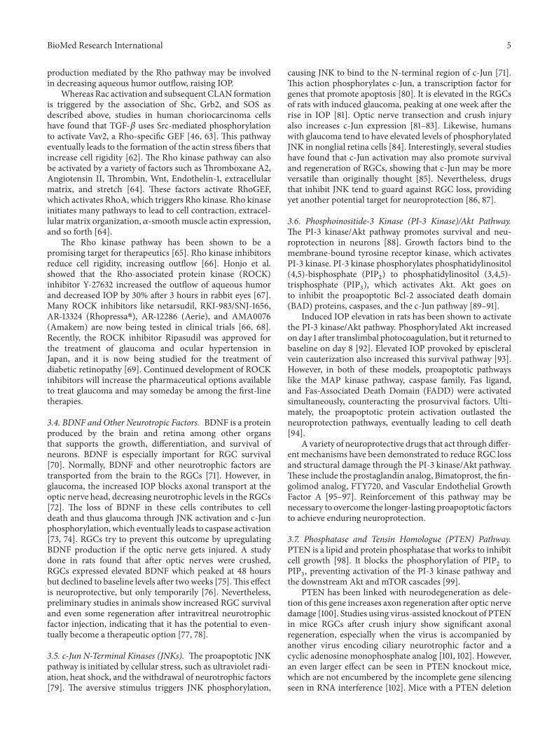
3.8. Bcl-2 Pathway. Bcl-2 is a protein that protects againstapoptosis by inhibiting proapoptotic proteins. DNA damageor cellular stress activates BH3-only proteins, which includeBim, Bid, and BAD [71]. These proteins stimulate Bcl-2 asso-ciated X protein (BAX) and Bak, which increase mitochon-drial membrane permeability and the release of CytochromeC from the mitochondria (Figure 2). Cytochrome C goes onto activate the caspases that eventually lead to apoptosis. Bcl-2stops this process by inhibiting BAX and Bak activation [71].
Levkovitch-Verbin et al. showed that optic nerve transec-tion-induced glaucoma and NMDA eye injections increasedthe expression of BAX and BAD, while downregulating Bcl-2, ultimately leading to cell death [106]. However, overex-pression of Bcl-2 leads to neuron preservation, which caneven save permanently infarcted brain tissue [107]. In fact,the antibiotic minocycline and the monoamine oxidase Binhibitor rasagiline promote neuroprotection in RGCs byincreasing Bcl-2 levels [108, 109]. A number of experi-mental drugs also protect RGCs through this antioxidantand antiapoptotic pathway, supplying even more targets forintervention [110–112].
3.9. Caspases. Caspases are protease enzymes involved inextrinsic and intrinsic apoptotic pathways. In the extrinsicpathway, tumor necrosis factor (TNF) or Fas ligand will bindto the Fas receptor, which is bound to FADD (Figure 2).This activates Caspase 8, which goes on to activate effectorCaspases 3, 6, and 7, and these eventually cause apoptosis[113]. In the intrinsic pathway, Cytochrome C released by themitochondria due to DNA damage or ROS activates Caspase9. Caspase 9 then activates the same effector caspases, leadingto cell death. Apoptosis inhibitor proteins (IAPs) like XIAP, c-IAP, and c-IAP2 inhibit Caspase 3 and Caspase 9, halting thisprocess [113].
Optic nerve transection, crush, and degeneration allresult in an increase in Caspase 3 and Caspase 9 activity[114–116]. Caspase 7 knockout mice preserve more RGCs
after optic nerve crush than wild-type mice, indicating thatblocking caspases may be neuroprotective [117]. Indeed,drugs that inhibit caspases delay RGC death, but they donot help axon regeneration [118]. Neuroprotection is alsoprovided by IAPs, which are upregulated after transectionof the optic nerve or induced glaucoma [119]. However, thisupregulation only occurs in younger animals and lasts shorterthan the concurrently increased expression of proapoptoticgenes, causing cells to ultimately die [120].
3.10. Calcium-Calpain Pathway. Disruptions in calciumhomeostasis occur in many neurodegenerative diseases,including glaucoma [121]. The increased IOP in this disorderintensifies the influx of extracellular calcium into RGCs [122].Calcium activates calpain, a cysteine protease that cleavescalcineurin [123]. Calcineurin goes on to trigger apoptosisin RGCs via dephosphorylation of BAD and the release ofCytochrome C [124].
Theoptic nerve crushmodel has elucidated the role of cal-cium in axonal degeneration. Axotomy in rat models breaksneuronal membranes and may open voltage-gated calciumchannels, allowing the influx of extracellular calcium andthe initiation of degeneration [125]. Calcium likely activatesthe proapoptotic JNK/c-Jun pathway while inhibiting theprosurvival Akt pathway [126]. Calcium ionophores speed upthis process [127]. However, the topical addition of calciumchannel blockers onto the optic nerve has been shown toblock the rise in intracellular calcium, preventing acutesuperficial axonal destruction [127].
Huang et al. confirmed calpain’s role in glaucoma byinjecting hypertonic saline into rat eyes and employingimmunohistochemistry to look for evidence of calpain acti-vation [123]. They found that the retinas of rats with elevatedIOP had a 55 kDa autocatalytic active form of calpain aswell as cleaved spectrin and calcineurin, both substratesof calpain. This group also demonstrated that cleaved cal-pain makes the protease constitutively active, causing it tocontinuously stimulate the apoptotic pathway in rats withelevated IOP [124]. The inhibition of calpain was shown todecrease RGC death after axonal trauma [126]. When ratswere given FK506, a calcineurin inhibitor, therewas amarkeddecrease in BAD dephosphorylation and Cytochrome Crelease, which in turn promoted survival of RGCs and theoptic nerve. Interestingly, optic nerve crush alone did notlead to an increase in calcineurin cleavage [124]. This impliesthat calcineurin cleavage is not merely triggered by generalapoptosis, but rather that it is due to an increase in IOP.
Although the Calcium-Calpain pathway is implicated inaxonal degeneration, it is also a necessary factor in growthcone formation that occurs after destruction. New growthcones appear within 24 hours of axonal trauma, and thesecones always develop in regions of increased calcium concen-tration [128, 129].The proteolytic activity of calpain triggeredby increased calcium is necessary to break down proteinsneeded for regeneration [130]. For example, calpain degradesspectrin, a protein that attaches the intracellular cytoskeletonto the plasmamembrane.Thismay expedite exocytosis whichis needed for successful growth cone assembly [125]. Thebenefits of calcium or calpain inhibition are still unclear

BioMed Research International 7
FADD
Caspase 8
BID
BH3-only protein BAX Bak
Bcl-2
Cytochrome C
DNA damage or ER stress
Fas ligand
FADD
Caspase 9
Caspase 3
Caspase 6
Caspase 7
Apoptosis
IAPs
or TGF-𝛽
Figure 2: Extrinsic and intrinsic pathways of apoptosis. In the extrinsic pathway, Fas ligand or TGF-𝛽 binds to its receptor, which is bound byFADD.This activates Caspase 8, which activates the effector Caspases 3, 6, and 7.These caspases all lead to apoptosis. Caspase 8 also activatesBid, a protein that turns on BAX and Bak. The intrinsic pathway also triggers these same proteins by working through BH3-only proteins.BAX and Bak increase the permeability of the mitochondrial membrane, releasing Cytochrome C. This protein activates Caspase 9, whichactivates the same effector caspases, causing apoptosis. Bcl-2 inhibits this pathway by blocking the activation of BAX and Bak, while IAPsinhibit Caspases 3, 6, 7, and 9.
as they will inhibit growth cone formation and likely onlyprevent the acute axonal degeneration that occurs in a smallarea around the lesion, but further research is needed toinvestigate this thoroughly [131].
4. Conclusions
Glaucoma is a complex disease that can lead to irreversibleblindness in many people worldwide. The condition is gov-erned by genetic and environmental factors, and emergingresearch now suggests a role for epigenetics. These all workthrough a variety of signaling cascades, including the TGF-𝛽,MAPkinase, Rho kinase, BDNF, JNK, PI-3/Akt, PTEN, Bcl-2,Caspase, and Calcium-Calpain pathways. Understanding the
molecular players in these pathways is essential for creatingnew neuroprotective therapeutics that may ultimately helppreserve vision.
Competing Interests
The authors declare that there is no conflict of interestsregarding the publication of this paper and they do not haveproprietary interests related to the content of this paper.
Acknowledgments
Thisworkwas funded in part by anunrestricted departmentalgrant from Research to Prevent Blindness (RPB), Inc.

8 BioMed Research International
References
[1] Y.-C. Tham, X. Li, T. Y. Wong, H. A. Quigley, T. Aung, and C.-Y. Cheng, “Global prevalence of glaucoma and projections ofglaucoma burden through 2040. A systematic review andmeta-analysis,” Ophthalmology, vol. 121, no. 11, pp. 2081–2090, 2014.
[2] A. C. Gauthier and J. Liu, “Neurodegeneration and neuropro-tection in glaucoma,” Yale Journal of Biology and Medicine, vol.89, no. 1, pp. 73–79, 2016.
[3] J. L. Wiggs, “The cell and molecular biology of complex formsof glaucoma: updates on genetic, environmental, and epigeneticrisk factors,” Investigative Ophthalmology & Visual Science, vol.53, no. 5, pp. 2467–2469, 2012.
[4] R. C. W. Wolfs, C. C. W. Klaver, R. S. Ramrattan, C. M. VanDuijn, A. Hofman, and P. T. V. M. De Jong, “Genetic riskof primary open-angle glaucoma: population-based familialaggregation study,” Archives of Ophthalmology, vol. 116, no. 12,pp. 1640–1645, 1998.
[5] X. Wang, J. Harmon, N. Zabrieskie et al., “Using the UtahPopulation Database to assess familial risk of primary openangle glaucoma,” Vision Research, vol. 50, no. 23, pp. 2391–2395,2010.
[6] S. S. Verma, J. N. Cooke Bailey, A. Lucas et al., “Epistaticgene-based interaction analyses for glaucoma in eMERGE andNEIGHBOR consortium,” PLOS Genetics, vol. 12, no. 9, ArticleID e1006186, 2016.
[7] J. L. Wiggs, “Genetic etiologies of glaucoma,” Archives ofOphthalmology, vol. 125, no. 1, pp. 30–37, 2007.
[8] L. R. Pasquale and J. H. Kang, “Lifestyle, nutrition, andglaucoma,” Journal of Glaucoma, vol. 18, no. 6, pp. 423–428,2009.
[9] F. Ko, M. V. Boland, P. Gupta et al., “Diabetes, triglyceridelevels, and other risk factors for glaucoma in the national healthand nutrition examination survey 2005–2008,” InvestigativeOpthalmology & Visual Science, vol. 57, no. 4, pp. 2152–2157,2016.
[10] L. Xu, H. Wang, Y. Wang, and J. B. Jonas, “Intraocular pressurecorrelated with arterial blood pressure: The Beijing Eye Study,”American Journal of Ophthalmology, vol. 144, no. 3, pp. 461–462,2007.
[11] D. Zhao, J. Cho, M. H. Kim, and E. Guallar, “The associationof blood pressure and primary open-angle glaucoma: a meta-analysis,”American Journal of Ophthalmology, vol. 158, no. 3, pp.615.e9–627.e9, 2014.
[12] M. E. Charlson, C. G. de Moraes, A. Link et al., “Nocturnal sys-temic hypotension increases the risk of glaucoma progression,”Ophthalmology, vol. 121, no. 10, pp. 2004–2012, 2014.
[13] L. R. Pasquale, J. H. Kang, J. E. Manson, W. C. Willett, B.A. Rosner, and S. E. Hankinson, “Prospective study of type 2diabetes mellitus and risk of primary open-angle glaucoma inwomen,” Ophthalmology, vol. 113, no. 7, pp. 1081–1086, 2006.
[14] C. A. Girkin, G. McGwin Jr., S. F. McNeal, P. P. Lee, and C.Owsley, “Hypothyroidism and the development of open-angleglaucoma in a male population,” Ophthalmology, vol. 111, no. 9,pp. 1649–1652, 2004.
[15] C. Lin, C. Hu, J. Ho, H. Chiu, and H. Lin, “Obstructive SleepApnea and Increased Risk of Glaucoma,” Ophthalmology, vol.120, no. 8, pp. 1559–1564, 2013.
[16] D. Zhao, J. Cho, M. H. Kim, D. S. Friedman, and E. Guallar,“Diabetes, fasting glucose, and the risk of glaucoma: a meta-analysis,” Ophthalmology, vol. 122, no. 1, pp. 72–78, 2015.
[17] W. R. Coward, K. Watts, C. A. Feghali-Bostwick, A. Knox,and L. Pang, “Defective histone acetylation is responsible forthe diminished expression of cyclooxygenase 2 in idiopathicpulmonary fibrosis,”Molecular and Cellular Biology, vol. 29, no.15, pp. 4325–4339, 2009.
[18] T. Hardy and D. A. Mann, “Epigenetics in liver disease: frombiology to therapeutics,”Gut, vol. 65, no. 11, pp. 1895–1905, 2016.
[19] G. Tezel and M. B. Wax, “Hypoxia-inducible factor 1𝛼 inthe glaucomatous retina and opticnerve head,” Archives ofOphthalmology, vol. 122, no. 9, pp. 1348–1356, 2004.
[20] J. A. Watson, C. J. Watson, A.-M. McCrohan et al., “Generationof an epigenetic signature by chronic hypoxia in prostate cells,”HumanMolecular Genetics, vol. 18, no. 19, pp. 3594–3604, 2009.
[21] K. Pennington and M. DeAngelis, “Epigenetic mechanisms ofthe aging human retina,” Journal of Experimental Neuroscience,vol. 92, supplement 2, pp. 51–79, 2015.
[22] F.McDonnell, C.O’Brien, andD.Wallace, “The role of epigenet-ics in the fibrotic processes associated with glaucoma,” Journalof Ophthalmology, vol. 2014, Article ID 750459, 13 pages, 2014.
[23] K. Kimura, M. Iwano, D. F. Higgins et al., “Stable expression ofHIF-1𝛼 in tubular epithelial cells promotes interstitial fibrosis,”American Journal of Physiology—Renal Physiology, vol. 295, no.4, pp. F1023–F1029, 2008.
[24] R. C. Rao, A. K. Hennig, M. T. Malik, D. F. Chen, and S. Chen,“Epigenetic regulation of retinal development and disease,”Journal of Ocular Biology, Diseases, and Informatics, vol. 4, no.3, pp. 121–136, 2011.
[25] H. R. Pelzel, C. L. Schlamp, and R. W. Nickells, “Histone H4deacetylation plays a critical role in early gene silencing duringneuronal apoptosis,” BMC Neuroscience, vol. 11, no. 1, 2010.
[26] S. He, X. Li, N. Chan, and D. R. Hinton, “Review: epigeneticmechanisms in ocular disease,” Molecular Vision, vol. 19, pp.665–674, 2013.
[27] B. R. Schwechter, L. E. Millet, and L. A. Levin, “Histonedeacetylase inhibition-mediated differentiation of RGC-5 cellsand interaction with survival,” Investigative Ophthalmology &Visual Science, vol. 48, no. 6, pp. 2845–2857, 2007.
[28] X. Guo, A. Kimura, Y. Azuchi et al., “Caloric restrictionpromotes cell survival in a mouse model of normal tensionglaucoma,” Scientific Reports, vol. 6, Article ID 33950, 2016.
[29] F. S. McDonnell, S. A. McNally, A. F. Clark, C. J. O’Brien, and D.M.Wallace, “Increased global DNAmethylation and decreasedTGF𝛽1 promoter methylation in glaucomatous lamina cribrosacells,” Journal of Glaucoma, vol. 25, no. 10, pp. e834–e842, 2016.
[30] W. Bechtel, S. McGoohan, E. M. Zeisberg et al., “Methylationdetermines fibroblast activation and fibrogenesis in the kidney,”Nature Medicine, vol. 16, no. 5, pp. 544–550, 2010.
[31] A. Junemann, B. Lenz,U. Reulbach et al., “Genomic (epigenetic)DNA methylation in patients with open-angle glaucoma,” ActaOphthalmologica, vol. 87, no. s244, 2009.
[32] M. Molasy, A. Walczak, J. Szaflik, J. P. Szaflik, and I. Majsterek,“MicroRNAs in glaucoma and neurodegenerative diseases,”Journal of Human Genetics, 2016.
[33] A. Izzotti, C. Ceccaroli, M. G. Longobardi et al., “Moleculardamage in glaucoma: from anterior to posterior eye segment.The MicroRNA role,”MicroRNA, vol. 4, no. 1, pp. 3–17, 2015.
[34] H. Jayaram, W. O. Cepurna, E. C. Johnson, and J. C. Morrison,“MicroRNA expression in the glaucomatous retina,” Investiga-tive Opthalmology & Visual Science, vol. 56, no. 13, pp. 7971–7982, 2015.

BioMed Research International 9
[35] W. Shen, Y. Han, B. Huang et al., “MicroRNA-483-3p inhibitsextracellular matrix production by targeting smad4 in humantrabecularmeshwork cells,” InvestigativeOpthalmology&VisualScience, vol. 56, no. 13, pp. 8419–8427, 2015.
[36] Y. Tanaka, S. Tsuda, H. Kunikata et al., “Profiles of extracellularmiRNAs in the aqueous humor of glaucoma patients assessedwith a microarray system,” Scientific Reports, vol. 4, article no.5089, 2014.
[37] G. Villarreal, D. Oh, M. H. Kang, and D. J. Rhee, “Coordinatedregulation of extracellular matrix synthesis by the microRNA-29 Family in the trabecularmeshwork,” Investigative Opthalmol-ogy & Visual Science, vol. 52, no. 6, pp. 3391–3397, 2011.
[38] S. H. Paylakhi, H.Moazzeni, S. Yazdani et al., “FOXC1 in humantrabecular meshwork cells is involved in regulatory pathwaythat includes miR-204, MEIS2, and ITG𝛽1,” Experimental EyeResearch, vol. 111, pp. 112–121, 2013.
[39] N. Kong, X. Lu, and B. Li, “Downregulation of microRNA-100 protects apoptosis and promotes neuronal growth in retinalganglion cells,” BMCMolecular Biology, vol. 15, no. 1, 2014.
[40] B. S. Clark and S. Blackshaw, “Long non-coding RNA-dependent transcriptional regulation in neuronal developmentand disease,” Frontiers in Genetics, vol. 5, article no. 164, 2014.
[41] F. Li, X. Wen, H. Zhang, and X. Fan, “Novel insights into therole of long noncoding RNA in ocular diseases,” InternationalJournal of Molecular Sciences, vol. 17, no. 4, article 478, 2016.
[42] A. Congrains, K. Kamide, M. Ohishi, and H. Rakugi, “ANRIL:molecular mechanisms and implications in human health,”International Journal of Molecular Sciences, vol. 14, no. 1, pp.1278–1292, 2013.
[43] L. R. Pasquale, S. J. Loomis, J. H. Kang et al., “CDKN2B-AS1genotype–glaucoma feature correlations in primary open-angleglaucoma patients from the United States,” American Journal ofOphthalmology, vol. 155, no. 2, pp. 342.e5–353.e5, 2013.
[44] K. P. Burdon, S. MacGregor, A. W. Hewitt et al., “Genome-wideassociation study identifies susceptibility loci for open angleglaucoma at TMCO1 and CDKN2B-AS1,” Nature Genetics, vol.43, no. 6, pp. 574–578, 2011.
[45] W.D. Ramdas, L.M. vanKoolwijk, H. G. Lemij et al., “Commongenetic variants associated with open-angle glaucoma,”HumanMolecular Genetics, vol. 20, no. 12, pp. 2464–2471, 2011.
[46] C. L. Pervan, “Smad-independent TGF-𝛽2 signaling path-ways in human trabecular meshwork cells,” Experimental EyeResearch, 2016.
[47] M. Inatani, H. Tanihara, H. Katsuta, M. Honjo, N. Kido, andY. Honda, “Transforming growth factor-𝛽
2levels in aqueous
humor of glaucomatous eyes,” Graefe’s Archive for Clinical andExperimental Ophthalmology, vol. 239, no. 2, pp. 109–113, 2001.
[48] A. A. Ozcan, N. Ozdemir, and A. Canataroglu, “The aqueouslevels of TGF-𝛽2 in patients with glaucoma,” InternationalOphthalmology, vol. 25, no. 1, pp. 19–22, 2004.
[49] D. L. Fleenor, A. R. Shepard, P. E. Hellberg, N. Jacobson, I. Pang,and A. F. Clark, “TGF𝛽2-induced changes in human trabecularmeshwork: implications for intraocular pressure,” InvestigativeOpthalmology &Visual Science, vol. 47, no. 1, pp. 226–234, 2006.
[50] J. Gottanka, D. Chan, M. Eichhorn, E. Lutjen-Drecoll, andC. R. Ethier, “Effects of TGF-𝛽2 in perfused human eyes,”Investigative Ophthalmology & Visual Science, vol. 45, no. 1, pp.153–158, 2004.
[51] Y. Mu, S. K. Gudey, and M. Landstrom, “Non-Smad signalingpathways,” Cell and Tissue Research, vol. 347, no. 1, pp. 11–20,2012.
[52] C. M. McDowell, H. E. Tebow, R. J. Wordinger, and A. F.Clark, “Smad3 is necessary for transforming growth factor-beta2 induced ocular hypertension in mice,” Experimental EyeResearch, vol. 116, pp. 419–423, 2013.
[53] Y. E. Zhang, “Non-Smad pathways in TGF-𝛽 signaling,” CellResearch, vol. 19, no. 1, pp. 128–139, 2009.
[54] H. Han, T. Wecker, F. Grehn, and G. Schlunck, “Elasticity-dependent modulation of TGF-𝛽 responses in human tra-becular meshwork cells,” Investigative Opthalmology & VisualScience, vol. 52, no. 6, pp. 2889–2896, 2011.
[55] P. B. Liton,G. Li, C. Luna, P.Gonzalez, andD. L. Epstein, “Cross-talk between TGF-𝛽1 and IL-6 in human trabecular meshworkcells,”Molecular Vision, vol. 15, pp. 326–334, 2009.
[56] M. H. Kang, D. Oh, J. Kang, and D. J. Rhee, “Regulation ofSPARC by transforming growth factor 𝛽2 in human trabecularmeshwork,” InvestigativeOpthalmology&Visual Science, vol. 54,no. 4, pp. 2523–2532, 2013.
[57] A. D. Bradshaw and E. H. Sage, “SPARC, a matricellular proteinthat functions in cellular differentiation and tissue response toinjury,” Journal of Clinical Investigation, vol. 107, no. 9, pp. 1049–1054, 2001.
[58] M.Yamashita, K. Fatyol, C. Jin, X.Wang, Z. Liu, andY. E. Zhang,“TRAF6 mediates Smad-independent activation of JNK andp38 by TGF-𝛽,”Molecular Cell, vol. 31, no. 6, pp. 918–924, 2008.
[59] A. Sorrentino, N. Thakur, S. Grimsby et al., “The type I TGF-𝛽receptor engages TRAF6 to activate TAK1 in a receptor kinase-independent manner,” Nature Cell Biology, vol. 10, no. 10, pp.1199–1207, 2008.
[60] M. S. Filla, M. K. Schwinn, N. Sheibani, P. L. Kaufman,and D. M. Peters, “Regulation of cross-linked actin network(CLAN) formation in human trabecular meshwork (HTM)cells by convergence of distinct 𝛽1 and 𝛽3 integrin pathways,”Investigative Opthalmology & Visual Science, vol. 50, no. 12, pp.5723–5731, 2009.
[61] A. F. Clark, D. Brotchie, A. T. Read et al., “Dexamethasone altersF-actin architecture and promotes cross-linked actin networkformation in human trabecular meshwork tissue,” Cell Motilityand the Cytoskeleton, vol. 60, no. 2, pp. 83–95, 2005.
[62] P. P. Pattabiraman and P. V. Rao, “Mechanistic basis of RhoGTPase-induced extracellular matrix synthesis in trabecularmeshwork cells,” AJP: Cell Physiology, vol. 298, no. 3, pp. C749–C763, 2010.
[63] E. Papadimitriou, D. Kardassis, A. Moustakas, and C.Stournaras, “TGF𝛽-induced early activation of the smallGTPase RhoA is smad2/3-independent and involves Srcand the guanine nucleotide exchange factor Vav2,” CellularPhysiology and Biochemistry, vol. 28, no. 2, pp. 229–238, 2011.
[64] P. V. Rao, P. P. Pattabiraman, and C. Kopczynski, “Role of theRhoGTPase/Rho kinase signaling pathway in pathogenesis andtreatment of glaucoma: bench to bedside research,” Experimen-tal Eye Research, 2016.
[65] T. Inoue and H. Tanihara, “Rho-associated kinase inhibitors: anovel glaucoma therapy,” Progress in Retinal and Eye Research,vol. 37, pp. 1–12, 2013.
[66] G. Prasanna, B. Li, M. Mogi, and D. S. Rice, “Pharmacologyof novel intraocular pressure-lowering targets that enhanceconventional outflow facility: pitfalls, promises and what liesahead?” European Journal of Pharmacology, vol. 787, pp. 47–56,2016.
[67] M. Honjo, H. Tanihara, M. Inatani et al., “Effects of Rho-associated protein kinase inhibitor Y-27632 on intraocular

10 BioMed Research International
pressure and outflow facility,” Investigative Ophthalmology andVisual Science, vol. 42, no. 1, pp. 137–144, 2001.
[68] J. M. Sturdivant, S. M. Royalty, C. Lin et al., “Discovery ofthe ROCK inhibitor netarsudil for the treatment of open-angleglaucoma,” Bioorganic & Medicinal Chemistry Letters, vol. 26,no. 10, pp. 2475–2480, 2016.
[69] K. P. Garnock-Jones, “Ripasudil: first global approval,” Drugs,vol. 74, no. 18, pp. 2211–2215, 2014.
[70] J. E. Johnson, Y.-A. Barde, M. Schwab, and H.Thoenen, “Brain-derived neurotrophic factor supports the survival of culturedrat retinal ganglion cells,” Journal of Neuroscience, vol. 6, no. 10,pp. 3031–3038, 1986.
[71] H. Levkovitch-Verbin, “Retinal ganglion cell apoptotic pathwayin glaucoma: initiating and downstream mechanisms,” Progressin Brain Research, vol. 220, pp. 37–57, 2015.
[72] D. R. Anderson and A. Hendrickson, “Effect of intraocularpressure on rapid axoplasmic transport inmonkey optic nerve,”Investigative Ophthalmology, vol. 13, no. 10, pp. 771–783, 1974.
[73] M. Almasieh, A. M. Wilson, B. Morquette, J. L. Cueva Vargas,and A. Di Polo, “The molecular basis of retinal ganglion celldeath in glaucoma,” Progress in Retinal and Eye Research, vol.31, no. 2, pp. 152–181, 2012.
[74] J. Yuan and B. A. Yankner, “Apoptosis in the nervous system,”Nature, vol. 407, no. 6805, pp. 802–809, 2000.
[75] H. Gao, X. Qiao, F. Hefti, J. G. Hollyfield, and B. Knusel,“Elevated mRNA expression of brain-derived neurotrophicfactor in retinal ganglion cell layer after optic nerve injury,”Investigative Ophthalmology and Visual Science, vol. 38, no. 9,pp. 1840–1847, 1997.
[76] A. Di Polo, L. J. Aigner, R. J. Dunn, G.M. Bray, andA. J. Aguayo,“Prolonged delivery of brain-derived neurotrophic factor byadenovirus-infected Muller cells temporarily rescues injuredretinal ganglion cells,” Proceedings of the National Academy ofSciences, vol. 95, no. 7, pp. 3978–3983, 1998.
[77] N. Nafissi and M. Foldvari, “Neuroprotective therapies in glau-coma: I. Neurotrophic factor delivery,” Wiley InterdisciplinaryReviews: Nanomedicine and Nanobiotechnology, vol. 8, no. 2, pp.240–254, 2016.
[78] Y. Liu, Z. Gong, L. Liu, and H. Sun, “Combined effect ofolfactory ensheathing cell (OEC) transplantation and glial cellline-derived neurotrophic factor (GDNF) intravitreal injectiononoptic nerve injury in rats,”MolecularVision, vol. 16, pp. 2903–2910, 2010.
[79] A. Eilers, J. Whitfield, B. Shah, C. Spadoni, H. Desmond,and J. Ham, “Direct inhibition of c-Jun N-terminal kinase insympathetic neurones prevents c-jun promoter activation andNGF withdrawal-induced death,” Journal of Neurochemistry,vol. 76, no. 5, pp. 1439–1454, 2001.
[80] J. Ham, A. Eilers, J. Whitfield, S. J. Neame, and B. Shah, “c-Jun and the transcriptional control of neuronal apoptosis,”Biochemical Pharmacology, vol. 60, no. 8, pp. 1015–1021, 2000.
[81] H. Levkovitch-Verbin, H. A. Quigley, K. R. G.Martin et al., “Thetranscription factor c-jun is activated in retinal ganglion cells inexperimental rat glaucoma,” Experimental Eye Research, vol. 80,no. 5, pp. 663–670, 2005.
[82] S. Isenmann and M. Bahr, “Expression of c-Jun protein indegenerating retinal ganglion cells after optic nerve lesion in therat,” Experimental Neurology, vol. 147, no. 1, pp. 28–36, 1997.
[83] M. Takeda, H. Kato, A. Takamiya, A. Yoshida, and H. Kiyama,“Injury-specific expression of activating transcription factor-3 in retinal ganglion cells and its colocalized expression with
phosphorylated c-Jun,” Investigative Ophthalmology and VisualScience, vol. 41, no. 9, pp. 2412–2421, 2000.
[84] G. Tezel, B. C. Chauhan, R. P. LeBlanc, and M. B. Wax,“Immunohistochemical assessment of the glial mitogen-activated protein kinase activation in glaucoma,” InvestigativeOphthalmology and Visual Science, vol. 44, no. 7, pp. 3025–3033,2003.
[85] E. C. Johnson, Y. Guo, W. O. Cepurna, and J. C. Morrison,“Neurotrophin roles in retinal ganglion cell survival: lessonsfrom rat glaucoma models,” Experimental Eye Research, vol. 88,no. 4, pp. 808–815, 2009.
[86] H. Sun, Y. Wang, IH. Pang et al., “Protective effect of a JNKinhibitor against retinal ganglion cell loss induced by acutemoderate ocular hypertension,” Molecular Vision, vol. 17, pp.864–875, 2011.
[87] G. Tezel, X. Yang, J. Yang, and M. B. Wax, “Role of tumornecrosis factor receptor-1 in the death of retinal ganglion cellsfollowing optic nerve crush injury in mice,” Brain Research, vol.996, no. 2, pp. 202–212, 2004.
[88] H.Dudek, S. R.Datta, T. F. Franke et al., “Regulation of neuronalsurvival by the serine-threonine protein kinase Akt,” Science,vol. 275, no. 5300, pp. 661–665, 1997.
[89] S. R. Datta, H. Dudek, X. Tao et al., “Akt phosphorylationof BAD couples survival signals to the cell-intrinsic deathmachinery,” Cell, vol. 91, no. 2, pp. 231–241, 1997.
[90] H. Zhou, X. Li, J. Meinkoth, and R. N. Pittman, “Akt regulatescell survival and apoptosis at a postmitochondrial level,” TheJournal of Cell Biology, vol. 151, no. 3, pp. 483–494, 2000.
[91] M. K. Barthwal, “Negative regulation of mixed lineage kinase3 by protein kinase B/AKT leads to cell survival,” Journal ofBiological Chemistry, vol. 278, no. 6, pp. 3897–3902, 2003.
[92] H. Levkovitch-Verbin, N. Harizman, R. Dardik, Y. Nisgav,S. Vander, and S. Melamed, “Regulation of cell death andsurvival pathways in experimental glaucoma,” Experimental EyeResearch, vol. 85, no. 2, pp. 250–258, 2007.
[93] H. S. Kim and C. K. Park, “Retinal ganglion cell death is delayedby activation of retinal intrinsic cell survival program,” BrainResearch, vol. 1057, no. 1-2, pp. 17–28, 2005.
[94] S. Vander and H. Levkovitch-Verbin, “Regulation of cell deathand survival pathways in secondary degeneration of the opticnerve a long-term study,” Current Eye Research, vol. 37, no. 8,pp. 740–748, 2012.
[95] N. Takano, K. Tsuruma, Y. Ohno, M. Shimazawa, and H.Hara, “Bimatoprost protects retinal neuronal damage via Aktpathway,” European Journal of Pharmacology, vol. 702, no. 1–3,pp. 56–61, 2013.
[96] Y. You, V. K. Gupta, J. C. Li, N. Al-Adawy, A. Klistorner,and S. L. Graham, “FTY720 protects retinal ganglion cells inexperimental glaucoma,” Investigative Opthalmology & VisualScience, vol. 55, no. 5, pp. 3060–3066, 2014.
[97] R. H. Foxton, A. Finkelstein, S. Vijay et al., “VEGF-A isnecessary and sufficient for retinal neuroprotection in modelsof experimental glaucoma,”The American Journal of Pathology,vol. 182, no. 4, pp. 1379–1390, 2013.
[98] R. L. Zhu, K. S. Cho, C. Y. Guo, J. Chew, D. F. Chen, and L. Yang,“Intrinsic determinants of optic nerve regeneration,” ChineseMedical Journal, vol. 126, no. 13, pp. 2543–2547, 2013.
[99] L. I. Benowitz, Z. He, and J. L. Goldberg, “Reaching the brain:advances in optic nerve regeneration,” Experimental Neurology,vol. 287, pp. 365–373, 2017.

BioMed Research International 11
[100] Y. Koriyama, M. Kamiya, K. Arai, K. Sugitani, K. Ogai, andS. Kato, “Nipradilol promotes axon regeneration through S-nitrosylation of PTEN in retinal ganglion cells,” Advances inExperimental Medicine and Biology, vol. 801, pp. 751–757, 2014.
[101] K. K. Park, K. Liu, Y. Hu et al., “Promoting axon regenerationin the adult CNS by modulation of the PTEN/mTOR pathway,”Science, vol. 322, no. 5903, pp. 963–966, 2008.
[102] B. J. Yungher, X. Luo, Y. Salgueiro, M. G. Blackmore, and K. K.Park, “Viral vector-based improvement of optic nerve regener-ation: characterization of individual axons’ growth patterns andsynaptogenesis in a visual target,” Gene Therapy, vol. 22, no. 10,pp. 811–821, 2015.
[103] F. Sun, K. K. Park, S. Belin et al., “Sustained axon regenerationinduced by co-deletion of PTEN and SOCS3,” Nature, vol. 480,no. 7377, pp. 372–375, 2011.
[104] F. Bei, H. Lee, X. Liu et al., “Restoration of visual function byenhancing conduction in regenerated axons,” Cell, vol. 164, no.1-2, pp. 219–232, 2016.
[105] S. Li, Q. He, H. Wang et al., “Injured adult retinal axonswith Pten and Socs3 co-deletion reform active synapses withsuprachiasmatic neurons,” Neurobiology of Disease, vol. 73, pp.366–376, 2015.
[106] H. Levkovitch-Verbin, D.Makarovsky, and S. Vander, “Compar-ison between axonal and retinal ganglion cell gene expressionin various optic nerve injuries including glaucoma,” MolecularVision, vol. 19, pp. 2526–2541, 2013.
[107] J.-C. Martinou, M. Dubois-Dauphin, J. K. Staple et al., “Over-expression of BCL-2 in transgenic mice protects neurons fromnaturally occurring cell death and experimental ischemia,”Neuron, vol. 13, no. 4, pp. 1017–1030, 1994.
[108] H. Levkovitch-Verbin, S. Vander, and S. Melamed, “Rasagiline-induced delay of retinal ganglion cell death in experimentalglaucoma in rats,” Journal of Glaucoma, vol. 20, no. 5, pp. 273–277, 2011.
[109] H. Levkovitch-Verbin, Y.Waserzoog, S. Vander, D.Makarovsky,and P. Ilia, “Minocycline mechanism of neuroprotectioninvolves the Bcl-2 gene family in optic nerve transection,”International Journal of Neuroscience, vol. 124, no. 10, pp. 755–761, 2014.
[110] H.Wang, C. Zhang, D. Lu et al., “Oligomeric proanthocyanidinprotects retinal ganglion cells against oxidative stress-inducedapoptosis,”Neural RegenerationResearch, vol. 8, no. 25, pp. 2317–2326, 2013.
[111] H. Cheng, Y. Ding, R. Yu, J. Chen, and C.Wu, “Neuroprotectionof a novel cyclopeptide C∗HSDGIC∗ from the cyclization ofPACAP (1–5) in cellular and rodent models of retinal ganglioncell apoptosis,” PLoS ONE, vol. 9, no. 10, Article ID e108090,2014.
[112] Z. Wang, X. Pan, D. Wang et al., “Protective effects of proto-catechuic acid on retinal ganglion cells from oxidative damageinduced by H
2O2,”Neurological Research, vol. 37, no. 2, pp. 159–
166, 2014.[113] H. Marzban, M. R. Del Bigio, J. Alizadeh, S. Ghavami, R.
M. Zachariah, and M. Rastegar, “Cellular commitment in thedeveloping cerebellum,” Frontiers in Cellular Neuroscience, vol.8, article 450, 2015.
[114] P. Kermer, N. Klocker, M. Labes, S. Thomsen, A. Srinivasan,and M. Bahr, “Activation of caspase-3 in axotomized rat retinalganglion cells in vivo,” FEBS Letters, vol. 453, no. 3, pp. 361–364,1999.
[115] P. Kermer, R. Ankerhold, N. Klocker, S. Krajewski, J. C. Reed,and M. Bahr, “Caspase-9: involvement in secondary death of
axotomized rat retinal ganglion cells in vivo,” Molecular BrainResearch, vol. 85, no. 1-2, pp. 144–150, 2000.
[116] H. Levkovitch-Verbin, R. Dardik, S. Vander, and S. Melamed,“Mechanism of retinal ganglion cells death in secondary degen-eration of the optic nerve,” Experimental Eye Research, vol. 91,no. 2, pp. 127–134, 2010.
[117] S. Choudhury, Y. Liu, A. F. Clark, and I.-H. Pang, “Caspase-7: acritical mediator of optic nerve injury-induced retinal ganglioncell death,” Molecular Neurodegeneration, vol. 10, article 40,2015.
[118] V. Vigneswara, M. Berry, A. Logan, and Z. Ahmed, “Pharma-cological inhibition of caspase-2 protects axotomised retinalganglion cells from apoptosis in adult rats,” PLoS ONE, vol. 7,no. 12, Article ID e53473, 2012.
[119] H. Levkovitch-Verbin, R. Dardik, S. Vander, Y. Nisgav, M.Kalev-Landoy, and S. Melamed, “Experimental glaucoma andoptic nerve transection induce simultaneous upregulation ofproapoptotic and prosurvival genes,” Investigative Ophthalmol-ogy and Visual Science, vol. 47, no. 6, pp. 2491–2497, 2006.
[120] H. Levkovitch-Verbin, S. Vander, D. Makarovsky, and F. Lavin-sky, “Increase in retinal ganglion cells’ susceptibility to elevatedintraocular pressure and impairment of their endogenousneuroprotective mechanism by age,” Molecular Vision, vol. 19,pp. 2011–2022, 2013.
[121] U. Wojda, E. Salinska, and J. Kuznicki, “Calcium ions inneuronal degeneration,” IUBMB Life, vol. 60, no. 9, pp. 575–590,2008.
[122] R. M. Sappington, T. Sidorova, D. J. Long, and D. J. Calkins,“TRPV1: contribution to retinal ganglion cell apoptosis andincreased intracellular Ca2+ with exposure to hydrostatic pres-sure,” Investigative Opthalmology & Visual Science, vol. 50, no.2, pp. 717–728, 2009.
[123] W. Huang, J. Fileta, I. Rawe, J. Qu, and C. L. Grosskreutz,“Calpain activation in experimental glaucoma,” InvestigativeOphthalmology & Visual Science, vol. 51, no. 6, pp. 3049–3054,2010.
[124] W. Huang, J. B. Fileta, A. Dobberfuhl et al., “Calcineurincleavage is triggered by elevated intraocular pressure, andcalcineurin inhibition blocks retinal ganglion cell death inexperimental glaucoma,” Proceedings of the National Academyof Sciences, vol. 102, no. 34, pp. 12242–12247, 2005.
[125] L. F.Gumy,C. L. Tan, and J.W. Fawcett, “The role of local proteinsynthesis and degradation in axon regeneration,” ExperimentalNeurology, vol. 223, no. 1, pp. 28–37, 2010.
[126] V. T. Ribas and P. Lingor, “Calcium channel inhibition-mediatedaxonal stabilization improves axonal regeneration after opticnerve crush,” Neural Regeneration Research, vol. 11, no. 8, pp.1245–1246, 2016.
[127] J. Knoferle, J. C. Koch, T. Ostendorf et al., “Mechanisms of acuteaxonal degeneration in the optic nerve in vivo,” Proceedings oftheNational Academy of Sciences, vol. 107, no. 13, pp. 6064–6069,2010.
[128] M. Kerschensteiner, M. E. Schwab, J. W. Lichtman, and T. Mis-geld, “In vivo imaging of axonal degeneration and regenerationin the injured spinal cord,” Nature Medicine, vol. 11, no. 5, pp.572–577, 2005.
[129] N. E. Ziv and M. E. Spira, “Localized and transient elevationsof intracellular Ca2+ induce the dedifferentiation of axonalsegments into growth cones,” Journal of Neuroscience, vol. 17, no.10, pp. 3568–3579, 1997.
[130] D. Gitler and M. E. Spira, “Real time imaging of calcium-induced localized proteolytic activity after axotomy and its

12 BioMed Research International
relation to growth cone formation,” Neuron, vol. 20, no. 6, pp.1123–1135, 1998.
[131] P. Lingor, J. C. Koch, L. Tonges, andM. Bahr, “Axonal degenera-tion as a therapeutic target in theCNS,”Cell andTissue Research,vol. 349, no. 1, pp. 289–311, 2012.

Submit your manuscripts athttps://www.hindawi.com
Stem CellsInternational
Hindawi Publishing Corporationhttp://www.hindawi.com Volume 2014
Hindawi Publishing Corporationhttp://www.hindawi.com Volume 2014
MEDIATORSINFLAMMATION
of
Hindawi Publishing Corporationhttp://www.hindawi.com Volume 2014
Behavioural Neurology
EndocrinologyInternational Journal of
Hindawi Publishing Corporationhttp://www.hindawi.com Volume 2014
Hindawi Publishing Corporationhttp://www.hindawi.com Volume 2014
Disease Markers
Hindawi Publishing Corporationhttp://www.hindawi.com Volume 2014
BioMed Research International
OncologyJournal of
Hindawi Publishing Corporationhttp://www.hindawi.com Volume 2014
Hindawi Publishing Corporationhttp://www.hindawi.com Volume 2014
Oxidative Medicine and Cellular Longevity
Hindawi Publishing Corporationhttp://www.hindawi.com Volume 2014
PPAR Research
The Scientific World JournalHindawi Publishing Corporation http://www.hindawi.com Volume 2014
Immunology ResearchHindawi Publishing Corporationhttp://www.hindawi.com Volume 2014
Journal of
ObesityJournal of
Hindawi Publishing Corporationhttp://www.hindawi.com Volume 2014
Hindawi Publishing Corporationhttp://www.hindawi.com Volume 2014
Computational and Mathematical Methods in Medicine
OphthalmologyJournal of
Hindawi Publishing Corporationhttp://www.hindawi.com Volume 2014
Diabetes ResearchJournal of
Hindawi Publishing Corporationhttp://www.hindawi.com Volume 2014
Hindawi Publishing Corporationhttp://www.hindawi.com Volume 2014
Research and TreatmentAIDS
Hindawi Publishing Corporationhttp://www.hindawi.com Volume 2014
Gastroenterology Research and Practice
Hindawi Publishing Corporationhttp://www.hindawi.com Volume 2014
Parkinson’s Disease
Evidence-Based Complementary and Alternative Medicine
Volume 2014Hindawi Publishing Corporationhttp://www.hindawi.com