REVIEW - York University · Abhijit Guha, M.D. Arthur and Sonia Labatt Brain Tumour Center,...
17
PROTEOMICS: PRESENT AND FUTURE IMPLICATIONS IN NEURO-ONCOLOGY PROTEOMICS, IN ITS broadest mandate, is the study of proteins and their functions. As the “workhorses” of the genome, proteins govern normal cellular structure and function. Protein function is not just a reflection of its expression level; it is also the cumulative result of many post-transcriptional (splicing) and post-translational events that together determine cellular localization, interactions, and longevity.The composition and vari- ability of the proteome is vastly more complex than the corresponding genome. It is this proteome variation that helps define an organism and the unique characteristics that separate one individual from another. Aberrations in protein function, which alter normal cellular structure and function, are the ultimate basis of disease, including can- cer. Therefore, an understanding of protein networks through a systems biology approach of proteomics is necessary to understand normal and abnormal cellular function, with the goal of performing rational therapeutic interventions. In this review, we focus on two emerging proteomic technologies: mass spectrometry and bioluminescence reso- nance energy transfer. In addition to reviewing the principles and potential utilization of these two techniques, we highlight their application in neuro-oncology research. KEY WORDS: Differential mass spectrometry, Mass spectrometry, Protein regulation, Proteomics, Resonance energy transfer techniques Neurosurgery 62:539–555, 2008 DOI: 10.1227/01.NEU.0000297124.77570.B9 www.neurosurgery-online.com NEUROSURGERY VOLUME 62 | NUMBER 3 | MARCH 2008 | 539 REVIEW Johann Micallef, B.Sc.H. Arthur and Sonia Labatt Brain Tumour Center, Hospital for Sick Children’s Research Institute, University of Toronto, Toronto, Canada Aaron Gajadhar, B.Sc.H. Arthur and Sonia Labatt Brain Tumour Center, Hospital for Sick Children’s Research Institute, University of Toronto, Toronto, Canada Joseph Wiley, M.Sc. Arthur and Sonia Labatt Brain Tumour Center, Hospital for Sick Children’s Research Institute, University of Toronto, Toronto, Canada Leroi V. DeSouza, Ph.D. Department of Chemistry and Centre for Research in Mass Spectrometry, York University, Toronto, Canada K.W. Michael Siu, Ph.D. Department of Chemistry and Centre for Research in Mass Spectrometry, York University, Toronto, Canada Abhijit Guha, M.D. Arthur and Sonia Labatt Brain Tumour Center, Hospital for Sick Children’s Research Institute, and Division of Neurosurgery, Western Hospital, University of Toronto, Toronto, Canada Reprint requests: Abhijit Guha, M.D., 4W-446, Western Hospital, 399 Bathurst Street, Toronto, Ontario, M5T 2S8, Canada. Email: [email protected] Received, June 12, 2007. Accepted, September 24, 2007. T he elucidation of the human genome, which harbors approximately 23,000 genes that encode for 100,000 to 150,000 different transcripts, has led to an increased understanding of disease processes such as cancer at the molecular level (34, 72). How- ever, alterations at the level of the genome and transcriptome manifest their effects through aberrations of the proteome. The challenge in understanding the complexity of the proteome stems not only from the number of different proteins, which is estimated to be more than 1,000,000 in humans, but also from the seemingly infinite number of regulatory levels of protein expression and activity that have evolved to maintain normal cellular and tissue homeostasis. Functional modules such as molecular com- plexes, signaling networks, and whole organelles often regulate cellular processes. Proteins are the individual components of these functional modules, with multiple levels of regulation that include protein turnover (both recycling and degradation), post-trans- lational modifications (PTMs) involving diverse molecular groups such as carbohy- drates and phosphates, subcellular localiza- tion and compartmentalization, and protein- protein interactions that lead to the formation of complexes such as those involved in cell sig- naling or cytoarchitecture. It is an enormous challenge, but the ultimate goal in cancer research is to integrate results from large-scale genomics- and transcriptome-based studies into a proteomic profile of the cancer cell to develop a “cell map.” Proteomics is an evolv- ing technology that has benefited from tech- nological developments in mass spectrometry (MS) and other high-throughput analytical tools in conjunction with proteome-wide bioin- formatic analysis. Results to date are extremely encouraging and are complementary to those obtained via examinations at the genome and transcriptome levels. As a consequence, it is now possible for us to begin understanding diseases such as nervous system tumors from a perspective that includes alterations of pro- tein expression, interaction, and function. The potential benefits of this proteomic-based understanding are immense and include the
Transcript of REVIEW - York University · Abhijit Guha, M.D. Arthur and Sonia Labatt Brain Tumour Center,...

PROTEOMICS: PRESENT AND FUTURE IMPLICATIONSIN NEURO-ONCOLOGY
PROTEOMICS, IN ITS broadest mandate, is the study of proteins and their functions. Asthe “workhorses” of the genome, proteins govern normal cellular structure and function.Protein function is not just a reflection of its expression level; it is also the cumulativeresult of many post-transcriptional (splicing) and post-translational events that togetherdetermine cellular localization, interactions, and longevity. The composition and vari-ability of the proteome is vastly more complex than the corresponding genome. It isthis proteome variation that helps define an organism and the unique characteristicsthat separate one individual from another. Aberrations in protein function, which alternormal cellular structure and function, are the ultimate basis of disease, including can-cer. Therefore, an understanding of protein networks through a systems biology approachof proteomics is necessary to understand normal and abnormal cellular function, withthe goal of performing rational therapeutic interventions. In this review, we focus ontwo emerging proteomic technologies: mass spectrometry and bioluminescence reso-nance energy transfer. In addition to reviewing the principles and potential utilizationof these two techniques, we highlight their application in neuro-oncology research.
KEY WORDS: Differential mass spectrometry, Mass spectrometry, Protein regulation, Proteomics, Resonanceenergy transfer techniques
Neurosurgery 62:539–555, 2008 DOI: 10.1227/01.NEU.0000297124.77570.B9 www.neurosurgery-online.com
NEUROSURGERY VOLUME 62 | NUMBER 3 | MARCH 2008 | 539
REVIEW
Johann Micallef, B.Sc.H.Arthur and Sonia LabattBrain Tumour Center,Hospital for Sick Children’sResearch Institute,University of Toronto,Toronto, Canada
Aaron Gajadhar, B.Sc.H.Arthur and Sonia LabattBrain Tumour Center,Hospital for Sick Children’sResearch Institute,University of Toronto,Toronto, Canada
Joseph Wiley, M.Sc.Arthur and Sonia LabattBrain Tumour Center,Hospital for Sick Children’sResearch Institute,University of Toronto,Toronto, Canada
Leroi V. DeSouza, Ph.D.Department of Chemistry andCentre for Research inMass Spectrometry,York University,Toronto, Canada
K.W. Michael Siu, Ph.D.Department of Chemistry andCentre for Research inMass Spectrometry,York University,Toronto, Canada
Abhijit Guha, M.D.Arthur and Sonia LabattBrain Tumour Center,Hospital for Sick Children’sResearch Institute, andDivision of Neurosurgery,Western Hospital,University of Toronto,Toronto, Canada
Reprint requests:Abhijit Guha, M.D.,4W-446, Western Hospital,399 Bathurst Street,Toronto, Ontario, M5T 2S8, Canada.Email: [email protected]
Received, June 12, 2007.
Accepted, September 24, 2007.
The elucidation of the human genome,which harbors approximately 23,000genes that encode for 100,000 to 150,000
different transcripts, has led to an increasedunderstanding of disease processes such ascancer at the molecular level (34, 72). How-ever, alterations at the level of the genomeand transcriptome manifest their effectsthrough aberrations of the proteome. Thechallenge in understanding the complexity ofthe proteome stems not only from the numberof different proteins, which is estimated to bemore than 1,000,000 in humans, but also fromthe seemingly infinite number of regulatorylevels of protein expression and activity thathave evolved to maintain normal cellular andtissue homeostasis.
Functional modules such as molecular com-plexes, signaling networks, and wholeorganelles often regulate cellular processes.Proteins are the individual components ofthese functional modules, with multiple levelsof regulation that include protein turnover(both recycling and degradation), post-trans-lational modifications (PTMs) involving
diverse molecular groups such as carbohy-drates and phosphates, subcellular localiza-tion and compartmentalization, and protein-protein interactions that lead to the formationof complexes such as those involved in cell sig-naling or cytoarchitecture. It is an enormouschallenge, but the ultimate goal in cancerresearch is to integrate results from large-scalegenomics- and transcriptome-based studiesinto a proteomic profile of the cancer cell todevelop a “cell map.” Proteomics is an evolv-ing technology that has benefited from tech-nological developments in mass spectrometry(MS) and other high-throughput analyticaltools in conjunction with proteome-wide bioin-formatic analysis. Results to date are extremelyencouraging and are complementary to thoseobtained via examinations at the genome andtranscriptome levels. As a consequence, it isnow possible for us to begin understandingdiseases such as nervous system tumors froma perspective that includes alterations of pro-tein expression, interaction, and function. Thepotential benefits of this proteomic-basedunderstanding are immense and include the
ability to diagnose, classify, prognosticate, and evaluate thera-peutic effectiveness, to eventually lead to the possibility ofdeveloping personalized medicine based on a patient’s pro-teome. The following discussion of protein regulation and com-plexes highlights areas of investigation that can be exploitedusing present and developing proteomic techniques.
Protein Regulation
Protein TurnoverIn vivo, a cellular protein is in a dynamic state of turnover, and
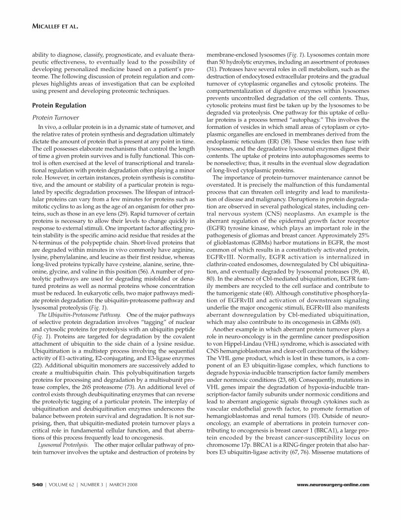
the relative rates of protein synthesis and degradation ultimatelydictate the amount of protein that is present at any point in time.The cell possesses elaborate mechanisms that control the lengthof time a given protein survives and is fully functional. This con-trol is often exercised at the level of transcriptional and transla-tional regulation with protein degradation often playing a minorrole. However, in certain instances, protein synthesis is constitu-tive, and the amount or stability of a particular protein is regu-lated by specific degradation processes. The lifespan of intracel-lular proteins can vary from a few minutes for proteins such asmitotic cyclins to as long as the age of an organism for other pro-teins, such as those in an eye lens (29). Rapid turnover of certainproteins is necessary to allow their levels to change quickly inresponse to external stimuli. One important factor affecting pro-tein stability is the specific amino acid residue that resides at theN-terminus of the polypeptide chain. Short-lived proteins thatare degraded within minutes in vivo commonly have arginine,lysine, phenylalanine, and leucine as their first residue, whereaslong-lived proteins typically have cysteine, alanine, serine, thre-onine, glycine, and valine in this position (56). A number of pro-teolytic pathways are used for degrading misfolded or dena-tured proteins as well as normal proteins whose concentrationmust be reduced. In eukaryotic cells, two major pathways medi-ate protein degradation: the ubiquitin-proteasome pathway andlysosomal proteolysis (Fig. 1).
The Ubiquitin-Proteasome Pathway. One of the major pathwaysof selective protein degradation involves “tagging” of nuclearand cytosolic proteins for proteolysis with an ubiquitin peptide(Fig. 1). Proteins are targeted for degradation by the covalentattachment of ubiquitin to the side chain of a lysine residue.Ubiquitination is a multistep process involving the sequentialactivity of E1-activating, E2-conjugating, and E3-ligase enzymes(22). Additional ubiquitin monomers are successively added tocreate a multiubiquitin chain. This polyubiquitination targetsproteins for processing and degradation by a multisubunit pro-tease complex, the 26S proteasome (73). An additional level ofcontrol exists through deubiquitinating enzymes that can reversethe proteolytic tagging of a particular protein. The interplay ofubiquitination and deubiquitination enzymes underscores thebalance between protein survival and degradation. It is not sur-prising, then, that ubiquitin-mediated protein turnover plays acritical role in fundamental cellular function, and that aberra-tions of this process frequently lead to oncogenesis.
Lysosomal Proteolysis. The other major cellular pathway of pro-tein turnover involves the uptake and destruction of proteins by
membrane-enclosed lysosomes (Fig. 1). Lysosomes contain morethan 50 hydrolytic enzymes, including an assortment of proteases(31). Proteases have several roles in cell metabolism, such as thedestruction of endocytosed extracellular proteins and the gradualturnover of cytoplasmic organelles and cytosolic proteins. Thecompartmentalization of digestive enzymes within lysosomesprevents uncontrolled degradation of the cell contents. Thus,cytosolic proteins must first be taken up by the lysosomes to bedegraded via proteolysis. One pathway for this uptake of cellu-lar proteins is a process termed “autophagy.” This involves theformation of vesicles in which small areas of cytoplasm or cyto-plasmic organelles are enclosed in membranes derived from theendoplasmic reticulum (ER) (38). These vesicles then fuse withlysosomes, and the degradative lysosomal enzymes digest theircontents. The uptake of proteins into autophagosomes seems tobe nonselective; thus, it results in the eventual slow degradationof long-lived cytoplasmic proteins.
The importance of protein-turnover maintenance cannot beoverstated. It is precisely the malfunction of this fundamentalprocess that can threaten cell integrity and lead to manifesta-tion of disease and malignancy. Disruptions in protein degrada-tion are observed in several pathological states, including cen-tral nervous system (CNS) neoplasms. An example is theaberrant regulation of the epidermal growth factor receptor(EGFR) tyrosine kinase, which plays an important role in thepathogenesis of gliomas and breast cancer. Approximately 25%of glioblastomas (GBMs) harbor mutations in EGFR, the mostcommon of which results in a constitutively activated protein,EGFRvIII. Normally, EGFR activation is internalized inclathrin-coated endosomes, downregulated by Cbl ubiquitina-tion, and eventually degraded by lysosomal proteases (39, 40,80). In the absence of Cbl-mediated ubiquitination, EGFR fam-ily members are recycled to the cell surface and contribute tothe tumorigenic state (40). Although constitutive phosphoryla-tion of EGFRvIII and activation of downstream signalingunderlie the major oncogenic stimuli, EGFRvIII also manifestsaberrant downregulation by Cbl-mediated ubiquitination,which may also contribute to its oncogenesis in GBMs (60).
Another example in which aberrant protein turnover plays arole in neuro-oncology is in the germline cancer predispositionto von Hippel-Lindau (VHL) syndrome, which is associated withCNS hemangioblastomas and clear-cell carcinoma of the kidney.The VHL gene product, which is lost in these tumors, is a com-ponent of an E3 ubiquitin-ligase complex, which functions todegrade hypoxia-inducible transcription factor family membersunder normoxic conditions (23, 68). Consequently, mutations inVHL genes impair the degradation of hypoxia-inducible tran-scription-factor family subunits under normoxic conditions andlead to aberrant angiogenic signals through cytokines such asvascular endothelial growth factor, to promote formation ofhemangioblastomas and renal tumors (10). Outside of neuro-oncology, an example of aberrations in protein turnover con-tributing to oncogenesis is breast cancer 1 (BRCA1), a large pro-tein encoded by the breast cancer-susceptibility locus onchromosome 17p. BRCA1 is a RING-finger protein that also har-bors E3 ubiquitin-ligase activity (67, 76). Missense mutations of
540 | VOLUME 62 | NUMBER 3 | MARCH 2008 www.neurosurgery-online.com
MICALLEF ET AL.

BRCA1 clustered in the Zn2+-binding residues of the RING-finger domain, which is critical for ubiquitin-ligase function, leadto predisposition for development of breast cancers (9, 57).
Post-translational ModificationsAfter protein translation, a newly synthesized protein must
undergo several modifications before becoming fully func-tional. These modifications are known as PTMs and includeprotein folding, proteolytic cleavage, assembly of polypeptidesinto a quaternary structure, formation of disulfide bonds, phos-phorylation of specific amino acids, and glycosylation.
Glycosylation is one of the most common PTMs. This PTMcan occur in the lumen of the ER or in the Golgi cisternae. Thetwo general types of protein glycosylation are N- and O-linkedglycosylation. N-linked oligosaccharides are attached to theamide nitrogen of asparagine via a unique N-glycosyl bond.The consensus sequence at which this occurs is N-X-cysteine/serine/threonine, where X can be any amino acid except pro-line. N-linked oligosaccharides are between 7 and 25 mono-
saccharide units in length.They all contain mannose andN-acetylglucosamine, andthey typically consist of sev-eral branches (30). O-linkedoligosaccharides are attachedto the hydroxyl group ofhydroxylysine via galactose orto serine or threonine via N-acetylgalactosamine (51). Incontrast with N-linked oligo-saccharides, there is no univer-sal consensus sequence atwhich O-linked glycosylationoccurs. O-linked oligosaccha-rides are much shorter than N-linked oligosaccharides andtypically contain only one tofour sugar units. The synthe-sis of O-linked oligosaccha-rides also differs from that ofN-linked oligosaccharides. Theformer occurs through sequen-tial addition of single sugarunits, whereas the latter occursvia the addition of a 14-unitoligosaccharide and subse-quent removal or addition ofspecific sugar units (63).
Glycosylation has manyimportant roles, including pro-tein folding and stability, cell-to-cell and cell-to-matrix adhe-sion, and maintenance ofproper protein function. Alter-ations in protein glycosylationare a common feature of gli-
omas. Yamamoto et al. (78) have shown that transfection of theα2, 6-sialyltransferase gene, which is involved in the synthesisof N-linked oligosaccharides, results in decreased invasion andalterations in adhesion. These changes were explained, at leastin part, by the glycosylation of the α3β1 integrin receptor. It hasalso been shown that overexpression of various glycosyltrans-ferases in a glioma cell line can lead to increased sensitivity tocell death when cells are exposed to chemotherapeutic agentssuch as etoposide (12). These results indicate that alterations inglycosylation contribute to chemotherapeutic resistance.
Another common PTM is protein phosphorylation, whichinvolves binding of phosphate groups to three specific aminoacids: serine, threonine, and tyrosine. These reactions are mediatedby kinases that recognize specific sequences within a given pro-tein. In addition, amino acids at distant sites as well as the pro-tein’s conformation may influence phosphorylation. Proteindephosphorylation is mediated by a second class of enzymes, thephosphatases. The activity of a target protein can be highly regu-lated through cycles of phosphorylation and dephosphorylation.
NEUROSURGERY VOLUME 62 | NUMBER 3 | MARCH 2008 | 541
PROTEOMICS IN NEURO-ONCOLOGY
FIGURE 1. Two major pathways of protein degradation. The ubiquitin-proteasome pathway involves a ubiquitinationenzyme cascade “tagging” a cytosolic or nuclear protein, followed by processing and ultimate proteolysis by protea-somal machinery. Lysosomal proteolysis is mediated through nonselective formation of autophagosomes entrappingcytosolic proteins and organelles. Fusion of this vesicular intermediate with a primary lysosome initiates the diges-tive process resulting in hydrolytic destruction of the autophagic vacuole.

These cycles can occur rapidly (within seconds) or over severalhours to activate or stop signaling cascades that regulate a widevariety of cellular events including cell proliferation and apop-tosis. Aberrant regulation of these phosphorylation anddephosphorylation cycles, such as in the constitutively phos-phorylated mutant EGFRvIII in GBMs, contributes to in-creased proliferation, migration, invasion, and angiogenesis(4, 33, 42, 53). Small-molecule inhibitors that block kinases toinhibit phosphorylation of receptors such as EGFRvIII, orthose that may promote dephosphorylation of such activatedreceptors, are, therefore, prime candidates for potential ther-apeutic intervention (35, 43, 47, 79).
Protein Trafficking and Intracellular LocalizationThe movement or trafficking of proteins from one intracellu-
lar compartment to another, or even the secretion of proteininto the extracellular space, is critical for the formation ofappropriate multiprotein complexes that can then act upon rel-evant functional targets. The process of directing a newly syn-thesized protein to a particular target destination is oftenreferred to as protein localization or protein sorting. The firstsorting event occurs during the initial translation of polypep-tide chains by cytoplasmic ribosomes. Proteins destined forservice in particular organelles or for export from the cell aresynthesized with an N-terminal leader peptide (58). These sig-nal sequences serve as cellular addresses in directing the nas-cent protein toward the ER to complete synthesis, before pass-ing onto the Golgi apparatus for subsequent sorting anddispatching to predetermined cellular destinations (59). Thereare at least 10 main subcellular localizations in eukaryotes, sev-eral of which can be further subdivided into intraorganellecompartments. Proteins are shuttled from the ER and Golgiapparatus by transport vesicles that are formed by buddingfrom a membranous compartment (36). These vesicles trans-port cellular proteins through a cascade of vesicular budding,movement along filamentous cytoskeletal tracks, and dockingand fusion events between donor and acceptor organelle mem-branes (36). Coordination of vesicle docking and fusion isachieved in large measure by a subfamily of Ras-like small Gproteins, the Rab GTPases (49). In addition, transport vesiclespossess tethering complexes and fusion assemblies known assoluble N-ethylmaleimide-sensitive factor-attachment proteinreceptors (SNAREs) to impart additional transport specificity.In brief, integral membrane vesicle-SNAREs (v-SNAREs) selec-tively interact with the target membrane SNARE (t-SNAREs) tofacilitate binding and trigger the fusion process itself (54).Indeed, protein-sorting processes are so vital to the organiza-tion and function of a cell that disruption of protein transporthas proven to be a key event behind several human diseases,including cancer and Alzheimer’s disease (8, 20, 62).
Protein InteractionsProteins accomplish myriad functions through direct interac-
tions with target proteins, phospholipids, nucleic acids, andsmall-molecule second messengers. These interactions are facil-itated by the presence of conserved amino acid sequences that
form adaptor or interaction domains that recognize specifictopographic epitopes on their binding partners (Fig. 2). Duringthe past 10 years, knowledge of these domains has increasedexponentially, and with this has come a better understanding ofintegrative cellular signaling. A high degree of signaling com-plexity is achieved by diverse combinations of these interactiondomains within a single polypeptide (52). This complexity pro-vides the ability for a single activated receptor complex to elicitvaried responses, depending on the specific target-moleculeinteractions that occur. Initiation of the cell cycle; regulation ofgene expression; protein turnover, modification, and transport;DNA repair; cell migration; and apoptosis are all end results ofthe cellular signaling that occurs via these interaction domains.
The list of interaction domains and their target binding mol-ecules is expanding continually, enabling the classification ofthese domains into groups (Fig. 2): Src homology-2 (SH2) andphosphotyrosine-binding domains recognize phosphotyrosines(44, 65); Src homology-3 and WW recognize proteins withstretches of proline (32); and 14-3-3, forkhead-associated, andWD40 domains recognize phosphoserine- and phosphothreo-nine-containing proteins (77). The postsynaptic density-95(PSD95), Drosophila disc large tumor suppressor (DlgA), andzonula occludens-1 (ZO-1) homology domains bind to theextreme C-terminus of proteins (64). In addition, interaction
542 | VOLUME 62 | NUMBER 3 | MARCH 2008 www.neurosurgery-online.com
MICALLEF ET AL.
FIGURE 2. A, subset of interacting protein domains and their aminoacid target sequences. B, protein c-Src as an example of the functional pos-sibilities of different domain combinations. In this instance, the SH2 andSrc homology-3 domains allow recruitment of a target protein for c-Src’skinase function. C, generic layout of protein-protein interactions as facil-itated by interaction domains. Upon ligand binding, receptor tyrosinekinases such as epidermal growth factor receptor (EGFR), undergo a con-formational change resulting in the formation of hetero- and homo-dimers,which autophosphorylate tyrosine residues. Phosphorylated tyrosines arebound by the SH2 domain. In the case of phosphorylated EGFR receptor,the SH2 containing adaptor Grb2 is recruited. Grb2 in turn recruits Sosvia an interaction between the Src homology-3 domain of Grb2 and theproline rich motif on Sos. Sos is a Guanine Exchange factor and functionsto switch out guanosine 5’-diphosphate to guanosine 5’-triphosphate(GTP) bound to small GTPases, such as Ras. When bound to GTP, Rasadopts an active conformation, thereby conveying the signal downstream.
AB
C

domains exist not only for specific protein epitopes, but also forphospholipids. The pleckstrin homology and Fab-1, YGL 023,Vps, and EFA1 domains all recognize phosphoinositides in amanner specific to the phosphorylation site of the inositol head-group (17, 37). A detailed discussion of the mechanisms andtargets of the various domains mentioned is not possible in thisreview; however, it is exemplified by a more in-depth examina-tion of the well-characterized module, the SH2 domain.
The function of receptor tyrosine kinases (RTKs) is interest-ing, particularly in the field of oncology. Targets of RTK signal-ing commonly contain the SH2 domain despite vastly differentfunctions. Extracellular ligands bind to RTKs and induce a con-formational change that result in receptor dimerization andautophosphorylation of tyrosine residues. The SH2 domain-containing proteins bind to these phosphotyrosine residues andserve as adaptors to recruit additional proteins to the activatedRTK complex. These target proteins help integrate and propa-gate intracellular signals. Although all SH2 domains recognizethe phosphotyrosine residue, the specificity of binding is influ-enced by the downstream amino acid sequence. The residues inthe +1 to +3 positions are important for recognition by the SH2domain-containing protein, and the specificity is determined bythe extent of the fit into specific binding pockets of the respec-tive SH2 modules (65).
As an example, normal EGFR upon binding its natural lig-and, EGF or transforming growth factor-α, undergoes a confor-mational change thereby allowing the receptor to dimerize andautophosphorylate its tyrosine residues (69). This phosphory-lation causes the SH2 domain-containing adaptor protein,growth factor receptor-bound protein 2 (Grb2), to bind theamino acid sequence pTyr-X-Asn-X (where X is any aminoacid), as found starting from tyrosine 1068 of EGFR (5). Grb2also contains an Src homology-3 domain that recognizes theproline-rich sequence of the son of sevenless (Sos) protein (52).Sos functions as a guanine-nucleotide exchange factor and con-verts inactive Ras-guanosine diphosphate to active Ras-guano-sine triphosphate. Ras-guanosine triphosphate, in turn, initiatesa protein-serine/threonine kinase cascade that terminates inthe phosphorylation of transcription factors by mitogen-activated protein kinase (26). This represents a single signaling“branch” from a single phosphorylated residue; the presence ofmultiple epitopes on activated receptors results in the forma-tion of various multiple-protein complexes that are capable ofeliciting a range of cellular responses. A particular responsethen arises from the specific cell state and the combination ofactivated binding sites present on the RTK. Replication of thisbinding mode in diverse protein systems is an elegant meansby which proteins conserve and yet manage activation of avariety of signaling pathways and biological functions.
Proteomic Analyses: An OverviewProteomic analyses can be broadly categorized into those
that identify and/or quantify proteins that are expressed indifferent physiological and disease states and those that mapprotein-protein interactions and post-translational modifica-tions. Western blot and immunohistochemical (IHC) analyses
are traditional techniques used for the detection, quantifica-tion, interactions, and PTM of proteins but are dependent onthe availability of well-characterized antibodies. In addition,these techniques also require relatively large amounts of tissuelysates and are typically limited to analysis of a few samples ata time. Present and emerging proteomic techniques have thepotential to overcome some of these obstacles to identify andcharacterize low-abundance proteins not only from the dis-eased tissue, but also from bodily fluids (plasma, urine, cere-brospinal fluid [CSF]), in a high-throughput, nonbiased man-ner. Hence, proteomic analyses have the potential to allow us todiscover, identify, and quantify markers for early cancer detec-tion, diagnosis, and prognosis, and lead us toward developingand guiding molecular targets for more effective therapeuticinterventions.
The following sections review established and emergingproteomic techniques used in oncology, highlighting thestrengths and drawbacks of each. The focus is on emergingtechnologies and, especially, applications to neuro-oncology.These include MS and bioluminescence resonance energytransfer (BRET) techniques.
Two-dimensional Polyacrylamide Gel ElectrophoresisFor many years, the foundation of protein-expression profiling
has been two-dimensional polyacrylamide gel electrophoresis(2D-PAGE), which allows investigators to identify proteins thatare up- or downregulated in comparable samples. Proteins arefirst separated and characterized on the basis of charge. This isachieved by applying a crude protein-lysate mixture to a “first-dimension” pH gradient acrylamide gel strip and then exposingthe strip to an electric field. This separates the proteins on thebasis of isoelectric point (pI), which is the pH value at which aprotein is electrically neutral. As the protein molecules movedown the gel, they experience the pH gradient and migrate topositions that correspond to their respective pI values. Once aprotein arrives at the position of its pI, it has no charge, and,thus, moves no further. When the isoelectric focusing is com-plete, the gel strip is reduced, alkylated, and transferred to asodium-dodecyl sulfate-polyacrylamide gel. In this “seconddimension,” proteins are denatured and separated according tomolecular mass. This two-dimensional separation allows visual-ization of a substantial range of proteins in a single experiment.With 2D-PAGE, 3000 to 10,000 protein spots can be resolved pergel, depending on the method of spot detection (24). Proteindetection is accomplished by staining the gels with dyes such asCoomassie brilliant blue, silver stain, or various fluorescent dyes(11). Silver staining is highly sensitive but incompatible withsome downstream applications such as MS. Use of colloidalCoomassie blue G-250 when staining can improve detection sen-sitivity (48). After staining is complete, the resulting protein spotsare recorded and quantified using sophisticated image-analysissoftware. Gels are compared, and biological differences betweenthe samples are identified. Spots of interest are then excised andsubjected to mass spectrometric analysis.
The use of 2D-PAGE as an expression-profiling tool is limitedby several issues. At first glance, the resolving capability of
NEUROSURGERY VOLUME 62 | NUMBER 3 | MARCH 2008 | 543
PROTEOMICS IN NEURO-ONCOLOGY

2D-PAGE is impressive, but it is still inadequate compared withthe enormous dynamic range of cellular protein expression.Abundant proteins such as actin are present at a concentra-tion of approximately 108 molecules per cell, whereas less-abundant transcription factors are represented by approxi-mately 100 to 1000 copies, thereby creating a 106 dynamic rangein protein concentration, which is clearly beyond the 104 rangeassociated with 2D-PAGE (55). Enrichment or prefractionationstrategies must be used to locate the less plentiful proteins.Then, there is a problem of protein solubility, especially forhydrophobic-membrane and nuclear proteins. This problem islinked with the isoelectric step of 2D-PAGE, and such proteinsusually do not enter the second dimension of the gel. Further-more, it is sometimes problematic to use two-dimensional gelsto detect proteins with molecular masses greater than 100 kDaor pI values that are less than 4 or greater than 9. This problemcan be circumvented to a degree by spreading the pH rangeacross several gels, using so-called “zoom gels,” which allowresolution of 5000 to 10,000 protein spots.
Additionally, a recent study has shown that 2D-PAGE detectsonly the most abundant proteins (74). Typical detection of 3000to 10,000 protein spots represents those protein species withmore than 10,000 molecules per cell (74). Traditional detectionmethods cannot distinguish protein spots of less than 1000 mol-ecules per cell; as such, it is doubtful that all of the cell or tissueproteins are resolved. Approximately 50 to 75% of the proteinson the gel, the low-abundance proteins, are not detected (74).Another serious issue is the inadequacy of the technology forquantification. The predominant protein-staining methods, forinstance, are either not sensitive enough (e.g., Coomassie bril-liant blue) or have limited linearity (e.g., silver staining), and,thus, impede a truly quantitative protein-expression analysis(70). The inherent gel-to-gel variability of 2D-PAGE compro-mises quantitative comparison of protein-expression levels.
An emerging method known as fluorescent two-dimensionaldifferential gel electrophoresis (2D-DIGE) has proven to be ofenormous importance for quantification accuracy. In brief,Samples A and B are labeled with two different fluorescentdyes (i.e., Cy3 or Cy5). These labeled samples are mixed andapplied to one 2D-DIGE gel. After electrophoresis, this gel issuccessively illuminated with the excitation wavelengths ofCy3 and Cy5. The resulting protein spot patterns can then becompared and analyzed for differential protein expression bythe construction of image overlays or by using software specif-ically designed for 2D-DIGE (70). Despite the aforementionedshortcomings, 2D-PAGE continues to be an essential tool inproteomics research. Improvements in sensitivity, reproducibil-ity, and quantitative precision will solidify this status.
Mass Spectrometry-based ProteomicsOverview. MS is arguably the most important and powerful
technology for proteomics. Here, we provide an introduction ofMS to neuro-oncologists; as such, our overview is necessarilyselective in nature. For a comprehensive treatise of MS-basedproteomics, excellent reviews are available (1, 2). Most proteinsare too complex for direct analysis by MS; consequently, enzy-
matic digestion (typically using trypsin) of proteins into pep-tides is required with subsequent separation and identification ofproteins via tryptic peptide mass or via the partial sequencing ofone (and typically multiple) peptides after a search of nonre-dundant protein databases. These searches are usually adminis-tered automatically via powerful, commercially available soft-ware such as Mascot (Matrix Science, London, England) andProteinPilot (Applied Biosystems, Foster City, CA) and continu-ally updated public databases such as the National Center forBiotechnology Information and SwissProt. The exception to thisapproach of identifying proteins from its constituent peptides(referred to as the “bottom-up approach”) is whole-proteinanalysis (the “top-down approach”). The top-down approachinvariably involves the use of Fourier-transform MS, with unpar-alleled mass accuracy and resolution required to manage thecomplexity associated with whole-protein analysis. In general,MS can only contend with relatively simple mixtures at any giventime; hence, efficient up-front separation of the proteins or pep-tides is of paramount importance.
Sample Preparation and Separation. Biological samples suchas cultured cells or biopsy samples are very complex in natureand are composed of not only proteins, but also lipids andnucleic acids. In specimens, there is the added complexity of avariety of cell types, which may require microisolation tech-niques such as laser-capture microdissection, although thequantity of such proteins obtained may make analysis imprac-tical. Protein or peptide separation can be undertaken usingsingle-dimensional gels, or, if required, multi-dimensional gelsmay be used to maximize resolution and increase the probabil-ity and efficiency of detecting low-abundance proteins.Typically, proteins are first extracted using detergent to solubi-lize proteins and separate them from lipids. Subsequent stepsthen involve separating this protein-enriched mixture as muchas possible into its components. A commonly used techniquefor protein separation is gel electrophoresis, either one-dimensional, which separates proteins according to molecularweight, or two-dimensional, which separates proteins firstaccording to pI value and then on the basis of molecularweights. The separated proteins are stained with dyes that arecompatible with MS detection, e.g., Coomassie blue, or undercertain conditions, silver stain, to allow visualization. Thevisualized protein spots or bands are then excised, trypsinized,and analyzed.
An alternative separation technique that is increasingly beingused in tandem with MS detection is liquid chromatography(LC), frequently in multiple dimensions. The most commonpractice involves separation at the peptide level. Proteins aretrypsinized before chromatography, typically first via strongcation exchange and then by reverse phase, thus resolving thepeptides largely by charge and molecular weight, respectively. Inmost applications, peptides resolved by the reverse-phase sepa-ration are analyzed directly by means of online nano-electro-spray ionization (ESI) tandem MS. Alternatively, the effluentfrom the reverse-phase column is collected and fractionated ona sample target for subsequent offline matrix-assisted laser des-orption/ionization (MALDI) MS/MS. Many arguments have
544 | VOLUME 62 | NUMBER 3 | MARCH 2008 www.neurosurgery-online.com
MICALLEF ET AL.

been forwarded in favor of and against gel electrophoresis ver-sus LC, and within LC, ESI versus MALDI MS/MS. Althoughthere is almost always an element of truth in all of these argu-ments, most reasoning can be boiled down to personal prefer-ence. Combining the results of more than one methodologyalmost invariably increases the number of identified proteinsand/or the confidence of the identification. Thus, the aforemen-tioned technologies are complementary rather than competitive.
Fundamentals of Mass Spectrometry. Put into the simplestterms, a mass spectrometer is an electronic balance that weighsan ionized molecule by its mass-to-charge (m/z) ratio. As shownschematically in Figure 3A, each MS instrument has an ion sourcethat produces ions from the sample, a mass analyzer thatresolves peptide ions on the basis of m/z ratio, and a detectorthat reveals these ions resolved by the mass analyzer to displayion abundances versus the m/z values in the mixture of ions toyield the mass spectrum. If the mixture of ions is the result of a
mixture of tryptic peptides from a given protein, the mass spec-trum is sometimes referred to as a tryptic peptide map. Tosequence the peptides, tandem MS analysis is required, whichinvolves isolation of the tryptic peptide ions in the first massspectrometer (actually the MS stage), fragmentation by collidingwith a neutral gas (typically nitrogen or argon), and mass analy-sis of the fragments using a second mass spectrometer. The m/zratios of the fragment ions can be analyzed using computerizeddatabase search engines (e.g., Mascot, ProteinPilot, X!Tandem[The Global Proteome Machine Organization], OMSSA [OpenMass Spectrometry Search Algorithm; National Center forBiotechnology Information, Bethesda, MD]) to reveal the identi-ties of peptides and their parent proteins.
Source. A fundamental requirement for a molecule to be ana-lyzed via MS is that it must be ionized or charged at the source.Ionization of peptides for modern MS analysis is based on two newionization technologies, termed ESI and MALDI (Fig. 3, B and C),
which earned their inventors ashare of the 2002 Nobel Prize inChemistry. Ionization of pep-tides is typically achieved viaprotonation (giving positivecharges) that occurs concomitantwith transfer of the peptides intothe gas phase (ion desorption).In ESI mode, this process isinduced by the application of,typically, a few kilovolts on anemitter in the form of an ESIneedle, through which the sam-ple is sprayed; this is often aidedby a coaxial nebulizer gas flowof nitrogen (Fig. 3B). For a tryp-tic peptide, the presence of abasic C-terminal lysine or argi-nine residue frequently results ina doubly protonated peptide;the presence of a second or thirdbasic residue may lead to tripleand even quadruple protona-tion. MALDI produces proto-nated peptides as well, albeit bydifferent mechanisms (Fig. 3C),and in contrast to ESI, results inpredominately single proto-nated peptides. As mentionedpreviously, MALDI MS/MS isparticularly suited to offlineanalysis of fractionated samplesfrom LC separation, as it permitsextended and targeted analysesof key fractions. In contrast, ESIMS/MS is typically used inonline analysis after LC separa-tion. As previously mentioned,although each method has its
NEUROSURGERY VOLUME 62 | NUMBER 3 | MARCH 2008 | 545
PROTEOMICS IN NEURO-ONCOLOGY
FIGURE 3. A, schematic representation of a mass spectrometer. Each instrument has a source that produces ions fromthe sample, a mass analyzer that resolves peptide ions based on their mass to charge ratio (m/z) and a detector thatdetects ions resolved by the mass analyzer. B, electrospray ionization (ESI) source: a liquid containing a peptide mix-ture passes through a capillary tube under high voltage. As the liquid exits the capillary tube, charged droplets con-taining peptides are formed. A gas, such as nitrogen, is passed over the charged droplets, resulting in evaporation ofthe solvent (desolvation) and the accumulation of charged peptides. C, matrix-assisted laser desorption/ionization(MALDI) source: a sample is mixed with a chemical matrix that absorbs light at a specific wavelength. This admix-ture is placed onto a glass slide and the solvent is allowed to evaporate. After evaporation, a crystal lattice in whichthe peptides are integrated is left on the slide. This is placed into the source and a laser is directed onto the sample.The matrix will absorb the photons of the laser and become energetically excited. This energy is then transferred tothe peptides, which are ejected from the matrix into a gas phase. MALDI-TOF-MS: These peptides, which are nowionized in the gas phase, are directed under an applied voltage to the time-of-flight (TOF) analyzer. The detector willcalculate the time it takes ions to fly from one end of the TOF analyzer to the other end.
A
B
C

individual merits and limitations, often both are used to augmentthe overall MS analysis.
Mass Analyzer. Ions that are formed in the ion source, irre-spective of whether the source is ESI or MALDI, must be trans-ferred into the mass analyzer. This transfer is arguably morechallenging for ESI than for MALDI processes because the ESIion source is at atmospheric pressure, and, thus, presents a com-paratively larger pressure differential between the ion sourceand the analyzer. In the sampling process, ions are selectivelyenriched over neutral (mainly nitrogen and solvent) molecules.The ions are then dispersed according to their m/z values in themass analyzer region. To obtain sequence information, twomass analyzers are required in tandem with a region in between(typically a collision cell) where fragmentation of mass-selectedions can occur over a sequence of timed events.
The mass analyzer can take many forms and operate on dif-ferent principles. The most common mass analyzers affect m/zseparation by means of a time-dependent electrical field. In thequadrupole mass filter, the time-dependent electrical field hasboth alternating current (AC) and direct current (DC) compo-nents, with mass separation performed by judicious combina-tion of the AC and DC fields by increasing the AC and DC mag-nitudes simultaneously while maintaining a constant ratio ofthe two. Selected ions that are transmitted exit the quadrupolefilter axially. In a single-stage mass spectrometer, ions transmit-ted from the mass analyzer are intercepted downstream by adetector and are measured. In a tandem mass spectrometer, thefirst stage can act as a mass filter that permits only selected ionsof interest into the second stage, which is the collision cell. Thecollision cell is similar to the first stage in its ability to confinethe ions radially for effective collision, but unlike the first stage,it performs no mass filtering. Instead, this section of the massspectrometer is maintained at a greater pressure than the previ-ous section, thereby increasing the probability of transmittedions colliding with neutral gas molecules. As mentioned earlier,these collisions cause fragmentation of the ions; the fragmentsare then transmitted into another mass analyzer downstream,they are detected, and their sequence is determined.
In the 3-D ion trap, application of only an AC field confinesall ions within the trap. Mass scanning is achieved by rampingthe AC amplitude, causing oscillations of increasing magni-tude in the axial direction for eventual ejection of ions inincreasing m/z value. The ions are then evaluated by thedetector. An increasingly more common mass analyzer ofgreater mass accuracy and resolution is the time-of-flight(TOF) mass analyzer (Fig. 3C). In this system, ions of all m/zvalues are injected into one end of the zero-field flight tubewith a nearly identical kinetic energy; each ion arrives at theother end of the tube with a flight time that is proportional tothe square root of its mass. TOF MS are highly accurate (massaccuracy, 10 ppm) with a mass resolution of 10,000, whichmeans that for most tryptic peptides that are measured, thesecond decimal place of their m/z values is a significant figurethat should be reported. A type of tandem MS that has enjoyedparticular commercial success is the hybrid QqTOF, whichuses a quadrupole mass filter as the first mass-analyzer stage
(Q), an AC-only quadrupole as the collision cell (q), and TOFas the second mass-analyzer stage (2).
Detector. Detection of ions in modern MS relies almostexclusively on ion counting. The channel electron multiplier isthe most common detector used with quadrupole and ion-trap instrumentation. The most important element of thechannel electron multiplier is a cylindrical electron-emissivesurface that has a potential decrease of typically 2 to 3 kV inthe axial direction. An ion striking the surface near theentrance of the channel electron multiplier causes emission ofa number of electrons into the cylindrical space. These elec-trons are accelerated toward the anode end of the cylinder(the channel), and as these electrons travel down the cylinder,they repeatedly strike the electron emissive surface to releasesecondary electrons. When working properly, this multiplica-tion process results in the generation of 100 million electronsper incident ion. The arrival of the electron pulse at the anoderegisters as a single ion count.
Mass Tagging and Differential Mass SpectrometryOne of the most effective ways to discover disease biomark-
ers is to perform differential analysis, in which the expressedproteins in the diseased and normal states are compared. Asmultiple stages of separation are used to resolve the proteins ortheir tryptic peptides before MS analysis, losses of the proteinsand peptides during the separation steps are unavoidable. Theonly practical method for ensuring that the losses are identicalin the normal and diseased samples (and, hence, the compari-son is valid) is to tag the proteins or peptides using reagentsthat are chemically identical but different in mass (by havingdifferent isotopes of the same element, typically 12C and 13C,14N and 15N, and 16O and 18O) as early in the process as possi-ble, and combining the two samples before any separation isperformed. Tagging of the proteins or peptides typically makesuse of time-honored derivatization reactions that involve a par-ticular functional group, e.g., the sulfhydryl group of cysteineby isotope-coded affinity tag (ICAT) or the amino group oflysine by isobaric tags for relative and absolute quantification(iTRAQ).
The Isotope-coded Affinity TagThe ICAT reagent was developed to tag proteins via their
sulfhydryl groups (18). The reagent is composed of three parts(Fig. 4A), including:1) an iodoacetamide functional group that reacts selectively
with sulfhydryl groups of proteins; 2) a linker region that is different between the light and heavy
versions of the region. In the original ICAT reagent, theheavy tag contains strategically placed hydrogen and deu-terium atoms in the linker region. However, the resultinglight- and heavy-tagged peptides often showed small differ-ences in retention times during reverse-phase chromatogra-phy, thus complicating quantification. This problem waseliminated in the second-generation ICAT reagent that has alinker region using 12C and 13C for differentiation; and
546 | VOLUME 62 | NUMBER 3 | MARCH 2008 www.neurosurgery-online.com
MICALLEF ET AL.

3) a biotin moiety that permits affinity purification via avidinbinding.
The two samples for analysis are treated with either the lightor heavy ICAT reagent and then combined for trypsinization.The resulting tryptic peptides are separated for eventual onlineESI MS/MS analysis. A typical separation strategy involves afirst dimension of strong cation-exchange chromatography inwhich the tryptic peptides are dispersed into a number(approximately 30) of fractions. The labeled peptides in eachfraction are then selectively preconcentrated on a seconddimension of affinity chromatography (via the biotin-avidinchemistry) and then separated on a third dimension of reverse-phase separation with online nano-ESI MS/MS. The assump-tion is that the ICAT tags disturb the chemistry of the labeledpeptides identically, and that the relative abundances of theMS peaks reflect the relative abundances of the proteins in thesamples. The question arises as to how much the relative abun-dance needs to deviate from 1 in an ICAT-labeling experimentbefore differential expression is denoted to be significant. Mostpublications have used a minimum factor of 2 in comparisonsinvolving small sample sizes to avoid false positives (14).
ICAT tagging is consideredto be more selective than othermass-tagging technology, e.g.,iTRAQ, as cyste ine i s anuncommon residue. If a pro-tein contains only one cysteineresidue, ICAT tagging willresult in only one labeled tryp-tic peptide, which may com-promise the confidence inunambiguous identification ofthis protein. However, thisselectivity significantly reducesthe number of peptides foronline MS/MS analysis, whichdecreases the extent of overlap-ping peaks, and increases theprobability of identifying differ-entially expressed peptides oflow abundance (13).
Isobaric Tags for Relativeand Absolute Quantification
iTRAQ is a relatively newmass-tagging reagent that tar-gets the much more abundantamino group on the side chainof lysine residues and the N-terminus of the tryptic pep-tides (Fig. 4B). Because pro-teins with modified lysineresidues are no longer cleavedefficiently by trypsin, trypticdigestion needs to occur be-fore iTRAQ tagging is per-
formed. In addition, trypsin digestion before tagging also gen-erates a free primary amine group at the N-terminus of eachpeptide and ensures that peptides lacking lysine residues canalso be tagged and used in the analysis. The iTRAQ reagent ismore versatile in that the first-generation reagent came withfour different tags, and the next-generation reagent (that willsoon be available) will come with eight different tags. iTRAQdiffers from ICAT in that relative quantification is performedat the tandem-MS stage via the relative abundances of reporterions that are collision-induced dissociation products. Each tagyields a reporter ion of a different m/z value. In the four-plexreagent, the reporter ions are 114, 115, 116, and 117 Da (Fig. 4B).The linker region’s isotopic composition (12C and 13C and 16Oand 18O) compensates for the differences in these reporter ionmasses such that peptides labeled with the four tags have thesame m/z values. Collision-induced dissociation of these pep-tides yields ions that yield sequence information as well asthe reporter ions, whose abundance ratios yield relative quan-tification data among the four samples.
The aforementioned techniques, particularly those involvingthe use of LC-MS analysis, frequently generate large amounts of
NEUROSURGERY VOLUME 62 | NUMBER 3 | MARCH 2008 | 547
PROTEOMICS IN NEURO-ONCOLOGY
FIGURE 4. A, structure of isotope-coded affinity tag (ICAT). There are two ICAT molecules, a light chain and aheavy chain. Each molecule consists of three groups; 1) a biotin group, which allows affinity purification by bindingto avidin; 2) a linker region that contains hydrogen (light chain) or deuterium (heavy chain) or 12C (light chain) or13C (heavy chain); and 3) an iodoacetamide functional group that reacts with -SH groups of proteins. B, structureof isobaric tags for relative and absolute quantification (iTRAQ). Each tag contains 1) a reporter group that variesin molecular weight (114, 115, 116, and 117kDa); 2) a linker group that varies in composition (12C and 13C; and16O and 18O) and compensates for the differences in these reporter ion masses; and 3) a peptide reactive group thatreacts with primary amines, i.e., lysine side chains and N-terminus of peptides.
A
B

data. Interpretation of volumes of data such as these necessitatesthe use of powerful software that is constantly being improvedand updated. Mining of the results produced by such softwareis another aspect that can require a significant commitment ofresources. This commitment would be in the form of bioinfor-matics packages and personnel who are familiar with thenuances and limitations of the types of mass spectrometricanalysis and search algorithms used in the data generation, aswell as the implications and biological significance of theresults produced. Once the data from these kinds of pro-teomic analyses are compiled and appropriately verified, theycan be coordinated with data from genomic analysis in a “sys-tems biological” approach that could then provide a betterunderstanding into mechanisms involved in the biologicalprocesses of interest.
Examples of MS-based Analysis in Neuro-oncology. MS-basedanalytical tools have been used to decipher the proteome of thenormal and diseased brain, including primary brain tumors(16, 21, 50). In addition, MS-based techniques have been usedto decipher potential biomarkers from CSF of patients harbor-ing brain tumors (28). Our group is presently examining the tis-sues and sera of patients with primary brain tumors and com-paring them with the tumor proteomes to determine whetheror not serum biomarkers can augment our diagnostic and ther-apeutic capabilities in a noninvasive manner. The ability ofiTRAQ analysis to discover and identify tissue biomarkers inendometrial cancer was recently described in reports from ourgroup (13, 15).
We have also used MS-based approaches to increase ourmechanistic understanding of genetic alterations relevant toneuro-oncology; two examples are detailed here. The first area ofinvestigation centers on increasing our knowledge of interactingproteins with neurofibromin, the protein encoded by the neurofi-bromatosis Type 1 (NF1) gene. Mutations of NF1, including lossor deletion of the second allele and, thereby, neurofibrominexpression, which leads to increased Ras-GTP activity as a resultof a loss of neurofibromin-mediated Ras-GAP function, is thelikely basis of the tumors associated with neurofibromatosisType 1. The increased Ras-GTP activity is secondary to the lossof neurofibromin binding to Ras-GTP through its GAP-relateddomain (GRD), which normally results in inactive Ras-GDP (Fig.5A). However, many of the non-oncological manifestations ofneurofibromatosis Type 1 occur without loss of neurofibrominexpression from the second normal allele, including those lead-ing to learning difficulties caused by developmental abnormali-ties of the CNS. We and others have postulated that becauseneurofibromin is an extremely large protein with severaldomains (Fig. 5A), many of these clinical manifestations in neu-rofibromatosis Type 1 may arise because of the loss of additionalnormal interactions of neurofibromin outside of the GRD (53a).
The problem associated with deciphering additional neu-rofibromin-interacting proteins using standard protein-proteininteraction techniques such as coimmunoprecipitation andyeast-two-hybrid is that because of its large size (2818 aminoacids, approximately 240 kDa), full-length neurofibromin hasnot been exogenously expressed. As a result of this limitation,
we have used an MS-based approach to decipher interactingproteins with specific neurofibromin domains, with our firstexperiments directed at what proteins interact with the tubu-lin-binding domain (TBD), which is adjacent to the GRD ofneurofibromin (Fig. 5A). The smaller domains of neurofi-bromin, such as the TBD, were subcloned and expressed witha glutathione-S-transferase (GST) tag in the N-terminal end ofthe fusion protein. After the GST-fusion protein was immobi-lized on a glutathione column, cell lysate from human NF1null Schwann cells (ST8814) was applied, and proteins thatassociate with the fusion domain were eluted, separated viamass on a denaturing SDS-PAGE gel, and then visualized witha colloidal Coomassie brilliant blue stain (Fig. 5B). Differen-tially expressed proteins in the GST-TBD versus GST-negativecontrol lane appeared as bands (Fig. 5B, arrows) and wereexcised, trypsin digested, and used for MS analysis.
LC-coupled MS/MS and analysis has yielded the aminoacid sequence of the extracted peptide fragments. These frag-ment sequences were searched against databases to identifythe candidate interactors. Leucine-rich pentatricopeptide-repeat cassette (LRPPRC) is one of the identified novel neu-rofibromin TBD-interacting proteins that we are examiningwith interest. This mitochondrial protein, speculated to beinvolved in vesicular transport (perhaps by its interactionwith tubulin) and thereby in regulating mRNA stability, ismutated in the Leigh syndrome-French Canadian subtype,which is an inherited syndrome with neurological manifesta-tions that have some overlap with those observed in neurofi-bromatosis Type 1 patients. The discovery of this novel inter-action using MS technology is additionally verified by moretraditional proteomic techniques including Western blotanalysis, which demonstrates specific physical interactions ofLRPPRC with the TBD but not other domains of neurofi-bromin (Fig. 5C). Co-immunofluorescence experiments alsosuggest colocalization of neurofibromin and LRPPRC in themitochondria of cells (Fig. 5D). Present experiments arefocused on deciphering the functional role of the LRPPRC-neurofibromin interaction to improve our understanding ofthe molecular pathogenesis of neurofibromatosis Type 1.
In addition to our ongoing work in deciphering serum bio-markers from patients with CNS tumors, similar to ourapproach for endometrial cancers (13), we have used differen-tial MS analysis to increase our knowledge of aberrant EGFR-mediated signaling in human GBMs. We undertook ICAT-based analysis with the goal of identifying differentiallyexpressed proteins that may confer a growth advantage toGBMs that harbor amplification and/or mutations in EGFRcompared with GBMs that do not (Fig. 6). Using ICAT-labeledlysates from human GBM explant xenografts characterizedfor their positive or negative EGFR status as identified withthe use of fluorescence in situ hybridization and IHC (Fig.6A), we detected peptide fragments that were differentiallyincreased 10- to 25-fold in the positive-EGFR GBMs (Fig. 6B).
Using protein bioinformatics software, we discovered thatthese peptides mapped to a protein called MARCKS, a proteinkinase C substrate that has been implicated in erbB2 (a close
548 | VOLUME 62 | NUMBER 3 | MARCH 2008 www.neurosurgery-online.com
MICALLEF ET AL.

family member of EGFR-erbB1) signaling in breast cancer(25). Increased MARCKS expression was verified in theexplant xenografts by the use of traditional Western blotanalysis (Fig. 6C), and also in a larger panel of GBM cell lines,in explant xenografts and operative specimens with positiveor negative EGFR expression. Functional studies with MAR-CKS siRNA knockdown and transfection-mediated overex-pression have demonstrated a significant biological role ofMARCKS in mediating aberrant EGFR signaling in GBMs.The two aforementioned areas of research currently beingundertaken in our laboratories are examples of how MS-basedanalysis can be used to discover novel protein interactions
and to identify proteins involved in altered signaling path-ways in neuro-oncology.
Serum and Tumor Proteomics in Neuro-oncologyThe potential utility of proteomics-based analysis is being
realized at an incredible rate; however, this technology, specif-ically in the field of neuro-oncology, is only beginning to beexploited. For example, much effort and study have beenfocused on using CSF as an indirect indicator of disease type,status, and response. Diseases of the CNS are often associatedwith changes in the CSF protein composition; therefore, profil-ing this fluid could enable us to detect, diagnose, and monitor
NEUROSURGERY VOLUME 62 | NUMBER 3 | MARCH 2008 | 549
PROTEOMICS IN NEURO-ONCOLOGY
FIGURE 5. Demonstration of use of mass spectrometric analysis to identifyinteracting proteins in neuro-oncology: Neurofibromin, the gene productencoded by the neurofibromatosis Type 1 gene, is too large to express in total.A, six domains of neurofibromin were epitope tagged with an amino termi-nal glutathione-S-transferase (GST) tag, for use in coimmunoprecipitationstudies: cysteine/serine-rich domain (CSRD), tubulin-binding domain(TBD), GAP-related domain (GRD) syndecan binding domain (Synbd1 andSynbd2), and the SEC14 domain. B, for example, the GST-TBD bait proteinidentified a novel TBD interacting protein (arrows-leucine-rich pentatri-copeptide-repeat cassette [LRPPRC]), from lysates of the ST88 to 14 humanNF-1 malignant peripheral nerve sheath tumor (MPNST) cell line. Several
interacting protein bands were isolated by gel separation, and peptidesequences were identified with MS analysis with subsequent identification ofleucine-rich pentatricopeptide-repeat cassette (LRPPRC) from bioinformaticanalysis of protein data bases. C and D, verification of identifying LRPPRCas a novel neurofibromin TBD interacting protein is demonstrated by west-ern blot analysis demonstrating LRPPRC interacts specifically with the GST-TBD and not the parental GST, GST-CSRD, or GST-SEC14 (C) andimmunofluorescence co-localization of LRPPRC (green) with neurofibromin(red) in a perinuclear pattern (yellow), which likely represents mitochondriawhere LRPPRC has been predominantly demonstrated (D).
A
B C
D

this disease. To this end, Khwaja et al. (27) used a strong cation-exchange separation combined with surface-enhanced laserdesorption ionization (SELDI)-TOF-MS of CSF to uncover con-served protein “fingerprints” for neoplastic disease comparedwith reactive/inflammatory CNS diseases. This group has also
shown that CSF analysis via2D-GE and cleavable ICAT aresensitive methods for identify-ing tumor- and grade-specificsignature proteins in high-grade astrocytoma CSF sam-ples (28). Likewise, the use ofpatient sera to profile pro-teomic differences betweendiseased and non-diseasedindividuals holds equal prom-ise. Indeed, Zhang et al. (81)demonst ra ted the use o fSELDI-TOF-MS to identifyseven astrocytoma-specificserum biomarkers that areabsent in the serum of nonma-lignant individuals. One grouphas already demonstrated theability to generate protein-biomarker patterns by use ofSELDI-TOF-MS serum screen-ing, thus enabling the dis-crimination of gliomas fromhealthy individuals (41). More-over, they could further distin-guish Grade I and II astro-cytomas from high-gradegliomas with high levels ofaccuracy and specificity.
Proteomic-based analysis ontumor biopsies also holdsgreat promise as a more pre-cise, nonsubjective, and sys-tematic method than the pres-ent pathology-based methods.Schwartz et al. (61) developeda method to directly profileprotein-expression patternsobtained from tumor biopsiesusing MALDI-MS. This pro-tein profile allowed for the dis-tinction between primarygliomas and normal brain tis-sue as well as between the dif-ferent grades of gliomas. Inaddition, they identified prog-nostic-specific protein patternsthat classified the gliomapatient population into twoprognostic groups, namely,
short- and long-term survival groups. In addition, patients withGBM could also be grouped into short- and long-term survivalgroups based on protein profiling. These examples highlightthe effectiveness of using proteomic analysis to screen andidentify tumors. In the future, these techniques may represent
550 | VOLUME 62 | NUMBER 3 | MARCH 2008 www.neurosurgery-online.com
MICALLEF ET AL.
FIGURE 6. Demonstration of use of ICAT mass spectrometric-based techniques to elucidate differentially expressedproteins in 2 subsets of human GBMs. A, two GBM explant xenografts (+EGFR and �EGFR) were characterizedwith fluorescence in situ hybridization and EGFRvIII by immunohistochemistry (IHC). Fluorescence in situhybridization analysis with a red probe against EGFR and a green probe against Chromosome #7 shows onexenograft is amplified for EGFR (top left) and the other xenograft is not (bottom left). IHC shows only thexenograft that is amplified for EGFR is positive for EGFRvIII (top right). B, ICAT labeling of lysates from +EGFRand-EGFR GBM xenografts (see Fig. 4) led to identification of differentially expressed peptide fragments correspon-ding to the MARCKS protein, that were increased several fold in the +EGFR compared to the �EGFR xenograft.The relative abundance ratio between the +EGFR and �EGFR xenograft for each peptide fragment is listed in righthand column. C, verification of MARCKS overexpression in the +EGFR xenograft versus the �EGFR xenograft,by the use of western blot analysis with an antibody that recognizes both wtEGFR and EGFRvIII. The +EGFRxenograft expresses both wtEGFR and EGFRvIII, whereas the �EGFR xenograft does not. Western blot analysisusing a MARCKS antibody shows the +wtEGFR andEGFRvIII xenograft to express much greater levels of MAR-CKS compared with the wtEGFR and EGFRvIII xenograft. Beta-actin expression confirms equal loading. Currentstudies are aimed at deciphering the role of differentially expressed MARCKS in biology of human GBMs that doexpress amplified and mutated EGFR.
A
B C

If the aforementioned oxidation by Rluc occurs in close prox-imity with another fluorophore, such as enhanced green fluores-cent protein (GFP), with an overlapping absorption spectrum tothe emitted bioluminescence, there is resultant energy transferto the GFP with an additional emission at 510 nm (Fig. 7, A andB) (46). The distance between the donor Rluc and the acceptorfluorophore for effective resonance energy transfer to occurshould range between 10 to 100 ≈ (6), a requisite that allows usto use this technique to determine whether two proteins such asmonomers of wild-type (wt) and mutant (m) EGFR subtypesexpressed in GBMs physically interact with one another (Fig.7C). In brief, we are expressing fusion between wtEGFR:Rlucand mEGFR:GFP proteins in GBM cells and adding the coelen-terazine substrate. If the two members do not interact, only onesignal emitted by the Rluc-oxidizing coelenterazine at 395 nmwill be detected. If the wt- or mEGFRs physically interact toform dimers, resonance energy transfer will occur between Rlucand GFP, with an emitted signal by GFP that can be quanti-tated and visualized by spectrophotometry.
A predecessor to BRET was fluorescence resonance energytransfer (FRET), in which cyan fluorescent protein was the donor
and yellow fluorescent proteinwas the acceptor. In FRET, thedonor fluorophore must beexcited using monochromaticlight at the appropriate wave-length. In BRET, the donor mol-ecule is the enzyme Rluc withresultant bioluminescence byaddition of the substrate coe-lenterazine; this negates therequirement of external excita-tion, and increases the sensitiv-ity by approximately a factor of10 compared with FRET (3).The external excitation of thedonor also adds to the back-ground noise in FRET becausethere can be a degree of directexcitation of the acceptor ratherthan excitation through reso-nance energy transfer. In addi-tion, the external excitation canalso result in photo bleachingof the donor in FRET, withresultant signal loss over time.
However, FRET has theadvantage that the fluoromet-ric emission, as a result ofenergy transfer between twoproteins, can be visualized inliving cells under direct micro-scopy, which is not as readilyundertaken with BRET be-cause of the low intensity ofluciferase light emission (6).
new approaches for the clinical diagnosis, prognosis, and treat-ment of CNS diseases.
Resonance Energy Transfer TechniquesProteins usually do not work in isolation; they interact with
other proteins by protein modules to form complexes or pro-tein networks. Presently, studies of protein interactions usingimmunoblot or immunofluorescence do not allow study andquantification of these interactions in living cells. This is theprimary advantage of a relatively new proteomic-based tech-nique termed BRET. Resonance energy transfer occurs whenpart of the energy of an excited donor is transferred to anacceptor fluorophore, which re-emits light at another wave-length. This transfer only takes place if the emission spectrumof the donor molecule and the absorption spectrum of theacceptor molecule overlap sufficiently. BRET was developedon the basis of observations of marine animals such as the seapansy (Renilla reniformis), where intrinsically produced coelen-terazine can be oxidized by Renilla luciferase (Rluc) to coelen-teramide, and the resultant emitted bioluminescence occurs ata wavelength of 395 nm.
NEUROSURGERY VOLUME 62 | NUMBER 3 | MARCH 2008 | 551
PROTEOMICS IN NEURO-ONCOLOGY
FIGURE 7. The use of BRET2 to investigate protein-protein interactions in live cells. A, absence of interactionbetween the protein partners results in emission solely from the luciferase upon addition of coelenterazine, the Renillaluciferase (Rluc) substrate. B, physical interaction between tagged protein partners (proximity of 1–10 nm) allows bio-luminescence resonance energy transfer to excite the GFP with subsequent light emission at 510 nm. C, use of BRETmethodology to query oligomerization between wtEGFR and mutant EGFRs, such as EGFRvIII, in GBMs. In the eventthat a dimerization event occurs (EGF induced or not) between the tagged receptors, a measurable BRET signal willbe generated.
A B
C D

This property has led to FRET being used for visualization ofprotein interactions involving EGFR-Grb-2, activation of Rasand Rap1 in neurons, and oligomerization of human histidinereceptors (19, 45, 66, 71). To overcome the problems with visu-alization, a second-generation BRET2 assay with improved spec-tral overlap between Rluc (390–400 nm) and GFP2 (a codonhumanized form of wtGFP that emits at 510 nm) has beendeveloped. Using BRET2, interaction of circadian proteins incyanobacteria, measurement of growth hormone-receptor activa-tion, and interactions of insulin and opioid receptors have beenundertaken (7, 75).
Examples of Resonance Energy Transfer-based Analysis inNeuro-oncology. Our interest in application of BRET2 is to in-vestigate various characteristics of EGFR family members,including the 2 most common mutants, EGFRvIII andEGFRc958, which are found in GBMs. Currently, there is stillmuch controversy surrounding whether or not these mutantEGFRs signal as monomers or form dimers with themselvesor other erbB family members, the kinetics of their internaliza-tion and turnover, the differential signaling networks that areinduced by their activity and how all of these may influenceGBM biology, and ultimately therapeutic strategies targetingEGFR. BRET2 will facilitate examination of some of thesequeries in a novel manner in living cells. For example, wehave constructed fusion proteins of wtEGFR, EGFRvIII, andEGFRc-958 with Rluc and GFP2 and are currently investigat-ing whether they form homo- or hetero-dimers (Fig. 7C). Ournext set of experiments will be to visualize the subcellularlocalization of these activated mutant dimers using BRET2-live imaging in GBM cells lines, to provide insights into recep-tor internalization and turnover. These novel BRET2 experi-ments will be independently verified using current standardproteomic and molecular techniques.
CONCLUSION
The promise of proteomics is the precise definition of thefunction of every protein in the cell and how that functionchanges in different environmental conditions, with differentmodification states of the protein, in different cellular locales,and with different interacting partners. A comprehensive com-pilation of protein behavior and activity would allow a snap-shot of the cell in different developmental or disease contexts.The goal is ultimately to define molecular profiles that repre-sent the physiological status of a cell or tissue and allow com-parison between the normal and diseased states. Precise pat-terns of protein expression could generate unique molecularprofiles specific to the neoplastic stage of the disease andunique to an individual patient. Because similarly classifiedtumors can follow quite different clinical outcomes, the prom-ise of individualized molecular medicine could result, yieldingimprovements in drug development and patient-tailored drugregimens. Such molecular profiles would be useful for far morethan disease classification and diagnosis; their greatest utilitywill be in identifying molecular targets for therapy and subse-quently permitting tangible measurement of response and/or
regression. Although generally applicable to cancer research,proteomics approaches are particularly relevant to neuro-oncol-ogy research, where much remains to be elucidated in the treat-ment of these clinically difficult tumors.
REFERENCES
1. Aebersold R, Goodlett DR: Mass spectrometry in proteomics. Chem Rev101:269–295, 2001.
2. Aebersold R, Mann M: Mass spectrometry-based proteomics. Nature422:198–207, 2003.
3. Arai R, Nakagawa H, Tsumoto K, Mahoney W, Kumagai I, Ueda H,Nagamune T: Demonstration of a homogeneous noncompetitive immunoas-say based on bioluminescence resonance energy transfer. Anal Biochem289:77–81, 2001.
4. Batra SK, Castelino-Prabhu S, Wikstrand CJ, Zhu X, Humphrey PA, FriedmanHS, Bigner DD: Epidermal growth factor ligand-independent, unregulated,cell-transforming potential of a naturally occurring human mutant EGFRvIIIgene. Cell Growth Differ 6:1251–1259, 1995.
5. Batzer AG, Rotin D, Ureña JM, Skolnik EY, Schlessinger J: Hierarchy of bind-ing sites for Grb2 and Shc on the epidermal growth factor receptor. Mol CellBiol 14:5192–5201, 1994.
6. Boute N, Jockers R, Issad T: The use of resonance energy transfer in high-throughput screening: BRET versus FRET. Trends Pharmacol Sci 23:351–354,2002.
7. Brown RJ, Adams JJ, Pelekanos RA, Wan Y, McKinstry WJ, Palethorpe K,Seeber RM, Monks TA, Eidne KA, Parker MW, Waters MJ: Model for growthhormone receptor activation based on subunit rotation within a receptordimer. Nat Struct Mol Biol 12:814–821, 2005.
8. Bryant DM, Stow JL: The ins and outs of E-cadherin trafficking. Trends CellBiol 14:427–434, 2004.
9. Brzovic PS, Meza JE, King MC, Klevit RE: BRCA1 RING domain cancer-pre-disposing mutations. Structural consequences and effects on protein-proteininteractions. J Biol Chem 276:41399–41406, 2001.
10. Clifford SC, Cockman ME, Smallwood AC, Mole DR, Woodward ER,Maxwell PH, Ratcliffe PJ, Maher ER: Contrasting effects on HIF-1alpha reg-ulation by disease-causing pVHL mutations correlate with patterns oftumourigenesis in von Hippel-Lindau disease. Hum Mol Genet10:1029–1038, 2001.
11. Corthals GL, Wasinger VC, Hochstrasser DF, Sanchez JC: The dynamic rangeof protein expression: A challenge for proteomic research. Electrophoresis21:1104–1115, 2000.
12. Dawson G, Moskal JR, Dawson SA: Transfection of 2,6 and 2,3-sialyltrans-ferase genes and GlcNAc-transferase genes into human glioma cell line U-373MG affects glycoconjugate expression and enhances cell death. J Neurochem89:1436–1444, 2004.
13. DeSouza L, Diehl G, Rodrigues MJ, Guo J, Romaschin AD, Colgan TJ, SiuKW: Search for cancer markers from endometrial tissues using differentiallylabeled tags iTRAQ and cICAT with multidimensional liquid chromatogra-phy and tandem mass spectrometry. J Proteome Res 4:377–386, 2005.
14. DeSouza L, Diehl G, Yang EC, Guo J, Rodrigues MJ, Romaschin AD, ColganTJ, Siu KW: Proteomic analysis of the proliferative and secretory phases of thehuman endometrium: Protein identification and differential protein expres-sion. Proteomics 5:270–281, 2005.
15. DeSouza LV, Grigull J, Ghanny S, Dubé V, Romaschin AD, Colgan TJ, SiuKW: Endometrial carcinoma biomarker discovery and verification using dif-ferentially tagged clinical samples with multidimensional liquid chromatogra-phy and tandem mass spectrometry. Mol Cell Proteomics 6:1170–1182, 2007.
16. Dumont D, Noben JP, Verhaert P, Stinissen P, Robben J: Gel-free analysis of thehuman brain proteome: Application of liquid chromatography and massspectrometry on biopsy and autopsy samples. Proteomics 6:4967–4977, 2006.
17. Ferguson KM, Lemmon MA, Sigler PB, Schlessinger J: Scratching the surfacewith the PH domain. Nat Struct Biol 2:715–718, 1995.
18. Gygi SP, Rist B, Gerber SA, Turecek F, Gelb MH, Aebersold R: Quantitativeanalysis of complex protein mixtures using isotope-coded affinity tags. NatBiotechnol 17:994–999, 1999.
552 | VOLUME 62 | NUMBER 3 | MARCH 2008 www.neurosurgery-online.com
MICALLEF ET AL.

19. Harpur AG, Wouters FS, Bastiaens PI: Imaging FRET between spectrally sim-ilar GFP molecules in single cells. Nat Biotechnol 19:167–169, 2001.
20. Hartmann T, Bergsdorf C, Sandbrink R, Tienari PJ, Multhaup G, Ida N, BiegerS, Dyrks T, Weidemann A, Masters CL, Beyreuther K: Alzheimer’s diseasebetaA4 protein release and amyloid precursor protein sorting are regulatedby alternative splicing. J Biol Chem 271:13208–13214, 1996.
21. He S, Wang Q, He J, Pu H, Yang W, Ji J: Proteomic analysis and comparisonof the biopsy and autopsy specimen of human brain temporal lobe.Proteomics 6:4987–4996, 2006.
22. Hershko A, Ciechanover A: The ubiquitin system. Annu Rev Biochem67:425–479, 1998.
23. Huang LE, Gu J, Schau M, Bunn HF: Regulation of hypoxia-inducible factor1alpha is mediated by an O2-dependent degradation domain via the ubiqui-tin-proteasome pathway. Proc Natl Acad Sci U S A 95:7987–7992, 1998.
24. Issaq HJ: The role of separation science in proteomics research.Electrophoresis 22:3629–3638, 2001.
25. Jin Cho S, La M, Ahn JK, Meadows GG, Joe CO: Tob-mediated cross-talkbetween MARCKS phosphorylation and ErbB-2 activation. Biochem BiophysRes Commun 283:273–277, 2001.
26. Kapoor GS, O’Rourke DM: Mitogenic signaling cascades in glial tumors.Neurosurgery 52:1425–1434, 2003.
27. Khwaja FW, Nolen JD, Mendrinos SE, Lewis MM, Olson JJ, Pohl J, Van MeirEG, Ritchie JC, Brat DJ: Proteomic analysis of cerebrospinal fluid discrimi-nates malignant and nonmalignant disease of the central nervous systemand identifies specific protein markers. Proteomics 6:6277–6287, 2006.
28. Khwaja FW, Reed MS, Olson JJ, Schmotzer BJ, Gillespie GY, Guha A, GrovesMD, Kesari S, Pohl J, Meir EG: Proteomic identification of biomarkers in thecerebrospinal fluid (CSF) of astrocytoma patients. J Proteome Res 6:559–570,2007.
29. King RW, Peters JM, Tugendreich S, Rolfe M, Hieter P, Kirschner MW: A 20Scomplex containing CDC27 and CDC16 catalyzes the mitosis-specific conju-gation of ubiquitin to cyclin B. Cell 81:279–288, 1995.
30. Kornfeld R, Kornfeld S: Assembly of asparagine-linked oligosaccharides.Annu Rev Biochem 54:631–664, 1985.
31. Kroemer G, Jäättelä M: Lysosomes and autophagy in cell death control. NatRev Cancer 5:886–897, 2005.
32. Kuriyan J, Cowburn D: Modular peptide recognition domains in eukaryoticsignaling. Annu Rev Biophys Biomol Struct 26:259–288, 1997.
33. Lal A, Glazer CA, Martinson HM, Friedman HS, Archer GE, Sampson JH,Riggins GJ: Mutant epidermal growth factor receptor up-regulates moleculareffectors of tumor invasion. Cancer Res 62:3335–3339, 2002.
34. Lander ES, Linton LM, Birren B, Nusbaum C, Zody MC, Baldwin J, Devon K,Dewar K, Doyle M, FitzHugh W, Funke R, Gage D, Harris K, Heaford A,Howland J, Kann L, Lehoczky J, LeVine R, McEwan P, McKernan K, MeldrimJ, Mesirov JP, Miranda C, Morris W, Naylor J, Raymond C, Rosetti M, SantosR, Sheridan A, Sougnez C, Stange-Thomann N, Stojanovic N, Subramanian A,Wyman D, Rogers J, Sulston J, Ainscough R, Beck S, Bentley D, Burton J, CleeC, Carter N, Coulson A, Deadman R, Deloukas P, Dunham A, Dunham I,Durbin R, French L, Grafham D, Gregory S, Hubbard T, Humphray S, HuntA, Jones M, Lloyd C, McMurray A, Matthews L, Mercer S, Milne S, MullikinJC, Mungall A, Plumb R, Ross M, Shownkeen R, Sims S, Waterston RH,Wilson RK, Hillier LW, McPherson JD, Marra MA, Mardis ER, Fulton LA,Chinwalla AT, Pepin KH, Gish WR, Chissoe SL, Wendl MC, Delehaunty KD,Miner TL, Delehaunty A, Kramer JB, Cook LL, Fulton RS, Johnson DL, MinxPJ, Clifton SW, Hawkins T, Branscomb E, Predki P, Richardson P, Wenning S,Slezak T, Doggett N, Cheng JF, Olsen A, Lucas S, Elkin C, Uberbacher E,Frazier M, Gibbs RA, Muzny DM, Scherer SE, Bouck JB, Sodergren EJ, WorleyKC, Rives CM, Gorrell JH, Metzker ML, Naylor SL, Kucherlapati RS, NelsonDL, Weinstock GM, Sakaki Y, Fujiyama A, Hattori M, Yada T, Toyoda A, ItohT, Kawagoe C, Watanabe H, Totoki Y, Taylor T, Weissenbach J, Heilig R,Saurin W, Artiguenave F, Brottier P, Bruls T, Pelletier E, Robert C, Wincker P,Smith DR, Doucette-Stamm L, Rubenfield M, Weinstock K, Lee HM, DuboisJ, Rosenthal A, Platzer M, Nyakatura G, Taudien S, Rump A, Yang H, Yu J,Wang J, Huang G, Gu J, Hood L, Rowen L, Madan A, Qin S, Davis RW,Federspiel NA, Abola AP, Proctor MJ, Myers RM, Schmutz J, Dickson M,Grimwood J, Cox DR, Olson MV, Kaul R, Raymond C, Shimizu N, KawasakiK, Minoshima S, Evans GA, Athanasiou M, Schultz R, Roe BA, Chen F, PanH, Ramser J, Lehrach H, Reinhardt R, McCombie WR, de la Bastide M,
Dedhia N, Blöcker H, Hornischer K, Nordsiek G, Agarwala R, Aravind L,Bailey JA, Bateman A, Batzoglou S, Birney E, Bork P, Brown DG, Burge CB,Cerutti L, Chen HC, Church D, Clamp M, Copley RR, Doerks T, Eddy SR,Eichler EE, Furey TS, Galagan J, Gilbert JG, Harmon C, Hayashizaki Y,Haussler D, Hermjakob H, Hokamp K, Jang W, Johnson LS, Jones TA, KasifS, Kaspryzk A, Kennedy S, Kent WJ, Kitts P, Koonin EV, Korf I, Kulp D,Lancet D, Lowe TM, McLysaght A, Mikkelsen T, Moran JV, Mulder N, PollaraVJ, Ponting CP, Schuler G, Schultz J, Slater G, Smit AF, Stupka E,Szustakowski J, Thierry-Mieg D, Thierry-Mieg J, Wagner L, Wallis J, WheelerR, Williams A, Wolf YI, Wolfe KH, Yang SP, Yeh RF, Collins F, Guyer MS,Peterson J, Felsenfeld A, Wetterstrand KA, Patrinos A, Morgan MJ, de Jong P,Catanese JJ, Osoegawa K, Shizuya H, Choi S, Chen YJ, The InternationalHuman Genome Sequencing Consortium: Initial sequencing and analysis ofthe human genome. Nature 409:860–921, 2001.
35. Lassman AB, Abrey LE, Gilbert MR: Response of glioblastomas to EGFRkinase inhibitors. N Engl J Med 354:525–526, 2006.
36. Lee MC, Miller EA, Goldberg J, Orci L, Schekman R: Bi-directional proteintransport between the ER and Golgi. Annu Rev Cell Dev Biol 20:87–123,2004.
37. Lemmon MA: Phosphoinositide recognition domains. Traffic 4:201–213, 2003.38. Levine B, Klionsky DJ: Development by self-digestion: Molecular mecha-
nisms and biological functions of autophagy. Dev Cell 6:463–477, 2004.39. Levkowitz G, Waterman H, Ettenberg SA, Katz M, Tsygankov AY, Alroy I,
Lavi S, Iwai K, Reiss Y, Ciechanover A, Lipkowitz S, Yarden Y: Ubiquitin lig-ase activity and tyrosine phosphorylation underlie suppression of growth fac-tor signaling by c-Cbl/Sli-1. Mol Cell 4:1029–1040, 1999.
40. Levkowitz G, Waterman H, Zamir E, Kam Z, Oved S, Langdon WY, BeguinotL, Geiger B, Yarden Y: C-Cbl/Sli-1 regulates endocytic sorting and ubiquiti-nation of the epidermal growth factor receptor. Genes Dev 12:3663–3674,1998.
41. Liu J, Zheng S, Yu JK, Zhang JM, Chen Z: Serum protein fingerprinting cou-pled with artificial neural network distinguishes glioma from healthy popu-lation or brain benign tumor. J Zhejiang Univ Sci B 6:4–10, 2005.
42. Luwor RB, Zhu HJ, Walker F, Vitali AA, Perera RM, Burgess AW, Scott AM,Johns TG: The tumor-specific de2-7 epidermal growth factor receptor (EGFR)promotes cells survival and heterodimerizes with the wild-type EGFR.Oncogene 23:6095–6104, 2004.
43. Mamot C, Drummond DC, Greiser U, Hong K, Kirpotin DB, Marks JD, ParkJW: Epidermal growth factor receptor (EGFR)-targeted immunoliposomesmediate specific and efficient drug delivery to EGFR- and EGFRvIII-overex-pressing tumor cells. Cancer Res 63:3154–3161, 2003.
44. Margolis B: The PTB Domain: The Name Doesn’t Say It All. TrendsEndocrinol Metab 10:262–267, 1999.
45. Mochizuki N, Yamashita S, Kurokawa K, Ohba Y, Nagai T, Miyawaki A,Matsuda M: Spatio-temporal images of growth-factor-induced activation ofRas and Rap1. Nature 411:1065–1068, 2001.
46. Morin JG, Hastings JW: Energy transfer in a bioluminescent system. J CellPhysiol 77:313–318, 1971.
47. Nagane M, Narita Y, Mishima K, Levitzki A, Burgess AW, Cavenee WK,Huang HJ: Human glioblastoma xenografts overexpressing a tumor-specificmutant epidermal growth factor receptor sensitized to cisplatin by theAG1478 tyrosine kinase inhibitor. J Neurosurg 95:472–479, 2001.
48. Neuhoff V, Arold N, Taube D, Ehrhardt W: Improved staining of proteins inpolyacrylamide gels including isoelectric focusing gels with clear backgroundat nanogram sensitivity using Coomassie Brilliant Blue G-250 and R-250.Electrophoresis 9:255–262, 1988.
49. Novick P, Brennwald P: Friends and family: The role of the Rab GTPases invesicular traffic. Cell 75:597–601, 1993.
50. Park YM, Kim JY, Kwon KH, Lee SK, Kim YH, Kim SY, Park GW, Lee JH, LeeB, Yoo JS: Profiling human brain proteome by multi-dimensional separationscoupled with MS. Proteomics 6:4978–4986, 2006.
51. Paulson JC: Glycoproteins: What are the sugar chains for? Trends BiochemSci 14:272–276, 1989.
52. Pawson T, Schlessinger J: SH2 and SH3 domains. Curr Biol 3:434–442, 1993.53. Pedersen MW, Tkach V, Pedersen N, Berezin V, Poulsen HS: Expression of a
naturally occurring constitutively active variant of the epidermal growth fac-tor receptor in mouse fibroblasts increases motility. Int J Cancer 108:643–653,2004.
NEUROSURGERY VOLUME 62 | NUMBER 3 | MARCH 2008 | 553
PROTEOMICS IN NEURO-ONCOLOGY

53a. Perrin, R, Guha A: Management of malignant peripheral nerve tumors.Neurosurg Clin N Am 15:203–216, 2004.
54. Pfeffer SR: Transport vesicle docking: SNAREs and associates. Annu RevCell Dev Biol 12:441–461, 1996.
55. Rabilloud T: Two-dimensional gel electrophoresis in proteomics: Old, oldfashioned, but it still climbs up the mountains. Proteomics 2:3–10, 2002.
56. Rechsteiner M, Rogers SW: PEST sequences and regulation by proteolysis.Trends Biochem Sci 21:267–271, 1996.
57. Ruffner H, Joazeiro CA, Hemmati D, Hunter T, Verma IM: Cancer-predis-posing mutations within the RING domain of BRCA1: Loss of ubiquitin pro-tein ligase activity and protection from radiation hypersensitivity. Proc NatlAcad Sci U S A 98:5134–5139, 2001.
58. Rusch SL, Kendall DA: Protein transport via amino-terminal targetingsequences: Common themes in diverse systems. Mol Membr Biol12:295–307, 1995.
59. Schatz G, Dobberstein B: Common principles of protein translocation acrossmembranes. Science 271:1519–1526, 1996.
60. Schmidt MH, Furnari FB, Cavenee WK, Bögler O: Epidermal growth factorreceptor signaling intensity determines intracellular protein interactions,ubiquitination, and internalization. Proc Natl Acad Sci U S A 100:6505–6510, 2003.
61. Schwartz SA, Weil RJ, Thompson RC, Shyr Y, Moore JH, Toms SA, JohnsonMD, Caprioli RM: Proteomic-based prognosis of brain tumor patients usingdirect-tissue matrix-assisted laser desorption ionization mass spectrometry.Cancer Res 65:7674–7681, 2005.
62. Shurety W, Merino-Trigo A, Brown D, Hume DA, Stow JL: Localization andpost-Golgi trafficking of tumor necrosis factor-alpha in macrophages. J Interferon Cytokine Res 20:427–438, 2000.
63. Silberstein S, Gilmore R: Biochemistry, molecular biology, and genetics of theoligosaccharyltransferase. Faseb J 10:849–858, 1996.
64. Songyang Z, Fanning AS, Fu C, Xu J, Marfatia SM, Chishti AH, CromptonA, Chan AC, Anderson JM, Cantley LC: Recognition of unique carboxyl-ter-minal motifs by distinct PDZ domains. Science 275:73–77, 1997.
65. Songyang Z, Shoelson SE, Chaudhuri M, Gish G, Pawson T, Haser WG,King F, Roberts T, Ratnofsky S, Lechleider RJ: SH2 domains recognize spe-cific phosphopeptide sequences. Cell 72:767–778, 1993.
66. Sorkin A, McClure M, Huang F, Carter R: Interaction of EGF receptor andgrb2 in living cells visualized by fluorescence resonance energy transfer(FRET) microscopy. Curr Biol 10:1395–1398, 2000.
67. Starita LM, Parvin JD: The multiple nuclear functions of BRCA1:Transcription, ubiquitination and DNA repair. Curr Opin Cell Biol15:345–350, 2003.
68. Tanimoto K, Makino Y, Pereira T, Poellinger L: Mechanism of regulation ofthe hypoxia-inducible factor-1 alpha by the von Hippel-Lindau tumor sup-pressor protein. EMBO J 19:4298–4309, 2000.
69. Ullrich A, Schlessinger J: Signal transduction by receptors with tyrosinekinase activity. Cell 61:203–212, 1990.
70. Van den Bergh G, Arckens L: Fluorescent two-dimensional difference gelelectrophoresis unveils the potential of gel-based proteomics. Curr OpinBiotechnol 15:38–43, 2004.
71. van Rijn RM, Chazot PL, Shenton FC, Sansuk K, Bakker RA, Leurs R:Oligomerization of recombinant and endogenously expressed human hista-mine H(4) receptors. Mol Pharmacol 70:604–615, 2006.
72. Venter JC, Adams MD, Myers EW, Li PW, Mural RJ, Sutton GG, Smith HO,Yandell M, Evans CA, Holt RA, Gocayne JD, Amanatides P, Ballew RM,Huson DH, Wortman JR, Zhang Q, Kodira CD, Zheng XH, Chen L, SkupskiM, Subramanian G, Thomas PD, Zhang J, Gabor Miklos GL, Nelson C,Broder S, Clark AG, Nadeau J, McKusick VA, Zinder N, Levine AJ, RobertsRJ, Simon M, Slayman C, Hunkapiller M, Bolanos R, Delcher A, Dew I,Fasulo D, Flanigan M, Florea L, Halpern A, Hannenhalli S, Kravitz S, LevyS, Mobarry C, Reinert K, Remington K, Abu-Threideh J, Beasley E, BiddickK, Bonazzi V, Brandon R, Cargill M, Chandramouliswaran I, Charlab R,Chaturvedi K, Deng Z, Di Francesco V, Dunn P, Eilbeck K, Evangelista C,Gabrielian AE, Gan W, Ge W, Gong F, Gu Z, Guan P, Heiman TJ, HigginsME, Ji RR, Ke Z, Ketchum KA, Lai Z, Lei Y, Li Z, Li J, Liang Y, Lin X, Lu F,Merkulov GV, Milshina N, Moore HM, Naik AK, Narayan VA, Neelam B,Nusskern D, Rusch DB, Salzberg S, Shao W, Shue B, Sun J, Wang Z, WangA, Wang X, Wang J, Wei M, Wides R, Xiao C, Yan C, Yao A, Ye J, Zhan M,
Zhang W, Zhang H, Zhao Q, Zheng L, Zhong F, Zhong W, Zhu S, Zhao S,Gilbert D, Baumhueter S, Spier G, Carter C, Cravchik A, Woodage T, Ali F,An H, Awe A, Baldwin D, Baden H, Barnstead M, Barrow I, Beeson K,Busam D, Carver A, Center A, Cheng ML, Curry L, Danaher S, DavenportL, Desilets R, Dietz S, Dodson K, Doup L, Ferriera S, Garg N, GluecksmannA, Hart B, Haynes J, Haynes C, Heiner C, Hladun S, Hostin D, Houck J,Howland T, Ibegwam C, Johnson J, Kalush F, Kline L, Koduru S, Love A,Mann F, May D, McCawley S, McIntosh T, McMullen I, Moy M, Moy L,Murphy B, Nelson K, Pfannkoch C, Pratts E, Puri V, Qureshi H, Reardon M,Rodriguez R, Rogers YH, Romblad D, Ruhfel B, Scott R, Sitter C, SmallwoodM, Stewart E, Strong R, Suh E, Thomas R, Tint NN, Tse S, Vech C, Wang G,Wetter J, Williams S, Williams M, Windsor S, Winn-Deen E, Wolfe K, ZaveriJ, Zaveri K, Abril JF, Guigó R, Campbell MJ, Sjolander KV, Karlak B,Kejariwal A, Mi H, Lazareva B, Hatton T, Narechania A, Diemer K,Muruganujan A, Guo N, Sato S, Bafna V, Istrail S, Lippert R, Schwartz R,Walenz B, Yooseph S, Allen D, Basu A, Baxendale J, Blick L, Caminha M,Carnes-Stine J, Caulk P, Chiang YH, Coyne M, Dahlke C, Mays A,Dombroski M, Donnelly M, Ely D, Esparham S, Fosler C, Gire H, GlanowskiS, Glasser K, Glodek A, Gorokhov M, Graham K, Gropman B, Harris M, HeilJ, Henderson S, Hoover J, Jennings D, Jordan C, Jordan J, Kasha J, Kagan L,Kraft C, Levitsky A, Lewis M, Liu X, Lopez J, Ma D, Majoros W, McDanielJ, Murphy S, Newman M, Nguyen T, Nguyen N, Nodell M, Pan S, Peck J,Peterson M, Rowe W, Sanders R, Scott J, Simpson M, Smith T, Sprague A,Stockwell T, Turner R, Venter E, Wang M, Wen M, Wu D, Wu M, Xia A,Zandieh A, Zhu X: The sequence of the human genome. Science291:1304–1351, 2001.
73. Voges D, Zwickl P, Baumeister W: The 26S proteasome: A molecularmachine designed for controlled proteolysis. Annu Rev Biochem68:1015–1068, 1999.
74. Vuong GL, Weiss SM, Kammer W, Priemer M, Vingron M, Nordheim A,Cahill MA: Improved sensitivity proteomics by postharvest alkylation andradioactive labelling of proteins. Electrophoresis 21:2594–2605, 2000.
75. Wang D, Sun X, Bohn LM, Sadée W: Opioid receptor homo- and het-erodimerization in living cells by quantitative bioluminescence resonanceenergy transfer. Mol Pharmacol 67:2173–2184, 2005.
76. Xia Y, Pao GM, Chen HW, Verma IM, Hunter T: Enhancement of BRCA1 E3ubiquitin ligase activity through direct interaction with the BARD1 protein.J Biol Chem 278:5255–5263, 2003.
77. Yaffe MB, Elia AE: Phosphoserine/threonine-binding domains. Curr OpinCell Biol 13:131–138, 2001.
78. Yamamoto H, Kaneko Y, Rebbaa A, Bremer EG, Moskal JR: Alpha2,6-Sialyltransferase gene transfection into a human glioma cell line (U373 MG)results in decreased invasivity. J Neurochem 68:2566–2576, 1997.
79. Yang W, Barth RF, Wu G, Ciesielski MJ, Fenstermaker RA, Moffat BA, RossBD, Wikstrand CJ: Development of a syngeneic rat brain tumor modelexpressing EGFRvIII and its use for molecular targeting studies with mon-oclonal antibody L8A4. Clin Cancer Res 11:341–350, 2005.
80. Yokouchi M, Kondo T, Houghton A, Bartkiewicz M, Horne WC, Zhang H,Yoshimura A, Baron R: Ligand-induced ubiquitination of the epidermalgrowth factor receptor involves the interaction of the c-Cbl RING fingerand UbcH7. J Biol Chem 274:31707–31712, 1999.
81. Zhang H, Wu G, Tu H, Huang F: Discovery of serum biomarkers in astro-cytoma by SELDI-TOF MS and proteinchip technology. J Neurooncol84:315–323, 2007.
AcknowledgmentsWe thank the Hospital for Sick Children’s Research Institute proteomic facilities
with our ongoing research and collaboration. Johann Micallef, B.Sc.H., and AaronGajadhar, B.Sc.H., are funded by RESTRACOMP graduate student support fromSick Children’s. This work is being funded by NCIC and CIHR grants to AbhijitGuha, M.D., and NCIC and NSERC grants to K.W. Michael Siu, Ph.D., JohannMicallef, B.Sc.H., and Aaron Gajadhar, B.Sc.H., contributed equally to this work.
COMMENTS
Micallef et al. present a comprehensive, well-referenced overview ofthe role of proteomics in neuro-oncology, with a focus on two spe-
554 | VOLUME 62 | NUMBER 3 | MARCH 2008 www.neurosurgery-online.com
MICALLEF ET AL.

cific technologies: mass spectrometry and bioluminescence resonanceenergy transfer. Despite some necessary technical jargon, they presentsome important and complex information in an easy-to-read manner.“Proteomics,” “genomics,” and “immunomics” will become increas-ingly important in the future of neuro-oncology, so a basic understand-ing of these concepts will be a fundamental tool for neurosurgeons toadequately interpret the vast explosion of articles on these topics. Forthis reason, this review is an important contribution to the neurosurgi-cal literature and may serve as a resource for neurosurgeons interestedin learning more about this field.
Linda M. LiauLos Angeles, California
In this review, the group led by Dr. Guha provides an elegant andcomprehensive review of proteomics and its applications to neuro-
oncology. The tools of molecular biology have expanded at a recordpace over the past decade. We can now describe the complexity of bio-logical systems by studying changes of entire sets of genes rather thanone gene at a time. However, genes only provide partial informationand the role of their genetic products (i.e., proteins) in provision ofinformation through processing after translation (post-translationalprocessing), turnover, trafficking/localization, and protein-proteininteractions is now recognized as the essential component of normaland abnormal cellular function. Proteomics studies such complexity inits entirety and thus complements and greatly expands the knowledgegained by gene expression profile analyses.
This review is timely and truly provides us with an appreciation forthe basic and fundamental concepts needed to study this field.
E. Antonio ChioccaColumbus, Ohio
This is a comprehensive report on a complex subject. For the major-ity of practicing neurosurgeons, this may be more than they want
to know, but a review of proteomics is important for neurosurgeons tohave at their disposal. Understanding the rapidly evolving field ofneuroscience requires some familiarity with cellular function, molecu-lar biology, and genomics. In order for us to understand how these dis-ciplines will bring innovation to patient care, we must first gain anappreciation of the basics. Articles such as this are not the norm for asurgical journal. Publishing this high-quality material distinguishesNeurosurgery as the “journal of record” for our discipline.
Joseph M. PiepmeierNew Haven, Connecticut
Understanding the composition of the human genome has given usgreat hope and reason to anticipate further definition of the intri-
cacies of cancer. In particular, we have learned much in recent yearsregarding the brain tumor genome; yet, as we learn more, we havecome to realize that despite the many genomic aberrations seen inbrain tumors, there can be many more abnormal proteins associatedwith a given aberrant gene. That said, as we continue to find thesegenomic aberrations, we must pay particular attention to the alter-ations of the brain tumor proteome. Changes in protein function canclearly affect not only cellular composition, but also function, and willprovide new clues as well as unique therapeutic targets for combatingbrain tumors. Thus, this is a timely article to describe the field of pro-tein function and networks and focus on the technologies used to deter-mine proteomic composition, namely, mass spectrometry and biolumi-
nescence. In addition to the tremendous information that will be gainedwith these new technologies in this emerging field, we will need atremendous effort in the bioinformatics field to put all this new infor-mation together and make sense of it as it relates to neuro-oncology.Micallef et al. are to be commended for putting this review together sothat we can better understand the potential role proteomics will play inour complex field of brain tumor biology.
Mitchel S. BergerSan Francisco, California
This is a well-written, detailed, and timely review of the relativelynew field of proteomics as it applies to neuro-oncology research.
Not only do the authors provide an informative review of the variousprotein modifications that occur in normal cells, but they also illus-trate how these processes can be dysregulated in neurological dis-eases and cancer. The discussion of the various techniques for per-forming proteomic analysis is aided by a frank analysis of thelimitations that are associated with each technique. This part of thereview will be an extremely valuable reference for other investigatorslooking to develop expertise in this area. Ultimately, akin to genomicanalysis, proteomic techniques produce a vast wealth of informationthat requires the development and use of novel tools and statisticalmethods for analysis; one can literally drown in data if these tech-niques are not carefully applied and are instead turned into veryexpensive fishing expeditions. I applaud Micallef et al. for sharingwith us the knowledge and experience that they gained as they devel-oped their expertise in this area.
Michael A. VogelbaumCleveland, Ohio
This review of current technologies available for research in the neu-rosciences comes at a good time as very little has been done so far
with these techniques. For some time to come, the search for targets forselective therapies in neuro-oncology will still be based on gene expres-sion analysis, as this is still the more widely applicable technique.Eventually, however, gene expression may not reflect the actual level offunctional proteins made by a given cell because of RNA-stability,translation efficacy, protein stability, or secondary modification. Thedetermination of shifts in protein levels which can be achieved by somemechanisms other than alterations of gene expression may truly reflectthe adaptive response of a tumor cell to therapy and the pathwaysinvolved in that response. Gene expression analysis by array technol-ogy and the proteomic techniques will eventually need to validate eachother. As with all modern techniques which generate massive amountsof data, however, the true value will be determined by the bioinformat-ics behind it, an aspect which is somewhat neglected in this technol-ogy-focused article. Proteomics as a discovery tool will be implementedin only a few centers because, not only does it need a highly sophisti-cated and expensive technology, but also intelligent experiments to befed into the analysis and, finally, very close interaction with biomathe-maticians to interpret the data. The article has its merits in the thor-oughness with which the authors demonstrate that some of the verycurrent biological questions concerning the function of heavily tar-geted molecules in glioblastoma can be advanced further with the helpof these elaborate and sophisticated proteomics techniques and areintegral to present drug discovery and development.
Manfred WestphalHamburg, Germany
NEUROSURGERY VOLUME 62 | NUMBER 3 | MARCH 2008 | 555
PROTEOMICS IN NEURO-ONCOLOGY


![arXiv:1801.04161v2 [cs.CV] 24 Nov 2018 · QuickNAT: A Fully Convolutional Network for Quick and Accurate Segmentation of Neuroanatomy Abhijit Guha Roya;b, Sailesh Conjetib;c, Nassir](https://static.fdocuments.net/doc/165x107/601f4305ff81b6591a45fe02/arxiv180104161v2-cscv-24-nov-2018-quicknat-a-fully-convolutional-network-for.jpg)


![New Arrivals January 2016 - Amrita Vishwa Vidyapeetham · New Arrivals – Central Library ... examinations / Abhijit Guha. : McGraw Hill, 2014 001 P0;Q4 [63201] Click here for more](https://static.fdocuments.net/doc/165x107/5b3a20887f8b9a1a678d46a8/new-arrivals-january-2016-amrita-vishwa-vidyapeetham-new-arrivals-central.jpg)