Review Weathering of rocks and neogenesis of minerals
28
Ž . Applied Clay Science 16 2000 229–256 www.elsevier.nlrlocaterclay Review Weathering of rocks and neogenesis of minerals associated with lichen activity Paola Adamo ) , Pietro Violante Dipartimento di Scienze Chimico-Agrarie, UniÕersita di Napoli ‘‘Federico II’’, Portici, Italy ` Received 9 March 1999; accepted 11 October 1999 Abstract The weathering action of lichens on rocks and the biogeophysical and biogeochemical alteration of rock-forming minerals, their influence in dissolution and precipitation reactions and in bioformation of new minerals are reviewed. The intimate adhesion of lichen thalli to the rock surface and the hyphal penetration in less coherent areas of the rock cause a physical disaggrega- tion and fragmentation of the mineral surface. Chemical weathering is essentially due to the excretion of organic acids. Depending on the nature of the minerals, etching patterns and decomposition features are formed by biosolubilisation processes. The presence of calcium, magnesium, manganese and copper oxalate crystals at the rock–lichen interface and in the lichen thalli suggests that oxalic acid, secreted by the mycobiont, is one of the most active agents of chemical alteration. Transformation of minerals to siliceous relicts, precipitation of iron oxides and hydroxides and formation of alumino-silicates are presumably related to the activity of organic acids in complexing and removing elements from the substrate. The involvement of the so-called ‘lichen acids’, a group of mainly polyphenolic compounds, as bioweathering agents has been only recently well documented. Present and further progresses in the study of the lichen–rock relationship rely on application of modern instrumental and analytical techniques. The relevance of lichen weathering to the biodeterioration of stoneworks of artistic value is considered. q 2000 Elsevier Science B.V. All rights reserved. Keywords: lichens; bioweathering; oxalates; lichen acids; mineral neogenesis ) Corresponding author. Fax: q 39-081-7755130; E-mail: [email protected] 0169-1317r00r$ - see front matter q 2000 Elsevier Science B.V. All rights reserved. Ž . PII: S0169-1317 99 00056-3
Transcript of Review Weathering of rocks and neogenesis of minerals

Ž .Applied Clay Science 16 2000 229–256www.elsevier.nlrlocaterclay
Review
Weathering of rocks and neogenesis of mineralsassociated with lichen activity
Paola Adamo ), Pietro ViolanteDipartimento di Scienze Chimico-Agrarie, UniÕersita di Napoli ‘‘Federico II’’, Portici, Italy`
Received 9 March 1999; accepted 11 October 1999
Abstract
The weathering action of lichens on rocks and the biogeophysical and biogeochemicalalteration of rock-forming minerals, their influence in dissolution and precipitation reactions andin bioformation of new minerals are reviewed. The intimate adhesion of lichen thalli to the rocksurface and the hyphal penetration in less coherent areas of the rock cause a physical disaggrega-tion and fragmentation of the mineral surface. Chemical weathering is essentially due to theexcretion of organic acids. Depending on the nature of the minerals, etching patterns anddecomposition features are formed by biosolubilisation processes. The presence of calcium,magnesium, manganese and copper oxalate crystals at the rock–lichen interface and in the lichenthalli suggests that oxalic acid, secreted by the mycobiont, is one of the most active agents ofchemical alteration. Transformation of minerals to siliceous relicts, precipitation of iron oxidesand hydroxides and formation of alumino-silicates are presumably related to the activity oforganic acids in complexing and removing elements from the substrate. The involvement of theso-called ‘lichen acids’, a group of mainly polyphenolic compounds, as bioweathering agents hasbeen only recently well documented. Present and further progresses in the study of the lichen–rockrelationship rely on application of modern instrumental and analytical techniques. The relevanceof lichen weathering to the biodeterioration of stoneworks of artistic value is considered. q 2000Elsevier Science B.V. All rights reserved.
Keywords: lichens; bioweathering; oxalates; lichen acids; mineral neogenesis
) Corresponding author. Fax: q39-081-7755130; E-mail: [email protected]
0169-1317r00r$ - see front matter q 2000 Elsevier Science B.V. All rights reserved.Ž .PII: S0169-1317 99 00056-3

( )P. Adamo, P. ViolanterApplied Clay Science 16 2000 229–256230
1. Introduction
1.1. Main characteristics of lichens
Lichens are composite organisms comprising a fungal component, the myco-biont, and an alga or cyanobacteria, the photobiont. Most of the mycobionts areascomycetes and do not occur in a non-lichenized state. Several photobionts,members of chlorophyta or of cyanobacteria, can be encountered in a free-livingcondition. The two bionts live in symbiotic relationship forming a heterogeneousstructure, the thallus, with a distinct anatomy, morphology and physiologyŽ .Ozenda and Clauzade, 1970; Ahmadjan and Hale, 1973 .
Most lichens have a stratified structure. The photobionts are restricted gener-ally to a particular layer in the thallus. Besides the algal zone there is themedulla, which consists of loosely interwoven hyphae. A cortical layer, formedby closely organised hyphae, always covers the upper side of the thallus andsometimes also the lower surface.
On the basis of the growth form lichens are divided into three main groups:crustose, foliose and fruticose. Crustose lichens never possess a lower cortex.They are firmly attached to soil, rock, or tree bark by the hyphae of the medulla.Species growing inside rock are called endolithic. The thallus of foliose lichensis formed by flattened lobes. It adheres more or less firmly to the substrata bybundles of tendentially parallel aligned hyphae called rhizines or rhizoidalhyphae. Either the whole lower surface is in contact with the substrate or themargin of the lobes becomes free and bends upwards. Fruticose lichens havestrap-shaped or threadlike lobes. The thalli are attached to the substrata with thebase and can be branched, erect, ascending or pendulous. In some lichens thethallus consists of a horizontal part lying on the substrate and of a vertical,fruticose part, bearing the fruiting bodies. The horizontal thallus may disappearas the lichen matures. The fruticose stalk is called podetium or pseudopodetiumwhen formed from the generative or vegetative primary thallus tissue, respec-tively. Squamulose or placodioid thalli are intermediate between crustose andfoliose lichens.
1.2. Rock weathering induced by lichens
The effectiveness of lichens as agents of rock weathering and soil formationŽhas long been recognised Syers and Iskandar, 1973; Jones, 1988; Jones and
Wilson, 1985; Gehrmann et al., 1988; Ascaso and Wierzchos, 1995; Wilson,.1995 . Unlike the situation in soil, where there are more complicating and
interacting factors, the zone of contact between saxicolous lichens and their rocksubstrate provides an ideal environment for studying the biological weatheringof minerals.
The close and intimate contact by the fungus with the underlying substratumand the location of the algal cells in the upper layers of the lichen thallus suggest

( )P. Adamo, P. ViolanterApplied Clay Science 16 2000 229–256 231
Žthat the weathering ability of lichens is essentially due to the mycobiont Wilson.and Jones, 1983 . Differences in thallus morphology related to more or less firm
adhesion to the substrate do not necessarily imply differences in the capacity oflichens to alter the substrate, which more likely are related to physiological
Ž .differences among species Adamo et al., 1993 . Some squamulose or placodioidŽlichens, however, produce significant root-like structures rhizomorphs in sensu
. Žlato that may greatly extend the lichen–substrate contact zone Sanders et al.,.1994 .
The weathering action of lichens involves both biogeophysical and biogeo-Ž .chemical processes Syers and Iskandar, 1973 . Rhizine and rhizoid exploration
and adhesion or, more generally, fungal hyphae penetration and thallus expan-Žsion and contraction a consequence of the wetting and drying of its gelatinous
.or mucilaginous substances are the most important mechanisms involved inphysical weathering. The excretion by the mycobiont of low molecular weightorganic carboxylic acids, such as oxalic, citric, gluconic, lactic acids, withcombined chelating and acidic properties, and the production of slightly water-soluble polyphenolic compounds called ‘lichen acids’, 1 able to form complexeswith the metal cations present in the rock-forming minerals, are phenomena ofhigh local intensity. These substances promote the chemical processes by meansof which lichens are able to decompose lithic constituents. The ability of lichensto absorb and retain water allows chemical weathering reactions to proceed for
1 Depsides and depsidones, usually referred to as ‘lichen acids’, although not all of them are infact acids, are the most commonly encountered secondary products of lichen metabolism. Theymay account for up to 8% of the dry weight of the lichen and are usually present in the medullaŽ .Syers and Iskandar, 1973 . Several studies have indicated that some of these substances areinvolved in biological weathering. Depsides and depsidones are esters and oxidative coupling
Žproducts of variously substituted phenolic acids Sundholm and Huneck, 1980; Huneck and. Ž .Yoshimura, 1996 . Their general structure may be of type A which form the orcinol series or B
Ž 1 .which form the b-orcinol series with a one-carbon substituent, R , in the 3-position .
The simplest depside of the orcinol series is lecanoric acid. The most frequently encountereddepside in the b-orcinol series is atranorin. Depsidones derive from oxidative ring closure ofdepsides. The cyclization usually takes place between the C-2 and C-5X positions. A number of
Ž .depsidones fumarprotocetraric acid, stictic acid, norstictic acid, psoromic acid and salazanic acid ,reported as able to affect the lichenised rock substrate, are aldehydes.
Ž .The occurrence in ortho adjacent positions of certain electron donors polar groups, such as -OH,-COOH and -CHO, largely determine the water solubility and the metal complexing capacity of
Ž .the ‘lichen acids’ acting as biogeochemical weathering agents Iskandar and Syers, 1972 .
( )P. Adamo, P. ViolanterApplied Clay Science 16 2000 229–256232
longer than on bare rock. The dissolution of respiratory carbon dioxide inabsorbed water, leading to the formation of carbonic acid, seems to play only a
Žminor role in the weathering occurring beneath encrusting lichens Syers and.Iskandar, 1973 .
Lichen–substrate interactions result in the disruption of the rock surface, inextensive etch markings on rock minerals and in the extracellular andror
Žintracellular formation of a range of biogenic minerals Jones and Wilson,.1986 . Due to the abundance of biomolecules, crystallisation processes are
extremely slow at the rock–lichen interface. Typically, weathering induced bylichens is considered to be combined with the presence of non-crystalline or
Žpoorly-ordered secondary products and organo-mineral complexes Wilson and.Jones, 1983 . Nevertheless, the neoformation of crystalline phases may result
from differentiation of the contact zone between rock and lichen in micrositesŽwith particular pH, humidity and redox potential conditions Adamo et al.,
.1997 .The weathering effects caused by lichens have been most extensively studied
using microscopic, submicroscopic and analytical methods. In particular, frac-tured surface lichen-encrusted rock samples and polished surfaces, thin andultra-thin sections of undisturbed resin-embedded samples have allowed theobservation in situ of the rock–lichen contact zone, showing its complexity and
Ž . Ž .uniqueness. Optical microscopy OM , electron microscopy SEMrTEM ,Ž . Ž .equipped with diffraction accessory ED and microprobe EDXRA , X-ray
Ž .diffractometry XRD and IR spectrometry have revealed the nature and compo-sition of the secondary products formed at the rock–lichen interface or, indeed,
Žwithin the lichen thallus itself Jones and Wilson, 1985; Jones et al., 1981;Modenesi and Lajolo, 1988; Ascaso et al., 1990; Purvis et al., 1990; Nimis and
.Tretiach, 1995; Adamo et al., 1997 . Additional information has recently beenŽ .revealed by the applications of SEM in the back-scattered electron BSE
Žemission mode Ascaso and Wierzchos, 1994; Wierzchos and Ascaso, 1994,. Ž .1996 and using high-resolution transmission electron microscopy HRTEM
Ž .Wierzchos and Ascaso, 1998 .In this chapter, some aspects of the bioweathering phenomena are described
and data regarding the biominerals resulting from the growth of lichens onvarious rock substrates are reported.
2. Biogeophysical weathering
Due to the lack of suitable instruments to investigate microscale chemicaltransformations, earlier researches on rock alteration by lichens were mainlyfocused on physical weathering, which was considered as more important than
Žchemical decomposition Mellor, 1923; Fry, 1924, 1927; Polynov, 1945; Jones,.1959 .
( )P. Adamo, P. ViolanterApplied Clay Science 16 2000 229–256 233
The mechanical action of lichen thalli on the rock generally consists of amore or less extensive disaggregation and fragmentation of the lithic surfaceimmediately below the lichen crust. The intensity of disintegration is a result of
Žboth the physico-chemical properties of the rock compactness, hardness, lami-.nation or preexisting surface alteration , and the nature of the lichen thallus.
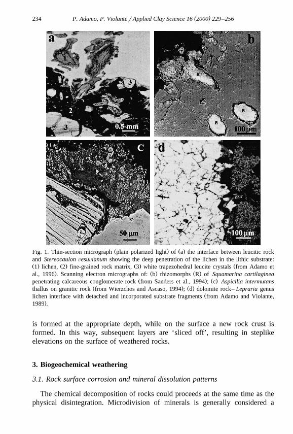
Ž .For example, the presence in leucitic lavic rock of Mt. Vesuvius Italy ofmany vesicles and less coherent areas makes the penetration of the lichen
Ž .Stereocaulon ÕesuÕianum in the substrate easier Adamo and Violante, 1991 . Inthin sections, the organ of adhesion of the lichen — the pseudopodetium — and
Žits ramifications were observed to penetrate down to 30 mm in the rock Adamo. Ž .et al., 1997 Fig. 1a .
In the case of Squamarina cartilaginea growing on a calcareous rock fromŽ .the hills surrounding the town of Cuenca Spain , elaborations of the thallus —
the rhizomorphs — and their ramifications penetrate the substrate preferentiallyalong the interfaces of rock and mineral fragments embedded within theconglomerate and appear to bore unimpeded through the extensive calcareous
Ž . Ž .cement Sanders et al., 1994 Fig. 1b . On siliceous schist or when the thalluscovers a zone of rock rich in micaceous material, hyphal penetration mainlyoccurs between laminae, which are increasingly separated with continued hyphal
Ž . Ž .proliferation Sanders et al., 1994; Wierzchos and Ascaso, 1994, 1996 Fig. 1c .The detachment of mineral fragments and their incorporation into the thallus
is often observed as a result of hyphal interpenetration of the substrate particlesŽand of thallus swelling and shrinking with hydration cycles Jones et al., 1981;
Adamo and Violante, 1989; Ascaso et al., 1990; Sanders et al., 1994; Wierzchos. Ž .and Ascaso, 1994, 1996 Fig. 1d . These forces are often cited as an explana-
tion for physical desegregation of mineral grains, but apparently their strengthhas never been measured. For rock surfaces the mechanical processes of
Ž .disintegration e.g., freeze–thaw cycles and subsequent colonisation of freshlyexposed minerals along grain boundaries are probably also important mecha-nisms of biogeophysical weathering.
Substrate disaggregation is not always more pronounced under crustoselichens, firmly and closely attached to the substrate via the entirety of the lowersurface of the thallus. The adhesion of foliose species, by distinct clusters of
Ž . Ž .hyphae rhizine , can be equally strong Adamo et al., 1993 . The indeterminategrowth and the proliferation of the rhizomorph system of some species ofsquamulose lichens, characterised by the presence of small scales or squamules,
Ž .may produce an extensive substratic network of hyphae Sanders et al., 1994 . Inthe frigid desert of the Antarctic dry valleys the activity of cryptoendolithiclichens, able to grow embedded within the sandstone rock matrix between andaround the crystals of the porous substrate, results in a characteristic exfoliative
Ž .weathering pattern Friedmann, 1982 . The substance cementing the sandstonegrains is apparently solubilized at the level of the lichen, which is exposed as theupper rock crust peels off. Hyphae then penetrate deeper and a new lichen zone
( )P. Adamo, P. ViolanterApplied Clay Science 16 2000 229–256234
Ž . Ž .Fig. 1. Thin-section micrograph plain polarized light of a the interface between leucitic rockand Stereocaulon ÕesuÕianum showing the deep penetration of the lichen in the lithic substrate:Ž . Ž . Ž . Ž1 lichen, 2 fine-grained rock matrix, 3 white trapezohedral leucite crystals from Adamo et
. Ž . Ž .al., 1996 . Scanning electron micrographs of: b rhizomorphs R of Squamarina cartilagineaŽ . Ž .penetrating calcareous conglomerate rock from Sanders et al., 1994 ; c Aspicilia intermutans
Ž . Ž .thallus on granitic rock from Wierzchos and Ascaso, 1994 ; d dolomite rock– Lepraria genusŽlichen interface with detached and incorporated substrate fragments from Adamo and Violante,
.1989 .
is formed at the appropriate depth, while on the surface a new rock crust isformed. In this way, subsequent layers are ‘sliced off’, resulting in steplikeelevations on the surface of weathered rocks.
3. Biogeochemical weathering
3.1. Rock surface corrosion and mineral dissolution patterns
The chemical decomposition of rocks could proceeds at the same time as thephysical disintegration. Microdivision of minerals is generally considered a
( )P. Adamo, P. ViolanterApplied Clay Science 16 2000 229–256 235
result of the mechanical action of the thalli, however, the participation of sometype of chemical action cannot be discarded. In addition, mechanical fragmenta-tion, increasing the surface area of the mineral or rock, accelerates chemicaldecomposition.
Dissolution processes, mainly by organic acids, occur at the microsites wherelichens adhere to the rocks. These are manifested by extensive surface etching ofthe grains incorporated into the lichen thallus and of the rock surfaces immedi-ately below the lichen thallus.
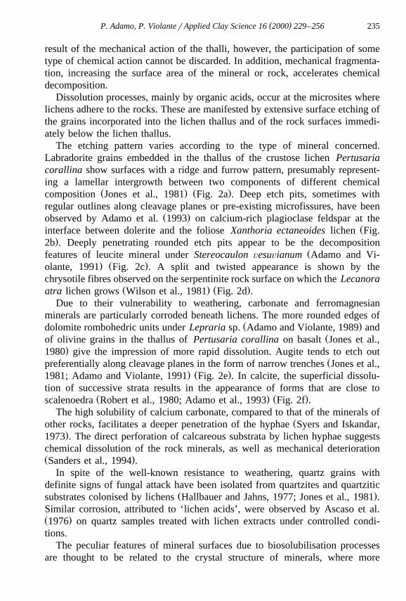
The etching pattern varies according to the type of mineral concerned.Labradorite grains embedded in the thallus of the crustose lichen Pertusariacorallina show surfaces with a ridge and furrow pattern, presumably represent-ing a lamellar intergrowth between two components of different chemical
Ž . Ž .composition Jones et al., 1981 Fig. 2a . Deep etch pits, sometimes withregular outlines along cleavage planes or pre-existing microfissures, have been
Ž .observed by Adamo et al. 1993 on calcium-rich plagioclase feldspar at theŽinterface between dolerite and the foliose Xanthoria ectaneoides lichen Fig.
.2b . Deeply penetrating rounded etch pits appear to be the decompositionŽfeatures of leucite mineral under Stereocaulon ÕesuÕianum Adamo and Vi-
. Ž .olante, 1991 Fig. 2c . A split and twisted appearance is shown by thechrysotile fibres observed on the serpentinite rock surface on which the Lecanora
Ž . Ž .atra lichen grows Wilson et al., 1981 Fig. 2d .Due to their vulnerability to weathering, carbonate and ferromagnesian
minerals are particularly corroded beneath lichens. The more rounded edges ofŽ .dolomite rombohedric units under Lepraria sp. Adamo and Violante, 1989 and
Žof olivine grains in the thallus of Pertusaria corallina on basalt Jones et al.,.1980 give the impression of more rapid dissolution. Augite tends to etch out
Žpreferentially along cleavage planes in the form of narrow trenches Jones et al.,. Ž .1981; Adamo and Violante, 1991 Fig. 2e . In calcite, the superficial dissolu-
tion of successive strata results in the appearance of forms that are close toŽ . Ž .scalenoedra Robert et al., 1980; Adamo et al., 1993 Fig. 2f .
The high solubility of calcium carbonate, compared to that of the minerals ofŽother rocks, facilitates a deeper penetration of the hyphae Syers and Iskandar,
.1973 . The direct perforation of calcareous substrata by lichen hyphae suggestschemical dissolution of the rock minerals, as well as mechanical deteriorationŽ .Sanders et al., 1994 .
In spite of the well-known resistance to weathering, quartz grains withdefinite signs of fungal attack have been isolated from quartzites and quartzitic
Ž .substrates colonised by lichens Hallbauer and Jahns, 1977; Jones et al., 1981 .Similar corrosion, attributed to ‘lichen acids’, were observed by Ascaso et al.Ž .1976 on quartz samples treated with lichen extracts under controlled condi-tions.
The peculiar features of mineral surfaces due to biosolubilisation processesare thought to be related to the crystal structure of minerals, where more
( )P. Adamo, P. ViolanterApplied Clay Science 16 2000 229–256236
Ž .Fig. 2. Scanning electron micrographs showing surface etching patterns of: a labradorite grainŽembedded in the thallus of crustose lichen Pertusaria corallina on basalt from Jones et al.,
. Ž .1981 ; b calcium-rich plagioclase feldspar localised at the dolerite rock– Xanthoria ectaneoidesŽ . Ž . Žinterface from Adamo et al., 1993 ; c leucite mineral under Stereocaulon ÕesuÕianum from
. Ž . ŽAdamo et al., 1991 ; d chrysotile fibres beneath Lecanora atra on serpentinite from Wilson et. Ž . Žal., 1981 ; e augite found at the vesuvite rock– Stereocaulon ÕesuÕianum interface from Adamo
. Ž . Ž .et al., 1991 ; f calcite under Xanthoria ectaneoides interface from Adamo et al., 1993 .
unstable areas of higher strain energy due to some kind of structural dislocation,Žbehave as sites of preferential dissolution Wilson, 1995; Wilson and Jones,
.1983 .
( )P. Adamo, P. ViolanterApplied Clay Science 16 2000 229–256 237
The amount of rock removed by corrosion caused by lichens can make asignificant contribution to the small-scale formation of fine-grained material
Ž .deposits. Recently, Garty 1992 , taking into account the total volume of holesand pits produced by the growth of different lithobiontic microorganisms onchalk rock from a burnt forest area of the Carmel Mountains in the Beit-Oren
Ž .Nature Reserve Israel and the specific weight of chalk, has estimated theamount of CaCO removed by only one kind of endolithic lichen to yield up to3
some 1740 kg hay1 of rock. Such amount, regarded by the author as irrelevantfor the specific small study area in the Beit-Oren region, can give an idea of thepossible contribution of endoliths to pedogenesis in the Mount Carmelo area aswell as in other Mediterranean ecosystems in Israel or in similar ecosystems inthe Mediterranean region. Findings of the same study indicate the importance ofcorrosion patterns in the postfire recolonisation of rock outcrops by lithobionts,because of the water-holding capacity of empty holes and pits and the possibledeposition of soil and rock particles, organic matter and fire ash in thismicrorelief.
4. Neogenesis of minerals
In the weathered material localised at the rock–lichen interface and in thethallus itself, secondary products can be formed by dissolving and chelatingactions of the biological processes associated with lichen growth. In thefollowing paragraphs we report about the principal biominerals groups men-tioned in the literature as formed as a result of lichen growth on rocks. It shouldbe emphasised that unless ‘‘in situ’’ techniques are not applied to detect anddetermine these new mineral phases, two potential sources of misinterpretedresults should always be taken into account. The first is the possible wind- orrainwater-born ‘‘contamination’’ along with the serious difficulty in distinguish-ing between weathering and bio-weathering products. The second could arisefrom procedures for samples preparation. Typically, the removal of organic
Žmatter through H O treatment which leads to oxalic acid production Farmer2 2.and Mitchell, 1963; Jackson, 1975 . In lichen–mineral studies the formation of
insoluble oxalates might be possible as a result of this treatment.
4.1. Oxalates
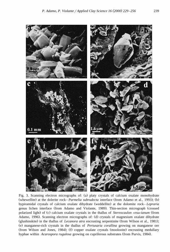
The reaction of oxalic acid secreted by many lichen-forming fungi with theminerals of the rock leads to the precipitation of oxalates. A close relationshipexists between the chemical composition of the substratum and the type ofinsoluble oxalate accumulating immediately beneath or within the thallus.
On calcareous rocks, such as limestone and dolomite, as well as on rockscontaining calcium-bearing minerals, calcium oxalate is predominant, usually
Ž . Žwhewellite, CaC O PH O, sometimes weddellite, CaC O P 2qx H O, Syers2 4 2 2 4 2
( )P. Adamo, P. ViolanterApplied Clay Science 16 2000 229–256238
and Iskandar, 1973; Jones et al., 1980; Ascaso et al., 1982, 1990; Vidrich et al.,1982; Adamo and Violante, 1989; Adamo et al., 1993; Wierzchos and Ascaso,
.1994 . The monohydrate form has monoclinic symmetry and a flat, platyŽ .morphology Fig. 3a . Its main diagnostic X-ray reflections are at 0.593, 0.365
and 0.297 nm. The polyhydrate has tetragonal symmetry, tends to show bipyra-Ž .midal- or tetragonal-prismatic habits Fig. 3b with strong reflections at 0.618,
0.442 and 0.278 nm.The occurrence of calcium oxalates on the outer surface of hyphae within the
lichen thallus or on the upper cortex suggests the extracellular formation of thecrystals. As observed in several higher plants, it has been suggested that thecrystals are initially formed intracellularly, within the wall of the hyphae, and, asthey increase in length, their distal ends protrude through the hyphal wallŽ .Pinna, 1983 . In lichenized rock thin sections calcium oxalate crystals in thethallus and in the contact zone can be easily detected with the optical micro-
Ž .scope by their high interference colours in crossed polarised light Fig. 3c .A need for the lichen to dispose of an excess of calcium is probably the main
reason for the formation of calcium oxalate. Nevertheless, there is evidence ofcalcium oxalate occurrence in lichens colonising substrates, including brick,
Ž .wood and bark, where calcium is almost absent Wadsten and Moberg, 1985 .These cases might be due to a reaction between oxalic acid produced by thelichen and calcium present in run-off.
The factors determining the formation of either the monohydrate or thepolyhydrate phase are yet to be clarified. Whewellite is the stable phase of thesystem calcium oxalaterwater. Weddellite is metastable. It tends to disappearfrom the system if ‘stored’ in water. A solid-state transformation of thepolyhydrate structure into that of the monoclinic monohydrate structure is notpossible. Weddellite may transform into whewellite only through dissolution of
Ž . Ž .the polyhydrate crystals Frey-Wyssling, 1981 . Horner et al. 1985 havesuggested that high pH and a high Caroxalic acid ratio are generally necessaryfor weddellite formation. At low pH the polyhydrate dissolves and is reprecipi-
Ž .tated as whewellite. According to Ascaso et al. 1982 the two forms are relatedto the amount of water. In the absence of hydration water on the rocks, calciumoxalate monohydrate rather than calcium oxalate polyhydrate is preferentiallyformed.
The various hydrates may have some role in the water balance of the lichens,Ž .known to be very tolerant to desiccation Wadsten and Moberg, 1985 . Weddel-
lite accommodates in the structure mobile zeolitic water molecules, which canbe lost becoming available to the lichen. The presence of the polyhydrate phasein dry sites may therefore serve as a source of water.
From substrate rocks, where calcium is present in low amounts, other oxalatesmay originate.
Ž .In the Grampian Region Scotland on outcrop of serpentinite, a rockconsisting almost entirely of magnesium silicate minerals with very low calcium
( )P. Adamo, P. ViolanterApplied Clay Science 16 2000 229–256 239
Ž .Fig. 3. Scanning electron micrographs of: a platy crystals of calcium oxalate monohydrateŽ . Ž . Ž .whewellite at the dolerite rock– Parmelia subrudecta interface from Adamo et al., 1993 ; b
Ž .bypiramidal crystals of calcium oxalate dihydrate weddellite at the dolomite rock– LeprariaŽ . Žgenus lichen interface from Adamo and Violante, 1989 . Thin-section micrograph crossed
. Ž . Žpolarized light of c calcium oxalate crystals in the thallus of Stereocaulon ÕesuÕianum from. Ž .Adamo, 1996 . Scanning electron micrographs of: d crystals of magnesium oxalate dihydrate
Ž . Ž .glushinskite in the thallus of Lecanora atra encrusting serpentinite from Wilson et al., 1981 ;Ž .e manganese-rich crystals in the thallus of Pertusaria corallina growing on manganese oreŽ . Ž . Ž .from Wilson and Jones, 1984 ; f copper oxalate crystals moolooite encrusting medullary
Ž .hyphae within Acarospora rugulosa growing on cupriferous substrates from Purvis, 1984 .
( )P. Adamo, P. ViolanterApplied Clay Science 16 2000 229–256240
content, appreciable amounts of crystalline magnesium oxalate dihydrate — theŽ .mineral glushinskite Wilson et al., 1980 — were found in the thallus of
Ž .Lecanora atra as well as at the rock–lichen interface Wilson et al., 1981 . Themineral occurs as tiny crystals ranging from 2 to 5 mm in size, the majority
Ž .showing a distorted pyramidal form Fig. 3d . Its main X-ray reflections arefound at 0.489, 0.317, 0.238, 0.204 and 0.186 nm. Microprobe analysis of theglushinskite shows that it contained significant amounts of nickel, iron andmanganese. More recently MgC O P2H O has been described from the Island2 4 2
of Rhum in the Inner Hebrides of Scotland where it may form by lichen activityŽ .on magnesium-rich rocks Wilson and Bayliss, 1987 .
Accumulations of poorly formed sub-equant, blocky manganese-rich crystals,which have been proved to be manganese oxalate dihydrate by X-ray diffractionŽ .main X-ray powder reflections at 0.483, 0.472, 0.301 and 0.267 nm , haveresulted from the interaction between a manganese ore, consisting of hard
Ž . Ž .cryptomelane, KMn O , and powdery lithiophorite, Al,Li MnO OH and8 16 2 2Ž . Ž .the lichen Pertusaria corallina Fig. 3e Wilson and Jones, 1984 .
The occurrence of vivid blue inclusions of copper oxalate hydrate, laterŽ .recognized as the mineral moolooite Chisholm et al., 1987 , in lichens growing
Ž .on cupriferous substrates has been revealed by the work of Purvis 1984 . Theblue crystalline material consists of aggregates of platy crystals 1–3 mm in
Ž .diameter encrusting medullary hyphae Fig. 3f . The X-ray diffraction pattern ischaracterised by a very strong reflection at 0.388 nm and medium intensityreflections at 0.194, 0.177 and 0.171 nm, all other reflections being weak orvery weak.
In principle, it seems likely that a range of previously unreported oxalateminerals may exist where oxalic acid-secreting lichens have colonised substratesof appropriate composition. On the basis of the crystallographic studies of
Ž . Ž . Ž .Lagier et al. 1969 and Dubernat and Pezerat 1974 , Wilson et al. 1980; 1981suggested that magnesium could be substituted by nickel, cobalt, iron, zinc andmanganese in the glushinskite structure, similar to, and isomorphous with, the
Ž .dihydrated oxalates of these elements. Purvis 1984 also considers the feasibil-ity of the precipitation of the relatively insoluble oxalates of barium, lead andsilver.
Ž .So far, only the detection of non-hydrated ferric oxalate C O Fe , giving6 12 2
X-ray peaks at 0.530, 0.438 and 0.348 nm, in Caloplaca callopisma growing onŽ .Fe-rich dolomite has been reported Ascaso et al., 1982 . Apparently ferrous
iron oxalate, the already well-known mineral humboldtine, is absent in theweathering zone between lichen and rock. The oxidation, possibly microbial, oforganic molecules complexing Fe2qrFe3q, with subsequent hydrolysis andprecipitation of more or less crystalline iron oxides has been claimed as a
Žprobable process to which the finding may be ascribed Jones and Wilson, 1986;.Adamo et al., 1997 .

( )P. Adamo, P. ViolanterApplied Clay Science 16 2000 229–256 241
The incorporation of heavy metal ions into oxalates within the lichen thallus,but external to the protoplasm, seems to be related with the well-known ability
Žof lichens to avoid the effects of toxic elements Jones and Wilson, 1985;.Wilson, 1995; Purvis, 1996; Purvis and Halls, 1996 . Recently, extracellular
immobilisation of Zn and Pb as oxalate salts in the lichen metal hyperaccumula-Ž .tor Diploschistes muscorum, collected in the vicinity of a Zn,Pb S smelter
located at Auby in the North of France, has been revealed by Sarret et al.Ž . Ž .1998 , coupling powder X-ray diffraction XRD by extended X-ray absorption
Ž .fine structure EXAFS spectroscopy.
4.2. Iron oxides and hydroxides
The biogenic formation of iron oxides and hydroxides minerals is obviousfrom the colour of the rock surface beneath the lichen thallus. In 1970, Jacksonand Keller found a considerable enrichment in Fe of the reddish weatheringcrust of Stereocaulon Õulcani-covered Hawaii lava flows. An unidentified,poorly crystallised form of ferric oxide, metastable with respect of hematite, anddistinctly different from the iron oxide occurring in the lichen-free rock crustwas detected. Possibly this amorphous ferruginous oxide may be the actuallywell known mineral ferrihydrite, more recently reported in a thin ochreous layerat the interface between Pertusaria corallina and a weathered basalt substrate in
Ž .Western Scotland Jones et al., 1980 and in the rusty ferruginous materialfrequently located in the zone of contact between the thallus of Stereocaulon
Ž .ÕesuÕianum and the leucite-bearing rock of Mt. Vesuvius Adamo et al., 1997 .Ferrihydrite is a short-range order iron oxyhydroxide structurally resemblinghematite. It forms very small spherical particles, 3 to 7 nm in diameter, which
Ž .usually are highly aggregated Fig. 4a . According to its crystallinity, it givesrise to XRD and electron diffraction patterns characterised by a variable number
Ž .of very broad peaks at about 0.25, 0.22, 0.197, 0.173 and 0.147 nm Fig. 4b .Unlike most other Fe oxides it is nearly completely soluble in acid ammonium
Ž .oxalate in the dark. Differential X-ray diffraction DXRD of an untreated andan oxalate-treated sample may be required for positive identification. Thedifficulty of distinguishing between ferrihydrite and feroxyhite, with similarstructures and X-ray lines, implies the possibility that this iron oxyhydroxidemay also be significantly present among the secondary products formed as aresult of lichen weathering.
Goethite is by far the most common form of crystalline iron oxide occurringŽ .at the rockrlichen interface. On several occasions Ascaso et al. 1976 have
observed twinned crystals of a-FeOOH beneath the thallus of RhizocarponŽ .geographicum growing on granite Fig. 4c . An aluminium-containing goethite
Ž .has been detected by Jones et al. 1981 in an ochreous coating on the surface ofŽ .Tremolecia atrata as ‘‘Lecidea dicksonii’’ encrusting a biotite chlorite schist.Ž . Ž .Again, Galvan et al. 1981 find considerable amounts of iron oxides goethites

( )P. Adamo, P. ViolanterApplied Clay Science 16 2000 229–256242
Ž . Ž .Fig. 4. Transmission electron micrograph a and electron diffraction pattern b of ferrihydriteŽfrom the rusty interface between Stereocaulon ÕesuÕianum and volcanic rock from Adamo et al.,
. Ž .1997 . c TEM of twinned goethite at the interface between the thallus of RhizocarponŽ . Ž .geographicum and granite substrate from Ascaso et al., 1976 . d Diagrammatic representation
Ž .of iron complexation and translocation under lichen thallus from Adamo et al., 1993 . ScanningŽ .electron micrographs of: e fibrous silica gel at interface between serpentinite rock and lichen
Ž . Ž .Lecanora atra from Jones et al., 1981 ; f Silica-rich microdiscs at serpentinite rock– CaloplacaŽ .sp. interface from Adamo et al., 1993 .

( )P. Adamo, P. ViolanterApplied Clay Science 16 2000 229–256 243
in the material from the interfaces between various lichens and a garnet-chlo-ritoid and quarzite schist. However, the authors do not ascribe any significanceto this finding because the mineral was already present in the parent rock.
Ž .Recently, small 0.1–0.3 mm hexagonal plates of hematite, as well asŽ .acicular shaped goethite crystals, have been found by Adamo et al. 1997 in the
iron-rich material surrounding the basal part of Stereocaulon ÕesuÕianum thal-lus.
In iron-rich dark sandstones of the Antarctic cold desert iron solubilisationŽhas been observed to take place in the cryptoendolithic lichen zone Friedmann,
.1982 . As a result, the thin crust above the lichen and the rock substrate a fewmillimeters below appear darker because of iron deposition. Precipitated iron
Ž .compounds probably hematite or goethite, or both have been found to coverthe colourless fungal hyphae penetrating the rock substrate.
It seems likely that the organic acids produced by lichens play a key role inthe formation and enrichment of poorly-ordered and crystalline Fe phases at therock–lichen interface. Presumably the biomolecules complex Fe, primarily
Ž .released from Fe II silicates on weathering, and link the small ferrihydriteparticles, preventing, in both cases, the formation of more crystalline iron oxidesŽ .Schematization shown in Fig. 4d . Processes of transformation of poorly-orderedoxyhydroxides as well as reactions of oxidationrprecipitation of Fe2q wouldaccount for the neoformation of more crystalline phases. Differentiation of therock–lichen interface into microsites each with separate pH, humidity and redoxpotential conditions may result in the genesis of either goethite or hematiteŽ .Schwertmann et al., 1986 .
4.3. Siliceous relicts
The formation of siliceous relicts as a result of the intense mineral decompo-Žsition produced by lichens has been widely reported Ascaso et al., 1976; Jones
.et al., 1981; Wilson et al., 1981 . The preferential extraction of structuralmagnesium from the silicate chrysotile by oxalic acid secreted by the mycobiontof Lecanora atra left behind an X-ray amorphous silica gel often retaining the
Ž . Ž .fibrous morphology of the parent mineral Fig. 4e Wilson et al., 1981 .Microprobe analysis of individual flakes of biotite, incorporated into a culture
medium of an oxalic acid producing fungus, shows that the decomposition of thelayer silicate resulted in the removal of all elements, with the exception ofsilicon, and reveals that acid attack generally proceeded from the edge of the
Ž .flake and progressed towards the centre Jones et al., 1981 .Amorphous silica has been found to be associated with Parmelia conspersa
growth on granite and gneiss. Thallus fragments from the same lichen are evenable to generate in vitro SiO from the three primary rock forming minerals,2
Ž .quartz, micas and feldspars Ascaso et al., 1976 .

( )P. Adamo, P. ViolanterApplied Clay Science 16 2000 229–256244
Silica in the form of microdiscs a few microns in diameter has been observedat the interface between quartz and Acarospora hospitans and between serpenti-nite rock and Caloplaca sp., suggesting, as for phytoliths, dissolution, absorp-
Ž . Žtion and excretion of the element Fig. 4f Robert et al., 1983; Adamo et al.,.1993 .
4.4. Alumino-silicates
Poorly ordered alumino-silicates, intimately admixed with rock-forming min-erals, phyllosilicates and ferrihydrite, have been found in the weathering crust at
Ž .the rock–lichen interface Jones et al., 1980; Adamo and Violante, 1991 .Ž .Electron micrographs Fig. 5a show that these amorphous Al–Si materials
consist of microaggregates of very finegrained particles yielding a diffuse andŽ .poorly defined electron diffraction pattern Adamo and Violante, 1991 . Accord-
Ž .ing to Wilson and Jones 1983 their formation is presumably related to theeffectiveness of lichens in complexing and removing aluminium from thesubstrate minerals by organic compounds. The biotic oxidation of these organo-mineral complexes would liberate Al in a reactive form to combine with silica.
Ž .Farmer 1979 reports a similar mechanism of formation in the podzolic Bhorizons of an X-ray amorphous alumino-silicate complex called proto-im-ogolite allophane. Recently, allophanes and imogolite fibres have been detectedby electron microscopy under the thallus of Xanthoria elegans growing on
Ž .volcanic andesite in maritime Antarctica Ascaso et al., 1990 .ŽMany authors Jackson and Keller, 1970; Jones et al., 1980; Wilson et al.,
.1981; Vidrich et al., 1982 suggest caution in considering possible the neogene-sis of well-ordered alumino-silicates in the zone of contact between lichen androck substrate. ‘Biochemical’ weathering, differently from ‘geochemical’, oc-
Ž .Fig. 5. Transmission electron micrographs of: a amorphous alumino-silicates of the fine clayŽ . Žfraction B-0.5 mm from the volcanic rock– Stereocaulon ÕesuÕianum interface material from
. Ž . Ž .Adamo and Violante, 1991 ; b kaolinite K crystals at the interface between the thallus ofŽ .Parmelia conspersa and granite substrate from Ascaso et al., 1976 .
( )P. Adamo, P. ViolanterApplied Clay Science 16 2000 229–256 245
curs through the mediation of complexing organic acids and this conditionwould severely limit the crystallisation to clay minerals leading rather to thepreferential formation of poorly ordered phases.
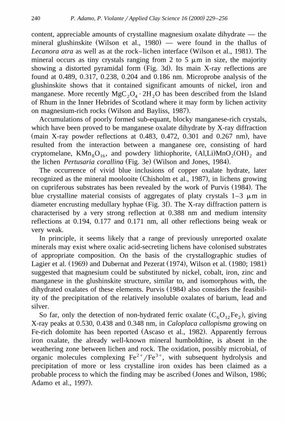
On the other hand, various phyllosilicates have been identified in the weath-ered material accumulated at the rock–lichen interface. Halloysite and kaolinite
Ž .have been frequently noted by Ascaso et al. 1976 under the thalli of Parmeliaconspersa and Rhizocarpon geographicum either on granite or gneiss substrateŽ .Fig. 5b . Furthermore, these newly formed minerals and montmorillonite, havebeen generated in laboratory experiments by incubating fragments of the lichen
Žthalli or extracts of selected lichen compounds atranorin, usnic acid, stictic acid. Žand norstictic acid with either rock samples or their primary minerals albite,
. Žorthoclase, biotite and muscovite Ascaso and Galvan, 1976; Ascaso et al.,.1976 . Some micas of the illite type, which may be degradation products of
various phyllosilicates in the rock, have been identified beneath of the thallus ofLecidea lapicida collected from volcanic andesite in South Shetland IslandsŽ .Ascaso et al., 1990 . X-ray diffractometer traces of the fine fraction separated
Ž . Ž .Fig. 6. X-ray diffractometer traces CoK radiation of the clay fraction B-2.0 mm from thea
weathered material beneath the lichen Stereocaulon ÕesuÕianum colonising leucitic rock of Mt.Ž .Vesuvius from Adamo, 1996 .
( )P. Adamo, P. ViolanterApplied Clay Science 16 2000 229–256246
from the weathered material beneath the lichen Stereocaulon ÕesuÕianumcolonising leucitic rock of Mt. Vesuvius revealed the occurrence of various clayminerals. Ethylene glycol solvation and heating at 5508C demonstrated kaolinite,
Ž . Žillite and a 1.4 nm intergrade mineral presence Fig. 6 Adamo, 1996; Adamo.and Violante, 1991 . These clay minerals commonly occur in Andisols of
temperate climate regions and particularly have been found in the clays of soilsdeveloped on central-southern Italy volcanic materials of petrological composi-tion similar to that of Mt. Vesuvius. With the exception of illite, probablyinherited from mica in the parent material, they are believed to originate to alarge extent by neoformation reactions, rather than by transformation of pre-ex-
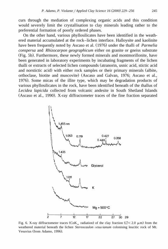
Ž .isting phyllosilicate structures Violante and Wilson, 1983 . Recently, Wierz-Ž .chos and Ascaso 1996 have observed distinct depletion of interlaminar potas-
sium in adhesion zones of Parmelia conspersa and Aspicilia intermutans thalli,Ž .on granitic biotite sheets Fig. 7a and b . On the bases of the geochemical mass
Ž .Fig. 7. SEM back-scattered electron image of a the interface zone between the ParmeliaŽ . Ž .conspersa thallus and granitic biotite sheets. b X-ray distribution map of K in a . HRTEM
Ž .images of lattice fringes of octadecylammonium ion treated c unaltered biotite revealing the 10˚ ˚Ž . Ž .A basal spacing and d ordered bioweathered biotite, interstratified biotite 10 A and vermiculite
˚Ž . Ž .14–30 A phases from the lichen–biotite contact area from Wierzchos and Ascaso, 1996, 1998 .

( )P. Adamo, P. ViolanterApplied Clay Science 16 2000 229–256 247
balance of the K-rich and K-poor biotite zones, the authors suggest the transfor-mation of K-rich biotite to scarcely altered biotite with a biotite–vermiculiteŽ .hydrobiotite-like intermediate interstratified phase. In a more recent high
Ž .resolution transmission electron microscopy HRTEM study of the carefullyŽ .extracted mineral material Wierzchos and Ascaso 1998 have further demon-
˚ ( )strated the biogenetic vermiculitization of biotite. A homogenous 10 A d 001 -space, unaffected by octadecylammonium chloride treatment, was observed forunweathered biotite samples within and on the surface of the fresh parent
Ž .granitic rock Fig. 7c . Nevertheless, HRTEM image of lattice fringes of biotitetaken from the lichen–biotite contact zone after ODA treatment revealed large
˚ ˚Ž . Ž .area of both unexpanded 10 A and expanded 14–30 A layers of phyllosili-Ž .cates identified as interstratified biotitervermiculite Fig. 7d .
It is difficult to be certain whether clay minerals are newly formed in therock–lichen contact area or whether they derive from extraneous sources in theform of wind-borne dust trapped by lichen thalli. Their ordered nature makes itunlikely their formation in the same environment that favours the formation ofamorphous and poorly ordered materials. Nevertheless, the possibility that at therock–lichen interface the accumulation of various cations and organic com-pounds, with peculiar number and location of hydroxyl and carboxyl groups andstability of their complexes, combined with specific physico-chemical parame-ters might create conditions favorable to phyllosilicate genesis cannot be dis-counted entirely.
4.5. Carbonates
Calcite, in form of rhizomorphic features and cytomorphic sands, has beenŽfrequently observed at the surface of hyphae or roots and inside hyphae Robert
.and Berthelin, 1986 ; however, until recently, only two cases of carbonatesformation by lichen activity on rocks have been reported. The first is the
w Ž . Ž . xdetection of hydrocerussite, a basic lead carbonate Pb CO OH , in the3 3 2 2
thallus of Stereocaulon ÕesuÕianum growing on siliceous limestone in the ruinsŽ .of a flue from a lead-smelting mill Jones et al., 1982 . Although no obvious
crystalline form yielding a lead signal on probing was discernible by scanningelectron microscopy, the X-ray diffraction pattern obtained from tufts ofmycelium sampled near the rock surface, almost identical to that of the mineral
Ž .hydrocerussite, proves the occurrence of the carbonate mineral Table 1 . Thesecond example is the identification by X-ray analysis and IR spectroscopy of asmall amount of calcite in the contact area between the endemic Antarctic lichenBacidia stipata and its rock substrate, a volcanigenic sediment classified as
Ž .clastic Ascaso et al., 1990 . In this study many bacteria were found underneathand inside the lichen thallus in the weathered area of the rock. This observation
Žcould suggest a contribution of the co-existing microorganisms bacteria,.cyanobacteria, algae or fungi to the alteration of the substrate. Lichens,
( )P. Adamo, P. ViolanterApplied Clay Science 16 2000 229–256248
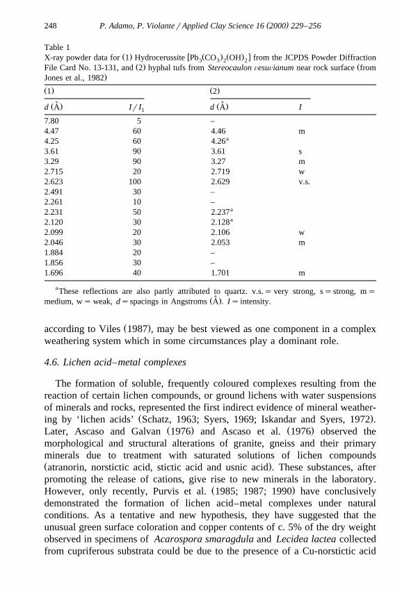
Table 1Ž . w Ž . Ž . xX-ray powder data for 1 Hydrocerussite Pb CO OH from the JCPDS Powder Diffraction3 3 2 2
Ž . ŽFile Card No. 13-131, and 2 hyphal tufs from Stereocaulon ÕesuÕianum near rock surface from.Jones et al., 1982
Ž . Ž .1 2
˚ ˚Ž . Ž .d A Ir I d A I1
7.80 5 –4.47 60 4.46 m
a4.25 60 4.263.61 90 3.61 s3.29 90 3.27 m2.715 20 2.719 w2.623 100 2.629 v.s.2.491 30 –2.261 10 –
a2.231 50 2.237a2.120 30 2.128
2.099 20 2.106 w2.046 30 2.053 m1.884 20 –1.856 30 –1.696 40 1.701 m
aThese reflections are also partly attributed to quartz. v.s.s very strong, ssstrong, ms˚Ž .medium, ws weak, dsspacings in Angstroms A . Is intensity.
Ž .according to Viles 1987 , may be best viewed as one component in a complexweathering system which in some circumstances play a dominant role.
4.6. Lichen acid–metal complexes
The formation of soluble, frequently coloured complexes resulting from thereaction of certain lichen compounds, or ground lichens with water suspensionsof minerals and rocks, represented the first indirect evidence of mineral weather-
Ž .ing by ‘lichen acids’ Schatz, 1963; Syers, 1969; Iskandar and Syers, 1972 .Ž . Ž .Later, Ascaso and Galvan 1976 and Ascaso et al. 1976 observed the
morphological and structural alterations of granite, gneiss and their primaryminerals due to treatment with saturated solutions of lichen compoundsŽ .atranorin, norstictic acid, stictic acid and usnic acid . These substances, afterpromoting the release of cations, give rise to new minerals in the laboratory.
Ž .However, only recently, Purvis et al. 1985; 1987; 1990 have conclusivelydemonstrated the formation of lichen acid–metal complexes under naturalconditions. As a tentative and new hypothesis, they have suggested that theunusual green surface coloration and copper contents of c. 5% of the dry weightobserved in specimens of Acarospora smaragdula and Lecidea lactea collectedfrom cupriferous substrata could be due to the presence of a Cu-norstictic acid
( )P. Adamo, P. ViolanterApplied Clay Science 16 2000 229–256 249
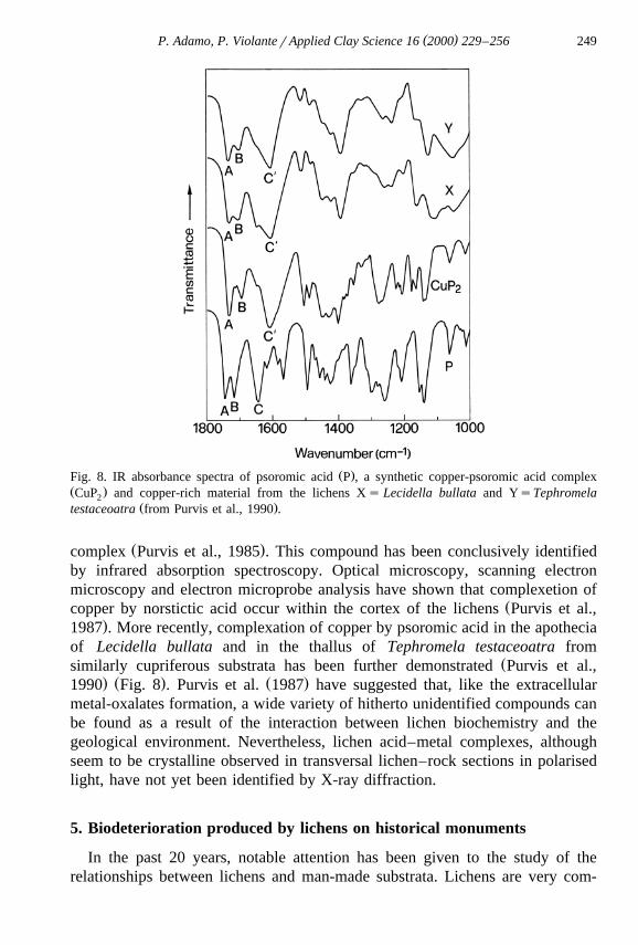
Ž .Fig. 8. IR absorbance spectra of psoromic acid P , a synthetic copper-psoromic acid complexŽ .CuP and copper-rich material from the lichens Xs Lecidella bullata and YsTephromela2
Ž .testaceoatra from Purvis et al., 1990 .
Ž .complex Purvis et al., 1985 . This compound has been conclusively identifiedby infrared absorption spectroscopy. Optical microscopy, scanning electronmicroscopy and electron microprobe analysis have shown that complexetion of
Žcopper by norstictic acid occur within the cortex of the lichens Purvis et al.,.1987 . More recently, complexation of copper by psoromic acid in the apothecia
of Lecidella bullata and in the thallus of Tephromela testaceoatra fromŽsimilarly cupriferous substrata has been further demonstrated Purvis et al.,
. Ž . Ž .1990 Fig. 8 . Purvis et al. 1987 have suggested that, like the extracellularmetal-oxalates formation, a wide variety of hitherto unidentified compounds canbe found as a result of the interaction between lichen biochemistry and thegeological environment. Nevertheless, lichen acid–metal complexes, althoughseem to be crystalline observed in transversal lichen–rock sections in polarisedlight, have not yet been identified by X-ray diffraction.
5. Biodeterioration produced by lichens on historical monuments
In the past 20 years, notable attention has been given to the study of therelationships between lichens and man-made substrata. Lichens are very com-

( )P. Adamo, P. ViolanterApplied Clay Science 16 2000 229–256250
mon on artistic stoneworks and contribute to their deterioration, frequentlycreating serious problems for their recovery, restoration and conservation.Lichen coverage alters monuments aesthetically, inducing colour changes andobscuring detail of sculpture and paintwork. Recent literature on this topic has
Ž .been reviewed by Piervittori et al. 1994; 1996; 1998 .Biogeophysical and biogeochemical deterioration has been mainly identified
in the fracturing of substratum surfaces and in the build-up of encrustationsformed as a result of the reaction between lichen by-products and the minerals inthe stone. Extensive erosion of the material has been often observed.
Conventional and Fourier Transform Raman spectroscopic methods, withvisible and near infrared laser excitation, have been proved to be effective in theinterpretation and characterisation of both the physical and chemical effects onhistoric monuments, frescoes and other works of art brought about by the action
Žof certain aggressive lichens Edwards et al., 1991, 1992, 1997; Seaward and.Edwards, 1995, 1997; Seaward et al., 1995 . These techniques, which use very
small amounts of material, in the nanogram–picogram range, and low laserpower for sample illumination, are non-destructive of the valuable samples; theyhave permitted, for example, microscopical investigation of the chemical natureof the gradient through the lichen thallus, the substratum, and their interface.The detection of complex chelating ‘‘lichen acids’’ and the identification of thestate of hydration of calcium oxalate in lichen encrustations have been achieved.In addition, the presence of incorporated material, such as calcite, gypsum andpaint pigments has been shown.
The ambient urban climate and associated atmospheric pollutants dramaticallyaffect the lichen flora. Strong evidence has been produced to suggest thatmodified environmental conditions have been conducive to increasing detrimen-tal invasion by certain aggressive lichen species such as Dirina massiliensisforma sorediata. This lichen is extending its ecological range due to itsreproductive strategy and its ability to exploit diverse substrata, facilitated bynew environmental regimes, including qualitative changes in atmospheric pollu-tion, which have frequently allowed it to dominate in the wake of the rapiddisappearance of other more pollution-sensitive species. It is now commonly
Ž .found on a range of works of art throughout Europe Edwards et al., 1997 .Ž .In their first review of the literature, Piervittori et al. 1994 drew attention to
the need to avoid generalization about the effects that lichens may have onstonework substrata. In interpreting and evaluating the role of lichens in thebiodeterioration of monuments the species-specific differences in weatheringability, the physical and chemical nature of the substrata and the microclimaticconditions, particularly in terms of water retention, have to be carefully takeninto consideration. The principal causes of lichen colonization, the factors thatmay accelerate the growth rate of the species involved, their reproductivestrategies and the consequences arising from their elimination have to besystematically gathered.
( )P. Adamo, P. ViolanterApplied Clay Science 16 2000 229–256 251
It cannot always be said that lichen encrustations are detrimental to theirimmediate substrates. Indeed, it has been sometimes suggested that the organ-
Ž .isms may play a protective role in this respect Seaward et al., 1989 . Biodeteri-oration and bioprotection are in an unstable equilibrium which can be unbal-anced by environmental conditions, the substratum and the type of organismscolonising the monument. This has been exemplified in the sandstone pavement
Ž .of the forum of the Roman city of Baelo Claudia Cadiz, Spain , where theflagstones without lichen cover show higher deterioration than those colonised
Ž .by lichens Arino et al., 1995 . In this particularly aggressive environment the˜combined effects of wind, salt and water easily disintegrate a fragile substratum.Although the lichen–sandstone interface shows some weathering, namely disag-gregation, calcium oxalate deposition and crystal etching, biodeterioration is amuch slower process than physical and chemical deterioration: sometimeslichens can even represent a protective cover for the stones. In a porous
Žsubstrate, like the sandstone of the Baelo Claudia forum pavement Arino et al.,˜.1995 , the presence of lichen retards rainwater absorption, partially lessening
dissolution and precipitation processes, and it also prevents the abrasive actionproduced by airborne sand particles, the impact of raindrops and changes intemperature.
Extensive, homogeneous, yellow-brown films mainly composed of calciumŽ . Ž .oxalate, both monohydrate whewellite and bihydrate weddellite are fre-
quently observed on artefacts of historic and artistic interest, on buildings,sculptures and archaeological remains independently of the substrate. One of themain hypotheses proposed for the formation of the films involves the productionof oxalic acid by lichens which could have colonised the monuments presum-
Žably favoured by the unpolluted atmosphere of the past Del Monte, 1991; DelMonte and Sabbioni, 1987; Del Monte and Ferrari, 1989; Seaward and Edwards,
.1997 . However, according to many authors, only in a limited number of casesŽcan the origin of oxalate films be related to lichen activity Franzini et al., 1984;
.Matteini and Moles, 1986; Alessandrini et al., 1989 .
6. Conclusions
The weathering ability of lichens with respect to their mineral substrates haslong been recognised and many papers have been published in this centurydocumenting the physical and chemical processes involved. The bioweathering
Ž .seems to consists of: a a more or less intense disaggregation and fragmentationof the rock surface immediately below the lichen by surface adhesion and
Ž . Ž .hyphal penetration, b dissolution processes and c the precipitation andformation of new minerals. The detailed understanding of the mechanisms andprocesses involved in lichens weathering of mineral surfaces has given a most

( )P. Adamo, P. ViolanterApplied Clay Science 16 2000 229–256252
significant contribution to explain the biodeterioration phenomena of stoneworkof monuments and other archeological materials.
Many of the papers referred to in this review indicate that oxalic acid and‘lichen acids’ must be considered biomolecules extremely active as weatheringagents. Oxalates, whose nature depends upon the composition of the substrate,are the best studied lichen biominerals and are commonly found in the thallusandror at the rockrlichen interface. The suspected involvement of ‘lichenacids’ has been conclusively demonstrated in the last decade. Crystalline salts of‘‘lichen acids’’ containing metal cations derived from the mineral substrate havebeen shown to occur in the thallus. It is likely that more oxalater‘lichenacid’-derived minerals could be described in the future. Furthermore, other
Ž .simple low-molecular-weight organic acids e.g., citric, lactic and tartaric acid ,known to be produced by fungi, are presumably excreted by lichen mycobiontsin much the same way as oxalic acids. Humic and fulvic acids could beproduced from the decomposition of lichen residues. The effectiveness of thesecompounds in the decomposition of soil minerals by both the acidic effect andcomplex formation or chelation is well known. Although the lack of experimen-tal evidence, they are expected to play an analogous important role in lichenweathering. Hence, much remains to be studied to fully elucidate the interactionbetween lichens and mineral surfaces. The application of more specialisedinstrumental and analytical techniques and the close collaboration among biolo-gists, chemists and mineralogists are fundamental requisites for future progress.
Acknowledgements
The authors would like to thank Dr. Carmen Ascaso of the Centro deCiencias Medioambientales of Madrid, Dr. David Jones of the Macaulay LandUse Research Institute of Aberdeen, Dr. O.W. Purvis of the Natural HistoryMuseum of London and Dr. Jacek Wierzchos of the Servei de MicroscopiaElectronica of the Lleida University who kindly provided some of the SEM andTEM micrographs reported in this review. Thanks are due to Dr. H.A. Viles andto the anonymous referee for comments which helped to improve the presenta-tion of the text. Mr. Maurizio Clumez is also thanked for his skilful technicalassistance in the electronic preparation of the figures. This work was supportedby grants from the Italian Ministry for University and Scientific and Technologi-
Žcal Research PRIN project ‘‘Cryptogams as Biomonitors in Terrestrial Ecosys-. Ž .tems’’ . Contribution No. 184 DISCA .
References
Adamo, P., 1996. Ruolo dell’attivita dei licheni nell’alterazione di substrati rocciosi e nella`neogenesi di entita mineralogiche. Atti XIII Convegno Nazionale SICA, pp. 13–26.`
( )P. Adamo, P. ViolanterApplied Clay Science 16 2000 229–256 253
Adamo, P., Colombo, C., Violante, P., 1997. Iron oxides and hydroxides in the weatheringinterface between Stereocaulon ÕesuÕianum and volcanic rock. Clay Minerals 32, 275–283.
Adamo, P., Marchetiello, A., Violante, P., 1993. The weathering of mafic rocks by lichens.Ž .Lichenologist 25 3 , 285–297.
Adamo, P., Violante, P., 1989. Bioalterazione di roccia dolomitica operata da una specie lichenicadel genere Lepraria. Agricoltura Mediterranea 119, 460–464.
Adamo, P., Violante, P., 1991. Weathering of volcanic rocks from Mt. Vesuvius associated withthe lichen Stereocaulon ÕesuÕianum. Pedobiologia 35, 209–217.
Ahmadjan, V., Hale, M.E., 1973. The Lichens. Academic Press, London, pp. 697.Alessandrini, G., Bonecchi, R., Peruzzi, R., Toniolo, L., 1989. Caratteristiche composizionali e
morfologiche di pellicole ad ossalato: studio comparato su substrati lapidei di diversa natura.In: Proc. Symp. Le pellicole ad ossalato: origine e significato nella conservazione delle opered’arte, Centro CNR Gino Bozza, Milano, pp. 137–150.
Arino, X., Ortega-Calvo, J.J., Gomez-Bolea, A., Saiz-Jimenez, C., 1995. Lichen colonization of˜Ž .the Roman pavement at Baelo Claudia Cadiz, Spain : biodeterioration vs. bioprotection. The
Science of the Total Environment 167, 353–363.Ascaso, C., Galvan, J., 1976. Studies on the pedogenic action of lichen acids. Pedobiologia 16,
321–331.Ascaso, C., Wierzchos, J., 1994. Structural aspects of the lichen–rock interface using back-scattered
electron imaging. Botanica Acta 107, 251–256.Ascaso, C., Wierzchos, J., 1995. Study of the biodeterioration zone between the lichen thallus and
the substrate. Cryptogamic Botany 5, 270–281.Ascaso, C., Galvan, J., Ortega, C., 1976. The pedogenetic action of Parmelia conspersa,
Rhizocarpon geographicum and Umbilicaria pustulata. Lichenologist 8, 151–171.Ascaso, C., Galvan, J., Rodriguez-Pascual, C., 1982. The weathering of calcareous rocks by
lichens. Pedobiologia 24, 219–229.Ascaso, C., Sancho, L.G., Rodriguez-Pascal, C., 1990. The weathering action of saxicolous
lichens in maritime Antarctica. Polar Biology 11, 33–39.Chisholm, J.E., Jones, G.C., Purvis, O.W., 1987. Hydrated copper oxalate, moolooite, in lichens.
Mineralogical Magazine 51, 766–803.Del Monte, M., 1991. Trajan’s column: lichens don’t live here anymore. Endeavour, New Series
Ž .15 2 , 86–92.Del Monte, M., Ferrari, A., 1989. Patine da biointerazione alla luce delle superfici marmoree. In:
Proc. Symp. Le pellicole ad ossalato: origine e significato nella conservazione delle opered’arte, Centro CNR Gino Bozza, Milano, pp. 171–182.
Del Monte, M., Sabbioni, C., 1987. A study of the patina called ‘‘scialbatura’’ on imperialRoman marbles. Studies in Conservation 32, 114–121.
Dubernat, P.J., Pezerat, H., 1974. Fautes d’empilement dans les oxalates dihydrates des metaux´ ´Ž .divalents de la series magnesienne Mg, Fe, Co, Ni, Zn, Mn . Journal of Applied Crystallogra-´ ´
phy 7, 387–394.Edwards, H.G.M., Farwell, D.W., Seaward, M.R.D., Giacobini, C., 1991. Preliminary Raman
microscopic analysis of a lichen encrustation involved in the biodeterioration of Renaissancefrescoes in central Italy. International Biodeterioration 27, 1–9.
Edwards, H.G.M., Farwell, D.W., Jenkins, R., Seaward, M.R.D., 1992. Vibrational RamanSpectroscopic Studies of calcium oxalate monohydrate and dihydrate in lichen encrustations onRenaissance frescoes. Journal of Raman Spectroscopy 23, 185–189.
Edwards, H.G.M., Farwell, D.W., Seaward, M.R.D., 1997. FT-Raman spectroscopy of DirinaŽ .massiliensis f. sorediata encrustations growing on diverse substrata. Lichenologist 29 1 ,
83–90.ŽFarmer, V.C., 1979. Possible roles of a mobile hydroxyaluminium orthosilicate complex proto-
( )P. Adamo, P. ViolanterApplied Clay Science 16 2000 229–256254
.imogolite in podzolization. In: Migrations organo-minerales dans les sols temperes. Interna-´ ´ ´tional Colloquium of C.N.R.S., Nancy, No. 303, pp. 275–279.
Farmer, V.C., Mitchell, B.D., 1963. Occurrence of oxalates in soils clays following hydrogenperoxide treatment. Soil Science 96, 221–229.
Franzini, M., Gratzui, C., Wicks, E., 1984. Patine ad ossalato di calcio sui monumenti marmorei.Societa Italiana di Mineralogia e Petrologia 39, 59–70.`
Frey-Wyssling, A., 1981. Crystallography of the two hydrates of crystalline calcium oxalate inplants. American Journal of Botany 68, 130–141.
Friedmann, E.I., 1982. Endolithic organisms in the Antarctic cold desert. Science 215, 1045–1053.Fry, E.J., 1924. A suggested explanation of the mechanical action of lithophytic lichens on rocks
Ž .shale . Annals of Botany 38, 175–196.Fry, E.J., 1927. The mechanical action of crustaceous lichens on substrata of shale, schist, gneiss,
limestone and obsidian. Annals of Botany 41, 437–460.Galvan, J., Rodriguez, C., Ascaso, C., 1981. The pedogenetic action of lichens on metamorphic
rocks. Pedobiologia 21, 60–73.Garty, J., 1992. The postfire recovery of rock-inhabiting algae, microfungi and lichens. Canadian
Journal of Botany 70, 301–312.Gehrmann, C., Krumbein, W.E., Petersen, K., 1988. Lichen weathering activities on mineral and
rock surfaces. Studia Geobotanica 8, 33–45.Hallbauer, D.K., Jahns, H.M., 1977. Attack of lichens on quartzitic rock surfaces. Lichenologist 9,
119–122.Horner, H.T., Tiffany, L.H., Cody, A.M., 1985. Calcium oxalate bipyramidal crystals on the
Ž .Basidiocarps of Geastrum minus Lycoperdales . Proc. Iowa Acad. Sci. 92, 70–77.Huneck, S., Yoshimura, I., 1996. Identification of Lichen Substances. Springer Verlag, Berlin.Iskandar, I.K., Syers, J.K., 1972. Metal complex formation by lichen compounds. Journal of Soil
Science 23, 255–265.Jackson, M.L., 1975. Soil Chemical Analysis — Advanced Course, 2nd edn., 10th printing.
Published by the author, Madison, WI.Jackson, T.A., Keller, W.D., 1970. A comparative study of the role of lichens and ‘‘inorganic’’
processes in the chemical weathering of recent Hawaiian lava flows. American Journal ofScience 269, 446–466.
Ž .Jones, D., 1988. Lichens and pedogenesis. In: Galun, M. Ed. , Handbook of Lichenology, CRCPress, Boca Raton, pp. 109–124.
Jones, D., Wilson, M.J., 1985. Chemical activity of lichens on mineral surfaces — a review.International Biodeterioration 21, 99–104.
Jones, D., Wilson, M.J., 1986. Biomineralization in crustose lichens. A Review. In: Leadbeater,Ž .B.S.C., Riding, R. Eds. , The Systematics Association Symposium on Biomineralization in
Lower Plants and Animals. Oxford Univ. Press, Oxford, pp. 91–105.Jones, D., Wilson, M.J., McHardy, W.J., 1981. Lichen weathering of rock-forming minerals:
application of scanning electron microscopy and microprobe analysis. Journal of Microscopy124, 95–104.
Jones, D., Wilson, M.J., Tait, J.M., 1980. Weathering of a basalt by Pertusaria corallina.Lichenologist 12, 277–289.
Jones, D., Wilson, M.J., Laundon, J.R., 1982. Observations on the location and form of lead inStereocaulon ÕesuÕianum. Lichenologist 14, 281–286.
Jones, R.J., 1959. Lichen hyphae in limestone. Lichenologist 1, 119.Lagier, J.P., Pezerat, H., Dubernat, J., 1969. Oxalates dihydrates de Mg, Mn, Fe, Co, Ni et Zn´
divalants: evolution vers la forme la plus ordonnee des composes presentant des fautes´ ´de’empilement. Rev. Chim. Miner. 6, 1081–1093.
Matteini, M., Moles, A., 1986. Le patine di ossalato sui manufatti in marmo, In: Restauro delmarmo-Opere e Problemi. Numero speciale OPD Restauro, Opus Libri, Firenze, pp. 65–73.
( )P. Adamo, P. ViolanterApplied Clay Science 16 2000 229–256 255
Mellor, E., 1923. Lichens and their action on the glass and leadings of church windows. Nature25, 299–300.
Modenesi, P., Lajolo, L., 1988. Microscopical investigation on a marble encrusting lichen. StudiaGeobotanica 8, 47–64.
Nimis, P.L., Tretiach, M., 1995. Studies on the biodeterioration potential of lichens, withŽ .particular reference to endolithic forms. In: De Cleene, M. Eds. , Interactive physical
weathering and bioreceptivity study on building stones, monitored by computerized X-RayŽ .Tomography CT as a potential non-destructive research tool. Protection and Conservation of
the European Cultural Heritage, Research Report N. 2, University of Ghent, pp. 63–122.Ozenda, P., Clauzade, G., 1970. Les Lichens — Etude Biologique et Flore Illustree, Masson,´
Paris, pp. 801.Piervittori, R., Salvadori, O., Laccisaglia, A., 1994. Literature on lichens and biodeterioration of
Ž .stoneworks I. Lichenologist 26 2 , 171–192.Piervittori, R., Salvadori, O., Laccisaglia, A., 1996. Literature on lichens and biodeterioration of
stoneworks II. Lichenologist 28, 471–483.Piervittori, R., Salvadori, O., Isocrono, D., 1998. Literature on lichens and biodeterioration of
Ž .stoneworks III. Lichenologist 30 3 , 263–277.Pinna, D., 1983. Fungal physiology and the formation of calcium oxalate films on stone
monuments. Aerobiologia 9, 157–167.Polynov, B.B., 1945. The first stages of soil formation on massive crystalline rocks. Pochvovede-
nie 7, 325–339.Purvis, O.W., 1984. The occurrence of copper oxalate in lichens growing on copper sulphide-
bearing rocks in Scandinavia. Lichenologist 16, 197–204.Ž .Purvis, O.W., 1996. Interactions of lichens with metals. Science Progress 79 4 , 283–309.
Purvis, O.W., Halls, C., 1996. A review of lichens in metal-enriched environments. LichenologistŽ .28 6 , 571–601.
Purvis, O.W., Gilbert, O.L., James, P.W., 1985. The influence of copper mineralization onAcarospora smaragdula. Lichenologist 17, 111–116.
Purvis, O.W., Elix, J.A., Broomhead, J.A., Jones, G.C., 1987. The occurrence of copper-norsticticacid in lichens from cupriferous substrata. Lichenologist 19, 193–203.
Purvis, O.W., Elix, J.A., Gaul, L., 1990. The occurrence of copper-psoromic acid in lichens fromcupriferous substrata. Lichenologist 22, 345–354.
Robert, M., Berthelin, J., 1986. Role of biological and biochemical factors in soil mineralŽ .weathering, In: Huang, P.M., Schnitzer, M. Eds. , Interactions of Soil Minerals with Natural
Organics and Microbes, Soil Sci. Soc. Am., Madison, WI, pp. 453–495.Robert, M., Veneau, G., Berrier, J., 1980. Solubilisation comparee des silicates carbonates et´
hydroxydes en fonction del conditions du milieu. Bull. Miner. 103, 324–329.Robert, M., Berrier, J., Eyralde, J., 1983. Role des etres vivants dans les premiers stadesˆ ˆ
d’alteration des mineraux. Colloq. Int. CNRS Petrologie des alterations et des sols, Paris. Sci.´ ´ ´ ´Geol. 73, 95–103.
Sanders, W.B., Ascaso, C., Wierzchos, J., 1994. Physical interactions of two rhizomorph-forminglichens with their rock substrate. Botanica Acta 107, 432–439.
Sarret, G., Manceau, A., Cuny, D., Van Haluwyn, C., Deruelle, S., Hazemann, J., Soldo, Y.,´Eybert-Berard, L., Menthonnex, J., 1998. Mechanisms of lichen resistance to metallic pollu-tion. Environmental Science and Technology 32, 3325–3330.
Schatz, A., 1963. Soil microorganisms and soil chelation. The pedogenic action of lichens andlichen acids. Agricultural and Food Chemistry 11, 112–118.
Schwertmann, U., Kodama, H., Fischer, W.R., 1986. Mutual Interactions between organics andŽ .iron oxides, In: Huang, P.M., Schnitzer, M. Eds. , Interactions of Soil Minerals with Natural
Organics and Microbes, Soil Sci. Soc. Am., Madison, WI, pp. 223–250.
( )P. Adamo, P. ViolanterApplied Clay Science 16 2000 229–256256
Seaward, M.R.D., Edwards, H.G.M., 1995. Lichen-substratum interface studies, with particularreference to Raman microscopic analysis: 1. Deterioration of works of art by Dirina massilien-sis forma sorediata. Cryptogamic Botany 5, 282–287.
Seaward, M.R.D., Edwards, H.G.M., 1997. Biological origin of major chemical disturbances onecclesiastical architecture studied by Fourier Transform Raman Spectroscopy. Journal ofRaman Spectroscopy 28, 691–696.
Seaward, M.R.D., Edwards, H.G.M., Farwell, D.W., 1995. FT-Raman microscopic studies ofHaematomna ochroleucum var. porphyrium. In: Knoph, J.G., Schrufer, K., Sipman, H.J.M.¨Ž .Eds. , Studies in Lichenology with Emphasis on Chemotaxonomy, Geography and Phyto-chemistry, Berlin-Stuttgart, Bibliotheca Lichenologica, Vol. 57, pp. 395–407.
Seaward, M.R.D., Giacobini, C., Giuliani, M.R., Roccardi, A., 1989. The role of lichens in thebiodeterioration of ancient monuments with particular reference to Central Italy. InternationalBiodeterioration 25, 49–55.
Sundholm, E.G., Huneck, S., 1980. 13C NMR-spectra of lichen depsides, depsidones anddepsones. Chemica Scripta 16, 197–200.
Syers, I.K., 1969. Chelating ability of fumarprotocetraric acid and Parmelia conspersa. Plant andSoil 31, 205–208.
Syers, J.K., Iskandar, I.K., 1973. Pedogenetic significance of lichens, In: Ahmadjian, V., Hall,Ž .M.E. Eds. , The Lichens. Academic Press, London, pp. 225–248.
Vidrich, V., Cecconi, C.A., Ristori, G.C., Fusi, P., 1982. Verwitterung Toskanischer Gesteineunter Mitwirkung von Flechten. Zeitschrift fur Pflanzenerneahrung und Bodenkunde 145,¨384–389.
Viles, H.A., 1987. A quantitative scanning electron microscope study of evidence for lichenweathering of limestone, Mendip Hills, Somerset. Earth Surface Processes and Landforms 12,467–473.
Violante, P., Wilson, M.J., 1983. Mineralogy of some italian andosols with special reference tothe origin of the clay fraction. Geoderma 29, 157–174.
Wadsten, T., Moberg, R., 1985. Calcium oxalate hydrates on the surface of lichens. Lichenologist17, 239–245.
Wierzchos, J., Ascaso, C., 1994. Application of back-scattered electron imaging to the study of thelichen–rock interface. Journal of Microscopy 175, 54–59.
Wierzchos, J., Ascaso, C., 1996. Morphological and chemical features of bioweathered graniticbiotite induced by lichen activity. Clays and Clay Minerals 44, 652–657.
Wierzchos, J., Ascaso, C., 1998. Mineralogical transformation of bioweathered granitic biotitestudied by HRTEM: evidence for a new pathway in lichen activity. Clays and Clay Minerals
Ž .46 3 , 446–452.Wilson, M.J., 1995. Interactions between lichens and rocks; a review. Cryptogamic Botany 5,
299–305.Wilson, M.J., Bayliss, P., 1987. Mineral nomenclature: glushinskite. Mineralogical Magazine 51,
327–328.Wilson, M.J., Jones, D., 1983. Lichen weathering of minerals and implications for pedogenesis,
Ž .In: Wilson, R.C.L. Ed. , Residual Deposits: Surface Related Weathering Processes andMaterials, Special Publication of the Geological Society, Blackwell, London, pp. 5–12.
Wilson, M.J., Jones, D., 1984. The occurrence and significance of manganese oxalate inPertusaria corallina. Pedobiologia 26, 373–379.
Wilson, M.J., Jones, D., McHardy, W.J., 1981. The weathering of serpentinite by Lecanora atra.Lichenologist 13, 167–176.
Wilson, M.J., Jones, D., Russell, J.D., 1980. Glushinskite, a naturally occurring magnesiumoxalate. Mineralogical Magazine 43, 837–840.