Review Twisting and stretching single DNA molecules -...
26
Progress in Biophysics & Molecular Biology 74 (2000) 115–140 Review Twisting and stretching single DNA molecules Terence Strick a, *, Jean-Francois Allemand a,b , Vincent Croquette a , David Bensimon a a LPS, ENS, UMR 8550 CNRS, 24 rue Lhomond, 75231 Paris Cedex 05, France b De ´partement de Chimie, ENS, UMR 8640 CNRS, 24 rue Lhomond, 75231 Paris, Cedex 05, France Abstract The elastic properties of DNA are essential for its biological function. They control its bending and twisting as well as the induction of structural modifications in the molecule. These can affect its interaction with the cell machinery. The response of a single DNA molecule to a mechanical stress can be precisely determined in single-molecule experiments which give access to an accurate measurement of the elastic parameters of DNA. # 2000 Published by Elsevier Science Ltd. All rights reserved. Contents 1. Introduction .......................................... 116 2. Single molecule micromanipulation ............................... 117 2.1. End-specific anchoring of DNA and molecular combing ................ 117 2.2. DNA manipulation and force measurements ....................... 120 3. Models of polymer elasticity .................................. 122 3.1. The Kratky–Porod model ................................ 123 3.2. The freely jointed chain model .............................. 124 3.3. The worm like chain model ............................... 124 3.4. Self-avoidance effects ................................... 126 3.5. Beyond the entropic regime ............................... 127 3.6. Elasticity of heterogeneous polymers ........................... 128 4. DNA under torsion ....................................... 128 4.1. Topological properties of coiled DNA .......................... 128 *Corresponding author. 0079-6107/00/$ - see front matter # 2000 Published by Elsevier Science Ltd. All rights reserved. PII:S0079-6107(00)00018-3
Transcript of Review Twisting and stretching single DNA molecules -...
Progress in Biophysics & Molecular Biology 74 (2000) 115–140
Review
Twisting and stretching single DNA molecules
Terence Stricka,*, Jean-Francois Allemanda,b, Vincent Croquettea,David Bensimona
aLPS, ENS, UMR 8550 CNRS, 24 rue Lhomond, 75231 Paris Cedex 05, FrancebDepartement de Chimie, ENS, UMR 8640 CNRS, 24 rue Lhomond, 75231 Paris, Cedex 05, France
Abstract
The elastic properties of DNA are essential for its biological function. They control its bending andtwisting as well as the induction of structural modifications in the molecule. These can affect its interactionwith the cell machinery. The response of a single DNA molecule to a mechanical stress can be preciselydetermined in single-molecule experiments which give access to an accurate measurement of the elasticparameters of DNA. # 2000 Published by Elsevier Science Ltd. All rights reserved.
Contents
1. Introduction . . . . . . . . . . . . . . . . . . . . . . . . . . . . . . . . . . . . . . . . . . 116
2. Single molecule micromanipulation . . . . . . . . . . . . . . . . . . . . . . . . . . . . . . . 1172.1. End-specific anchoring of DNA and molecular combing . . . . . . . . . . . . . . . . 117
2.2. DNA manipulation and force measurements . . . . . . . . . . . . . . . . . . . . . . . 120
3. Models of polymer elasticity . . . . . . . . . . . . . . . . . . . . . . . . . . . . . . . . . . 1223.1. The Kratky–Porod model . . . . . . . . . . . . . . . . . . . . . . . . . . . . . . . . 1233.2. The freely jointed chain model . . . . . . . . . . . . . . . . . . . . . . . . . . . . . . 124
3.3. The worm like chain model . . . . . . . . . . . . . . . . . . . . . . . . . . . . . . . 1243.4. Self-avoidance effects . . . . . . . . . . . . . . . . . . . . . . . . . . . . . . . . . . . 1263.5. Beyond the entropic regime . . . . . . . . . . . . . . . . . . . . . . . . . . . . . . . 127
3.6. Elasticity of heterogeneous polymers . . . . . . . . . . . . . . . . . . . . . . . . . . . 128
4. DNA under torsion . . . . . . . . . . . . . . . . . . . . . . . . . . . . . . . . . . . . . . . 1284.1. Topological properties of coiled DNA . . . . . . . . . . . . . . . . . . . . . . . . . . 128
*Corresponding author.
0079-6107/00/$ - see front matter # 2000 Published by Elsevier Science Ltd. All rights reserved.
PII: S 0 0 7 9 - 6 1 0 7 ( 0 0 ) 0 0 0 1 8 - 3
4.2. The mechanical buckling instability . . . . . . . . . . . . . . . . . . . . . . . . . . . 1294.3. DNA under torsion: the rod like chain model . . . . . . . . . . . . . . . . . . . . . . 129
4.4. Torque induced transitions in DNA . . . . . . . . . . . . . . . . . . . . . . . . . . . 1314.5. Twisting rigidity measured through the critical torque of
denaturation . . . . . . . . . . . . . . . . . . . . . . . . . . . . . . . . . . . . . . . 132
5. DNA–protein interactions . . . . . . . . . . . . . . . . . . . . . . . . . . . . . . . . . . . 135
Acknowledgements . . . . . . . . . . . . . . . . . . . . . . . . . . . . . . . . . . . . . . . . . 136
References . . . . . . . . . . . . . . . . . . . . . . . . . . . . . . . . . . . . . . . . . . . . . . 136
1. Introduction
The past seven years have witnessed the emergence of a wealth of new techniques and tools forthe study of single-molecule biophysics. Methods as diverse as optical and magnetic tweezers,microfibers and atomic force microscopy are now used in many labs to manipulate (displace,stretch or twist) single biomolecules (DNA, proteins, carbohydrates, etc.). In parallel opticalmethods based on fluorescence (by energy transfer or directly with evanescent wave, two-photonsor confocal configurations) have also been developed to study biochemical processes at the single-molecule level. Currently, many groups are actively trying to combine both aspects, i.e. tovisualize the displacement and activity of a single-molecule (myosin, RNA-polymerase, etc.)under stress.In this paper we shall focus on the manipulation of single DNA molecules and on
the measurement of their elastic properties in particular. After a brief review of the relevanceof these properties to the biology of DNA, we shall describe the techniques involved in themanipulation of DNA. First, the anchoring of the molecule’s ends to appropriate surfaces and theuse of a meniscus to stretch and align a DNA molecule on a surface. This technique known asmolecular combing has found many applications in the field of genomics. We shall then reviewvarious techniques for the manipulation of DNA and in particular the magnetic trap which allowsone to both pull and twist a single-molecule. We will then present the various models used todescribe DNA under tension and discuss their adequacy with the experimental results. Thestretching of twisted DNA will be reviewed with an emphasis on the measurement of the torsionalconstant of the molecule and a description of the torque-induced structural phase transitions.Finally we shall review some of the experiments studying DNA=protein interactions at the single-molecule level.DNA is one of the longest molecule in nature. A human chromosome for example is a few
centimeters long. To squeeze such a lengthy molecule in a micron-size nucleus DNA is stronglybent and wrapped around histones, forming the bead on a string structure of chromatin, itselffurther compactified by extensive coiling. The bending and torsional properties of DNA (andchromatin) are therefore essential to an understanding of its compactification in the nucleus.DNA is a polymer, i.e. a linear chain made of repeating structural units. These consist of a
ribose-phosphate to which four different groups can be linked: adenine (A), guanine (G), cytosine(C) or thymine (T). DNA differs from most polymers in that it is formed by the winding aroundeach other of two ribose-phosphate polymer chains (a DNA strand) locked by hydrogen bonding
T. Strick et al. / Progress in Biophysics & Molecular Biology 74 (2000) 115–140116

between their complementary bases: adenine (guanine) on one strand with thymine (cytosine) onthe other. This double helical structure prevents the relaxation of torsional stress by rotationabout a single covalent bond as common with man-made polymers. Moreover, the stacking of thebases on top of each other confers unto DNA an unusually large flexional rigidity.This structure also poses some formidable mechanical problems to the cellular machinery which
has to read, transcribe and replicate the instructions of the genetic code buried inside the doublehelix. To make the code accessible to the DNA or RNA polymerase enzymes, the molecule has tobe unwound and the two strands separated. Thus, as an RNA polymerase proceeds along themolecule the DNA upstream of the transcription complex is overwound, whereas downstream it isunderwound (Liu and Wang, 1987). The regulation of the winding and torsional stresses involvedin those processes is performed by a battery of enzymes known as topoisomerases (Wang et al.,1998). To study the function of these DNA-associated molecular motors one has to firstunderstand the mechanical response of DNA under stress.
2. Single molecule micromanipulation
There are by now many new techniques to manipulate single molecules: optical (Simmonset al., 1996) or magnetic tweezers (Amblard et al., 1996; Gosse and Croquette, 1999) and traps(Smith et al., 1996; Strick et al., 1996), atomic force cantilevers (Florin et al., 1994), microfibers(Ishijima et al., 1991; Cluzel et al., 1996) and hydrodynamic drag (Smith et al. 1992). In all thesetechniques, a DNA molecule (but also a protein or some other polymer) is first anchored to asurface at one end and to a force sensor at the other. The force sensor is usually a trapped micron-sized bead or a cantilever whose displacements are used to measure the force, see Fig. 1. Differentforce range and measurement time scales are afforded by these techniques. Magnetic traps andmicrofibers further allow twisting of the molecule by rotating the magnets or the fibers where thebead is attached.The upper bound for force measurements in micromanipulation experiments is the tensile
strength of a covalent bound, on the order of eV=A or about 1000 pN (1 pN ¼ 10ÿ12 N). Thesmallest measurable force is set by the Langevin force which is responsible for the Brownianmotion of the sensor. Because of its random nature, the Langevin force is a noise density in forcewhich is simply written as fn ¼
ffiffiffiffiffiffiffiffiffiffiffiffiffiffiffiffiffiffiffiffiffi4kBT6pZr
p(Z is the viscosity of the medium, r is the radius of the
particle). For a 1ÿmm diameter bead in water, fn �0:017 pN=ffiffiffiffiffiffiffiHzp
. In between those two extremeslies the forces typical of the molecular scale, which are of order kBT=nm �4 pN. This is roughlythe stall force of a single-molecular motor such as myosin (4 pN; Finer et al., 1994) or RNA-polymerase (15–30 pN; Yin et al., 1995; Wang et al., 1998). It is also the typical force needed tounpair the DNA bases (about 15 pN; Essevaz-Roulet et al., 1997).
2.1. End-specific anchoring of DNA and molecular combing
The first step in any DNA manipulation experiment is to anchor the DNA (preferentially via itsextremities) to appropriately treated surfaces. Many different methods have been developedto achieve specific DNA binding to surfaces. They have found useful applications, from genemapping, sequencing and analysis (Chee et al., 1996) to the development of very sensitive
T. Strick et al. / Progress in Biophysics & Molecular Biology 74 (2000) 115–140 117
immunological assays (immuno-PCR) (Sano et al., 1992). Most of these applications achieve therequired binding specificity via biochemical reactions between a (possibly modified) DNAmolecule and an appropriately treated surface. For example, the extremity of the molecule can befunctionalized with biotine (or digoxigenine) which can interact specifically with streptavidine (oran antibody to digoxigenine) bound to a surface (Smith et al., 1992). Similarly, surfaces coatedwith oligonucleotides can be used to recognize the complementary extremity of DNA molecules.
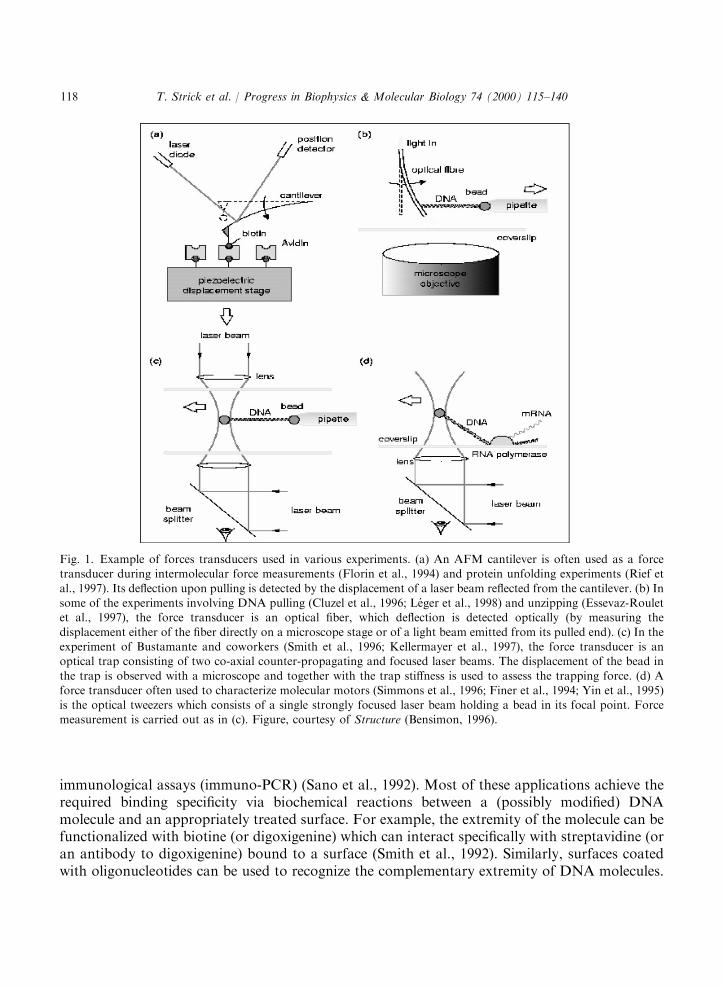
Fig. 1. Example of forces transducers used in various experiments. (a) An AFM cantilever is often used as a forcetransducer during intermolecular force measurements (Florin et al., 1994) and protein unfolding experiments (Rief et
al., 1997). Its deflection upon pulling is detected by the displacement of a laser beam reflected from the cantilever. (b) Insome of the experiments involving DNA pulling (Cluzel et al., 1996; Leger et al., 1998) and unzipping (Essevaz-Rouletet al., 1997), the force transducer is an optical fiber, which deflection is detected optically (by measuring the
displacement either of the fiber directly on a microscope stage or of a light beam emitted from its pulled end). (c) In theexperiment of Bustamante and coworkers (Smith et al., 1996; Kellermayer et al., 1997), the force transducer is anoptical trap consisting of two co-axial counter-propagating and focused laser beams. The displacement of the bead in
the trap is observed with a microscope and together with the trap stiffness is used to assess the trapping force. (d) Aforce transducer often used to characterize molecular motors (Simmons et al., 1996; Finer et al., 1994; Yin et al., 1995)is the optical tweezers which consists of a single strongly focused laser beam holding a bead in its focal point. Forcemeasurement is carried out as in (c). Figure, courtesy of Structure (Bensimon, 1996).
T. Strick et al. / Progress in Biophysics & Molecular Biology 74 (2000) 115–140118
Finally, there exist a large range of chemical methods to anchor (with various degrees ofspecificity) the extremities of DNA to surfaces bearing reactive groups (e.g. primary or secondaryamine, carboxyl or thiol moieties).An interesting alternative which does not require any modification of the molecule relies on the
specific adsorption of DNA by its ends on hydrophobic surfaces at a pH �5:5 (Allemand et al.,1997). On many different hydrophobic materials (teflon, polystyrene, graphite, silanised glass,etc.) DNA was observed to adhere strongly and non-specifically at low pH and weakly or not atall at high pH. In between there exists a narrow pH range (pH ¼ 5:5� 0:2) where DNA binds tothe surface by its extremities only.Once DNA is anchored to a surface by its end(s), a very easy way to stretch it is to drain the
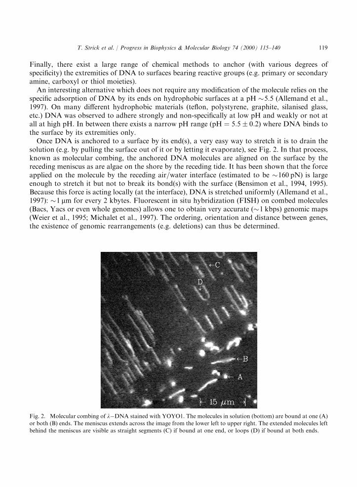
solution (e.g. by pulling the surface out of it or by letting it evaporate), see Fig. 2. In that process,known as molecular combing, the anchored DNA molecules are aligned on the surface by thereceding meniscus as are algae on the shore by the receding tide. It has been shown that the forceapplied on the molecule by the receding air=water interface (estimated to be �160 pN) is largeenough to stretch it but not to break its bond(s) with the surface (Bensimon et al., 1994, 1995).Because this force is acting locally (at the interface), DNA is stretched uniformly (Allemand et al.,1997): �1 mm for every 2 kbytes. Fluorescent in situ hybridization (FISH) on combed molecules(Bacs, Yacs or even whole genomes) allows one to obtain very accurate (�1 kbps) genomic maps(Weier et al., 1995; Michalet et al., 1997). The ordering, orientation and distance between genes,the existence of genomic rearrangements (e.g. deletions) can thus be determined.
Fig. 2. Molecular combing of lÿDNA stained with YOYO1. The molecules in solution (bottom) are bound at one (A)or both (B) ends. The meniscus extends across the image from the lower left to upper right. The extended molecules leftbehind the meniscus are visible as straight segments (C) if bound at one end, or loops (D) if bound at both ends.
T. Strick et al. / Progress in Biophysics & Molecular Biology 74 (2000) 115–140 119

Observation of the combing of DNA molecules grafted at both ends to a surface is instructive,see Fig. 3. First as the meniscus moves past the anchoring points of the molecule it stretches itstwo anchored segments (legs) perpendicular to the interface. The portion of the molecule insolution decreases until it spans the distance between the two legs. It is then stretched parallel tothe contact line, its length diminishing as the meniscus recedes thus forming a loop. Then as thetension in the molecule increases the loop breaks. The length of the DNAmolecule at the breakingpoint lb can be measured and compared with the unstretched length lb;0, see Fig. 3. It turns outthat lb=lb;0 ¼ 2:14� 0:2 (Bensimon et al., 1995): DNA can be extended to more than twice itslength before breaking! As we shall see below, this huge deformation of DNA (it is similar to theratio of the interphosphate distance to the distance between base-pairs) has to be accommodatedby a profound restructuring of the Watson–Crick double helix.
2.2. DNA manipulation and force measurements
Once DNA has been bound to a surface, it can be manipulated by displacing the anchoringobject. The first manipulations of DNA molecules involved translating with the help of opticaltweezers a small streptavidine-coated bead to which a single biotynilated DNA had been anchored(Chu, 1991; Perkins et al., 1994a,b). The molecule stained with a fluorescent dye was stretched bythe hydrodynamic drag and its relaxation in various conditions was studied. In particular whenstretched in a dilute solution of other DNA molecules the relaxation of the molecule provided thefirst direct observation of the reptation dynamics of an entangled polymer.By anchoring a DNA at both ends to different surfaces (typically a coverslip, a microbead or a
microneedle), a force can be exerted on the molecule by displacing the surfaces relative to eachother. Thus, DNA can be anchored to an AFM cantilever (or microfiber) at one end and to a
Fig. 3. Fragment of the genomic DNA, of one of the authors combed on a hydrophobic surface. The DNA bound atboth ends form a typical broken loop. From measurements on such figures we deduce the extension of DNA at the
breaking point: the stretched molecule length is lb ¼ BC, its unstretched length lb;0 is deduced by dividing the lengthABþDC by the extension factor observed for a straight molecule combed on an hydrophobic surface (1 mm for 2kbytes).
T. Strick et al. / Progress in Biophysics & Molecular Biology 74 (2000) 115–140120
treated surface at the other (Engel et al., 1999; Rief et al., 1999, 1997; Florin et al., 1994). Bydisplacing the surface, see Fig. 1, the molecule is stretched and pulls on the cantilever. Knowing itsspring constant, the force on the molecule can be measured. Very high forces (many thousands ofpN) can be achieved and measured in these experiments with a precision of �10 pN and a spatialresolution of Oð0:1 nm).By using two co-axial counter-propagating laser beams a small transparent bead can be
trapped, see Fig. 1 with a force of Oð100 pN) and a spatial resolution �10 nm. The force exertedon the bead can be deduced from the displacement of the trapping beam due to its refraction inthe bead, i.e. by directly measuring the momentum transfer (Smith, 1998). This absolute measurebypasses the need for a calibration of this optical trap.Another popular technique involves the stretching by an intensely focused laser beam (optical
tweezers (Simmons et al., 1996)) of a transparent bead of radius r of Oð1 mm) anchored by a DNAmolecule to a surface, see Fig. 1. Forces F550 pN can be achieved and measured with a precisionof �0:1 pN by following the displacement dx of the trapped bead from its equilibrium (zero force)position: F ¼ ktrapdx, where ktrap is the elastic stiffness of the optical trap. dx can be measuredwith a precision of O(1 nm) (Simmons et al., 1996), enough to resolve a single step of myosin(Finer et al., 1994; Saito et al., 1994) or kinesin (Schnitzer and Block, 1997; Hua et al., 1997). ktraphas to be determined prior to any force measurement, for example by pulling on the bead with aknown force such as the hydrodynamic drag Fs of a fluid (of viscosity Z) flowing with velocity v
around the bead: Fs ¼ 6pZrv. Alternatively, one may determine ktrap by measuring the intensity ofthe Brownian fluctuations hdx2i of the trapped bead. By the equipartition theorem they have tosatisfy (Simmons et al., 1996; Einstein, 1956; Reif, 1965):
ktraphdx2i2
¼ kBT
2: ð1Þ
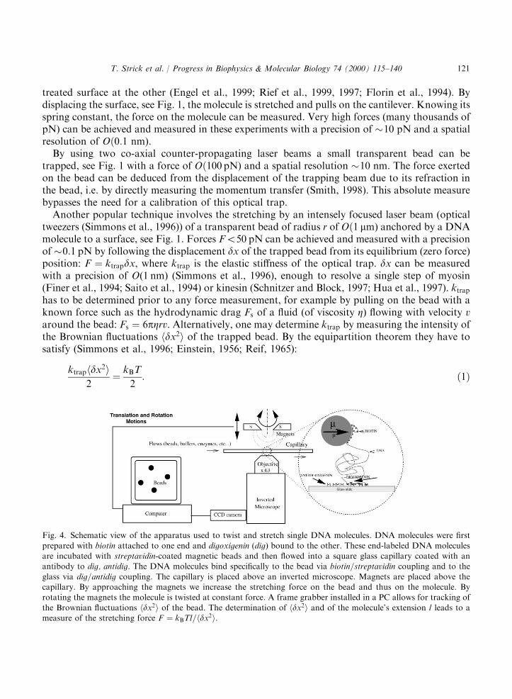
Fig. 4. Schematic view of the apparatus used to twist and stretch single DNA molecules. DNA molecules were first
prepared with biotin attached to one end and digoxigenin (dig) bound to the other. These end-labeled DNA moleculesare incubated with streptavidin-coated magnetic beads and then flowed into a square glass capillary coated with anantibody to dig, antidig. The DNA molecules bind specifically to the bead via biotin=streptavidin coupling and to the
glass via dig=antidig coupling. The capillary is placed above an inverted microscope. Magnets are placed above thecapillary. By approaching the magnets we increase the stretching force on the bead and thus on the molecule. Byrotating the magnets the molecule is twisted at constant force. A frame grabber installed in a PC allows for tracking of
the Brownian fluctuations hdx2i of the bead. The determination of hdx2i and of the molecule’s extension l leads to ameasure of the stretching force F ¼ kBTl=hdx2i.
T. Strick et al. / Progress in Biophysics & Molecular Biology 74 (2000) 115–140 121
To twist and stretch a DNA molecule and study its interactions with proteins, a magnetictrapping technique (Strick et al., 1996) has proved particularly convenient. Briefly, it consists instretching a single DNA molecule bound at one end to a surface and at the other to a magneticmicro-bead (1–4:5 mm in diameter), see Fig. 4. Small magnets, whose position and rotation can becontrolled, are used to pull on and rotate the micro-bead and thus stretch and twist the molecule.This system allows one to apply and measure forces ranging from a few fN ð10ÿ3pNÞ to nearly 100pN (see Strick et al., 1998b) with a relative accuracy of �10%.In contrast with other techniques, this force measurement is absolute and does not require a
calibration of the sensor. It is based on the analysis of the Brownian fluctuations of the tetheredbead, which is completely equivalent to a damped pendulum of length l ¼ hzi pulled by amagnetic force F (along the z-axis). Its longitudinal ðdz2 ¼ hz2i ÿ hzi2Þ and transverse dx2
fluctuations are characterized by effective rigidities kjj ¼ @zF and k? ¼ F=l. By the equipartitiontheorem they satisfy (Einstein, 1956; Reif, 1965)
dz2 ¼ kBT
kjj¼ kBT
@zF; ð2Þ
dx2 ¼ kBT
k?¼ kBTl
F: ð3Þ
Thus from the bead’s Brownian fluctuations ðdx2; dy2Þ one can extract the force pulling on themolecule (the smaller the fluctuations the greater F) and from dz2 one obtains its first derivative,@zF . This measurement method can be used with magnetic (but not optical) traps because thevariation of the trapping gradients occurs on a scale (Oð1 mmÞ) much larger than the scale onwhich the elasticity of the molecule changes (Oð0:1 mmÞ). In other words, the stiffness of theoptical trap is very large compared to F=l. A further bonus of the magnetic trap technique is thatmeasurements on DNA at constant force are trivial (just keep the position of the magnets fixed).With cantilevers or optical tweezers to work at constant force requires an appropriate feedback toensure that the displacement of the sensor is kept constant. However, because its stiffness dependson the force, the magnetic trap technique has at weak forces ð51 pNÞ a lower spatial resolution ofOð10 nmÞ, than the other manipulation methods. Finally notice that by using electro-magnets, afaster and more versatile magnetic tweezers system has recently been developed (Gosse andCroquette, 1999).
3. Models of polymer elasticity
Just like any polymer in solution, free DNA adopts a random coil conformation whichmaximizes its entropy (de Gennes, 1979). Pulling on the molecule reduces this entropy and costsenergy. The associated entropic forces result from a reduction of the number of possibleconfigurations of the system consisting of the molecule (be it a polymer, DNA or a protein) and itssolvent (water, ions), so that at full extension there is but one configuration left: a straight polymerlinking both ends. To reach that configuration work has to be done against entropy, a force has tobe applied. The entropic forces are rather weak, typically 510 pN. Beyond this regime and up toabout 70 pN DNA stretches like any spring: it is in an enthalpy dominated regime.
T. Strick et al. / Progress in Biophysics & Molecular Biology 74 (2000) 115–140122
3.1. The Kratky–Porod model
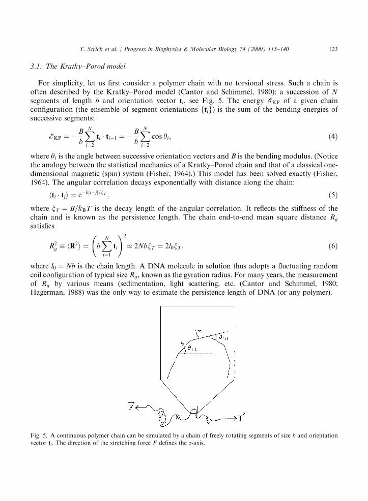
For simplicity, let us first consider a polymer chain with no torsional stress. Such a chain isoften described by the Kratky–Porod model (Cantor and Schimmel, 1980): a succession of Nsegments of length b and orientation vector ti, see Fig. 5. The energy EKP of a given chainconfiguration (the ensemble of segment orientations ftig) is the sum of the bending energies ofsuccessive segments:
EKP ¼ ÿB
b
XNi¼2
ti � tiÿ1 ¼ ÿB
b
XNi¼2
cos yi; ð4Þ
where yi is the angle between successive orientation vectors and B is the bending modulus. (Noticethe analogy between the statistical mechanics of a Kratky–Porod chain and that of a classical one-dimensional magnetic (spin) system (Fisher, 1964).) This model has been solved exactly (Fisher,1964). The angular correlation decays exponentially with distance along the chain:
hti � tji ¼ eÿbjiÿjj=xT ; ð5Þ
where xT ¼ B=kBT is the decay length of the angular correlation. It reflects the stiffness of thechain and is known as the persistence length. The chain end-to-end mean square distance Rg
satisfies
R2g � hR2i ¼ b
XNi¼1
ti
!2
’ 2NbxT ¼ 2l0xT ; ð6Þ
where l0 ¼ Nb is the chain length. A DNA molecule in solution thus adopts a fluctuating randomcoil configuration of typical size Rg, known as the gyration radius. For many years, the measurementof Rg by various means (sedimentation, light scattering, etc. (Cantor and Schimmel, 1980;Hagerman, 1988) was the only way to estimate the persistence length of DNA (or any polymer).
Fig. 5. A continuous polymer chain can be simulated by a chain of freely rotating segments of size b and orientationvector ti. The direction of the stretching force F defines the z-axis.
T. Strick et al. / Progress in Biophysics & Molecular Biology 74 (2000) 115–140 123
The stretching of a single DNA molecule now provides a much more precise way of measuringxT . To model the behavior of a polymer chain under tension, it suffices to add to Eq. (4) a termrepresenting the work W ¼ ÿF � R ¼ ÿFb
Pti;z ¼ ÿFb
PcosYi done by a force F acting on the
chain along the zÿaxis (Yi is the angle between ti and the zÿaxis):
EKP ¼ ÿB
b
XNi¼2
ti � tiÿ1 ÿ FbXNi¼1
ti;z ¼ ÿB
b
XNi¼2
cos yi ÿ FbXNi¼1
cosYi: ð7Þ
Unfortunately, this model can be solved only for small forces, where the mean extension of thechain l5l0 (Fisher, 1964) is
l ¼ 2FxT3kBT
l0 ¼F
3kBTR2
g: ð8Þ
To compute the elastic response of a chain at higher forces one has to resort to numericalcalculations (e.g. transfer matrix methods) or to various approximations of the Kratky–Porodmodel.
3.2. The freely jointed chain model
An interesting limit is the freely jointed chain (FJC) model, which consists in setting B ¼ 0 inEq. (7). It models a chain whose segments are unrestricted in their respective orientation andcorresponds to a discretization of a polymer with segments of length b ¼ 2xT (the so-called Kuhnlength). In the FJC model the energy of a given chain configuration ftig is thus EFJC ¼W ¼ÿFb
PcosYi. The partition function Z is
Z ¼Xti
eÿEFJC=kBT ¼Xti
YNi¼1
eFb cos Yi=kBT ð9Þ
¼Z
dO eFb cos Y=kBT� �N
¼ 2pkBTFb
sinhFb
kBT
� �N: ð10Þ
From the free energy F ¼ ÿkBT logZ, one can compute the mean extension of the chain l
l ¼ ÿ @F@F¼ l0 coth
Fb
kBTÿ kBT
Fb
� �: ð11Þ
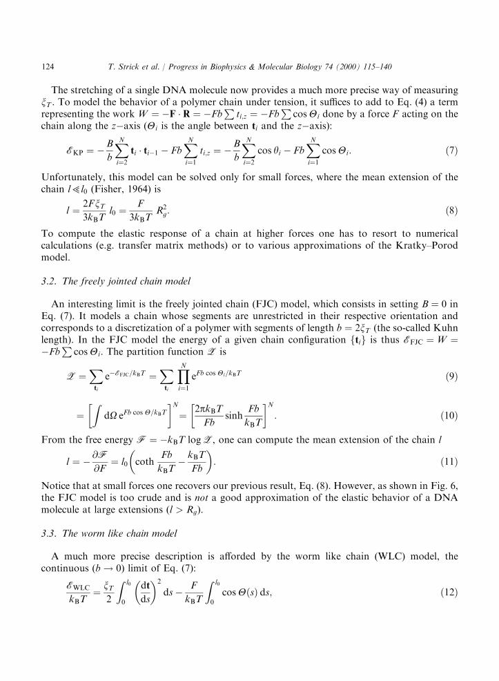
Notice that at small forces one recovers our previous result, Eq. (8). However, as shown in Fig. 6,the FJC model is too crude and is not a good approximation of the elastic behavior of a DNAmolecule at large extensions (l > Rg).
3.3. The worm like chain model
A much more precise description is afforded by the worm like chain (WLC) model, thecontinuous (b! 0) limit of Eq. (7):
EWLC
kBT¼ xT
2
Z l0
0
dt
ds
� �2
dsÿ F
kBT
Z l0
0
cosYðsÞ ds; ð12Þ
T. Strick et al. / Progress in Biophysics & Molecular Biology 74 (2000) 115–140124
where s is the curvilinear coordinate along the chain. The calculation of the partition function Zand the free energyF of that model calls upon an analogy with the quantum mechanical problemof a dipole in an electric field, which is beyond the scope of this paper. It has however been solvedby Marko and Siggia (Fixman and Kovac, 1973; Bustamante et al., 1994; Vologodskii, 1994;Marko and Siggia, 1995a,b). Though there is no analytic formula equivalent to Eq. (11) for the
Fig. 6. Force versus extension curves of single DNA molecules obtained by different groups. (A) The dots correspondto several experiments performed over a wide range of forces. The force was measured using the Brownian fluctuation
technique (Strick et al., 1996). The full line curve is a best fit to the WLC model for forces smaller than 5 pN. Thedashed curve is the result of the FJC model with the same persistence length (it is clearly a worse description of thebehavior of DNA under stress than the WLC model). At high forces, the molecule first elongates slightly, as would any
material in its elastic regime. Above 70 pN, the length abruptly increases, corresponding to the appearance of a newstructure called S-DNA. (B) The same transition observed by J.-F. Leger and D. Chatenay using a glass needledeflection on a nicked molecule and an unnicked molecule (the transition occurs for a higher force). (C) The transition
is also observed by S. Smith and C. Bustamante using optical tweezers. (D) Finally, H. Clausen-Schaumann andH. Gaub observe also the transition using an AFM. (We thank C. Bustamante, D. Chatenay, H. Clausen-Schaumann,H. Gaub, J.-F. Leger and S. Smith for sharing their data).
T. Strick et al. / Progress in Biophysics & Molecular Biology 74 (2000) 115–140 125

force vs. extension behavior of a WLC, a simple and efficient numerical solution was recentlyprovided by Bouchiat et al. (1999), who gave an approximation better than 0.1%:
F ¼ ðkBT=xTÞgðxÞ; ð13Þwhere x ¼ l=l0 and
gðxÞ ¼ xÿ 1
4þ 1
4ð1ÿ xÞ2þX7i¼2
aixi ð14Þ
with a2 ¼ ÿ0:5164228; a3 ¼ ÿ2:737418; a4 ¼ 16:07497; a5 ¼ ÿ38:87607; a6 ¼ 39:49944; a7¼ ÿ14:17718 (Bouchiat et al., 1999). Notice that at small relative extensions x51, as for theFJC model we recover Eq. (8). However, when compared over the whole extension range, theWLC model is a much better description of the behavior of DNA than the FJC model. As shownin Fig. 6 the WLC model fits extremely well the measured data and allows a very preciseestimation of the DNA’s persistence length: xT ¼ 52� 2 nm in physiological conditions (10 mMphosphate buffer (pH ¼ 7:5), 10 mM NaCl).
3.4. Self-avoidance effects
In the theoretical models described previously, the fact that a real polymer cannot intersectitself was not taken into consideration. For example in the computation of the partition functionZ, Eq. (10), we included these unrealistic configurations. In that section we shall try to justify thisapproach. A treatment of the self-avoidance effects of a stretched polymer exists only at lowextensions (l5l0), where a heuristic argument due to Flory (1975) works remarkably well. Due toself-avoidance one expects a real chain to occupy a larger volume than the so-called Gaussian(intersecting or phantom) chains considered previously, which occupied a volume R3
g. That ofcourse cost some entropic energy which is, however, compensated by a reduction in theprobability of interaction. In this approach the free energy of a FJC polymer is (recall that at lowextensions all models are equivalent):
F
kBT¼ 3l2
2R2g
þ vN2
l3: ð15Þ
The first term on the right describes the energy cost associated with a swelling of the polymer(�RF dl, with F given by Eq. (8)). The second term accounts for the self-avoidance. It is
proportional to the probability that two segments of the FJC of volume v ¼ pr2b will share thesame location in the space l3 occupied by the chain (the segments are assumed cylindrical with aradius r and length b). That repulsive term decreases as the chain swells. By minimizing F withrespect to l, one obtains the equilibrium (Flory’s) radius of a self-avoiding polymer:
RF ¼ ðvb2Þ1=5N3=5: ð16ÞExcluded volume interactions will become important when in the volume explored by one
monomer, typically b3 there is another monomer, i.e. when the monomer concentration c ¼N=R3
F ¼ 1=b3. Self-avoidance thus becomes non-negligible when N > ðb=rÞ3=2=p3=4. For a DNAmolecule with r ¼ 1 nm and b ¼ 100 nm, this corresponds to N�400, i.e. a molecular length:l0;F ¼ Nb�40 mm. Almost all manipulations of DNA molecules so far have been in a
T. Strick et al. / Progress in Biophysics & Molecular Biology 74 (2000) 115–140126
regime l05l0;F, where self-avoidance is totally negligible. That is except in torsionallyconstrained experiments where self-avoidance is crucial as it stabilizes plectonemic loops (seeSection 4 below).
3.5. Beyond the entropic regime
Beyond the entropic regime, i.e. from �6 pN to about 70 pN, DNA behaves like an elastic rodwith stretch modulus EA�1000 pN (Smith et al., 1996; Wang et al., 1997) (where E is the Youngmodulus of DNA and A its effective cross-sectional area (Hogan and Austin, 1987)). Neglectingentropic contributions, the force vs. extension curve follows a simple Hookean law (as any elasticmaterial): F ¼ EAðxÿ 1Þ (with x ¼ l=l0 > 1). Some ad hoc formulas exist, interpolating betweenthe entropic and Hookean regimes, e.g. replacing the term ð1ÿ xÞ2 in Eq. (14) by ð1ÿ xþ F=EAÞ2(Wang et al., 1997).Finally at about 70 pN a surprising transition has recently been discovered where DNA
stretches to about 1.7 times its crystallographic length (Smith et al., 1996; Cluzel et al., 1996). Ascharacteristic of the first-order transitions in nature (e.g. boiling) that transition is highlycooperative: a small change in force results in a large change in extension (see Fig. 6). Aphenomenological description of that transition has been proposed (Cluzel et al., 1996; Cizeauand Viovy, 1997; Ahsan et al., 1998; Marko, 1998), where the force plays the same role as themagnetic field in a ferromagnetic context. In this model, the observed sharpness of the transition(its high cooperativity) is associated with a large interfacial energy between the phases, suggestingthat the typical domain size is about 100 bases long (Cizeau and Viovy, 1997).To address the possible structural modification in the molecule resulting from pulling on it, a
numerical energy minimization of DNA under stress was performed by R. Lavery andcollaborators (Cluzel et al., 1996; Lebrun and Lavery, 1996). Its results reveal the existence of anew conformation called S-DNA indeed 70% longer than B-DNA, whose exact structure dependson which extremities of the DNA are being pulled (30–30 or 50–50). If both 30 extremities are beingpulled the double helix unwinds upon stretching. The final structure resembles a ladder. If both 50
ends are pulled a helical structure is preserved. It is characterised by a strong base pair inclination,a narrow minor groove and a diameter roughly 30% less than that of B-DNA. In both cases therupture of the molecule (by unpairing of the bases) occurs as observed during molecular combing(Bensimon et al., 1994, 1995) when its extension is more than twice that of B-DNA (Wilkins et al.,1951).These numerical results are supported by experiments done almost 50 years ago by Wilkins
et al. (1951) before the double helix structure of DNA was even proposed. Those suggested thatstretched DNA fibers indeed undergo a transition to a structure with tilted bases about twicelonger than the relaxed molecule. However, recent experiments by Leger et al. ð1999Þ stretchingtorsionally constrained single molecules have shown that the S-DNA phase has a helical pitch of22 nm with 38 bases per right-handed turn, which would make it look more like a slightly twistedladder.The existence of a new stable form of DNA at high extension might have considerable interest
for the study of DNA=protein interactions. Thus RecA is known to induce a 60% extension of B-DNA (Stasiak and Di Capua, 1982; Stasiak et al., 1983) and facilitate the formation of a triplehelix, a putative intermediate during recombination. Smith et al. (1996) calculated that the
T. Strick et al. / Progress in Biophysics & Molecular Biology 74 (2000) 115–140 127
existence of an extended S-form of DNA reduced the energetics of RecA binding to DNA by asmuch as 15kBT (9 kcal=mol) per complex. Recent experiments (Leger et al., 1998; Shivashankaret al., 1999; Hegner et al., 1999) have indeed shown that the polymerisation of RecA on a dsDNAwas facilitated by stretching the molecule. This implies that the barriers to nucleation andaccretion of a RecA fiber on DNA is lowered by the presence of a S-DNA sub-phase. Numericalmodeling further suggest that its structure is closer to the RecA=DNA complex than regular B-DNA (Lebrun and Lavery, 1997).
3.6. Elasticity of heterogeneous polymers
In the preceding discussion, the polymer properties were assumed homogenous, i.e. wedid not consider possible sequence specific effects on the elastic behavior of DNA.The Kratky–Porod model allows one to treat those to some extent. It is thus easy tointroduce some local preferred bending (i.e. orientational angle ci between successivesegments) by replacing the term cos yi in Eq. (7) by cosðyi ÿ ciÞ. The case where the ci’s arefixed randomly along the chain, thus defining a static (disorder induced) angular correlation xd,has been analysed by two groups using slightly different models (Bensimon et al., 1998; Nelson,1998). Surprisingly, it turns out that the elastic behavior of such a heterogeneous chain is to allpractical purposes identical to that of an homogenous WLC with an effective persistence lengthxeff which is a model-dependent function of xd and xT . Therefore, one does not expect sequencespecific effects to alter drastically the results of the WLC model described previously. It is worthemphasizing the theoretical point that sequence inhomogeneities must be treated as ‘quenched’,i.e. the free energy of the system should be averaged over the disorder (Binder and Young, 1986).Some results claiming to describe sequence disorder effects (Nelson, 1998; Trifonov et al., 1987;Schellman and Harvey, 1995) actually employ an ‘annealed’ averaging of the partition functionover disorder which is mathematically simpler but is irrelevant to experiment where the sequenceis fixed.
4. DNA under torsion
4.1. Topological properties of coiled DNA
To describe DNA under torsional stress it is first necessary to introduce some topologicalconcepts. The first is the twist (Tw), the number of helical turns along the molecule. For atorsionally unconstrained B-DNA, Tw ¼ Tw0 ¼ N=h where N is the number of base-pairs andh ¼ 10:4 is the number of base-pairs per turn of the helix. The second topological quantity ofinterest is the writhe (Wr) of the molecule. Wr is a measure of the coiling of the DNA axis aboutitself, as a twisted phone cord which forms interwound structures in order to relieve its torque. Ifthe DNA molecule is torsionally constrained, then the total number of times the two strands ofthe helix cross each other (either by twist or writhe) is a topological invariant of the system calledthe linking number Lk ¼ TwþWr (White, 1969). For relaxed linear DNA molecules, assumingthe absence of any spontaneous local curvature, Lk ¼ Lk0 ¼ Tw0. The relative difference inlinking number between the supercoiled and relaxed forms of DNA is called the degree of
T. Strick et al. / Progress in Biophysics & Molecular Biology 74 (2000) 115–140128
supercoiling, s:
s ¼ ðLkÿ Lk0Þ=Lk0 ¼ DLk=Lk0: ð17Þ
The value of s for most circular molecules isolated from cells or virions is roughly ÿ0:06. Anotable exception occurs in hyperthermophilic archeabacteria (Woese and Fox, 1977; Woese etal., 1990) who have a positively coiled DNA (Kikuchi and Asai, 1984; Forterre, 1996). In theexperiments described here, provided that the anchoring of the DNA molecule is achieved atmultiple points at both ends, a torsional constraint can be applied on the molecule by simplyrotating the magnets. As one turn of the magnets implies a change of one turn of the molecule, wehave simply DLk ¼ �n, where �n is the number of turns by which the magnets are made to rotate.Note that at fixed Lk the ratio Tw=Wr will depend on the force stretching the molecule, the writhebeing suppressed by high forces. As a consequence, pulling on a coiled molecule increases thetorque twisting it, until its writhe reaches zero.
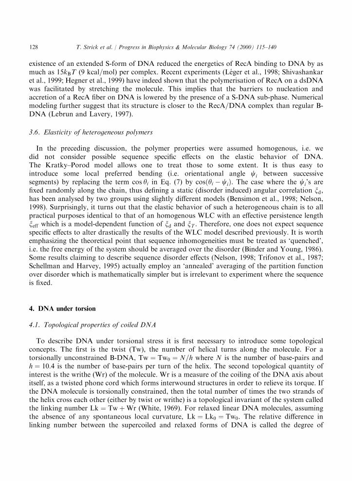
4.2. The mechanical buckling instability
Twisting DNA leads to a torsional buckling instability analogous to that observed on telephonecords or rubber tubes. This instability leads to the formation of interwound structures known assupercoils or plectonemes. Of course, a DNA molecule is also animated by very strong thermalfluctuations which play an important role. However, it is instructive to first consider the purelymechanical (zero-temperature) instability of a rubber tube of length l and torsional modulus C. Ifwe firmly hold one end of the tube while simultaneously rotating and pulling on the second endwith a force F , we observe the following phenomenon (see Fig. 7): when the twist constraint issmall, the associated torque G increases linearly with the twist angle y, G ¼ Cy=l and the tuberemains straight. As the tube is further twisted, a critical twist angle yc;b and torque Gc;b arereached where the tube ceases to be straight: it locally buckles and forms a small loop of radiusRc;b. The torsional energy thus gained is 2pGc;b, whereas the energy cost (due to bending and workagainst F) is (see Eq. (12)): E ¼ pB=Rþ 2pRF . The cost is minimized for a loop of radiusRc;b ¼
ffiffiffiffiffiffiffiffiffiffiffiffiB=2F
p. The critical torque for the formation of plectonemes is controlled by the balance
between energy gain and cost, i.e. by the stretching force:
Gc;b ¼ Emin=2p ¼ffiffiffiffiffiffiffiffiffi2BFp
: ð18Þ
As we twist the tube further, we increase the length of the plectonemes but the torque in the tuberemains basically fixed at its critical value Gc;b.
4.3. DNA under torsion: the rod like chain model
For DNA, the picture is pretty much the same (Bouchiat and Mezard, 1998; Moroz andNelson, 1998; Vologodskii and Marko, 1997). The thermal fluctuations which will be mostimportant near the mechanical instability at yc;b will tend to round it off. Hence as one is coiling aDNA molecule under fixed force F , one observes the following behavior (see Fig. 7): at lowdegrees of supercoiling jsj the molecule’s extension varies little. Beyond a critical value sc (whichdepends on the force), the molecule shortens continuously as it is twisted further. A theoreticaltreatment of this behavior is given by the RLC model developed by Bouchiat and Mezard (1998,
T. Strick et al. / Progress in Biophysics & Molecular Biology 74 (2000) 115–140 129
1999) which consists in describing a DNA molecule not as a chain free to rotate but as a rod witha finite torsional modulus C. The energy of this rod like chain (RLC) model ERLC is obtained byadding to the energy of the WLC model, Eq. (12), an energy of twist ET:
ERLC ¼ EWLC þ ET ¼ EWLC þC
2
Z l0
0
dsO2ðsÞ; ð19Þ
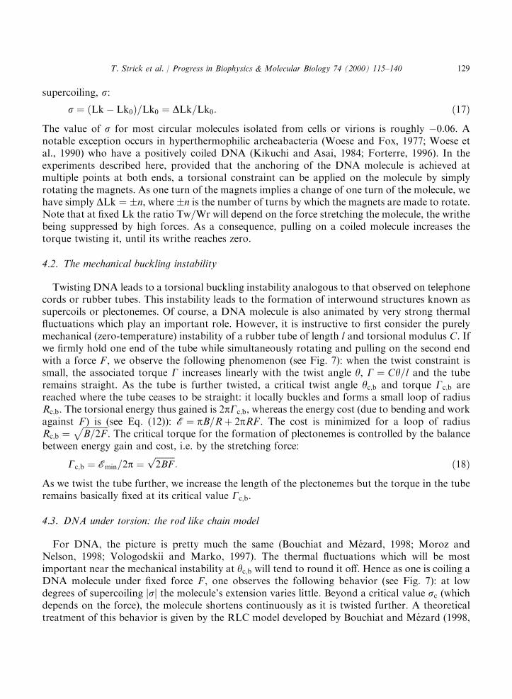
where OðsÞ is the local twist of the chain. A detailed solution of this model which is beyond thescope of the present paper can be found in Bouchiat and Mezard (1998, 1999) and Moroz andNelson (1998a,b). A particular mathematical subtely of this model is that the twist integrant inEq. (19) is singular. That singularity has to be regularized either by a truncation of its seriesexpansion (Moroz and Nelson, 1998a,b) or by the reintroduction of the short length cut-off b ofthe Kratky–Porod model (Bouchiat and Mezard, 1998; Bouchiat et al., 1999). Although the RLCmodel neglects the chain’s self-avoidance, which stabilizes the plectonemes, its predictions fitremarkably well with our observations at low forces (50:4 pN) where as the molecule is coiled itsend to end distance decreases, see Fig. 8. From a fit of the RLC predictions to our data, one canextract a value for the torsional modulus C of DNA: C=kBT ¼ 86� 5 nm (Bouchiat and Mezard,
Fig. 7. LEFT: schematic view of the buckling transition for a twisted rubber tube (dotted line) or a DNA molecule(solid line). Below a critical number of turns nc;b the rubber tube’s torque increases linearly as it stores twisting energy.When nc;b turns have been added the system abruptly exchanges twisting energy for bending energy and plectonemesbegin to form. The plectonemes grow linearly with subsequent twisting and the torque remains constant thereafter. In
the case of DNA the same picture holds, except for the fact that thermal fluctuation round off the transition which takesplace at nc;b. RIGHT: Results from the RLC model corresponding to a stretching force of F ¼ 0:33 pN. The x-axisrepresents the supercoiling variable Z ¼ 2pnxT=l0 ’ 95s (Bouchiat and Mezard, 1998), and the y-axis is in arbitrary
units. The long-dash curve represents the torque acting on the DNA: as decribed above, it increases linearly untilZc;b �1 (s �0:01) and remains essentially constant thereafter. The short-dash curve represents the ratio of writhe totwist: note that the writhe is never zero and increases rapidly as Z > 1. Finally, the full line measures the fraction of
plectonemes in DNA: stable supercoiled structures only appear after the torsional buckling transition has been passed.
T. Strick et al. / Progress in Biophysics & Molecular Biology 74 (2000) 115–140130
1998). This result depends weakly on the magnitude of b which best-fitted value of �6 nm isroughly equal to twice the DNA pitch.A number of groups have attempted to go beyond the model defined in Eq. (19) by introducing
a coupling between the stretch on the double helix and its twist (Moroz and Nelson, 1998b;Marko, 1997; Nelson et al., 1997). The approach is sensible, but the comparison with theexperimental data is very problematic due to the existence of structural transitions in DNA (seebelow) and the smallness of the predicted effect. Much theoretical work has also been done on thecoupling between the intrinsic curvature and the twisting of the molecule (Schlick and Olson,1992; Olson, 1996; Chirico and Langowski, 1996; Garrivier and Fourcade, 2000). As manyregulating factors are supposed to bend the molecule, a coupling to its twist could modulate theinteraction of distant sites along the DNA. This very interesting and important problem, deservesto be studied with the new tools now at our disposal. Finally, the dynamical aspects related torelaxation or transport of torsional stress along DNA (Nelson, 1999) need also to be addressedexperimentally.
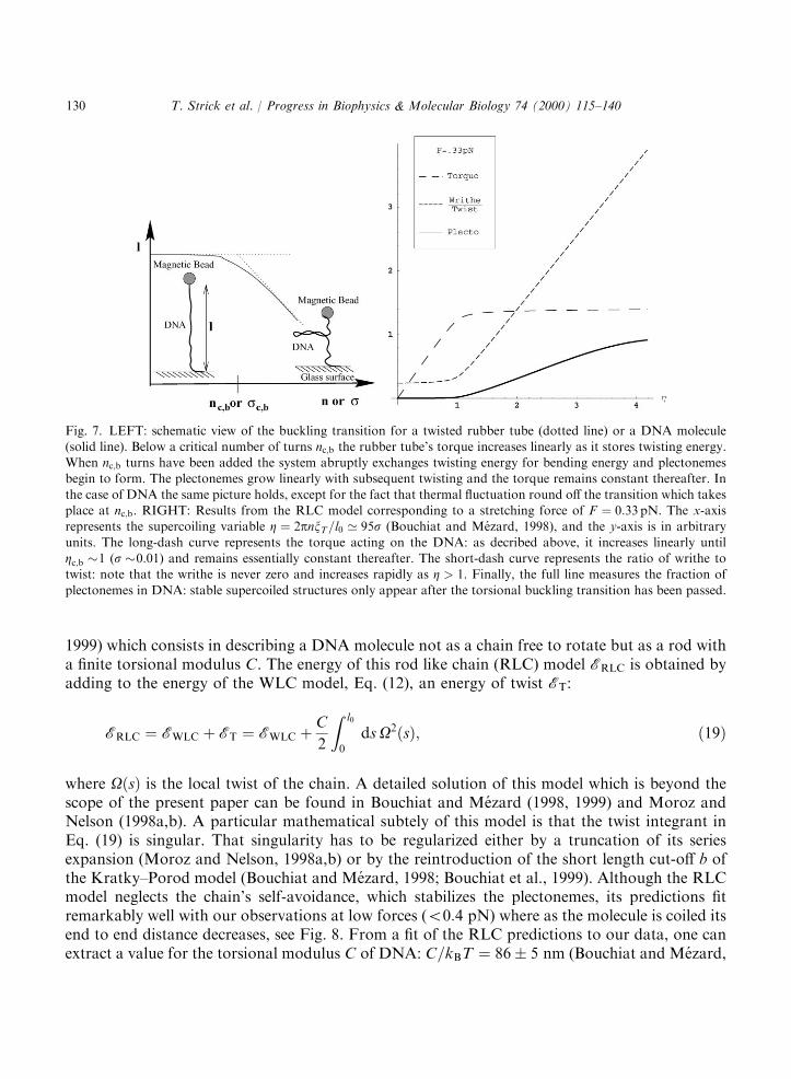
4.4. Torque induced transitions in DNA
The torsional buckling instability just described treats the DNA molecule as a continuouselastic tube. It ignores the underlying double-helical structure of the molecule, and its relevanceis therefore limited to very low forces (F50:4 pN) or low degrees of supercoiling(ÿ0:0155s50:037). For higher forces and degrees of supercoiling, the buildup of torque in the
Fig. 8. Relative extension of a DNA molecule vs. the degree of supercoiling Z ¼ 2pnxT=l0 ’ 95s for various stretchingforces. For the three curves obtained at low force, the behavior is symmetrical under s! ÿs. The shorteningcorresponds to the formation of plectonemes upon writhing. For these low forces the comparison between the
experimental data (points) and the rod like chain model with C=kBT ¼ 86 nm (full-line) is very good. When the force isincreased above 0.5 pN, the curve becomes asymmetric: supercoils still form for positive coiling while local denaturationadsorbs the torsional stress for negative s. At forces larger than 3 pN no plectonemes are observed: the torsional stress
is adsorbed not by writhe but in local structural changes of the molecule.
T. Strick et al. / Progress in Biophysics & Molecular Biology 74 (2000) 115–140 131

molecule can be large enough to actually modify its internal structure (Strick et al., 1999). This isevidenced by breaking of the s! ÿs symmetry in the extension vs. supercoiling curves, see Fig. 8.As a critical force is reached (ipso facto a critical torque), the molecule undergoes a transitionfrom a contracted state (plectonemic B-DNA) to a highly extended one (Strick et al., 1998). Thisstate is characterized by the coexistence of B-DNA with denatured DNA for s50 and with a newphase called P-DNA for s > 0 (Allemand et al., 1998). This new P-DNA structure has an intrinsicdegree of coiling sp ¼ 3, i.e. it has 2.6 bps=turn (Allemand et al., 1998; Leger et al., 1999).Numerical simulations, supported by chemical reactivity studies, suggest that in P-DNA thephosphate backbone is winding inside the structure and the bases are exposed in solution.These results show that at very low forces and low degrees of supercoiling DNA can locally
undergo major structural transitions. These transitions might be relevant to the activity of RNA-polymerase, which is known to exert forces as high as 35 pN and to under(over)-wind themolecule up(down)-stream. It is furthermore worthwhile to notice that a structure very similar toP-DNA has apparently been observed in the packaging of DNA in the Pf1 virus. There thisunusual structure is stabilised by the proteins of the virus’ coat (Liu and Day, 1994).
4.5. Twisting rigidity measured through the critical torque of denaturation
The denaturation transition offers a second way to evaluate the elastic torsional persistencelength C using a very simple model (Strick et al., 1999). It consists in measuring the difference inwork done while stretching a single DNA molecule wound either positively or negatively by thesame number of turns.Fig. 8 shows the molecular extension as a function of supercoiling for various forces. At a low
force (F �0:2 pN) the elastic behavior of DNA is symmetric under s! ÿs. Pulling on themolecule removes the writhe and thus increases the twist and the torque on the molecule. Forunderwound molecules (in 10 mM phosphate buffer) above the critical force (Fc �0:5 pN) and itsassociated critical torque (Gc �9 pNnm) writhing becomes energetically unfavorable. Themolecule elongates, see Fig. 8 as plectonemes (which are used to absorb twist) are convertedlocally into melted (denatured) regions of DNA. For positive supercoilings the critical force andtorque are significantly higher. Thus, we can easily find values of jsj such that denaturation occursat ÿjsj whereas plectonemes remain at jsj.In the following, we shall use the force vs. extension measurements on DNA supercoiled by �n
turns, i.e. with the same jsj, to estimate the torsional constant, C, the critical torque atdenaturation Gc and the energy of denaturation per base pair (bp), ed.Consider the case of a DNA of contour length l0 at an initial extension lAþð5l0). Let us coil the
molecule by n > 0 turns to state Aþ, (Fig. 9), requiring a torsional energy TAþ (F50:2 pN) andthen extend it to state Bþ (F ¼ 4 pN), so as to pull out its plectonemes and eliminate its writhe.Alternatively, state Bþ could be reached by first stretching the torsionally relaxed DNA and thentwisting it. In that case its torsional energy TBþ is purely twist. The free energy of a stretchedcoiled DNA being a state variable, the mechanical works performed on the molecule by stretchingit from thermodynamic state Aþ to Bþ along these two different paths should be equal:
TAþ þ DWABþ ¼ TBþ ¼C
2l0ð2pnÞ2: ð20Þ
T. Strick et al. / Progress in Biophysics & Molecular Biology 74 (2000) 115–140132
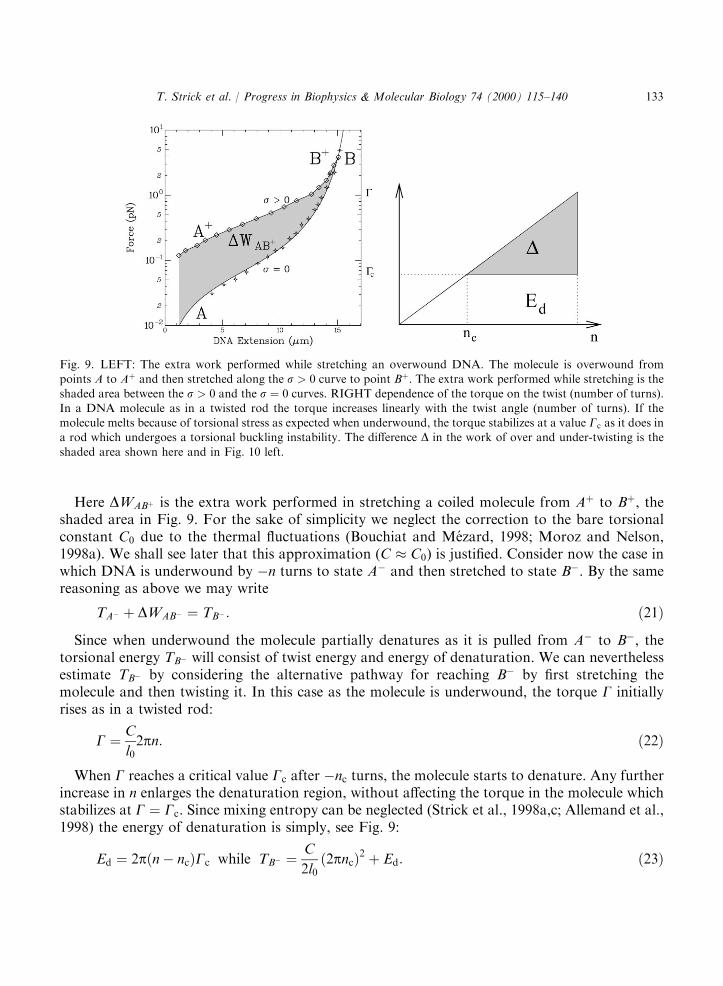
Here DWABþ is the extra work performed in stretching a coiled molecule from Aþ to Bþ, theshaded area in Fig. 9. For the sake of simplicity we neglect the correction to the bare torsionalconstant C0 due to the thermal fluctuations (Bouchiat and Mezard, 1998; Moroz and Nelson,1998a). We shall see later that this approximation (C � C0) is justified. Consider now the case inwhich DNA is underwound by ÿn turns to state Aÿ and then stretched to state Bÿ. By the samereasoning as above we may write
TAÿ þ DWABÿ ¼ TBÿ : ð21Þ
Since when underwound the molecule partially denatures as it is pulled from Aÿ to Bÿ, thetorsional energy TBÿ will consist of twist energy and energy of denaturation. We can neverthelessestimate TBÿ by considering the alternative pathway for reaching Bÿ by first stretching themolecule and then twisting it. In this case as the molecule is underwound, the torque G initiallyrises as in a twisted rod:
G ¼ C
l02pn: ð22Þ
When G reaches a critical value Gc after ÿnc turns, the molecule starts to denature. Any furtherincrease in n enlarges the denaturation region, without affecting the torque in the molecule whichstabilizes at G ¼ Gc. Since mixing entropy can be neglected (Strick et al., 1998a,c; Allemand et al.,1998) the energy of denaturation is simply, see Fig. 9:
Ed ¼ 2pðnÿ ncÞGc while TBÿ ¼C
2l0ð2pncÞ2 þ Ed: ð23Þ
Fig. 9. LEFT: The extra work performed while stretching an overwound DNA. The molecule is overwound frompoints A to Aþ and then stretched along the s > 0 curve to point Bþ. The extra work performed while stretching is the
shaded area between the s > 0 and the s ¼ 0 curves. RIGHT dependence of the torque on the twist (number of turns).In a DNA molecule as in a twisted rod the torque increases linearly with the twist angle (number of turns). If themolecule melts because of torsional stress as expected when underwound, the torque stabilizes at a value Gc as it does ina rod which undergoes a torsional buckling instability. The difference D in the work of over and under-twisting is the
shaded area shown here and in Fig. 10 left.
T. Strick et al. / Progress in Biophysics & Molecular Biology 74 (2000) 115–140 133
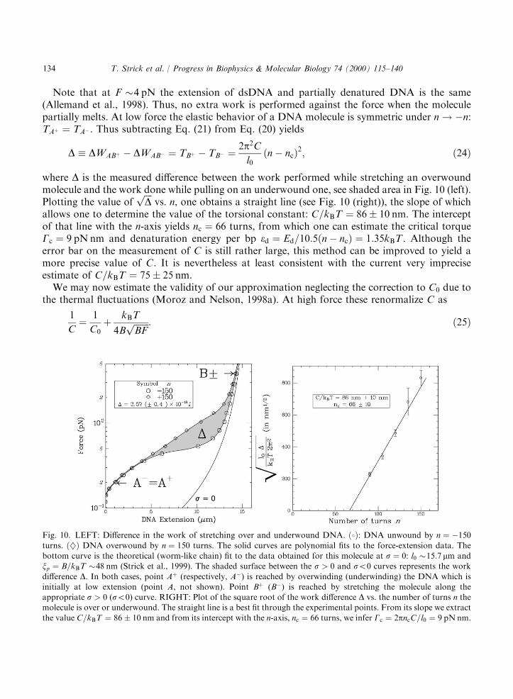
Note that at F �4 pN the extension of dsDNA and partially denatured DNA is the same(Allemand et al., 1998). Thus, no extra work is performed against the force when the moleculepartially melts. At low force the elastic behavior of a DNA molecule is symmetric under n! ÿn:TAþ ¼ TAÿ . Thus subtracting Eq. (21) from Eq. (20) yields
D � DWABþ ÿ DWABÿ ¼ TBþ ÿ TBÿ ¼2p2Cl0ðnÿ ncÞ2; ð24Þ
where D is the measured difference between the work performed while stretching an overwoundmolecule and the work done while pulling on an underwound one, see shaded area in Fig. 10 (left).Plotting the value of
ffiffiffiffiDp
vs. n, one obtains a straight line (see Fig. 10 (right)), the slope of whichallows one to determine the value of the torsional constant: C=kBT ¼ 86� 10 nm. The interceptof that line with the n-axis yields nc ¼ 66 turns, from which one can estimate the critical torqueGc ¼ 9 pN nm and denaturation energy per bp ed ¼ Ed=10:5ðnÿ ncÞ ¼ 1:35kBT . Although theerror bar on the measurement of C is still rather large, this method can be improved to yield amore precise value of C. It is nevertheless at least consistent with the current very impreciseestimate of C=kBT ¼ 75� 25 nm.We may now estimate the validity of our approximation neglecting the correction to C0 due to
the thermal fluctuations (Moroz and Nelson, 1998a). At high force these renormalize C as
1
C¼ 1
C0þ kBT
4BffiffiffiffiffiffiffiBFp : ð25Þ
Fig. 10. LEFT: Difference in the work of stretching over and underwound DNA. ð8Þ: DNA unwound by n ¼ ÿ150turns. ð}Þ DNA overwound by n ¼ 150 turns. The solid curves are polynomial fits to the force-extension data. Thebottom curve is the theoretical (worm-like chain) fit to the data obtained for this molecule at s ¼ 0: l0 �15:7 mm andxp ¼ B=kBT �48 nm (Strick et al., 1999). The shaded surface between the s > 0 and s50 curves represents the work
difference D. In both cases, point Aþ (respectively, Aÿ) is reached by overwinding (underwinding) the DNA which isinitially at low extension (point A, not shown). Point Bþ (Bÿ) is reached by stretching the molecule along theappropriate s > 0 (s50) curve. RIGHT: Plot of the square root of the work difference D vs. the number of turns n themolecule is over or underwound. The straight line is a best fit through the experimental points. From its slope we extract
the value C=kBT ¼ 86� 10 nm and from its intercept with the n-axis, nc ¼ 66 turns, we infer Gc ¼ 2pncC=l0 ¼ 9 pN nm.
T. Strick et al. / Progress in Biophysics & Molecular Biology 74 (2000) 115–140134

The last term on the right implies a correction of only 5% to the value of C at F ¼ 4 pN;smaller than our experimental uncertainty. It is interesting to note that the value of C determinedhere is in good agreement with the one obtained from the measurement of the molecular extensionversus s at constant force (Bouchiat and Mezard, 1998), a totally independent measurement basedon the model of a rod like chain polymer, see above.
5. DNA–protein interactions
The understanding gained on the manipulation and mechanical properties of a stretched andcoiled DNA, allows one to use these measurements as a tool to probe DNA=protein interactionsat the level of a single molecule, e.g. chromatin (Chatenay et al., 1997; Cui and Bustamante, 2000).For example, the lactose repressor-mediated loop formation in a single DNA molecule was
followed by studying the decrease in the Brownian motion of a DNA tethered bead (Finzi andGelles, 1995), as its molecular leash gets shorter. Subjecting that bead to a force F should yield thefree energy dG for the formation of the complex repressor=DNA. Indeed when the formation andbreakdown of the loop are equiprobable: dG ¼ Fdl (Marko and Siggia, 1997), where dl is the looplength.By using the stretching force as a control parameter the polymerization of RecA on a double-
stranded DNA has been induced (Leger et al., 1998; Shivashankar et al., 1999; Hegner et al.,1999). As we have seen previously, stretching a molecule with a force of �70 pN induces atransition to S-DNA, a structure of DNA presumably similar to the one adopted by the double-stranded molecule when interacting with RecA (Cluzel et al., 1996) or the TATA box-bindingprotein (Lebrun et al., 1997). By applying a force (even if less than 70 pN) on the molecule, oneincreases the probability of nucleation of an S-DNA region in the regular B-DNA helix. This S-DNA bubble has a higher affinity for RecA than B-DNA and serves as a nucleation center for thegrowth of a RecA fiber. As the complex RecA=DNA is about 60% longer than B-DNA, thekinetics of RecA polymerization can be followed as a function of time (at various forces) bymonitoring the change in the molecular extension. Whether this stretched induced polymerizationof RecA on dsDNA actually occurs in vivo remains an interesting suggestion.The progression of an E. coli RNA-polymerase on a dsDNA can be followed by tethering the
molecule to a small bead (Schafer et al., 1991; Yin et al., 1994) and anchoring the molecule at itsother end to a RNA-pol bound to a surface. As the DNA is transcribed, the ‘‘leash’’ binding thebead to the surface gets shorter. The extent of its fluctuations is thus reduced and can be used tomonitor the progression of the enzyme (Schafer et al., 1991). By trapping the bead with an opticaltweezers, a force can be applied on a single polymerase. Its speed, pauses and the force to stall it(about 35 pN) can thus be determined (Wang et al., 1998; Yin et al., 1995).Similarly, the replication of a single-strand DNA by a single DNA-polymerase could be
observed by following the change in extension of a stretched template as a new strand issynthesized (Wuite et al., 2000; Maier et al., 2000). From the variation of the replication rate withthe applied force, one could argue that a few bases (2 for T7-DNA polymerase, 4 for the Klenowfragment of polI) had to be fit between the two strands in the replication site for the enzyme toproceed. These results are in agreement with structural data and support the ‘‘induced-fit kineticmechanism’’ for replication (Wong et al., 1991).
T. Strick et al. / Progress in Biophysics & Molecular Biology 74 (2000) 115–140 135
Finally, the relaxation of DNA supercoiling by a single topoisomerase (Wang, 1998) couldbe monitored (Strick et al., 2000) and individual enzymatic cycles observed at low ATPconcentrations. From the distribution of the time intervals between successive cycles we coulddeduce that a single ATP was apparently burned per turnover. By averaging over many singleenzymatic reactions, we could regain the kinetic behavior of topoII known from bulkmeasurements. Surprisingly, stretching the supercoiled DNA resulted in a lower enzymaticactivity, indicating that the DNA gate religation step might be rate limiting. Finally in absence ofATP, the enzyme was observed to stabilise DNA crossovers.These experiments open the way to a study of single mechanical enzymes, with a possibility of a
detailed analysis of their cycle. Future experiments will certainly combine these manipulationtechniques with single molecule observations (Kitamura et al., 1999; Harada et al., 1999; Funatsuet al., 1995; Yasuda et al., 1998) using fluorescence polarisation (Sase et al., 1997) or energytransfer (Weiss, 1999) to monitor the enzyme’s structural deformation and energy consumptionduring its cycle.
Acknowledgements
We thank C. Bustamante, D. Chatenay, H. Clausen-Schaumann, H. Gaub, J.-F. Leger and S.Smith for sharing their data. We also thank C. Bouchiat, B. Maier, M. Mezard, for stimulatingdiscussions.
References
Ahsan, A., Rudnick, J., Bruinsma, R., 1998. Elasticity theory of the B-DNA to S-DNA transition. Biophys. J. 74,132–137.
Allemand, J.-F., Bensimon, D., Jullien, L., Bensimon, A., Croquette, V., 1997. pH-dependent specific binding andcombing of DNA. Biophys. J. 73, 2064–2070.
Allemand, J.-F., Bensimon, D., Lavery, R., Croquette, V., 1998. Stretched and overwound DNA form a Pauling-like
structure with exposed bases. Proc. Natl. Acad. Sci. USA 95, 14,152–14,157.Amblard, F., Yurke, B., Pargellis, A., Leibler, S., 1996. A magnetic manipulator for studying local rheology andmicromechanical properties of biological system. Rev. Sci. Instrum. 67 (2), 1–10.
Bensimon, A., 1996. Force: a new structural control parameter? Structure 4, 885–889.Bensimon, D., Dohmi, D., Mezard, M., 1998. Stretching a heteropolymer. Europhys. Lett. 42, 97–102.Bensimon, A., Simon, A.J., Chiffaudel, A., Croquette, V., Heslot, F., Bensimon, D., 1994. Alignment and sensitivedetection of DNA by a moving interface. Science 265, 2096–2098.
Bensimon, D., Simon, A.J., Croquette, V., Bensimon, A., 1995. Stretching DNA with a receding meniscus: experimentsand models. Phys. Rev. Lett. 74, 4754–4757.
Binder, K., Young, A.P., 1986. Spin glasses: experimental facts, theoretical concepts and open questions. Rev. Mod.
Phys. 58 (4), 801–976.Bouchiat, C., Mezard, M., 1998. Elasticity theory of a supercoiled DNA molecules. Phys. Rev. Lett. 80, 1556–1559.Bouchiat, C., Mezard, M., 1999. Elasticity rod model of supercoiled DNA molecules. condmat=9904018.Bouchiat, C., Wang, M.D., Block, S.M., Allemand, J.-F., Strick, T.R., Croquette, V., 1999. Estimating the persistencelength of a worm-like chain molecule from force-extension measurements. Biophys. J. 76, 409–413.
Bustamante, C., Marko, J.F., Siggia, E.D., Smith, S., 1994. Entropic elasticity of l-phage DNA. Science 265,
1599–1600.
T. Strick et al. / Progress in Biophysics & Molecular Biology 74 (2000) 115–140136
Cantor, C.R., Schimmel, P.R., 1980. Biophysical Chemistry, Part III: The Behaviour of Biological Macromolecules.
W.H. Freemann, San Francisco.Chatenay, D., Houchmanzadeh, B., Marko, J.F., Libchaber, A., 1997. Elasticity and structure of eukaryotechromosomes studied by micromanipulation and micropipette aspiration. J. Cell. Biol. 139, 1–12.
Chee, M., Yang, R., Hubbell, E., Berno, A., Huang, X.C., Stern, D., Winkler, J., Lockhart, D.J., Morris, M.S., Fodor,S.P.A., 1996. Accessing genetic information with high-density DNA arrays. Science 274, 610–614.
Chirico, G., Langowski, J., 1996. Brownian dynamics simulations of supercoiled DNA with bent sequences. Biophys. J.
71, 955–971.Chu, S., 1991. Laser manipulation of atoms and particles. Science 253, 861–866.Cizeau, P., Viovy, J.L., 1997. Modeling extreme extension of DNA. Biopolymers 42, 383–385.
Cluzel, P., Lebrun, A., Heller, C., Lavery, R., Viovy, J.-L., Chatenay, D., Caron, F., 1996. DNA: an extensiblemolecule. Science 271, 792–794.
Cui, Y., Bustamante, C., 2000. Pulling a single chromatin fiber reveals the forces that maintain its higher orderstructure. Proc. Natl. Acad. Sci. (USA) 97, 127–132.
de Gennes, P.G., 1979. Scaling concepts in Polymer Physics. Cornell University Press, Ithaca.Einstein, A., 1956. Investigation of the Brownian Theory of Movement. Dover Publication, New York.Engel, A., Gaub, H.E., Muller, D.J., 1999. Atomic force microscopy: a forceful way with single molecules. Curr. Biol. 9,
R133–R136.Essevaz-Roulet, B., Bockelmann, U., Heslot, F., 1997. Mechanical separation of the complementary strands of DNA.Proc. Natl. Acad. Sci. (USA) 94, 11,935–11,940.
Finer, J.T., Simmons, R.M., Spudich, J.A., 1994. Single myosin molecule mechanics: piconewton forces and nanometresteps. Nature 368, 113–119.
Finzi, L., Gelles, J., 1995. Measurement of lactose repressor-mediated loop formation and breakdown in single DNAmolecules. Science 267, 378–380.
Fisher, M.E., 1964. Magnetism in one-dimensional systems } the Heisenberg model for infinite spin. Am. J. Phys. 32,343–346.
Fixman, M., Kovac, J., 1973. Polymer conformational statistics III: modified Gaussian models of the stiff chains. J.
Chem. Phys. 58, 1564–1568.Florin, E.L., Moy, V.T., Gaub, H.E., 1994. Adhesion force between individual ligand–receptor pairs. Science 264,415–417.
Flory, P., 1975. Principles of Polymer Chemistry. Cornell University Press, Ithaca.Forterre, P., 1996. A hot topic: the origin of hyperthermophiles (minireview). Cell 85, 789–792.Funatsu, T., Harada, Y., Tokunaga, M., Saito, K., Yanagida, T., 1995. Imaging of single fluorescent molecules and
individual ATP turnovers by single myosin molecules in aqueous solution. Nature 374, 555–559.Garrivier, D., Fourcade, B., 2000. Twisting DNA with variable intrinsic curvature. Europhys. Lett. 49, 390–395.Gosse, C., Croquette, V., 1999. Magnetic tweezers. Rev. Sci. Instrum., in preparation.Hagerman, P.J., 1988. Flexibility of DNA. Ann. Rev. Biophys. Chem. 17, 265–268.
Harada, Y., Funatsu, T., Murakami, K., Nonoyama, Y., Ishihama, A., Yanagida, T., 1999. Single-molecule imaging ofRNA polymerase-DNA interactions in real time. Biophys. J. 76, 709–715.
Hegner, M., Smith, S.B., Bustamante, C., 1999. Polymerization and mechanical properties of single RecA-DNA
filaments. Proc. Natl. Acad. Sci. USA 96, 10,109–10,114.Hogan, M.E., Austin, R.H., 1987. Importance of DNA stiffness in protein-DNA binding specificity. Nature 329,263–266.
Hua, W., Young, E.C., Fleming, M.L., Gelles, J., 1997. Coupling of kinesin steps to ATP hydrolysis. Nature 388, 390–393.Ishijima, A., Doi, T., Sakurada, K., Yanagida, T., 1991. Sub-piconewtown force fluctuations of actomyosin in vitro.Nature 352, 301–306.
Kellermayer, M.S.Z., Smith, S.B., Granzier, H.L., Bustamante, C., 1997. Folding–unfolding transition in single titinmolecules characterized with laser tweezers. Science 276, 1112–1116.
Kikuchi, A., Asai, K., 1984. Reverse-gyrase } a topoisomerase which introduces positive superhelical turns into DNA.Nature 309, 677–681.
T. Strick et al. / Progress in Biophysics & Molecular Biology 74 (2000) 115–140 137

Kitamura, K., Tokunaga, M., Iwane, A.H., Yanagida, T., 1999. A single myosin head moves along an actin filament
with regular steps of 5.3 nanometers. Nature 397, 129–134.Lebrun, A., Lavery, R., 1996. Modelling extreme deformations of DNA. Nucl. Acids Res. 24, 2260.Lebrun, A., Lavery, R., 1997. Unusual DNA conformations. Curr. Op. Struct. Biol. 7, 348–354.
Lebrun, A., Shakked, Z., Lavery, R., 1997. Local DNA stretching mimics the distortion caused by the TATA box-binding protein. Proc. Natl. Acad. Sci. USA 94, 2993–2998.
Leger, J.F., Robert, J., Bourdieu, L., Chatenay, D., Marko, J.F., 1998. RecA binding to a single double-stranded DNA
molecule: a possible role of DNA conformational fluctuations. Proc. Natl. Acad. Sci. USA 95, 12,295–12,296.Leger, J.F., Romano, G., Sarkar, A., Robert, J., Bourdieu, L., Chatenay, D., Marko, J.F., 1999. Structural transitionsof a twisted and stretched DNA molecule. Phys. Rev. Lett. 83, 1066–1069.
Liu, D.J., Day, L.A., 1994. Pf1 virus structure: helical coat protein and DNA with paraxial phosphates. Science 265,671–674.
Liu, L.F., Wang, J.C., 1987. Supercoiling of the DNA template during transcription. Proc. Natl. Acad. Sci. USA 84,7024–7027.
Maier, B., Bensimon, D., Croquette, V., 2000. Replication by a single DNA-polymerase of a stretched single strandDNA. Proc. Natl. Acad. Sci. USA, accepted for publication.
Marko, J.F., 1997. Stretching must twist DNA. Europhys. Lett. 38, 183–188.
Marko, J.F., 1998. DNA under high tension: overstreching undertwisting and relaxation dynamics. Phys. Rev. E 57 (2),2134–2149.
Marko, J.F., Siggia, E., 1995a. Statistical mechanics of supercoiled DNA. Phys. Rev. E 52 (3), 2912–2938.
Marko, J.F., Siggia, E., 1995b. Stretching DNA. Macromolecules 28 (26), 8759–8770.Marko, J.F., Siggia, E.D., 1997. Driving proteins off DNA using applied tension. Biophys. J. 73, 2173–2178.Moroz, J.D., Nelson, P., 1998a. Torsional directed walks, entropic elasticity and DNA twist stiffness. Proc. Natl. Acad.Sci (USA) 94, 14,418–14,422.
Moroz, J.D., Nelson, P., 1998b. Entropic elasticity of twist-storing polymers. Macromolecules 31, 6333–6347.Michalet, X., Ekong, R., Fougerousse, F., Rousseaux, S., Schurra, C., Povey, S., Beckmann, J.S., Bensimon, A, 1997.Dynamic molecular combing: stretching the whole human genome for high resolution studies. Science 277, 1518–
1523.Nelson, P., 1998. Sequence-disorder effects on DNA entropic elasticity. Phys. Rev. Lett. 80, 5810–5812.Nelson, P., 1999. Transport of torsional stress in DNA. Proc. Natl. Acad. Sci. 96, 14,342–14,347.
Nelson, P., Kamien, R.D., Lubensky, T.C., O’Hern, C.S., 1997. Direct determination of DNA twist-stretch coupling.Europhys. Lett. 38, 237–242.
Olson, W.K., 1996. Simulating DNA at low resolution. Curr. Op. Struct. Biol. 6, 242–256.
Perkins, T.T., Smith, D.E., Chu, S., 1994a. Direct observation of tube-like motion of a single polymer chain. Science264, 819–822.
Perkins, T.T., Quake, S.R., Smith, D.E., Chu, S., 1994b. Relaxation of a single DNA molecule observed by opticalmicroscopy. Science 264, 822–826.
Reif, F., 1965. Fundamentals of Statistical and Thermal Physics. McGraw-Hill, New York.Rief, M., Clausen-Schaumann, H., Gaub, H.E., 1999. Sequence-dependent mechanics of single DNA molecules. NatureStruct. Bio. 6, 346–349.
Rief, M., Gautel, M., Oesterhelt, F., Fernandez, J.M., Gaub, H.E., 1997. Reversible unfolding of individual titinimmunoglobulin domains by AFM. Science 276, 1109–1112.
Saito, K., Takaaki Aoki, Toshiaki Aoki, Yanagida, T., 1994. Movement of single myosin filaments and myosin step size
on an actin filament suspended in solution by a laser trap. Biophys. J. 66, 769–777.Sano, T., Smith, C.L., Cantor, C.R., 1992. Immuno-PCR: very sensitive antigen detection by means of specificantibody-DNA conjugates. Science 258, 120–122.
Sase, I., Miyata, H., Ishiwata, S., Kinosita, K., 1997. Axial rotation of sliding actin filaments revealed by single-fluorophore imaging. Proc. Natl. Acad. Sci. USA 94, 5646–5650.
Schafer, D.A., Gelles, J., Sheetz, M.P., Landick, R., 1991. Transcription by single molecules of RNA polymeraseobserved by light microscopy. Nature 352, 444–448.
T. Strick et al. / Progress in Biophysics & Molecular Biology 74 (2000) 115–140138
Schellman, J.A., Harvey, S.C., 1995. Static contributions to the persistence length of DNA and dynamic contributions
to DNA curvature. Biophys. Chem. 55, 95–114.Schlick, T., Olson, W.K., 1992. Supercoiled DNA energetics and dynamics by computer simulation. J. Mol. Biol. 223,1089–1119.
Schnitzer, M.J., Block, S.M., 1997. Kinesin hydrolyses one ATP per 8-nm step. Nature 388, 386–390.Shivashankar, G.V., Feingold, M., Kritchevsky, O., Libchaber, A., 1999. RecA polymerization on double-strandedDNA by using single-molecule manipulation: the role of ATP hydrolysis. Proc. Natl. Acad. Sci. USA 96, 7916–7921.
Simmons, R.M., Finer, J.T., Chu, S., Spudich, J.A., 1996. Quantitative measurements of force and displacement usingan optical trap. Biophys. J. 70, 1813–1822.
Smith, S.B., 1998. Stretch transitions observed in single biopolymer molecules (DNA or protein) using laser tweezers.
Thesis, U. Twente, Netherlands.Smith, S.B., Cui, Y., Bustamante, C., 1996. Overstretching B-DNA: the elastic response of individual double-strandedand single-stranded DNA molecules. Science 271, 795–799.
Smith, S.B., Finzi, L., Bustamante, C., 1992. Direct mechanical measurements of the elasticity of single DNA molecules
by using magnetic beads. Science 258, 1122–1126.Stasiak, A., Di Capua, E., 1982. The helicity of DNA in complexes with Rec-A protein. Nature 299, 185–187.Stasiak, A., Di Capua, E., Koller, T., 1983. Unwinding of duplex DNA in complexes with Rec-A protein. Cold Spring
Harbor Symposium on Quantitative Biology, Vol. 47, pp. 811–820.Strick, T., Allemand, J.F., Bensimon, D., Bensimon, A., Croquette, V., 1996. The elasticity of a single supercoiled DNAmolecule. Science 271, 1835–1837.
Strick, T., Allemand, J.-F., Bensimon, D., Croquette, V., 1998a. The behavior of supercoiled DNA. Biophys. J. 74,2016–2028.
Strick, T., Allemand, J.-F., Croquette, V., Bensimon, D., 1998b. Physical approaches to the study of DNA. J. Stat.Phys. 93, 647–672.
Strick, T., Croquette, V., Bensimon, D., 1998c. Homologous pairing in streched supercoiled DNA. Proc. Natl. Acad.Sci. (USA) 95, 10,579–10,583.
Strick, T., Allemand, J.-F., Bensimon, D., Lavery, R., Croquette, V., 1999. Phase coexistence in a single DNA
molecule. Physica A 263, 392–404.Strick, T., Bensimon, D., Croquette, V., 1999. Micro-mechanical measurement of the torsional modulus of DNA.Genetica, Proceedings of NATO ARW on Structural Biology and Functional Genomics, 1998, Vol. 106, pp. 57–62.
Strick, T.R., Croquette, V., Bensimon, D., 2000. Single-molecule analysis of DNA uncoiling by a type II topoisomerase.Nature 404, 901–904.
Trifonov, E.N., Tan, R.K.-Z., Harvey, S.C., 1987. Static Persistence Length of DNA. Adenine Press.
Vologodskii, A.V., 1994. DNA extension under the action of an external force. Macromolecules 27, 5623–5625.Vologodskii, A., Marko, J.F., 1997. Extension of torsionally stressed DNA by external force. Biophys. J. 73, 123–132.Wang, J.C., 1998. Moving one DNA double helix through another by a type II DNA topoisomerase: the story of asimple molecular machine. Quart. Rev. Biophys. 31, 107–144.
Wang, M.D., Schnitzer, M.J., Yin, H., Landick, R., Gelles, J., Block, S., 1998. Force and velocity measured for singlemolecules of RNA polymerase. Science 282, 902–907.
Wang, M.D., Yin, H., Landick, R., Gelles, J., Block, S., 1997. Stretching DNA with optical tweezers. Biophys. J. 72,
1335–1346.Weier, H.-U.G., Wang, M., Mullikin, J.C., Zhu, Y., Cheng, J.-F., Greulich, K.M., Bensimon, A., Gray, J.M., 1995.Quantitative DNA fiber mapping. Human Mol. Gen. 4, 1903–1910.
Weiss, S., 1999. Fluorescence spectroscopy of single biomolecules. Science 283, 1676–1683.White, J.H., 1969. Self linking and the gauss integral in higher dimensions. Am. J. Math. 91, 693–728.Wilkins, M.H.F., Gosling, R.G., Seeds, W.E., 1951. Nucleic acid: an extensible molecule? Nature 167, 759–760.
Woese, C.R., Fox, G.E., 1977. Phylogenetic structure of the prokaryotic domain: the primary kingdoms. Proc. Natl.Acad. Sci. USA 74, 5088–5090.
Woese, C.R., Kandler, O., Wheelis, M.L., 1990. Towards a natural system of organisms: proposal for the domain:archaea, bacteria and eucarya. Proc. Natl. Acad. Sci. USA 87, 4576–4579.
T. Strick et al. / Progress in Biophysics & Molecular Biology 74 (2000) 115–140 139
Wong, I., Patel, S.S., Johnson, K.A., 1991. An induced-fit kinetic mechanism for DNA replication fidelity: direct
measurement by single-turnover kinetics. Biochemistry 30, 526–537.Wuite, G.J.L., Smith, S.B., Young, M., Keller, D., Bustamante, C., 2000. Single-molecule studies of the effect oftemplate tension on T7 DNA polymerase activity. Nature 404, 103–106.
Yasuda, R., Noji, H., Kinosita, K., Yoshida, M., 1998. F1-ATPase is a highly efficient molecular motor that rotateswith discrete 1208 steps. Cell 93, 1117–1124.
Yin, H., Landick, R., Gelles, J., 1994. Tethered particle motion method for studying transcript elongation by a single
RNA polymerase molecule. Biophys. J. 67, 2468–2478.Yin, H., Wang, M.D., Svoboda, K., Landick, R., Block, S., Gelles, J., 1995. Transcription against a applied force.Science 270, 1653–1657.
T. Strick et al. / Progress in Biophysics & Molecular Biology 74 (2000) 115–140140