Review series The cell biology of vision
11
The Rockefeller University Press $30.00 J. Cell Biol. Vol. 190 No. 6 953–963 www.jcb.org/cgi/doi/10.1083/jcb.201006020 JCB 953 JCB: Review Correspondence to Ching-Hwa Sung: [email protected] Abbreviations used in this paper: CC, connecting cilium; IFT, intraflagellar transport; IS, inner segment; OS, outer segment; PI3P, phosphatidylinositol 3-phosphate; RPE, retinal pigment epithelium; SARA, Smad anchor for recep- tor activation. Looking at the big picture: the retina is a neural circuit composed of different cell types The eye is an optical device that transmits and focuses light onto the neural retina (Fig. 1 A). The retina, a 0.2-mm-thick central nervous tissue, is the first station of the visual system. In addition to acting as a light receiver, the retina carries out considerable image processing through circuits that involve five main classes of cells (i.e., photoreceptors, bipolar cells, amacrine cells, hori- zontal cells, and ganglion cells; Fig. 1 B). These processes col- lectively amplify, extract, and compress signals to preserve relevant information before it gets transmitted to the midbrain and the thalamus through the optical nerves (axons of the ganglion cells; for review see Baccus, 2007). The retinal infor- mation received by the midbrain is processed to control eye move- ment, pupil size, and circadian photoentrainment (Huberman et al., 2008). Only the retinal input that terminates at the lateral geniculate nucleus of the thalamus is processed for visual per- ception and gets sent to the visual cortex. There, information about shade, color, relative motion, and depth are all combined to result in one’s visual experience. The vertebrate retina has an orderly laminated structure. The nuclei and processes of the retinal cells are segregated into alternate, anatomically distinctive layers (see legends in Fig. 1 C). Photoreceptors lie in the outer part of the retina, the region far- thest from incoming light. Light passes through transparent inner retinal layers before it can be captured by the photoreceptor. Though such an organization may seem counterintuitive, it allows the retinal pigment epithelial (RPE) cells that lie juxtaposed to the apical side of the photoreceptors to absorb scattered light or light unabsorbed by the photoreceptors. Visual perception begins when the captured photon isom- erizes the chromophore conjugated with the visual pigment in the photoreceptor cell. The photoexcited visual pigment then initiates a signal transduction cascade that amplifies the signal and leads to the closure of cation channels on the plasma mem- branes. As a result, the cells become hyperpolarized. The change in membrane potential is sensed by the synapses, which react by releasing fewer neurotransmitters (see more in Box 1; for review see Yau and Hardie, 2009). This information is relayed to the bipolar cells, and subsequently, the ganglion cells via a forward pathway. This information is also modified by their lateral interactions with the interneurons-amacrine cells and horizontal cells. Humans and other higher primates (but not other mam- mals) have foveas that are 700-µm-diameter foci near the cen- tral retina. The fovea has the highest visual acuity in comparison with other parts of the retina due to several structural and com- positional modifications of this region. For example, the cell bodies of the proximal retinal neurons have been shifted to the side, enabling light paths to enter photoreceptors with minimal distortion (Fig. 1 A, boxed area). In addition, the fovea primarily consists of cone photoreceptors. The cone system has a higher resolution; each fovea cone is connected to only one bipolar and one ganglion cell. In other areas of the retina, each bipolar cell and ganglion cell serves multiple photoreceptors. Cones and rods are the two types of photoreceptors. Cones mediate vision in bright light, including color vision. Cones are named after their conically shaped light-sensing outer segments (OS). The OS is a modified cilium, a light-sensing organelle in which phototransduction occurs. The cone OS is com- monly considered as a structure containing an orderly stack of membranous laminella that are continuous to the plasma membrane and form a highly convoluted surface membrane. Humans possess the remarkable ability to perceive color, shape, and motion, and to differentiate between light intensities varied by over nine orders of magnitude. Photo- transduction—the process in which absorbed photons are converted into electrical responses—is the first stage of visual processing, and occurs in the outer segment, the light-sensing organelle of the photoreceptor cell. Studies of genes linked to human inherited blindness have been crucial to understanding the biogenesis of the outer seg- ment and membrane-trafficking of photoreceptors. Review series The cell biology of vision Ching-Hwa Sung and Jen-Zen Chuang Dyson Vision Research Institute, Department of Ophthalmology, Department of Cell and Developmental Biology, Weill Cornell Medical College, New York, NY 10065 © 2010 Sung and Chuang This article is distributed under the terms of an Attribution– Noncommercial–Share Alike–No Mirror Sites license for the first six months after the pub- lication date (see http://www.rupress.org/terms). After six months it is available under a Creative Commons License (Attribution–Noncommercial–Share Alike 3.0 Unported license, as described at http://creativecommons.org/licenses/by-nc-sa/3.0/). THE JOURNAL OF CELL BIOLOGY Downloaded from http://rupress.org/jcb/article-pdf/190/6/953/1348176/jcb_201006020.pdf by guest on 12 March 2022
Transcript of Review series The cell biology of vision
The Rockefeller University Press $30.00J. Cell Biol. Vol. 190 No. 6 953–963www.jcb.org/cgi/doi/10.1083/jcb.201006020 JCB 953
JCB: Review
Correspondence to Ching-Hwa Sung: [email protected] used in this paper: CC, connecting cilium; IFT, intraflagellar transport; IS, inner segment; OS, outer segment; PI3P, phosphatidylinositol 3-phosphate; RPE, retinal pigment epithelium; SARA, Smad anchor for recep-tor activation.
Looking at the big picture: the retina is a neural circuit composed of different cell typesThe eye is an optical device that transmits and focuses light onto the neural retina (Fig. 1 A). The retina, a 0.2-mm-thick central nervous tissue, is the first station of the visual system. In addition to acting as a light receiver, the retina carries out considerable image processing through circuits that involve five main classes of cells (i.e., photoreceptors, bipolar cells, amacrine cells, hori-zontal cells, and ganglion cells; Fig. 1 B). These processes col-lectively amplify, extract, and compress signals to preserve relevant information before it gets transmitted to the midbrain and the thalamus through the optical nerves (axons of the ganglion cells; for review see Baccus, 2007). The retinal infor-mation received by the midbrain is processed to control eye move-ment, pupil size, and circadian photoentrainment (Huberman et al., 2008). Only the retinal input that terminates at the lateral geniculate nucleus of the thalamus is processed for visual per-ception and gets sent to the visual cortex. There, information about shade, color, relative motion, and depth are all combined to result in one’s visual experience.
The vertebrate retina has an orderly laminated structure. The nuclei and processes of the retinal cells are segregated into alternate, anatomically distinctive layers (see legends in Fig. 1 C).
Photoreceptors lie in the outer part of the retina, the region far-thest from incoming light. Light passes through transparent inner retinal layers before it can be captured by the photoreceptor. Though such an organization may seem counterintuitive, it allows the retinal pigment epithelial (RPE) cells that lie juxtaposed to the apical side of the photoreceptors to absorb scattered light or light unabsorbed by the photoreceptors.
Visual perception begins when the captured photon isom-erizes the chromophore conjugated with the visual pigment in the photoreceptor cell. The photoexcited visual pigment then initiates a signal transduction cascade that amplifies the signal and leads to the closure of cation channels on the plasma mem-branes. As a result, the cells become hyperpolarized. The change in membrane potential is sensed by the synapses, which react by releasing fewer neurotransmitters (see more in Box 1; for review see Yau and Hardie, 2009). This information is relayed to the bipolar cells, and subsequently, the ganglion cells via a forward pathway. This information is also modified by their lateral interactions with the interneurons-amacrine cells and horizontal cells.
Humans and other higher primates (but not other mam-mals) have foveas that are 700-µm-diameter foci near the cen-tral retina. The fovea has the highest visual acuity in comparison with other parts of the retina due to several structural and com-positional modifications of this region. For example, the cell bodies of the proximal retinal neurons have been shifted to the side, enabling light paths to enter photoreceptors with minimal distortion (Fig. 1 A, boxed area). In addition, the fovea primarily consists of cone photoreceptors. The cone system has a higher resolution; each fovea cone is connected to only one bipolar and one ganglion cell. In other areas of the retina, each bipolar cell and ganglion cell serves multiple photoreceptors.
Cones and rods are the two types of photoreceptors. Cones mediate vision in bright light, including color vision. Cones are named after their conically shaped light-sensing outer segments (OS). The OS is a modified cilium, a light-sensing organelle in which phototransduction occurs. The cone OS is com-monly considered as a structure containing an orderly stack of membranous laminella that are continuous to the plasma membrane and form a highly convoluted surface membrane.
Humans possess the remarkable ability to perceive color, shape, and motion, and to differentiate between light intensities varied by over nine orders of magnitude. Photo-transduction—the process in which absorbed photons are converted into electrical responses—is the first stage of visual processing, and occurs in the outer segment, the light-sensing organelle of the photoreceptor cell. Studies of genes linked to human inherited blindness have been crucial to understanding the biogenesis of the outer seg-ment and membrane-trafficking of photoreceptors.
Review series
The cell biology of vision
Ching-Hwa Sung and Jen-Zen Chuang
Dyson Vision Research Institute, Department of Ophthalmology, Department of Cell and Developmental Biology, Weill Cornell Medical College, New York, NY 10065
© 2010 Sung and Chuang This article is distributed under the terms of an Attribution–Noncommercial–Share Alike–No Mirror Sites license for the first six months after the pub-lication date (see http://www.rupress.org/terms). After six months it is available under a Creative Commons License (Attribution–Noncommercial–Share Alike 3.0 Unported license, as described at http://creativecommons.org/licenses/by-nc-sa/3.0/).
TH
EJ
OU
RN
AL
OF
CE
LL
BIO
LO
GY
Dow
nloaded from http://rupress.org/jcb/article-pdf/190/6/953/1348176/jcb_201006020.pdf by guest on 12 M
arch 2022

JCB • VOLUME 190 • NUMBER 6 • 2010 954
signaling, the visual cycle, and photoreceptor fate determination (please refer to http://www.sph.uth.tmc.edu/retnet for a sum-mary of the genes implicated in retinal degeneration). Interest-ingly, several of these genes encode molecules that play a significant role in morphogenesis and the vesicular trafficking of photoreceptors. These findings have drawn considerable at-tention to the fascinating cell biology questions brought about by these neuronal cells. Here, we present an updated overview of these findings with an emphasis on vertebrate rods.
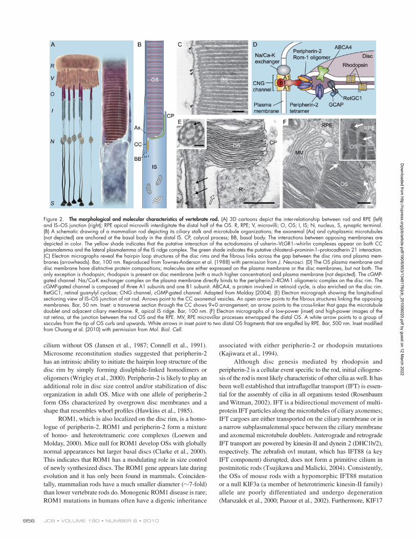
The vertebrate rod: elegance and efficiencyRods have evolved a unique structure to detect and process light with high sensitivity and efficiency; human rods can detect sin-gle photons (Hecht et al., 1942; Baylor et al., 1979). Each rod contains four morphologically distinguishable compartments: the OS, inner segment (IS), nucleus, and axon/synaptic terminal (Fig. 2 A). The length of the rod OS ranges from 30 to 60 µm in length (and 1.4–10 µm in diameter), depending on the spe-cies. Basically, the rod OS is a cylindrically shaped membrane sac filled with 1,000 flattened, lamellar-shaped membrane discs that are orderly arrayed perpendicular to the axis of the OS (Sung and Tai, 2000; Molday, 2004). These discs appear to be floating freely, although filamentous structures bridging adja-cent discs and disc rims to the nearby plasma membrane do exist (Fig. 2, C and D; Roof and Heuser, 1982; Molday, 2004). The visual pigment of the rod, rhodopsin, comprises 95% of the total amount of disc protein; it is densely packed within the
Electron micrographs depicting cone OSs containing closed discs have also been observed (De Robertis and Lasansky, 1958; Dowling, 1965; Cohen, 1970). Three cone pigments with maximal absorption for different wavelengths of light are packed into the OS disc membranes of different types of cone cells.
Rods mediate vision in dim light. Unlike other retinal cells, there is only one kind of rod photoreceptor, and it is the predominant cell type in the retina. Humans have 130 million photoreceptors, 5 million bipolar cells, and 1 million gan-glion cells. Rods outnumber cones by 20-fold, and are dis-tributed throughout the retina with the exception of the fovea region. More is known about rod photoreceptors than any other type of retinal cell. The signal transduction, visual cycle, and electrophysiology of the rod have all been studied comprehen-sively (Box 1). The unusual structure of rod photoreceptor cells has also become a major source of interest in the field of cell bi-ology as early as half a century ago, though limited accessibility in the past to these cells in vivo has made the study of these photoreceptor cells quite difficult. Genetic and mouse model studies from the past two decades have pinpointed the death of rod cells as the root cause of several inherited human retinal degeneration diseases, such as retinitis pigmentosa. Retinitis pigmentosa is a common form of progressive rod-predominant dystrophy that occurs in 1 in every 4,000 people. Patients with retinitis pigmentosa have impaired night vision and peripheral vision that precede the loss of central vision. Many causative genes for retinitis pigmentosa encode key players in phototransduction
Figure 1. The visual sense organ. (A) Diagrams of the eye; an enlarged diagram of the fovea is shown in the box. Retina forms the inner lining of the most of the posterior part of the eye. The RPE is sandwiched between the retina and choroids, a vascularized and pigmented connective tissue. (B) Diagram of the organization of retinal cells. R, rod; C, cone; B, bipolar cell; H, horizontal cell; A, amacrine cell; G, ganglion cells; M, Müller cell. (C) An H&E-stained transverse section of human retina. Retina has laminated layers. The nuclei of the photoreceptors constitute the outer nuclear layer (ONL). The nuclei of the bipolar cells, amacrine cells, horizontal cells, and Müller glial cells are found in the inner nuclear layer (INL), and the nuclei of ganglion cells form the gan-glion cell layer (GCL). The outer plexiform layer (OPL) contains the processes and synaptic terminals of photoreceptors, horizontal cells, and bipolar cells. The inner plexiform layer (IPL) contains the processes and terminals of bipolar cells, amacrine cells, and ganglion cells. The processes of Müller glial cells fill all space in the retina that is not occupied by neurons and blood vessels. Reproduced from Swaroop and Zack (2002), published by BioMed Central.
Dow
nloaded from http://rupress.org/jcb/article-pdf/190/6/953/1348176/jcb_201006020.pdf by guest on 12 M
arch 2022
955The cell biology of vision • Sung and Chuang
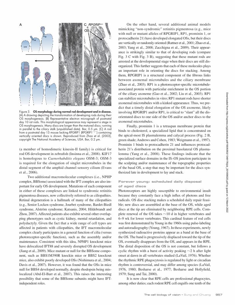
development until near the end. It begins with the extension of a primitive cilium from the basal body anchored on the plasma membrane (Fig. 3 A). This rudimentary cilium has the morpho-logical appearance of a typical primary cilium. The apical end of a primitive cilium gradually becomes swollen and filled with a variety of membranous vesicles, tubules, and sacs. At postnatal days 8–10, disc-like membranous cisternae begin to fill the developing OS; however, these discs are often excessively long and highly disorganized (Fig. 3 B; De Robertis, 1956, 1960; Besharse et al., 1985). Many of them are aligned parallel to or at an oblique angle relative to the ciliary stalk. The next phase of OS differentiation involves a major remodeling that reorganizes the discs to align perpendicular to the ciliary stalk, making them stackable. The terminal phase of rod differentiation is the elon-gation of the OS; the lengths of OSs containing orderly disc stacks increase at an almost linear rate during this period until the OSs reach their mature size (LaVail, 1973).
Somewhat unexpectedly, rhodopsin has an indispensable role in disc formation in addition to its well-known role in signal transduction. Rods of rhodopsin knockout mice form primitive cilia without disc-containing OSs (Jansen et al., 1987; Connell et al., 1991; Lee et al., 2006). Mice lacking one allele of rhodopsin develop OSs that adopt a relatively normal appearance, but have lower levels of rhodopsin. Furthermore, transgenic mice that overexpress rhodopsin develop OSs with increased diameters (Wen et al., 2009).
Peripherin-2 (also called peripherin/rds) is a photoreceptor- specific tetraspan membrane protein confined to the rim region of the rod discs, which has a characteristic hairpin loop appear-ance (Fig. 2, C and D; Molday et al., 1987, 2004). Like rhodop-sin null mice, peripherin-2 null (or rds/rds) mice form a bare
disc lamellae (i.e., 25,000 molecules/µm2). The high density of rhodopsin, together with its ordered alignment with respect to the light path, increases the probability of capturing an inci-dent photon.
Viewed in its longitudinal section, the asymmetric appear-ance of the OS resembles a hair comb (Fig. 2 B). The “spine” of the comb is the ciliary stalk, which contains axonemal micro-tubules that extend distally to about half the length of the OS. The axonemal microtubules are anchored at the basal body located at the distal end of the IS, and traverse through the connecting cilium (CC). The CC is a narrow stalk (0.3–1.2 µm in length; 0.3 µm in diameter) that acts as the single physical bridge link-ing the IS and the OS. The rod axoneme contains nine outer microtubule doublets and lacks the central pair of microtubules seen in motile cilia (i.e., 9+0; Fig. 2 E, inset). There are fibrous structures cross-linking each microtubule doublet to the overly-ing plasma membrane, and rows of intramembrane particles in the proximal CC (i.e., ciliary necklace); these ciliary structures are speculated to serve as diffusion barriers (for review see Sung and Tai, 2000). The distal OS axoneme primarily contains micro-tubule singlets and no fibrous cross-linker. In terms of both morphology and molecular composition, the CC shares similar features with primary cilia. Primary cilia are hair-like cell pro-trusions present on most cells in vertebrate organisms, and are typically 3–6 µm in length. These cilia sense extracellular sig-nals by facing a fluid-filled luminal cavity (Rosenbaum and Witman, 2002).
The IS is where metabolism, biosynthesis, and endocytosis take place. The photoreceptors have a very high energy demand and contain abundant mitochondria. Several specialized sub-domains of the IS have recently received much attention. The api-cal portion of the IS forms a ridge-like structure that surrounds the circumference of the CC (Fig. 2 E). The apical surface of the IS ridge is closely apposed to the basal OS surface (Fig. 2, B and E). In addition, prominent microvilli extending from the apex of the IS ridge surround the proximal OS. Resembling the calyx of a flower, these protrusions are named calycal processes (Fig. 2, B and E). The lateral surface of the IS ridge (or periciliary plasma membrane) is closely apposed to the CC, forming a groove-like structure (Fig. 2, B and E). Supramolecular complexes, which are made up of scaffolding and/or adhesion molecules (e.g., usherin, VLGR1, whirlin, SANS), are enriched in the specialized (sub)plasmalemma of the lateral IS ridge and/or CC (Fig. 2 B, yellow; Maerker et al., 2008). It has been proposed that the long ectodomains of the adhesion molecules, usherin and VLGR1, tether and form fibrous structures between the apposing mem-branes of these subdomains. These fibrous links are analogous to the ankle links between adjacent stereocilia in developing hair cells. Consistent with this idea, mutations in each of these proteins lead to Usher syndrome, a human deaf-blind disease (Kremer et al., 2006; Williams, 2008).
Seeing is believing: OS morphogenesis in developing rodsRod differentiation in rodents starts postnatally, and it takes 2–3 wk for the OS to fully mature (LaVail, 1973). OS morpho-genesis among the rods is not completely synchronized throughout
Box 1
Phototransduction. Phototransduction occurs in the outer segments (OS) of photoreceptors. Light initiates the isomerization of the 11-cis retinal, a chromophore covalently conjugated to rhodopsin embedded within the disc membranes. Photoactivated rhodopsin activates the heterotrimeric G protein transducin by catalyzing the exchange of GDP for GTP. The dissociated -subunit of transducin then activates cGMP-phosphodiesterase, which rapidly hydrolyzes cytoplasmic cGMP. The decrease in cGMP concentration causes closure of cGMP-gated cation channels on the plasma membrane (Fig. 2 D). As a result, the cells are hyperpolarized and release less glutamate transmitters to their connected bipolar cells.
In the recovery phase, photoactivated rhodopsin is desensitized by phosphorylation and arrestin binding. Retinal guanylyl cyclase and guanylyl cyclase activator help to replenish cGMP levels in a Ca2+-dependent manner. Na/Ca-K exchanger, which resides on the plasma membrane, regulates the Ca2+ homeostasis of the OS (Molday, 1998). All these components are highly integrated in close proximity on the disc membranes and/or adjacent plasma membranes for fast processing (Fig. 2 D).
Visual cycle. Photoisomerization converts 11-cis retinal to all trans-retinal, and finally to all-trans-retinol in the OS. All-trans-retinol leaves rods and “diffuses” to the RPE, where it undergoes a series of enzymatic reactions to be converted back to 11-cis-retinal. 11-cis-retinal returns back to the OS where it regenerates rhodopsin and completes the visual cycle (Lamb and Pugh, 2004). Considerable progress has been made in characterizing all the major components of this visual cycle; however, the cell biology of retinoid trafficking remains largely unknown.
Dow
nloaded from http://rupress.org/jcb/article-pdf/190/6/953/1348176/jcb_201006020.pdf by guest on 12 M
arch 2022
JCB • VOLUME 190 • NUMBER 6 • 2010 956
associated with either peripherin-2 or rhodopsin mutations (Kajiwara et al., 1994).
Although disc genesis mediated by rhodopsin and peripherin-2 is a cellular event specific to the rod, initial ciliogene-sis of the rod is most likely characteristic of other cilia as well. It has been well established that intraflagellar transport (IFT) is essen-tial for the assembly of cilia in all organisms tested (Rosenbaum and Witman, 2002). IFT is a bidirectional movement of multi-protein IFT particles along the microtubules of ciliary axonemes; IFT cargoes are either transported on the ciliary membrane or in a narrow subplasmalemmal space between the ciliary membrane and axonemal microtubule doublets. Anterograde and retrograde IFT transport are powered by kinesin-II and dynein 2 (DHC1b/2), respectively. The zebrafish ovl mutant, which has IFT88 (a key IFT component) disrupted, does not form a primitive cilium in postmitotic rods (Tsujikawa and Malicki, 2004). Consistently, the OSs of mouse rods with a hypomorphic IFT88 mutation or a null KIF3a (a member of heterotrimeric kinesin-II family) allele are poorly differentiated and undergo degeneration (Marszalek et al., 2000; Pazour et al., 2002). Furthermore, KIF17
cilium without OS (Jansen et al., 1987; Connell et al., 1991). Microsome reconstitution studies suggested that peripherin-2 has an intrinsic ability to initiate the hairpin loop structure of the disc rim by simply forming disulphide-linked homodimers or oligomers (Wrigley et al., 2000). Peripherin-2 is likely to play an additional role in disc size control and/or stabilization of disc organization in adult OS. Mice with one allele of peripherin-2 form OSs characterized by overgrown disc membranes and a shape that resembles whorl profiles (Hawkins et al., 1985).
ROM1, which is also localized on the disc rim, is a homo-logue of peripherin-2. ROM1 and peripherin-2 form a mixture of homo- and heterotetrameric core complexes (Loewen and Molday, 2000). Mice null for ROM1 develop OSs with globally normal appearances but larger basal discs (Clarke et al., 2000). This indicates that ROM1 has a modulating role in size control of newly synthesized discs. The ROM1 gene appears late during evolution and it has only been found in mammals. Coinciden-tally, mammalian rods have a much smaller diameter (7-fold) than lower vertebrate rods do. Monogenic ROM1 disease is rare; ROM1 mutations in humans often have a digenic inheritance
Figure 2. The morphological and molecular characteristics of vertebrate rod. (A) 3D cartoons depict the inter-relationship between rod and RPE (left) and IS–OS junction (right); RPE apical microvilli interdigitate the distal half of the OS. R, RPE; V, microvilli; O, OS; I, IS; N, nucleus, S, synaptic terminal. (B) A schematic drawing of a mammalian rod depicting its ciliary stalk and microtubule organizations; the axonemal (Ax) and cytoplasmic microtubules (not depicted) are anchored at the basal body in the distal IS. CP, calycal process; BB, basal body. The interactions between opposing membranes are depicted in color. The yellow shade indicates that the putative interaction of the ectodomains of usherin–VLGR1–whirlin complexes appear on both CC plasmalemma and the lateral plasmalemma of the IS ridge complex. The green shade indicates the putative chlosterol–prominin-1–protocadherin 21 interaction. (C) Electron micrographs reveal the hairpin loop structures of the disc rims and the fibrous links across the gap between the disc rims and plasma mem-branes (arrowheads). Bar, 100 nm. Reproduced from Townes-Anderson et al. (1988) with permission from J. Neurosci. (D) The OS plasma membrane and disc membrane have distinctive protein compositions; molecules are either expressed on the plasma membrane or the disc membranes, but not both. The only exception is rhodopsin; rhodopsin is present on disc membrane (with a much higher concentration) and plasma membrane (not depicted). The cGMP-gated channel: Na/Ca-K exchanger complex on the plasma membrane directly binds to the peripherin-2–ROM-1 oligomeric complex on the disc rim. The cGMP-gated channel is composed of three A1 subunits and one B1 subunit. ABCA4, a protein involved in retinoid cycle, is also enriched on the disc rim. RetGC1, retinal guanylyl cyclase; CNG channel, cGMP-gated channel. Adapted from Molday (2004). (E) Electron micrograph showing the longitudinal sectioning view of IS–OS junction of rat rod. Arrows point to the CC axonemal vesicles. An open arrow points to the fibrous structures linking the opposing membranes. Bar, 50 nm. Inset: a transverse section through the CC shows 9+0 arrangement; an arrow points to the cross-linker that gaps the microtubule doublet and adjacent ciliary membrane. R, apical IS ridge. Bar, 100 nm. (F) Electron micrographs of a low-power (inset) and high-power images of the rat retina, at the junction between the rod OS and the RPE. MV, RPE microvillar processes enwrapped the distal OS. A white arrow points to a group of saccules from the tip of OS curls and upwards. White arrows in inset point to two distal OS fragments that are engulfed by RPE. Bar, 500 nm. Inset modified from Chuang et al. (2010) with permission from Mol. Biol. Cell.
Dow
nloaded from http://rupress.org/jcb/article-pdf/190/6/953/1348176/jcb_201006020.pdf by guest on 12 M
arch 2022
957The cell biology of vision • Sung and Chuang
On the other hand, several additional animal models mimicking “non-syndromic” retinitis pigmentosa (e.g., mice with null or mutant alleles of RPGRIP1, RP1, prominin 1, or protocadherin 21) have developed elongated OSs, but their discs are vertically or randomly oriented (Rattner et al., 2001; Zhao et al., 2003; Yang et al., 2008; Zacchigna et al., 2009). Their appear-ance is strikingly similar to that of developing rods (compare Fig. 3 C with Fig. 3 B), suggesting that these mutant rods are arrested at the developmental stage when their discs are still dis-organized. This further suggests that each of these molecules plays an important role in orienting the discs for stacking. Among them, RPGRIP1 is a structural component of the fibrous links between axonemal microtubules and the ciliary membrane (Zhao et al., 2003). RP1 is a photoreceptor-specific microtubule-associated protein with particular enrichment in the OS portion of the ciliary axoneme (Gao et al., 2002; Liu et al., 2003). RP1 can stabilize microtubules in vitro; RP1 mutant rods have shorter axonemal microtubules with a kinked appearance. Thus, we pre-dict that a timely distal elongation of the OS axoneme, likely involving RPGRIP1 and/or RP1, is critical to “slant” all the dis-orientated discs to one side of the OS and/or anchor discs to the axonemal microtubules.
Finally, prominin 1 is a tetraspan membrane protein that binds to cholesterol, a specialized lipid that is concentrated on the apical-most IS plasmalemma and calycal process (Fig. 2 B, green shade; Andrews and Cohen, 1983; Weigmann et al., 1997). Prominin 1 binds to protocadherin 21 and influences protocad-herin 21’s distribution on the proximal basolateral OS plasma-lemma (Yang et al., 2008). These findings indicate that the specialized surface domains in the IS–OS junction participate in the sculpting and/or maintenance of the topographic properties of the basal OS, a step that may be important for the discs syn-thesized late in development to lay and stack.
Forever young: scheduled daily disposal of aged discsPhotoreceptors are highly susceptible to environmental insult because they constantly face a high influx of photons and free radicals. OS disc stacking makes a scheduled daily repair feasi-ble; new discs are assembled at the base of the OS, while aged discs at the tip are eliminated by neighboring RPE cells. Com-plete renewal of the OS takes 10 d in higher vertebrates and 6–9 wk for lower vertebrates. This cardinal feature of rod cells was first demonstrated by Young in the 1960s using pulse-labeling and autoradiography (Young, 1967). In these experiments, newly synthesized radioactive proteins appear as a band at the base of the OS. The band is progressively displaced toward the tip of the OS, eventually disappears from the OS, and appears in the RPE. The distal disposition of the OS is not constant, but follows a cyclic rhythm with a burst of activity peaking 2 h after light onset at dawn in all vertebrates studied (LaVail, 1976). Whether the rhythmic RPE phagocytosis is regulated by light or circadian rhythm is controversial, and may differ among species (LaVail, 1976, 1980; Besharse et al., 1977; Besharse and Hollyfield, 1979; Sung and Tai, 2000).
It is now clear that RPE cells are professional phagocytes, among other duties; each rodent RPE cell engulfs one tenth of the
(a member of homodimeric kinesin-II family) is critical for rod OS development in zebrafish (Insinna et al., 2008). KIF17 is homologous to Caenorhabditis elegans OSM-3; OSM-3 is required for the elongation of singlet microtubules in the distal segment of the amphid channel sensory cilium (Evans et al., 2006).
Two additional macromolecular complexes (i.e., NPHP complex, BBSome) associated with the IFT complex are also im-portant for early OS development. Mutations of each component in either of these complexes are linked to syndromic retinitis pigmentosa diseases, now collectively referred to as ciliopathies. Retinal degeneration is a hallmark of many of the ciliopathies (e.g., Senior Locken syndrome, Jourbet syndrome, Bardet-Biedl syndrome, Alström syndrome; Katsanis, 2004; Hildebrandt and Zhou, 2007). Affected patients also exhibit several other overlap-ping phenotypes such as cystic kidney, mental retardation, and polydactyly. Given the fact that a wide range of cell types are affected in patients with ciliopathies, the IFT macromolecular complex clearly participates in a general function of cilia (versus photoreceptor-specific function), such as the assembly and/or maintenance. Consistent with this idea, NPHP1 knockout mice have delocalized IFT88 and severely disrupted OS development (Jiang et al., 2009). Mice mutant or null for the BBSome compo-nent, such as BBS1M390R knockin mice or BBS2 knockout mice, also exhibit poorly developed OSs (Nishimura et al., 2004; Davis et al., 2007). However, it was found that the OSs in mice null for BBS4 developed normally, despite rhodopsin being mis-localized (Abd-El-Barr et al., 2007). This raises the interesting possibility that some of the BBSome subunits might have IFT- independent roles.
Figure 3. OS morphology during normal rod development and in disease. (A) A drawing depicting the transformation of developing rods during their OS morphogenesis. (B) Representative electron micrograph of postnatal day 10 rat rods. This morphological appearance may represent a stage in OS morphogenesis. Many discs are longer than the matured discs; running in parallel to the ciliary stalk (unpublished data). Bar, 0.5 µm. (C) A rod from a postnatal day 15 mouse lacking RPGRIP1 (RPGRIP1/) containing vertically oriented discs is shown. Reproduced from Zhao et al. (2003), copyright The National Academy of Sciences, USA. Bar, 0.2 µm.
Dow
nloaded from http://rupress.org/jcb/article-pdf/190/6/953/1348176/jcb_201006020.pdf by guest on 12 M
arch 2022
JCB • VOLUME 190 • NUMBER 6 • 2010 958
targeting assay, and OS renewal in amphibians and mammals have resulted in inconsistent conclusions (Sung and Tai, 2000; Ding and Naash, 2006; Chuang et al., 2007; Baker et al., 2008; Kwok et al., 2008), indicating that the morphological differences between these dissimilar species might play an important role in their divergent strategies to adapt to their respective cellular physiologies and habitats.
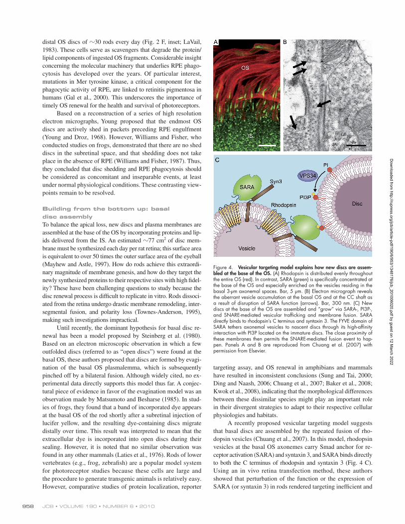
A recently proposed vesicular targeting model suggests that basal discs are assembled by the repeated fusion of rho-dopsin vesicles (Chuang et al., 2007). In this model, rhodopsin vesicles at the basal OS axonemes carry Smad anchor for re-ceptor activation (SARA) and syntaxin 3, and SARA binds directly to both the C terminus of rhodopsin and syntaxin 3 (Fig. 4 C).Using an in vivo retina transfection method, these authors showed that perturbation of the function or the expression of SARA (or syntaxin 3) in rods rendered targeting inefficient and
distal OS discs of 30 rods every day (Fig. 2 F, inset; LaVail, 1983). These cells serve as scavengers that degrade the protein/lipid components of ingested OS fragments. Considerable insight concerning the molecular machinery that underlies RPE phago-cytosis has developed over the years. Of particular interest, mutations in Mer tyrosine kinase, a critical component for the phagocytic activity of RPE, are linked to retinitis pigmentosa in humans (Gal et al., 2000). This underscores the importance of timely OS renewal for the health and survival of photoreceptors.
Based on a reconstruction of a series of high resolution electron micrographs, Young proposed that the endmost OS discs are actively shed in packets preceding RPE engulfment (Young and Droz, 1968). However, Williams and Fisher, who conducted studies on frogs, demonstrated that there are no shed discs in the subretinal space, and that shedding does not take place in the absence of RPE (Williams and Fisher, 1987). Thus, they concluded that disc shedding and RPE phagocytosis should be considered as concomitant and inseparable events, at least under normal physiological conditions. These contrasting view-points remain to be resolved.
Building from the bottom up: basal disc assemblyTo balance the apical loss, new discs and plasma membranes are assembled at the base of the OS by incorporating proteins and lip-ids delivered from the IS. An estimated 77 cm2 of disc mem-brane must be synthesized each day per rat retina; this surface area is equivalent to over 50 times the outer surface area of the eyeball (Mayhew and Astle, 1997). How do rods achieve this extraordi-nary magnitude of membrane genesis, and how do they target the newly synthesized proteins to their respective sites with high fidel-ity? These have been challenging questions to study because the disc renewal process is difficult to replicate in vitro. Rods dissoci-ated from the retina undergo drastic membrane remodeling, inter-segmental fusion, and polarity loss (Townes-Anderson, 1995), making such investigations impractical.
Until recently, the dominant hypothesis for basal disc re-newal has been a model proposed by Steinberg et al. (1980). Based on an electron microscopic observation in which a few outfolded discs (referred to as “open discs”) were found at the basal OS, these authors proposed that discs are formed by evagi-nation of the basal OS plasmalemma, which is subsequently pinched off by a bilateral fusion. Although widely cited, no ex-perimental data directly supports this model thus far. A conjec-tural piece of evidence in favor of the evagination model was an observation made by Matsumoto and Besharse (1985). In stud-ies of frogs, they found that a band of incorporated dye appears at the basal OS of the rod shortly after a subretinal injection of lucifer yellow, and the resulting dye-containing discs migrate distally over time. This result was interpreted to mean that the extracellular dye is incorporated into open discs during their sealing. However, it is noted that no similar observation was found in any other mammals (Laties et al., 1976). Rods of lower vertebrates (e.g., frog, zebrafish) are a popular model system for photoreceptor studies because these cells are large and the procedure to generate transgenic animals is relatively easy. However, comparative studies of protein localization, reporter
Figure 4. Vesicular targeting model explains how new discs are assem-bled at the base of the OS. (A) Rhodopsin is distributed evenly throughout the entire OS (red). In contrast, SARA (green) is specifically concentrated at the base of the OS and especially enriched on the vesicles residing in the basal 3-µm axonemal spaces. Bar, 5 µm. (B) Electron micrograph reveals the aberrant vesicle accumulation at the basal OS and at the CC shaft as a result of disruption of SARA function (arrows). Bar, 300 nm. (C) New discs at the base of the OS are assembled and “grow” via SARA-, PI3P-, and SNARE-mediated vesicular trafficking and membrane fusion. SARA directly binds to rhodopsin’s C terminus and syntaxin 3. The FYVE domain of SARA tethers axonemal vesicles to nascent discs through its high-affinity interaction with PI3P located on the immature discs. The close proximity of these membranes then permits the SNARE-mediated fusion event to hap-pen. Panels A and B are reproduced from Chuang et al. (2007) with permission from Elsevier.
Dow
nloaded from http://rupress.org/jcb/article-pdf/190/6/953/1348176/jcb_201006020.pdf by guest on 12 M
arch 2022
959The cell biology of vision • Sung and Chuang
distance between the trans-Golgi network and the distal IS is quite far; rods with cytoplasmic dynein (DHC1) suppressed had aberrant post-Golgi vesicle accumulation in the IS (Insinna et al., 2010). Several C-terminal retinitis pigmentosa mutant rhodopsins display a reduced affinity for Tctex-1, which may explain their mislocalization in rods (Tai et al., 1999).
Besides rhodopsin, photoreceptor retinol dehydrogenase also uses a (V/I)XPX motif for its OS targeting (Luo et al., 2004). No other major OS proteins are known to contain a similar motif, suggesting that a variety of OS targeting signals and/or strategies must be operating in rods. For example, the piggyback/hitch-hiking model is likely to be a common strategy for OS trafficking by other molecules simply interacting with rhodopsin or rhodopsin- bearing vesicles, which are abundant in rods (Concepcion et al., 2002). A selective retention strategy is also used to trap mol-ecules via high affinity protein–protein (or protein–lipid) inter-actions. As an example, photoactivated rhodopsin snatches
led to aberrant accumulation of vesicles at the base of the OS, the axonemal shaft, and the CC (Fig. 4 B). These results suggest the interaction between the FYVE domain of SARA (on axo-nemal vesicles) and phosphatidylinositol 3-phosphate (PI3P; on basal nascent discs) primes the axonemal vesicles to discs for SNARE-mediated fusion (Fig. 4 C). These findings also provide convincing pieces of evidence that the precursors of the discs are membranous vesicles.
The vesicular targeting model suggests that the disc incor-poration of rhodopsin and the formation of nascent discs occur concomitantly. SARA (Fig. 4 A), syntaxin3, and PI3P all dis-play a gradient of expression along the proximal 3–8-µm OS, with the strongest signal toward the base. Spatial restriction of these proteins could function as a programmed timer that deter-mines both the proper size and the number of new discs to be generated per day.
High resolution EM results provided by Chuang et al. (2007) also showed that the basal OS plasma membrane en-wraps the entire set of disc stacks, with no “open discs” ob-served (Fig. 2 E; Fig. 4 B). A similar conclusion can be drawn by using cryofixation techniques, namely, rapid freezing or high pressure freezing combined with freeze substitution (Obata and Usukura, 1992). Thus, the open disc–like structures are con-ceivable artifacts due to the histological damage of the basal OS plasmalemma.
Getting to the top: targeting of rhodopsin and other proteins to the OSRhodopsin has an autonomous OS targeting signal located within its cytoplasmic C-terminal tail. The VXPX motif within this targeting signal is critical. This was first shown by the ob-servation that the transgenically expressed retinitis pigmentosa rhodopsin mutant, which lacks the last five residues of the pro-tein, loses its polarized OS distribution (Sung et al., 1994). The C-terminal region of rhodopsin is highly conserved among species, and is a hot spot for human retinitis pigmentosa muta-tions (Sung and Tai, 2000). The sufficiency of rhodopsin’s C terminus for OS targeting was later demonstrated in transgenic frogs and zebrafish using a reporter assay (Tam et al., 2000; Perkins et al., 2002).
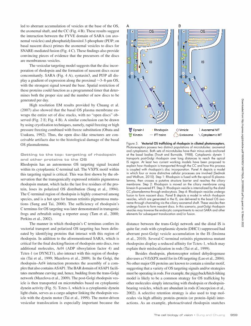
The manner in which rhodopsin’s C terminus confers its vectorial transport and polarized OS targeting has been delin-eated by identifying proteins that interact with this region of rhodopsin. In addition to the aforementioned SARA, which is critical for the final docking/fusion of rhodopsin onto discs, two additional molecules, Arf4 (ADP ribosylation factor 4) and Tctex-1 (or DYNLT1), also interact with this region of rhodop-sin (Tai et al., 1999; Mazelova et al., 2009). In the Golgi, the rhodopsin–Arf4 interaction promotes the assembly of a com-plex that also contains ASAP1. The BAR domain of ASAP1 facili-tates membrane curving and, hence, budding from the trans-Golgi network (Mazelova et al., 2009). The post-Golgi rhodopsin ves-icle is then transported on microtubules based on cytoplasmic dynein activity (Fig. 5). Tctex-1, which is a cytoplasmic dynein light chain, serves as a cargo adaptor linking the rhodopsin ves-icle with the dynein motor (Tai et al., 1999). The motor-driven vesicular translocation is especially important because the
Figure 5. Vectorial OS trafficking of rhodopsin in ciliated photoreceptors. Photoreceptors possess two distinct populations of microtubules: axonemal and cytoplasmic. Both sets of microtubules have their minus ends anchored at the basal bodies (Troutt and Burnside, 1988). Cytoplasmic dynein 1 transports post-Golgi rhodopsin over long distances to reach the apical IS region. At least two current working models have been proposed to explain how rhodopsin is transported through the CC and how this process is coupled with rhodopsin’s disc incorporation. Panel A depicts a model in which four or more distinctive cellular processes are involved (Sedmak and Wolfrum, 2010). Step 1: Rhodopsin is fused with the apical IS plasma-lemma, then crosses a putative structure barrier and reaches the ciliary membrane. Step 2: Rhodopsin is moved on the ciliary membrane using kinesin II–powered IFT. Step 3: Rhodopsin vesicle is internalized by the distal CC plasmalemma through endocytosis. Step 4: Rhodopsin vesicles undergo fusion to form nascent discs. Panel B depicts a model in which rhodopsin vesicles, which are generated in the IS, are delivered to the basal OS axo-neme through channeling via the ciliary axonemal shaft. These vesicles then undergo fusion to form nascent discs. In this scenario, post-Golgi rhodopsin vesicles may traverse the endocytic compartments to recruit SARA and other elements for subsequent translocation and/or fusion.
Dow
nloaded from http://rupress.org/jcb/article-pdf/190/6/953/1348176/jcb_201006020.pdf by guest on 12 M
arch 2022
JCB • VOLUME 190 • NUMBER 6 • 2010 960
To the best of our knowledge, rhodopsin has not been shown to be a cargo of IFT. In addition, there is currently no evidence supporting the idea that rhodopsin is transported through IFT. The improper rhodopsin distribution seen in the abnormally dif-ferentiated and rapidly degenerated hypomorphic IFT88 and null KIF3A rods should not be used to conclude IFT’s role in rhodop-sin transport (Marszalek et al., 2000; Pazour et al., 2002). This is because rhodopsin is not completely polarized in the OS until PN12, and rhodopsin fails to integrate efficiently once the OS is shortened and/or degenerated. A recent study using a different strain of rod-specific KIF3A knockout that has a slower degener-ation rate demonstrated that rhodopsin’s OS targeting is KIF3A independent (Avasthi et al., 2009). The same study also showed that KIF3A is not required for OS plasmalemma localization of the cGMP-gated channel. Thus, the nature of the membrane cargo(es) transported by IFT in photoreceptors remains to be identified. In this regard, a biochemical study showed that retinal guanylyl cyclase binds (indirectly) to IFT88, making it a potential IFT cargo (Bhowmick et al., 2009).
Assuming that rhodopsin is transported on the ciliary mem-brane, a mechanism must exist to pinch off the membrane to gen-erate discs. In addition to the evagination model discussed before, the pinching off of internalized ciliary membrane has also been proposed (Fig. 5 A; Obata and Usukura, 1992; Usukura and Obata, 1995). Evidence for the latter is limited to studying fluid phase marker uptake performed in dissociated immature rods.
An attractive alternative model is that rhodopsin vesicles are shipped directly from the IS through the axonemal shaft (Fig. 5 B). The SARA+syntaxin3+ axonemal vesicles then un-dergo fusion at the base of the OS to produce new discs, as sug-gested by the vesicular targeting model (Fig. 4 C). This model is appealing because rods may have evolved from other ciliated cells to develop a means to effectively move cargo in bulk and quickly generate disc membranes. The presence of small vesi-cles in CC (Fig. 2 E; Dowling, 1967; Chuang et al., 2007), but not other types of cilia, supports this model.
No IFT retrograde transport is needed to return the OS trans-membrane proteins back to the cell body for degradation. How-ever, three major signaling components (i.e., arrestin, transducin, and recoverin), which are all peripheral membrane proteins, shut-tle back and forth between the IS and OS in a light-dependent manner. This is one remarkable mechanism that is unique to photo-receptors, and is used for light adaptation (Strissel et al., 2004). A parallel light-dependent translocation is also conserved in fly photoreceptors, in which the light-sensing organelles are actin-based microvilli called rhabdomeres. Light-dependent protein transport in photoreceptors has been comprehensively reviewed elsewhere (Strissel et al., 2004).
Our vision: concluding remarksCompared with other types of polarized cells, the high spatial and temporal resolution of photoreceptors has much to offer for those who are interested in cell polarity, vesicular trafficking, and organ-elle biogenesis. Many discoveries and new working models de-scribed in this review provide a starting point for advancement. There is much more to be learned about protein transport and turn-over in cone photoreceptors, which are also rich in membranes.
arrestin from its IS localization upon illumination (Strissel et al., 2004). It was also shown that the OS plasmalemma loca-tion of the cGMP-gated channel relies on its direct binding with a membrane skeletal protein ankryin-G (Kizhatil et al., 2009). It is plausible that many of these mechanisms occur redundantly and/or successively to ensure the fidelity of the targeting of these molecules to their respective OS membrane domains.
Finally, a strategy completely independent of rhodopsin’s targeting must be used for the disc rim targeting of peripherin-2 because no mislocalized peripherin-2 has been found in any mouse model in which rhodopsin is mislocalized (Hagstrom et al., 2001; Zhao et al., 2003; Abd-El-Barr et al., 2007). It was sug-gested that a “checkpoint” system selectively permits peripherin-2 prepacked as homotetramers or even higher oligomers, but not dimers, into the OS (Loewen et al., 2003). The ability of peripherin-2 to further oligomerize with ROM-1 and to interact with the cGMP-gated channel adjacent to the OS may further promote and/or stabilize its disc rim distribution (Fig. 2 D).
Crossing the narrow bridge: ciliary transport of rhodopsinOne of the most pressing questions pertinent to rhodopsin traffick-ing is how rhodopsin passes through the bottleneck-shaped CC. Rhodopsin immunoreactivity has rarely been detected anywhere on the CC. This might be a consequence of several technical limita-tions, such as the high concentration of rhodopsin in the OS or poor penetration of reagents into the CC, among other possibilities (Besharse and Horst, 1990; Liu et al., 1999; Wolfrum and Schmitt, 2000). The presence of rhodopsin on the ciliary membrane has, however, been detected when a heavy-etching technique and a mixture of anti-rhodopsin antibodies were used in combination (Wolfrum and Schmitt, 2000). A study of shaker1 mice, in which the myosin VIIa gene is disrupted, found an increased rhodopsin signal on the CC membrane (Liu et al., 1999; Wolfrum and Schmitt, 2000). The importance and direct nature of myosin VIIa on the CC’s transport of rhodopsin remain unclear because rhodopsin was not detectably mislocalized in the IS and other parts of the rods. Furthermore, CC is largely devoid of microfilaments, although G-actin immunoreactivity is detected (Wolfrum and Schmitt, 2000). Instead, the actin meshwork of rods is prominent at both the proximal and distal ends of the CC (Sung and Tai, 2000).
Many reviews have suggested that rhodopsin at the apical IS plasmalemma is able to laterally diffuse and then traverse through the ciliary membrane by using kinesin-motored IFT anterograde transport (Fig. 5 A). This proposition is analogous to a working model for the transmembrane protein that travels on primary cilia (Rosenbaum and Witman, 2002). Rab8 GTPase has a proposed role in docking/fusing cargoes near the base of primary cilia to re-plenish membrane materials for cilia elongation (Nachury et al., 2007; Yoshimura et al., 2007). In agreement with this notion, trans-genic frog rods that overexpress dominant-negative Rab8 have ab-errant vesicular accumulation at the base of the CC (Moritz et al., 2001). Paradoxically, rhodopsin cannot be efficiently targeted to the primary cilia in epithelial cultures (Chuang and Sung, 1998). Rhodopsin is actually largely absent from the base of the primary cilium, a specialized lipid zone that prevents free diffusion between the apical surface and cilium (Vieira et al., 2005).
Dow
nloaded from http://rupress.org/jcb/article-pdf/190/6/953/1348176/jcb_201006020.pdf by guest on 12 M
arch 2022
961The cell biology of vision • Sung and Chuang
Besharse, J.C., J.G. Hollyfield, and M.E. Rayborn. 1977. Photoreceptor outer segments: accelerated membrane renewal in rods after exposure to light. Science. 196:536–538. doi:10.1126/science.300504
Besharse, J.C., D.M. Forestner, and D.M. Defoe. 1985. Membrane assembly in retinal photoreceptors. III. Distinct membrane domains of the connecting cilium of developing rods. J. Neurosci. 5:1035–1048.
Bhowmick, R., M. Li, J. Sun, S.A. Baker, C. Insinna, and J.C. Besharse. 2009. Photoreceptor IFT complexes containing chaperones, guanylyl cyclase 1 and rhodopsin. Traffic. 10:648–663. doi:10.1111/j.1600-0854.2009 .00896.x
Burnside, B. 1976. Microtubules and actin filaments in teleost visual cone elon-gation and contraction. J. Supramol. Struct. 5:257–275. doi:10.1002/jss .400050302
Chuang, J.-Z., and C.-H. Sung. 1998. The cytoplasmic tail of rhodopsin acts as a novel apical sorting signal in polarized MDCK cells. J. Cell Biol. 142:1245–1256. doi:10.1083/jcb.142.5.1245
Chuang, J.Z., Y. Zhao, and C.H. Sung. 2007. SARA-regulated vesicular target-ing underlies formation of the light-sensing organelle in mammalian rods. Cell. 130:535–547. doi:10.1016/j.cell.2007.06.030
Chuang, J.Z., S.Y. Chou, and C.H. Sung. 2010. Chloride intracellular channel 4 is critical for the epithelial morphogenesis of RPE cells and retinal attachment. Mol. Biol. Cell. 21:3017–3028. 10.1091/mbc.E09-10-0907
Clarke, G., A.F. Goldberg, D. Vidgen, L. Collins, L. Ploder, L. Schwarz, L.L. Molday, J.S. Rossant, A. Szél, R.S. Molday, et al. 2000. Rom-1 is re-quired for rod photoreceptor viability and the regulation of disk morpho-genesis. Nat. Genet. 25:67–73. doi:10.1038/75621
Cohen, A.I. 1970. Further studies on the question of the patency of saccules in outer segments of vertebrate photoreceptors. Vision Res. 10:445–453. doi:10.1016/0042-6989(70)90001-5
Concepcion, F., A. Mendez, and J. Chen. 2002. The carboxyl-terminal domain is essential for rhodopsin transport in rod photoreceptors. Vision Res. 42:417–426. doi:10.1016/S0042-6989(01)00195-X
Connell, G., R. Bascom, L. Molday, D. Reid, R.R. McInnes, and R.S. Molday. 1991. Photoreceptor peripherin is the normal product of the gene responsible for retinal degeneration in the rds mouse. Proc. Natl. Acad. Sci. USA. 88:723–726. doi:10.1073/pnas.88.3.723
Davis, R.E., R.E. Swiderski, K. Rahmouni, D.Y. Nishimura, R.F. Mullins, K. Agassandian, A.R. Philp, C.C. Searby, M.P. Andrews, S. Thompson, et al. 2007. A knockin mouse model of the Bardet-Biedl syndrome 1 M390R mu-tation has cilia defects, ventriculomegaly, retinopathy, and obesity. Proc. Natl. Acad. Sci. USA. 104:19422–19427. doi:10.1073/pnas.0708571104
De Robertis, E. 1956. Morphogenesis of the retinal rods; an electron microscope study. J. Biophys. Biochem. Cytol. 2(4, Suppl):209–218. doi:10.1083/ jcb.2.4.209
De Robertis, E. 1960. Some observations on the ultrastructure and morphogen-esis of photoreceptors. J. Gen. Physiol. 43:1–13. doi:10.1085/jgp.43.6.1
De Robertis, E., and A. Lasansky. 1958. Submicroscopic organization of retinal cones of the rabbit. J. Biophys. Biochem. Cytol. 4:743–746. doi:10.1083/ jcb.4.6.743
Ding, X.Q., and M.I. Naash. 2006. Transgenic animal studies of human retinal dis-ease caused by mutations in peripherin/rds. Adv. Exp. Med. Biol. 572:141–146. doi:10.1007/0-387-32442-9_21
Dowling, J.E. 1965. Foveal receptors of the monkey retina: fine structure. Science. 147:57–59. doi:10.1126/science.147.3653.57
Dowling, J.E. 1967. The organization of vertebrate visual receptors. In Molecular Organization and Biological Function. J.M. Allen, editor. Harper & Row, New York. 186–210.
Evans, J.E., J.J. Snow, A.L. Gunnarson, G. Ou, H. Stahlberg, K.L. McDonald, and J.M. Scholey. 2006. Functional modulation of IFT kinesins extends the sen-sory repertoire of ciliated neurons in Caenorhabditis elegans. J. Cell Biol. 172:663–669. doi:10.1083/jcb.200509115
Gal, A., Y. Li, D.A. Thompson, J. Weir, U. Orth, S.G. Jacobson, E. Apfelstedt-Sylla, and D. Vollrath. 2000. Mutations in MERTK, the human orthologue of the RCS rat retinal dystrophy gene, cause retinitis pigmentosa. Nat. Genet. 26:270–271. doi:10.1038/80002
Gao, J., K. Cheon, S. Nusinowitz, Q. Liu, D. Bei, K. Atkins, A. Azimi, S.P. Daiger, D.B. Farber, J.R. Heckenlively, et al. 2002. Progressive pho-toreceptor degeneration, outer segment dysplasia, and rhodopsin mis-localization in mice with targeted disruption of the retinitis pigmentosa-1 (Rp1) gene. Proc. Natl. Acad. Sci. USA. 99:5698–5703. doi:10.1073/ pnas.042122399
Hagstrom, S.A., M. Adamian, M. Scimeca, B.S. Pawlyk, G. Yue, and T. Li. 2001. A role for the Tubby-like protein 1 in rhodopsin transport. Invest. Ophthalmol. Vis. Sci. 42:1955–1962.
Hawkins, R.K., H.G. Jansen, and S. Sanyal. 1985. Development and degeneration of retina in rds mutant mice: photoreceptor abnormalities in the hetero-zygotes. Exp. Eye Res. 41:701–720. doi:10.1016/0014-4835(85)90179-4
The cell biology study of other neurons in the visual sys-tem is still in its infancy. The inner retinal neurons come in a striking diversity. For example, 20 and 30 types of ganglion cells and amacrine cells, respectively, have been identified based on their different shapes, molecular, and electrophysiological properties (for review see Masland, 2004). Each of these indi-vidual cell types appears to be specialized in distinct functions that are not completely understood. How are their diversified morphologies established? How do these cells develop their or-derly stratified architecture? How are the processes of neurons that are involved in visual perception wired in such a complex yet remarkably precise manner? How do retinal neurons undergo morphological adaptation in response to environmental cues (e.g., light, circadian), and what are their functional implications (Burnside, 1976; Vollrath and Spiwoks-Becker, 1996; Spiwoks-Becker et al., 2004)? The recent completion of the inventory of all retinal neuron types (Wässle, 2004) and our increasing capa-bility to target, manipulate, and visualize specific cell types in the visual system provide great promise for our deeper under-standing of the cell biology of our vision system.
We apologize to those authors whose works have not been cited due to space limitation. Recent findings are referenced; the older evidence is discussed in the reviews cited and the RetNet website. We thank Sheila Nirenberg, Jarema Malicki, Robert Molday, Enrique Rodriguez-Boulan, and Uwe Wolfrum for valuable comments on the manuscript. We thank Ann Milam and Robert Molday for the original images presented in Fig. 1 C, and Fig. 2 D, respec-tively. We thank Ms. Fannie Liu, Dr. Chang-Juang Lai, and Ms. Jocelyn Chuang for the illustrations of Fig. 1 C, Fig. 2 A, and Fig. 3 A, respectively.
This work was supported by grants from the National Institutes of Health (EY11307, EY016805, EY015656), RPB, Ruth and Melton Steinbach Macular Degeneration Foundation, Irma Hirschl Trust, Foundation Fighting Blindness, and Research to Prevent Blindness.
Other reviews in this series are: The cell biology of hearing (Schwander et al. 2010. J. Cell Biol. doi:10.1083/jcb.201001138) and The cell bi-ology of taste (Chaudhari and Roper. 2010. J. Cell Biol. doi: 10.1083/ jcb.201003144).
Submitted: 3 June 2010Accepted: 31 August 2010
ReferencesAbd-El-Barr, M.M., K. Sykoudis, S. Andrabi, E.R. Eichers, M.E. Pennesi,
P.L. Tan, J.H. Wilson, N. Katsanis, J.R. Lupski, and S.M. Wu. 2007. Impaired photoreceptor protein transport and synaptic transmission in a mouse model of Bardet-Biedl syndrome. Vision Res. 47:3394–3407. doi:10.1016/j.visres.2007.09.016
Andrews, L.D., and A.I. Cohen. 1983. Freeze-fracture studies of photoreceptor membranes: new observations bearing upon the distribution of choles-terol. J. Cell Biol. 97:749–755. doi:10.1083/jcb.97.3.749
Avasthi, P., C.B. Watt, D.S. Williams, Y.Z. Le, S. Li, C.K. Chen, R.E. Marc, J.M. Frederick, and W. Baehr. 2009. Trafficking of membrane proteins to cone but not rod outer segments is dependent on heterotrimeric kinesin-II. J. Neurosci. 29:14287–14298. doi:10.1523/JNEUROSCI.3976-09.2009
Baccus, S.A. 2007. Timing and computation in inner retinal circuitry. Annu. Rev. Physiol. 69:271–290. doi:10.1146/annurev.physiol.69.120205.124451
Baker, S.A., M. Haeri, P. Yoo, S.M. Gospe III, N.P. Skiba, B.E. Knox, and V.Y. Arshavsky. 2008. The outer segment serves as a default destination for the trafficking of membrane proteins in photoreceptors. J. Cell Biol. 183:485–498. doi:10.1083/jcb.200806009
Baylor, D.A., T.D. Lamb, and K.W. Yau. 1979. Responses of retinal rods to sin-gle photons. J. Physiol. 288:613–634.
Besharse, J.C., and J.G. Hollyfield. 1979. Turnover of mouse photoreceptor outer segments in constant light and darkness. Invest. Ophthalmol. Vis. Sci. 18:1019–1024.
Besharse, J.C., and C.J. Horst. 1990. The photoreceptor connecting cilium, a model for the transition zone. In Ciliary and Flagellar Membranes. R.A. Bloodgood, editor. Plenum Press, New York, NY. 389–417.
Dow
nloaded from http://rupress.org/jcb/article-pdf/190/6/953/1348176/jcb_201006020.pdf by guest on 12 M
arch 2022
JCB • VOLUME 190 • NUMBER 6 • 2010 962
A novel Usher protein network at the periciliary reloading point between molecular transport machineries in vertebrate photoreceptor cells. Hum. Mol. Genet. 17:71–86. doi:10.1093/hmg/ddm285
Marszalek, J.R., X. Liu, E.A. Roberts, D. Chui, J.D. Marth, D.S. Williams, and L.S. Goldstein. 2000. Genetic evidence for selective transport of opsin and arrestin by kinesin-II in mammalian photoreceptors. Cell. 102:175–187. doi:10.1016/S0092-8674(00)00023-4
Masland, R.H. 2004. Neuronal cell types. Curr. Biol. 14:R497–R500. doi:10.1016/j.cub.2004.06.035
Matsumoto, B., and J.C. Besharse. 1985. Light and temperature modulated staining of the rod outer segment distal tips with Lucifer yellow. Invest. Ophthalmol. Vis. Sci. 26:628–635.
Mayhew, T.M., and D. Astle. 1997. Photoreceptor number and outer segment disk membrane surface area in the retina of the rat: stereological data for whole organ and average photoreceptor cell. J. Neurocytol. 26:53–61. doi:10.1023/A:1018563409196
Mazelova, J., L. Astuto-Gribble, H. Inoue, B.M. Tam, E. Schonteich, R. Prekeris, O.L. Moritz, P.A. Randazzo, and D. Deretic. 2009. Ciliary targeting motif VxPx directs assembly of a trafficking module through Arf4. EMBO J. 28:183–192. doi:10.1038/emboj.2008.267
Molday, R.S. 1998. Photoreceptor membrane proteins, phototransduction, and retinal degenerative diseases. The Friedenwald Lecture. Invest. Ophthalmol. Vis. Sci. 39:2491–2513.
Molday, R.S. 2004. Molecular organization of rod outer segments. In Photo-receptor Cell Biology and Inherited Retinal Degenerations. D.S. Williams, editor. World Scientific, London. 259–300.
Molday, R.S., D. Hicks, and L. Molday. 1987. Peripherin. A rim-specific mem-brane protein of rod outer segment discs. Invest. Ophthalmol. Vis. Sci. 28:50–61.
Molday, R.S., L.L. Molday, and C.J. Loewen. 2004. Role of subunit assembly in autosomal dominant retinitis pigmentosa linked to mutations in peripherin 2. Novartis Found. Symp. 255:95–112, discussion :113–116: 177–178. doi:10.1002/0470092645.ch8
Moritz, O.L., B.M. Tam, L.L. Hurd, J. Peränen, D. Deretic, and D.S. Papermaster. 2001. Mutant rab8 Impairs docking and fusion of rhodopsin-bearing post-Golgi membranes and causes cell death of transgenic Xenopus rods. Mol. Biol. Cell. 12:2341–2351.
Nachury, M.V., A.V. Loktev, Q. Zhang, C.J. Westlake, J. Peränen, A. Merdes, D.C. Slusarski, R.H. Scheller, J.F. Bazan, V.C. Sheffield, and P.K. Jackson. 2007. A core complex of BBS proteins cooperates with the GTPase Rab8 to promote ciliary membrane biogenesis. Cell. 129:1201–1213. doi:10.1016/j.cell.2007.03.053
Nishimura, D.Y., M. Fath, R.F. Mullins, C. Searby, M. Andrews, R. Davis, J.L. Andorf, K. Mykytyn, R.E. Swiderski, B. Yang, et al. 2004. Bbs2-null mice have neurosensory deficits, a defect in social dominance, and reti-nopathy associated with mislocalization of rhodopsin. Proc. Natl. Acad. Sci. USA. 101:16588–16593. doi:10.1073/pnas.0405496101
Obata, S., and J. Usukura. 1992. Morphogenesis of the photoreceptor outer seg-ment during postnatal development in the mouse (BALB/c) retina. Cell Tissue Res. 269:39–48. doi:10.1007/BF00384724
Pazour, G.J., S.A. Baker, J.A. Deane, D.G. Cole, B.L. Dickert, J.L. Rosenbaum, G.B. Witman, and J.C. Besharse. 2002. The intraflagellar transport pro-tein, IFT88, is essential for vertebrate photoreceptor assembly and main-tenance. J. Cell Biol. 157:103–113. doi:10.1083/jcb.200107108
Perkins, B.D., P.M. Kainz, D.M. O’Malley, and J.E. Dowling. 2002. Transgenic expression of a GFP-rhodopsin COOH-terminal fusion protein in zebra-fish rod photoreceptors. Vis. Neurosci. 19:257–264.
Rattner, A., P.M. Smallwood, J. Williams, C. Cooke, A. Savchenko, A. Lyubarsky, E.N. Pugh, and J. Nathans. 2001. A photoreceptor-specific cadherin is essential for the structural integrity of the outer segment and for photoreceptor survival. Neuron. 32:775–786. doi:10.1016/S0896- 6273(01)00531-1
Roof, D.J., and J.E. Heuser. 1982. Surfaces of rod photoreceptor disk mem-branes: integral membrane components. J. Cell Biol. 95:487–500. doi:10 .1083/jcb.95.2.487
Rosenbaum, J.L., and G.B. Witman. 2002. Intraflagellar transport. Nat. Rev. Mol. Cell Biol. 3:813–825. doi:10.1038/nrm952
Sedmak, T., and U. Wolfrum. 2010. Intraflagellar transport molecules in ciliary and nonciliary cells of the retina. J. Cell Biol. 189:171–186. doi:10.1083/jcb.200911095
Spiwoks-Becker, I., M. Glas, I. Lasarzik, and L. Vollrath. 2004. Mouse photo-receptor synaptic ribbons lose and regain material in response to illumi-nation changes. Eur. J. Neurosci. 19:1559–1571. doi:10.1111/j.1460-9568 .2004.03198.x
Steinberg, R.H., S.K. Fisher, and D.H. Anderson. 1980. Disc morphogenesis in vertebrate photoreceptors. J. Comp. Neurol. 190:501–508. doi:10.1002/ cne.901900307
Hecht, S., S. Shlaer, and M.H. Pirenne. 1942. Energy, quanta, and vision. J. Gen. Physiol. 25:819–840. doi:10.1085/jgp.25.6.819
Hildebrandt, F., and W. Zhou. 2007. Nephronophthisis-associated ciliopathies. J. Am. Soc. Nephrol. 18:1855–1871. doi:10.1681/ASN.2006121344
Huberman, A.D., M.B. Feller, and B. Chapman. 2008. Mechanisms underlying de-velopment of visual maps and receptive fields. Annu. Rev. Neurosci. 31:479–509. doi:10.1146/annurev.neuro.31.060407.125533
Insinna, C., N. Pathak, B. Perkins, I. Drummond, and J.C. Besharse. 2008. The homodimeric kinesin, Kif17, is essential for vertebrate photo-receptor sensory outer segment development. Dev. Biol. 316:160–170. doi:10.1016/j.ydbio.2008.01.025
Insinna, C., L.M. Baye, A. Amsterdam, J.C. Besharse, and B.A. Link. 2010. Analysis of a zebrafish dync1h1 mutant reveals multiple functions for cytoplasmic dynein 1 during retinal photoreceptor development. Neural Dev. 5:12. doi:10.1186/1749-8104-5-12
Jansen, H.G., S. Sanyal, W.J. De Grip, and J.J. Schalken. 1987. Development and degeneration of retina in rds mutant mice: ultraimmunohistochemi-cal localization of opsin. Exp. Eye Res. 44:347–361. doi:10.1016/S0014- 4835(87)80170-7
Jiang, S.T., Y.Y. Chiou, E. Wang, Y.L. Chien, H.H. Ho, F.J. Tsai, C.Y. Lin, S.P. Tsai, and H. Li. 2009. Essential role of nephrocystin in photorecep-tor intraflagellar transport in mouse. Hum. Mol. Genet. 18:1566–1577. doi:10.1093/hmg/ddp068
Kajiwara, K., E.L. Berson, and T.P. Dryja. 1994. Digenic retinitis pigmentosa due to mutations at the unlinked peripherin/RDS and ROM1 loci. Science. 264:1604–1608. doi:10.1126/science.8202715
Katsanis, N. 2004. The oligogenic properties of Bardet-Biedl syndrome. Hum. Mol. Genet. 13:R65–R71. doi:10.1093/hmg/ddh092
Kizhatil, K., S.A. Baker, V.Y. Arshavsky, and V. Bennett. 2009. Ankyrin-G pro-motes cyclic nucleotide-gated channel transport to rod photoreceptor sensory cilia. Science. 323:1614–1617. doi:10.1126/science.1169789
Kremer, H., E. van Wijk, T. Märker, U. Wolfrum, and R. Roepman. 2006. Usher syndrome: molecular links of pathogenesis, proteins and pathways. Hum. Mol. Genet. 15:R262–R270. doi:10.1093/hmg/ddl205
Kwok, M.C., J.M. Holopainen, L.L. Molday, L.J. Foster, and R.S. Molday. 2008. Proteomics of photoreceptor outer segments identifies a subset of SNARE and Rab proteins implicated in membrane vesicle traffick-ing and fusion. Mol. Cell. Proteomics. 7:1053–1066. doi:10.1074/mcp .M700571-MCP200
Lamb, T.D., and E.N. Pugh Jr. 2004. Dark adaptation and the retinoid cycle of vision. Prog. Retin. Eye Res. 23:307–380. doi:10.1016/j.preteyeres.2004.03.001
Laties, A.M., D. Bok, and P. Liebman. 1976. Procion yellow: a marker dye for outer segment disc patency and for rod renewal. Exp. Eye Res. 23:139–148. doi:10.1016/0014-4835(76)90197-4
LaVail, M.M. 1973. Kinetics of rod outer segment renewal in the developing mouse retina. J. Cell Biol. 58:650–661. doi:10.1083/jcb.58.3.650
LaVail, M.M. 1976. Rod outer segment disk shedding in rat retina: relationship to cyclic lighting. Science. 194:1071–1074. doi:10.1126/science.982063
LaVail, M.M. 1980. Circadian nature of rod outer segment disc shedding in the rat. Invest. Ophthalmol. Vis. Sci. 9:407–411.
LaVail, M.M. 1983. Outer segment disc shedding and phagocytosis in the outer retina. Trans. Ophthalmol. Soc. U. K. 103:397–404.
Lee, E.S., B. Burnside, and J.G. Flannery. 2006. Characterization of peripherin/rds and rom-1 transport in rod photoreceptors of transgenic and knock-out animals. Invest. Ophthalmol. Vis. Sci. 47:2150–2160. doi:10.1167/ iovs.05-0919
Liu, X., I.P. Udovichenko, S.D. Brown, K.P. Steel, and D.S. Williams. 1999. Myosin VIIa participates in opsin transport through the photoreceptor cilium. J. Neurosci. 19:6267–6274.
Liu, Q., A. Lyubarsky, J.H. Skalet, E.N. Pugh Jr., and E.A. Pierce. 2003. RP1 is required for the correct stacking of outer segment discs. Invest. Ophthalmol. Vis. Sci. 44:4171–4183. doi:10.1167/iovs.03-0410
Loewen, C.J., and R.S. Molday. 2000. Disulfide-mediated oligomerization of Peripherin/Rds and Rom-1 in photoreceptor disk membranes. Implica-tions for photoreceptor outer segment morphogenesis and degeneration. J. Biol. Chem. 275:5370–5378. doi:10.1074/jbc.275.8.5370
Loewen, C.J., O.L. Moritz, B.M. Tam, D.S. Papermaster, and R.S. Molday. 2003. The role of subunit assembly in peripherin-2 targeting to rod photorecep-tor disk membranes and retinitis pigmentosa. Mol. Biol. Cell. 14:3400–3413. doi:10.1091/mbc.E03-02-0077
Luo, W., N. Marsh-Armstrong, A. Rattner, and J. Nathans. 2004. An outer segment localization signal at the C terminus of the photoreceptor- specific retinol dehydrogenase. J. Neurosci. 24:2623–2632. doi:10.1523/ JNEUROSCI.5302-03.2004
Maerker, T., E. van Wijk, N. Overlack, F.F. Kersten, J. McGee, T. Goldmann, E. Sehn, R. Roepman, E.J. Walsh, H. Kremer, and U. Wolfrum. 2008.
Dow
nloaded from http://rupress.org/jcb/article-pdf/190/6/953/1348176/jcb_201006020.pdf by guest on 12 M
arch 2022
963The cell biology of vision • Sung and Chuang
Strissel, K.J., M. Sokolov, and V.Y. Arshavsky. 2004. Light-dependent transloca-tion of signaling proteins in vertebrate and invertebrate photoreceptors. In Photoreceptor Cell Biology and Inherited Retinal Degenerations. D.S. Williams, editor. World Scientific, London. 163–194.
Sung, C.-H., and A.W. Tai. 2000. Rhodopsin trafficking and its role in retinal dystrophies. Int. Rev. Cytol. 195:215–267. doi:10.1016/S0074-7696 (08)62706-0
Sung, C.-H., C. Makino, D. Baylor, and J. Nathans. 1994. A rhodopsin gene mutation responsible for autosomal dominant retinitis pigmentosa results in a protein that is defective in localization to the photoreceptor outer seg-ment. J. Neurol. 14:5818–5833.
Swaroop, A., and D.J. Zack. 2002. Transcriptome analysis of the retina. Genome Biol. 3:S1022. doi:10.1186/gb-2002-3-8-reviews1022
Tai, A.W., J.-Z. Chuang, C. Bode, U. Wolfrum, and C.-H. Sung. 1999. Rhodopsin’s carboxy-terminal cytoplasmic tail acts as a membrane recep-tor for cytoplasmic dynein by binding to the dynein light chain Tctex-1. Cell. 97:877–887. doi:10.1016/S0092-8674(00)80800-4
Tam, B.M., O.L. Moritz, L.B. Hurd, and D.S. Papermaster. 2000. Identification of an outer segment targeting signal in the COOH terminus of rho-dopsin using transgenic Xenopus laevis. J. Cell Biol. 151:1369–1380. doi:10.1083/jcb.151.7.1369
Townes-Anderson, E. 1995. Intersegmental fusion in vertebrate rod photo-receptors. Rod cell structure revisited. Invest. Ophthalmol. Vis. Sci. 36:1918–1933.
Townes-Anderson, E., R.F. Dacheux, and E. Raviola. 1988. Rod photoreceptors dissociated from the adult rabbit retina. J. Neurosci. 8:320–331.
Troutt, L.L., and B. Burnside. 1988. Microtubule polarity and distribution in teleost photoreceptors. J. Neurosci. 8:2371–2380.
Tsujikawa, M., and J. Malicki. 2004. Intraflagellar transport genes are essential for differentiation and survival of vertebrate sensory neurons. Neuron. 42:703–716. doi:10.1016/S0896-6273(04)00268-5
Usukura, J., and S. Obata. 1995. Morphogenesis of photoreceptor outer seg-ments in retinal development. Prog. Retin. Eye Res. 15:113–125. doi:10 .1016/1350-9462(95)00006-2
Vieira, O.V., P. Verkade, A. Manninen, and K. Simons. 2005. FAPP2 is involved in the transport of apical cargo in polarized MDCK cells. J. Cell Biol. 170:521–526. doi:10.1083/jcb.200503078
Vollrath, L., and I. Spiwoks-Becker. 1996. Plasticity of retinal ribbon syn-apses. Microsc. Res. Tech. 35:472–487. doi:10.1002/(SICI)1097-0029 (19961215)35:6<472::AID-JEMT6>3.0.CO;2-K
Wässle, H. 2004. Parallel processing in the mammalian retina. Nat. Rev. Neurosci. 5:747–757. doi:10.1038/nrn1497
Weigmann, A., D. Corbeil, A. Hellwig, and W.B. Huttner. 1997. Prominin, a novel microvilli-specific polytopic membrane protein of the api-cal surface of epithelial cells, is targeted to plasmalemmal protrusions of non-epithelial cells. Proc. Natl. Acad. Sci. USA. 94:12425–12430. doi:10.1073/pnas.94.23.12425
Wen, X.H., L. Shen, R.S. Brush, N. Michaud, M.R. Al-Ubaidi, V.V. Gurevich, H.E. Hamm, J. Lem, E. Dibenedetto, R.E. Anderson, and C.L. Makino. 2009. Overexpression of rhodopsin alters the structure and photo-response of rod photoreceptors. Biophys. J. 96:939–950. doi:10.1016/ j.bpj.2008.10.016
Williams, D.S. 2008. Usher syndrome: animal models, retinal function of Usher proteins, and prospects for gene therapy. Vision Res. 48:433–441. doi:10.1016/j.visres.2007.08.015
Williams, D.S., and S.K. Fisher. 1987. Prevention of rod disk shedding by detachment from the retinal pigment epithelium. Invest. Ophthalmol. Vis. Sci. 28:184–187.
Wolfrum, U., and A. Schmitt. 2000. Rhodopsin transport in the membrane of the connecting cilium of mammalian photoreceptor cells. Cell Motil. Cytoskeleton. 46:95–107. doi:10.1002/1097-0169(200006)46:2<95::AID- CM2>3.0.CO;2-Q
Wrigley, J.D., T. Ahmed, C.L. Nevett, and J.B. Findlay. 2000. Peripherin/rds in-fluences membrane vesicle morphology. Implications for retinopathies. J. Biol. Chem. 275:13191–13194. doi:10.1074/jbc.C900853199
Yang, Z., Y. Chen, C. Lillo, J. Chien, Z. Yu, M. Michaelides, M. Klein, K.A. Howes, Y. Li, Y. Kaminoh, et al. 2008. Mutant prominin 1 found in patients with macular degeneration disrupts photoreceptor disk morphogenesis in mice. J. Clin. Invest. 118:2908–2916. doi:10.1172/ JCI35876
Yau, K.W., and R.C. Hardie. 2009. Phototransduction motifs and variations. Cell. 139:246–264. doi:10.1016/j.cell.2009.09.029
Yoshimura, S., J. Egerer, E. Fuchs, A.K. Haas, and F.A. Barr. 2007. Functional dissection of Rab GTPases involved in primary cilium formation. J. Cell Biol. 178:363–369. doi:10.1083/jcb.200703047
Young, R.W. 1967. The renewal of photoreceptor cell outer segments. J. Cell Biol. 33:61–72. doi:10.1083/jcb.33.1.61
Young, R.W., and B. Droz. 1968. The renewal of protein in retinal rods and cones. J. Cell Biol. 39:169–184. doi:10.1083/jcb.39.1.169
Zacchigna, S., H. Oh, M. Wilsch-Bräuninger, E. Missol-Kolka, J. Jászai, S. Jansen, N. Tanimoto, F. Tonagel, M. Seeliger, W.B. Huttner, et al. 2009. Loss of the cholesterol-binding protein prominin-1/CD133 causes disk dysmorphogenesis and photoreceptor degeneration. J. Neurosci. 29:2297–2308. doi:10.1523/JNEUROSCI.2034-08.2009
Zhao, Y., D.H. Hong, B. Pawlyk, G. Yue, M. Adamian, M. Grynberg, A. Godzik, and T. Li. 2003. The retinitis pigmentosa GTPase regulator (RPGR)-interacting protein: subserving RPGR function and participat-ing in disk morphogenesis. Proc. Natl. Acad. Sci. USA. 100:3965–3970. doi:10.1073/pnas.0637349100
Dow
nloaded from http://rupress.org/jcb/article-pdf/190/6/953/1348176/jcb_201006020.pdf by guest on 12 M
arch 2022