Review Selectin ligands - Proceedings of the National Academy
8
Proc. Nati. Acad. Sci. USA Vol. 91, pp. 7390-7397, August 1994 Review Selectin ligands Ajit Varki Gfro bg Program, Cancer Cente, and Division of Cellular and Molecular Medicine, Univert of California, San Diego, La Jolla, CA 92093 ABSTRACT The selectins initiate many critical interactions among blood cells. The volume of information and di- versity of opinions on the nature of the biologically relevant ligands for selectins is remarkable. This review analyzes the mat- ter and suggests the hypothesis that at least some of the specificity may involve recog- nition of "clustered saccharide patches." Five years ago, the cloning of three vas- cular proteins resulted in identification of the selectin family of cell adhesion mol- ecules (1-9). A shared N-terminal carbo- hydrate-recognition domain homologous to other Ca2+-dependent (C-type) mam- malian lectins (10) strengthened predic- tions from functional studies (for review, see ref. 1) that the cognate ligands for these receptors would be cell-surface carbohydrates (Fig. 1). Several groups then reported that previously known mammalian oligosaccharides bearing Fuc and sialic acid (Sia) are recognized in a Ca2+-dependent manner by the selectins (11-24). Evidence for the function of se- lectins in leukocyte trafficking, thrombo- sis and inflammation, as well as the pos- sibility for therapeutic intervention in reperfusion injury, inflammation, al- lergy, autoimmunity, and cancer (for re- view, see refs. 1-9) attracted additional investigators (>500 articles in the last 5 yr).* More recently, specific macromo- lecular ligands for the selectins have been reported (25-38). Molecular cloning of the polypeptide backbones of some of these molecules indicates that ligand for- mation is indeed glycosylation-depen- dent (28, 33, 34, 37, 39, 40). Some studies suggest that small sialylated, fucosylated oligosaccharides such as sialyl-Lewisx (SLex) and sialyl-Lewisa (SLea) (11, 12, 20, 22-24, 41-44) and/or their sulfated equivalents (45-47) are the ligands, whereas others indicate that complex- clustered saccharide motifs on specific glycoconjugates are required for biolog- ically relevant recognition (28, 31). Some have even reported selectin ligands that lack Sia or Fuc (36, 43, 48-51). Because of recent excellent reviews on other as- pects of selectin biology (3-9), this dis- cussion will focus exclusively upon the nature of the selectin ligands. Selectin Ligands: Historical Back- ground. Early evidence for Ca2+-depen- dent carbohydrate recognition by L-se- | L-SELECTIN yE-SELECTIN P-SELECTIN FIG. 1. Location and topology of the selectin family of cell adhesion molecules and their carbohydrate ligands. Expression of the selectins and their ligands may be constitutive or induced, depending upon the cell type, the tissue, and the biological circumstance. The selectins can also be shed from cell surfaces by proteolysis. lectin came from the inhibitory effects of phosphorylated monosaccharides and a phosphorylated yeast mannan on the in- teraction of lymphocytes with high- endothelial venules (HEV) in lymph nodes (1). These studies were inspired by work on a different class of mammalian lectins, the mannose 6-phosphate recep- tors (52). However, sialidase treatment of lymph node sections abolished lym- phocyte-HEV interaction, indicating a critical role for Sia (53). Subsequently, the selective inhibitory properties of var- ious phosphorylated and sulfated saccha- rides (1) confirmed a lectin-like interac- tion. It is now evident that the selectin ligands are not themselves phosphory- lated (nor always sulfated) and that the inhibitors worked either because of high concentrations or high charge density. In parallel, others (3) described monoclonal antibodies (mAbs) that recognized spe- cific "addressins" involved in lympho- cyte trafficking. The peripheral node ad- dressin recognized by mAb MECA-79 (54) is a family of glycoproteins (gps), some of which interact with L-selectin in a Sia-dependent manner (25). Likewise, the mAb HECA-452 recognizes Sia- dependent epitopes of a proposed cuta- neous lymphocyte antigen, which medi- ates recognition of skin-seeking lympho- cytes by E-selectin (27). Others defined the ligands operationally, showing loss of selectin-mediated binding upon sialidase or protease treatment of the target cells (15, 18, 55-57). Taken together, these data indicated that specific carbohydrate ligands are recognized by selecting. Many Diverse Carbohydrates Are Rec- ognized by Selectins. A plethora of simple and complex carbohydrates have been reported as recognized by the selecting, on the basis of either direct binding ex- periments or inhibitory properties (see Table 1). Most, but not all, carry sialy- lated, sulfated, and/or fucosylated se- quences normally found at the nonreduc- ing termini of N-linked or O-linked oli- gosaccharides, or on glycosphingolipids. Such sequences are presented in Table 2 and contrasted with related sequences recognized by two other mammalian blood cell adhesion molecules, CD22 and sialoadhesin. The common feature of most of those recognized by selectins is a lactosamine backbone of either type 1 (Galp1-3GlcNAc) or type 2 (Gal,81- 4GlcNAc). When present, Sia is usually in a2-3 linkage, Fuc is in either al-3 or al-4 linkage, and the location of the Fuc residue relative to the Sia residue (deter- mined by fucosyltransferase and cell type) is important (43, 58-60) (see Table 2). Conformational analyses suggest that a specific face of the SLeX tetrasaccha- Abbreviations: SLex, sialyl-Lewisx; SLea, si- alyl-Lewisa; HEV, high-endothelial venule(s); mAbs, monoclonal antibodies; Sia, sialic acid; gp, glycoprotein. *Because of space constraints the bibliogra- phy presented is necessarily incomplete. 7390
Transcript of Review Selectin ligands - Proceedings of the National Academy

Proc. Nati. Acad. Sci. USAVol. 91, pp. 7390-7397, August 1994
Review
Selectin ligandsAjit VarkiGfro bg Program, Cancer Cente, and Division of Cellular and Molecular Medicine, Univert of California, San Diego, La Jolla, CA 92093
ABSTRACT The selectins initiatemany critical interactions among bloodcells. The volume of information and di-versity of opinions on the nature of thebiologically relevant ligands for selectins isremarkable. This review analyzes the mat-ter and suggests the hypothesis that at leastsome of the specificity may involve recog-nition of "clustered saccharide patches."
Five years ago, the cloning of three vas-cular proteins resulted in identification ofthe selectin family of cell adhesion mol-ecules (1-9). A shared N-terminal carbo-hydrate-recognition domain homologousto other Ca2+-dependent (C-type) mam-malian lectins (10) strengthened predic-tions from functional studies (for review,see ref. 1) that the cognate ligands forthese receptors would be cell-surfacecarbohydrates (Fig. 1). Several groupsthen reported that previously knownmammalian oligosaccharides bearing Fucand sialic acid (Sia) are recognized in aCa2+-dependent manner by the selectins(11-24). Evidence for the function of se-lectins in leukocyte trafficking, thrombo-sis and inflammation, as well as the pos-sibility for therapeutic intervention inreperfusion injury, inflammation, al-lergy, autoimmunity, and cancer (for re-view, see refs. 1-9) attracted additionalinvestigators (>500 articles in the last 5yr).* More recently, specific macromo-lecular ligands for the selectins have beenreported (25-38). Molecular cloning ofthe polypeptide backbones of some ofthese molecules indicates that ligand for-mation is indeed glycosylation-depen-dent (28, 33, 34, 37, 39, 40). Some studiessuggest that small sialylated, fucosylatedoligosaccharides such as sialyl-Lewisx(SLex) and sialyl-Lewisa (SLea) (11, 12,20, 22-24, 41-44) and/or their sulfatedequivalents (45-47) are the ligands,whereas others indicate that complex-clustered saccharide motifs on specificglycoconjugates are required for biolog-ically relevant recognition (28, 31). Somehave even reported selectin ligands thatlack Sia or Fuc (36, 43, 48-51). Becauseof recent excellent reviews on other as-pects of selectin biology (3-9), this dis-cussion will focus exclusively upon thenature of the selectin ligands.
Selectin Ligands: Historical Back-ground. Early evidence for Ca2+-depen-dent carbohydrate recognition by L-se-
| L-SELECTIN yE-SELECTIN P-SELECTIN
FIG. 1. Location and topology of the selectin family of cell adhesion molecules and theircarbohydrate ligands. Expression of the selectins and their ligands may be constitutive orinduced, depending upon the cell type, the tissue, and the biological circumstance. The selectinscan also be shed from cell surfaces by proteolysis.
lectin came from the inhibitory effects ofphosphorylated monosaccharides and aphosphorylated yeast mannan on the in-teraction of lymphocytes with high-endothelial venules (HEV) in lymphnodes (1). These studies were inspired bywork on a different class of mammalianlectins, the mannose 6-phosphate recep-tors (52). However, sialidase treatmentof lymph node sections abolished lym-phocyte-HEV interaction, indicating acritical role for Sia (53). Subsequently,the selective inhibitory properties of var-ious phosphorylated and sulfated saccha-rides (1) confirmed a lectin-like interac-tion. It is now evident that the selectinligands are not themselves phosphory-lated (nor always sulfated) and that theinhibitors worked either because of highconcentrations or high charge density. Inparallel, others (3) described monoclonalantibodies (mAbs) that recognized spe-cific "addressins" involved in lympho-cyte trafficking. The peripheral node ad-dressin recognized by mAb MECA-79(54) is a family of glycoproteins (gps),some ofwhich interact with L-selectin ina Sia-dependent manner (25). Likewise,the mAb HECA-452 recognizes Sia-dependent epitopes of a proposed cuta-neous lymphocyte antigen, which medi-ates recognition of skin-seeking lympho-cytes by E-selectin (27). Others definedthe ligands operationally, showing loss ofselectin-mediated binding upon sialidaseor protease treatment of the target cells
(15, 18, 55-57). Taken together, thesedata indicated that specific carbohydrateligands are recognized by selecting.Many Diverse Carbohydrates Are Rec-
ognized by Selectins. A plethora of simpleand complex carbohydrates have beenreported as recognized by the selecting,on the basis of either direct binding ex-periments or inhibitory properties (seeTable 1). Most, but not all, carry sialy-lated, sulfated, and/or fucosylated se-quences normally found at the nonreduc-ing termini of N-linked or O-linked oli-gosaccharides, or on glycosphingolipids.Such sequences are presented in Table 2and contrasted with related sequencesrecognized by two other mammalianblood cell adhesion molecules, CD22 andsialoadhesin. The common feature ofmost of those recognized by selectins is alactosamine backbone of either type 1(Galp1-3GlcNAc) or type 2 (Gal,81-4GlcNAc). When present, Sia is usuallyin a2-3 linkage, Fuc is in either al-3 oral-4 linkage, and the location of the Fucresidue relative to the Sia residue (deter-mined by fucosyltransferase and celltype) is important (43, 58-60) (see Table2). Conformational analyses suggest thata specific face of the SLeX tetrasaccha-
Abbreviations: SLex, sialyl-Lewisx; SLea, si-alyl-Lewisa; HEV, high-endothelial venule(s);mAbs, monoclonal antibodies; Sia, sialic acid;gp, glycoprotein.*Because of space constraints the bibliogra-phy presented is necessarily incomplete.
7390

Proc. Natl. Acad. Sci. USA 91 (1994) 7391
ride involved in recognition is sharedwith the isomer SLea (22, 24, 61). Signif-icant differences in binding can be gen-erated by modifications ofthe GlcNAc orSia N-acetyl groups (24, 62) or specificOH groups on the Fuc or Gal residues(63). In contrast, truncation of the Siaside chain has little effect upon recogni-tion (24, 64).Such data suggest a high specificity for
the interaction with tetrasaccharidessuch as SLeX. However, sulfate esters oruronic acids can substitute for Sia resi-dues, and some neutral fucosylatedchains can be recognized, at high densi-ties or under specific experimental con-ditions (13, 20, 43, 65). Also, some se-quences that lack Fuc residues are rec-ognized (see Table 2), and P-selectin mayrecognize Sia residues in a2-6 linkage(63, 66). Thus, there now appears to be awide and diverse range of candidate oli-gosaccharide ligands. However, selec-tive inhibition ofN- or O-linked glycosy-lation (38, 66-68) suggests that the bio-logically relevant ligands may not becarried on all glycoconjugate classes. Afurther difficulty is that if all three selec-tins recognized the same carbohydratestructure (e.g., SLex), the situation couldbe impractical because selectin and li-gand might appear on the same cell (seeFig. 1). To explain these confusing data,one must carefully consider the differ-ence between what can bind to a selectin
in an in vitro assay and what does actuallybind in vivo.Importance ofAssay Conditions Used to
Identify Potential Ligands. In vitro, cer-tain assay conditions can show selectinbinding that may not be biologically rel-evant. For example, the use of TLC todetect potential ligands has the advan-tage that natural or artificial glycolipidscan be separated into distinct molecularspecies, all of which can be probed si-multaneously, with a visual readout.However, apotential disadvantage is thatthe oligosaccharides might be presentedin an unnaturally high density, givingavid binding that might not occur if thesame molecules were presented as minorcomponents of a natural lipid bilayer.Likewise, the use of highly multivalentarrays of either selectins (e.g., as immu-nocomplexes) or of potential oligosac-charide ligands (e.g., as neoglycopro-teins) can give a positive result that maynot be relevant to the natural situation.The same is true ofthe use oftransfectedcell lines expressing very high densitiesof selectins. Also, static cell adhesionassays with long interaction times canshow in vitro binding that may not berelevant in the dynamic flow situationsencountered in vivo. In contrast, assaysthat use soluble molecules and/or strin-gent washing (e.g., direct binding of mo-nomeric selectins, immunoprecipitation,affinity chromatography) are more likely
to reveal biologically relevant binding ofspecific molecules. Likewise, cell inter-action studies incorporating biologicallyrelevant shear forces (e.g., in flow cham-bers or on rocker platforms) are morelikely to reveal specific binding.When Is Recognition by Selectins Bio-
logically Relevant? Selectin ligands musthave both the opportunity (be located atthe right place at the right time), as wellas adequate affinity or avidity (bindstrongly enough to mediate specific bio-logical recognition under natural circum-stances). The selectins may not normallyhave the opportunity in vivo to recognizeall structures listed in Table 1. Thus,some candidate oligosaccharides (e.g.,SLea) have not been found on any of therelevant vascular cells (although type 1chains could generate pathological li-
gands in carcinomas). Regarding bindingstrength, most studies of monomeric li-gands (Table 1) report IC50 values (roughindicators of binding constants) in thePM-mM range (20, 51, 62, 63, 69, 70).One reported exception (71) might beexplained by a 60g force used to induceinitial cell binding. In contrast, the abilityto use stringent techniques such as prob-ing of blots, flow cytometry analysis,precipitation, and affinity chromatogra-phy (26, 29-33, 35, 36, 38, 40, 69, 72, 73)indicates that interactions with certainligands involve much tighter binding. In-deed, in the few direct-binding studies
Table 1. General classes of molecules reported to be recognized by selectinsRecognized by
Class of molecules Sources E-selectin P-selectin L-selectin CommentsSialylated fucosylated Widespread on N- and + + + + + See Table 2; freelactosamine oligosaccharides O-linked oligosaccharides, oligosaccharides inhibit
and on glycolipids at high concentrationsSulfated fucosylated Tumor mucins, some + ++ ++ See Table 2; freelactosamine oligosaccharides normal mucins oligosaccharides inhibit
at high concentrations3-Sulfated glucuronosyl lipids Brain glycolipids; some - + + + See Table 2; probably
gps? low-affinity ligandsSulfatide Various sources - ++ + See Table 2; not Ca2+
dependent?Heparin (commercial) Pig gut or bovine lung - ++/- ++/- Batch-to-batch variation in
inhibitory effects?Heparan sulfate proteoglycans Cultured calf and human - ++++ ++++ See Table 3
endothelial cellsChondroitin sulfate Various sources - Weak inhibition at high
concentrationsPolyphosphomannan Phosphorylated yeast - - + + Also ligand for mannose
polysaccharide 6-phosphate receptorsFucoidan Sulfated fucosylated - + + + + Sulfate and Fuc required
seaweed polysaccharide for inhibitory actionDextran sulfate Semi-synthetic sulfated - + + + + Sulfate required for
polysaccharide inhibitory actionSialylated fucosylated Human and murine + + + + + + See Table 3mucin-type gps neutrophils, HL-60 cells
Sialylated, sulfated, fucosyl- Mouse lymph nodes + + + + + + See Table 3ated mucin-type gps
Sialoglycoproteins, type Human, bovine, and murine ++++ ++++ See Table 3unspecified leukocytesRecognition (based on binding or inhibitory capacity) by the selectins is indicated on an arbitrary scale of + + + + (best) to none (-).
Review: Varki

Proc. Natl. Acad. Sci. USA 91 (1994)
Table 2. Some anionic oligosaccharide sequences recognized by mammalian lectinsRecognized by
Common name Structure E-selectin P-selectin L-selectin CD22 SialoadhesinSialyl Tn Siaa2-6GalNAcal-O-Ser/ThrSialyl T Siaa2-3Gal#1-3GalNAcal-Rl/P1-R2 ++6' Sialyl-LN Siaa2-6Gall-4GlcNAcl-R3 - +/- - ++3' Sialyl-LN Siaa2-3Ga4Il-4GlcNAc#1-R3 - - - -SLex Siaa2-3Galf81-4(Fucal-3)GlcNAcl-R3 + + + + +VIM-2 (CD65) Siaa2-3Gal81-4GlcNAc,81-3Galpl-4(Fucal-3)GlcNAc81-R3 +/- + ?+Sialyl dimeric Lewisx Siaa2-3Gal,81-4(Fucal-3)GlcNAc,81-3Gal1-4(Fucal-3)- + + ++ ?+
GlcNAc,81-R3SLea Siaa2-3Gal81-3(Fucal4)GlcNAc,81-R3 + + + + +3' Sulfo-Lewisx SO4-3Gal81-4(Fucal-3)GlcNAc,81-R3 + ++ ++3' Sulfo-Lewisa SO4-3Galf81-3(Fucal4)GlcNAc,81-R3 + ++ ++Sulfatide SO4-3Gal,91-'1-ceramide - + ++3' Sulfo-LN S04-3Gal#1-4GlcNAc,81-R3SGNL (HNK-1) S04-3GlcA81-4GalIl-4GlcNAc#l-R3 +GNL GlcA,1-4Gal,14GlcNAc31-R3 +
Recognition by the individual selectins is indicated on an arbitrary scale of ++ (best) to none (-). R1, O-linked oligosaccharide; R2,ceramide-linked oligosaccharide; R3, O-linked, N-linked, or ceramide-linked oligosaccharide; GicA, glucuronic acid.
with soluble selectins, affinities in thelow nanomolar range are reported (18,48, 73). Although the data set is incom-plete, it seems to predict that biologicallyrelevant recognition involves higher or-der structures that can generate a suffi-cient affinity (or avidity) to function invivo.
Sialylated Fucosylated LactosaminesAre Necessary Components of Selectin Li-gands in Hmns. In a human geneticdisorder called leukocyte adhesion defi-ciency type II (74), a general failure offucosylation reduces expression of allfucosylated lactosamines and is associ-ated with markedly diminished selectin-mediated binding both in vitro and in vivo(74, 75). While the patients have otherunrelated abnormalities probably due tothe Fuc deficiency, this experiment ofnature indicates that fucosylated lac-tosamines are critical components ofmany biologically relevant selectin li-gands in humans. However, the clinicalabnormality is not complete, and someleukocyte trafficking can still occur inthese patients, possibly due to alternateligands that lack Fuc (36, 49-51).Lack of Conservation of Selectin Li-
gands Among Species. Recognition of li-gands by selectins is preserved across avariety of mammalian species (76, 77).Thus, L-selectin from one species canrecognize lymph node HEV ligands inother species (36, 64, 76, 78), and cellsfrom one species can show selectin-dependent interactions within the vascu-lature of others (75, 79, 80). However,studies using mAbs indicate that sialy-lated fucosylated lactosamines such asSLex are not expressed on the neutro-phils of many mammals, including somerodents and nonhuman primates (81, 82).Despite this, in vivo blocking studies withSLex-based oligosaccharides (83-85) inrats indicate that their endogenous selec-tins can recognize these ligands. One
explanation is that the nonhuman oligo-saccharide ligands are masked from mAbrecognition by Sia modifications or byother unknown subtleties of structure(86, 87). However, these experimentswere done with an extensive mAb panelof overlapping specificities (82). A betterexplanation is that during evolution thebinding pocket ofa selectin (derived fromamino acids) is less likely to have di-verged than the ligands which (based onoligosaccharide sequences) are under in-dependent selective pressures. Sincesugar chains are more flexible than poly-peptides, they may also be more capableof accommodating structural changes,while still preserving critical three-dimensional features for recognition.Thus, the conserved binding pockets ofthe selectins could cross-react with dif-ferent carbohydrate ligands in differentspecies, each of which can generate thesame three-dimensional recognitionstructure. These observations indicatethat the biologically relevant selectin li-gands are unlikely to be linear, well-defined oligosaccharide sequences andmay be complex structures that can begenerated in more ways than one.Mechanisms by Which Monovalent Oi-accharide Chains Could Generate Se-
lectin Linds With Enhnc Affinity orAvidity. Studies ofmany plant and animallectins (10, 88) indicate that monovalentcarbohydrate ligands usually do not havehigh affinity. Similarly, most reportedIC5o values for monovalent selectin li-gands are rather high (20, 51, 62, 63, 69,70). There are several ways in whichmonovalent oligosaccharide chains couldgenerate the enhanced binding seen withbiologically relevant selectin ligands(Fig. 2). Simple multivalency of botholigosaccharide and selectin by presen-tation on intact cell surfaces could en-hance avidity (Fig. 2A). Oligosaccharidemultivalency could be more efficiently
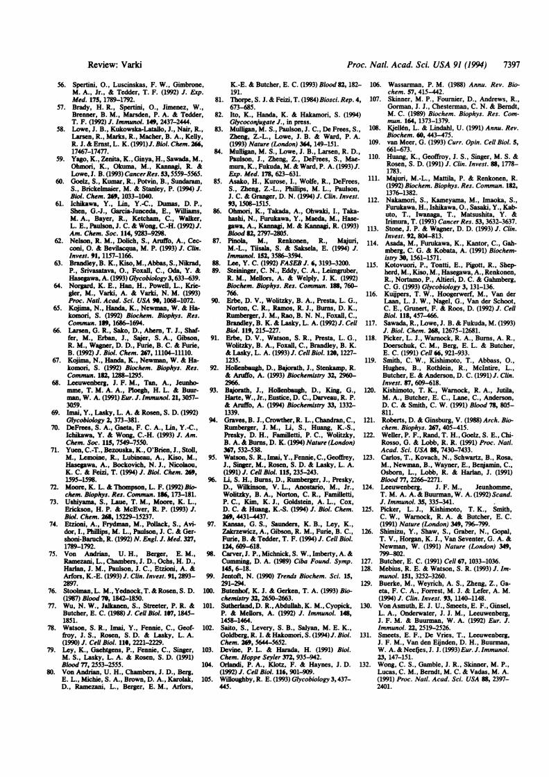
generated by a polypeptide backbone(Fig. 2A). This has indeed been suggestedfor some high-affinity selectin ligandsthat are mucins (28, 31, 33, 34) whichhave many oligosaccharides, closelyspaced to one another. However, theIC50 of chemically synthesized dimericSLex for E-selectin showed only a 5-foldimprovement over the monomeric form(70). Further, P- and L-selectin do notbind to some cell types that express con-siderable amounts of SLex, and myeloidcell recognition by P-selectin is de-stroyed by the enzyme O-sialoglycopro-tease (31, 73, 89), even though the vastmajority of surface SLex remains intact(31). Thus, simple ligand multivalencyseems insufficient to explain biologicallyrelevant selectin binding. Another possi-bility is multivalent aggregation of theselectin (Fig. 2B). However, high-affinitybinding of soluble monomeric P-selectinto cell surfaces (73) indicates that this isnot essential. Also, there is no publishedevidence for naturally occurring multi-meric selectins. Ligand multivalencyprovided by cell surfaces or specific poly-peptides could also be recognized bymultiple binding sites within a single se-lectin lectin domain (Fig. 2C). However,this is negated by studies of the lectindomains of the selectins by epitope map-ping, mutagenesis, and homology criteria(90-93), which indicate binding sites forcarbohydrates that are quite small; thishas been recently confirmed for E-selec-tin by x-ray crystallography (94). Fig. 3shows that there is barely enough room inthe E-selectin lectin-binding domain toaccommodate one copy of a SLex tet-rasaccharide, let alone many closelyspaced units. Thus, simple ligand or se-lectin multivalency does not appear suf-ficient to explain high-affinity recogni-tion. Another possibility is a combinationof both a basic oligosaccharide sequenceand an adjacent peptide-recognition site
7392 Review: Varki

Proc. Nadl. Acad. Sci. USA 91 (1994) 7393
(Fig. 2D). Some studies involving muta-genesis, domain swapping, or deletion ofthe other protein domains of selectinsseem to support this (95-97). However,the persistence of high-affinity recogni-tion after extensive denaturation or pro-teolytic digestion of some biologicallyrelevant selectin ligands (28, 29, 31, 36,69) is against this. These observationsalso make it unlikely that the peptidecarrier forces a monovalent oligosaccha-ride into an unusual conformation fa-vored for recognition (Fig. 2E). By ex-clusion, the remaining possibility is thathigh-affinity binding involves recognitionof a "clustered saccharide patch" (31)created by a peptide carrying multipleoligosaccharides (Fig. 2 F and G).
Free oligosaccharides in solution or atsingle attachment sites have significantfreedom of mobility and show limitedinteractions with associated proteins(98). However, oligosaccharides packedclosely together (particularly in mucins)can form rigid rod-like structures (99,100) that must have less mobility and yetare relatively unaffected by denaturationof the polypeptide backbone. Such clus-tering of common oligosaccharides couldpresent uncommon "clustered saccha-ride patches" generated by multiple oli-gosaccharides that are closely spacedenough to restrict their motion (31). Thepatch recognized by a selectin could begenerated in two possible ways. In the
A
first case (Fig. 2charide with low(e.g., SLe') mfsurrounding sugaconformation farecognition. Thihstabilized by pethe adjacent sugsugars might alsoformational chamdomain itself. II2G), the bindingby combinationgroups from muli.e., a "disconttope." Thus, theselectin might ilhydroxyl group facetyl group froand a carboxyl ithird chain. In btide need not trecognition site.crucial for preseithe correct arr;affinity recognitiarrangement is (vidual sugar chaiable affinity forexplain why mo:selectin ligands .macromoleculescharides releasenot bind with recsame selectin (34
B
F
D
riz Selectin* Basic
ol igosaccha ride
ligand
F), a common oligosac- would be distinct from improved bindingaffinity for the selectin of certain lectins to multiantennary N-
ght be forced by other linked oligosaccharides or to glycopep-wr chains into an unusual tides with widely spaced O-linked oligo-vored fQr high-affinity saccharides (88). This results from cor-s could also be aided or rect spacing of terminal sugar residues,,ptide interactions with each of which are still recognized as,ar chains. The adjacent distinct components of individual chains.induce a favorable con- The hypothesis also predicts that struc-
ige in the selectin lectin tural analyses of O-linked oligosaccha-the second case (Fig. rides released from a specific selectin
site would be generated ligand might not uncover unique se-is of side chains and quences required for recognition. There-ltiple oligosaccharides- fore, it may be necessary to completelyLiguous saccharide epi- elucidate both the polypeptide chain se-patch recognized by the quence and the oligosaccharide struc-nclude, for example, a tures, as well as their specific sites ofFrom one sugar chain, an attachment on the protein. Ultimately,im another sugar chain, one must reconstruct the ligand and de-or sulfate group from a fine the position of each of its compo-Both cases, the polypep- nents in three-dimensional space.be part of the selectin There is, in fact, much prior evidenceHowever, it would be for recognition of "clustered saccharide
nting the sugar chains in patches." The enzyme O-sialoglycopro-angement. Thus, high- tease (101), which has been of particularion would be lost if the value in identifying high-affinity selectindisrupted, and the indi- ligands, will only recognize and cleaveins may not have detect- mucins with large numbers of closelythe selectin. This might spaced 0-linked oligosaccharides (31).st reported high-affinity Many mAbs that detect specific glyco-are heavily glycosylated proteins have "sialidase-sensitive" or, and why free oligosac- "Sia-dependent" epitopes-e.g., cancerd from such ligands do mucins, CD24, CD45, and CD43 (see ref.ognizable affinity to the 31 for citations). With some heavily gly-6, 69). Such recognition cosylated O-linked gps, almost all avail-
able mAbs require glycosylation for spe-C cific recognition. Many such proteins
with sialidase-sensitive epitopes are ei-ther mucins with clustered 0-linked oli-gosaccharides or have large numbers ofN-linked oligosaccharides. However,when the sialyl-oligosaccharides on suchproteins are examined, they are not un-usual in structure compared with thosefrom other proteins of the same cells. Ina well-documented example, the recog-nition of a common sialylated oligosac-charide sequence by different mAbs dif-fered depending upon the type of glyco-
G conjugate attached (102). Other examplesinclude the reactivity of a mucin-specificlectin from Sambucus sieboldiana (103),
-] recognition of clustered O-linked oligo-saccharides on glycophorin A by the in-vasion receptor of Plasmodium falci-parum (104), the specific binding of ro-taviruses to intact mucins (105), andrecognition of the sperm receptor on themucin-type zonapellucidaglycoprotein 3ofthe mouse egg (106). In such instances,unique clustering of relatively commonoligosaccharides can explain the speci-ficity of recognition.
_Polypeptide Hypothesis: A Clustered S ridePatch Could Be Shared bySeiy Dis-prate GlycocouJuptes. Selectins can
FIG. 2. Models that could explain improved binding of selectirelative to the basic oligosaccharide ligands they can recognize.
possibilities A-E cannot explain many observations concerningpresent the concept of "clustered saccharide patches" that could
ins to their natural ligands also recognizeglycoan chainsAs discussed in the text, in a Ca2+-dependent manner (23, 36, 48,selectin binding. F and G 51, 107). Glycosaminoglycans have re-reconcile these data. peating disaccharide structures that do
Review: Varki
..............
.I

Proc. Nati. Acad. Sci. USA 91 (1994)
1W97 Y -1 1 e 9 797o
FIG. 3. Size comparison of the E-selectin lectin domain and SLex (courtesy of B. Graves,based on ref. 94). Stereo diagram of a "top" view of the E-selectin lectin domain, alongside anSLeX tetrasaccharide. The protein is depicted as a ribbon and tube diagram, with explicit atomsshown for the side chains ofthe eight amino acid residues known to affect carbohydrate bindingand for the calcium ion (with a van der Waals dot surface around it).
not typically contain Sia or Fuc (108) buthave variable numbers of sulfate estersand uronic acids. Competition experi-ments and Ca2+ dependence suggest thatthe binding site is the same or overlap-ping with that for sialylated fucosylatedoligosaccharides. How could two suchdisparate structures be recognized by thesame binding site? One explanation isthat a "clustered saccharide patch" rec-ognized by a selectin can be generated bydifferent types of glycoconjugates. Thus,the patch generated by a cluster ofO-linked oligosaccharides on a mucintype glycoprotein might be mimicked bya proper arrangement of carboxyl, hy-droxyl, and sulfate groups on a heparansulfate glycosaminoglycan chain. In thelatter case, the chain must generate therecognition site itself, because releasefrom the polypeptide backbone does notcause loss of binding (36). This hypoth-esis could also explain why relativelyhigh-affinity binding of selectins is seenwhen glycosphingolipids are immobilizedat high density on plates or onTLC plates(43, 46). Here, the clustered saccharidepatch might be artificially recreated onthe immobile surface because of a highdensity not normally found on cell sur-faces. However, because glycosphin-golipids can coaggregate in patches oncell surfaces (109), this could also be anatural mechanism for generating spe-cific selectin recognition.
Selectin Ligands That Are Probably Bi-ologically Relevant. From the foregoing,most, ifnot all ofthe biologically relevantselectin ligands appear to be complexmacromolecules. Table 3 lists such can-didate ligands chosen based on expres-sion in the right cell type and demon-strated ability to bind with high affinity toselectins in a Ca2+- and carbohydrate-dependent way. The definition of "highaffinity" is somewhat arbitrary andbased upon ligand binding surviving ex-tensive washing during procedures suchas direct binding to cells, affinity chro-
matography, or detection by blotting.Most, but not all, of these ligands aremucin-type gps. Less well-characterizedligands not listed in the table include mul-tiple sulfated gps found in a rat-HEVderived cell line Ax (30), 230-kDa and130-kDa P-selectin ligands reported onmurine myeloid cells and HL-60 cells (38),a sialidase-resistant ligand found on cen-tral nervous system myelin (110), siali-dase-sensitive ligands on activated endo-thelial cells (55-57), and a variety of car-cinoma-associated ligands (44, 111-113).Also not included are various neutrophilgps bearing SLeX that are recognized byE- and/or P-selectin in vitro, includingLFA-1 (114, 115), nonspecific cross-reacting antigens CD66/CD67 (116),LAMP-1 (117), and L-selectin itself (118).Like most other neutrophil proteins, thesepolypeptides have SLex-bearing oligosac-charides. Thus, when purified in quantityand immobilized on artificial surfaces,they can support cell adhesion via E-and/or P-selectin. However, their role inpresenting high-affinity, biologically rele-vant ligands in the natural situation isunclear. A particularly confusing case isthat of the L-selectin molecule on neutro-phils, which has been suggested as a majorpresenter of ligands for P- and E-selectin(118). Indeed, anti-L-selectin antibodiespartially block binding in systems whereE- and P-selectin recognition are impor-tant (119, 120). Also, L-selectin isolatedfrom neutrophils carries SLex and can berecognized by E- and P-selectin whenpurified and immobilized on a surface(118). However, neutrophils that haveshed L-selectin continue to bind well (18),as do many cells (e.g., HL-60) that lackL-selectin. Thus, an alternative explana-tion may lie in the unusual distribution ofL-selectin on the tips of neutrophilpseudopodia (118). Perhaps the true li-
gands for E- and/or P-selectin are alsolocated in the same favored site for inter-actions; if so, anti-L-selectin antibodiesmay act by locally hindering the recogni-
tion of these other ligands. It is also dif-ficult to know whether to include specificglycosphingolipids among the biologicallyrelevant ligands. Molecules such as sul-fatide show binding (47, 49) to some se-lectins, but this is not always Ca2+-dependent, and a large number of unre-lated adhesion proteins are known to bindto this small sulfated glycolipid (121).SLex-bearing glycosphingolipids canclearly support E-selectin-based bindingwhen purified and immobilized on artifi-cial surfaces. However, whether they canattain similar densities on cell surfaces isunknown.Nomenclature Difficulties. The nomen-
clature for the low-affinity monomericoligosaccharide structures recognized byselectins is straightforward and is simplya description oftheir structures (Table 2).There is also no problem with the nomen-clature of ligands defined by mAbsrecognizing multiple and/or unknownspecies with unusual carbohydrate epi-topes-e.g., peripheral node addressin(MECA-79) (25), cutaneous lymphocyteantigen (HECA-452) (27), and SLex-var(2F3) (86). These antibodies were impor-tant in understanding selectin-mediatedrecognition-e.g., MECA-79 reacts withcells that bear L-selectin ligands (25, 34)and with ligands from murine lymphnodes (26, 69). However, they may alsoreact with related carbohydrate epitopeson other molecules that are not ligandsfor the selectins. Regarding the discretebiological ligands for the selectins (Table3), there are several difficulties with cur-rent nomenclature. (i) The differentnames and terms do not relate well toeach other. (ii) A polypeptide that acts asa scaffolding for generating a selectinligand in one tissue may not do so inanother tissue (39), and some may havecompletely independent functions (34).(iii) It appears likely that there are sig-nificant differences in functionally activeligands among different animal species.(iv) Different classes of glycoconjugates(N-linked gps, mucin-type gps, proteo-glycans) appear to function as ligands.The issue of nomenclature for these li-gands deserves more attention.
Is Weak Bindig Also Bologlcally Im-portant? This discussion has favored thenotion that macromolecules with com-plex oligosaccharide arrangements arethe critical high-affinity ligands for theselectins. However, in vitro studies showthat high-copy numbers of oligosaccha-rides such as SLeX on cell surfaces cangive weak, but detectable, binding be-tween cells. Is such binding biologicallyrelevant in vivo? For example, the highlevel of SLeX on neutrophils could actu-ally be responsible for the initial recog-nition of a neutrophil by E- or P-selectinon an endothelial cell. This could benecessary, but not sufficient, to initiaterolling, serving as the first recognition
7394 Review: Varki

Proc. Nail. Acad. Sci. USA 91 (1994) 7395
Table 3. Natural selectin ligands likely to be of biological relevanceRecognition
Polypeptide Natural requiresOriginal name Source name ligand for Sia Fuc S04 Comments Refs.
sgp5OHEV L-selectin Mouse peripheral lymph GlyCAM-1 L-selectin + +? + Primarily secreted-a natural 26, 28ligand node antiadhesin? Susceptible to
O-sialoglycoprotease120-kDa myeloid Human neutrophil, HL-60 PSGL-1 P-selectin + + Cell surface homodimer-minor 29, 31, 37
P-selectin ligand cells component of total surface gps.Susceptible to O-sialoglycoprotease.Murine homologue?
150-kDa myeloid Mouse bone marrow cells Polypeptide E-selectin + N-linked oligosaccharides required for 32, 38E-selectin ligand (80%o neutrophils) unknown binding. Resistant to O-sialoglyco-
protease. Human homologue?sgp90 sulfated Mouse peripheral lymph CD34 L-selectin + + Polypeptide also present in 33
L-selectin ligand node nonlymphoid endothelium-not aligand in these locations
MAdCAM-1 Mouse mesenteric lymph MAdCAM-1 L-selectin + Polypeptide also present in Peyers 34node patch HEV-not a major ligand in
this location because glycosylationis not appropriate
250-kDa E-selectin Bovine y/8 lymphocytes Polypeptide E-selectin + Isolated by affinity chromatography 35ligand unknown
Heparan sulfate Bovine and human Core L-selectin - + Isolated by affinity chromatography 36endothelial cells protein from cultured cells-subcellular
not location and function in intactrequired tissues not proven
160-kDa myeloid Mouse bone marrow cells Polypeptide P-selectin + Susceptible to O-sialoglycoprotease 32, 38P-selectin ligand (80%o neutrophils) unknown N-linked oligosaccharides also
required for binding? Homologue inhuman HL-60 cells?
sgp, Sulfated gp; PSGL-1, P-selectin gp ligand 1; MAdCAM, mucosal addressin cell adhesion molecule; GlyCAM-1, glycosylation-dependentcell adhesion molecule 1.
step that permits subsequent interactionswith the higher-affinity (but lower copynumber) macromolecular ligands on thesame cell. Such recognition could also beimportant pathologically in carcinomacells, which have high levels of SLexand/or SLea on their cell surfaces (44,111-113).Are Some "Ligands" Actuafly "Antiad-
hesins"? In at least two instances a por-tion of the ligand synthesized by cells invitro is found in the culture medium-i.e.,glycosylation-dependent cell adhesionmolecule (28) and heparan sulfate proteo-glycans (36) (K. Norgard-Sumnicht andA.V., unpublished work). These solubleligands could act as inhibitors of selectinbinding rather than as adhesion mole-cules. Whether their potential antiadhe-sive function is biologically relevant re-mains to be seen.Are the Lgands Shared Between Differ-
ent Selectins and Between Different Leu-kocyte Types? To date, most attention hasbeen directed toward the recognition ofhigh-endothelial venule ligands by L-se-lectin on lymphocytes and of neutrophilligands by endothelial E- and P-selectin.However, monocytes and eosinophilspossess ligands for E-selectin, which arefunctional in interaction with immobi-lized selectins or activated endotheliumin vitro (56, 122-124), and distinct subsetsof lymphocytes carry ligands for P- or
E-selectin (27, 72, 87, 125, 126). Thenature of these ligands, their structuralrelationship to the neutrophil ligands,and their importance in the in vivo situ-ation remain uncertain. If the ligands areshared among different leukocytes, thedifferential trafficking of cell types mightbe explained by the multistep combina-torial nature of leukocyte extravasation(127).Most studies have focused upon the
interaction of a single selectin with asingle ligand from a particular cell type.However, the overlap in recognition ofsmall oligosaccharides by the selectins(20, 41, 42, 51, 62) predicts that overlapsmay also exist with regard to the macro-molecular ligands. While some overlapshave been seen (38, 66, 73, 91, 128),interactions with a particular ligand arenot comparably strong between the se-lectins. Detailed comparisons of the rec-ognition of specific ligands by the threeselectins are needed.
Therapeutic Inhibition of Selectins inVivo by Lignd Analogs Problems andProspects. The development of selectinligand analogs that can interfere in vivohas obvious therapeutic potential in avariety of pathological processes (3-9).The use of sugar analogs or related struc-tures is attractive also because of theirrelative lack of immune reactivity, aswell as their ease of handling for thera-
peutic use. The small SLex-based thera-peutics currently entering clinical trials(J. Paulson, personal communication)have the potential to partially block thefunctions of all three selectins. Studies ofthe complex macromolecular ligandslisted in Table 3 could lead to design ofspecific analogs that discriminate be-tween selectins and permit selectiveblockade in specific disease states. Ad-ditional considerations include a betterhalf-life in the circulation and enoughaffinity to use small amounts for sus-tained blockade of selectin function invivo.Future Directions. There are many
other unexplained mysteries and interest-ing questions regarding selectin ligandsthat deserve investigation. The fact thatselectin ligands survive treatments suchas heating, ionic detergents, proteasetreatment, and organic extraction (26, 28,29, 31, 36-, 69) suggests that primary rec-ognition involves carbohydrates alone.However, other domains of the selectinscould be recognizing other as-yet-unidentified polypeptides via protein-protein interactions. These could be im-portant in facilitating or modulating somerecognition events mediated by the se-lectins. The epidermal growth factor-likeand complement-regulatory-like domainsof the selectins are candidates for suchinteractions. The "variant" forms of
Rev iew: Varki

Proc. Natl. Acad. Sci. USA 91 (1994)
SLex on blood cell surfaces detected byspecific mAbs such as HECA-452 (22)and 2F3 (86) remain unexplained. An-other mystery is the fact that concentra-tions of small oligosaccharides that seeminadequate to block selectin function invitro are surprisingly successful in abro-gating some selectin-mediated patholog-ical reactions in the intact animal (83, 84,129). They may function by an alternativemechanism in vivo-e.g., by inducingdown-regulation of selectin expressionon cell surfaces (130, 131). Regarding thehigh-affinity cell-specific macromolecu-lar ligands, it is possible that they haveother biological functions mediated bytheir ligation-e.g., the binding of P-se-lectin to neutrophils causes biologicalresponses (132). Another area of confu-sion is the description of "partially Ca2+-dependent" ligands, seen particularly inthe interactions of P- and L-selectin withsulfated ligands. Thus, addition ofexcessEDTA to chelate Ca2+ sometimes maynot completely reverse a selectin-basedbinding. This may occur despite the factthat the primary binding requires Ca2+.One explanation is that ligand-dependentactivation or an "induced-fit" might oc-cur, such that Ca:2+ is required for initialbinding but not for subsequent mainte-nance of the complex. Alternatively, aCa2+ could form a very tight complexwith the ligand after initial binding, whichmight then be very difficult to removewithout prolonged EDTA exposure. Re-gardless, these and other data (47, 49, 93,97) suggest that P- and L-selectin mayhave a sulfate-recognition site whosefunction is not strictly Ca2+-dependent.Finally, most studies on the role of se-lectins in neutrophil traffic have focusedupon pathological situations. It is of in-terest to know if the selectins are in-volved in normal turnover and/or "mar-gination" of neutrophils, which havevery short half-lives in circulation.Summary and Conclusions. Much evi-
dence suggests that the biologically rele-vant ligands for the selectins are diverseand complex macromolecules that sharein common certain types of anionic car-bohydrate structures. While details re-garding these molecules begin to emerge,it remains to be seen which ligands areimportant for specific biological pro-cesses involving the selectins. High-affinity recognition of seemingly dispar-ate oligosaccharides may involve the for-mation of similar "clustered saccharidepatches." If so, fully understanding se-lectin-ligand interactions will require de-fining the precise three-dimensionalstructures of both partners. Such knowl-edge could also permit the design of amore specific and efficacious therapeuticligand analog to be used for interventionin the many disease processes involvingselectins.
I acknowledge many investigators for shar-ing work in press and thank H. Freeze, M.Ginsberg, N. Varki, J. Paulson, and T. Koganfor helpful comments and for their criticalreview of the manuscript.
1. Yednock, T. A. & Rosen, S. D. (1989) Adv.Immunol. 44, 313-378.
2. Stoolman, L. M. (1989) Cell 56, 907-910.3. Picker, L. J. & Butcher, E. C. (1992) Annu.
Rev. Immunol. 10, 561-591.4. Lasky, L. A. (1992) Science 258, 964-969.5. Varki3 A. (1992) Curr. Opin. Cell Biol. 4,
257-266.6. Rosen, S. D. (1993) Semin. Immunol. 5, 237-
247.7. Bevilacqua, M. P. & Nelson, R. M. (1993) J.
Clin. Invest. 91, 379-387.8. McEver, R. P. (1994) Curr. Opin. Immunol. 6,
75-84.9. Lefer, A. M., Weyrich, A. S. & Buerke, M.
(1994) Cardiovasc. Res. 28, 289-294.10. Drickamer, K. (1993) Curr. Opin. Struct. Biol.
3, 393-400.11. Phillips, M. L., Nudelman, E., Gaeta,
F. C. A., Perez, M., Singhal, A. K., Hako-mori, S. & Paulson, J. C. (1990) Science 250,1130-1132.
12. Walz, G., Aruffo, A., Kolanus, W., Bevilac-qua, M. & Seed, B. (1990) Science 250, 1132-1135.
13. Larsen, E., Palabrica, T., Sajer, S., Gilbert,G. E., Wagner, D. D., Furie, B. C. & Funie,B. (1990) CeU 63, 467-474.
14. Lowe, J. B., Stoolman, L. M., Nair, R. P.,Larsen, R. D., Berhend, T. L. & Marks,R. M. (1990) Cell 63, 475-484.
15. Corral, L., Singer, M. S., Macher, B. A. &Rosen, S. D. (1990) Biochem. Biophys. Res.Commun. 172, 1349-1356.
16. True, D. D., Singer, M. S., Lasky, L. A. &Rosen, S. D. (1990) J. Cell Biol. 111, 2757-2764.
17. Goelz, S. E., Hession, C., Goff, D., Griffiths,B., Tizard, R., Newman, B., Chi-Rosso, G. &Lobb, R. (1990) Cell 63, 1349-1356.
18. Moore, K. L., Varki, A. & McEver, R. P.(1991) J. CeU Biol. 112, 491-499.
19. Tiemeyer, M., Swiedler, S., Ishihara, M.,Moreland, M., Schweingruber, H., Hirtzer, P.& Brandley, B. K. (1991) Proc. Natl. Acad.Sci. USA 88, 1138-1142.
20. Polley, M. J., Phillips, M. L., Wayner, E.,Nudelman, E., Sighl, A. K., Hakomori, S.& Paulson, J. C. (1991) Proc. Natl. Acad. Sci.USA 88, 6224-6228.
21. Zhou, Q., Moore, K. L., Smith, D. F., Varki,A., McEver, R. P. & Cummings, R. D. (1991)J. CeU Biol. 115, 557-564.
22. Berg, E. L., Robinson, M. K., Mansson, O.,Butcher, E. C. & Magnani, J. L. (1991) J.Biol. Chem. 266,14869-14872.
23. Handa, K., Nudelman, E. D., Stroud, M. R.,Shiozawa, T. & Hakomori, S. (1991) Biochem.Biophys. Res. Commun. 181, 1223-1230.
24. Tyrrell, D., James, P., Rao, N., Foxall, C.,Abbas, S., Dasgupta, F., Nashed, M., Hase-gawa, A., Kiso, M., Asa, D., Kidd, J. &Brandley, B. K. (1991) Proc. Natl. Acad. Sci.USA 88, 10372-10376.
25. Berg, E. L., Robinson, M. K., Warnock,R. A. & Butcher, E. C. (1991) J. Cell Biol.114, 343-349.
26. Imai, Y., Singer, M. S., Fennie, C., Lasky,L. A. & Rosen, S. D. (1991) J. Cell Biol. 113,1213-1222.
27. Berg, E. L., Yoshino, T., Rott, L., Robinson,M., Warnock, R., Kishimoto, T. K., Picker,L. J. & Butcher, E. C. (1991) J. Exp. Med.174, 1461-1466.
28. Lasky, L. A., Singer, M. S., Dowbenko, D.,Imai, Y., Henzel, W. J., Grimley, C., Fennie,C., Gillett, N., Watson, S. R. & Rosen, S. D.(1992) Cell 69, 927-938.
29. Moore, K. L., Stults, N. L., Diaz, S., Smith,D. F., Cummings, R. D., Varki, A. &
McEver, R. P. (1992) J. CeU Biol. 118, 445-456.
30. Tamatani, T., Kuida, K., Watanabe, T.,Koike, S. & Miyasaka, M. (1993) J. Immunol.158, 1735-1745.
31. Norgard, K. E., Moore, K. L., Diaz, S.,Stults, N. L., Ushiyama, S., McEver, R. P.,Cummings, R. D. & Varki, A. (1993) J. Biol.Chem. 268, 12764-12774.
32. Levinovitz, A., Mdhlhoff, J., Isenmann, S. &Vestweber, D. (1993) J. Cel Biol. 121, 449-459.
33. Baumhueter, S., Singer, M. S., Henzel, W.,Hemmerich, S., Renz, M., Rosen, S. D. &Lasky, L. A. (1993) Science 262, 436-438.
34. Berg, E. L., McEvoy, L. M., Berlin, C., Bar-gatze, R. F. & Butcher, E. C. (1993) Nature(London) 366, 695-698.
35. Walcheck, B., Watts, G. & Jutila, M. A.(1993) J. Exp. Med. 178, 853-863.
36. Norgard-Sumnicht, K. E., Varki, N. M. &Varki, A. (1993) Science 261, 480-483.
37. Sako, D., Chang, X.-J., Barone, K. M.,Vachino, G., White, H. M., Shaw, G., Veld-man, G. M., Bean, K. M., Ahern, T. J., Fb-rie, B., Cumming, D. A. & Larsen, G. R.(1993) Cel 75, 1179-1186.
38. Lenter, M., Levinovitz, A., Isenmann, S. &Vestweber, D. (1994) J. Cell Biol. 12S, 471-481.
39. Dowbenko, D., Kikuta, A., Fennie, C., Gil-lett, N. & Lasky, L. A. (1993) J. Clin. Invest.92, 952-960.
40. Imai, Y., Lasky, L. A. & Rosen, S. D. (1993)Nature (London) 361, 555-557.
41. Berg, E. L., Magnani, J., Warnock, R. A.,Robinson, M. K. & Butcher, E. C. (1992)Bio-chem. Biophys. Res. Commun. 184, 1048-1055.
42. Foxall, C., Watson, S. R., Dowbenko, D.,Fennie, C., Lasky, L. A., Kiso, M., Hase-gawa, A., Asa, D. & Brandley, B. K. (1992) J.Cell Biol. 117, 895-902.
43. Larkin, M., Ahern, T. J., Stoll, M. S., Shaf-fer, M., Sako, D., O'Brien, J., Yuen, C.-T.,Lawson, A. M., Childs, R. A., Barone,K. M., Langer-Safer, P. R., Hasegawa, A.,Kiso, M., Larsen, G. R. & Feizi, T. (1992) J.Biol. Chem. 267, 13661-13668.
44. Takada, A., Ohmori, K., Yoneda, T.,Tsuyuoka, K., Hasegawa, A., Kiso, M. &Kannagi, R. (1993) Cancer Res. 53, 354-361.
45. Green, P. J., Tamatani, T., Watanabe, T.,Miyasaka, M., Hasegawa, A., Kiso, M.,Yuen, C.-T., Stoll, M. S. & Feizi, T. (1992)Biochem. Biophys. Res. Commun. 18, 244-251.
46. Yuen, C.-T., Lawson, A. M., Chai, W., Lar-kin, M., Stoll, M. S., Suart, A. C., Sulivan,F. X., Ahern, T. J. & Feizi, T. (1992) Bio-chemistry 31, 9126-9131.
47. Suzuki, Y., Toda, Y., Tamatani, T., Wata-nabe, T., Suzuki, T., Nakao, T., Murase, K.,Kiso, M., Hasegawa, A., Tadano-Anitomi, K.,Ishizuka, I. & Miyasaka, M. (1993) Biochem.Biophys. Res. Commun. 19, 426-434.
48. Skinner, M. P., Lucas, C. M., Bums, G. F.,Chesterman, C. N. & Berndt, M. C. (1991) J.Biol. Chem. 266, 5371-5374.
49. Aruffo, A., Kolanus, W., Walz, G., Fredman,P. & Seed, B. (1991) CeU 67, 35-44.
50. Needham, L. K. & Schnaar, R. L. (1993)Proc. Natl. Acad. Sci. USA ", 1359-1363.
51. Nelson, R. M., Cecconi, O., Roberts, W. G.,Aruffo, A., Linhardt, R. J. & Bevilacqua,M. P. (1993) Blood 82, 3253-3258.
52. Kormfeld, S. (1992) Annu. Rev. Biochem. 61,307-330.
53. Rosen, S. D., Singer, M. S., Yednock, T. A.& Stoolman, L. M. (1985) Science 228, 1005-1007.
54. Streeter, P. R., Rouse, B. T.&Butcher, E. C.(1968)J. Cel Biol. 107, 1853-1862.
55. Spertini, O., Luscinskas, F. W., Kansas,G. S., Munro, J. M., Griffin, J. D., Gimbrone,M. A., Jr., &Tedder, T. F. (1991)J. Immunol.147, 2565-2573.
7396 Review: Varki
Proc. Natl. Acad. Sci. USA 91 (1994) 7397
56. Spertini, O., Luscinskas, F. W., Gimbrone,M. A., Jr., & Tedder, T. F. (1992) J. Exp.Med. 175, 1789-1792.
57. Brady, H. R., Spertini, O., Jimenez, W.,Brenner, B. M., Marsden, P. A. & Tedder,T. F. (1992) J. Immunol. 149, 2437-2444.
58. Lowe, J. B., Kukowska-Latallo, J., Nair, R.,Larsen, R., Marks, R., Macher, B. A., Kelly,R. J. & Ernst, L. K. (1991)J. Biol. Chem. 26,17467-17477.
59. Yago, K., Zenita, K., Ginya, H., Sawada, M.,Ohmori, K., Okuma, M., Kannagi, R. &Lowe, J. B. (1993) CancerRes. 53,5559-5565.
60. Goelz, S., Kumar, R., Potvin, B., Sundaram,S., Brickelmaier, M. & Stanley, P. (1994) J.Biol. Chem. 269, 1033-1040.
61. Ichikawa, Y., Lin, Y.-C., Dumas, D. P.,Shen, G.-J., Garcia-Junceda, E., Williams,M. A., Bayer, R., Ketcham, C., Walker,L. E., Paulson, J. C. & Wong, C.-H. (1992) J.Am. Chem. Soc. 114, 9283-9298.
62. Nelson, R. M., Dolich, S., Aruffo, A., Cec-coni, 0. & Bevilacqua, M. P. (1993) J. Clin.Invest. 91, 1157-1166.
63. Brandley, B. K., Kiso, M., Abbas, S., Nikrad,P., Srivasatava, O., Foxall, C., Oda, Y. &Hasegawa, A. (1993) Glycobiology 3, 633-639.
64. Norgard, K. E., Han, H., Powell, L., Krie-gler, M., Varki, A. & Varki, N. M. (1993)Proc. Nad. Acad. Sci. USA ", 1068-1072.
65. Kojima, N., Handa, K., Newman, W. & Ha-komori, S. (1992) Biochem. Biophys. Res.Commun. 189, 1686-1694.
66. Larsen, G. R., Sako, D., Ahern, T. J., Shaf-fer, M., Erban, J., Sajer, S. A., Gibson,R. M., Wagner, D. D., Furie, B. C. & Furie,B. (1992) J. Biol. Chem. 267, 11104-11110.
67. Kojima, N., Handa, K., Newman, W. & Ha-komori, S. (1992) Biochem. Biophys. Res.Commun. 182, 1288-195.
68. Leeuwenberg, J. F. M., Tan, A., Jeunho-mme, T. M. A. A., Ploegh, H. L. & Buur-man, W. A. (1991) Eur. J. Immunol. 21, 3057-3059.
69. Imai, Y., Lasky, L. A. & Rosen, S. D. (1992)Glycobiology 2, 373-381.
70. DeFrees, S. A., Gaeta, F. C. A., Lin, Y.-C.,Ichikawa, Y. & Wong, C.-H. (1993) J. Am.Chem. Soc. 115, 7549-7550.
71. Yuen, C.-T., Bezouska, K., O'Brien, J., Stoll,M., Lemoine, R., Lubineau, A., Kiso, M.,Hasegawa, A., Bockovich, N. J., Nicolaou,K. C. & Feizi, T. (1994)J. Biol. Chem. 269,1595-1598.
72. Moore, K. L. & Thompson, L. F. (1992) Bio-chem. Biophys. Res. Commun. 186, 173-181.
73. Ushiyama, S., Laue, T. M., Moore, K. L.,Erickson, H. P. & McEver, R. P. (1993) J.Biol. Chem. 268, 15229-15237.
74. Etzioni, A., Frydman, M., Pollack, S., Avi-dor, I., Phillips, M. L., Paulson, J. C. & Ger-shoni-Baruch, R. (1992) N. Engl. J. Med. 327,1789-1792.
75. Von Andrian, U. H., Berger, E. M.,Ramezani, L., Chambers, J. D., Ochs, H. D.,Harlan, J. M., Paulson, J. C., Etzioni, A. &Arfors, K.-E. (1993) J. Clin. Invest. 91, 2893-2897.
76. Stoolman, L. M., Yednock, T. & Rosen, S. D. 1(197) Blood 70, 1842-1850.
77. Wu, N. W., Jalkanen, S., Streeter, P. R. & 1Butcher, E. C. (1988) J. Cell Biol. 1W7, 1845-1851.
78. Watson, S. R., Imai, Y., Fennie, C., Geof- 11froy, J. S., Rosen, S. D. & Lasky, L. A.(1990) J. Cell Biol. 110, 2221-2229.
79. Ley, K., Gaehtgens, P., Fennie, C., Singer, 11M. S., Lasky, L. A. & Rosen, S. D. (1991)Blood 77, 2553-2555. 1(
80. Von Andrian, U. H., Chambers, J. D., Berg,E. L., Michie, S. A., Brown, D. A., Karolak, 1(D., Ramezani, L., Berger, E. M., Arfors,
K.-E. & Butcher, E. C. (1993) Blood 82, 182-191.
81. Thorpe, S. J. & Feizi, T. (1984)Biosci. Rep. 4,673-685.
82. Ito, K., Handa, K. & Hakamori, S. (1994)Glycocoijugate J., in press.
83. Mulligan, M. S., Paulson, J. C., De Frees, S.,Zheng, Z.-L., Lowe, J. B. & Ward, P. A.(1993) Nature (London) 364, 149-151.
84. Mulligan, M. S., Lowe, J. B., Larsen, R. D.,Paulson, J., Zheng, Z., DeFrees, S., Mae-mura, K., Fukuda, M. & Ward, P. A. (1993)J.Exp. Med. 178, 623-631.
85. Asako, H., Kurose, I., Wolfe, R., DeFrees,S., Zheng, Z.-L., Phillips, M. L., Paulson,J. C. & Granger, D. N. (1994) J. Clin. Invest.93, 1508-1515.
86. Ohmori, K., Takada, A., Ohwaki, I., Taka-hashi, N., Furukawa, Y., Maeda, M., Hase-gawa, A., Kannagi, M. & Kannagi, R. (1993)Blood 82, 2797-2805.
87. Pinola, M., Renkonen, R., Majuri,M.-L., Tiisala, S. & Saksela, E. (1994) J.Ininunol. 1S2, 3586-3594.
88. Lee, Y. C. (1992) FASEB J. 6, 3193-3200.89. Steininger, C. N., Eddy, C. A., Leimgruber,
R. M., Mellors, A. & Welply, J. K. (1992)Blochem. Biophys. Res. Commun. 188, 760-766.
90. Erbe, D. V., Wolitzky, B. A., Presta, L. G.,Norton, C. R., Ramos, R. J., Burs, D. K.,Rumberger, J. M., Rao, B. N. N., Foxall, C.,Brandley, B. K. & Lasky, L. A. (1992)J. CellBiol. 119, 215-227.
91. Erbe, D. V., Watson, S. R., Presta, L. G.,Wolitzky, B. A., Foxall, C., Brandley, B. K.& Lasky, L. A. (1993) J. Cell Biol. 120, 1227-1235.
92. Hollenbaugh, D., Bajorath, J., Stenkamp, R.& Anrffo, A. (1993) Biochemistry 32, 2960-2966.
93. BaJorath, J., Hollenbaugh, D., King, G.,Harte, W., Jr., Eustice, D. C., Darveau, R. P.& Aruflo, A. (1994) Biochemistry 33, 1332-1339.
94. Graves, B. J., Crowther, R. L., Chandran, C.,Rumberger, J. M., Li, S., Huang, K.-S.,Presky, D. H., Familletti, P. C., Wolitzky,B. A. & Burns, D. K. (1994) Nature (London)367, 532-538.
95. Watson,S. R., Imai, Y., Fennie, C., Geoffrey,J., Singer, M., Rosen, S. D. & Lasky, L. A.(1991) J. Cell Bidol. 115, 235-243.
96. Li, S. H., Burns, D., Rumberger, J., Presky,D., Wilkinson, V. L., Anostario, M., Jr.,Wolitzky, B. A., Norton, C. R., Familletti,P. C., Kim, K. J., Goldstein, A. L., Cox,D. C. & Huang, K.-S. (1994) J. Biol. Chem. 1269, 4431-4437.
97. Kansas, G. S., Saunders, K. B., Ley, K.,Zakrzewicz, A., Gibson, R. M., Furie, B. C., IFurie, B. & Tedder, T. F. (1994) J. Cell Biol.124, 609-618.
98. Carver, J. P., Michnick, S. W., Imberty, A. &Cumning, D. A. (1989) Ciba Found. Symp. 1145, 6-18. 1
99. Jentoft, N. (1990) Trends Biochem. Sci. 15,291-294. 1
LOO. Butenhof, K. J. & Gerken, T. A. (1993) Bio-ch/mistry 32, 2650-2663.
LO1. Sutherland,D. R., Abdullah, K. M., Cyopick, 1P. & Mellors, A. (1992) J. Immunol. 148,1458-1464.
02. Saito, S., Levery, S. B., Salyan, M. E. K.,Goldberg, R. I. & Hakomori, S. (1994)J. Biol. 1:Chem. 269, 5644-5652.
03. Devine, P. L. & Harada, H. (1991) Biol.Chem. Hoppe Seyler 372, 935-942.
04. Orlandi, P. A., Klotz, F. & Haynes, J. D. 1(1992) J. Cell Biol. 116, 901-909.
)5. Willoughby, R. E. (1993) Glycobiology 3, 437-445.
106. Wassarman, P. M. (1988) Annu. Rev. Bio-chem. 57, 415-442.
107. Skinner, M. P., Fournier, D., Andrews, R.,Gorman, J. J., Chesterman, C. N. & Berndt,M. C. (1989) Biochem. Biophys. Res. Com-mun. 164, 1373-1379.
108. Kjelln, L. & Lindahl, U. (1991) Annu. Rev.Biochem. 60, 443-475.
109. van Meer, G. (1993) Curr. Opin. Cell Biol. 5,661-673.
110. Huang, K., Geoffroy, J. S., Singer, M. S. &Rosen, S. D. (1991) J. Clin. Invest. 88, 1778-1783.
111. Majuri, M.-L., Mattila, P. & Renkonen, R.(1992) Biochem. Biophys. Res. Commun. 182,1376-1382.
112. Nakamori, S., Kameyama, M., Imaoka, S.,Furukawa, H., Ishikawa, O., Sasaki, Y., Kab-uto, T., Iwanaga, T., Matsushita, Y. &Irimura, T. (1993) Cancer Res. 53, 3632-3637.
113. Stone, J. P. & Wagner, D. D. (1993) J. Clin.Invest. 92, 804-813.
114. Asada, M., Furukawa, K., Kantor, C., Gah-mberg, C. G. & Kobata, A. (1991) Biochem-istry 30, 1561-1571.
115. Kotovuori, P., Tontti, E., Pigott, R., Shep-herd, M., Kiso, M., Hasegawa, A., Renkonen,R., Nortamo, P., Altieri, D. C. & Gahmberg,C. G. (1993) Glycobiology 3, 131-136.
116. Kujpers, T. W., Hoogerwerf, M., Van derLaan, L. J. W., Nagel, G., Van der Schoot,C. E., Grunert, F. & Roos, D. (1992) J. CellBiol. 118, 457-466.
117. Sawada, R., Lowe, J. B. & Fukuda, M. (1993)J. Biol. Chem. 268, 12675-12681.
118. Picker, L. J., Warnock, R. A., Burns, A. R.,Doerschuk, C. M., Berg, E. L. & Butcher,E. C. (1991) Cell 66, 921-933.
119. Smith, C. W., Kishimoto, T., Abbass, O.,Hughes, B., Rothlein, R., McIntire, L.,Butcher, E. & Anderson, D. C. (1991)J. Clin.Invest. 87, 609-618.
120. Kishimoto, T. K., Warnock, R. A., Jutila,M. A., Butcher, E. C., Lane, C., Anderson,D. C. & Smith, C. W. (1991) Blood 78, 805-811.
121. Roberts, D. & Ginsburg, V. (1988) Arch. Bio-chem. Biophys. 267, 405-415.
122. Weller, P. F., Rand, T. H., Goelz, S. E., Chi-Rosso, G. & Lobb, R. R. (1991) Proc. Natd.Acad. Sci. USA 88, 7430-7433.
123. Carlos, T., Kovach, N., Schwartz, B., Rosa,M., Newman, B., Wayner, E., Benjamin, C.,Osborn, L., Lobb, R. & Harlan, J. (1991)Blood 77, 2266-2271.
124. Leeuwenberg, J. F. M., Jeunhomme,T. M. A. A. & Buurman, W. A. (1992) Scand.J. Immunol. 35, 335-341.
125. Picker, L. J., Kishimoto, T. K., Smith,C. W., Warnock, R. A. & Butcher, E. C.(1991) Nature (London) 349, 79-799.
126. Shimizu, Y., Shaw, S., Graber, N., Gopal,T. V., Horgan, K. J., Van Seventer, G. A. &Newman, W. (1991) Nature (London) 349,799-802.
127. Butcher, E. C. (1991) Cell 67, 1033-1036.28. Mebius, R. E. & Watson, S. R. (1993) J. Im-
munol. 151, 3252-3260.129. Buerke, M., Weyrich, A. S., Zheng, Z., Ga-
eta, F. C. A., Forrest, M. J. & Lefer, A. M.(1994) J. Clin. Invest. 93, 1140-1148.
L30. Von Asmuth, E. J. U., Smeets, E. F., Ginsel,L. A., Onderwater, J. J. M., Leeuwenberg,J. F. M. & Buurman, W. A. (1992) Eur. J.Immunol. 22, 2519-2526.
31. Smeets, E. F., De Vries, T., Leeuwenberg,J. F. M., Van den Eijnden, D. H., Buurman,W. A. &Neeoes, J. J. (1993)Eur. J. Immunol.23, 147-151.
32. Wong, C. S., Gamble, J. R., Skinner, M. P.,Lucas, C. M., Berndt, M. C. & Vadas, M. A.(1991) Proc. Natd. Acad. Sci. USA 88, 2397-2401.
Rkeview: Varki