REVIEW RTICLE Neuropathology of Charcot-Marie-Tooth and ...
20
REVIEW ARTICLE Neuropathology of Charcot-Marie-Tooth and Related Disorders J. Michael Schröder* Department of Neuropathology, University Hospital, RWTH Aachen, Germany Received August 22, 2005; Revised November 18, 2005; Accepted November 30, 2005 Abstract The peripheral nervous system (PNS), with all its branches and connections, is so complex that it is impossible to study all components at the light or electron microscopic level in any individual case; nevertheless, in certain diseases a simple nerve biopsy may suffice to arrive at a precise diagnosis. Structural changes of the PNS in neuropathies of the Charcot-Marie-Tooth (CMT) type and related disorders comprise various components of the PNS. These include peripheral motor, sensory, and autonomous neurons with their axons, Schwann cells, and myelin sheaths in the radicular and peripheral nerves as well as satellite cells in spinal and autonomous ganglia. Astrocytes, oligodendroglial cells, and microglial cells around motor neurons in the anterior horn and around sensory neurons in other areas of the spinal cord are also involved. In addition, connective tissue elements such as endoneurial, perineurial, and epineurial com- ponents including blood and lymph vessels play an important role. This review focuses on the cellular components and organelles involved, that is, myelin sheaths, axons with their micro- tubules and neurofilaments; nuclei, mitochondria, endoplasmic reticulum, and connective tissue including the perineurium and blood vessels. A major role is attributed to recent progress in the pathomorphology of various types of CMT1, 2, 4, CMTX, and HMNSL, based on light and electron microscopic findings, morphometry, teased fiber studies, and new immunohisto- chemical results such as staining of certain periaxin domains in CMT4F. doi: 10.1385/NMM:8:1–2:23 Index Entries: Neuropathology of CMT; CMT subtypes; Schwann cells; demyelination and remyelination; axonal degeneration and regeneration; blood vessels; connective tissue; electron microscopy; fine structure. NeuroMolecular Medicine Copyright © 2006 Humana Press Inc. All rights of any nature whatsoever reserved. ISSN0895-8696/06/08:23–42/$30.00 (Online) 1559-1174 doi: 10.1385/NMM:8:1–2:23 NeuroMolecular Medicine 23 Volume 8, 2006 *Author to whom all correspondence and reprint requests should be addressed. E-mail: [email protected] Introduction Compared with the large and growing number of mutations in genes and chromosomal loci known to cause a Charcot-Marie-Tooth (CMT) type of neuropathy (De Jonghe et al., 1997, 1998; Nelis et al., 1999), there is only a limited number of struc- tural changes in peripheral nerves including small nerve fascicles in muscle and skin biopsies. The main changes consist in segmental demyelination
Transcript of REVIEW RTICLE Neuropathology of Charcot-Marie-Tooth and ...
REVIEW ARTICLE
Neuropathology of Charcot-Marie-Tooth and Related Disorders
J. Michael Schröder*
Department of Neuropathology, University Hospital, RWTH Aachen, Germany
Received August 22, 2005; Revised November 18, 2005; Accepted November 30, 2005
Abstract
The peripheral nervous system (PNS), with all its branches and connections, is so complexthat it is impossible to study all components at the light or electron microscopic level in anyindividual case; nevertheless, in certain diseases a simple nerve biopsy may suffice to arrive ata precise diagnosis. Structural changes of the PNS in neuropathies of the Charcot-Marie-Tooth(CMT) type and related disorders comprise various components of the PNS. These includeperipheral motor, sensory, and autonomous neurons with their axons, Schwann cells, and myelinsheaths in the radicular and peripheral nerves as well as satellite cells in spinal and autonomousganglia. Astrocytes, oligodendroglial cells, and microglial cells around motor neurons in theanterior horn and around sensory neurons in other areas of the spinal cord are also involved.In addition, connective tissue elements such as endoneurial, perineurial, and epineurial com-ponents including blood and lymph vessels play an important role. This review focuses on thecellular components and organelles involved, that is, myelin sheaths, axons with their micro-tubules and neurofilaments; nuclei, mitochondria, endoplasmic reticulum, and connective tissueincluding the perineurium and blood vessels. A major role is attributed to recent progress inthe pathomorphology of various types of CMT1, 2, 4, CMTX, and HMNSL, based on light andelectron microscopic findings, morphometry, teased fiber studies, and new immunohisto-chemical results such as staining of certain periaxin domains in CMT4F.
doi: 10.1385/NMM:8:1–2:23
Index Entries: Neuropathology of CMT; CMT subtypes; Schwann cells; demyelinationand remyelination; axonal degeneration and regeneration; blood vessels; connective tissue;electron microscopy; fine structure.
NeuroMolecular MedicineCopyright © 2006 Humana Press Inc.All rights of any nature whatsoever reserved.ISSN0895-8696/06/08:23–42/$30.00 (Online) 1559-1174doi: 10.1385/NMM:8:1–2:23
NeuroMolecular Medicine 23 Volume 8, 2006
*Author to whom all correspondence and reprint requests should be addressed. E-mail: [email protected]
Introduction
Compared with the large and growing numberof mutations in genes and chromosomal lociknown to cause a Charcot-Marie-Tooth (CMT) type
of neuropathy (De Jonghe et al., 1997, 1998; Neliset al., 1999), there is only a limited number of struc-tural changes in peripheral nerves including smallnerve fascicles in muscle and skin biopsies. Themain changes consist in segmental demyelination
03_Schroder 3/30/06 5:50 PM Page 23

24 Schröder
NeuroMolecular Medicine Volume 8, 2006
and remyelination, or axonal degeneration andregeneration finally resulting in loss of nerve fibers.Distally accentuated reduction of the number ofnerve fibers (“dying back”) is usually involved andneeds quantification using morphometric technics.
Yet the traditional basic neuropathological dis-tinction between primarily “axonal” and primar-ily “demyelinating” disorders of the peripheralnervous system (PNS) needs further subdivision:
1. according to the topographical site of attack onthe neuron (motor, sensory, and/or autonomousneurons);
2. in respect to the perikaryon (cell body with itsnucleus), a. its distal peripheral, b. proximal peripheral, c. central peripheral, and central proximal
processes (neurite, axon), d. its axonal and dendritic terminals or synapses
contacting postsynaptic elements; and 3. concerning the myelin sheath with its paranode,
internode, Schmidt-Lanterman incisures, and thecomplicated attachment zone of myelin loops tothe axolemma.
In addition, differentiation is needed accordingto the organelle that is primarily affected: nucleus,mitochondria, endoplasmic reticulum of theperikaryon and axoplasmic reticulum, Golgi com-plex, pinocytotic vesicles, coated vesicles, lyso-somes, peroxisomes, microtubules, intermediatefilaments (neurofilaments), microfilaments (actin)and others. Furthermore, the metabolism of lipids,carbohydrates, iron, copper, sodium, potassium,and calcium ions may be affected within neurons,Schwann cells, endoneurial fibroblasts, and peri-neurial cells as well as endoneurial and epineurialblood vessels including endothelial and smoothmuscle cells, and rarely lymph vessels. All theseelements may be primarily or secondarily affected.Logically, all these structures, as far as we know,may give rise to disorders, in man or under exper-imental conditions, inherited or sporadic (acquired).Some of these disorders are restricted to the PNS;others affect the PNS and other systems to a moreor less severe degree.
Examples with representative changes are illus-trated (Figs. 1–5). More illustrations are presentedin recent reviews, handbooks, or atlases (King, 1999;
Schröder, 1999, 2001; Dyck and Thomas, 2005). Theoverwhelming proportion of changes are nonspe-cific and not pathognostic. But in certain diseases,a single nerve biopsy allows a specific diagnosis.Nevertheless, many changes are group-specific andoffer ways to estimate the severity and time-courseof the disease.
Changes of Myelin Sheaths
More or less, severe segmental demyelination is abasic reaction of nerve fibers in all conditions pri-marily or secondarily affecting the myelin sheath(Fig. 1A–F). It usually starts with paranodal demyeli-nation and spreads to the complete internode. Theonset of demyelination at the paranode is particu-larly impressive in CMTX in which microvesicula-tion of terminal myelin loops represents one of themajor changes before other pleomorphic changesat this site lead to segmental demyelination. Becauseof the limited space in journals, the wide spectrumof fine structural changes seen at the paranode hasnot yet been sufficiently covered despite a consi-derable number of studies on this subject (Bergoffenet al., 1993; Scherer, 1996; Sander et al., 1998; Schereret al., 1998; Senderek et al., 1998, 1999; Hahn et al.,2001; Vital et al., 2001). A classification of develop-mental, pathological, and artificial changes has beenprovided (Schröder, 1996). It is important to knowthat some paranodal myelin loops are not attachedto the axon causing an erroneous impression ofpathological “axoglial dysjunction” (Thomas et al.,1996). The number of these “detached” loopsincreases in proportion to the growing thickness ofthe myelin sheaths forming the so-called braceletsof Nageotte, whereas the paranodal myelin attach-ment zone is not proportionately increasing inlength (Bertram and Schröder, 1993). Its minimallength is about 2.3 µm at birth and not longer than4–7.8 µm at 2–17 mo of age, despite an increase ofthe number of myelin lamellae from 40 to 125 duringthis period of development in human sural nerves.This needs to be considered when discussingchanges of the paranode in any type of neuropathy.
The traditional classification of neuropathies ofthe CMT type starts with demyelinating diseases(Dyck and Thomas, 2005). The most frequent oneis CMT1A or “hereditary motor and sensory neu-ropathy type Ia” (HMSN Ia) owing to duplication
03_Schroder 3/30/06 5:50 PM Page 24

Neuropathology of CMT 25
NeuroMolecular Medicine Volume 8, 2006
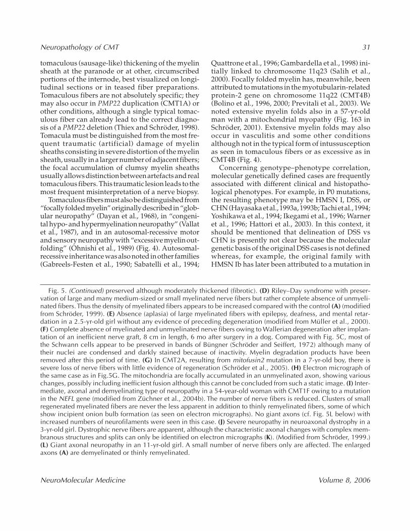
Fig. 1. Neuropathies of predominantly demyelinating type. (A) CMT1A owing to PMP22 duplication showingmultiple onion bulb formations. Nerve fiber density is severely reduced. Instead, the number of Schwann cells andthe connective tissue is increased in amount “hypertrophic neuropathy.” (B) Tomaculous neuropathy (neuropathywith liability to pressure palsies) owing to PMP22 deletion showing only one tomaculous fiber (arrow). (C) Largeonion bulb formations and abnormally shaped and large nuclei (arrows) in neurofibromatous neuropathy type 2(NF2). (D) Congenital hypomyelination neuropathy (CHN) owing to P0 mutation showing numerous hypomyeli-nated or demyelinated fibers and severe reduction of the number of nerve fibers. There is endoneurial edema andthe number of Schwann cells is increased (dark nuclei). The light halo around thinly remyelinated fibers indicates“basal lamina onion bulb formation” (arrows). (E) CHN in PRX mutation showing similar changes as in Fig. 1D,but with some thickly remyelinated fibers. (F) Adult polyglucosan body (PGB) neuropathy resulting from a muta-tion in the branching enzyme gene (GBE). The myelin sheaths around axons containing PGB (arrows) are too thinothers are irregular in contour because of axonal shrinkage. PGB in Schwann cells and perineurial cells are notapparent in this plane of section.
03_Schroder 3/30/06 5:50 PM Page 25

26 Schröder
NeuroMolecular Medicine Volume 8, 2006
of PMP22 on chromosome 17p11.2-p12 (Lupski et al.,1991; Raeymaekers et al., 1991) (Fig. 1A). This typeof neuropathy is structurally characterized by onionbulb formations that were shown by electron
microscopy to be made up of supernumerarySchwann cells concentrically arranged arounddemyelinated and remyelinated axons (Webster etal.,1967). “Onion bulbs” are formed by proliferation of
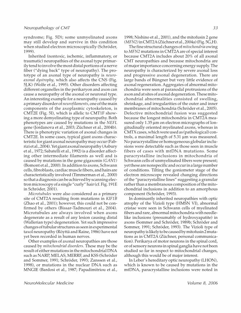
Fig. 2. Optic-electronic evaluation of a cross-sectional area of a semithin section from the sural nerve of a15-yr-old boy (A,C) and his mother (D) with CMTX shown at the highest available light microscopic resolutionwith a planapochromat objective lens (×100) in comparison with an age-matched control (B). The number of nervefibers is reduced and the myelin sheaths are disproportionately thin in relation to the size of their axons and arematched rather precisely by pseudocolors using the KS 300 system of Zeiss-Kontron (Oberkochen, Germany)(Schröder, 1998). The three dimensional diagrams (B–D) illustrate the large scatter of myelin thickness in relationto the axonal dimensions, especially in the boy’s nerve. (Modified from Senderek et al., 1998.)
03_Schroder 3/30/06 5:50 PM Page 26
Neuropathology of CMT 27
NeuroMolecular Medicine Volume 8, 2006
Schwann cells during de- and remyelination,whereas the basal lamina of the demyelinated nervefiber acts as a scaffold to keep the Schwann cells inplace close to the central axon and causing their semi-circular or sometimes even circular arrangement.The number of Schwann cells increases by a factorof 8–14, depending on the size of the degeneratingmyelin sheath, when demyelination occurs, for exam-ple, in experimental allergic neuritis (Schröder, 1968b;Schröder and Krücke, 1970). This figure probablyequals the number of Schwann cells increasing aftersegmental demyelination in hereditary neuropathiesof man. The typical aspect of onion bulb formationsmay be mitigated if CMT1Ais combined with chronicdiabetic neuropathy or other conditions; this mayfinally lead to a severe neuronal type of neuropathywith rather complete loss of myelinated nerve fibers(Thiex and Schröder, 1998; Beckmann and Schröder,2000). The collagen between the layers of Schwanncells is also increasing. The thickness of the collagen
filaments and the amount of collagen tends to bedirectly related to the duration of the disease and thenumber of fibroblasts involved.
Myelin thickness and internodal length of thelargest remyelinated fibers remains usually reduced.Thus on cross-sections the ratio between axon caliberand myelin thickness appears to be increased and,as measured in longitudinal sections or teased fiberpreparations, internodal length is shortened toabout 250–300 µm. These features are of diagnosticimportance for identifying chronic and slowly pro-gressive demyelinating types of neuropathy. Forestimating myelin thickness it must be consideredthat the axon/myelin ratio changes toward thickermyelin sheaths during development as determinedin sural, femoral, radial, and facial nerves reachingthe adult value around puberty (15 yr of age)(Schröder et al., 1978, 1988). Thus myelin sheathsin infants and children are relatively thin. Thenormal range must therefore be kept in mind before
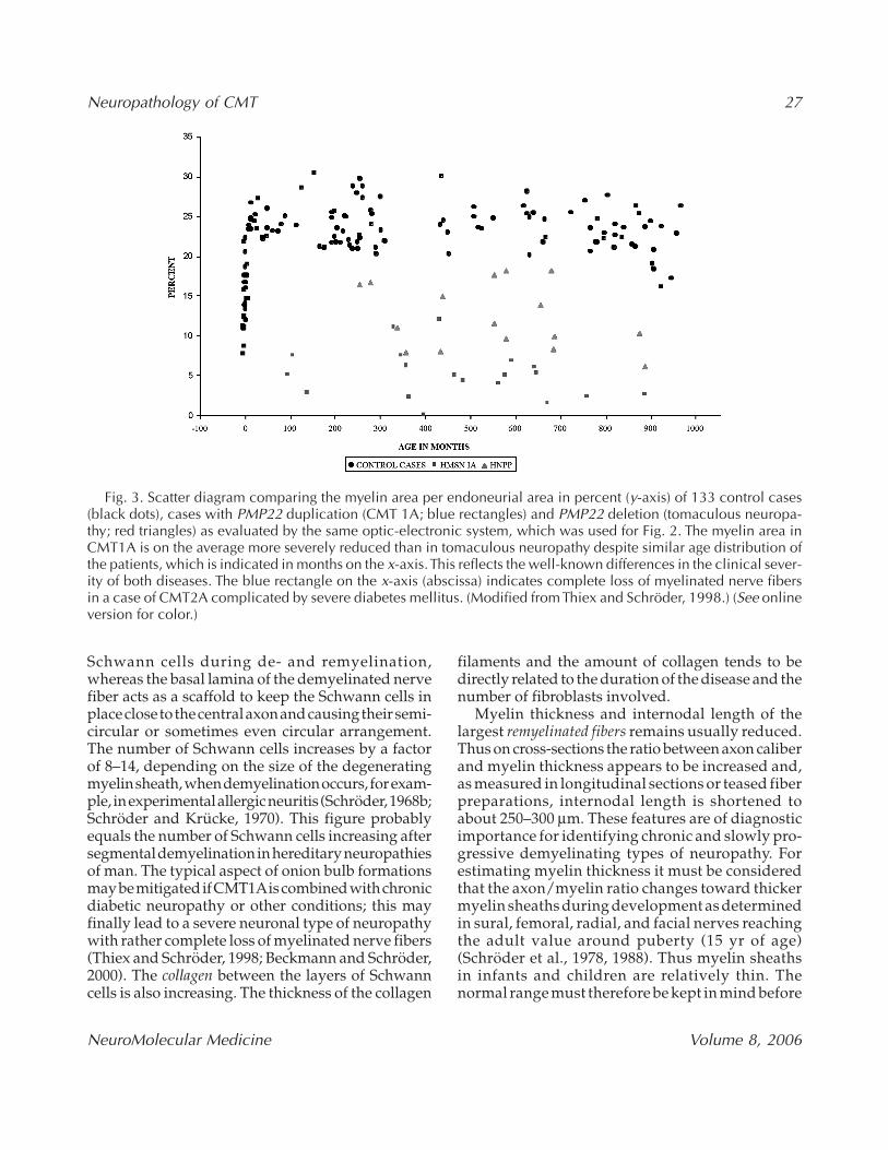
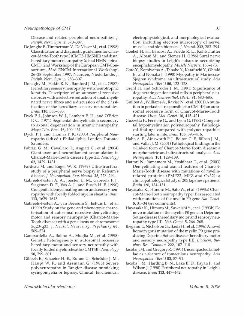
Fig. 3. Scatter diagram comparing the myelin area per endoneurial area in percent (y-axis) of 133 control cases(black dots), cases with PMP22 duplication (CMT 1A; blue rectangles) and PMP22 deletion (tomaculous neuropa-thy; red triangles) as evaluated by the same optic-electronic system, which was used for Fig. 2. The myelin area inCMT1A is on the average more severely reduced than in tomaculous neuropathy despite similar age distribution ofthe patients, which is indicated in months on the x-axis. This reflects the well-known differences in the clinical sever-ity of both diseases. The blue rectangle on the x-axis (abscissa) indicates complete loss of myelinated nerve fibersin a case of CMT2A complicated by severe diabetes mellitus. (Modified from Thiex and Schröder, 1998.) (See onlineversion for color.)
03_Schroder 3/30/06 5:50 PM Page 27

28 Schröder
NeuroMolecular Medicine Volume 8, 2006
diagnosing hypomyelination (see Developmental Dis-orders of the Myelin Sheath section). Followingaxonal degeneration and regeneration the myelinsheaths of the largest nerve fibers do not reach theirregular thickness or a normal axon/myelin ratio,respectively (Schröder, 1972); they remain thinnerso that an increased axon/myelin ratio does notnecessarily indicate develomental delay or pre-ceding segmental demyelination and remyelina-tion. This is particularly important to realize when
isolated nerve fibers have degenerated and regen-erated without apparent formation of typical clus-ters, i.e., bundles of regenerated fibers of unevensize indicating overshooting regeneration (“hyper-neurotization” of bands of Büngner) (Schröder,1968a). Myelin sheaths may also become dispro-portionately thin when axons increase in diameteras seen in polyglucosan body neuropathy (Fig. 1F),or giant axonal neuropathy (Asbury et al., 1972)(Fig. 5L).
Fig. 4. Severe reduction of the number of nerve fibers, multiple onion bulb formations with demyelinated orthinly remyelinated axons in their center, and five typical, focally folded myelin sheaths (arrows in A; electronmicrographs in B and C) in the sural nerve of a 8-yr-old boy with CMT4B. The axons (A) in B and C are thinlyremyelinated despite severe “hypertrophy” of their myelin sheaths. (B and C modified from Schröder, 1999.)
03_Schroder 3/30/06 5:50 PM Page 28

Neuropathology of CMT 29
NeuroMolecular Medicine Volume 8, 2006
Progressive demyelination is a common featurein “congenital hypomyelination neuropathy”(CHN) (Kennedy, 1977; Guzzetta et al., 1982). CHNis clinically characterized by a Dejerine Sottas syn-drome (DSS) with severely reduced conductionvelocity (few m/s) and structurally characterizedby demyelinated axons, disproportionately thinmyelin sheaths following remyelination, and onionbulb formations of a special type, i.e., “basal laminaonion bulbs” Fig. 1D,E). In these, the circularly ori-ented Schwann cells processes have degenerated,whereas their basal laminae are persisting. Pro-gressive CHN may be caused by point mutations inthe following genes: PMP22 (Simonati et al., 1999),myelin protein zero (MPZ; P0) (Warner et al., 1996)(Fig. 1D), early growth response gene (EGR2)(Warner et al., 1998; Timmerman et al., 1999), peri-axin (PRX) (CMT4F; Fig. 1E) (Boerkoel et al., 2001;Guilbot et al., 2001; Takashima et al., 2002), N-mycdownstream-regulated gene 1 (NDRG1) (HMSNLom) (Baethmann et al., 1998; Kalaydjieva et al.,1998; Merlini et al., 1998; King et al., 1999; Chandleret al., 2000; Colomer et al., 2000; Kalaydjieva et al.,2000), and KIAA1985 (CMT4C) (Senderek et al.,2003b). Distinguishing features between these dif-ferent forms of CHN were elaborated for the firsttime by immunohistochemical technics for stainingsubunits of periaxin (Takashima et al., 2002) allow-ing a specific morphological diagnosis. Immuno-histochemical diagnosis has thus far not beenachieved in other hereditary neuropathies exceptfor amyloidosis (Linke, 1982). In CMT4C, thinextended Schwann cell processes were described asa distinguishing feature (Kessali et al., 1997;Gabreels-Festen et al., 1999; Senderek et al., 2003b),although this does not appear to be specific; clini-cally thoracic scoliosis appears to be a more char-acteristic feature in KIAA1985 mutations.
It is of interest that onion bulb formation in auto-immune disorders such as Guillain Barré syndromeand chronic inflammatory demyelinating neuropa-thy (CIDP) do not result in basal lamina onion bulbformations so typical for CHN. Schwann cells inearly manifesting, genetically determined neu-ropathies are obviously more liable to die and towaste away, forming empty basal lamina onion bulbsthan in disorders in which the myelin sheath isattacked by immunological pathomechanisms lead-ing, for example, to chronic inflammatory demyeli-nating neuropathy or multifocal hypertrophic
neuropathy with palpably enlarged nerves owingto increased numbers of Schwann cells, fibroblasts,and collagen (Webster et al., 1967).
Cell “viability” is even more obvious in neoplasticdisorders such as neurofibromatosis type 2, in whichonion bulb formations appear to be more cellularand larger in size showing prominent nuclei withirregular shape (Fig. 1C) (Sperfeld et al., 2002).
The myelin sheath may also be disproportion-ately thick in relation to the size of the axon. Thepathomechanism underlying this type of dispropor-tionately increased myelin thickness or myelin “hyper-trophy” is not definitely clarified, but can best beexplained by a model of concentric slippage of thespiral of the myelin lamella around the axon fol-lowing reduction of the axonal perimeter. It is seenmore or less frequently in nearly all types of neu-ropathy because of axonal atrophy and may beowing to primary disorders of neurons or axons, ormay follow secondarily to segmental demyelina-tion in a series of proximal segments (Fig. 1A). Onthe other hand, axonal atrophy may cause secondarysegmental demyelination, which can only be verifiedmorphometrically using statistical analysis of teasedfiber preparations as exemplified in uremic neu-ropathy and Friedreich’s disease (Dyck et al., 1971;Thomas et al., 1971). Because this needs laboriousverification of an increased number of demyelinatedor remyelinated segments on very long teased nervefibers in primarily axonal types of neuropathy, thismethod did not gain widespread application. Nev-ertheless, secondary segmental demyelination mustalways be considered when, on cross-section, dis-proportionately thin myelin sheaths are seen in aprimarily neuronal or axonal type of neuropathy.
“Hypertrophy” of myelin sheaths is also seen in“tomaculous neuropathy” or “hereditary neuropa-thy with liability to pressure palsies” (Behse et al.,1972; Madrid and Bradley, 1975; Jacobs and Gregory,1991; Malandrini et al., 1992; Thomas et al., 1994;Mancardi et al., 1995; Tachi et al., 1997). It is usuallycaused by PMP22 deletion (Chance et al., 1993) andtends to be less severe than CMT1Acaused by PMP22duplication (Fig. 3). Typical tomaculous fibers (Fig.1B) are the result of a pathomechanism of intus-susception or invagination of a hypertrophicmyelin sheath by a double, complete ring of addi-tional myelin around itself thus causing three com-plete layers of myelin sheaths on cross-sections(Madrid and Bradley, 1975). This results in focal
03_Schroder 3/30/06 5:50 PM Page 29

30 Schröder
NeuroMolecular Medicine Volume 8, 2006
Fig. 5. Representative axonal types neuropathy in comparison with a normal control (A). (B) Complete aplasiaof myelinated nerve fibers in a 14-yr-old girl without any evidence of preceding degeneration of myelinated nervefibers and with corresponding hypoplasia of this nerve fascicle (most unmyelinated fibers are preserved) (modi-fied from Schröder et al., 1993a). (C) Complete degeneration of myelinated and unmyelinated nerve fibers includ-ing their Schwann cells in the sural nerve of a case with Tangier disease resulting from a mutation in the ABC1gene (case V:2 in Züchner et al., 2003). There are only several fibroblasts and a single blood vessel left over withinthis sclerotic nerve fascicle, which is filled with collagen (fibrosis). The perineurium is remarkably well
03_Schroder 3/30/06 5:50 PM Page 30
Neuropathology of CMT 31
NeuroMolecular Medicine Volume 8, 2006
tomaculous (sausage-like) thickening of the myelinsheath at the paranode or at other, circumscribedportions of the internode, best visualized on longi-tudinal sections or in teased fiber preparations.Tomaculous fibers are not absolutely specific; theymay also occur in PMP22 duplication (CMT1A) orother conditions, although a single typical tomac-ulous fiber can already lead to the correct diagno-sis of a PMP22 deletion (Thiex and Schröder, 1998).Tomacula must be distinguished from the most fre-quent traumatic (artificial) damage of myelinsheaths consisting in severe distortion of the myelinsheath, usually in a larger number of adjacent fibers;the focal accumulation of clumsy myelin sheathsusually allows distinction between artefacts and realtomaculous fibers. This traumatic lesion leads to themost frequent misinterpretation of a nerve biopsy.
Tomaculous fibers must also be distinguished from“focally folded myelin” originally described in “glob-ular neuropathy” (Dayan et al., 1968), in “congeni-tal hypo- and hypermyelination neuropathy” (Vallatet al., 1987), and in an autosomal-recessive motorand sensory neuropathy with “excessive myelin out-folding” (Ohnishi et al., 1989) (Fig. 4). Autosomal-recessive inheritance was also noted in other families(Gabreels-Festen et al., 1990; Sabatelli et al., 1994;
Quattrone et al., 1996; Gambardella et al., 1998) ini-tially linked to chromosome 11q23 (Salih et al.,2000). Focally folded myelin has, meanwhile, beenattributed to mutations in the myotubularin-relatedprotein-2 gene on chromosome 11q22 (CMT4B)(Bolino et al., 1996, 2000; Previtali et al., 2003). Wenoted extensive myelin folds also in a 57-yr-oldman with a mitochondrial myopathy (Fig. 163 inSchröder, 2001). Extensive myelin folds may alsooccur in vasculitis and some other conditionsalthough not in the typical form of intussusceptionas seen in tomaculous fibers or as excessive as inCMT4B (Fig. 4).
Concerning genotype–phenotype correlation,molecular genetically defined cases are frequentlyassociated with different clinical and histopatho-logical phenotypes. For example, in P0 mutations,the resulting phenotype may be HMSN I, DSS, orCHN (Hayasaka et al., 1993a, 1993b; Tachi et al., 1994;Yoshikawa et al., 1994; Ikegami et al., 1996; Warneret al., 1996; Hattori et al., 2003). In this context, itshould be mentioned that delineation of DSS vsCHN is presently not clear because the moleculargenetic basis of the original DSS cases is not definedwhereas, for example, the original family withHMSN Ib has later been attributed to a mutation in
Fig. 5. (Continued) preserved although moderately thickened (fibrotic). (D) Riley–Day syndrome with preser-vation of large and many medium-sized or small myelinated nerve fibers but rather complete absence of unmyeli-nated fibers. Thus the density of myelinated fibers appears to be increased compared with the control (A) (modifiedfrom Schröder, 1999). (E) Absence (aplasia) of large myelinated fibers with epilepsy, deafness, and mental retar-dation in a 2.5-yr-old girl without any evidence of preceding degeneration (modified from Müller et al., 2000).(F) Complete absence of myelinated and unmyelinated nerve fibers owing to Wallerian degeneration after implan-tation of an inefficient nerve graft, 8 cm in length, 6 mo after surgery in a dog. Compared with Fig. 5C, most ofthe Schwann cells appear to be preserved in bands of Büngner (Schröder and Seiffert, 1972) although many oftheir nuclei are condensed and darkly stained because of inactivity. Myelin degradation products have beenremoved after this period of time. (G) In CMT2A, resulting from mitofusin2 mutation in a 7-yr-old boy, there issevere loss of nerve fibers with little evidence of regeneration (Schröder et al., 2005). (H) Electron micrograph ofthe same case as in Fig.5G. The mitochondria are focally accumulated in an unmyelinated axon, showing variouschanges, possibly including inefficient fusion although this cannot be concluded from such a static image. (I) Inter-mediate, axonal and demyelinating type of neuropathy in a 54-year-old woman with CMT1F owing to a mutationin the NEFL gene (modified from Züchner et al., 2004b). The number of nerve fibers is reduced. Clusters of smallregenerated myelinated fibers are never the less apparent in addition to thinly remyelinated fibers, some of whichshow incipient onion bulb formation (as seen on electron micrographs). No giant axons (cf. Fig. 5L below) withincreased numbers of neurofilaments were seen in this case. (J) Severe neuropathy in neuroaxonal dystrophy in a3-yr-old girl. Dystrophic nerve fibers are apparent, although the characteristic axonal changes with complex mem-branous structures and splits can only be identified on electron micrographs (K). (Modified from Schröder, 1999.)(L) Giant axonal neuropathy in an 11-yr-old girl. A small number of nerve fibers only are affected. The enlargedaxons (A) are demyelinated or thinly remyelinated.
03_Schroder 3/30/06 5:50 PM Page 31

32 Schröder
NeuroMolecular Medicine Volume 8, 2006
the P0 gene (Bird et al., 1997). As outlined earlier,CHN may be caused by at least six different genes.
Developmental Disorders of the Myelin Sheath
These consist of apparently extremely rare con-ditions with complete absence of myelin only in theperipheral nervous system (PNS; Palix and Coignet,1978; Charnas et al., 1988), or in the PNS and cen-tral nervous system (CNS; Schröder and Bohl, 1978)(Fig. 5B). Disproportionately thin myelin sheathswithout any or very little evidence of progressionhave been reported, in addition to the conditionsmentioned earlier and also in systemic disorderswith presumably anabolic disturbance of myelina-tion (Schröder, 1982a) such as Leigh’s syndrome(Goebel et al., 1986; Peiffer et al., 1988; Jacobs et al.,1990), Cockayne syndrome (Moosa and Dubowitz,1970; Ohnishi et al., 1987; Schröder, 2001), andmuscle phosphoglycerate kinase deficiency(Schröder et al., 1996). It is not settled whether dis-proportionately thin myelin sheath, such as seen incertain cases with Cx32 mutation (CMTX; Fig. 2),are caused by a developmental disturbance or earlydemyelination and remyelination.
There is a great variety of other fine structural changesof myelin sheaths. Although recommended, the com-plete spectrum of myelin changes is only rarelyevaluated because a large number of electron micro-graphs at different magnifications are needed (cf.,Table 3 in Senderek et al., 2000). Which of these finestructural changes of the myelin sheath are associ-ated with which and how many mutations is notyet settled. For example, dissociation of terminalmyelin loops from the axon and loss of transversebands was defined as the initial change in periaxinneuropathy (CMT4F) (Takashima et al., 2002). Yetthe paranodium is the site of initial changes in otherdemyelinating neuropathies such as diphtheric neu-ropathy (Webster et al., 1961), although exactly sim-ilar changes have not been documented at theelectron microscopic level in diphtheritic and otherneuropathies.
Changes of Axons
Neuropathies of “axonal” or “neuronal” type(Fig. 5A–L) are 3.2-fold more frequent in the pre-sent author’s data bank of 8052 sural nerve biopsies,collected over 40 yr, than those of a “demyelinating”
type (4500:1406). Of these, 578 were classified as“mixed axonal and demyelinating” in type. The othercases were not classified using this simplifyingscheme.
The distinction between an axonal and a neuronaltype of neuropathy may be arbitrary. But we con-sider neuropathies of axonal vs neuronal type whenthere is evidence of regeneration, i.e., when thereare clusters of regenerated or regenerating fibers;these clusters indicate persisting viability of the cellbody (perikaryon of the neuron). When there is lossof nerve fibers with no evidence of regeneration, aneuronal type of neuropathy is assumed which ini-tially does not exclude more proximal viability ofthe axon and neuron. However, in a chronic type ofneuropathy of several years duration at least someregenerating sprouts forming clusters within theiroriginal basal lamina or band of Büngner shouldhave reached the distal (sural) site of the nervebiopsy if the proximal axon and neuron have per-sisted. The preponderance of axonal types of neu-ropathy is presumably owing to the extraordinarylength of the axon. An axon depends on the integrityof its perikaryon as well as on adequate blood supplyfor nutrition, and a proper endoneurial milieu alongits length.
Developmental disturbances of peripheral neurons areseen in several rare conditions. The most severe formincludes total absence of myelinated fibers asalready mentioned when describing disorders ofmyelination. Aselective involvement of certain neu-ronal systems is apparent in the following diseases,which need to be ascertained using morphometry. Oneof these conditions is characterized by autosomal-recessive deficiency of large myelinated nerve fibers inthe sural nerve (Sabatelli et al., 1998) with a corre-sponding deficiency of large neurons in spinalganglia and in the spinal cord (Müller et al., 2000)(Fig. 5E). The condition is nearly nonprogressive,and is associated with epilepsy, deafness, and mentalretardation. The underlying molecular genetic back-ground has not yet been analyzed. Ahereditary sen-sory neuropathy with selective reduction of smallmyelinated nerve fibers has also been described (Don-aghy et al., 1987). This condition may be difficult todifferentiate from cases with nonhereditary “smallfiber neuropathy,” which is increasingly studiedusing skin biopsies. Rather complete absence ofunmyelinated fibers is seen in hereditary sensory auto-nomic neuropathy type III (HSAN III; Riley–Day
03_Schroder 3/30/06 5:50 PM Page 32
Neuropathology of CMT 33
NeuroMolecular Medicine Volume 8, 2006
syndrome; Fig. 5D); some unmyelinated axonsmay still develop and survive in this conditionwhen studied electron microscopically (Schröder,1999).
Inherited (nontoxic, ischemic, inflammatory, ortraumatic) neuropathies of the axonal type primar-ily tend to involve the most distal portions of a nervefiber (“dying back” type of neuropathy). The pro-totype of an axonal type of neuropathy is neuro-axonal dystrophy, which also affects the CNS (Fig.5J,K) (Wolfe et al., 1995). Other disorders affectingdifferent organelles in the perikaryon and axon cancause a neuropathy of the axonal or neuronal type.An interesting example for a neuropathy caused bya primary disorder of neurofilaments, one of the maincomponents of the axoplasmic cytoskeleton, isCMT2E (Fig. 5I), which is allelic to CMT1F show-ing a more demyelinating type of neuropathy. Bothphenotypes are caused by mutations in the NEFLgene (Jordanova et al., 2003; Züchner et al., 2004b).There is phenotypic variation of axonal changes inCMT2E. In some cases, typical giant axons charac-teristic for giant axonal neuropathy may occur (Fab-rizi et al., 2004). Yet giant axonal neuropathy (Asburyet al., 1972; Sabatelli et al., 1992) is a disorder affect-ing other intermediate filaments as well and iscaused by mutations in the gene gigaxonin (GAN1)(Bomont et al., 2000). In addition to axons, Schwanncells, fibroblasts, cardiac muscle fibers, and hairs arecharacteristically involved (Timmerman et al., 2000)so that a diagnosis can be achieved by scanning elec-tron microscopy of a single “curly” hair (cf. Fig. 191Ein Schröder, 2001).
Microtubules were also considered as a primarysite of CMT2A resulting from mutations in KIF1B(Zhao et al., 2001); however, this could not be con-firmed by others (Bissar-Tadmouri et al., 2004).Microtubules are always involved when axonsdegenerate as a result of any lesion causing distal(Wallerian type) degeneration. Yet such impressivechanges of tubular structures as seen in experimentaltaxol neuropathy (Röyttä and Raine, 1986) have notyet been recorded in human nerves.
Other examples of axonal neuropathies are thosecaused by mitochondrial disorders. These may be theresult of either mutations in the mitochondrial DNAsuch as NARP, MELAS, MERRF, and KSS (Schröderand Sommer, 1991; Schröder, 1993; Zanssen et al.,1998), or mutations in the nuclear DNA such asMNGIE (Bardosi et al., 1987; Papadimitriou et al.,
1998; Nishino et al., 2001), and the mitofusin 2 gene(MFN2) in CMT2A(Züchner et al., 2004a) (Fig. 5G,H).
The fine structural changes of mitochondria owingto MFN2 mutations in CMT2A are of special interestbecause CMT2A includes about 20% of all axonalCMT neuropathies and because mitochondria areof major importance concerning energy supply. Theneuropathy is characterized by severe axonal lossand progressive axonal degeneration. There arelarge bands of Büngner but very little evidence ofaxonal regeneration. Aggregates of abnormal mito-chondria were seen at paranodal protrusions of theaxon and at sites of axonal degeneration. These mito-chondrial abnormalities consisted of swelling,shrinkage, and irregularities of the outer and innermembranes of mitochondria (Schröder et al., 2005).Defective mitochondrial fusion was suggestedbecause the longest mitochondria in CMT2A mea-sured only 1.35 µm on electron micrographs of lon-gitudinally oriented myelinated axons, whereas inCMTX cases, which were used as (pathological) con-trols, a maximal length of 5.31 µm was measured.No paracrystalline or homogeneous globular inclu-sions were detectable such as those seen in musclefibers of cases with mtDNA mutations. Someparacrystalline inclusions in mitochondria ofSchwann cells of unmyelinated fibers were present;yet these are nonspecific and occur in a large numberof conditions. Tilting the goniometer stage of theelectron microscope revealed changing directionsof the “paracrystalline lines” suggesting a granularrather than a membranous composition of the mito-chondrial inclusions in addition to an amorphouscomponent (Schröder, 1993).
In dominantly inherited neuropathies with opticatrophy of the Vizioli type (HMSN VI), abnormalcristae were seen in Schwann cells of myelinatedfibers and rare, abnormal mitochondria with needle-like inclusions (presumably of hydroxyapatite) inaxons (Sommer and Schröder, 1989b; Schröder andSommer, 1991; Schröder, 1993). The Vizioli type ofneuropathy is likely to be caused by mitofusin 2 muta-tions as in CMT2A (Züchner, personal communica-tion). Perikarya of motor neurons in the spinal cord,or of sensory neurons in spinal ganglia have not beenstudied so far in respect to mitochondrial changes,although this would be of major interest.
In Leber’s hereditary optic neuropathy (LHON),which is known to be caused by mutations in themtDNA, paracrystalline inclusions were noted in
03_Schroder 3/30/06 5:50 PM Page 33

34 Schröder
NeuroMolecular Medicine Volume 8, 2006
Schwann cells of unmyelinated fibers in which theyhave already been seen in various neuropathies(Lyon and Evrard, 1970) including Refsum’s dis-ease (Fardeau and Engel, 1969) and mitochondrialmyopathy of undefined cause (Yiannikas et al.,1983). In Schwann cells of myelinated fibers, closelyopposed mitochondrial cristae with paracrystallinematerial were an interesting finding not seen in con-trols (Sommer and Schröder, 1989b). It would beworthwhile to study DNAin archival material fromthese cases to classify these disorders with molec-ular genetic techniques as has been done in othercases (Thiex and Schröder, 1998; Senderek et al.,1998, 1999, 2000, 2003a; Schröder et al., 1999b;Züchner et al., 2003).
Intermediate (Axonal andDemyelinating) Forms of Neuropathy
An increasing number of neuropathies are nei-ther typically demyelinating (conduction velocities<45 m/s) nor typically axonal (conduction veloci-ties ranging from normal to 25 m/s). These are des-ignated as “intermediate” and occur followingmutations, for example, in Cx32 (Rouger et al., 1997;Senderek et al., 1999a), NEFL (Züchner et al., 2004b)(Fig. 5I) and GDAP1 (Senderek et al., 2003a) andother dominant intermediate forms of CMT (DI-CMT) such as those caused by mutations of the pleck-strin homology domain of dynamin 2 (DNM2)(Züchner et al., 2005). Nerve biopsies from the lattertype of neuropathy are not available as long as nobiopsies, previously performed and stored inarchives of certain laboratories, have been attributedto such a mutation. Biopsies in cases with GDAP1mutations revealed evidence of axonal degenerationand regeneration as well as segmental demyelina-tion and remyelination with a moderate degree ofonion bulb formation (Senderek et al., 2003a). In P0mutations, a predominantly axonal type of neu-ropathy may be encountered (Senderek et al., 1999),although P0 is the major component of peripheralmyelin sheaths.
Endoneurial Connective Tissue Changes
The endoneurial connective tissue is regularly,although seemingly always secondarily, involved
in all types of peripheral neuropathy. These neu-ropathies are made up of various forms of (heredi-tary) amyloidosis that can be delineated anddifferentiated immunohistochemically and electronmicroscopically by their characteristic fibrillarendoneurial deposits (for references see Sommer andSchröder, 1989a).
The endoneurial collagen is particularly increasedin chronic “hypertrophic” types of neuropathywhere an interaction between proliferated Schwanncells and fibroblasts may lead to a severe increaseof the endoneurial collagen. As mentioned earlier,collagen fibers are, in chronic cases, a major com-ponent of onion bulb formations. The increase ofcollagen may be most prominent in multifocalhypertrophic neuropathy (Webster et al., 1967). Asillustrated in Fig. 5C, collagen may persist and maybe the major endoneurial component after allparenchymal elements (axons and Schwann cells)have disappeared.
Degenerating endoneurial fibroblasts are a fre-quent phenomenon in a large spectrum of periph-eral neuropathies, including inherited neuropathies.They are particularly frequent in vasculitic neu-ropathy (Grehl and Schröder, 1991).
Neuropathy as a result of neurofibromatosis type 2(Fig. 1C; v. s.) may be included in this group of dis-eases, although Schwann cells, which are consideredas parenchymal cells, appear to be more severelyaffected in this disease than fibroblasts which, asidefrom blood vessels, are the main cellular compo-nent of the connective tissue. Quiescent or activeendoneurial macrophages may also be considered asa component of the connective tissue. They play amajor role in various storage diseases or in remov-ing debris from degenerating nerve fibers.Macrophages with large vacuoles occur in chronichereditary neuropathies and are nonspecificallyindicating chronicity. Matrix metalloproteinases as aprimary site of a genetic disorder have thus far notbeen delineated as a cause of hereditary peripheralneuropathy, although they form an important com-ponent in peripheral nerves.
Perineurial Changes
Arare, presumably heterogeneous and autosomal-recessive, “complicated” type of neuropathy is asso-ciated with mental retardation and cataract. The
03_Schroder 3/30/06 5:50 PM Page 34

Neuropathology of CMT 35
NeuroMolecular Medicine Volume 8, 2006
hallmark are perineurial alterations consisting ofunusual, abnormally proliferated, indented, andextended perineurial cells in the endoneurium(Schröder et al., 1999a; Thomas et al., 2000; Wolfeet al., 2000). These are unlike those seen in “mini-fascicles” and perineurioma (Schröder, 1999).
Similar perineurial cell alterations were notnoted in Marinesco-Sjögren syndrome, which isalso characterized by mental retardation andcataracts but additionally by short stature. It hasrecently been attributed to mutations in SIL1(Senderek et al., 2005) and shows a nonprogres-sive or very slowly progressive neuropathy of amixed, axonal, and demyelinating type ( Serratriceet al., 1973; Alexianu et al., 1983); this is supportedby muscle biopsies revealing some degree of neuro-genic atrophy (Zimmer et al., 1992). Yet the impres-sive and seemingly specific fine structural changesinvolving sarcolemmal nuclei (Schröder, 1982b;Goto et al., 1990; Sewry et al., 1988) were not seenin peripheral nerves.
Vascular Changes
Certain hereditary diseases are associated withgeneralized vascular changes. These diseases maybe diagnosed by a peripheral nerve biopsy becauseof characteristic alterations in endothelial, smoothmuscle, or perivascular cells. Such diseases includeneuraminidase A and B (Sandhoff ’s disease)(Schröder, 2001), CADASIL (Schröder et al., 1995;Schröder et al., 2005), and ferritinopathy ( Schröder,2005), which has previously been described as“granular nuclear inclusion body disease” (Schröderet al., 1985).
Hereditary Multisystem (Metabolic)Diseases With Characteristic ChangesAffecting Peripheral Nerves More or Less Severely
As already mentioned, the most severe neu-ropathy in our whole series of 8052 sural nerve biop-sies was neuronal in type with complete loss of axonsas well as Schwann cells in a case of Tangier disease(Fig. 5C). This disease is caused by a mutation inthe ATPase binding cassette transporter gene 1
(ABC1) gene (Züchner et al., 2003). There were onlya few surviving blood vessels and some macro-phages with characteristic lipid storage products inthe endoneurium, whereas the perineurium and theepineurium were rather well preserved. Usually,following Wallerian degeneration and definite lossof axons, Schwann cells tend to survive in bands ofBüngner despite lack of reinnervation (Fig. 5F).Other cases were less severely affected showingcharacteristic endoneurial macrophages andSchwann cells with lipid inclusions that are notmembrane-bound (Gibbels et al., 1985). When thelipids are extracted during the embedding proce-dure these inclusions may be misinterpreted as“membrane-bound vacuoles.”
Fabry’s disease, Sandhoff ’s disease (neu-raminidase Aand B deficiency), gangliosidosis types1 and 2, Niemann Pick’s disease, and various formsof mucopolysaccharidosis (Chen et al., 1999) shouldalso be mentioned because of characteristic fine struc-tural changes allowing a specific diagnosis (for illus-trations see Schröder, 2001). Among the latter,Sanfilippo’s disease may be associated with a severeform of peripheral neuropathy. In addition, polyglu-cosan body disease (branching enzyme deficiency;glycogenosis type IV) owing to mutations in thebranching enzyme gene may cause a peripheral neu-ropathy showing impressive and specific inclusions(polyglucosan bodies) in axons (Fig. 1F), Schwanncells, and perineurial cells (Busard et al., 1990;Schröder et al., 1993b; Ziemssen et al., 2000), even-tually also in epineurial smooth muscle cells. In perox-isomal diseases bilaminar or trilaminar inclusions inSchwann cells and other cells may allow a group-specific fine structural diagnosis (Schröder et al.,2004). These diseases, however, are not restricted tothe PNS, although PNS symptoms may predominate.
In conclusion, although most pathological changesin peripheral nerves are nonspecific, a large numberof those listed allow a more or less specific fine struc-tural diagnosis. In many cases, it remains a difficultneuropathological task to distinguish pathognosticfrom nonspecific and normal structures in developing,adult, and aging peripheral nerves.
Acknowledgments
The nerve specimen and molecular genetic diag-nosis to Fig. 1D (P0 mutation) were kindly provided
03_Schroder 3/30/06 5:50 PM Page 35

36 Schröder
NeuroMolecular Medicine Volume 8, 2006
by Dr. U. Schara and Prof. Dr. W. Mortier (Bochum,Germany). The diagnosis “CMT4C” for the caseillustrated in Fig. 4 is based on molecular geneticstudies (personal communication by Dr. JanSenderek, Institut für Humangenetik, Universität-sklinikum, RWTH Aachen, Germany). The other fig-ures were modified from published cases (Schröder,1999; Schröder, 2001) in the author’s archive.
References
Alexianu M. D., Christodoresc C., Vasilescu A., et al.(1983) Sensorimotor neuropathy in a patient withMarinesco-Sjögren syndrome. Eur. Neurol. 22,222–226.
Asbury A. K., Gale M. K., Cox S. C., Baringer J. R., andBerg B. O. (1972) Giant axonal neuropathy—aunique case with segmental neurofilamentousmasses. Acta Neuropathol. (Berl.) 20, 237–247.
Baethmann M., Göhlich-Ratmann G., Schröder J. M.,Kalaydjieva L., and Voit T. (1998) HMSNL in a 13-year-old Bulgarian girl. Neuromusc. Disord. 8,90–94.
Bardosi A., Creutzfeldt W., DiMauro S., et al. (1987)Myo-, neuro-, gastrointestinal encephalopathy(MNGIE syndrome) due to partial deficiency ofcytochrome-c-oxidase. Anew mitochondrial mul-tisystem disorder. Acta Neuropathol. (Berl.) 74,248–258.
Beckmann A. and Schröder J. M. (2000) Screening forCharcot-Marie-Tooth type 1Aand hereditary neu-ropathy with liability to pressure palsy in archivalnerve biopsy samples by direct-double-differen-tial PCR. Acta Neuropathol. 100, 459–463.
Behse F., Buchthal F., Carlsen F., and Knappeis G. G.(1972) Hereditary neuropathy with liability topressure palsies. Electrophysiological andhistopathological aspects. Brain 95, 777–794.
Bergoffen J., Scherer S. S., Wang S., et al. (1993) Con-nexin mutations in X-linked Charcot-Marie-Toothdisease. Science 262, 2039–2042.
Bertram M. and Schröder J. M. (1993) Developmentalchanges at the node and paranode in human suralnerves: morphometric and fine-structural evalu-ation. Cell Tissue Res. 273, 499–509.
Bird T. D., Kraft G. H., Lipe H. P., Kenney K. L., andSumi S. M. (1997) Clinical and pathological phe-notype of the original family with Charcot-Marie-Tooth type 1B: a 20-year study. Ann. Neurol. 41,463–469.
Bissar-Tadmouri N., Nelis E., Züchner S., et al. (2004)Absence of KIF1B mutation in a large Turkish
CMT2A family suggests involvement of a secondgene. Neurology 62, 1522–1525.
Boerkoel C. F., Takashima H., Stankiewicz P., et al. (2001)Periaxin mutations cause recessive Dejerine-Sottasneuropathy. Am. J. Hum. Genet. 68, 325–333.
Bolino A., Brancolini V., Bono F., et al. (1996) Local-ization of a gene responsible for autosomal reces-sive demyelinating neuropathy with focally foldedmyelin sheaths to chromosome 11q23 by homozy-gosity mapping and haplotype sharing. Hum. Mol.Genet. 5, 1051–1054.
Bolino A., Muglia M., Conforti F. L., et al. (2000) Char-cot-Marie-Tooth type 4B is caused by mutationsin the gene encoding myotubularin-related pro-tein-2. Nat. Genet. 25, 17–19.
Bomont P., Cavalier L., Blondeau F., et al. (2000) Thegene encoding gigaxonin, a new member of thecytoskeletal BTB/kelch repeat family, is mutatedin giant axonal neuropathy. Nat. Genet. 26, 370–374.
Busard H., Gabreels-Festen A., Van t’Hof M., RenierW., and Gabreels F. (1990) Polyglucosan bodies insural nerve biopsies. Acta Neuropathol. (Berl.) 80,554–557.
Chance P. F., Alderson M. K., Leppig K. A., et al. (1993)DNA deletion associated with hereditary neu-ropathy with liability to pressure palsies. Cell 72,143–151.
Chandler D., Angelicheva D., Heather L., et al. (2000)Hereditary motor and sensory neuropathy - lom(HMSNL): refined genetic mapping in romani(Gypsy) families from several european countries.Neuromuscul. Disord. 10, 584–591.
Charnas L., Trapp B., and Griffin J. (1988) Congenitalabsence of peripheral myelin: abnormal Schwanncell development causes lethal arthrogryposismultiplex congenita. Neurology. 38, 966–974.
Chen C. S., Patterson M. C., Wheatley C. L., O’BrienJ. F., and Pagano R. E. (1999) Broad screening testfor sphingolipid-storage diseases. Lancet 354,901–905 (see comments).
Colomer J., Iturriaga C., Kalaydjieva L., AngelichevaD., King R. H., and Thomas P. K. (2000) Hereditarymotor and sensory neuropathy-Lom (HMSNL) ina spanish family: clinical, electrophysiological,pathological and genetic studies.NeuromusculDisord. 10, 578–583.
Dayan A. D., Graveson G. S., Robinson P. K., and Wood-house M. A. (1968) Globular neuropathy. A disor-der of axons and Schwann cells. J. Neurol.Neurosurg. Psychiatry 31, 552–560.
De Jonghe P., Timmerman V., Nelis E., Martin J. J., andVan Broeckhoven C. (1997) Charcot-Marie-Tooth
03_Schroder 3/30/06 5:50 PM Page 36
Neuropathology of CMT 37
NeuroMolecular Medicine Volume 8, 2006
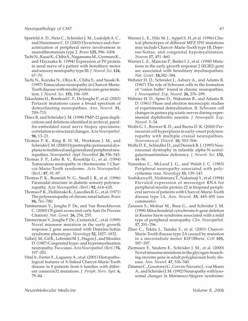
Disease and related peripheral neuropathies. J.Periph. Nerv. Syst. 2, 370–387.
De Jonghe P., Timmerman V., De Visser M., et al. (1998)Classification and diagnostic guidelines for Char-cot-Marie-Tooth type 2 (CMT2-HMSNII) and distalhereditary motor neuropathy (distal HMN-spinalCMT). 2nd Workshop of the European CMT-Con-sortium, 53rd ENCM International Workshop, 26–28 September 1997, Naarden, Niederlande. J.Periph. Nerv. Syst. 3, 283–307.
Donaghy M., Hakin R. N., Bamford J. M., et al. (1987)Hereditary sensory neuropathy with neurotrophickeratitis. Description of an autosomal recessivedisorder with a selective reduction of small myeli-nated nerve fibres and a discussion of the classi-fication of the hereditary sensory neuropathies.Brain 110, 563–583.
Dyck P. J., Johnson W. J., Lambert E. H., and O’BrienP. C. (1971) Segmental demyelination secondaryto axonal degeneration in uremic neuropathy.Mayo Clin. Proc. 46, 400–431.
Dyck, P. J. and Thomas P. K. (2005) Peripheral Neu-ropathy (4th ed.). Philadelphia, London, Toronto:Saunders.
Fabrizi G. M., Cavallaro T., Angiari C., et al. (2004)Giant axon and neurofilament accumulation inCharcot-Marie-Tooth disease type 2E. Neurology62, 1429–1431.
Fardeau M. and Engel W. K. (1969) Ultrastructuralstudy of a peripheral nerve biopsy in Refsum’sdisease. J. Neuropathol. Exp. Neurol. 28, 278–294.
Gabreels-Festen A. A., Joosten E. M., Gabreels F. J.,Stegeman D. F., Vos A. J., and Busch H. F. (1990)Congenital demyelinating motor and sensory neu-ropathy with focally folded myelin sheaths. Brain113, 1629–1643.
Gabreels-Festen A., van Beersum S., Eshuis L., et al.(1999) Study on the gene and phenotypic charac-terisation of autosomal recessive demyelinatingmotor and sensory neuropathy (Charcot-Marie-Tooth disease) with a gene locus on chromosome5q23-q33. J. Neurol. Neurosurg. Psychiatry 66,569–574.
Gambardella A., Bolino A., Muglia M., et al. (1998)Genetic heterogeneity in autosomal recessivehereditary motor and sensory neuropathy withfocally folded myelin sheaths (CMT4B). Neurology50, 799–801.
Gibbels E., Schaefer H. E., Runne U., Schröder J. M.,Haupt W. F., and Assmann G. (1985) Severepolyneuropathy in Tangier disease mimickingsyringomyelia or leprosy. Clinical, biochemical,
electrophysiological, and morphological evalua-tion, including electron microscopy of nerve,muscle, and skin biopsies. J. Neurol. 232, 283–294.
Goebel H. H., Bardosi A., Friede R. L., KohlschutterA., Albani M., and Siemes H. (1986) Sural nervebiopsy studies in Leigh’s subacute necrotizingencephalomyelopathy. Muscle Nerve 9, 165–173.
Goto Y., Komiyama A., Tanabe Y., Katafuchi Y., OhtakiE., and Nonaka I. (1990) Myopathy in Marinesco-Sjogren syndrome: an ultrastructural study. ActaNeuropathol. (Berl.) 80, 123–128.
Grehl H. and Schröder J. M. (1991) Significance ofdegenerating endoneurial cells in peripheral neu-ropathy. Acta Neuropathol. (Berl.) 81, 680–685.
Guilbot A., Williams A., Ravise N., et al. (2001) Amuta-tion in periaxin is responsible for CMT4F, an auto-somal recessive form of Charcot-Marie-Toothdisease. Hum. Mol. Genet. 10, 415–421.
Guzzetta F., Ferriere G., and Lyon G. (1982) Congeni-tal hypomyelination polyneuropathy. Pathologi-cal findings compared with polyneuropathiesstarting later in life. Brain 105, 395–416.
Hahn A. F., Ainsworth P. J., Bolton C. F., Bilbao J. M.,and Vallat J. M. (2001) Pathological findings in thex-linked form of Charcot-Marie-Tooth disease: amorphometric and ultrastructural analysis. ActaNeuropathol. 101, 129–139.
Hattori N., Yamamoto M., Yoshihara T., et al. (2003)Demyelinating and axonal features of Charcot-Marie-Tooth disease with mutations of myelin-related proteins (PMP22, MPZ and Cx32): aclinicopathological study of 205 Japanese patients.Brain 126, 134–151.
Hayasaka K., Himoro M., Sato W., et al. (1993a) Char-cot-Marie-Tooth neuropathy type 1B is associatedwith mutations of the myelin P0 gene Nat. Genet.5, 31–34 (see comments).
Hayasaka K., Himoro M., Sawaishi Y., et al. (1993b) Denovo mutation of the myelin P0 gene in Dejerine-Sottas disease (hereditary motor and sensory neu-ropathy type III). Nat. Genet. 5, 266–268.
Ikegami T., Nicholson G., Ikeda H., et al. (1996) Anovelhomozygous mutation of the myelin P0 gene pro-ducing Dejerine-Sottas disease (hereditary motorand sensory neuropathy type III). Biochem. Bio-phys. Res. Commun. 222, 107–110.
Jacobs J. M. and Gregory R. (1991) Uncompacted lamel-lae as a feature of tomaculous neuropathy. ActaNeuropathol. (Berl.) 83, 87–91.
Jacobs J. M., Harding B. N., Lake B. D., Payan J., andWilson J. (1990) Peripheral neuropathy in Leigh’sdisease. Brain 113, 447–462.
03_Schroder 3/30/06 5:50 PM Page 37

38 Schröder
NeuroMolecular Medicine Volume 8, 2006
Jordanova A., De Jonghe P., Boerkoel C. F., et al. (2003)Mutations in the neurofilament light chain gene(NEFL) cause early onset severe Charcot-Marie-Tooth disease. Brain 126, 590–597.
Kalaydjieva L., Gresham D., Gooding R., et al. (2000)N-myc downstream-regulated gene 1 is mutatedin hereditary motor and sensory neuropathy-Lom.Am. J. Hum. Genet. 67, 47–58.
Kalaydjieva L., Nikolova A., Turnev I., et al (1998)Hereditary motor and sensory neuropathy—Lom,a novel demyelinating neuropathy associated withdeafness in gypsies. Clinical, electrophysiologicaland nerve biopsy findings. Brain 121, 399–408.
Kennedy W. R., Sung J. H., and Berry J. F. (1977) A caseof congenital hypomyelinisation neuropathy. Clin-ical, morphological and chemical studies. Arch.Neurol. (Chic.) 34, 337–345.
Kessali M., Zemmouri R., Guilbot A., et al. (1997) Aclinical, electrophysiologic, neuropathologic, andgenetic study of two large Algerian families withan autosomal recessive demyelinating form ofCharcot-Marie-Tooth disease. Neurology 48,867–873.
King R. H. M. (1999) Atlas of Peripheral Nerve Disor-ders. London, Sydney, Auckland: Arnold, 217 pp.
King R. H. M., Tournev I., Colomer J., Merlini L., Kalay-djieva L., and Thomas P. K. (1999) Ultrastructuralchanges in peripheral nerve in hereditary motorand sensory neuropathy—Lom. Neuropathol. Appl.Neurobiol. 25, 306–312.
Linke R. P. (1982) Immunohistochemical identificationand cross reactions of amyloid fibril proteins insenile heart and amyloid in familial polyneu-ropathy. Lack of reactivity with cerebral amyloidin Alzheimer ’s disease. Clin. Neuropathol. 1,172–182.
Lupski J. R., de Oca-Luna R. M., Slaugenhaupt S., et al.(1991) DNA duplication associated with Char-cot-Marie-Tooth disease type 1A. Cell 66, 219–232.
Lyon G. and Evrard P. (1970) Sur la présence d’inclu-sions cristalline dans les cellules d Schwann dansdiverses neuropathies périphériques. C.R. Des. Sci.(Paris) D. Sci. Nat. D. 272, 1000–1002.
Madrid R. and Bradley W. G. (1975) The pathology ofneuropathies with focal thickening of the myelinsheath (tomaculous neuropathy). Studies on theformation of the abnormal myelin sheath. J. Neurol.Sci. 25, 415–448.
Malandrini A., Guazzi G. C., and Federico A. (1992)Sensory-motor chronic neuropathy in two siblings:atypical presentation of tomaculous neuropathy.Clin. Neuropathol. 11, 318–322.
Mancardi G.L., Mandich P., Nassani S., et al. (1995)Progressive sensory-motor polyneuropathy withtomaculous changes is associated to 17p11.2 dele-tion. J. Neurol. Sci. 131, 30–34.
Merlini L., Villanova M., Sabatelli P., et al. (1998) Hered-itary motor and sensory neuropathy Lom type inan Italian Gypsy family. Neuromuscul. Disord. 8,182–185.
Moosa A. and Dubowitz V. (1970) Peripheral neu-ropathy in Cockayne’s syndrome. Arch. Dis. Child45, 674–677.
Müller H. D., Mugler M., Ramaekers V. T., and SchröderJ. M. (2000) Hereditary motor and sensory neu-ropathy with absence of large myelinated fibersdue to absence of large neurons in dorsal root gan-glia and anterior horns, clinically associated withdeafness, mental retardation, and epilepsy(HMSN-ADM). J. Periph. Nerv. Syst. 5, 1–11.
Nelis E., Timmerman V., De Jonghe P., Van BroeckhovenC., and Rautenstrauss B. (1999) Molecular geneticsand biology of inherited peripheral neuropathies:a fast-moving field. Neurogenetics 2, 137–148.
Nishino I., Spinazzola A., and Hirano M. (2001)MNGIE: from nuclear DNA to mitochondrialDNA. Neuromuscul. Disord. 11, 7–10.
Ohnishi A., Mitsudome A., and Murai Y. (1987) Pri-mary segmental demyelination in the sural nervein Cockayne’s syndrome. Muscle Nerve 10,163–167.
Ohnishi A., Murai Y., Ikeda M., Fujita T., Furuya H.,and Kuroiwa Y. (1989) Autosomal recessive motorand sensory neuropathy with excessive myelinoutfolding. Muscle Nerve 12, 568–575.
Palix C. and Coignet J. (1978) Un cas de polyneu-ropathie péripherique neo-natale par amyélinisa-tion. Pediatrie 33, 201–207.
Papadimitriou A., Comi G. P., Hadjigeorgiou G. M.,et al. (1998) Partial depletion and multiple deletionsof muscle mtDNA in familial MNGIE syndrome.Neurology 51, 1086–1092.
Peiffer J., Kustermann-Kuhn B., Mortier W., et al. (1988)Mitochondrial myopathies with necrotizingencephalopathy of the Leigh type. Pathol. Res.Pract. 183, 706–716.
Previtali S. C., Zerega B., Sherman D. L., et al. (2003)Myotubularin-related 2 protein phosphatase andneurofilament light chain protein, both mutatedin CMT neuropathies, interact in peripheral nerve.Hum. Mol. Genet. 12, 1713–1723.
Quattrone A., Gambardella A., Bono F., et al. (1996)Autosomal recessive hereditary motor and sen-sory neuropathy with focally folded myelin
03_Schroder 3/30/06 5:50 PM Page 38

Neuropathology of CMT 39
NeuroMolecular Medicine Volume 8, 2006
sheaths: clinical, electrophysiologic, and geneticaspects of a large family. Neurology 46, 1318–1324.
Raeymaekers P., Timmerman V., Nelis E., et al. (1991)Duplication in chromosome 17p11.2 in Charcot-Marie-Tooth neuropathy type 1a (CMT 1a). TheHMSN Collaborative Research Group. Neuromus-cul. Disord. 1, 93–97.
Rouger H., LeGuern E., Birouk N., et al. (1997) Charcot-Marie-Tooth disease with intermediate motornerve conduction velocities: characterization of 14Cx32 mutations in 35 families. Hum. Mutat. 10,443–452.
Röyttä M. and Raine C. S. (1986) Taxol-induced neu-ropathy: chronic effects of local injection. J. Neu-rocytol. 15, 483–496.
Sabatelli M., Bertini E., Servidei S., Fernandez E.,Magi S., and Tonali P. (1992) Giant axonal neu-ropathy: report on a case with focal fiber loss. ActaNeuropathol. (Berl.) 83, 543–546.
Sabatelli M., Mignogna T., Lippi G., et al. (1994) Auto-somal recessive hypermyelinating neuropathy.Acta Neuropathol. (Berl.) 87, 337–342.
Sabatelli M., Mignogna T., Lippi G., et al. (1998) Hered-itary motor and sensory neuropathy with deaf-ness, mental retardation, and absence of sensorylarge myelinated fibers: confirmation of a newentity. Am. J. Med. Genet. 75, 309–313.
Salih M. A., Maisonobe T., Kabiraj M., et al. (2000) Auto-somal recessive hereditary neuropathy withfocally folded myelin sheaths and linked to chro-mosome 11q23: a distinct and homogeneous entityNeuromuscul. Disord. 10, 10–15.
Sander S., Nicholson G. A., Ouvrier R. A., McLeod J.G., and Pollard J. D. (1998) Charcot-Marie-Toothdisease: histopathological features of the periph-eral myelin protein (PMP22) duplication (CMT1A)and connexin32 mutations (CMTX1). Muscle Nerve21, 217–225.
Scherer S. S. (1996) Molecular specializations at nodesand paranodes in peripheral nerve. Microsc. Res.Tech. 34, 452–461.
Scherer S. S., Xu Y. T., Nelles E., Fischbeck K., WilleckeK., and Bone L. J. (1998) Connexin32-null micedevelop demyelinating peripheral neuropathy.Glia 24, 8–20.
Schröder J. M. (1968a) Die Hyperneurotisation Büngn-erscher Bänder bei der experimentellen Isoniazid-Neuropathie: Phasenkontrast- und elektronen-mikroskopische Untersuchungen. Virch. Arch. Abt.B Zellpath. 1, 131–156.
Schröder J. M. (1968b) Überzählige Schwannzellen beider Remyelinisation regenerierter und segmental
demyelinisierter Axone im peripheren Nerven.Verh. Dtsch. Ges. Pathol. 52, 222–228.
Schröder J. M. (1972) Altered ratio between axon diam-eter and myelin sheath thickness in regeneratednerve fibers. Brain Res. 45, 49–65.
Schröder J. M. (1982a) Feinstrukturell-morphome-trische Analyse anaboler Myelinisationsstörungenim peripheren Nerven. Verh. Dtsch. Ges. Pathol. 66,272–275.
Schröder J. M. (1982b) Pathologie der Muskulatur.Berlin, Heidelberg, New York: Springer pp. 813.
Schröder J. M. (1993) Neuropathy associated with mito-chondrial disorders. Brain Pathol. 3, 177–190.
Schröder J. M. (1996) Developmental and pathologi-cal changes at the node and paranode in humansural nerves. Microsc. Res. Tech. 34, 422–435.
Schröder J. M. (1998) Recommendations for the exam-ination of peripheral nerve biopsies. Virchows Arch.432, 199–205.
Schröder J. M. (1999) Pathologie peripherer Nerven.In Spezielle pathologische Anatomie. Vol. XIII/8.G. Seifert, (ed.), Berlin, Heidelberg, New York:Springer, pp. 862.
Schröder J. M. (2001) Pathology of peripheral nerves.An Atlas of Structural and Molecular Pathologi-cal Changes. Berlin, Heidelberg, New York:Springer, 380 pp.
Schröder J. M. (2005) Ferritinopathy: diagnosis bymuscle or nerve biopsy, with a note on othernuclear inclusion body diseases. Acta Neuropathol.(Berl.) 109, 109–114.
Schröder J. and Bohl J. (1978) Altered ratio betweenaxon caliber and myelin thickness in sural nervesof children In: Canal, N. (ed.) Peripheral Neu-ropathies, Amsterdam: Elsevier, pp. 49–62.
Schröder J. M. and Krücke W. (1970) Zur Feinstrukturder experimentell-allergischen Neuritis beim Kan-inchen. Acta Neuropathol. (Berl.) 14, 261–283.
Schröder J. M. and Seiffert K. E. (1972) Untersuchun-gen zur homologen Nerventransplantation. Mor-phologische Ergebnisse. Zentralbl. Neurochir. 33,103–118.
Schröder J. M. and Sommer C. (1991) Mitochondrialabnormalities in human sural nerves: fine struc-tural evaluation of cases with mitochondrialmyopathy, hereditary and non-hereditary neu-ropathies, and review of the literature. Acta Neu-ropathol. (Berl.) 82, 471–482.
Schröder J. M., Bohl J., and Brodda K. (1978) Changesof the ratio between myelin thickness and axondiameter in the human developing sural nerve.Acta Neuropathol (Berl.) 43, 169–178.
03_Schroder 3/30/06 5:50 PM Page 39

40 Schröder
NeuroMolecular Medicine Volume 8, 2006
Schröder J. M., Bohl J., and von Bardeleben U. (1988)Changes of the ratio between myelin thickness andaxon diameter in human developing sural,femoral, ulnar, facial, and trochlear nerves. ActaNeuropathol. (Berl.) 76, 471–483.
Schröder J. M., Krämer K. G., and Hopf H. C. (1985)Granular nuclear inclusion body disease: finestructure of tibial muscle and sural nerve. MuscleNerve 8, 52–59.
Schröder J. M., Sellhaus B., and Jörg J. (1995) Identifi-cation of the characteristic vascular changes in asural nerve biopsy of a case with cerebral autoso-mal dominant arteriopathy with subcorticalinfarcts and leukoencephalopathy (CADASIL).Acta Neuropathol. (Berl.).89, 116–121.
Schröder J. M., Senderek J., Bergmann C., and Hermanns B. (1999b) Identification of DNAmuta-tions in archival sural nerve biopsies confirmedby blood samples in HMSN X: occasional associationwith Becker ’s muscular dystrophy and colorblindness. J. Neurol. Sci. 166, 166, 167.
Schröder J. M., Dodel R., Weis J., Stefanidis I., andReichmann H. (1996) Mitochondrial changes inmuscle phosphoglycerate kinase deficiency. Clin.Neuropathol. 15, 34–40.
Schröder J. M., Heide G., Ramaekers V., and MortierW. (1993a) Subtotal aplasia of myelinated nervefibers in the sural nerve. Neuropediatrics 24,286–291.
Schröder J. M., Rollnik J. D., Schubert M., and DenglerR. (1999a) Demyelinating sensorimotor neuropa-thy with congenital cataract, mental retardation,and unique, dysplastic perineurial cells within theendoneurium. Acta Neuropathol. 98, 421–426.
Schröder J. M., Hackel V., Wanders R. J., Goehlich-Ratmann G., and Voit T. (2004) Optico-cochleo-dentate degeneration associated with severeperipheral neuropathy and caused by peroxiso-mal D-bifunctional protein deficiency. Acta Neu-ropathol. (Berl.) 108, 154–167.
Schröder J. M., May R., Shin Y. S., Sigmund M., andNase-Hüppmeier S. (1993b) Juvenile hereditarypolyglucosan body disease with complete branch-ing enzyme deficiency (type IV glycogenosis). ActaNeuropathol. (Berl.) 85, 419–430.
Schröder J. M., Verhoeven K., Van Hul M., De JongheP., and Timmerman V. (2005) Alterations of axonalmitochondria in CMT2Adue to mitofusin 2 muta-tion: caparison to cases with mitochondrial DNAmutation. J. Periph. Nervous Syst. 10, 84–85.
Schröder J. M., Züchner, S., Dichgans, M., et al. (2005)Peripheral nerve and skeletal muscle involve-
ment in CADASIL. Acta Neuropathol. (Berl.) 110,587–599.
Senderek J., Bergmann C., Quasthoff S., Ramaekers V.T., and Schröder J. M. (1998) X-linked dominantCharcot-Marie-Tooth disease: nerve biopsies allowmorphological evaluation and detection of con-nexin32 mutations (Arg15Trp, Arg22Gln). ActaNeuropathol. (Berl.) 95, 443–449.
Senderek J., Bergmann C., Ramaekers V. T., et al. (2003a)Mutations in the ganglioside-induced differenti-ation-associated protein- 1 (GDAP1) gene in inter-mediate type autosomal recessive Charcot-Marie-Tooth neuropathy. Brain 126, 642–649.
Senderek J., Bergmann C., Stendel C., et al. (2003b)Mutations in a gene encoding a novel SH3/TPRdomain protein cause autosomal recessive Char-cot-Marie-Tooth Type 4C Neuropathy. Am. J. Hum.Genet. 73, 1106–1119.
Senderek J., Hermanns B., Bergmann C., et al. (1999)X-Linked dominant Charcot-Marie-Tooth neu-ropathy: clinical, electrophysiological, and mor-phological phenotype in four families with differentconnexin32 mutations. J. Neurol. Sci. 167, 90–101.
Senderek J., Hermanns B., Lehmann U., et al. (2000)Charcot-Marie-Tooth neuropathy type 2 and P0point mutations: two novel amino acid substitu-tions (Asp61Gly; Tyr119Cys) and a possible“hotspot”on Thr124Met. Brain Pathol. 10, 235–248.
Senderek J., Krieger M., Stendel C., et al. (2005) Muta-tions in SIL1 cause Marinesco-Sjögren syndrome,a cerebellar ataxia with cataract and myopathy.Nat Gene. 37, 1312–1314.
Serratrice G., Gastaut J. L., and Dubois-Gambarelli D.(1973) Amyotrophie neurogéne périphérique aucours du syndrome de Marinesco-Sjögren. Rev.Neurol. (Paris) 128, 432–441.
Sewry C. A., Voit T., and Dubowitz V. (1988) Myopa-thy with unique ultrastructural feature in Mari-nesco-Sjögren syndrome. Ann. Neurol. 24, 576–580.
Simonati A., Fabrizi G. M., Pasquinelli A., et al. (1999)Congenital hypomyelination neuropathy withSer72Leu substitution in PMP22. Neuromuscul.Disord. 9, 257–261.
Sommer C. and Schröder J. M. (1989a) Amyloid neu-ropathy: immunocytochemical localization ofintra- and extracellular immunoglobulin lightchains. Acta Neuropathol. (Berl.) 79, 190–199.
Sommer C. and Schröder J. M. (1989b) Hereditary motorand sensory neuropathy with optic atrophy. Ultra-structural and morphometric observations on nervefibers, mitochondria, and dense-cored vesicles.Arch. Neurol. 46, 973–977.
03_Schroder 3/30/06 5:50 PM Page 40
Neuropathology of CMT 41
NeuroMolecular Medicine Volume 8, 2006
Sperfeld A. D., Hein C., Schröder J. M., Ludolph A. C.,and Hanemann C. O. (2002) Occurrence and char-acterization of peripheral nerve involvement inneurofibromatosis type 2. Brain 125, 996–1004.
Tachi N., Kasai K., Chiba S., Naganuma M., Uyemura K.,and Hayasaka K. (1994) Expression of P0 proteinin sural nerve of a patient with hereditary motorand sensory neuropathy type III. J. Neurol. Sci. 124,67–70.
Tachi N., Kozuka N., Ohya K., Chiba S., and Sasaki K.(1997) Tomaculous neuropathy in Charcot-Marie-Tooth disease with myelin protein zero gene muta-tion. J. Neurol. Sci. 153, 106–109.
Takashima H., Boerkoel C. F., De Jonghe P., et al. (2002)Periaxin mutations cause a broad spectrum ofdemyelinating neuropathies. Ann. Neurol. 51,709–715.
Thiex R. and Schröder J. M. (1998) PMP-22 gene dupli-cations and deletions identified in archival, paraf-fin-embedded sural nerve biopsy specimens:correlation to structural changes. Acta Neuropathol.96, 13–21.
Thomas P. K., King R. H. M., Workman J. M., andSchröder J. M. (2000) Hypertrophic perineurial dys-plasia in multifocal and generalized peripheral neu-ropathies. Neuropathol. Appl. Neurobiol. 26, 536–543.
Thomas F. P., Lebo R. V., Rosoklija G., et al. (1994)Tomaculous neuropathy in chromosome 1 Char-cot-Marie-Tooth syndrome. Acta Neuropathol.(Berl.) 87, 91–97.
Thomas P. K., Beamish N. G., Small J. R., et al. (1996)Paranodal structure in diabetic sensory polyneu-ropathy. Acta Neuropathol. (Berl.) 92, 614–620.
Thomas P. K., Hollinrake K., Lascelles R. G., et al. (1971)The polyneuropathy of chronic renal failure. Brain94, 761–780.
Timmerman V., Jonghe P. De, and Van BroeckhovenC. (2000) Of giant axons and curly hair (In ProcessCitation). Nat. Genet. 26, 254, 255.
Timmerman V., Jonghe P. De., Ceuterick C., et al. (1999)Novel missense mutation in the early growthresponse 2 gene associated with Dejerine-Sottassyndrome phenotype. Neurology 52, 1827–1832.
Vallat J. M., Gil R., Leboutet M. J., Hugon J., and MouliesD. (1987) Congenital hypo- and hypermyelinationneuropathy. Two cases. Acta Neuropathol. (Berl.) 74,197–201.
Vital A., Ferrer X., Lagueny A., et al. (2001) Histopatho-logical features of X-linked Charcot-Marie-Toothdisease in 8 patients from 6 families with differ-ent connexin32 mutations. J. Periph. Nerv. Syst. 6,79–84.
Warner L. E., Hilz M. J., Appel S. H.,et al. (1996) Clin-ical phenotypes of different MPZ (P0) mutationsmay include Charcot- Marie-Tooth type 1B, Dejer-ine-Sottas, and congenital hypomyelination.Neuron 17, 451–460.
Warner L. E., Mancias P., Butler I. J., et al. (1998) Muta-tions in the early growth response 2 (EGR2) geneare associated with hereditary myelinopathies.Nat. Genet. 18,382–384.
Webster H. D., Schröder J., Asbury A., and Adams R.(1967) The role of Schwann cells in the formationof “onion bulbs” found in chronic neuropathies.J. Neuropathol. Exp. Neurol. 26, 276–299.
Webster H. D., Spiro D., Waksman B., and Adams R.D. (1961) Phase and electron microscopic studiesof experimental demyelination. II. Schwann cellchanges in guinea pig sciatic nerves during exper-imental diphtheritic neuritis. J. Neuropath. Exp.Neurol. 5–34.
Wolfe G. I., Brower R. D., and Burns D. K. (2000) Per-ineurial cell hyperplasia in early-onset polyneu-ropathy with multiple cranial neuropathies.Neuromuscul. Disord. 10, 503–506.
Wolfe D. E., Schindler D., and Desnick R. J. (1995) Neu-roaxonal dystrophy in infantile alpha-N-acetyl-galactosaminidase deficiency. J. Neurol. Sci. 132,44–56.
Yiannikas C., McLeod J. G., and Walsh J. C. (1983)Peripheral neuropathy associated with poly-cythemia vera. Neurology 33, 139–143.
Yoshikawa H., Nishimura T., Nakatsuji Y., et al. (1994)Elevated expression of messenger RNA forperipheral myelin protein 22 in biopsied periph-eral nerves of patients with Charcot-Marie-Toothdisease type 1A. Ann. Neurol. 35, 445–450 (seecomments).
Zanssen S., Molnar M., Buse G., and Schröder J. M.(1998) Mitochondrial cytochrome b-gene deletionin Kearns Sayre syndrome associated with a mildtype of peripheral neuropathy. Clin. Neuropathol.17, 291–296.
Zhao C., Takita J., Tanaka Y., et al. (2001) Charcot-Marie-Tooth disease type 2A caused by mutationin a microtubule motor KIF1Bbeta. Cell 105,587–597.
Ziemssen F., Sindern E., Schröder J. M., et al. (2000)Novel missense mutations in the glycogen-branch-ing enzyme gene in adult polyglucosan body dis-ease. Ann. Neurol. 47, 536–540.
Zimmer C., Gosztonyi G., Cervós-Navarro J., von MoersA., and Schröder J. M. (1992) Neuropathy with lyso-somal changes in Marinesco-Sjögren syndrome:
03_Schroder 3/30/06 5:50 PM Page 41

42 Schröder
NeuroMolecular Medicine Volume 8, 2006
fine structural findings in skeletal muscle and con-junctiva. Neuropediatrics 23, 329–335.
Züchner S., Sperfeld A. D., Senderek J., Sellhaus B.,Hanemann C. O., and Schröder J. M. (2003) Anovelnonsense mutation in the ABC1 gene causes asevere syringomyelia-like phenotype of Tangierdisease. Brain 126, 920 –927.
Züchner S., Mersiyanova I. V., Muglia M., et al. (2004a)Mutations in the mitochondrial GTPase mitofusin2 cause Charcot-Marie-Tooth neuropathy type 2A.Nat. Genet. 36, 449–451.
Züchner S., Vorgerd J. M., Sindern E., and SchröderJ. M. (2004b) The novel neurofilament light(NEFL) mutation Glu397Lys is associated with aclinically and morphologically heterogeneoustype of Charcot-Marie-Tooth neuropathy. Neuro-muscul. Disord. 14, 147–157.
Züchner S., Noureddine M., Kennerson M., et al.(2005) Mutations in the pleckstrin homologydomain of dynamin 2 cause dominant interme-diate Charcot-Marie-Tooth disease. Nat. Genet.37, 289–294.
03_Schroder 3/30/06 5:50 PM Page 42