REVIEW Open Access Risk factors in the development of stem ... · stem cells (HSC) and endothelial...
14
REVIEW Open Access Risk factors in the development of stem cell therapy Carla A Herberts 1* , Marcel SG Kwa 2 , Harm PH Hermsen 1 Abstract Stem cell therapy holds the promise to treat degenerative diseases, cancer and repair of damaged tissues for which there are currently no or limited therapeutic options. The potential of stem cell therapies has long been recognised and the creation of induced pluripotent stem cells (iPSC) has boosted the stem cell field leading to increasing development and scientific knowledge. Despite the clinical potential of stem cell based medicinal products there are also potential and unanticipated risks. These risks deserve a thorough discussion within the perspective of current scientific knowledge and experience. Evaluation of potential risks should be a prerequisite step before clinical use of stem cell based medicinal products. The risk profile of stem cell based medicinal products depends on many risk factors, which include the type of stem cells, their differentiation status and proliferation capacity, the route of administration, the intended location, in vitro culture and/or other manipulation steps, irreversibility of treatment, need/possibility for concurrent tissue regeneration in case of irreversible tissue loss, and long-term survival of engrafted cells. Together these factors determine the risk profile associated with a stem cell based medicinal product. The identified risks (i.e. risks identified in clinical experience) or potential/theoretical risks (i.e. risks observed in animal studies) include tumour formation, unwanted immune responses and the transmission of adventitious agents. Currently, there is no clinical experience with pluripotent stem cells (i.e. embryonal stem cells and iPSC). Based on their characteristics of unlimited self-renewal and high proliferation rate the risks associated with a product containing these cells (e.g. risk on tumour formation) are considered high, if not perceived to be unacceptable. In contrast, the vast majority of small-sized clinical trials conducted with mesenchymal stem/stromal cells (MSC) in regenerative medicine applications has not reported major health concerns, suggesting that MSC therapies could be relatively safe. However, in some clinical trials serious adverse events have been reported, which emphasizes the need for additional knowledge, particularly with regard to biological mechanisms and long term safety. Introduction Stem cells are undifferentiated cells that have the capa- city to proliferate in undifferentiated cells both in vitro and in vivo (self-renewal) and to differentiate into mature specialized cells. The field of stem cell therapy is rapidly developing, and many clinical trials have been initiated exploring the use of stem/progenitor cells in the treatment of degenerative diseases and cancer and for the repair of damaged or lost tissues. Despite the great promise, there are still many questions regarding the safe application of stem cell therapy. In this paper we will focus on risks associated with stem cell therapy, based on both theore- tical concerns and examples of adverse observations. Based on their characteristics different stem cells types have been described (table 1). The distinctive feature of different stem cell types is based on the capability of the cells to differentiate along multiple lineages and produce derivatives of cell types of the three germ layers or to produce multiple cell types. Below different stem cell types are briefly described. Embryonal stem cells In the early sixties researchers isolated a single cell type from a teratocarcinoma, a tumour derived from a germ cell. These embryonal carcinoma cells are the stem cells of teratocarcinomas which can be considered the * Correspondence: [email protected] 1 Centre for Biological Medicines and Medical Technology, National Institute for Public Health and the Environment, A. v. Leeuwenhoeklaan 9, P.O.Box 1, 3720 BA, Bilthoven, The Netherlands Full list of author information is available at the end of the article Herberts et al. Journal of Translational Medicine 2011, 9:29 http://www.translational-medicine.com/content/9/1/29 © 2011 Herberts et al; licensee BioMed Central Ltd. This is an Open Access article distributed under the terms of the Creative Commons Attribution License (http://creativecommons.org/licenses/by/2.0), which permits unrestricted use, distribution, and reproduction in any medium, provided the original work is properly cited.
Transcript of REVIEW Open Access Risk factors in the development of stem ... · stem cells (HSC) and endothelial...
REVIEW Open Access
Risk factors in the development of stem celltherapyCarla A Herberts1*, Marcel SG Kwa2, Harm PH Hermsen1
Abstract
Stem cell therapy holds the promise to treat degenerative diseases, cancer and repair of damaged tissues forwhich there are currently no or limited therapeutic options. The potential of stem cell therapies has long beenrecognised and the creation of induced pluripotent stem cells (iPSC) has boosted the stem cell field leading toincreasing development and scientific knowledge. Despite the clinical potential of stem cell based medicinalproducts there are also potential and unanticipated risks. These risks deserve a thorough discussion within theperspective of current scientific knowledge and experience. Evaluation of potential risks should be a prerequisitestep before clinical use of stem cell based medicinal products.The risk profile of stem cell based medicinal products depends on many risk factors, which include the type ofstem cells, their differentiation status and proliferation capacity, the route of administration, the intended location,in vitro culture and/or other manipulation steps, irreversibility of treatment, need/possibility for concurrent tissueregeneration in case of irreversible tissue loss, and long-term survival of engrafted cells. Together these factorsdetermine the risk profile associated with a stem cell based medicinal product. The identified risks (i.e. risksidentified in clinical experience) or potential/theoretical risks (i.e. risks observed in animal studies) include tumourformation, unwanted immune responses and the transmission of adventitious agents.Currently, there is no clinical experience with pluripotent stem cells (i.e. embryonal stem cells and iPSC). Based ontheir characteristics of unlimited self-renewal and high proliferation rate the risks associated with a productcontaining these cells (e.g. risk on tumour formation) are considered high, if not perceived to be unacceptable. Incontrast, the vast majority of small-sized clinical trials conducted with mesenchymal stem/stromal cells (MSC) inregenerative medicine applications has not reported major health concerns, suggesting that MSC therapies couldbe relatively safe. However, in some clinical trials serious adverse events have been reported, which emphasizes theneed for additional knowledge, particularly with regard to biological mechanisms and long term safety.
IntroductionStem cells are undifferentiated cells that have the capa-city to proliferate in undifferentiated cells both in vitroand in vivo (self-renewal) and to differentiate intomature specialized cells.The field of stem cell therapy is rapidly developing,
and many clinical trials have been initiated exploringthe use of stem/progenitor cells in the treatment ofdegenerative diseases and cancer and for the repair ofdamaged or lost tissues. Despite the great promise, thereare still many questions regarding the safe application of
stem cell therapy. In this paper we will focus on risksassociated with stem cell therapy, based on both theore-tical concerns and examples of adverse observations.Based on their characteristics different stem cells types
have been described (table 1). The distinctive feature ofdifferent stem cell types is based on the capability of thecells to differentiate along multiple lineages and producederivatives of cell types of the three germ layers or toproduce multiple cell types. Below different stem celltypes are briefly described.
Embryonal stem cellsIn the early sixties researchers isolated a single cell typefrom a teratocarcinoma, a tumour derived from a germcell. These embryonal carcinoma cells are the stem cellsof teratocarcinomas which can be considered the
* Correspondence: [email protected] for Biological Medicines and Medical Technology, National Institutefor Public Health and the Environment, A. v. Leeuwenhoeklaan 9, P.O.Box 1,3720 BA, Bilthoven, The NetherlandsFull list of author information is available at the end of the article
Herberts et al. Journal of Translational Medicine 2011, 9:29http://www.translational-medicine.com/content/9/1/29
© 2011 Herberts et al; licensee BioMed Central Ltd. This is an Open Access article distributed under the terms of the CreativeCommons Attribution License (http://creativecommons.org/licenses/by/2.0), which permits unrestricted use, distribution, andreproduction in any medium, provided the original work is properly cited.

malignant counterparts of embryonic stem cells that ori-ginate from the inner cell mass of a blastocyst stageembryo. The embryonal carcinoma cells replicate andgrow in cell culture conditions.In 1981, embryonic stem cells (ES cells) were first
derived from mouse embryos [1,2]. Evans and Kaufman[1] revealed a new technique for culturing the mouseembryonic stem cells from embryos in the uterus toincrease cell numbers, allowing for the derivation of EScells from these embryos. Martin [2] showed thatembryos could be cultured in vitro and that ES cellscould be derived from these embryos. In 1998, Thomsonet al [3] developed a technique to isolate and growhuman embryonic stem cells in cell culture.Embryonal stem cells (ESC) are pluripotent cells that
have the ability to differentiate into derivatives of allthree germ layers (endoderm, mesoderm, and ectoderm).The most common assay for demonstrating pluripotencyis teratoma formation. However, pluripotent stem celllines must be able to fulfil several other specific features[4,5]. Stem cell lines have the ability to grow indefinitelyand express ESC markers and show ESC-like morphol-ogy. In addition, the cell line forms embryonic bodies
(in vitro) and/or teratomas (in vivo) containing all 3germlayers. In mice pluripotent stem cells have the abil-ity to form chimeras upon injection into early blasto-cysts [5].ESC are derived from totipotent cells of the inner cell
mass of the blastocyst, an early stage mammalianembryo. These cells are capable of unlimited, undiffer-entiated proliferation in vitro [3]. In mouse embryo chi-meras ESCs can differentiate into a range of adulttissues [6]. Also human ESCs have a large differentiationpotential and can form cells from all embryonic germlayers [7]. In 1998 Thomson et al indicated that ESCcell lines were expected to become useful in drug dis-covery [3].
Induced pluripotent stem cellsInduced pluripotent stem cells (iPSCs) are a type ofpluripotent stem cells artificially derived from an adultdifferentiated somatic cell that is non-pluripotent. Thetransformation of an adult somatic cell into a pluripo-tent stem cell (iPSC) was firstly achieved by inducing a“forced” expression of specific genes [8-14]. At thismoment it has been demonstrated that the forced
Table 1 Characteristics of different types of stem cells
ESC iPSC SSC
Derived from inner cell mass ofblastocyst
Derived from somatic cells Isolated from postnatal adult tissue
Allogenic material Autologous or allogenic material Autologous or allogenic material
Pluripotent Pluripotent Multipotent
Can differentiate in cell types of all threegerm lineages
Can differentiate in cell types of all three germlineages
Can differentiate in limited cell types depending on thetissue of origin
Ability to form chimeras Ability to form chimeras (maybe more difficultthan for ESCs)
Cannot form chimeras
Self-renewal Self-renewal Limited self-renewal
Require many steps to drivedifferentiation into the desired cell type
Require many steps to manufacture (e.g.genetic modification) and to drivedifferentiation into the desired cell type
Difficult to maintain in cell culture for long periods
High degree of proliferation onceisolated
High degree of proliferation Ease of access, yield and purification varies, dependingon the source tissue
Indefinite growth Indefinite growth Limited lifespan (population doublings)
Production of endless number of cells Production of endless number of cells Production of limited number of cells
Chromosome length is maintainedacross serial passage
Chromosomes tend to shorten with ageing Chromosomes tend to shorten with ageing
Significant teratoma risk Significant teratoma risk No teratoma risk
Serious ethical issues No ethical issues No ethical issues
Immuno-priviliged. Low level of MHC Iand II (also in ESC-derived cells)
Not immuno-priviliged when derived fromadult cells. Normal level of MHC I and IImolecules.
MSC have low immunogenicity and areimmunomodulatory.Not known for other somatic SC.
Cell lines will be allogenic Less chance immune rejection in case of HLA-matching
In case of autologous use, less chance of immunerejection, but immunogenicity in allogenic and non-homologous applications remains unpredictable
Donor history may be unknown for ‘old’cell lines (i.e. initially not intended forclinical application)
Targeted disease may still be present in stemcell in case of autologous use
Targeted disease may still be present in stem cell incase of autologous use
Herberts et al. Journal of Translational Medicine 2011, 9:29http://www.translational-medicine.com/content/9/1/29
Page 2 of 14

expression of a characterized set of transcription factors(Oct4, Sox2, c-Myc, Klf4, Nanog, and Lin28) can repro-gram human and mouse somatic cells into iPSCs[11,15]. Currently numerous alternative strategies formaking iPSC have been reported, these will be discussedin this paper in a section on genetic modification.For iPSC generation mostly fibroblasts are used, but
iPSC have also been derived from liver, pancreas b cellsand mature B cells [16]. Despite the difference in theirorigin, ESC and iPSC are very similar. They have highlysimilar growth characteristics, gene expression profiles,epigenetic modifications and developmental potential[16-18]. However, some differences in gene expressionhave been reported suggesting that reprogramming iniPSC is incomplete [17]. In addition, the generation ofchimeras from iPSC appears more difficult than forESCs and has been associated with a higher rate oftumour formation [19].
Somatic stem cellsMultipotent somatic or adult stem cells (SSC) are foundin differentiated tissues. The natural function of thesecells is the maintenance and regeneration of aged ordamaged tissue by replacing lost cells [20]. In generalthese undifferentiated cells are found throughout thebody in juvenile as well as adult animals and humans.SSC can be subdivided into different groups, dependingon their morphology, cell surface markers, differentia-tion potential, and/or tissue of origin. Examples are themesenchymal stem/stromal cells (MSC), haematopoieticstem cells (HSC) and endothelial progenitor cells (EPS).Scientific interest in somatic or adult stem cells hascentred on their ability to divide or self-renew indefi-nitely, and (with certain limitations) differentiate to yieldall the specialized cell types of the tissue from which itoriginated. For example, neural stem cells are self-renewing multipotent cells that generate mainly pheno-types of the nervous system (e.g. neurons, astrocytes andoligodendrocytes) [21]. These cells play an importantrole in neurogenesis [22].In principle SSC can be isolated from many tissues;
however cord blood and bone marrow are sourceswhich are often used as source of SSC for stem celltherapy. More recently, adipose tissue has also beenused. Neural stem cells have been isolated from variousareas of the adult brain and spinal cord.
Foetal stem cellsA relatively new stem cell type belongs to the group offoetal stem cells (FSC) [23] which can be derived eitherfrom foetus or from extra embryonic structures of foetalorigin. Foetal stem cells do not form teratomas. Varioussubtypes of foetal stem cells have been described based onthe tissues from which they are derived (i.e. amniotic fluid,
umbilical cord, Wharton’s jelly, amniotic membrane andplacenta). The relatively easy accessibility and high prolif-eration rate makes foetal stem cells ideal sources forregenerative medicine. Considering their features foetalstem cells can be considered a developmental and opera-tional intermediate between ESCs and SSCs [24-27].
Mesenchymal stem cellsThe first clinical trials with adult stem/progenitor cells torepair non-haematopoietic tissues were carried out withMSCs [28]. The initial clinical trials with MSCs were inosteogenesis imperfecta patients [29] and in patients suf-fering mucopolysaccharidoses [30]. Other indications forwhich clinical trials using SSC have been initiated aresuppression of GVHD severe autoimmune diseases,repair of skeletal tissue, amyotrophic lateral sclerosis,chronic spinal cord injury, non-healing chronic wounds,vascular disease, coronary artery disease and myocardialinfarction. Currently, the largest number of clinical trialsis in patients with heart disease with MSC [28].Most clinical trials studying stem cell therapy have
used MSC which were often derived from bone marrow[31,32]. This large interest in MSC applicability for clini-cal approaches relies on the ease of their isolation fromseveral human tissues, such as bone marrow, adiposetissue, placenta, and amniotic fluid, on their extensivecapacity for in vitro expansion (as many as 50 popula-tion doublings in about 10 weeks) and on their multipo-tential differentiation capacity (osteoblasts, chondrocytesand adipocytes) [32-34].
Risk factorsRisks associated with stem cell therapy depend on manyrisk factors. A risk is defined as a combination of theprobability of occurrence of harm and the severity ofthat harm [35,36]. A risk factor or hazard is defined as apotential source of harm [35,37]. Examples of risk fac-tors are the type of stem cells used, their procurementand culturing history, the level of manipulation and siteof injection. Because of the variety of risk factors, therisks associated with different stem cell based medicinalproducts may differ widely as well. For an adequate ben-efit/risk assessment of a stem cell based medicinal pro-duct, all important identified risks (i.e. risks or adverseevents identified in clinical experience) as well as poten-tial/theoretical risks (e.g. non-clinical safety concernsthat have not been observed in clinical experience) [38]should be thoroughly evaluated. Such an evaluation atthe start and during the development of a stem cellbased therapy may help to determine the extent andfocus of the product development and safety evaluationplans. Here we discuss several risks associated with stemcell based medicinal products, and the risk factors con-tributing to these risks.
Herberts et al. Journal of Translational Medicine 2011, 9:29http://www.translational-medicine.com/content/9/1/29
Page 3 of 14

Different categories of risk factors can be distin-guished (Table 2). Firstly, risk factors associated withthe intrinsic cellular properties of a particular cell typeor class of stem cells (Table 1); secondly extrinsic riskfactors introduced by procurement, handling, culturing,or storage of the cells; and finally the risk factors asso-ciated with the clinical characteristics (e.g. surgical pro-cedures, immunosuppression, site and mode ofadministration, or co-morbidities) will be discussed. It isimportant to realize that multiple risk factors from thesedifferent categories can contribute to the risk to thepatient. In principle, knowledge on potential risks andrisk factors obtained with other/existing stem cell basedmedicinal products may contribute to the risk evaluationof new stem cell based therapies.
The potential risk of tumour formation will be dis-cussed first. As multiple factors may contribute totumour formation the risk of tumour formation will bediscussed along the lines of these individual factors thatare discussed in separate paragraphs. Second are therisks associated with immune responses, particularly forallogeneic stem cell transplantation. Third, is the risk ofhuman pathogen transmission and adventitious agents.Finally, there may be potential other risk factors withyet unknown risks to patients.
Tumour formationStem cell features resemble some of the features of can-cer cells, such as long life span, relative apoptosis resis-tance and ability to replicate for extended periods of
Table 2 Overview of risk factors and risks associated with stem cell-based therapy
Risk factors or hazards Identified risks
Intrinsic factors - Origin of cells (e.g. autologous vs. allogenic, diseased vs.healthy donor/tissue)
- Rejection of cells
Cell characteristics - Differentiation status - Disease susceptibility
- Tumourigenic potential - Unwanted biological effect (e.g. in vivodifferentiation in unwanted cell type)
- Proliferation capacity - Toxicity
- Life span - neoplasm formation (benign or malignant)
- Long term viability
- Excretion patterns (e.g. growth factors, cytokines, chemokines)
Extrinsic factorsManufacturing andhandling
- Lack of donor history - Disease transmission
- Starting and raw materials - Reactivation of latent viruses
- Plasma derived materials - Cell line contamination (e.g. with unwanted cells,growth media components, chemicals)
- Contamination by adventitious agents (viral/bacterial/mycoplasma/fungi, prions, parasites)
- Mix-up of autologous patient material
- Cell handling procedures (e.g. procurement) - neoplasm formation (benign or malignant)
- Culture duration
- Tumourigenic potential (e.g. culture induced transformation,incomplete removal of undifferentiated cells)
- Non cellular components
- Pooling of allogenic cell populations
- Conservation (e.g. cryopreservatives)
- Storage conditions (e.g. failure of traceability, human materiallabelling)
- Transport conditions
Clinical characteristics - Therapeutic use (i.e. homologous or non-homologous) - Undesired immune response (e.g. GVHD)
- Indication - Unintended physiological and anatomicalconsequences (e.g. arrhythmia)
- Administration route - Engraftment at unwanted location
- Initiation of immune responses - Toxicity
- Use of immune supressives - Lack of efficacy
- Exposure duration - neoplasm formation (benign or malignant)
- Underlying disease
- Irreversibility of the treatment
Herberts et al. Journal of Translational Medicine 2011, 9:29http://www.translational-medicine.com/content/9/1/29
Page 4 of 14

time [39,40]. Therefore stem cells may be consideredpotential candidates for malignant transformation. Inaddition, similar growth regulators and control mechan-isms are involved in both cancer and stem cell mainte-nance [39]. This is probably why tumour formation isoften seen as a key obstacle to the safe use of stem-cellbased medicinal products.The potency of the stem cells (pluri- or multipotent)
is an essential factor contributing to the risk of tumourformation (Table 1). However the tumourigenic poten-tial of stem cell based medicinal products also dependson other intrinsic and extrinsic risk factors (Table 2),such as the site of administration (i.e. the local environ-ment of the stem cell in the recipient) and the need forin vitro culturing. The manipulation of the cells mayalso contribute to the tumourigenic potential.Recently a 13 year-old male ataxia telangiectasia
patient was diagnosed with a donor derived multifocalbrain tumour 4 years after receiving neural stem celltransplantation. The biopsied tumour was diagnosed asa glioneuronal neoplasm. Analysis showed that thetumour was of non-host origin suggesting it was derivedfrom the transplanted neural stem cells. Microsatelliteand HLA analysis demonstrated that the tumour wasderived from at least two donors [41]. The neural stemcells used were derived from periventricular tissue fromfetuses aborted at week 8-12. The cell population wasused after 3-4 passages with the total length of culturingwithin 12-16 days. 50-100 × 106 cells, obtained from 1-2fetuses were given in each treatment in 2-3 cc, either bydirect injection into the cerebellar white matter by openneurosurgical procedure or by injection into thepatient’s CSF by lumbar puncture. Although only karyo-typically normal fetuses were used for isolation and pre-paration of fetal neural stem cells details of the cellsafter culture are lacking. This anecdotal case reportillustrates that the risk of tumour formation of stem cellis not theoretical and should be carefully considered.Cellular characteristics and multi/pluripotencyRisk evaluation regarding the use of pluripotent stemcells (ESC or iPSC) should by definition include the pos-sible occurrence of teratomas (one of the hallmarks ofpluripotency). In animal models, not only benign terato-mas but also malignant teratocarcinomas have beenobserved following administration of human ESCs ormouse iPSCs [9,42]. In vitro differentiation of ESC/iPSCinto specific cell types is preferred as this will reducethe potency of the cells and may thus reduce the risk oftumour formation. However it should be noted that invitro culture should also be considered a tumourigenicrisk factor (see discussion below)Autologous SSC may play a role in the aetiology of
cancer where these cells may become tumourigenic [43].According to this cancer stem cell theory, only small
fraction of cells within a tumour, the so-called cancerstem cells, are capable of independent growth, and fulfilthe criteria described for (cancer) stem cells [39] (e.g.colony growth in soft agar in vitro or in spleen in vivo[44,45]. These cancer stem cells have metastatic poten-tial, form tumours in secondary hosts and are believedto be responsible for continuous renewal of cells withinthe tumour mass. However, despite the similaritiesbetween somatic stem cells and cancer stem cells (selfrenewal, asymmetric division and relative slow prolifera-tion) a direct link between somatic stem cells and can-cer stem cells remains to be shown.Multipotent, unaltered (non-cultured or differentiated)
SSC cells have been used extensively in the clinic fordecades. HSC are widely used for reconstitution ofimmune function [20]. Also bone marrow derivedmesenchymal stem/stromal cells (MSC) have been usedas supportive treatment in HSC transplantation. Theclinical experience with these therapies indicates thatthe i.v. administration of SSC did not reveal majorhealth concerns, and is generally not accompanied bytumour formation. However, limitations of the safetydatabase (i.e. number of patients treated) and lack oflong-term follow-up required to study potentially rareadverse events should be taken into account when eval-uating the tumourigenic potential of SSC. Autologousbone marrow derived stem cells have been identified asthe cell of origin of Helicobacter-induced gastric cancerin a mouse model [39,46]. Also osteogenic sarcoma hasbeen reported to originate from a mesenchymal stemcell [47]. In addition, donor-derived cells have beenshown to give rise to post-transplant Kaposi sarcoma[48], skin carcinoma [49] and oral squamous cell carci-noma [50]. Notably the incidence of solid tumours issignificantly increased in patients that have received abone marrow transplantation [51], and also recipients ofsolid organ transplants appear to have a higher inci-dence of secondary malignancy [39]. Supporting evi-dence is still lacking if the tumour is caused by the (co-)administered stem cells or by other aspects of the treat-ment (e.g. immune suppression, radiation or chemother-apy). Therefore also for SSC tumourigenicity may stillbe a concern, especially when these cells are used forother purposes than haematopoietic reconstitution.Site of administrationThe local environment in which the stem cell residesmay influence its tumourigenic potential. Removal ofthe cells from the context of a developing embryo andenforcing in vitro culture has been proposed as thecause for the increased tumourigenic potential of ESCwhen compared to the originator cells (the inner massof early blastocysts)[43]. The site of human ESC admin-istration in SCID mice is an important factor determin-ing the rate of teratoma formation [52]. In mouse it has
Herberts et al. Journal of Translational Medicine 2011, 9:29http://www.translational-medicine.com/content/9/1/29
Page 5 of 14

been shown that tumourigenicity of (mouse) ESCdepends on the host/species to which the cells are admi-nistrated. When transplanted into a homologous speciesmESC caused highly malignant teratocarcinomas at thesite of administration, while xenotransplantation in ratsresulted in migration and differentiation of the mESC[53]. Similar observations have been reported for humanESC by Shih et al. [42]. More aggressive tumour growthwas seen when hESC were injected in human foetal tis-sue engrafted in SCID mice, while differentiated terato-mas were formed when these cells were injected directlyin SCID mouse tissue.In vitro stem cell cultureFor ESC and iPSC in vitro differentiation is a require-ment for clinical application as these cells are inherentlytumourigenic when they are in their pluripotent state.Also for SSC ex vivo/in vitro proliferation and/or differ-entiation of stem cells prior to administration to apatient may be desirable in certain circumstances.In vitro expansion and culture of stem cells can
change the characteristics of the stem cell due to intra-cellular and extracellular influences. Every cell divisionhas a small chance of introducing deleterious mutationsand mechanisms to correct these alterations may notfunction as adequate (e.g. cell cycle arrest, DNA repair),or at all (e.g. immune recognition) occur during in vitroculture. Cell culture induced copy number changes andloss of heterozygosity have been reported for hESC lines[54]. In principle, such changes may cause transforma-tion of a cell into a tumourigenic phenotype and maycontribute to increased tumour formation. The clinicalrelevance (with regard to tumourigenic potential) ofthese alterations (e.g. chromosomal aberrations) stillremains a matter of debate [40]. Some reports indicatedthat the tumourigenicity of stem cells has been pre-dicted to increase proportionally with the length of invitro culturing [43]. In vitro ESC lines have beenreported to show a certain degree of deregulation of theso-called imprinted genes, also after differentiation [20].Spontaneous malignant transformation of mouse MSC
following long term in vitro culture has been described[55-57]. Also spontaneous transformation of mice neuralprecursor/stem cells has been reported [58]. Thesetransformed cells were detected already after ~10 pas-sages of cell culture, and produced tumours in vivoupon administration into rodent brains.Transformation of human MSC has also been investi-
gated. No supporting evidence for transformation ofhuman MSC has been found independently by severalauthors, even after extensive genetic characterisation[59-61]. Some publications have reported spontaneoustransformation of human MSC [62,63]. However, severalof these authors have reported that the occurrence oftransformed cells in their human MSC culture was due
to cross contamination of the original cell culture withtumour cells [64-66]. There is therefore still controversywhether, similar to mouse, also human MSC can trans-form into a malignant cell type after in vitro culture.Chromosomal alterations have been observed in MSCcultures [64,67] including in clinical grade cultures[61,67]. These karyotipic alterations often seem to con-cern aneuploidy [64], of chromosome 5 in particularand to a lesser extent of chromosome 8 and 20. It wassuggested that the occurrence of aneuploidy could bedonor dependent [61]. Interestingly, the abnormal kar-yotype did not always persist upon prolonged culturing[68]. Due to the delay in karyotype analysis, in a fewcases MSC with karyotypic alterations have beeninjected into human recipients and no tumour forma-tion has been observed (up to 2 years follow up) [61].Nevertheless, very limited patient numbers could explainlimited number of observations.It may therefore be concluded that although chromo-
somal aberrations have been observed after in vitro cul-ture of MSC, spontaneous in vitro malignanttransformation is still a matter of debate. At themoment, human MSC appear to be less prone to malig-nant transformation during in vitro culture when com-pared to murine MSC [61,69], but further studies areurgently needed.Genetic modificationSome stem cells (e.g. iPSC) may require extensive man-ufacturing steps, including genetic modification/repro-gramming prior to their clinical application. It isimportant to consider the different methods available togenerate iPSCs as depending on the used methodologyspecific risk factors can be relevant.Retroviruses and lentiviruses have been used to gener-
ate mouse or human iPSCs. These viruses were geneti-cally altered to encode the genes that are required fortransformation into an iPSC. Applying this geneticreprogramming, the used viruses can integrate into thecell genome. Consequently the cells may contain multi-ple viral integration sites in their genomes. The use ofretroviruses and lentiviruses raises safety issues similarto those that have been observed in the gene therapy ofpatients with X-linked severe combined immunodefi-ciency for which the occurrence of cancer has beenreported due to integration of therapeutic vectors acti-vating oncogenes [70,71]. It should be noted that iniPSC generation this risk factor may be controlled asviral integration sites can be determined in iPSC clones,which enables exclusion of clones that show unwanted(i.e. potentially hazardous) integration. A second riskfactor involved with the use of retroviruses and lenti-viruses is transgene reactivation. The reactivation of oneof the reprogramming factors, c-Myc, may result intumour formation which has been observed in
Herberts et al. Journal of Translational Medicine 2011, 9:29http://www.translational-medicine.com/content/9/1/29
Page 6 of 14
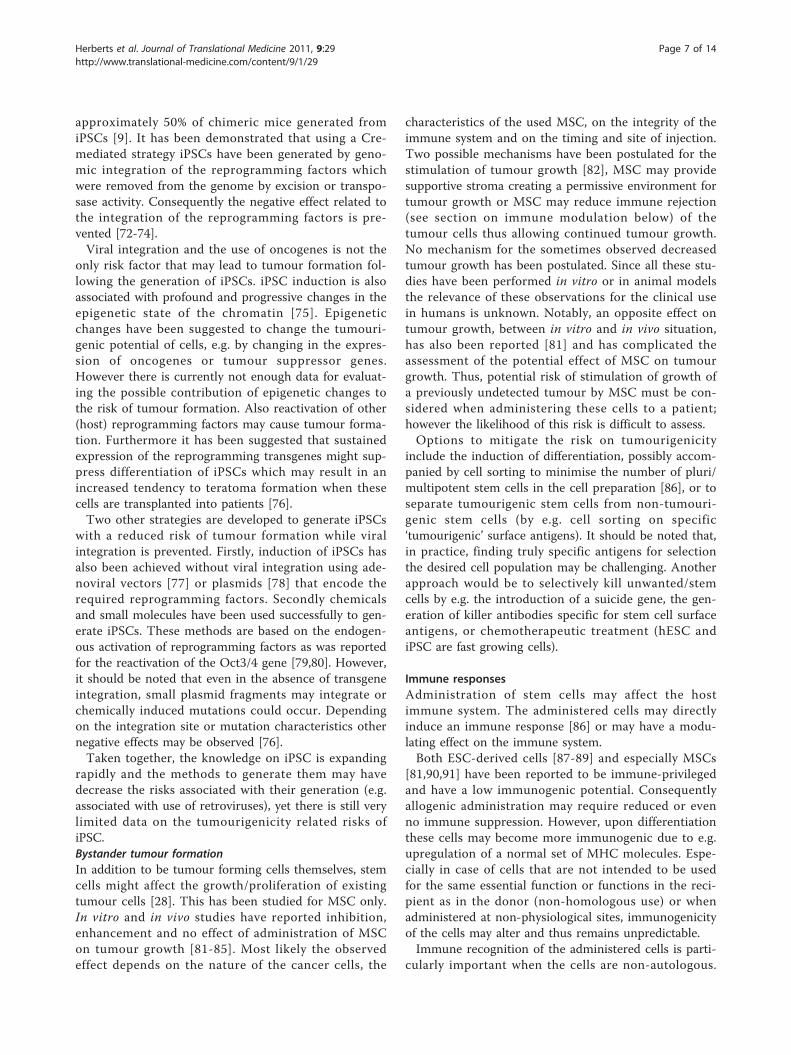
approximately 50% of chimeric mice generated fromiPSCs [9]. It has been demonstrated that using a Cre-mediated strategy iPSCs have been generated by geno-mic integration of the reprogramming factors whichwere removed from the genome by excision or transpo-sase activity. Consequently the negative effect related tothe integration of the reprogramming factors is pre-vented [72-74].Viral integration and the use of oncogenes is not the
only risk factor that may lead to tumour formation fol-lowing the generation of iPSCs. iPSC induction is alsoassociated with profound and progressive changes in theepigenetic state of the chromatin [75]. Epigeneticchanges have been suggested to change the tumouri-genic potential of cells, e.g. by changing in the expres-sion of oncogenes or tumour suppressor genes.However there is currently not enough data for evaluat-ing the possible contribution of epigenetic changes tothe risk of tumour formation. Also reactivation of other(host) reprogramming factors may cause tumour forma-tion. Furthermore it has been suggested that sustainedexpression of the reprogramming transgenes might sup-press differentiation of iPSCs which may result in anincreased tendency to teratoma formation when thesecells are transplanted into patients [76].Two other strategies are developed to generate iPSCs
with a reduced risk of tumour formation while viralintegration is prevented. Firstly, induction of iPSCs hasalso been achieved without viral integration using ade-noviral vectors [77] or plasmids [78] that encode therequired reprogramming factors. Secondly chemicalsand small molecules have been used successfully to gen-erate iPSCs. These methods are based on the endogen-ous activation of reprogramming factors as was reportedfor the reactivation of the Oct3/4 gene [79,80]. However,it should be noted that even in the absence of transgeneintegration, small plasmid fragments may integrate orchemically induced mutations could occur. Dependingon the integration site or mutation characteristics othernegative effects may be observed [76].Taken together, the knowledge on iPSC is expanding
rapidly and the methods to generate them may havedecrease the risks associated with their generation (e.g.associated with use of retroviruses), yet there is still verylimited data on the tumourigenicity related risks ofiPSC.Bystander tumour formationIn addition to be tumour forming cells themselves, stemcells might affect the growth/proliferation of existingtumour cells [28]. This has been studied for MSC only.In vitro and in vivo studies have reported inhibition,enhancement and no effect of administration of MSCon tumour growth [81-85]. Most likely the observedeffect depends on the nature of the cancer cells, the
characteristics of the used MSC, on the integrity of theimmune system and on the timing and site of injection.Two possible mechanisms have been postulated for thestimulation of tumour growth [82], MSC may providesupportive stroma creating a permissive environment fortumour growth or MSC may reduce immune rejection(see section on immune modulation below) of thetumour cells thus allowing continued tumour growth.No mechanism for the sometimes observed decreasedtumour growth has been postulated. Since all these stu-dies have been performed in vitro or in animal modelsthe relevance of these observations for the clinical usein humans is unknown. Notably, an opposite effect ontumour growth, between in vitro and in vivo situation,has also been reported [81] and has complicated theassessment of the potential effect of MSC on tumourgrowth. Thus, potential risk of stimulation of growth ofa previously undetected tumour by MSC must be con-sidered when administering these cells to a patient;however the likelihood of this risk is difficult to assess.Options to mitigate the risk on tumourigenicity
include the induction of differentiation, possibly accom-panied by cell sorting to minimise the number of pluri/multipotent stem cells in the cell preparation [86], or toseparate tumourigenic stem cells from non-tumouri-genic stem cells (by e.g. cell sorting on specific‘tumourigenic’ surface antigens). It should be noted that,in practice, finding truly specific antigens for selectionthe desired cell population may be challenging. Anotherapproach would be to selectively kill unwanted/stemcells by e.g. the introduction of a suicide gene, the gen-eration of killer antibodies specific for stem cell surfaceantigens, or chemotherapeutic treatment (hESC andiPSC are fast growing cells).
Immune responsesAdministration of stem cells may affect the hostimmune system. The administered cells may directlyinduce an immune response [86] or may have a modu-lating effect on the immune system.Both ESC-derived cells [87-89] and especially MSCs
[81,90,91] have been reported to be immune-privilegedand have a low immunogenic potential. Consequentlyallogenic administration may require reduced or evenno immune suppression. However, upon differentiationthese cells may become more immunogenic due to e.g.upregulation of a normal set of MHC molecules. Espe-cially in case of cells that are not intended to be usedfor the same essential function or functions in the reci-pient as in the donor (non-homologous use) or whenadministered at non-physiological sites, immunogenicityof the cells may alter and thus remains unpredictable.Immune recognition of the administered cells is parti-
cularly important when the cells are non-autologous.
Herberts et al. Journal of Translational Medicine 2011, 9:29http://www.translational-medicine.com/content/9/1/29
Page 7 of 14

Evidently, careful HLA-matching of donor and recipientmay diminish the risk on Graft-versus-Host disease(GVHD), but is often not readily achievable.Graft rejection may lead to loss-of-function of the
administered cells and consequently compromise thera-peutic activity. The use of immune suppressants maylimit this risk, but may elicit drug related adverse reac-tions. Other strategies to prevent immune rejection ofthe transplanted cells have been proposed and couldinclude banking ESC, iPSC or even SSC cells withdefined major histocompatibility complex backgroundsor genetically manipulating the stem cells to reduce oractively combat immune rejection [3,92].The immune modulatory effect of both ESC and MSC
has been described in multiple reports, mostly describ-ing in vitro experiments. MSC have been described tosuppress T cell proliferation, inhibit differentiation ofmonocyte and cord blood CD34+ cells into immaturemyeloid DC, affect DC function (skewing mature DCtowards immature state [90], inhibit TNF production,increase IL-10 production), and inhibit proliferation andcytotoxicity of resting NK cells and their cytokine pro-duction [65]. A direct effect of MSC on B cells is stillmatter of debate (conflicting results), however most stu-dies indicate that MSC can inhibit B cell proliferationand/or differentiation in vitro [65]. Recently, in vitrostudies have demonstrated that both human and mouseESC extracts retain the immune modulatory propertiesof ESCs and ESC derived factors can inhibit humanmDC maturation and function [89].In vivo control or limitation of GVHD by MSC has
been reported both in humans [93] and animal models[81,83]. In a small clinical study, MSC cotransplantationwith HSC of HLA-identical siblings the observeddecreased frequency in GVHD (acute and chronic) wasaccompanied by an increased frequency of relapse of thetreated of haematological malignancy [83].An immune suppressive effect of MSC has also been
observed in an animal model of rheumatoid arthritis[90]. In addition, MSCs have been shown to suppresslymphocyte proliferation to allogenic or xenogenic anti-gens [81,82,84] leading to acceptation of allo/xenotrans-plants in animal models [90]. In clinical studies MSChave been used to facilitate the engraftment of HSC anddecrease GVHD [81].Taken together the in vitro and some in vivo data sug-
gest that MSC can interact with cells of both the innateand adaptive immune system and can modulate theireffector functions leading to potent immunosuppressiveand anti-inflammatory effects. The secretion of varioussoluble factors by MSC [84] may enhance this effect. Ithas been described that MSC express Toll like receptors(TLRs) that after interaction induce proliferation, migra-tion and differentiation of the MSC and the secretion of
cytokines [81]. MSC may thus exert protective effectsresulting in e.g. effective stimulation or regeneration ofcells in situ or in a local immunosuppressive microen-vironment. Knowledge regarding mechanisms by whichMSC or ESC-derived cells exert their immune suppres-sive effect is still increasing [81]. Nevertheless, the extra-polation from animal or in vitro studies to human isrelatively unpredictable and both beneficial and adverseeffects should be considered.
Adventitious agentsManufacturing of cell based medicinal products inevita-bly does not include terminal sterilization, purification,viral removal and inactivation. Therefore, viral andmicrobial safety is a pivotal risk factor associated withthe use of non-autologous and/or cultured cells, includ-ing stem cells. These risk factors are not unique to stemcells and apply to all cell based medicinal products.Donor history is of particular importance for stem celllines which were initially intended for research purposes,rather than to be used in clinical application. The risk ofdonor-to-recipient transmission of bacterial, viral, fungalor prion pathogens may lead to life-threatening andeven fatal reactions. Disease transmission has beenreported after allograft transplantation [94,95]. Only lim-ited information is available on disease transmission viaadult somatic stem cells other than those routinely usedHCS. It has been shown that MSC are susceptible toboth CMV and HSV-1 infection in vitro. However,using sensitive PCR techniques no CMV DNA could bedetected in ex vivo expanded MSC derived from healthyCMV positive individuals [96]. No information on thesusceptibility for adventitious agents of pluripotent stemcells has been reported in the scientific literature.Although progress has been made in tissue culturing
techniques, both serum and feeder layers are occasion-ally still needed for the in vitro isolation and propaga-tion of (pluripotent and somatic) stem cells [91], Theuse of animal products in tissue culture (e.g. foetalbovine serum (FBS), or non-human feeder cells) alsomay introduce a risk of transmission of disease (e.g.prion) as well as activation of host immune system bybiomolecules [97] (e.g. non-human sialic acid) [69].Expansion of stem cells in medium supplemented withFBS has a potential risk of transmitting viral and priondiseases and causing immunological rejection. Autolo-gous or donor-derived plasma may be a safer substitutefor FBS and may still allow proper cell proliferation anddifferentiation. In fact, changing FBS to human plateletlysate has been described to result in accelerated/enhanced proliferation, without genetic abnormalities[69]. However, the use of autologous patient serum maybe less favourable because serum derived from agedindividuals has been reported to interfere with MSC
Herberts et al. Journal of Translational Medicine 2011, 9:29http://www.translational-medicine.com/content/9/1/29
Page 8 of 14

proliferation and differentiation capacity [69]. Whenpossible, cell feeder free isolation and culturing or theuse of a membrane between feeder cell and stem cellculture will enhance the viral safety of the stem cellbased medicinal product.Most of the ESC lines used today have been generated
for basic research, with the application in humans notyet in mind. These cell lines have not been isolatedunder FBS- and feeder cell free conditions. Now clinicalapplication for some of these ESC lines may be dawning,and potential contaminations with adventitious agentsbecomes a safety issue that should be thoroughlyaddressed. However, because each individual ESC linecan be considered as unique, ‘simple’ regeneration of anESC line under safer culturing conditions is not alwaysreadily achieved.Testing for adventitious agents will increase the safety
of stem cell based medicinal products. This may be fea-sible for products where the number of cells is not lim-ited, for example for ESC or iPSC cell lines withindefinite self-renewal capacity. However for individuallyprepared cell batches or SSC preparations there may notbe sufficient material to both test for the presence ofadventitious agents and to treat the patient(s).Another aspect of viral safety is the patient’s vulner-
ability to the contraction or reactivation of (latent)viruses due to immune suppression necessary for sometypes of stem cell therapy. In the case of allogenic stemcell therapy the use of immune suppressive agents maybe required leading to a (severe) compromised hostimmune system. In HSC transplantation, allogeneicstem cell transplantation is often complicated by reacti-vation of herpes viruses indicating that viral activation isnot only a theoretical risk.
Other risk factorsThere are several other risk factors which need to beconsidered before the clinical application of (stem) cells.For most of these factors only limited scientific evidenceis available.Biodistribution/Ectopic graftingAn important risk factor is the (bio)distribution of theadministered stem cells. MSC are known to home tospecific tissues e.g. the bone marrow, muscle, or spleen,particularly when the tissues are damaged or underpathological conditions such as ischemia or cancer[32,81,82,84,85]. The mechanism underlying the migra-tion of MSC remains to be clarified. Data suggests thatboth chemokines and their receptors and adhesionmolecules are involved. However, it has been reportedthat when used to treat myocardial infarction (MI) onlya few cells homed to the site of injury following intrave-nous administration, and engraftment rate appears to beextremely low even when injected at site of injury
(intramyocardial or intracoronary injection) [17,98]. It isunclear where the non-engrafted (stem) cells go to, andalso the risks associated with distribution to undesiredtissues are unknown. One possibility is the engraftmentof the stem cells at these distant or non-target sites. Asnoted earlier the local environment in which the stemcell resides in the recipient may influence the biologicalproperties of the stem cells, however only little is knownwhether these effects are potentially harmful or not.Given the limited data, the risk of such ectopic engraft-ment and its effects remains unpredictable and shouldbe taken into account.Mode and site of administrationAnother risk factor associated with the use of stem cellsmay be the potential high number of cells needed forthe beneficial effect. It is generally unknown how manycells are needed, however, given the (very) low rate ofretention and possible low cell survival, large number ofcells may be required for obtaining maximal clinicalbenefit. Injection of concentrated cells into tissue mayhave unwanted effects. Cells may form aggregates, parti-cularly if sheared by passage through small needles [28].These aggregates could cause pulmonary emboli orinfarctions after infusion. Injection in the portal veinmay partially circumvent this problem; however thisrequires specialised (surgical) procedures which mayintroduce other risks. Serious adverse events due to pro-cedural complications in combination with underlyingdisease conditions (e.g. veno-oclusive disease) have beenreported during clinical experience with HSC transplan-tation [99,100].Similarly, application of the cells at specific locations
(e.g. site of injury) may be desirable, e.g. intracardial, atsite of spinal cord injury or brain lesion, but also forthis specific procedures and/or surgery may be requiredwith associated risks.Unwanted (de)differentiationAs mentioned before, it is unlikely that undifferentiatediPSC or ESC will be used in the clinic, and that in vitrodifferentiation into a desired phenotype will be neces-sary prior to administration. However, it is unknown ifdedifferentiation of stem cells can occur in vitro or invivo. Dedifferentiation of somatic cells or redifferentia-tion into another cell type has been described [20],whether this has adverse clinical consequences remainsunclear. In addition, for MSC differentiation intounwanted mesenchymal cell types such as osteocytesand adipocytes has been described [101]. Encapsulatedstructures containing calcification and/or ossifications inthe heart have been seen in animals treated with BM-derived MSC for (induced) myocardial infarction [101].It can be concluded that unwanted differentiation istherefore not only a theoretical risk; however the factorscontributing to this risk are unknown. Differentiation or
Herberts et al. Journal of Translational Medicine 2011, 9:29http://www.translational-medicine.com/content/9/1/29
Page 9 of 14

culturing of stem cells could not only induce malignanttransformation but in theory may also induce cellularalterations such as altered excretion patterns or cell sur-face molecules which may influence the in vivo attri-butes of the administered cells. This may haveunexpected adverse or toxic consequences.Non-homologous useAlthough the use of MSC and HSC have a excellenttrack record in some routine clinical applications (bonemarrow transplantation or reconstituting of immuno-depleted patients) many of the potential risks discussedabove (e.g. ectopic grafting, unpredictable immune con-sequences, (de)differentiation) can also be relevant tothese type of cells in case they are not intended to beused for the same essential function or functions in therecipient as in the donor (so-called non-homologoususe). The potential unpredictable adverse effects clearlyneed further evaluation.Purity and identityAnother critical issue to address is the need for obtain-ing a pure population of the desired stem cells. Contam-ination with other types of cells could cause undesirableeffects [102], or in case of ESC derived cells undifferen-tiated ESC could be a potential source for tumour for-mation. In addition, several publications reporting onMSC to undergo spontaneous transformation eventshave recently been retracted since the reported observa-tions could not be reproduced [64,65,103]. It was con-firmed that the initial observations were based on crosscontaminated HT1080 human fibrosarcoma cells.Obviously such errors should be preventable by GoodManufacturing or Good Laboratory practices but theseexamples illustrate that even relatively simple risksshould be considered.(Lack of) functional characteristicsThere may also be risks associated with specific stemcell therapies. An example is the use of stem cell ther-apy in the treatment of myocardial infarction (MI). Oneof the main safety concerns is the occurrence ofarrhythmias [98,104]. These were seen in some, but notall trials using stem cell-based therapy in treatment ofheart failure or myocardial infarction [98]. The used celltype and route of administration may influence the riskon arrhythmias [98]. These arrhythmias may be causedby poor cell-cell coupling, incomplete differentiation(seen in vitro with MSC), an unexcitable state of theMSC, or a heterogeneous distribution of action potential[98,104]. Principally, cell therapy in the heart can bepredicted to have a multitude of electrical effects somepotential destabilizing and others clearly beneficial.Donor and recipient clinical characteristicsEvidently, if allogenic stem cells are used there is a riskof stem cell-tissue rejection which may be (partially)overcome by donor-patient matching, by immunological
sequestration or by the use of immune suppressants,which all have their own drawbacks.Numerous other factors can be identified which may
or may not contribute to a risk associated with the clini-cal application of a stem cell based medicinal product.These may be specific intrinsic characteristics of thestem cell based medicinal product or more extrinsic riskfactors related to e.g. the manufacturing or type ofapplication of the product. For example, when used inan autologous setting, the underlying disease, or medica-tion may have an impact on the number and functional-ity of the stem cells [34,105], which can induceunwanted side effect of stem cell therapy. Anotherexample may be the (unknown or unidentified) secretionof trophic factors and/or a variety of growth factors bythe stem cells [32].
ConclusionInitial clinical experience with somatic stem cell therapymay appear promising. However, many questionsregarding the potential risks have not yet beenanswered. The amount of data and the knowledge ofrisks associated with the use of stem cell therapy areexpanding. However, due to the large variation amongstthe studies (e.g. study protocol, patient population, het-erogeneity of the administered cell population, timing/location of injection) it is difficult to extrapolate resultsfrom one study to another, and also from one stem cellbased medicinal product to another. Currently, the mostextensive clinical experience has been obtained withhaematopoietic stem cells and mesenchymal stem/stro-mal cells. The clinical experience with endothelial pro-genitor cells is also growing.In most cases, irrespective of the treated condition or
mode of administration, MSC therapy appears relativelysafe [31,33,98,106]. However given the limited time offollow up, the low number of patients, the variation incell preparations and characterisation and mode ofdelivery, further studies on the safety of MSC are stillneeded, especially on long term effects such as tumouri-genicity. Autologous stem cell transplantation is per-ceived as non-harmful; however this only applies fornon-substantially manipulated stem cells. The risks asso-ciated with autologous stem cells that are substantiallymanipulated (e.g. by tissue culture or genetic modifica-tion) or cells that are not intended to be used for thesame essential function or functions in the recipient asin the donor do need further evaluation.In contrast to SSC, there is currently no clinical
experience with pluripotent stem cells. This is in parti-cular due to the assumption that the application ofthese cells is associated with a higher risk in particularrelated to tumourigenicity. Recent developments indi-cate that clinical experience with embryonic stem cells
Herberts et al. Journal of Translational Medicine 2011, 9:29http://www.translational-medicine.com/content/9/1/29
Page 10 of 14

become available in the near future. At this moment weare aware of 3 clinical trials using human ESC-derivedcells that have been approved by the FDA. The firstapproved trial is using oligodendrocyte progenitor cellsaimed at the treatment of spinal cord injury. This trialhas been temporarily put on hold before the first patientwas included due to non-clinical findings of microscopiccysts in the regenerating injury site [107]. However thehold has been lifted and recruitment is currentlyongoing. The two other trials have just been cleared bythe FDA, and aim to treat two eye diseases (Stargardt’smacular dystrophy and dry age-related macular degen-eration) with ESC-derived cells http://www.advancedcell.com.As discussed earlier, the perceived risk on tumour for-
mation is higher for iPSC than for ESC. Clinical applica-tion of iPSC is still relatively far away as the technique togenerate these cells is still quite new and the methods togenerate these cells more safely are rapidly developing.For iPSC, even the non-clinical information on thetumour formation in a context relevant to regenerativemedicine (focal injection or iv administration) is still verylimited (mouse iPSC) or essentially lacking (human iPSC)apart from their teratoma inducing capabilities [43].Overall stem cell therapy may represent great hope for
multiple diseases and degenerative conditions, but athorough evaluation of the risk factors and potential riskof a stem cell based medicinal product must be a prere-quisite step before wider clinical application and/orregistration can be accepted. For each stem cell basedmedicinal product the potential risks to the patientneeds to be adequately evaluated and should take intoaccount not only the specific intrinsic characteristics ofa specific stem cell but also the safety data alreadyobtained with similar type of products. In additionextrinsic risk factors like manufacturing, handling, sto-rage- and clinical or treatment related risk factors cancontribute to the overall risk to the patient. During therisk evaluation, knowledge of the safety of (similar) stemcell based medicinal products may be of great value.Documented/identified risks, and known risk factors aswell as potential/theoretical risks should be consideredin the risk evaluation. Table 2 presents a (non-exhaus-tive) overview of risk factors and risks. It should beclear that while tumour formation is an important riskassociated with stem cell therapy, other risks (e.g.adverse immune modulation) as well as strategies tominimize the risks should be should be carefully evalu-ated [35,38,108].Furthermore, for the successful development of a stem
cell based medicinal product more information on thebiological mechanism of stem cell therapy is needed aswell as sufficient characterisation of the cells and repro-ducible production of stem cell batches [109]. The
current knowledge on the mechanism of action of stemcell therapy is still limited and the cellular requirementsnecessary for a successful product are largely unknown[34,109]. Other issues such as choice of stem cells to beused, the need/possibility for concurrent tissue regenera-tion in case of irreversible tissue loss, the differentiationdegree and specific identity of the transplanted cells,and the long-term survival of engrafted cells in theabsence of a normal supportive tissue environmentshould be considered as well.
List of abbreviationsBM: bone marrow; DC: dendritic cells; EPS: endothelial progenitor cells; ESC:embryonic stem cells; FBS: foetal bovine serum; GVHD: graft versus hostdisease; HLA: human leukocyte antigen; HSC: Haematopoietic stem cells;iPSC: induced pluripotent stem cells; LVEF: left ventricular ejection fraction;MHC: major histocompatibility complex; MI: myocardial infarction; MSC:mesenchymal stem/stromal cells; SSC: somatic stem cells; TLR: toll likereceptor.
AcknowledgementsWe would like to thank Jan Willem van der Laan, Egbert Flory and AndreBerger for critically reading the manuscript.
Author details1Centre for Biological Medicines and Medical Technology, National Institutefor Public Health and the Environment, A. v. Leeuwenhoeklaan 9, P.O.Box 1,3720 BA, Bilthoven, The Netherlands. 2Department of Pharmacovigilance,Netherlands Medicines Evaluation Board, Kalvermarkt 53, 2511 CB, Den Haag,The Netherlands.
Authors’ contributionsAll authors contributed to the writing and discussion of the manuscript.The views expressed in this article are the personal views of the authors.
Competing interestsAll authors declare no competing interest. The views expressed in this articleare the personal views of the author(s) and may not be understood orquoted as being made on behalf of or reflecting the position of theNetherlands Medicines Evaluation Board.
Received: 20 July 2010 Accepted: 22 March 2011Published: 22 March 2011
References1. Evans MJ, Kaufman MH: Establishment in culture of pluripotential cells
from mouse embryos. Nature 1981, 292:154-156.2. Martin GR: Isolation of a pluripotent cell line from early mouse embryos
cultured in medium conditioned by teratocarcinoma stem cells.Proceedings of the National Academy of Sciences of the United States ofAmerica 1981, 78:7634-7638.
3. Thomson JA: Embryonic stem cell lines derived from human blastocysts.Science 1998, 282:1145-1147.
4. Solter D: From teratocarcinomas to embryonic stem cells and beyond: Ahistory of embryonic stem cell research. Nature Reviews Genetics 2006,7:319-327.
5. Lengner CJ: IPS cell technology in regenerative medicine. Book IPS celltechnology in regenerative medicine 2010, 1192:38-44, City; 38-44.
6. Bradley A, Evans M, Kaufman MH, Robertson E: Formation of germ-linechimaeras from embryo-derived teratocarcinoma cell lines. Nature 1984,309:255-256.
7. Reubinoff BE, Pera MF, Fong CY, Trounson A, Bongso A: Embryonic stemcell lines from human blastocysts: Somatic differentiation in vitro. NatureBiotechnology 2000, 18:399-404.
8. Yu J, Vodyanik MA, Smuga-Otto K, Antosiewicz-Bourget J, Frane JL, Tian S,Nie J, Jonsdottir GA, Ruotti V, Stewart R, et al: Induced pluripotent stemcell lines derived from human somatic cells. Science 2007, 318:1917-1920.
Herberts et al. Journal of Translational Medicine 2011, 9:29http://www.translational-medicine.com/content/9/1/29
Page 11 of 14
9. Okita K, Ichisaka T, Yamanaka S: Generation of germline-competentinduced pluripotent stem cells. Nature 2007, 448:313-317.
10. Takahashi K, Tanabe K, Ohnuki M, Narita M, Ichisaka T, Tomoda K,Yamanaka S: Induction of Pluripotent Stem Cells from Adult HumanFibroblasts by Defined Factors. Cell 2007, 131:861-872.
11. Takahashi K, Tanabe K, Ohnuki M, Narita M, Ichisaka T, Tomoda K,Yamanaka S: Induction of Pluripotent Stem Cells from Adult HumanFibroblasts by Defined Factors. Cell 2006, 131:861-872.
12. Aasen T, Raya A, Barrero MJ, Garreta E, Consiglio A, Gonzalez F, Vassena R,Bilic J, Pekarik V, Tiscornia G, et al: Efficient and rapid generation ofinduced pluripotent stem cells from human keratinocytes. NatureBiotechnology 2008, 26:1276-1284.
13. Dimos JT, Rodolfa KT, Niakan KK, Weisenthal LM, Mitsumoto H, Chung W,Croft GF, Saphier G, Leibel R, Goland R, et al: Induced pluripotent stemcells generated from patients with ALS can be differentiated into motorneurons. Science 2008, 321:1218-1221.
14. Hockemeyer D, Soldner F, Cook EG, Gao Q, Mitalipova M, Jaenisch R: ADrug-Inducible System for Direct Reprogramming of Human SomaticCells to Pluripotency. Cell Stem Cell 2008, 3:346-353.
15. Takahashi K, Yamanaka S: Induction of Pluripotent Stem Cells from MouseEmbryonic and Adult Fibroblast Cultures by Defined Factors. Cell 2006,126:663-676.
16. Yu J, Thomson JA: Pluripotent stem cell lines. Genes and Development2008, 22:1987-1997.
17. Saric T, Hescheler J: Stem cells and nuclear reprogramming. MinimallyInvasive Therapy and Allied Technologies 2008, 17:64-78.
18. Xu N, Papagiannakopoulos T, Pan G, Thomson JA, Kosik KS: MicroRNA-145Regulates OCT4, SOX2, and KLF4 and Represses Pluripotency in HumanEmbryonic Stem Cells. Cell 2009, 137:647-658.
19. Geoghegan E, Byrnes L: Mouse induced pluripotent stem cells.International Journal of Developmental Biology 2008, 52:1015-1022.
20. Pessina A, Gribaldo L: The key role of adult stem cells: Therapeuticperspectives. Current Medical Research and Opinion 2006, 22:2287-2300.
21. Koch P, Kokaia Z, Lindvall O, Brustle O: Emerging concepts in neural stemcell research: autologous repair and cell-based disease modelling. TheLancet Neurology 2009, 8:819-829.
22. Taupin P, Gage FH: Adult neurogenesis and neural stem cells of thecentral nervous system in mammals. Journal of Neuroscience Research2002, 69:745-749.
23. Pappa KI, Anagnou NP: Novel sources of fetal stem cells: Where do theyfit on the developmental continuum? Regenerative Medicine 2009,4:423-433.
24. De Coppi P, Bartsch Jr, Siddiqui MM, Xu T, Santos CC, Perin L,Mostoslavsky G, Serre AC, Snyder EY, Yoo JJ, et al: Isolation of amnioticstem cell lines with potential for therapy. Nature Biotechnology 2007,25:100-106.
25. Prusa AR, Marton E, Rosner M, Bernaschek G, HengstschlaÝĝer M: Oct-4-expressing cells in human amniotic fluid: A new source for stem cellresearch? Human Reproduction 2003, 18:1489-1493.
26. Karahuseyinoglu S, Cinar O, Kilic E, Kara F, Akay GG, Demiralp D, Tukun A,Uckan D, Can A: Biology of stem cells in human umbilical cord stroma: Insitu and in vitro surveys. Stem Cells 2007, 25:319-331.
27. Rhodes KE, Gekas C, Wang Y, Lux CT, Francis CS, Chan DN, Conway S,Orkin SH, Yoder MC, Mikkola HKA: The Emergence of Hematopoietic StemCells Is - Initiated in the Placental Vasculature in the Absence ofCirculation. Cell Stem Cell 2008, 2:252-263.
28. Prockop DJ, Olson SD: Clinical trials with adult stem/progenitor cells fortissue repair: Let’s not overlook some essential precautions. Blood 2007,109:3147-3151.
29. Horwitz EM, Prockop DJ, Gordon PL, Koo WWK, Fitzpatrick LA, Neel MD,McCarville ME, Orchard PJ, Pyeritz RE, Brenner MK: Clinical responses tobone marrow transplantation in children with severe osteogenesisimperfecta. Blood 2001, 97:1227-1231.
30. Koc ON, Day J, Nieder M, Gerson SL, Lazarus HM, Krivit W: Allogeneicmesenchymal stem cell infusion for treatment of metachromaticleukodystrophy (MLD) and Hurler syndrome (MPS-IH). Bone MarrowTransplantation 2002, 30:215-222.
31. Lasala GP, Minguell JJ: Bone Marrow-derived Stem/Progenitor Cells: TheirUse in Clinical Studies for the Treatment of Myocardial Infarction. HeartLung and Circulation 2009, 18:171-180.
32. Bieback K, Kluter H: Mesenchymal stromal cells from umbilical cordblood. Current Stem Cell Research and Therapy 2007, 2:310-323.
33. Giordano A, Galderisi U, Marino IR: From the laboratory bench to thepatient’s bedside: An update on clinical trials with Mesenchymal StemCells. Journal of Cellular Physiology 2007, 211:27-35.
34. Aranguren XL, Verfaillie CM, Luttun A: Emerging hurdles in stem celltherapy for peripheral vascular disease. Journal of Molecular Medicine2009, 87:3-16.
35. ISO/IEC Guide 51:1999. [http://www.iso.org/iso/iso_catalogue.htm].36. ISO 14971:2007. [http://www.iso.org/iso/iso_catalogue.htm].37. ICH Q9: Quality Risk Management. [http://www.ich.org/products/
guidelines.html].38. Guideline on risk management systems for medical products for human
use, EMEA/CHMP/96268/2005. [http://www.ema.europa.eu].39. Li HC, Soticov C, Rogers AB, Houghton JM: Stem cells and cancer:
Evidence for bone marrow stem cells in epithelial cancers. World Journalof Gastroenterology 2006, 12:363-371.
40. Werbowetski-Ogilvie TE, Bosse M, Stewart M, Schnerch A, Ramos-Mejia V,Rouleau A, Wynder T, Smith MJ, Dingwall S, Carter T, et al: Characterizationof human embryonic stem cells with features of neoplastic progression.Nature Biotechnology 2009, 27:91-97.
41. Amariglio N, Hirshberg A, Scheithauer BW, Cohen Y, Loewenthal R,Trakhtenbrot L, Paz N, Koren-Michowitz M, Waldman D, Leider-Trejo L, et al:Donor-derived brain tumor following neural stem cell transplantation inan ataxia telangiectasia patient. PLoS Medicine 2009, 6:0221-0231.
42. Shih CC, Forman SJ, Chu P, Slovak M: Human embryonic stem cells areprone to generate primitive, undifferentiated tumors in engraftedhuman fetal tissues in severe combined immunodeficient mice. StemCells and Development 2007, 16:893-902.
43. Knoepfler PS: Deconstructing stem cell tumorigenicity: A roadmap tosafe regenerative medicine. Stem Cells 2009, 27:1050-1056.
44. Hamburger AW, Salmon SE: Primary bioassay of human tumor stem cells.Science 1977, 197:461-463.
45. Bruce WR, Van Der Gaag H: A quantitative assay for the number ofmurine lymphoma cells capable of proliferation in vivo. Nature 1963,199:79-80.
46. Houghton J, Stoicov C, Nomura S, Rogers AB, Carlson J, Li H, Cai X, Fox JG,Goldenring JR, Wang TC: Gastric cancer originating from bone marrow-derived cells. Science 2004, 306:1568-1571.
47. Stark A, Aparisi T, Ericsson JLE: Human osteogenic sarcoma: Fine structureof the osteoblastic type. Ultrastructural Pathology 1983, 4:311-329.
48. Barozzi P, Luppi M, Faccheti F, Mecucci C, Alu M, Sarid R, Rasini V,Ravazzini L, Rossi E, Festa S, et al: Post-transplant Kaposi sarcomaoriginates from the seeding of donor-derived progenitors. NatureMedicine 2003, 9:554-561.
49. Aractingi S, Kanitakis J, Euvrard S, Le Danff C, Peguillet I, Khosrotehrani K,Lantz O, Carosella ED: Skin carcinoma arising from donor cells in a kidneytransplant recipient. Cancer Research 2005, 65:1755-1760.
50. Janin A, Murata H, Leboeuf C, Cayuela JM, Gluckman E, Legres L,Desveaux A, Varna M, Ratajczak P, Soulier J, et al: Donor-derived oralsquamous cell carcinoma after allogeneic bone marrow transplantation.Blood 2009, 113:1834-1840.
51. Ades L, Guardiola P, Socie G: Second malignancies after allogeneichematopoietic stem cell transplantation: New insight and currentproblems. Blood Reviews 2002, 16:135-146.
52. Prokhorova TA, Harkness LM, Frandsen U, Ditzel N, Schroder HD, Burns JS,Kassem M: Teratoma formation by human embryonic stem cells is sitedependent and enhanced by the presence of Matrigel. Stem Cells andDevelopment 2009, 18:47-54.
53. Erdo F, Buhrle C, Blunk J, Hoehn M, Xia Y, Fleischmann B, Focking M,Kustermann E, Kolossov E, Hescheler J, et al: Host-dependenttumorigenesis of embryonic stem cell transplantation in experimentalstroke. Journal of Cerebral Blood Flow and Metabolism 2003, 23:780-785.
54. Narva E, Autio R, Rahkonen N, Kong L, Harrison N, Kitsberg D, Borghese L,Itskovitz-Eldor J, Rasool O, Dvorak P, et al: High-resolution DNA analysis ofhuman embryonic stem cell lines reveals culture-induced copy numberchanges and loss of heterozygosity. Nature Biotechnology 2010,28:371-377.
55. Rodriguez R, Rubio R, Masip M, Catalina P, Nieto A, De La Cueva T,Arriero M, Martin NS, De La Cueva E, Balomenos D, et al: Loss of p53
Herberts et al. Journal of Translational Medicine 2011, 9:29http://www.translational-medicine.com/content/9/1/29
Page 12 of 14

induces tumorigenesis in p21-deficient mesenchymal stem cells.Neoplasia 2009, 11:397-407.
56. Li H, Fan X, Kovi RC, Jo Y, Moquin B, Konz R, Stoicov C, Kurt-Jones E,Grossman SR, Lyle S, et al: Spontaneous expression of embryonic factorsand p53 point mutations in aged mesenchymal stem cells: A model ofage-related tumorigenesis in mice. Cancer Research 2007, 67:10889-10898.
57. Miura Y, Gao Z, Miura M, Seo BM, Sonoyama W, Chen W, Gronthos S,Zhang L, Shi S: Mesenchymal stem cell-organized bone marrowelements: An alternative hematopoietic progenitor resource. Stem Cells2006, 24:2428-2436.
58. Siebzehnrubl FA, Jeske I, Muller D, Buslei R, Coras R, Hahnen E, Huttner HB,Corbeil D, Kaesbauer J, Appl T, et al: Spontaneous in vitro transformationof adult neural precursors into stem-like cancer cells. Brain Pathology2009, 19:399-408.
59. Kassem M, Burns JS, Castro JG, Munoz DR: Adult stem cells and cancer(multiple letters). Cancer Research 2005, 65:9601.
60. Bernardo ME, Zaffaroni N, Novara F, Cometa AM, Avanzini MA, Moretta A,Montagna D, Maccario R, Villa R, Daidone MG, et al: Human bone marrow-derived mesenchymal stem cells do not undergo transformation afterlong-term in vitro culture and do not exhibit telomere maintenancemechanisms. Cancer Research 2007, 67:9142-9149.
61. Tarte K, Gaillard J, Lataillade JJ, Fouillard L, Becker M, Mossafa H, Tchirkov A,Rouard H, Henry C, Splingard M, et al: Clinical-grade production of humanmesenchymal stromal cells: Occurrence of aneuploidy withouttransformation. Blood 2010, 115:1549-1553.
62. Rubio D, Garcia-Castro J, Martin MC, De La Fuente R, Cigudosa JC, Lloyd AC,Bernad A: Spontaneous human adult stem cell transformation. CancerResearch 2005, 65:3035-3039.
63. Rosland GV, Svendsen A, Torsvik A, Sobala E, McCormack E, Immervoll H,Mysliwietz J, Tonn JC, Goldbrunner R, Lonning PE, et al: Long-term culturesof bone marrow-derived human mesenchymal stem cells frequentlyundergo spontaneous malignant transformation. Cancer Research 2009,69:5331-5339.
64. Garcia S, Martin MC, De La Fuente R, Cigudosa JC, Garcia-Castro J,Bernad A: Pitfalls in spontaneous in vitro transformation of humanmesenchymal stem cells. Experimental Cell Research 2010, 316:1648-1650.
65. Torsvik A, Rosland GV, Svendsen A, Molven A, Immervoll H, McCormack E,Lonning PE, Primon M, Sobala E, Tonn JC, et al: Spontaneous malignanttransformation of human mesenchymal stem cells reflects cross-contamination: Putting the research field on track - Letter. CancerResearch 2010, 70:6393-6396.
66. De La Fuente R, Bernad A, Garcia-Castro J, Martin MC, Cigudosa JC:Retraction: Spontaneous human adult stem cell transformation (CancerResearch (2005) 1). Cancer Research 2010, 70:6682.
67. Lucas S: Chromosomal instability and mesenchymal stem cells. HumanGene Therapy 2009, 20:657-664.
68. Sensebe L, Tarte K, Lataillade JJ, Fouillard L, Rouard H, Tirchkov A,Vernant JP, Gorin NC: Clinical-grade mesenchymal stem/stromal cells:Aneuploidy is not transformation. Human Gene Therapy 2009, 20:657-664.
69. Lepperdinger G, Brunauer R, Jamnig A, Laschober G, Kassem M:Controversial issue: Is it safe to employ mesenchymal stem cells in cell-based therapies? Experimental Gerontology 2008, 43:1018-1023.
70. Bushman FD: Retroviral integration and human gene therapy. Journal ofClinical Investigation 2007, 117:2083-2086.
71. Hacein-Bey-Abina S, Von Kalle C, Schmidt M, McCormack MP, Wulffraat N,Leboulch P, Lim A, Osborne CS, Pawliuk R, Morillon E, et al: LMO2-Associated Clonal T Cell Proliferation in Two Patients after Gene Therapyfor SCID-X1. Science 2003, 302:415-419.
72. Kaji K, Norrby K, Paca A, Mileikovsky M, Mohseni P, Woltjen K: Virus-freeinduction of pluripotency and subsequent excision of reprogrammingfactors. Nature 2009, 458:771-775.
73. Soldner F, Hockemeyer D, Beard C, Gao Q, Bell GW, Cook EG, Hargus G,Blak A, Cooper O, Mitalipova M, et al: Parkinson’s Disease Patient-DerivedInduced Pluripotent Stem Cells Free of Viral Reprogramming Factors. Cell2009, 136:964-977.
74. Woltjen K, Michael IP, Mohseni P, Desai R, Mileikovsky M, Hamalainen R,Cowling R, Wang W, Liu P, Gertsenstein M, et al: PiggyBac transpositionreprograms fibroblasts to induced pluripotent stem cells. Nature 2009,458:766-770.
75. Esteban MA, Gan Y, Qin D, Pei D: Induced pluripotent stem cell (iPS)technology: Promises and challenges. Chinese Science Bulletin 2009, 54:2-8.
76. Yamanaka S: A Fresh Look at iPS Cells. Cell 2009, 137:13-17.77. Stadtfeld M, Nagaya M, Utikal J, Weir G, Hochedlinger K: Induced
pluripotent stem cells generated without viral integration. Science 2008,322:945-949.
78. Okita K, Nakagawa M, Hyenjong H, Ichisaka T, Yamanaka S: Generation ofmouse induced pluripotent stem cells without viral vectors. Science 2008,322:949-953.
79. Huangfu D, Osafune K, Maehr R, Guo W, Eijkelenboom A, Chen S,Muhlestein W, Melton DA: Induction of pluripotent stem cells fromprimary human fibroblasts with only Oct4 and Sox2. Nature Biotechnology2008, 26:1269-1275.
80. Shi Y, Tae Do J, Desponts C, Hahm HS, Scholer HR, Ding S: A CombinedChemical and Genetic Approach for the Generation of InducedPluripotent Stem Cells. Cell Stem Cell 2008, 2:525-528.
81. Uccelli A, Moretta L, Pistoia V: Mesenchymal stem cells in health anddisease. Nature Reviews Immunology 2008, 8:726-736.
82. Lazennec G, Jorgensen C: Concise review: Adult multipotent stromal cellsand cancer: Risk or benefit? Stem Cells 2008, 26:1387-1394.
83. Ning H, Yang F, Jiang M, Hu L, Feng K, Zhang J, Yu Z, Li B, Xu C, Li Y, et al:The correlation between cotransplantation of mesenchymal stem cellsand higher recurrence rate in hematologic malignancy patients:Outcome of a pilot clinical study. Leukemia 2008, 22:593-599.
84. Djouad F, Plence P, Bony C, Tropel P, Apparailly F, Sany J, Noel D,Jorgensen C: Immunosuppressive effect of mesenchymal stem cellsfavors tumor growth in allogeneic animals. Blood 2003,102:3837-3844.
85. Karnoub AE, Dash AB, Vo AP, Sullivan A, Brooks MW, Bell GW,Richardson AL, Polyak K, Tubo R, Weinberg RA: Mesenchymal stem cellswithin tumour stroma promote breast cancer metastasis. Nature 2007,449:557-563.
86. Nussbaum J, Minami E, Laflamme MA, Virag JAI, Ware CB, Masino A,Muskheli V, Pabon L, Reinecke H, Murry CE: Transplantation ofundifferentiated murine embryonic stem cells in the heart: Teratomaformation and immune response. FASEB Journal 2007, 21:1345-1357.
87. Li L, Baroja ML, Majumdar A, Chadwick K, Rouleau A, Gallacher L, Ferber I,Lebkowski J, Martin T, Madrenas J, Bhatia M: Human embryonic stem cellspossess immune-privileged properties. Stem Cells 2004, 22:448-456.
88. Drukker M, Katchman H, Katz G, Friedman SET, Shezen E, Hornstein E,Mandelboim O, Reisner Y, Benvenisty N: Human embryonic stem cells andtheir differentiated derivatives are less susceptible to immune rejectionthan adult cells. Stem Cells 2006, 24:221-229.
89. Mohib K, Allan D, Wang L: Human Embryonic Stem Cell-extracts Inhibitthe Differentiation and Function of Monocyte-derived Dendritic Cells.Stem Cell Reviews and Reports 2010, 6:611-621.
90. Nasef A, Ashammakhi N, Fouillard L: Immunomodulatory effect ofmesenchymal stromal cells: Possible mechanisms. Regenerative Medicine2008, 3:531-546.
91. Chamberlain G, Fox J, Ashton B, Middleton J: Concise review:Mesenchymal stem cells: Their phenotype, differentiation capacity,immunological features, and potential for homing. Stem Cells 2007,25:2739-2749.
92. Nakatsuji N, Nakajima F, Tokunaga K: HLA-haplotype banking and iPScells. Nature Biotechnology 2008, 26:739-740.
93. Le Blanc K, Frassoni F, Ball L, Locatelli F, Roelofs H, Lewis I, Lanino E,Sundberg B, Bernardo ME, Remberger M, et al: Mesenchymal stem cells fortreatment of steroid-resistant, severe, acute graft-versus-host disease: aphase II study. The Lancet 2008, 371:1579-1586.
94. Kainer MA, Linden JV, Whaley DN, Holmes HT, Jarvis WR, Jernigan DB,Archibald LK: Clostridium infections associated with musculoskeletal-tissue allografts. NEnglJMed 2004, 350:2564-2571.
95. Tugwell BD, Patel PR, Williams IT, Hedberg K, Chai F, Nainan OV,Thomas AR, Woll JE, Bell BP, Cieslak PR: Transmission of hepatitis C virusto several organ and tissue recipients from an antibody-negative donor.AnnInternMed 2005, 143:648-654.
96. Sundin M, Orvell C, Rasmusson I, Sundberg B, Ringden O, Le Blanc K:Mesenchymal stem cells are susceptible to human herpesviruses, butviral DNA cannot be detected in the healthy seropositive individual.Bone Marrow Transplantation 2006, 37:1051-1059.
97. Martin MJ, Muotri A, Gage F, Varki A: Human embryonic stem cellsexpress an immunogenic nonhuman sialic acid. Nature Medicine 2005,11:228-232.
Herberts et al. Journal of Translational Medicine 2011, 9:29http://www.translational-medicine.com/content/9/1/29
Page 13 of 14

98. Menasche P: Stem cell therapy for heart failure: Are arrhythmias a realsafety concern? Circulation 2009, 119:2735-2740.
99. Grigg A, Gibson R, Bardy P, Szer J: Acute portal vein thrombosis afterautologous stem cell transplantation. Bone Marrow Transplantation 1996,18:949-953.
100. Kikuchi K, Rudolph R, Murakami C, Kowdley K, McDonald GB: Portal veinthrombosis after hematopoietic cell transplantation: Frequency,treatment and outcome. Bone Marrow Transplantation 2002, 29:329-333.
101. Breitbach M, Bostani T, Roell W, Xia Y, Dewald O, Nygren JM, Fries JWU,Tiemann K, Bohlen H, Hescheler J, et al: Potential risks of bone marrowcell transplantation into infarcted hearts. Blood 2007, 110:1362-1369.
102. Carlsson T, Carta M, Winkler C, Bjorklund A, Kirik D: Serotonin neurontransplants exacerbate L-DOPA-induced dyskinesias in a rat model ofParkinson’s disease. JNeurosci 2007, 27:8011-8022.
103. Vogel G: To scientists’ dismay, mixed-up cell lines strike again. Science2010, 329:1004.
104. Ruckdeschel Smith R, Barile L, Messina E, Marban E: Stem cells in the heart:What’s the buzz all about? Part 2: Arrhythmic risks and clinical studies.Heart Rhythm 2008, 5:880-887.
105. Germani A, Di Campli C, Pompilio G, Biglioli P, Capogrossi MC:Regenerative therapy in peripheral artery disease. CardiovascularTherapeutics 2009, 27:289-304.
106. Tendera M, Wojakowski W: Cell therapy - Success does not come easy.European Heart Journal 2009, 30:640-641.
107. [http://www.fiercebiotech.com/story/geron-offers-reassurance-its-esc-safety-record/2009-08-27].
108. Guideline on the safety and efficacy follow-up - risk management ofadvanced therapy medicinal products, EMEA/149995/2008. [http://www.ema.europa.eu].
109. Pearson JD: Endothelial progenitor cells - Hype or hope? Journal ofThrombosis and Haemostasis 2009, 7:255-262.
doi:10.1186/1479-5876-9-29Cite this article as: Herberts et al.: Risk factors in the development ofstem cell therapy. Journal of Translational Medicine 2011 9:29.
Submit your next manuscript to BioMed Centraland take full advantage of:
• Convenient online submission
• Thorough peer review
• No space constraints or color figure charges
• Immediate publication on acceptance
• Inclusion in PubMed, CAS, Scopus and Google Scholar
• Research which is freely available for redistribution
Submit your manuscript at www.biomedcentral.com/submit
Herberts et al. Journal of Translational Medicine 2011, 9:29http://www.translational-medicine.com/content/9/1/29
Page 14 of 14