
REVIEW Open Access Microglial pathology
17
REVIEW Open Access Microglial pathology Wolfgang J Streit 1* , Qing-Shan Xue 1 , Jasmin Tischer 2 and Ingo Bechmann 2 Abstract This paper summarizes pathological changes that affect microglial cells in the human brain during aging and in aging-related neurodegenerative diseases, primarily Alzheimer’s disease (AD). It also provides examples of microglial changes that have been observed in laboratory animals during aging and in some experimentally induced lesions and disease models. Dissimilarities and similarities between humans and rodents are discussed in an attempt to generate a current understanding of microglial pathology and its significance during aging and in the pathogenesis of Alzheimer dementia (AD). The identification of dystrophic (senescent) microglia has created an ostensible conflict with prior work claiming a role for activated microglia and neuroinflammation during normal aging and in AD, and this has raised a basic question: does the brain’s immune system become hyperactive (inflamed) or does it become weakened (senescent) in elderly and demented people, and what is the impact on neuronal function and cognition? Here we strive to reconcile these seemingly contradictory notions by arguing that both low-grade neuroinflammation and microglial senescence are the result of aging-associated free radical injury. Both processes are damaging for microglia as they synergistically exhaust this essential cell population to the point where the brain’s immune system is effete and unable to support neuronal function. Keywords: Senescence, Oxidative damage, Chronic neuroinflammation, Glial dysfunction, Neurodegeneration Introduction The idea of “diseased microglia” represents a recent con- ceptual development in neuropathology that was first raised in the context of Creutzfeldt-Jakob disease [1]. Diseased microglia are different from activated micro- glia, in that they are incapacitated cells whereas activated microglia are cells capably responding to injury. Micro- glial disability and dysfunction have now captured the imagination of neuroscientists made apparent by a num- ber of excellent recent reviews with different perspec- tives on the subject [2-6]. Following the conceptual introduction of diseased microglia by v. Eitzen et al. [1], additional clues about the existence of unhealthy micro- glia came from immunohistochemical studies describing abnormal morphological features of microglial cells in the aged human brain [7], termed microglial dystrophy and thought to reflect microglial senescent degeneration. The microglial dysfunction hypothesis of Alzheimer’ s dis- ease (AD) claiming that neurofibrillary degeneration is the result of weakening microglial support was published in the same year [8]. Microglial morphology has been and continues to be of interest to neuroscientists due to its ever-changing nature and the initial morphological as- sessments suggesting pathological dysfunction have been strengthened by recent genetic discoveries, which are discussed in the above mentioned reviews. It seems a milestone has been reached in terms of a renewed ap- preciation of the importance of diminutive microglial cells whose main function in normal brain is to make sure neurons are well protected and connected [9-12]. As the idea of microglial dysfunction gains momentum and its implications for improved understanding of de- mentia pathogenesis are being realized, we consider it im- portant to summarize the current state of the art from a neuropathological perspective, as well as to discuss poten- tial causes and consequences of microglial degeneration. In this paper, we focus on microglial cell morphology and phenotype as seen by immunohistochemical staining of brain sections. Morphological plasticity is a character- istic feature of microglia that is evident even in the static images of traditional microscopy, as shown here. Mod- ern microscopy methods allowing live cell imaging have confirmed what has long been suspected from cell cul- ture studies [13], namely that microglia are on the move * Correspondence: [email protected] 1 Department of Neuroscience, University of Florida College of Medicine and McKnight Brain Institute, PO Box 100244, 32610-0244 Gainesville, FL, USA Full list of author information is available at the end of the article © 2014 Streit et al.; licensee BioMed Central Ltd. This is an Open Access article distributed under the terms of the Creative Commons Attribution License (http://creativecommons.org/licenses/by/4.0), which permits unrestricted use, distribution, and reproduction in any medium, provided the original work is properly credited. The Creative Commons Public Domain Dedication waiver (http://creativecommons.org/publicdomain/zero/1.0/) applies to the data made available in this article, unless otherwise stated. Streit et al. Acta Neuropathologica Communications 2014, 2:142 http://www.actaneurocomms.org/content/2/1/142
Transcript of REVIEW Open Access Microglial pathology

Streit et al. Acta Neuropathologica Communications 2014, 2:142http://www.actaneurocomms.org/content/2/1/142
REVIEW Open Access
Microglial pathologyWolfgang J Streit1*, Qing-Shan Xue1, Jasmin Tischer2 and Ingo Bechmann2
Abstract
This paper summarizes pathological changes that affect microglial cells in the human brain during aging and inaging-related neurodegenerative diseases, primarily Alzheimer’s disease (AD). It also provides examples of microglialchanges that have been observed in laboratory animals during aging and in some experimentally induced lesionsand disease models. Dissimilarities and similarities between humans and rodents are discussed in an attempt togenerate a current understanding of microglial pathology and its significance during aging and in the pathogenesisof Alzheimer dementia (AD). The identification of dystrophic (senescent) microglia has created an ostensible conflictwith prior work claiming a role for activated microglia and neuroinflammation during normal aging and in AD, andthis has raised a basic question: does the brain’s immune system become hyperactive (inflamed) or does it becomeweakened (senescent) in elderly and demented people, and what is the impact on neuronal function and cognition?Here we strive to reconcile these seemingly contradictory notions by arguing that both low-grade neuroinflammationand microglial senescence are the result of aging-associated free radical injury. Both processes are damaging formicroglia as they synergistically exhaust this essential cell population to the point where the brain’s immune system iseffete and unable to support neuronal function.
Keywords: Senescence, Oxidative damage, Chronic neuroinflammation, Glial dysfunction, Neurodegeneration
IntroductionThe idea of “diseased microglia” represents a recent con-ceptual development in neuropathology that was firstraised in the context of Creutzfeldt-Jakob disease [1].Diseased microglia are different from activated micro-glia, in that they are incapacitated cells whereas activatedmicroglia are cells capably responding to injury. Micro-glial disability and dysfunction have now captured theimagination of neuroscientists made apparent by a num-ber of excellent recent reviews with different perspec-tives on the subject [2-6]. Following the conceptualintroduction of diseased microglia by v. Eitzen et al. [1],additional clues about the existence of unhealthy micro-glia came from immunohistochemical studies describingabnormal morphological features of microglial cells inthe aged human brain [7], termed microglial dystrophyand thought to reflect microglial senescent degeneration.The microglial dysfunction hypothesis of Alzheimer’s dis-ease (AD) claiming that neurofibrillary degeneration is theresult of weakening microglial support was published in
* Correspondence: [email protected] of Neuroscience, University of Florida College of Medicine andMcKnight Brain Institute, PO Box 100244, 32610-0244 Gainesville, FL, USAFull list of author information is available at the end of the article
© 2014 Streit et al.; licensee BioMed Central LCommons Attribution License (http://creativecreproduction in any medium, provided the orDedication waiver (http://creativecommons.orunless otherwise stated.
the same year [8]. Microglial morphology has been andcontinues to be of interest to neuroscientists due to itsever-changing nature and the initial morphological as-sessments suggesting pathological dysfunction havebeen strengthened by recent genetic discoveries, whichare discussed in the above mentioned reviews. It seemsa milestone has been reached in terms of a renewed ap-preciation of the importance of diminutive microglialcells whose main function in normal brain is to makesure neurons are well protected and connected [9-12].As the idea of microglial dysfunction gains momentumand its implications for improved understanding of de-mentia pathogenesis are being realized, we consider it im-portant to summarize the current state of the art from aneuropathological perspective, as well as to discuss poten-tial causes and consequences of microglial degeneration.In this paper, we focus on microglial cell morphology
and phenotype as seen by immunohistochemical stainingof brain sections. Morphological plasticity is a character-istic feature of microglia that is evident even in the staticimages of traditional microscopy, as shown here. Mod-ern microscopy methods allowing live cell imaging haveconfirmed what has long been suspected from cell cul-ture studies [13], namely that microglia are on the move
td. This is an Open Access article distributed under the terms of the Creativeommons.org/licenses/by/4.0), which permits unrestricted use, distribution, andiginal work is properly credited. The Creative Commons Public Domaing/publicdomain/zero/1.0/) applies to the data made available in this article,

Streit et al. Acta Neuropathologica Communications 2014, 2:142 Page 2 of 17http://www.actaneurocomms.org/content/2/1/142
all the time [14,15]. In light of this restlessness it is prob-ably time to abandon the term “resting microglia”,although it may still serve the purpose of providing anantonym to “activated microglia”. It remains a challengeto translate morphological plasticity into more detailedfunctional plasticity and to further investigate the mean-ing of altered morphological appearances, such as hyper-trophy and dystrophy. While it is known, in general, thathypertrophy is associated with activation, i.e. increasedmetabolic and phagocytic activity in energized microglia,and that dystrophy is associated with senescent degener-ation in burned-out microglia, there are many stages inbetween these two extremes as both activation and sen-escence are progressive processes with many gradations[3,16]. Microglial activation ensues rapidly followingmost perturbations of CNS homeostasis, and the inten-sity and extent to which it occurs is commensurate withthe severity of the disturbance. For example, minimallyactivated microglia are also known as primed microgliaand they have been described mostly in experimentalmouse model systems [17-19], and to a lesser extent inhuman brain [20]. In contrast, maximally activatedmicroglia appear as rounded brain macrophages thatare also referred to as amoeboid microglia [21,22].Senescent changes in microglia may develop graduallyover decades during human aging and likely occur as aconsequence of multiple influences on the CNS micro-environment, notably free radical-mediated oxidativedamage. However, microglial pathology can also occur asa result of sudden extreme stress, as suggested by findingsfrom experimental animals. A good approach towards un-derstanding functional changes that occur in microglia asthey undergo morphological transformations in humanbrain is through characterization of their immunophe-notype, which can be quite heterogeneous due to up-and down-regulation of multiple proteins and receptors[23,24]. Microglia harbor a large assortment of intracel-lular and surface antigens and there exists an arsenal ofantibodies directed against various microglial antigens,some of which can be linked to specific functions [22].As the number of microglia-binding antibodies continuesto grow, it is likely that these reagents will facilitate anever more refined in situ assessment of microglial pheno-types which in conjunction with morphological changeswill provide information about cell function and dysfunc-tion in a variety of disease states. In situ description andevaluation of microglia in human brain in terms of theirmorphology and phenotype is essential for elucidating therole these cells play in neurodegenerative disease patho-genesis. Human histopathology is also important forestablishing relevance and represents the first line of in-vestigation for neurodegenerative conditions. Togetherwith human genetic studies, as well as experimentaltransgenic or other animal or cell culture models of
disease addressing molecular mechanisms, true insightsinto disease pathogenesis can be gained.
Microglial activation – is it pathological?The currently widespread opinion that activated micro-glia can be both beneficial and detrimental leaves openthe possibility that microglial activation is harmful undercertain conditions, and destructive microglial neurotox-icity as a possible factor in neurodegenerative diseasehas been discussed on numerous prior occasions (for re-views see [5,25-27]). However, both resting and activatedmicroglia perform beneficial neuron-supporting func-tions and they exert harmful effects only if they becomesenescent, diseased, or die unable to perform their nor-mal supportive roles. Microglial activation has beendescribed as a natural consequence to CNS injury incountless, acute experimental situation since the days ofdel Rio-Hortega [28] revealing how the brain respondsto injury and engages in wound healing. The purportedlydetrimental effects exerted by activated microglia in thecontext of chronic neuroinflammation are inferred fromstudies showing neurotoxic effects of rodent microgliain vitro under artificial conditions. Neither the timescales nor the microenvironment created in vitro arerepresentative of human brain [29], and while simplifiedin vitro systems may show what microglia are theoretic-ally capable of doing when stimulated to the maximalextent possible, these results are not validated by humanneuropathology and they cannot address the complexitiesof neurodegenerative disease pathogenesis with directrelevance. A major discrepancy between microglial activa-tion in vitro and in vivo is that in vitro experiments usethe proverbial sledgehammer method (LPS or similar) toachieve maximal microglial stimulation in vitro, producingmaximal and readily measurable effects, while at the sametime it is recognized that the intensity of microglial activa-tion in the normally aged and AD brain is consistent withonly minimal, low-level neuroinflammation [30-32]. Toequate such low-intensity microglial activation in humanbrain with artificially produced high-intensity microglialneurotoxicity in vitro constitutes a flaw in scientificreasoning explained possibly by immature judgment orpremature ambition.
Microglial pathology in the aged brainIt is currently thought that chronic, low-level inflamma-tion is associated with major degenerative diseases ofaging including those affecting the CNS [30,33]. Histo-pathological studies suggesting that microglial activationoccurs with normal brain aging have contributed towardsthis notion by showing increased microglial expressionof interleukin-1α and histocompatibility antigens (HLA-DR) in humans [16,34-36], seemingly consistent withobservations in experimental animals showing increased

Streit et al. Acta Neuropathologica Communications 2014, 2:142 Page 3 of 17http://www.actaneurocomms.org/content/2/1/142
expression of major histocompatibility complex (MHC)class II antigens and upregulation of interleukin-1 onacutely activated microglia and macrophages in a varietyof brain lesions [37-42]. Animal studies have also dem-onstrated increased expression of MHC class II antigenson microglia with aging, as well as morphological andphenotypic transition to a more macrophage-like ap-pearance [43-47]. Together with literature describingfluctuations and increases in pro-inflammatory cyto-kines during aging [18,48], the notion that chronic low-level neuroinflammation accompanies brain aging isthus firmly established. However, what is missing fromthis conception is the inciting stimulus, that is, a defini-tive cause of aging-associated chronic neuroinflamma-tion remains unidentified. This stands in contrast toother aging-related chronic, non-autoimmune inflamma-tory conditions outside the CNS which usually have anidentifiable cause. For example, in osteoarthritis there isloss/breakdown of articular cartilage which results in bonedamage, which in turn elicits a chronic inflammatory reac-tion, underscoring the point that in order for inflamma-tion to occur there must be damage or injury. Thus, ifbrain aging causes low-level chronic inflammation it is im-perative to understand the reasons for it.It has been hypothesized that free radical damage in
the aging brain causes inflammation [18] but free radicaldamage in neurons is subtle and non-specific, and there-fore difficult to demonstrate histopathologically in nor-mal human brain [49]. Nevertheless, assuming the freeradical theory of aging [50] is correct, free radical injuryoccurs inevitably in aerobic organisms as a consequenceof life-long oxygen exposure at all levels producing oxi-dized nucleic acids and proteins, damaged (senescent)cells and tissues, which is what ultimately accounts forthe development of most aging-related diseases. It there-fore makes sense that there would be a chronic low-levelinflammatory reaction because low-grade inflammationis the predicted tissue response to subtle and graduallydeveloping CNS injury regardless of its cause. Put an-other way, both chronic low-level inflammation andmicroglial senescence are indirect and direct results, re-spectively, of free radical injury associated with aging(Figure 1). Importantly, the mild reactive gliosis (neuro-inflammation) that occurs presumably as a result of freeradical injury is not an aggressive, neurotoxic type of re-sponse and is detrimental only in the sense that it mayover time contribute towards development of senescenceand dysfunction. The recent demonstration by Raj et al.[17] that DNA damage in a mouse model of genotoxicstress causes mild microglial activation (priming) offersclear support for the idea that free radical injury, whichoften produces DNA damage, elicits low-level neuroin-flammation. In addition, the fact that some measures ofaging-related, low-grade neuroinflammation can be
attenuated by caloric restriction [47,51] also supportsthe idea that neuroinflammation is due to free radicaldamage, since lowered food intake is associated with de-creased metabolic activity and concomitantly decreasedfree radical production [50].Regarding the microglial changes that occur during
aging, these can be seen as being consistent with bothmild activation and with senescence. While a rapidlychanging immunophenotype and dramatic morpho-logical transformations are part of high-intensity micro-glial activation after acute CNS lesions, such changesoccur only gradually during aging reflecting slowly pro-gressive evolution of a senescent morphology andphenotype, which is similar in its manifestation to low-grade activation described in models of mild CNS injury[52-54]. Microglial secretion of cytokines changes moredramatically during acute, severe injury situation than itdoes in less severe scenarios [55], and there are aging-associated changes in microglial cytokine production[56-58] which are consistent with the idea that microgliadevelop a senescence-associated secretory phenotype(SASP). Although thus far the notion of SASP has beendiscussed in terms of non-CNS cell senescence andtumor suppression rather than in a microglial context[59,60], current understanding of microglial secretoryactivity during aging and the fact that microglioma is anexceedingly rare tumor [61] are compatible with theSASP concept.Microglial mitosis, which is part of microglial activa-
tion in acute injury situations when there is a sudden de-mand for more cells to restore homeostasis, is absent orminimal during normal aging when such impromptu de-mands do not occur. However, when acute CNS injurieshappen in aged animals the ability of microglia to divideis unimpaired and even greater than in young animals[62] showing that microglial mitotic potential remainsrobust in the aged rodent brain. In contrast, replicativesenescence (reduced mitotic activity) occurs when thereis repetitive CNS injury forcing microglia to undergo re-peated bouts of mitosis over the course of one year [63].Microglial mitosis is difficult to measure reliably duringaging in human post-mortem brain, and the most directevidence for microglial senescence has come from mor-phological studies. The recognition of microglial dys-trophy has provided an indication of microglialsenescence in the aged human brain [7]. With aging anincreasing proportion of microglial cells display abnor-mal morphological features, such as shortened, gnarled,beaded, or fragmented cytoplasmic processes, as well asloss of fine ramifications and formation of spheroidalswellings, changes that are designated collectively asmicroglial dystrophy (Figure 2). Microglial dystrophy re-flects the abnormal morphology of senescent microgliabecause the number of dystrophic microglia increases
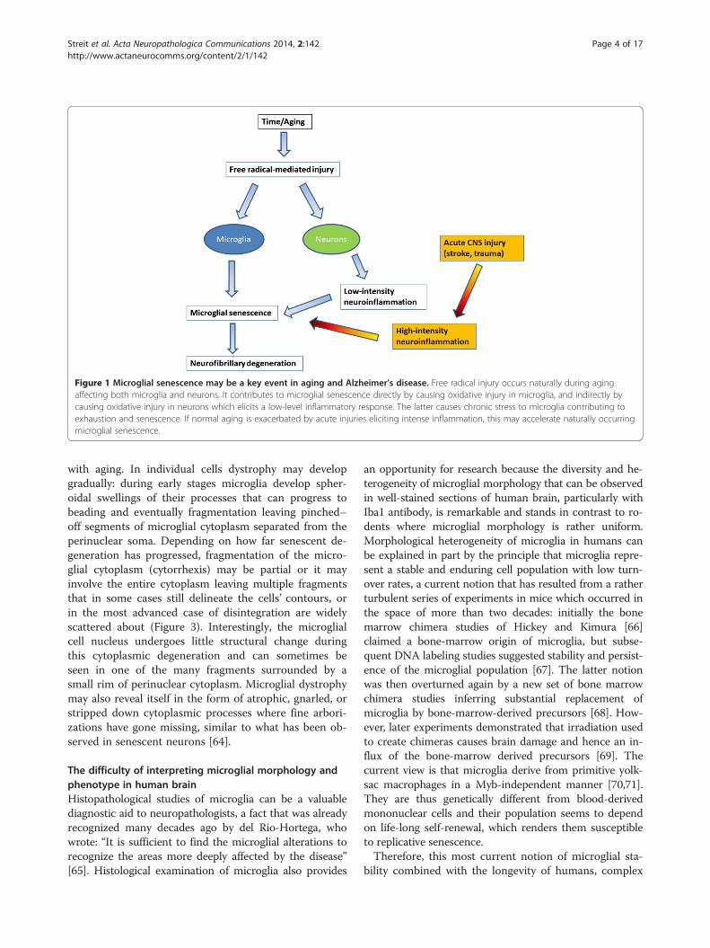
Figure 1 Microglial senescence may be a key event in aging and Alzheimer’s disease. Free radical injury occurs naturally during agingaffecting both microglia and neurons. It contributes to microglial senescence directly by causing oxidative injury in microglia, and indirectly bycausing oxidative injury in neurons which elicits a low-level inflammatory response. The latter causes chronic stress to microglia contributing toexhaustion and senescence. If normal aging is exacerbated by acute injuries eliciting intense inflammation, this may accelerate naturally occurringmicroglial senescence.
Streit et al. Acta Neuropathologica Communications 2014, 2:142 Page 4 of 17http://www.actaneurocomms.org/content/2/1/142
with aging. In individual cells dystrophy may developgradually: during early stages microglia develop spher-oidal swellings of their processes that can progress tobeading and eventually fragmentation leaving pinched–off segments of microglial cytoplasm separated from theperinuclear soma. Depending on how far senescent de-generation has progressed, fragmentation of the micro-glial cytoplasm (cytorrhexis) may be partial or it mayinvolve the entire cytoplasm leaving multiple fragmentsthat in some cases still delineate the cells’ contours, orin the most advanced case of disintegration are widelyscattered about (Figure 3). Interestingly, the microglialcell nucleus undergoes little structural change duringthis cytoplasmic degeneration and can sometimes beseen in one of the many fragments surrounded by asmall rim of perinuclear cytoplasm. Microglial dystrophymay also reveal itself in the form of atrophic, gnarled, orstripped down cytoplasmic processes where fine arbori-zations have gone missing, similar to what has been ob-served in senescent neurons [64].
The difficulty of interpreting microglial morphology andphenotype in human brainHistopathological studies of microglia can be a valuablediagnostic aid to neuropathologists, a fact that was alreadyrecognized many decades ago by del Rio-Hortega, whowrote: “It is sufficient to find the microglial alterations torecognize the areas more deeply affected by the disease”[65]. Histological examination of microglia also provides
an opportunity for research because the diversity and he-terogeneity of microglial morphology that can be observedin well-stained sections of human brain, particularly withIba1 antibody, is remarkable and stands in contrast to ro-dents where microglial morphology is rather uniform.Morphological heterogeneity of microglia in humans canbe explained in part by the principle that microglia repre-sent a stable and enduring cell population with low turn-over rates, a current notion that has resulted from a ratherturbulent series of experiments in mice which occurred inthe space of more than two decades: initially the bonemarrow chimera studies of Hickey and Kimura [66]claimed a bone-marrow origin of microglia, but subse-quent DNA labeling studies suggested stability and persist-ence of the microglial population [67]. The latter notionwas then overturned again by a new set of bone marrowchimera studies inferring substantial replacement ofmicroglia by bone-marrow-derived precursors [68]. How-ever, later experiments demonstrated that irradiation usedto create chimeras causes brain damage and hence an in-flux of the bone-marrow derived precursors [69]. Thecurrent view is that microglia derive from primitive yolk-sac macrophages in a Myb-independent manner [70,71].They are thus genetically different from blood-derivedmononuclear cells and their population seems to dependon life-long self-renewal, which renders them susceptibleto replicative senescence.Therefore, this most current notion of microglial sta-
bility combined with the longevity of humans, complex

Figure 2 Dystrophic microglia have many guises and can be stained with different antibodies. A, B Iba1 (red) and CD68 (green) doubleimmunohistochemical staining of dystrophic microglia in AD brain, as seen by 3-D confocal microscopy. Note fragmentation of cytoplasmicprocesses and punctate CD68 labeling indicating intracellular location of lysosomal antigen. C-E, anti-HLA-DR antigen immunohistochemistry(LN-3 antibody) shows dystrophic spheroid formation (C), atrophic deramified processes (D), and formation of microglial aggregate withspheroids (E). F, ferritin-positive fragmented microglia. G, Iba1-positive dystrophic microglia with beaded, fragmenting processes and spheroids.All images taken in cerebral cortex of AD subjects. Scale bars: 10 μm (A, B), 20 μm (C, D, G), 40 μm (E, F).
Streit et al. Acta Neuropathologica Communications 2014, 2:142 Page 5 of 17http://www.actaneurocomms.org/content/2/1/142
human genetics, and the many influences created by di-verse human lifestyles, diets, habits of drug consumption(both medicinal and recreational), latent infections,injuries, and systemic diseases, as well as subtle brainpathologies all of which may influence microglial behav-ior creates a tremendously heterogeneous picture that isreflected in the diverse microglial phenotypes encoun-tered. This morphological heterogeneity of the microglialpopulation can make it difficult to differentiate betweennon-activated, activated, and senescent (diseased) micro-glia since some features may not be unequivocal, and
thus morphological assessments are best combined withimmunophenotypic ones employing several microglialmarkers to characterize cells.A key issue is the reliable identification of and distinc-
tion between activated and diseased microglia, becausethis ultimately provides clues about the cells’ (dys) func-tional activities in any given region of interest. Since thereare no single histochemical markers to facilitate simpledifferentiation multiple markers together with morpho-logical assessments comprise a conclusive evaluation. Bothmicroglial activation and microglial dystrophy are defined

Figure 3 Schematic representation of cytorrhexis development. Ramified cells (1) develop spheroidal swellings (2), which progress tobeading and partial fragmentation of processes (3), which progresses to complete fragmentation while still maintaining cell contours (4),eventually ending up as scattered fragments (5). Note that the cell nucleus remains intact throughout.
Streit et al. Acta Neuropathologica Communications 2014, 2:142 Page 6 of 17http://www.actaneurocomms.org/content/2/1/142
originally by characteristic morphological appearances[7,72]. With the advent of Iba1 antibody [73] a robust re-agent has been made available that is resistant to variabletissue fixation and processing protocols and will label allmorphological variants of microglia, thereby eliminatinglargely the problem of inconsistent staining outcomes seenpreviously with other antibodies under different tissueprocessing conditions. Iba1 staining allows for excellent,unequivocal morphological differentiation of resting, acti-vated, phagocytic and dystrophic microglia (Figure 4).Staining for HLA-DR antigens is of particular interestsince many antibodies directed against these antigens areavailable and have been used widely to identify activatedmicroglia in human brain, based primarily on the fact thatstudies in experimental CNS lesions had shown an up-regulation of major histocompatibility complex (MHC)
Figure 4 Different functional states of microglia can be defined morpwith which they explore their surrounding microenvironment. B, activatedbuild-up and increased metabolic activity. C, phagocytic microglia often apcharacteristically display beaded, twisted or fragmented processes. Human
antigens by acutely activated microglia [38-40]. However,HLA-DR antigens are present on large numbers of non-activated, ramified microglia in normal human brain[74,75] and HLA-DR expression seems to be affected notonly by fixation but by a variety of factors, including gen-der, age, genotype, and possibly others [34,76]. In some ratstrains a high level of constitutive MHC II expression hasbeen reported [77] suggesting that MHC expression couldvary also with ethnicity in humans, but this has not beenstudied. One recent study shows no difference in HLA-DR expression between AD and control subjects [78].Overall the specific meaning of HLA-DR expression re-mains unknown and its use for demonstrating activatedmicroglia limited. Its use as a neuroinflammation markeris questionable for reasons already mentioned: widespreadexpression in normal CNS and high individual variability.
hologically. A, ramified microglia exhibit highly branched processesmicroglia retract processes and become enlarged due to organellepear as rounded brain macrophages. D, dystrophic microglia mostcerebral cortex stained with Iba1 antibody. Bar = 20 μm.

Streit et al. Acta Neuropathologica Communications 2014, 2:142 Page 7 of 17http://www.actaneurocomms.org/content/2/1/142
Senescent microglia in rodents and humansNeuroimmunological differences between rodent and hu-man microglia were discussed recently [26], and we wouldlike to not only support the perspectives conveyed bythese authors but add to them by pointing out thatmicroglial dystrophy as it appears with aging in humanbrain is not seen in the aged rodent brain. Dystrophy ispart of the aforementioned morphological heterogeneityof human microglia and the fact that it is absent in la-boratory rodents speaks to the vast differences in life-span and environment between rodents and humans.Brain aging happens differently in a short-lived animalraised in a nearly pathogen-free and unchanging environ-ment (incl. a never changing diet) than in a human beingwhere numerous environmental influences can play a role.The one thing common to rodents and men is the air theybreathe and the oxygen in it, and it would be safe to saythat free radical-mediated oxidative injury plays a role inboth rodent and human aging. Microglial cells in rodentsdo undergo aging-related changes and in light of the con-stancy of their environment these changes are likely to becaused exclusively by the free radical damage that occursover time. In the terms of grade-school arithmetic, aging-related changes in rodent microglia represent the leastcommon denominator. Vaughan and Peters provided thefirst and most comprehensive account of these changesusing both light and electron microscopy, and in essencetheir findings show an increase in cell size due to accu-mulation of dense inclusions closely resembling lipofuscindeposits [54]. Given the unchanging, sanitary, and uncon-taminated lives of laboratory rodents it would be reaso-nable to assume that occurrence of these lipofuscindeposits is purely a consequence of time passed and istherefore not pathological. Lipofuscin deposits can also befound in human microglia [79], but in addition humancells show those morphological abnormalities that we callmicroglial dystrophy which may well be a reflection of on-going pathology. With reference to earlier work [80], onecan thus speak of normal and pathological aging also interms of microglia (Figure 5).In the same vein, the term neuroinflammation when
applied to rodents in the context of normal aging is in-appropriate as inflammation is inherently tied to injuryand pathology, and it would be a stretch (in our minds)to call normal aging a disease. Perhaps more compellingis to point out that these purely aging-related alterationsin rodent microglia are not associated with neuronalpathology, unlike in humans where microglial dystrophyand neurofibrillary degeneration appear to be linked.
Microglial pathology and neurodegenerationNeurofibrillary degeneration (NFD), or tau pathology, andsynapse loss offer the best neuropathological correlates ofcognitive impairment and dementia in AD [81-85], and it
will be important to elucidate the role that microglial cellsplay in the development of AD-type neurodegeneration.For quite some time now it has been hypothesized thatneurodegenerative changes in AD are caused by patho-logically activated microglia thought of as autoaggressiveimmune effector cells exerting neurotoxic actions. Ac-cording to the amyloid cascade/neuroinflammation hy-pothesis, deposits of amyloid-beta (Aβ) protein incitechronic microglial activation and with it a detrimentalneuroinflammatory reaction characterized by elevatedproduction of proinflammatory cytokines, neurotoxins,and free radicals that cause neurodegeneration [86,87].However, the evidence in support of this theory is at bestindirect and at worst directly opposed to the idea, andsome of the caveats associated with it have already beendiscussed, i.e. the large discrepancy in the intensity ofmicroglial activation between in vitro and in vivo studies,the difficulty of reliable identification of activated micro-glia in human brain, and the fact that neuroinflammationin AD is quite mild and therefore unlikely to be destructive[88]. Additional concerns are raised by findings showingthat, a) tau pathology occurs in the absence of Aβ depositsand in the absence of microglial activation [89-91]; b) ad-ministration of anti-inflammatory drugs does not slow orreverse tau pathology or cognitive decline [92-94]; c) taupathology is not initiated or exacerbated by presence of se-vere neuroinflammation [95]; d) tau pathology does notoccur in transgenic animals overexpressing amyloid pre-cursor protein despite extensive Aβ deposition and associ-ated microglial activation [96-98]. For these reasons andothers discussed in this paper activated microglia are un-likely suspects in the causation of neuronal pathology as-sociated with AD. Instead it is more likely thatdegenerating (dystrophic) microglia are linked to NFD be-cause they increase in prevalence as NFD becomes morewidespread, they are co-localized with neurofibrillary tan-gles and senile plaques, and their occurrence precedes thespread of tau pathology [91,99]. The link between micro-glial and neuronal degeneration is in line also with the fun-damental idea of glial-neuronal interdependency, namely,that neuronal well-being is dependent on presence ofhealthy glial cells. The thought that microglial degener-ation is critically important in AD pathogenesis is sup-ported by additional observations showing high incidencesof microglial apoptosis in AD brain [100-102].Damaged neurons undergoing NFD are not “obvious
stimuli for inflammation” as previously thought [48] sincethere is little evidence for glial reactions to NFD or thephagocytic removal of neurofibrillary tangles in the ADbrain. In fact there is little evidence of any phagocyticactivity in cases of advanced AD or DS where morpho-logically intact microglia or brain macrophages are diffi-cult to discern (Figure 6). Because microglial and neuronalpathology are coincident in AD brain it is tempting to

Figure 5 Histopathological differences in microglial aging between laboratory rodents and humans provide a foundation for normaland pathological aging. Mice and rats experience only normal aging due to their short life spans and controlled environment, whereas humanswho live much longer lives are exposed to diverse environmental influences and lifestyles, including diets, physical and mental activities, drugs,pollutants, comorbidities and infections, all of which may affect the rates at which microglial senescence (dystrophy) occurs, indicated bydifferent trajectories of dashed red lines. As dystrophy occurs to varying degrees there is a decline in microglial neuroprotection and with thatneurofibrillary degeneration (NFD) increases at variable rates in different individuals.
Streit et al. Acta Neuropathologica Communications 2014, 2:142 Page 8 of 17http://www.actaneurocomms.org/content/2/1/142
speculate that there may be a causal connection, and sincethere is no obvious reactive response of microglia to NFDthe most likely scenario is that microglial dysfunction anddeath are a prelude to subsequent neuronal demise. Bothmicroglial dystrophy and NFD are considered to be patho-logical events in humans where they can be detected quiteearly in some instances albeit without apparent neuro-logical problems [7,89-91]. As NFD and microglial dys-trophy become more widespread over time theyeventually reach critical levels and become symptomatic.The progression of both processes is likely affected tovarying degrees by external influences (Figure 5) and bothdystrophy and NFD appear to be restricted largely to thehuman species and do not occur spontaneously even inaged laboratory rodents, where the main aging-related ef-fect on both neurons and microglia is accumulation oflipofuscin.The idea that detrimental microglial activation also
contributes to the degeneration of neurons in the sub-stantia nigra in Parkinson’s disease (PD) is firmly embed-ded in the literature yet much of it is speculative andextrapolated from animal and cell culture models[103-107]. In a thoughtfully delivered discussion, Croi-sier et al. state unequivocally that “there is no evidencethat microglia initiate neurodegeneration” [108], and itwould thus be fair to say that microglial involvement inPD remains a controversial and unresolved issue
[109,110]. One aspect that is clear is the ability of micro-glia to phagocytize neuronal debris [108,111,112], pro-viding a plausible explanation for presence of activatedmicroglia/macrophages in the substantia nigra of nor-mally aged humans [113], and certainly why microglialactivation is prominent in animal models of PD wherelesions and toxins are used to induce death of nigralneurons acutely [114-119]. With regard to the character-istic Lewy body pathology of PD, which is absent in mostPD animal models, studies in humans have shown a lackof microglial activation in the vicinity of Lewy bodies[120,121], a finding which we have been able to corrob-orate (Figure 7). However, these findings contrast withthose of others who do claim that activated microgliaare associated with Lewy bodies and Lewy neurites andcontribute neurotoxically to degeneration of nigral neu-rons [122,123]. It has not yet been determined whetheror not presence of Lewy pathology can be correlatedwith appearance of microglial dystrophy.
Microglia and amyloid-beta protein in human brainCurrent understanding of the relationship betweenmicroglia and amyloid-beta protein (Aβ), as well as themany questions that have arisen from it, have been dis-cussed in detail recently [3], and in the interest of non-redundancy we focus the current discussion on one par-ticular aspect of that relationship that has not yet

Figure 6 Widespread dystrophy and absence of brainmacrophages are evident in Down syndrome brain. Iba1-stainedsection of cerebral cortex from a subject with Down syndrome withBraak stage VI neurodegeneration. Normal microglial morphology isbarely discernable in only few cells and the field is mostly occupiedby microglial membrane fragments of varying shapes and sizes.Scale bar: 100 μm.
Streit et al. Acta Neuropathologica Communications 2014, 2:142 Page 9 of 17http://www.actaneurocomms.org/content/2/1/142
received much attention, namely, that Aβ can exert det-rimental effects on microglia. It is generally thought thatAβ activates microglia causing them to exert neurotoxiceffects, which is a key component of the amyloid cascadetheory of AD. However, and with reference to Figure 1in [3] showing activated microglia gathered in an Aβplaque in a transgenic mouse model, it is apparent thatany activating effect of Aβ, as evidenced by the micro-glial hypertrophy shown in this vivid image, is limited tocells located in the immediate vicinity and along the rimof the Aβ deposit. The activating effect of Aβ is highlylocalized affecting only those microglia that are in direct
Figure 7 Absence of activated microglia near neurons withLewy bodies. Double-label immunostaining for microglia (Iba1,brown) and α-synuclein (black) in a subject with PD, stage 5. Sectionof neocortex shows Lewy bodies in cortical neurons (arrows)surrounded by normal, ramified microglia.
physical contact with Aβ, whereas microglia located inthe neuropil between Aβ deposits are ramified and non-activated, a pattern that is also seen in human brain[124]. The image shown in Figure 1 [3] is strongly rem-iniscent of a foreign body reaction, where microglia aretrying to remove Aβ but are unable to do so. It may wellbe an example of so-called frustrated phagocytosis [87],but the consequences of this frustration may be differentthan thought. Traditional thinking has it that microgliaactivated and frustrated by contact with non-degradableamyloid plaques upregulate toxic inflammatory media-tors that cause neurodegeneration [87], but this is clearlynot the case in the transgenic mouse model (APPPS1)shown [3] where NFD does not occur. If NFD werepresent, one would expect to find it close to where acti-vated microglia are located. A different picture alsoemerges if one takes a closer look at microglia in andaround Aβ deposits in humans, where microglia appearas either ramified cells in diffuse (early) plaques or asdystrophic cells in neuritic (late) plaques [91] (Figure 8).In advanced plaques of the compact, dense-core, orneuritic type frustrated microglia trying to remove amyl-oid plaques become dystrophic and they can be seen toundergo cytorrhexis and burn out. As shown in Figure 8,dense-core plaques, also known as burned-out plaques[20], have a dense core that contains burned-out microgliaand thus although “burned-out plaque” was used beforeanybody knew about burned-out (dystrophic) microglia, itturns out that it was a very appropriate term to use. More-over, there are examples of previously published imagesshowing ostensibly activated microglia in and aroundamyloid deposits where the cells’ morphology is rathermore dystrophic than activated, e.g. [124-126], and theultrastructure of microglia within amyloid plaques inhumans is consistent with senescence revealing lipofuscindeposits, vacuolization of the cytoplasm, and swollenendoplasmic reticulum [109,124,127]. All this raises ques-tions about whether plaque-associated microglia even be-come activated or how long they may remain activatedbefore they degenerate. Head et al. [126] have shown thatoxidized Aβ is found within microglia that are ostensiblyactivated, but are really dystrophic according to themorphology shown, and that oxidized Aβ is present in98% of cored plaques, which supports the observationsshown in Figure 8. Oxidation of Aβ may be the result ofaging-related free radical damage, as it is for many otherproteins and nucleic acids, or it may occur because ofmicroglial actions as discussed in [126], but whatever themechanisms it seems clear that oxidation of Aβ acceler-ates Aβ aggregation and fibrillization [128,129]. Since dys-trophic microglia are associated with advanced plaquescontaining mostly insoluble fibrillar amyloid, the conclu-sion that amyloid has a detrimental or toxic effect onmicroglia is quite evident. Detrimental effects on

Figure 8 Ramified and dystrophic microglia are associated with amyloid-beta protein deposits. Double-label immunohistochemistry forAβ protein (brown reaction product; 4G8 antibody) and microglia (black reaction product; Iba1 antibody) in human cerebral cortex. A, diffuse Aβdeposits are colocalized with ramified, non-activated microglia, B, as Aβ deposits become more compact microglia begin to show signs ofdystrophy (arrow points to early fragmentation and spheroid formation). C, D show several advanced dense-core plaques where the dense coreis comprised of dystrophic microglial fragments (white arrows). Note multiple microglial fragments in the immediate vicinty of Aβ deposits.Bar = 50 μm.
Streit et al. Acta Neuropathologica Communications 2014, 2:142 Page 10 of 17http://www.actaneurocomms.org/content/2/1/142
microglia following exposure to Aβ peptides and to iso-lated human plaques have been described in vitro[130,131]. We therefore conclude that amyloid depositsrepresent an endogenous influence, in addition to thevarious external factors already mentioned (Figure 5),that contributes towards microglial degeneration. Ag-gregated Aβ in amyloid plaques together with other po-tentially toxic plaque components, notably iron[132-135], are likely to contribute pathologically to-wards dementia development by being toxic to micro-glia and promoting microglial degeneration which thenleads to neurofibrillary degeneration.
Causes of microglial pathologyOther than plain cellular senescence, our current under-standing of mechanisms that cause or aggravate microglialdystrophy and degeneration is in its infancy. As shown inFigure 5, pure non-pathological aging as it occurs in la-boratory rodents is not sufficient to trigger dystrophicchanges, and other factors related to human longevity, di-versity of living, and the complexity of human genetics arelikely to play a role [136]. Notwithstanding this uncer-tainty an important clue about potential triggers of micro-glial degeneration was provided by observations showingthat many dystrophic microglia are positive for the ironstorage protein, ferritin, and that such cells are prevalent
in AD brain [137,138]. Iron dyshomeostasis has long beenimplicated in the pathogenesis of neurodegeneration, butthere is incomplete understanding of the specific mecha-nisms that may underlie the involvement of iron[26,139-142]. Most authors see the detrimental role of ironin its potential to cause oxidative stress more or less dir-ectly to neurons thereby contributing to neurodegenera-tion. In light of the foregoing discussion about microglialpathology and the importance of neuronal-glial depend-ency, one might rethink the role of iron and consider thepossibility that the effects of iron-mediated oxidative stressmay not be directed primarily or exclusively towards neu-rons but to microglial cells as well. Presence of free iron inthe brain represents a potentially dangerous situation,which is why iron is sequestered through iron-bindingproteins, such as ferritin. In situations where there is sud-den entry of iron into the CNS parenchyma, for exampleduring intracerebral hemorrhages, it is of critical import-ance that the iron be sequestered rapidly and effectively,and this is a critical function of microglial first responders.Experimental studies in animals have shown that whenmicrohemorrhages occur or when there is CNS trauma,microglia not only become positive for ferritin they alsobecome dystrophic [143,144]. The strong correlationbetween ferritin positivity and dystrophy, reflected in find-ings from both human brains as well as from experimental

Streit et al. Acta Neuropathologica Communications 2014, 2:142 Page 11 of 17http://www.actaneurocomms.org/content/2/1/142
studies, strongly suggest that sequestration of iron is ahazardous activity that can cause damage to microglialcells. It is also an important mechanism by which micro-glia can protect neurons from oxidative damage, that is,microglia take the brunt of iron-mediated oxidative stressand deflect it away from neurons. Compared to neurons,microglia are relatively expendable cells since there is re-newal capacity from within the CNS (mitosis) as well asfrom bone marrow [66,68,145-149], and one might drawthe analogy to the game of chess where pawns are sacri-ficed to save the more valuable pieces. In terms of neuro-degenerative disease pathogenesis where stroke and/ormicrobleeds are clearly risk factors [150-152], one can seehow such vascular events can contribute to microglial de-generation and thus presumably to neurodegeneration.The situation may be aggravated further by the fact thatmicroglial renewal capacity in aged subjects is likely to bereduced [153].Cytorrhexis represents the most striking example of
microglial dystrophy and is indicative of an advanced, near-terminal state of cytoplasmic deterioration. Mouse modelsof AD do not show presence of fragmented microglia, al-though a recent study demonstrates other, more subtle al-terations in microglial morphology amounting to cellsbeing less ramified and possessing fewer branches and fineprocesses [154]. Cytoplasmic fragmentation of microglialcells has been reported in one transgenic model, thehuGFAP-CCL2 transgenic mouse where microglial func-tions are impaired and cells fail to undergo amoeboid trans-formation and instead show cytoplasmic fragmentation incortical slice preparations [155]. We have described exten-sive microglial cytorrhexis to occur in the SOD1G93A trans-genic rat, an animal model of motor neuron disease [156].In this model, microglial fragmentation occurs late in thecourse of the disease after a long-lasting neuroinflammatoryreaction and during the period of maximal motor neurondegeneration, thus supporting the coincidental and possiblylinked development of microglial and neuronal degener-ation. The fact that microglial cytorrhexis occurs afterprolonged microglial activation during symptomaticmotor neuron disease supports the idea that long-lastingmicroglial activation leads to immune exhaustion andmicroglial burn-out (Figure 9). However, microglial abnor-malities are present at all stages of motoneuron disease inthe SOD1G93A rat where they are evident as cellular aggre-gations and fusions. Most conspicuous among the latter isthe formation of Langhans-type multinucleated giant cells(MNGCs), which is a highly unusual phenomenon to ob-serve in rat brain. In humans, MNGCs are almost alwaysfound in disease states associated with immune suppres-sion, notably HIV encephalitis but also lymphomas andtuberculous meningitis, and are thought to occur viafusion of virus-infected microglia [157]. MNGCs representa spectacular example of microglial pathology where
diseased cells lose their normal contact inhibition andmelt together into a non-functional syncytium. Underscor-ing the idea of microglial dysfunction is the fact that inSOD1G93A transgenic rats MNGCs were coincident withanother bizarre event in rat brain, namely the presence ofbacillus bacteria (Figure 10). This observation helps con-solidate the point that severe microglial disability involvescompromise of both neuroprotective and immunological(bactericidal) capabilities.Microglial dystrophy (cytorrhexis) has also been de-
scribed in another experimental model of severe neuro-pathology induced in rats through administration of theG-series nerve agent, soman [158]. In this model, micro-glia undergo extensive fragmentation often in associationwith high expression of proinflammatory cytokines, suchas MCP-1 and IL-1 (Figure 11), and the onset of cytor-rhexis as well as cytokine expression occurs rapidlywithin 12 hours after soman exposure. The fact that fea-tures of microglial degeneration (cytorrhexis) and micro-glial activation (IL-1 and MCP-1 expression) occursimultaneously in the same cells underscores the severityof soman-induced CNS injury and also offers furthersupport in favor of microglial exhaustion, which in thiscase happens on a rather expedited schedule.Overall, it appears then that microglial dystrophy can be
induced experimentally in laboratory rodents under certainextreme conditions, such as genetically induced neurode-generative disease or acute intoxication. Ultrastructuralstudies showing severe mitochondrial abnormalities [159]support the idea that sudden and undue oxidative stressis a key factor in the development of cytorrhexis, andthus the animal findings are consistent with the sus-pected influences thought to trigger microglial degener-ation in humans. The idea of immune exhaustion, ormicroglial burn-out, is a central theme that will requirefurther study especially since it may lend itself to re-mediation and therapeutic intervention.
Significance (consequences) of microglial pathologyThe study of diseased glial cells and their potential im-portance for neurodegenerative disease pathogenesis isin its infancy [160]. Specifically, with regard to microgliait remains a challenge to reconcile understanding of dis-eased, truly disabled microglia with that of activatedmicroglia, the latter having been portrayed as overly ag-gressive and therefore pathogenic cells. For reasons dis-cussed in this paper and elsewhere [88,95] it seems quiteunlikely that microglial activation directly causes NFDthrough bystander damage, and we are inclined tobelieve that NFD happens as a result of gradually waningmicroglial neuroprotective abilities brought on in largepart by immunological exhaustion of microglia, which inturn may depend on multiple factors. Microglia can beneuroprotective in a number of ways, e.g. through free

Figure 9 Microglia in the spinal cord of SODG93A rats. A-C, double staining for microglia (Iba1) and neurons (NeuN) shows progressive loss ofneurons during symptomatic and end stages of motor neuron disease. Microglial activation during symptomatic disease (B) progresses tomicroglial dystrophy at 6 months of age when animals are terminal (C). Panel D shows close-up of fragmented microglia as seen with OX-42staining (brown color) during end stage disease. A few surviving motoneurons are stained blue with cresyl violet. Scale bars: 100 μm (A-C);20 μm (D).
Streit et al. Acta Neuropathologica Communications 2014, 2:142 Page 12 of 17http://www.actaneurocomms.org/content/2/1/142
iron sequestration, synapse maintenance, neurotrophicfactor production, elimination of debris, and it will beinteresting to determine which neuroprotective functionis most crucial such that that reduction or loss couldlead to NFD. However, rather than loss of a single func-tion, it is also conceivable and perhaps even more likelythat a generalized weakening in microglial capabilitiesbelow a certain minimal essential level may have to occurin order for NFD to develop. Since microglial dystrophycan be induced in animals under certain conditions asshown here, it seems possible through continued animalexperimentation to uncover what kind of external factorsare most effective in terms of promoting microglialexhaustion and influencing dystrophy development. Thesekinds of experiments may also be able to determine ifindeed NFD develops as a direct result of microglialdegeneration.Multiple lines of evidence have suggested that amyloid
plaques accumulate and persist because dysfunctionalmicroglia are unable to remove them [3,56,161-163].While this is clearly a possibility it does not explain whymicroglia should be phagocytically dysfunctional in thefirst place especially in young transgenic rodents overex-pressing APP where there is no a priori reason for suchimpairment. Moreover, in human brain there is no evi-dence showing that microglia are attempting to remove
soluble, non-fibrillized Aβ accumulating in diffuse pla-ques. The lack of amyloid removal may therefore not bedue to impaired phagocytic ability but instead be causedby toughness and indigestibility and of the amyloidfibrils resulting in the aforementioned “frustration”which wears down the cells and contributes to their ex-haustion together with other factors including naturallyoccurring senescence. Thus amyloid accumulation, un-like NFD development, may not be a consequence ofmicroglial dysfunction.Metaphorically speaking microglia are hybrids be-
tween neuroprotective glia and immunocompetent cellsand their functional deterioration plays out in dimin-ished neuroprotection as well as in decreased immuno-logical capabilities. The latter is strikingly illustrated inFigure 10 showing presence of bacteria within the CNSparenchyma. This is a highly unusual phenomenon sug-gesting profound immunological impairment. Togetherwith the coincident presence of MNGCs one thinks ofimmune deficiency diseases in humans, notably infec-tion with the human immunodeficiency virus-1 (HIV-1), where MNGCs represent a pathological hallmarkand microglial cells themselves are the target of the in-fectious agent [157,164]. It is reasonable to assume thatthe functionality of HIV-infected microglia is severelycompromised in terms of both their neuroprotective

Figure 10 Signs of microglial dysfunction. A, Langhans type giantcell consisting of fused microglia in the brainstem of a SOD1G93A rat, asshown by lectin histochemistry (brown color). Note the characteristicarrangement of nuclei along periphery. B, nidus of bacilli in anothersection from the same animal, possibly a reflection of impairedbactericidal activity of microglia. Cresyl violet stain. Scale bar = 20 μm.
Figure 11 Microglial pathology is evident in rats exposed to nerve agencytokines (MCP-1 in A; IL-1α in B) in soman exposed rats. Panel A shows sevecytoplasm, in the piriform cortex (arrows). MCP-1 immunoreactivity is localizedprocesses also reveal IL-1α immunoreactivity in their cytoplasm (arrow). These(cytorrhexis) are thought to represent transitional forms; they were found in theμm. Images provided by Erik A. Johnson, US Army Medical Research Institute of
Streit et al. Acta Neuropathologica Communications 2014, 2:142 Page 13 of 17http://www.actaneurocomms.org/content/2/1/142
and immunological properties, which could explainwhy patients with HIV/AIDS frequently suffer from theconsequences of neurodegeneration, i.e. HIV-associateddementia, as well as a high incidence of opportunisticCNS infections, notably toxoplasmosis [165,166].
ConclusionsThere are aging-related morphological and phenotypicchanges in rodent and human microglia that are con-sistent with both cell senescence and low-grade activa-tion. However, it is all but impossible to distinguishbetween low-grade neuroinflammatory and senescentchanges and these seemingly contradictory phenomenaare in fact one and the same. Since the focus is onaging when it comes to AD, it would be wise to stopusing the term neuroinflammation as this is suggestiveof pathology more severe than what really exists, andtherefore potentially misleading. In the context ofaging it makes sense to speak simply of microglial sen-escence, which in humans progresses to an advanced,pathological level, called dystrophy, that can be dir-ectly associated with neurofibrillary degeneration.Making this purely semantic change from inflamma-tion to senescence would help eliminate the ratherwidespread notion that microglia are aggressive im-mune effector cells, which is the unfortunate result ofprematurely overinterpreting and extrapolating in vitroobservations, and this has not been helpful for advan-cing understanding of AD pathogenesis. Microglialcells are not aggressors; they are victims of free radicaldamage like all cells. To help curb the ongoing demen-tia epidemic a major scientific challenge for the futurewill be to find ways of slowing or minimizing micro-glial senescent degeneration.
t. Double immunofluorescent staining for microglia (OX-42) andre dystrophy of microglia, evident as extensive fragmentation of the cells’in neurons. In Panel B, OX-42-positive microglia with fragmentedcells showing features of both activation (IL-1) and degenerationdentate gyrus. Survival time is 12 hours; DAPI counterstain. Scale bar: 50Chemical Defense (USAMRICD), Aberdeen Proving Ground, MD.
Streit et al. Acta Neuropathologica Communications 2014, 2:142 Page 14 of 17http://www.actaneurocomms.org/content/2/1/142
Competing interestsThe authors declare that they have no competing interests.
AcknowledgementsWe wish to thank the Cooper family of Indialantic, FL for their support ofdementia research. This work was supported in part by the HelmholtzAlliance ICEMET.
Author details1Department of Neuroscience, University of Florida College of Medicine andMcKnight Brain Institute, PO Box 100244, 32610-0244 Gainesville, FL, USA.2Institute of Anatomy, University of Leipzig, Leipzig, Germany.
Received: 10 September 2014 Accepted: 11 September 2014
References1. v Eitzen U, Egensperger R, Kosel S, Grasbon-Frodl EM, Imai Y, Bise K,
Kohsaka S, Mehraein P, Graeber MB (1998) Microglia and the developmentof spongiform change in Creutzfeldt-Jakob disease. J Neuropathol ExpNeurol 57(3):246–256
2. Mosher KI, Wyss-Coray T (2014) Microglial dysfunction in brain aging andAlzheimer’s disease. Biochem Pharmacol 88(4):594–604. doi:10.1016/j.bcp.2014.01.008
3. Prokop S, Miller KR, Heppner FL (2013) Microglia actions in Alzheimer’sdisease. Acta Neuropathol 126(4):461–477
4. Aguzzi A, Barres BA, Bennett ML (2013) Microglia: scapegoat, saboteur, orsomething else? Science 339(6116):156–161. doi:10.1126/science.1227901
5. Biber K, Owens T, Boddeke E (2014) What is microglia neurotoxicity (Not)?Glia 62(6):841–854. doi:10.1002/glia.22654
6. Derecki NC, Katzmarski N, Kipnis J, Meyer-Luehmann M (2014) Microglia as acritical player in both developmental and late-life CNS pathologies. ActaNeuropathol 128(3):333–345. doi:10.1007/s00401-014-1321-z
7. Streit WJ, Sammons NW, Kuhns AJ, Sparks DL (2004) Dystrophic microglia inthe aging human brain. Glia 45(2):208–212. doi:10.1002/glia.10319
8. Streit WJ (2004) Microglia and Alzheimer’s disease pathogenesis. J NeurosciRes 77(1):1–8. doi:10.1002/jnr.20093
9. Graeber MB (2010) Changing face of microglia. Science 330(6005):783–788.doi:10.1126/science.1190929
10. Kettenmann H, Kirchhoff F, Verkhratsky A (2013) Microglia: new roles for thesynaptic stripper. Neuron 77(1):10–18. doi:10.1016/j.neuron.2012.12.023
11. Streit WJ (2002) Microglia as neuroprotective, immunocompetent cells ofthe CNS. Glia 40(2):133–139. doi:10.1002/glia.10154
12. Hellwig S, Heinrich A, Biber K (2013) The brain’s best friend: microglialneurotoxicity revisited. Front Cell Neurosci 7:71. doi:10.3389/fncel.2013.00071
13. Booth PL, Thomas WE (1991) Evidence for motility and pinocytosis inramified microglia in tissue culture. Brain Res 548(1–2):163–171
14. Nimmerjahn A, Kirchhoff F, Helmchen F (2005) Resting microglial cells arehighly dynamic surveillants of brain parenchyma in vivo. Science308(5726):1314–1318. doi:10.1126/science.1110647
15. Hefendehl JK, Neher JJ, Suhs RB, Kohsaka S, Skodras A, Jucker M (2014)Homeostatic and injury-induced microglia behavior in the aging brain.Aging Cell 13(1):60–69. doi:10.1111/acel.12149
16. Streit WJ, Walter SA, Pennell NA (1999) Reactive microgliosis. Prog Neurobiol57(6):563–581
17. Raj DD, Jaarsma D, Holtman IR, Olah M, Ferreira FM, Schaafsma W, BrouwerN, Meijer MM, de Waard MC, van der Pluijm I, Brandt R, Kreft KL, Laman JD,de Haan G, Biber KP, Hoeijmakers JH, Eggen BJ, Boddeke HW (2014)Priming of microglia in a DNA-repair deficient model of accelerated aging.Neurobiol Aging 35(9):2147–2160. doi:10.1016/j.neurobiolaging.2014.03.025
18. Norden DM, Godbout JP (2013) Review: microglia of the aged brain: primedto be activated and resistant to regulation. Neuropathol Appl Neurobiol39(1):19–34. doi:10.1111/j.1365-2990.2012.01306.x
19. Perry VH (2010) Contribution of systemic inflammation to chronicneurodegeneration. Acta Neuropathol 120(3):277–286. doi:10.1007/s00401-010-0722-x
20. Sheng JG, Mrak RE, Griffin WS (1997) Neuritic plaque evolution inAlzheimer’s disease is accompanied by transition of activated microgliafrom primed to enlarged to phagocytic forms. Acta Neuropathol 94(1):1–5
21. Streit WJ, Graeber MB, Kreutzberg GW (1988) Functional plasticity ofmicroglia: a review. Glia 1(5):301–307. doi:10.1002/glia.440010502
22. Boche D, Perry VH, Nicoll JA (2013) Review: activation patterns of microgliaand their identification in the human brain. Neuropathol Appl Neurobiol39(1):3–18. doi:10.1111/nan.12011
23. Streit WJ, Graeber MB (1993) Heterogeneity of microglial and perivascularcell populations: insights gained from the facial nucleus paradigm. Glia7(1):68–74. doi:10.1002/glia.440070112
24. Gertig U, Hanisch UK (2014) Microglial diversity by responses andresponders. Front Cell Neurosci 8:101. doi:10.3389/fncel.2014.00101
25. Glass CK, Saijo K, Winner B, Marchetto MC, Gage FH (2010) Mechanismsunderlying inflammation in neurodegeneration. Cell 140(6):918–934.doi:10.1016/j.cell.2010.02.016
26. Smith AM, Dragunow M (2014) The human side of microglia. TrendsNeurosci 37(3):125–135. doi:10.1016/j.tins.2013.12.001
27. Block ML, Zecca L, Hong JS (2007) Microglia-mediated neurotoxicity:uncovering the molecular mechanisms. Nat Rev Neurosci 8(1):57–69.doi:10.1038/nrn2038
28. Kettenmann H, Hanisch UK, Noda M, Verkhratsky A (2011) Physiology ofmicroglia. Physiol Rev 91(2):461–553. doi:10.1152/physrev.00011.2010
29. Streit WJ (2010) Microglial activation and neuroinflammation in Alzheimer’sdisease: a critical examination of recent history. Front Aging Neurosci 2:22.doi:10.3389/fnagi.2010.00022
30. Howcroft TK, Campisi J, Louis GB, Smith MT, Wise B, Wyss-Coray T, AugustineAD, McElhaney JE, Kohanski R, Sierra F (2013) The role of inflammation inage-related disease. Aging 5(1):84–93
31. Giunta B, Fernandez F, Nikolic WV, Obregon D, Rrapo E, Town T, Tan J(2008) Inflammaging as a prodrome to Alzheimer’s disease. JNeuroinflammation 5:51. doi:10.1186/1742-2094-5-51
32. Veerhuis R (2011) Histological and direct evidence for the role of complementin the neuroinflammation of AD. Curr Alzheimer Res 8(1):34–58
33. Franceschi C, Capri M, Monti D, Giunta S, Olivieri F, Sevini F, Panourgia MP,Invidia L, Celani L, Scurti M, Cevenini E, Castellani GC, Salvioli S (2007)Inflammaging and anti-inflammaging: a systemic perspective on aging andlongevity emerged from studies in humans. Mech Ageing Dev 128(1):92–105.doi:10.1016/j.mad.2006.11.016
34. Overmyer M, Helisalmi S, Soininen H, Laakso M, Riekkinen P, Sr, Alafuzoff I(1999) Reactive microglia in aging and dementia: an immunohistochemicalstudy of postmortem human brain tissue. Acta Neuropathol97(4):383–392
35. Rogers J, Luber-Narod J, Styren SD, Civin WH (1988) Expression of immunesystem-associated antigens by cells of the human central nervous system:relationship to the pathology of Alzheimer’s disease. Neurobiol Aging9(4):339–349
36. Sheng JG, Mrak RE, Griffin WS (1998) Enlarged and phagocytic, but notprimed, interleukin-1 alpha-immunoreactive microglia increase with age innormal human brain. Acta Neuropathol 95(3):229–234
37. Morioka T, Kalehua AN, Streit WJ (1992) Progressive expression ofimmunomolecules on microglial cells in rat dorsal hippocampus followingtransient forebrain ischemia. Acta Neuropathol 83(2):149–157
38. Rao K, Lund RD (1993) Optic nerve degeneration induces the expression ofMHC antigens in the rat visual system. J Comp Neurol 336(4):613–627.doi:10.1002/cne.903360413
39. Popovich PG, Streit WJ, Stokes BT (1993) Differential expression of MHCclass II antigen in the contused rat spinal cord. J Neurotrauma 10(1):37–46
40. Streit WJ, Graeber MB, Kreutzberg GW (1989) Expression of Ia antigen onperivascular and microglial cells after sublethal and lethal motor neuroninjury. Exp Neurol 105(2):115–126
41. Giulian D, Chen J, Ingeman JE, George JK, Noponen M (1989) The role ofmononuclear phagocytes in wound healing after traumatic injury to adultmammalian brain. J Neurosci 9(12):4416–4429
42. Higgins GA, Olschowka JA (1991) Induction of interleukin-1 beta mRNA inadult rat brain. Brain Res Mol Brain Res 9(1–2):143–148
43. Perry VH, Matyszak MK, Fearn S (1993) Altered antigen expression of microgliain the aged rodent CNS. Glia 7(1):60–67. doi:10.1002/glia.440070111
44. Ogura K, Ogawa M, Yoshida M (1994) Effects of ageing on microglia in thenormal rat brain: immunohistochemical observations. Neuroreport5(10):1224–1226
45. Sheffield LG, Berman NE (1998) Microglial expression of MHC class IIincreases in normal aging of nonhuman primates. Neurobiol Aging19(1):47–55
46. Morgan TE, Xie Z, Goldsmith S, Yoshida T, Lanzrein AS, Stone D, Rozovsky I,Perry G, Smith MA, Finch CE (1999) The mosaic of brain glial hyperactivity
Streit et al. Acta Neuropathologica Communications 2014, 2:142 Page 15 of 17http://www.actaneurocomms.org/content/2/1/142
during normal ageing and its attenuation by food restriction. Neuroscience89(3):687–699
47. Wong AM, Patel NV, Patel NK, Wei M, Morgan TE, de Beer MC, de Villiers WJ,Finch CE (2005) Macrosialin increases during normal brain aging areattenuated by caloric restriction. Neurosci Lett 390(2):76–80. doi:10.1016/j.neulet.2005.07.058
48. Akiyama H, Barger S, Barnum S, Bradt B, Bauer J, Cole GM, Cooper NR,Eikelenboom P, Emmerling M, Fiebich BL, Finch CE, Frautschy S, Griffin WS,Hampel H, Hull M, Landreth G, Lue L, Mrak R, Mackenzie IR, McGeer PL,O’Banion MK, Pachter J, Pasinetti G, Plata-Salaman C, Rogers J, Rydel R,Shen Y, Streit W, Strohmeyer R, Tooyoma I, et al. (2000) Inflammation andAlzheimer’s disease. Neurobiol Aging 21(3):383–421
49. Good PF, Werner P, Hsu A, Olanow CW, Perl DP (1996) Evidence of neuronaloxidative damage in Alzheimer’s disease. Am J Pathol 149(1):21–28
50. Harman D (1956) Aging: a theory based on free radical and radiationchemistry. J Gerontol 11(3):298–300
51. Morgan TE, Wong AM, Finch CE (2007) Anti-inflammatory mechanisms ofdietary restriction in slowing aging processes. Interdiscip Top Gerontol35:83–97. doi:10.1159/000096557
52. Gehrmann J, Mies G, Bonnekoh P, Banati R, Iijima T, Kreutzberg GW,Hossmann KA (1993) Microglial reaction in the rat cerebral cortex inducedby cortical spreading depression. Brain Pathol 3(1):11–17
53. Wilson MA, Molliver ME (1994) Microglial response to degeneration ofserotonergic axon terminals. Glia 11(1):18–34. doi:10.1002/glia.440110105
54. Vaughan DW, Peters A (1974) Neuroglial cells in the cerebral cortex of ratsfrom young adulthood to old age: an electron microscope study.J Neurocytol 3(4):405–429
55. Streit WJ, Semple-Rowland SL, Hurley SD, Miller RC, Popovich PG, Stokes BT(1998) Cytokine mRNA profiles in contused spinal cord and axotomizedfacial nucleus suggest a beneficial role for inflammation and gliosis. ExpNeurol 152(1):74–87. doi:10.1006/exnr.1998.6835
56. Njie EG, Boelen E, Stassen FR, Steinbusch HW, Borchelt DR, Streit WJ (2012)Ex vivo cultures of microglia from young and aged rodent brain revealage-related changes in microglial function. Neurobiol Aging 33(1):195.e1–195.e12. doi:10.1016/j.neurobiolaging.2010.05.008
57. Sierra A, Gottfried-Blackmore AC, McEwen BS, Bulloch K (2007) Microgliaderived from aging mice exhibit an altered inflammatory profile. Glia 55(4):412–424. doi:10.1002/glia.20468
58. Ye SM, Johnson RW (1999) Increased interleukin-6 expression by microgliafrom brain of aged mice. J Neuroimmunol 93(1–2):139–148
59. Campisi J, d’Adda di Fagagna F (2007) Cellular senescence: when badthings happen to good cells. Nat Rev Mol Cell Biol 8(9):729–740.doi:10.1038/nrm2233
60. Coppe JP, Desprez PY, Krtolica A, Campisi J (2010) The senescence-associated secretory phenotype: the dark side of tumor suppression. AnnuRev Pathol 5:99–118. doi:10.1146/annurev-pathol-121808-102144
61. Hochberg FH, Miller DC (1988) Primary central nervous system lymphoma.J Neurosurg 68(6):835–853. doi:10.3171/jns.1988.68.6.0835
62. Conde JR, Streit WJ (2006) Effect of aging on the microglial response toperipheral nerve injury. Neurobiol Aging 27(10):1451–1461. doi:10.1016/j.neurobiolaging.2005.07.012
63. Miller KR, Streit WJ (2007) The effects of aging, injury and disease on microglialfunction: a case for cellular senescence. Neuron Glia Biol 3(3):245–253.doi:10.1017/S1740925X08000136
64. Scheibel ME, Lindsay RD, Tomiyasu U, Scheibel AB (1975) Progressivedendritic changes in aging human cortex. Exp Neurol 47(3):392–403
65. Del Rio-Hortega P (1932) Microglia. In: Penfield W (ed) Cytology and CellularPathology of the Nervous System. Hoeber, New York, pp 482–534
66. Hickey WF, Kimura H (1988) Perivascular microglial cells of the CNS are bonemarrow-derived and present antigen in vivo. Science 239(4837):290–292
67. Lawson LJ, Perry VH, Gordon S (1992) Turnover of resident microglia in thenormal adult mouse brain. Neuroscience 48(2):405–415
68. Priller J, Flugel A, Wehner T, Boentert M, Haas CA, Prinz M, Fernandez-KlettF, Prass K, Bechmann I, de Boer BA, Frotscher M, Kreutzberg GW, PersonsDA, Dirnagl U (2001) Targeting gene-modified hematopoietic cells to thecentral nervous system: use of green fluorescent protein uncovers microglialengraftment. Nat Med 7(12):1356–1361. doi:10.1038/nm1201-1356
69. Mildner A, Schmidt H, Nitsche M, Merkler D, Hanisch UK, Mack M,Heikenwalder M, Bruck W, Priller J, Prinz M (2007) Microglia in the adultbrain arise from Ly-6ChiCCR2+ monocytes only under defined hostconditions. Nat Neurosci 10(12):1544–1553. doi:10.1038/nn2015
70. Schulz C, Gomez Perdiguero E, Chorro L, Szabo-Rogers H, Cagnard N,Kierdorf K, Prinz M, Wu B, Jacobsen SE, Pollard JW, Frampton J, Liu KJ,Geissmann F (2012) A lineage of myeloid cells independent of Myb andhematopoietic stem cells. Science 336(6077):86–90. doi:10.1126/science.1219179
71. Ginhoux F, Greter M, Leboeuf M, Nandi S, See P, Gokhan S, Mehler MF,Conway SJ, Ng LG, Stanley ER, Samokhvalov IM, Merad M (2010) Fatemapping analysis reveals that adult microglia derive from primitivemacrophages. Science 330(6005):841–845. doi:10.1126/science.1194637
72. Oehmichen M, Huber H (1976) Reactive microglia with membrane featuresof mononuclear phagocytes. J Neuropathol Exp Neurol 35(1):30–39
73. Ito D, Imai Y, Ohsawa K, Nakajima K, Fukuuchi Y, Kohsaka S (1998) Microglia-specific localisation of a novel calcium binding protein, Iba1. Brain Res MolBrain Res 57(1):1–9
74. Gehrmann J, Banati RB, Kreutzberg GW (1993) Microglia in the immunesurveillance of the brain: human microglia constitutively express HLA-DRmolecules. J Neuroimmunol 48(2):189–198
75. Hayes GM, Woodroofe MN, Cuzner ML (1987) Microglia are the major cell typeexpressing MHC class II in human white matter. J Neurol Sci 80(1):25–37
76. Mattiace LA, Davies P, Dickson DW (1990) Detection of HLA-DR on microgliain the human brain is a function of both clinical and technical factors. Am JPathol 136(5):1101–1114
77. Sedgwick JD, Schwender S, Gregersen R, Dorries R, ter Meulen V (1993)Resident macrophages (ramified microglia) of the adult brown Norway ratcentral nervous system are constitutively major histocompatibility complexclass II positive. J Exp Med 177(4):1145–1152
78. Wojtera M, Sobow T, Kloszewska I, Liberski PP, Brown DR, Sikorska B (2012)Expression of immunohistochemical markers on microglia in Creutzfeldt-Jakob disease and Alzheimer’s disease: morphometric study and review ofthe literature. Folia Neuropathol 50(1):74–84
79. Samorajski T (1976) How the human brain responds to aging. J Am GeriatrSoc 24(1):4–11
80. Dickson DW, Crystal HA, Mattiace LA, Masur DM, Blau AD, Davies P, Yen SH,Aronson MK (1992) Identification of normal and pathological aging inprospectively studied nondemented elderly humans. Neurobiol Aging13(1):179–189
81. Arriagada PV, Growdon JH, Hedley-Whyte ET, Hyman BT (1992) Neurofibrillarytangles but not senile plaques parallel duration and severity of Alzheimer’sdisease. Neurology 42(3 Pt 1):631–639
82. Grober E, Dickson D, Sliwinski MJ, Buschke H, Katz M, Crystal H, Lipton RB(1999) Memory and mental status correlates of modified Braak staging.Neurobiol Aging 20(6):573–579
83. Riley KP, Snowdon DA, Markesbery WR (2002) Alzheimer’s neurofibrillarypathology and the spectrum of cognitive function: findings from the NunStudy. Ann Neurol 51(5):567–577
84. Braak H, Braak E (1991) Neuropathological stageing of Alzheimer-relatedchanges. Acta Neuropathol 82(4):239–259
85. DeKosky ST, Scheff SW (1990) Synapse loss in frontal cortex biopsies inAlzheimer’s disease: correlation with cognitive severity. Ann Neurol 27(5):457–464. doi:10.1002/ana.410270502
86. Hardy J, Selkoe DJ (2002) The amyloid hypothesis of Alzheimer’s disease: progressand problems on the road to therapeutics. Science 297(5580):353–356.doi:10.1126/science.1072994
87. Rogers J, Strohmeyer R, Kovelowski CJ, Li R (2002) Microglia andinflammatory mechanisms in the clearance of amyloid beta peptide. Glia 40(2):260–269
88. Filiou MD, Arefin AS, Moscato P, Graeber MB (2014) ‘Neuroinflammation’differs categorically from inflammation: transcriptomes of Alzheimer’sdisease, Parkinson’s disease, schizophrenia and inflammatory diseasescompared. Neurogenetics 15(3):201–212. doi:10.1007/s10048-014-0409-x
89. Braak H, Del Tredici K (2011) The pathological process underlyingAlzheimer’s disease in individuals under thirty. Acta Neuropathol121(2):171–181. doi:10.1007/s00401-010-0789-4
90. Braak H, Thal DR, Ghebremedhin E, Del Tredici K (2011) Stages of thepathologic process in Alzheimer disease: age categories from 1 to 100years. J Neuropathol Exp Neurol 70(11):960–969. doi:10.1097/NEN.0b013e318232a379
91. Streit WJ, Braak H, Xue QS, Bechmann I (2009) Dystrophic (senescent) ratherthan activated microglial cells are associated with tau pathology and likelyprecede neurodegeneration in Alzheimer’s disease. Acta Neuropathol118(4):475–485. doi:10.1007/s00401-009-0556-6

Streit et al. Acta Neuropathologica Communications 2014, 2:142 Page 16 of 17http://www.actaneurocomms.org/content/2/1/142
92. Halliday GM, Shepherd CE, McCann H, Reid WG, Grayson DA, Broe GA, Kril JJ(2000) Effect of anti-inflammatory medications on neuropathological findingsin Alzheimer disease. Arch Neurol 57(6):831–836.
93. Arvanitakis Z, Grodstein F, Bienias JL, Schneider JA, Wilson RS, Kelly JF,Evans DA, Bennett DA (2008) Relation of NSAIDs to incident AD, change incognitive function, and AD pathology. Neurology 70(23):2219–2225
94. ADAPT Research Group, Martin BK, Szekely C, Brandt J, Piantadosi S, BreitnerJC, Craft S, Evans D, Green R, Mullan M (2008) Cognitive function over timein the Alzheimer’s disease anti-inflammatory prevention trial (ADAPT): resultsof a randomized, controlled trial of naproxen and celecoxib. Arch Neurol65(7):896–905. doi:10.1001/archneur.2008.65.7.nct70006
95. Streit WJ, Xue QS, Braak H, del Tredici K (2014) Presence of severeneuroinflammation does not intensify neurofibrillary degeneration inhuman brain. Glia 62(1):96–105. doi:10.1002/glia.22589
96. Frautschy SA, Yang F, Irrizarry M, Hyman B, Saido TC, Hsiao K, Cole GM(1998) Microglial response to amyloid plaques in APPsw transgenic mice.Am J Pathol 152(1):307–317
97. Irizarry MC, McNamara M, Fedorchak K, Hsiao K, Hyman BT (1997) APPSwtransgenic mice develop age-related A beta deposits and neuropilabnormalities, but no neuronal loss in CA1. J Neuropathol Exp Neurol56(9):965–973
98. Irizarry MC, Soriano F, McNamara M, Page KJ, Schenk D, Games D, HymanBT (1997) Abeta deposition is associated with neuropil changes, but notwith overt neuronal loss in the human amyloid precursor protein V717F(PDAPP) transgenic mouse. J Neurosci 17(18):7053–7059
99. Xue QS, Streit WJ (2011) Microglial pathology in Down syndrome. ActaNeuropathol 122(4):455–466. doi:10.1007/s00401-011-0864-5
100. Yang F, Sun X, Beech W, Teter B, Wu S, Sigel J, Vinters HV, Frautschy SA,Cole GM (1998) Antibody to caspase-cleaved actin detects apoptosis indifferentiated neuroblastoma and plaque-associated neurons and microgliain Alzheimer’s disease. Am J Pathol 152(2):379–389
101. Lassmann H, Bancher C, Breitschopf H, Wegiel J, Bobinski M, Jellinger K,Wisniewski HM (1995) Cell death in Alzheimer’s disease evaluated by DNAfragmentation in situ. Acta Neuropathol 89(1):35–41
102. Jellinger KA, Stadelmann CH (2000) The enigma of cell death inneurodegenerative disorders. J Neural Transm Suppl 60:21–36
103. Halliday GM, Stevens CH (2011) Glia: initiators and progressors of pathologyin Parkinson’s disease. Mov Disord 26(1):6–17. doi:10.1002/mds.23455
104. Lee JK, Tran T, Tansey MG (2009) Neuroinflammation in Parkinson’s disease.J Neuroimmune Pharmacol 4(4):419–429. doi:10.1007/s11481-009-9176-0
105. Qian L, Flood PM, Hong JS (2010) Neuroinflammation is a key player inParkinson’s disease and a prime target for therapy. J Neural Transm117(8):971–979. doi:10.1007/s00702-010-0428-1
106. Mrak RE, Griffin WS (2007) Common inflammatory mechanisms inLewy body disease and Alzheimer disease. J Neuropathol Exp Neurol66(8):683–686. doi:10.1097/nen.0b013e31812503e1
107. McGeer PL, McGeer EG (2008) Glial reactions in Parkinson’s disease.Mov Disord 23(4):474–483. doi:10.1002/mds.21751
108. Croisier E, Moran LB, Dexter DT, Pearce RK, Graeber MB (2005) Microglialinflammation in the parkinsonian substantia nigra: relationship toalpha-synuclein deposition. J Neuroinflammation 2:14.doi:10.1186/1742-2094-2-14
109. Graeber MB, Streit WJ (2010) Microglia: biology and pathology. ActaNeuropathol 119(1):89–105. doi:10.1007/s00401-009-0622-0
110. Yokoyama H, Uchida H, Kuroiwa H, Kasahara J, Araki T (2011) Role of glialcells in neurotoxin-induced animal models of Parkinson’s disease. Neurol Sci32(1):1–7. doi:10.1007/s10072-010-0424-0
111. Depboylu C, Schafer MK, Arias-Carrion O, Oertel WH, Weihe E, HoglingerGU (2011) Possible involvement of complement factor C1q in theclearance of extracellular neuromelanin from the substantia nigra inParkinson disease. J Neuropathol Exp Neurol 70(2):125–132. doi:10.1097/NEN.0b013e31820805b9
112. Neumann H, Kotter MR, Franklin RJ (2009) Debris clearance by microglia: anessential link between degeneration and regeneration.Brain 132(Pt 2):288–295. doi:10.1093/brain/awn109
113. Beach TG, Sue LI, Walker DG, Lue LF, Connor DJ, Caviness JN, Sabbagh MN,Adler CH (2007) Marked microglial reaction in normal aging humansubstantia nigra: correlation with extraneuronal neuromelanin pigmentdeposits. Acta Neuropathol 114(4):419–424. doi:10.1007/s00401-007-0250-5
114. Cardenas H, Bolin LM (2003) Compromised reactive microgliosis in MPTP-lesioned IL-6 KO mice. Brain Res 985(1):89–97
115. Cho BP, Song DY, Sugama S, Shin DH, Shimizu Y, Kim SS, Kim YS, Joh TH(2006) Pathological dynamics of activated microglia following medialforebrain bundle transection. Glia 53(1):92–102. doi:10.1002/glia.20265
116. Cicchetti F, Drouin-Ouellet J, Gross RE (2009) Environmental toxins andParkinson’s disease: what have we learned from pesticide-induced animalmodels? Trends Pharmacol Sci 30(9):475–483. doi:10.1016/j.tips.2009.06.005
117. Gao HM, Jiang J, Wilson B, Zhang W, Hong JS, Liu B (2002) Microglialactivation-mediated delayed and progressive degeneration of rat nigraldopaminergic neurons: relevance to Parkinson’s disease. J Neurochem81(6):1285–1297
118. Kadiu I, Glanzer JG, Kipnis J, Gendelman HE, Thomas MP (2005)Mononuclear phagocytes in the pathogenesis of neurodegenerativediseases. Neurotox Res 8(1–2):25–50
119. Sherer TB, Betarbet R, Kim JH, Greenamyre JT (2003) Selective microglialactivation in the rat rotenone model of Parkinson’s disease. Neurosci Lett341(2):87–90
120. Rozemuller AJ, Eikelenboom P, Theeuwes JW, Jansen Steur EN, de Vos RA(2000) Activated microglial cells and complement factors are unrelated tocortical Lewy bodies. Acta Neuropathol 100(6):701–708
121. Iseki E, Marui W, Akiyama H, Ueda K, Kosaka K (2000) Degeneration processof Lewy bodies in the brains of patients with dementia with Lewy bodiesusing alpha-synuclein-immunohistochemistry. Neurosci Lett 286(1):69–73
122. Mackenzie IR (2000) Activated microglia in dementia with Lewy bodies.Neurology 55(1):132–134
123. Imamura K, Hishikawa N, Ono K, Suzuki H, Sawada M, Nagatsu T, Yoshida M,Hashizume Y (2005) Cytokine production of activated microglia anddecrease in neurotrophic factors of neurons in the hippocampus of Lewybody disease brains. Acta Neuropathol 109(2):141–150. doi:10.1007/s00401-004-0919-y
124. Itagaki S, McGeer PL, Akiyama H, Zhu S, Selkoe D (1989) Relationship ofmicroglia and astrocytes to amyloid deposits of Alzheimer disease.J Neuroimmunol 24(3):173–182
125. Kaneko Y, Kitamoto T, Tateishi J, Yamaguchi K (1989) Ferritinimmunohistochemistry as a marker for microglia. Acta Neuropathol79:129–136
126. Head E, Garzon-Rodriguez W, Johnson JK, Lott IT, Cotman CW, Glabe C(2001) Oxidation of Abeta and plaque biogenesis in Alzheimer’s disease andDown syndrome. Neurobiol Dis 8(5):792–806. doi:10.1006/nbdi.2001.0431
127. Perlmutter LS, Barron E, Chui HC (1990) Morphologic association betweenmicroglia and senile plaque amyloid in Alzheimer’s disease. Neurosci Lett119(1):32–36
128. Bieschke J, Zhang Q, Powers ET, Lerner RA, Kelly JW (2005) Oxidativemetabolites accelerate Alzheimer’s amyloidogenesis by a two-stepmechanism, eliminating the requirement for nucleation. Biochemistry44(13):4977–4983. doi:10.1021/bi0501030
129. Dyrks T, Dyrks E, Hartmann T, Masters C, Beyreuther K (1992)Amyloidogenicity of beta A4 and beta A4-bearing amyloid proteinprecursor fragments by metal-catalyzed oxidation. J Biol Chem267(25):18210–18217
130. Flanary BE, Sammons NW, Nguyen C, Walker D, Streit WJ (2007) Evidencethat aging and amyloid promote microglial cell senescence. RejuvenationRes 10(1):61–74. doi:10.1089/rej.2006.9096
131. Korotzer AR, Pike CJ, Cotman CW (1993) beta-Amyloid peptides inducedegeneration of cultured rat microglia. Brain Res 624(1–2):121–125
132. Grundke-Iqbal I, Fleming J, Tung YC, Lassmann H, Iqbal K, Joshi JG (1990)Ferritin is a component of the neuritic (senile) plaque in Alzheimerdementia. Acta Neuropathol 81(2):105–110
133. Smith MA, Perry G (1995) Free radical damage, iron, and Alzheimer’sdisease. J Neurol Sci 134(Suppl):92–94
134. LeVine SM (1997) Iron deposits in multiple sclerosis and Alzheimer’s diseasebrains. Brain Res 760(1–2):298–303
135. Lovell MA, Robertson JD, Teesdale WJ, Campbell JL, Markesbery WR (1998)Copper, iron and zinc in Alzheimer’s disease senile plaques. J Neurol Sci158(1):47–52
136. Barnard ND, Bush AI, Ceccarelli A, Cooper J, de Jager CA, Erickson KI, FraserG, Kesler S, Levin SM, Lucey B, Morris MC, Squitti R (2014) Dietary andlifestyle guidelines for the prevention of Alzheimer’s disease. NeurobiolAging 35(Suppl 2):S74–S78. doi:10.1016/j.neurobiolaging.2014.03.033
137. Lopes KO, Sparks DL, Streit WJ (2008) Microglial dystrophy in the aged andAlzheimer’s disease brain is associated with ferritin immunoreactivity.Glia 56(10):1048–1060. doi:10.1002/glia.20678

Streit et al. Acta Neuropathologica Communications 2014, 2:142 Page 17 of 17http://www.actaneurocomms.org/content/2/1/142
138. Simmons DA, Casale M, Alcon B, Pham N, Narayan N, Lynch G (2007)Ferritin accumulation in dystrophic microglia is an early event in thedevelopment of Huntington’s disease. Glia 55(10):1074–1084. doi:10.1002/glia.20526
139. Greenough MA, Camakaris J, Bush AI (2013) Metal dyshomeostasis andoxidative stress in Alzheimer’s disease. Neurochem Int 62(5):540–555.doi:10.1016/j.neuint.2012.08.014
140. Jellinger K, Paulus W, Grundke-Iqbal I, Riederer P, Youdim MB (1990) Brainiron and ferritin in Parkinson’s and Alzheimer’s diseases. J Neural TransmPark Dis Dement Sect 2(4):327–340
141. Smith MA, Zhu X, Tabaton M, Liu G, McKeel DW, Jr, Cohen ML, Wang X,Siedlak SL, Dwyer BE, Hayashi T, Nakamura M, Nunomura A, Perry G (2010)Increased iron and free radical generation in preclinical Alzheimer diseaseand mild cognitive impairment. J Alzheimers Dis 19(1):363–372. doi:10.3233/JAD-2010-1239
142. Zecca L, Youdim MB, Riederer P, Connor JR, Crichton RR (2004) Iron, brainageing and neurodegenerative disorders. Nat Rev Neurosci 5(11):863–873
143. Xue QS, Yang C, Hoffman PM, Streit WJ (2010) Microglial response tomurine leukemia virus-induced encephalopathy is a good indicator of neur-onal perturbations. Brain Res 1319:131–141. doi:10.1016/j.brainres.2009.12.089
144. Prasad A, Xue QS, Sankar V, Nishida T, Shaw G, Streit WJ, Sanchez JC (2012)Comprehensive characterization and failure modes of tungsten microwirearrays in chronic neural implants. J Neural Eng 9(5):056015. doi:10.1088/1741-2560/9/5/056015
145. Flugel A, Bradl M, Kreutzberg GW, Graeber MB (2001) Transformation ofdonor-derived bone marrow precursors into host microglia during auto-immune CNS inflammation and during the retrograde response to axotomy.J Neurosci Res 66(1):74–82. doi:10.1002/jnr.1198
146. Lassmann H, Schmied M, Vass K, Hickey WF (1993) Bone marrow derivedelements and resident microglia in brain inflammation. Glia 7(1):19–24
147. Simard AR, Soulet D, Gowing G, Julien J-P, Rivest S (2006) Bone marrow-derived microglia play a critical role in restricting senile plaque formation inAlzheimer’s disease. Neuron 49:489–502
148. Wirenfeldt M, Dissing-Olesen L, Anne Babcock A, Nielsen M, Meldgaard M,Zimmer J, Azcoitia I, Leslie RG, Dagnaes-Hansen F, Finsen B (2007) Populationcontrol of resident and immigrant microglia by mitosis and apoptosis. Am JPathol 171(2):617–631
149. Prinz M, Priller J, Sisodia SS, Ransohoff RM (2011) Heterogeneity of CNSmyeloid cells and their roles in neurodegeneration. Nat Neurosci 14(10):1227–1235. doi:10.1038/nn.2923
150. Iadecola C (2010) The overlap between neurodegenerative and vascularfactors in the pathogenesis of dementia. Acta Neuropathol 120(3):287–296.doi:10.1007/s00401-010-0718-6
151. Fotuhi M, Hachinski V, Whitehouse PJ (2009) Changing perspectivesregarding late-life dementia. Nat Rev Neurol 5(12):649–658. doi:10.1038/nrneurol.2009.175
152. Selnes OA, Vinters HV (2006) Vascular cognitive impairment. Nat Clin PractNeurol 2(10):538–547. doi:10.1038/ncpneuro0294
153. Serrano-Pozo A, Gomez-Isla T, Growdon JH, Frosch MP, Hyman BT (2013) Aphenotypic change but not proliferation underlies glial responses inAlzheimer disease. Am J Pathol 182(6):2332–2344. doi:10.1016/j.ajpath.2013.02.031
154. Baron R, Babcock AA, Nemirovsky A, Finsen B, Monsonego A (2014)Accelerated microglial pathology is associated with Abeta plaques in mousemodels of Alzheimer’s disease. Aging Cell 13(4):584–595. doi:10.1111/acel.12210
155. Huang D, Wujek J, Kidd G, He TT, Cardona A, Sasse ME, Stein EJ, Kish J, TaniM, Charo IF, Proudfoot AE, Rollins BJ, Handel T, Ransohoff RM (2005)Chronic expression of monocyte chemoattractant protein-1 in the centralnervous system causes delayed encephalopathy and impaired microglialfunction in mice. FASEB J 19(7):761–772. doi:10.1096/fj.04-3104com
156. Fendrick SE, Xue QS, Streit WJ (2007) Formation of multinucleated giantcells and microglial degeneration in rats expressing a mutant Cu/Znsuperoxide dismutase gene. J Neuroinflammation 4:9. doi:10.1186/1742-2094-4-9
157. Michaels J, Price RW, Rosenblum MK (1988) Microglia in the giant cellencephalitis of acquired immune deficiency syndrome: proliferation,infection and fusion. Acta Neuropathol 76(4):373–379
158. Johnson EA, Dao TL, Guignet MA, Geddes CE, Koemeter-Cox AI, Kan RK(2011) Increased expression of the chemokines CXCL1 and MIP-1alpha by
resident brain cells precedes neutrophil infiltration in the brain followingprolonged soman-induced status epilepticus in rats. J Neuroinflammation8:41. doi:10.1186/1742-2094-8-41
159. Streit WJ, Xue QS (2013) Microglial senescence. CNS Neurol Disord DrugTargets 12(6):763–767
160. Fellner L, Jellinger KA, Wenning GK, Stefanova N (2011) Glial dysfunction inthe pathogenesis of alpha-synucleinopathies: emerging concepts. ActaNeuropathol 121(6):675–693. doi:10.1007/s00401-011-0833-z
161. Fiala M, Lin J, Ringman J, Kermani-Arab V, Tsao G, Patel A, Lossinsky AS,Graves MC, Gustavson A, Sayre J, Sofroni E, Suarez T, Chiappelli F, Bernard G(2005) Ineffective phagocytosis of amyloid-beta by macrophages ofAlzheimer’s disease patients. J Alzheimers Dis 7(3):221–232. discussion255–262
162. Krabbe G, Halle A, Matyash V, Rinnenthal JL, Eom GD, Bernhardt U, Miller KR,Prokop S, Kettenmann H, Heppner FL (2013) Functional impairment ofmicroglia coincides with Beta-amyloid deposition in mice with Alzheimer-like pathology. PLoS One 8(4):e60921. doi:10.1371/journal.pone.0060921
163. Hickman SE, Allison EK, El Khoury J (2008) Microglial dysfunction anddefective beta-amyloid clearance pathways in aging Alzheimer’s diseasemice. J Neurosci 28(33):8354–8360. doi:10.1523/JNEUROSCI.0616-08.2008
164. Budka H (1989) Human immunodeficiency virus (HIV)-induced disease ofthe central nervous system: pathology and implications for pathogenesis.Acta Neuropathol 77(3):225–236
165. Mariani MM, Kielian T (2009) Microglia in infectious diseases of the centralnervous system. J Neuroimmune Pharmacol 4(4):448–461. doi:10.1007/s11481-009-9170-6
166. Anthony IC, Bell JE (2008) The Neuropathology of HIV/AIDS. Int RevPsychiatry 20(1):15–24. doi:10.1080/09540260701862037
doi:10.1186/s40478-014-0142-6Cite this article as: Streit et al.: Microglial pathology. Acta NeuropathologicaCommunications 2014 2:142.
Submit your next manuscript to BioMed Centraland take full advantage of:
• Convenient online submission
• Thorough peer review
• No space constraints or color figure charges
• Immediate publication on acceptance
• Inclusion in PubMed, CAS, Scopus and Google Scholar
• Research which is freely available for redistribution
Submit your manuscript at www.biomedcentral.com/submit


