Review Epithelial ultrastructure and cellular mechanisms...
14
1731 Introduction The insect gut carries out some of the most vital functions of nutrition and solute and water balance of the organism. It is the first line of defence against ingested pathogens and also the portal of entry of viruses and parasites for which insect species are major vectors. The gut epithelium, with its luminal contents, has a fascinating dynamic structure and nutrient circulation pattern, the importance of which is not completely understood. The diet of insects is of such wide variety in terms of texture, composition, fluidity and mechanical properties, from liquid plant sap to solid bark, from whole blood to decomposing insects, that the digestive system of each insect, in larval or adult form, male or female, appears specialized in overall structure and in biochemical machinery to handle its staple ingested material. The uptake of solutes and water by the gut is also vital for the maintenance of the composition, pH and osmolarity of the haemolymph within a permissible range. However, the integrated role of segments of the gut (midgut and hindgut) and the Malpighian tubules in overall fluid and electrolyte balance is far from clear in most insects. Whereas fluid secretion and excretion of normal nitrogenous metabolic end-products, toxins, pesticides, etc. by Malpighian tubules have been studied extensively (Beyenbach, 2001; Dow et al., 1997; Dow and Davies, 2001; Maddrell and O’Donnell, 1992; O’Donnell and Spring, 2000), the midgut and hindgut of most species have not been as amenable to study, possibly on account of their musculature. Insect organs such as the midgut are appealing as the epithelium is single-layered and the number and type of cells are limited. This permits in vitro physiological studies with excellent control of key variables: (1) solution composition at the apical or basal surfaces with minimum diffusion barriers, (2) hydrostatic pressure, particularly across the intercellular septa or tight-junctions, and (3) electrical potential (Tripathi and Boulpaep, 1989). Furthermore, the cells should be accessible with conventional and ion-selective microelectrodes, which permit evaluation of the cellular handling of ingested primary electrolytes such as K + , Na + , H + , Cl – , HCO 3 – , and possible organic substrates co-transported with these ions, and also patch-clamp and fluorescence studies of the cytosol. Combined with other studies such as imaging of transparent gut structures with pH-indicator dyes, localization of membrane transport proteins with fluorescent-labelled antibodies, and scanning of extracellular fluid adjacent to individual cells for ion gradients (the SIET The Journal of Experimental Biology 212, 1731-1744 Published by The Company of Biologists 2009 doi:10.1242/jeb.029306 Review Epithelial ultrastructure and cellular mechanisms of acid and base transport in the Drosophila midgut Shubha Shanbhag and Subrata Tripathi* Tata Institute of Fundamental Research, Colaba, Mumbai 400 005, India *Author for correspondence (e-mail: [email protected]) Accepted 16 March 2009 Summary There is a resurgence of interest in the Drosophila midgut on account of its potential value in understanding the structure, development and function of digestive organs and related epithelia. The recent identification of regenerative or stem cells in the adult gut of Drosophila has opened up new avenues for understanding development and turnover of cells in insect and mammalian gastrointestinal tracts. Conversely, the physiology of the Drosophila gut is less well understood as it is a difficult epithelial preparation to study under controlled conditions. Recent progress in microperfusion of individual segments of the Drosophila midgut, in both larval and adult forms, has enabled ultrastructural and electrophysiological study and preliminary characterization of cellular transport processes in the epithelium. As larvae are more active feeders, the transport rates are higher than in adults. The larval midgut has at least three segments: an anterior neutral zone, a short and narrow acid-secreting middle segment and a long and wider posterior segment (which is the best studied) that secretes base (probably HCO 3 – ) into the lumen. The posterior midgut has a lumen-negative transepithelial potential (35–45 mV) and a high resistance (800–1400 Ω. cm 2 ) that correlates with little or no lateral intercellular volume. The primary transport system driving base secretion into the lumen appears to be a bafilomycin-A 1 -sensitive, electrogenic H + V-ATPase located on the basal membrane, which extrudes acid into the haemolymph, as inferred from the extracellular pH gradients detected adjacent to the basal membrane. The adult midgut is also segmented (as inferred from longitudinal gradients of pH dye-indicators in the lumen) into anterior, middle and posterior regions. The anterior segment is probably absorptive. The middle midgut secretes acid (pH<4.0), a process dependent on a carbonic- anhydrase-catalysed H + pool. Cells of the middle segment are alternately absorptive (apically amplified by 9-fold, basally amplified by >90-fold) and secretory (apically amplified by >90-fold and basally by 10-fold). Posterior segment cells have an extensively dilated basal extracellular labyrinth, with a volume larger than that of anterior segment cells, indicating more fluid reabsorption in the posterior segment. The luminal pH of anterior and posterior adult midgut is 7–9. These findings in the larval and adult midgut open up the possibility of determining the role of plasma membrane transporters and channels involved in driving not only H + fluxes but also secondary fluxes of other solutes and water in Drosophila. Key words: stereology, microperfusion, H + V-ATPase, carbonic anhydrase, ion-selective microelectrodes, SIET, H + gradients, surface pH. THE JOURNAL OF EXPERIMENTAL BIOLOGY
Transcript of Review Epithelial ultrastructure and cellular mechanisms...

1731
IntroductionThe insect gut carries out some of the most vital functions ofnutrition and solute and water balance of the organism. It is the firstline of defence against ingested pathogens and also the portal ofentry of viruses and parasites for which insect species are majorvectors. The gut epithelium, with its luminal contents, has afascinating dynamic structure and nutrient circulation pattern, theimportance of which is not completely understood. The diet ofinsects is of such wide variety in terms of texture, composition,fluidity and mechanical properties, from liquid plant sap to solidbark, from whole blood to decomposing insects, that the digestivesystem of each insect, in larval or adult form, male or female,appears specialized in overall structure and in biochemicalmachinery to handle its staple ingested material. The uptake ofsolutes and water by the gut is also vital for the maintenance of thecomposition, pH and osmolarity of the haemolymph within apermissible range. However, the integrated role of segments of thegut (midgut and hindgut) and the Malpighian tubules in overallfluid and electrolyte balance is far from clear in most insects.Whereas fluid secretion and excretion of normal nitrogenousmetabolic end-products, toxins, pesticides, etc. by Malpighian
tubules have been studied extensively (Beyenbach, 2001; Dow etal., 1997; Dow and Davies, 2001; Maddrell and O’Donnell, 1992;O’Donnell and Spring, 2000), the midgut and hindgut of mostspecies have not been as amenable to study, possibly on account oftheir musculature.
Insect organs such as the midgut are appealing as the epitheliumis single-layered and the number and type of cells are limited. Thispermits in vitro physiological studies with excellent control of keyvariables: (1) solution composition at the apical or basal surfaceswith minimum diffusion barriers, (2) hydrostatic pressure,particularly across the intercellular septa or tight-junctions, and (3)electrical potential (Tripathi and Boulpaep, 1989). Furthermore, thecells should be accessible with conventional and ion-selectivemicroelectrodes, which permit evaluation of the cellular handlingof ingested primary electrolytes such as K+, Na+, H+, Cl–, HCO3
–,and possible organic substrates co-transported with these ions, andalso patch-clamp and fluorescence studies of the cytosol. Combinedwith other studies such as imaging of transparent gut structures withpH-indicator dyes, localization of membrane transport proteinswith fluorescent-labelled antibodies, and scanning of extracellularfluid adjacent to individual cells for ion gradients (the SIET
The Journal of Experimental Biology 212, 1731-1744Published by The Company of Biologists 2009doi:10.1242/jeb.029306
Review
Epithelial ultrastructure and cellular mechanisms of acid and base transport in theDrosophila midgut
Shubha Shanbhag and Subrata Tripathi*Tata Institute of Fundamental Research, Colaba, Mumbai 400 005, India
*Author for correspondence (e-mail: [email protected])
Accepted 16 March 2009
SummaryThere is a resurgence of interest in the Drosophila midgut on account of its potential value in understanding the structure,development and function of digestive organs and related epithelia. The recent identification of regenerative or stem cells in theadult gut of Drosophila has opened up new avenues for understanding development and turnover of cells in insect andmammalian gastrointestinal tracts. Conversely, the physiology of the Drosophila gut is less well understood as it is a difficultepithelial preparation to study under controlled conditions. Recent progress in microperfusion of individual segments of theDrosophila midgut, in both larval and adult forms, has enabled ultrastructural and electrophysiological study and preliminarycharacterization of cellular transport processes in the epithelium. As larvae are more active feeders, the transport rates are higherthan in adults. The larval midgut has at least three segments: an anterior neutral zone, a short and narrow acid-secreting middlesegment and a long and wider posterior segment (which is the best studied) that secretes base (probably HCO3
–) into the lumen.The posterior midgut has a lumen-negative transepithelial potential (35–45mV) and a high resistance (800–1400Ω.cm2) thatcorrelates with little or no lateral intercellular volume. The primary transport system driving base secretion into the lumen appearsto be a bafilomycin-A1-sensitive, electrogenic H+ V-ATPase located on the basal membrane, which extrudes acid into thehaemolymph, as inferred from the extracellular pH gradients detected adjacent to the basal membrane. The adult midgut is alsosegmented (as inferred from longitudinal gradients of pH dye-indicators in the lumen) into anterior, middle and posterior regions.The anterior segment is probably absorptive. The middle midgut secretes acid (pH<4.0), a process dependent on a carbonic-anhydrase-catalysed H+ pool. Cells of the middle segment are alternately absorptive (apically amplified by �9-fold, basallyamplified by >90-fold) and secretory (apically amplified by >90-fold and basally by �10-fold). Posterior segment cells have anextensively dilated basal extracellular labyrinth, with a volume larger than that of anterior segment cells, indicating more fluidreabsorption in the posterior segment. The luminal pH of anterior and posterior adult midgut is 7–9. These findings in the larvaland adult midgut open up the possibility of determining the role of plasma membrane transporters and channels involved indriving not only H+ fluxes but also secondary fluxes of other solutes and water in Drosophila.
Key words: stereology, microperfusion, H+ V-ATPase, carbonic anhydrase, ion-selective microelectrodes, SIET, H+ gradients, surface pH.
THE JOURNAL OF EXPERIMENTAL BIOLOGY

1732 S. Shanbhag and S. Tripathi
Anterior midgut Middle midgut Posterior midgut
Acid zone Alkaline zone
GC
Lumen
A
B CS
N N
LMSB BM
T
JC
LIS
CM
BEL
M
MV
PM
MT
HG
Fig. 1. (A) Drawing of larval midgut of Drosophila melanogaster showing various segments and the intraluminal pH zones. The luminal content of the anteriorsegment and the anterior part of the posterior segment is between neutral to mild alkalinity (pH>7 and <8; green), the middle segment is highly acidic(pH<3.0; magenta dots) and the posterior part of the posterior segment is highly alkaline (pH>10; blue dots). GC, gastric caeca; HG, hindgut; MT,Malpighian tubules. (B) Schematic representation of a cross-section of the posterior region of the larval posterior midgut showing the arrangement ofperitrophic membrane (magenta), epithelial cells (black), basement membrane (green), muscle fibres (red), regenerative cells or stem cells (light blue,marked S) and serosal barrier (brown). (C) General organization of two adjacent epithelial cells, lateral dimensions compressed. Colour coding is the sameas in B. The arrow denotes very small lateral intercellular spaces. BEL, basal extracellular labyrinth; BM, basement membrane; CM, circular muscle; JC,junctional complex; LIS, lateral intracellular septum; LM, longitudinal muscle; M, mitochondria; MV, microvilli; N, nucleus; PM, peritrophic membrane; SB,serosal barrier; T, tracheole.
Fig. 2. Unperfused larval posterior midgut. (A) Section through two adjacent enterocytes of unperfused posterior midgut of larva depicting the intercellularseptum from the junctional complex (JC) to the basal extracellular labyrinth (BEL). The lateral intracellular space is absent except for a few small dilations(arrows). The apical region of these cells contains microvilli (MV), and the basal region rests on the basement membrane (BM). An inner layer of circularmuscles (CM) is also seen; magnification �13,200; scale bar, 1μm. (B) Magnified view of an apical region with intercellular septal JC and dilations (arrows);magnification �37,300; scale bar, 0.5μm. (C) Apical region of epithelial cell showing MV separated from the gut lumen by a single peritrophic membrane(PM); magnification �21,000; scale bar, 0.5μm. (D) Cross-section of MV showing outer surfaces with fine fuzzy material and cytoplasmic surfaces containingportasome-like particles (arrowheads); magnification �84,000; scale bar, 0.5μm. (E) Section through a basal region of an epithelial cell showing anextensive BEL closely associated with mitochondria (M). The basement membrane (BM) is separated from the haemolymph by inner CM, outer longitudinalmuscle (LM) and the outermost thin serosal barrier (SB); magnification �16,700; scale bar, 0.5μm. The BEL is lined by particles of <20 nm diameter (smallarrows, in inset). The inset is a magnified view of the boxed region in E; magnification �54,000; inset scale bar, 0.25μm.
THE JOURNAL OF EXPERIMENTAL BIOLOGY

1733H+ transport in Drosophila midgut
Fig. 3. Perfused larval posterior midgut. (A–C) Sections of larval posterior midgut epithelial cell perfused with bilateral Hepes Ringer solution. (A) Microvillarbrush border (MV) and extensive basal extracellular labyrinth (BEL); magnification �11,700. (B) Removal of the peritrophic membrane ensures directaccess of intact microvilli (MV) to the luminal (L) perfusion solution. magnification �16,000. (C) Basal region of the cell shows basement membrane (BM),enclosing a significant BEL; magnification �17,400. (D–F) Sections of larval posterior midgut epithelial cell perfused with Hepes Ringer in the bath andNa+-free Hepes Ringer in the lumen. (D) The MV are intact, but BEL volume is reduced and several large intracellular vacuoles (V) are found in thecytoplasm; magnification �12,000. (E) MV have many vesicles budding off from them (arrows); magnification �16,700. (F) Magnified view of the basalregion of the cell with reduced BEL volume; magnification �18,700. (G–I) Perfusion with bilateral Na+-free Hepes Ringer. (G) The cell is filled with a largenumber of intracellular vacuoles (V); magnification �11,700. (H) MV height is reduced, and there are several dilated vesicles (arrows) budding off from MVtips; magnification �16,700. (I) Basal region of the cell is devoid of basal extracellular labyrinth (asterisk); magnification �14,000. CM, circular muscle; SB,serosal barrier; T, tracheole. Scale bars (A–I), 1.0μm.
THE JOURNAL OF EXPERIMENTAL BIOLOGY

1734 S. Shanbhag and S. Tripathi
Table 1. Quantitative ultrastructural analysis of larval posterior midgut segment in Drosophila melanogasterUnperfused Perfused, single-end cannulated
Luminal solution: HCO3–-R Hepes-R
Hepes-R Na+-free-R
Na+-free-R
Bath solution: HCO3–-R Hepes-R
Na+-free-R Hepes-R
Na+-free-R
Apical membrane gain by folding (per cell) 1.44 1.15 1.06 1.25 0.82
Mean microvillar amplification 44.76 34.22 38.75 29.37 15.56
Mean luminal membrane surface (excluding microvilli)(103 µm2 mm–1 length)
506.0 471.43 471.43 471.43 471.43
Mean apical membrane area (106 µm2 mm–1 length) 22.65 16.13 18.27 13.85 7.34
Mean basement membrane surface area (103 µm2 mm–1
length)597.14 534.29 534.29 534.29 534.29
Basal extracellular labyrinth amplification* 11.2±1.2 13.2±0.5 8.39±0.6 2.13±0.43 1.77±0.93
Mean cell volume (106 µm3 mm–1 length) 5.64 3.71 3.84 3.98 2.96
Mean basal extracellular labyrinth volume (106 µm3 mm–1
length)0.59
(10.5%)0.43
(11.6%)0.26 (6.8%) 0.05 (1.3%) 0.024 (0.8%)
Mean basal membrane area (106 µm2 mm–1 length) 6.69 7.05 4.48 1.14 0.95
Ratio of microvillar amplification/BEL amplification 3.99 2.59 4.62 13.79 8.79
Vacuolar volume* (106 µm3 mm–1 length) 0.04±0.01 0.07±0.01 0.28±0.07 0.37±0.06 0.34±0.12
Solutions present prior to fixation are shown in parentheses. Data are reported as means from at least 10 cells or at least 10 complete tubular midgut wallsections. *Data are means ± s.e.m. Fixation protocol and morphometric methods have been described in detail previously (Shanbhag and Tripathi,2005).
Anterior midgutAcidic midgut
Posterior midgut
SC
VAC
AC
A
B
C
G
N
EnS
LMSB
BM
T
JC
LIS
CM
BEL
M
MV
PM1
EcSPM2
MT
HG
ED
CH
*
*
Fig. 4. (A) Line diagram of adult gut showing the intraluminal pH in various midgut regions. The luminal contents of anterior and posterior midgut segmentsare mildly alkaline (pH 7–9; green dots), while the middle midgut segment is acidic (pH<4.0; magenta dots). C, cardia; H, head; HG, hindgut; MT, Malpighiantubules. (B–E) General organization of adjacent epithelial cells of various segments of adult midgut showing the arrangement of peritrophic membrane(magenta), epithelial cells (black), basement membrane (green), muscle fibres (red), regenerative cells or stem cells (light blue, marked *) and serosalbarrier (brown). (B,C) Anterior and posterior region of the anterior midgut, respectively. (D) Acidic middle midgut. The apical region of the secretory cell (SC)extrudes a large number of electron-lucent vesicles (V) and vesicles containing electron-dense granules (G). (E) Posterior midgut. AC, absorptive cell; BEL,basal extracellular labyrinth; BM, basement membrane; CM, circular muscle; EcS, ectoperitrophic space; EnS, endoperitrophic space; G, granules; JC,junctional complex; LIS, lateral intracellular septum; LM, longitudinal muscle; M, mitochondria; MV, microvilli; N, nucleus; PM1 and PM2, peritrophicmembranes; SB, serosal barrier; T, tracheole.
THE JOURNAL OF EXPERIMENTAL BIOLOGY

1735H+ transport in Drosophila midgut
Table 2. Quantitative ultrastructural analysis of bilaterally perfused anterior and posterior midgut segments in adult DrosophilamelanogasterAnterior segment Posterior segment
Unperfused Perfused Unperfused Perfused
Apical membrane gain by folding (per cell)
~2 ~1.73 ~1.6 ~2.37
Mean microvillar amplification* 142.9±7.4 (N=8) 35.1±2.3 (N=15) 98.1±4.5 (N=6) 64.9±5.2 (N=17)
Mean luminal membrane surface (excluding microvilli) (103 µm2 mm–1 length)
661.3 766.4 590.9 880.0
Mean apical membrane area (106 µm2 mm–1 length)
94.5 26.9 58.0 57.1
Mean basement membrane surface area(103 µm2 mm–1 length)
440.0 422.5 502.9 468.9
Basal extracellular labyrinth amplification* 43.4±2.7 (N=11) 26.0±1.3 (N=21) 25.7±2.1 (N=14) 25.9±3.4 (N=6)
Mean cell volume (106 µm3 mm–1 length)
6.5 7.7 5.5 6.2
Mean basal extracellular labyrinth volume(106 µm3 mm–1 length)
0.65 (10%) 1.2 (15.1%) 1.25 (22.9%) 1.0 (15.7%)
Mean basal membrane area (106 µm2 mm–1 length)
19.1 11.0 12.9 12.2
Ratio of microvillar amplification/BEL amplification 3.29 1.35 3.82 2.51
*Data are means ± s.e.m.
Fig. 5. Unperfused adult anterior midgut. (A,B) Light micrographs of cross-sections of anterior and posterior regions, respectively, of an adult anterior midgut.Epithelial cells show large spherical or oval nuclei at the centre. (A) Enterocytes of the anterior region are taller, with a central cytoplasmic dome into thelumen (arrowheads); magnification �400. (B) Posterior cells are large, cuboidal cells; magnification �400. Scale bars (A,B), 25μm. (C) A single epithelial cellsectioned through the anterior region of the anterior adult midgut has a large oval-shaped nucleus (N) and a central cytoplasmic dome. The apical region ofthe cell contains a well-developed microvillar (MV) brush-border. Arrowheads denote the region from where the intracellular junctional complexes begin;magnification �4400; scale bar, 2μm. (D) Section through double-layered peritrophic membranes (PM), an inner thick one (PM1) that surrounds the food,and an outer thin one (PM2) that bounds the ectoperitrophic space (asterisk) with the MV; star, ectoperitrophic space; magnification �18,700; scale bar,0.5μm. (E) Cross-sections of MV showing small portasome-like particles on their cytoplasmic side (arrows); magnification �100,000; scale bar, 0.1μm.(F) An elongated regenerative or stem cell (S) located between the two enterocytes; magnification �8100; scale bar, 2.0μm. (G) Longitudinal section througha portion of brush-border with MV; the apical region of the cell has several large mitochondria (M); magnification �20,100; scale bar, 0.5μm. (H) Sectionthrough a basal region of an enterocyte showing infoldings of the basal extracellular labyrinth (BEL). The basement membrane (BM) is separated from thehaemolymph by inner circular muscle (CM) and outer longitudinal muscle (LM). T, tracheole; magnification �8800; scale bar, 0.5μm.
THE JOURNAL OF EXPERIMENTAL BIOLOGY

1736
technique), it is possible to get valuable information on thefunctional organization of the gut, its developmental changes andits ultrastructural design (Boudko et al., 2001a; Onken et al., 2006).
The midgut of several insect species with a wide variety of diethas been studied at a cellular level (Anderson and Harvey, 1966;Cioffi, 1979; Clements, 1992; Dow, 1984; Dow, 1986; Dow andPeacock, 1989; Lehane and Billingsley, 1996; Moffett andCummings, 1994; Okech et al., 2008; Onken et al., 2008; Patricket al., 2006; Smith et al., 2007; Smith et al., 2008; Volkmann andPeters, 1989a; Volkmann and Peters, 1989b; Zhuang et al., 1999).These studies have revealed large longitudinal gradients of pHalong the length of the gut. However, the cellular basis ofgeneration of these gradients and their biological significance is notcompletely understood, with several competing models proposed.It is therefore of interest to know if the midgut of Drosophila, givenits potential in genomics, provides an opportunity for testing thesehypotheses.
Despite the wealth of data that has made Drosophila aninvaluable model system for studying development of structure andfunction of organ systems, there have been few studies on thestructure (Dimitriadis, 1991; Dimitriadis and Kastritsis, 1984;
Filshie et al., 1971; Gartner, 1985) and function (Romero et al.,2000; Sciortino et al., 2001) of the gut. The recent resurgence ofinterest in the structure and function of Drosophila midgut hascoincidentally occurred on two fronts. Microperfusion of themidgut and stereological analysis of ultrastructure under controlledconditions (Shanbhag and Tripathi, 2005) has enabled descriptionof the transport properties of the epithelium and analysis of thecellular basis of generation of axial H+ gradients in differentsegments. The identification of stem cells in adult midgut andhindgut segments (Lin et al., 2008; Micchelli and Perrimon, 2006;Ohlstein and Spradling, 2006; Takashima et al., 2008) has shownthat Drosophila midgut epithelial development and maintenance isanalogous to mammalian intestinal crypt development. In thisreview, we focus on the structural specialization of the epitheliumof various regions of the midgut and the mechanisms of acid–basetransport by this epithelium in larval and adult stages.
Structure of midgut regionsInsight into the structural organization and regional specializationof the Drosophila midgut was first obtained by Filshie et al. (Filshieet al., 1971). Gartner (Gartner, 1985) attempted the first
S. Shanbhag and S. Tripathi
Fig. 6. Perfused adult anterior midgut. (A) Epithelium showing centrally located nucleus (N) and many apically located mitochondria (M). The basalextracellular labyrinth (BEL) extends up to almost half the cell height. L, lumen; MV, microvilli; JC, junctional complex; magnification �2700; scale bar,5.0μm. (B) Section through two adjacent epithelial cells of a perfused adult anterior midgut shows a basal regenerative cell (S) between them;magnification �3780; scale bar, 5.0μm. (C) Apical region of the epithelial cell of perfused adult anterior midgut cells showing MV that are in direct accessto the luminal solution; magnification �12,000; scale bar, 1.0μm. (D) Basal region has many mitochondria (M). BM, basement membrane; CM, circularmuscle; LM, longitudinal muscle; SB, serosal barrier; T, tracheole; �10,000; scale bar, 1.0μm.
THE JOURNAL OF EXPERIMENTAL BIOLOGY

1737H+ transport in Drosophila midgut
stereological analysis of the anterior part of the adult epitheliumand observed the presence of regenerative cells and the primaryepithelial cells of the anterior midgut only, concluding that thesewere the only two cell types lining the epithelium. Dimitriadis andKastritsis (Dimitriadis and Kastritsis, 1984; Dimitriadis, 1991),however, analysed the entire midgut in detail and identified at leastthree specialized regions of the midgut and made the first attemptsto correlate the structure of the individual segments and cell typeswith function. The identification of the middle segment as an acid-secreting part has been verified and has invited immediatecomparison with mammalian epithelia, particularly the gastricepithelium containing parietal cells (Baumann, 2001; Dubreuil,2004; Dubreuil et al., 1998; Dubreuil et al., 2001; Yao and Forte,2003).
Larval midgutFor any epithelium, a controlled study requires access to both sidesof the epithelium, and for a tubular epithelium requires perfusionof the lumen. This has been achieved for the larval Drosophilamidgut (Shanbhag and Tripathi, 2005), allowing stereologicalanalysis of epithelial geometry simultaneously with physiological
studies. Fig.1 shows the basic organization of the midgut into atleast three separate parts: an anterior near-neutral part, a stronglyacidic middle segment and a third zone of increasing alkalinity. Theposterior midgut epithelium has been best studied, but in principleall segments are amenable to perfusion studies. The epithelium istight because of long septa between cells (Fig.2A). Notable amongits properties is the amplification of the apical and basal membranes(Fig.3A,B,C, Table1). The value of ultrastructure is further shownin its sensitivity in detecting changes produced by physiologicalmanoeuvres such as Na+ replacement, which can produce massiveand irreversible loss of plasma membrane (Fig.3D–I), particularlywhen done on the basal side. The apical and basal membranes haveportasome-like particles (Fig.2D,E, inset) indicative of H+ V-ATPase location on both membranes.
Adult midgutThe adult midgut is segmented into anterior, middle and posteriorregions (Fig. 4). The basic membrane areas and volumes of theanterior and posterior midgut of the adult epithelium (Table 2)reveal that their transport role is modest and correlate with smallerpH gradients. The anterior segment is probably absorptive and
Fig. 7. Unperfused adult posterior midgut. (A) Single layer of cuboidal cells, inner circular muscle (CM) and outer longitudinal muscle (LM); magnification�680; scale bar, 25μm. (B) The basal region of the cell shows highly dilated basal extracellular labyrinth (BEL); magnification �5700; scale bar, 2.0μm.(C) Densely packed microvilli (MV); asterisk, ectoperitrophic space; magnification �28,500; scale bar, 0.5μm. (D) Basal aspect of an adult posterior midgutepithelial cell with highly dilated BEL. BM, basement membrane; CM, circular muscle; SB, serosal barrier; T, tracheole; magnification �15,000; scale bar,0.5μm.
THE JOURNAL OF EXPERIMENTAL BIOLOGY
1738 S. Shanbhag and S. Tripathi
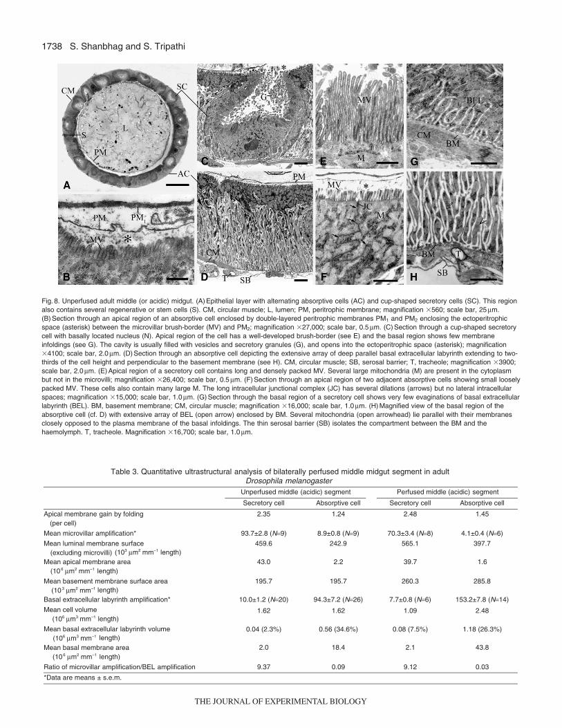
Fig. 8. Unperfused adult middle (or acidic) midgut. (A) Epithelial layer with alternating absorptive cells (AC) and cup-shaped secretory cells (SC). This regionalso contains several regenerative or stem cells (S). CM, circular muscle; L, lumen; PM, peritrophic membrane; magnification �560; scale bar, 25μm.(B) Section through an apical region of an absorptive cell enclosed by double-layered peritrophic membranes PM1 and PM2 enclosing the ectoperitrophicspace (asterisk) between the microvillar brush-border (MV) and PM2; magnification �27,000; scale bar, 0.5μm. (C) Section through a cup-shaped secretorycell with basally located nucleus (N). Apical region of the cell has a well-developed brush-border (see E) and the basal region shows few membraneinfoldings (see G). The cavity is usually filled with vesicles and secretory granules (G), and opens into the ectoperitrophic space (asterisk); magnification�4100; scale bar, 2.0μm. (D) Section through an absorptive cell depicting the extensive array of deep parallel basal extracellular labyrinth extending to two-thirds of the cell height and perpendicular to the basement membrane (see H). CM, circular muscle; SB, serosal barrier; T, tracheole; magnification �3900;scale bar, 2.0μm. (E) Apical region of a secretory cell contains long and densely packed MV. Several large mitochondria (M) are present in the cytoplasmbut not in the microvilli; magnification �26,400; scale bar, 0.5μm. (F) Section through an apical region of two adjacent absorptive cells showing small looselypacked MV. These cells also contain many large M. The long intracellular junctional complex (JC) has several dilations (arrows) but no lateral intracellularspaces; magnification �15,000; scale bar, 1.0μm. (G) Section through the basal region of a secretory cell shows very few evaginations of basal extracellularlabyrinth (BEL). BM, basement membrane; CM, circular muscle; magnification �16,000; scale bar, 1.0μm. (H) Magnified view of the basal region of theabsorptive cell (cf. D) with extensive array of BEL (open arrow) enclosed by BM. Several mitochondria (open arrowhead) lie parallel with their membranesclosely opposed to the plasma membrane of the basal infoldings. The thin serosal barrier (SB) isolates the compartment between the BM and thehaemolymph. T, tracheole. Magnification �16,700; scale bar, 1.0μm.
Table 3. Quantitative ultrastructural analysis of bilaterally perfused middle midgut segment in adult Drosophila melanogaster
Unperfused middle (acidic) segment Perfused middle (acidic) segment
Secretory cell Absorptive cell Secretory cell Absorptive cell
Apical membrane gain by folding (per cell)
2.35 1.24 2.48 1.45
Mean microvillar amplification* 93.7±2.8 (N=9) 8.9±0.8 (N=9) 70.3±3.4 (N=8) 4.1±0.4 (N=6)
Mean luminal membrane surface (excluding microvilli) (103 µm2 mm–1 length)
459.6 242.9 565.1 397.7
Mean apical membrane area (106 µm2 mm–1 length)
43.0 2.2 39.7 1.6
Mean basement membrane surface area (10 3 µm2 mm–1 length)
195.7 195.7 260.3 285.8
Basal extracellular labyrinth amplification* 10.0±1.2 (N=20) 94.3±7.2 (N=26) 7.7±0.8 (N=6) 153.2±7.8 (N=14)
Mean cell volume (106 µm3 mm–1 length)
2.0 18.4 2.1 43.8
Mean basal extracellular labyrinth volume (106 µm3 mm–1 length)
1.62 1.62 1.09 2.48
Mean basal membrane area (106 µm2 mm–1 length)
0.04 (2.3%) 0.56 (34.6%) 0.08 (7.5%) 1.18 (26.3%)
Ratio of microvillar amplification/BEL amplification 9.37 0.09 9.12 0.03
*Data are means ± s.e.m.
THE JOURNAL OF EXPERIMENTAL BIOLOGY

1739H+ transport in Drosophila midgut
Fig. 9. Unperfused adult middle midgut. (A,C) Low-magnification electron micrographs of a secretory cell (SC) located between two absorptive cells (AC) inclosed and open configurations (open arrow). The secretory cell cavity in the open state contains electron-dense granules and vesicles that are liberatedinto the ectoperitrophic space (asterisk) of the midgut; magnification �2600; scale bar, 5.0μm. (B,D) Diagrammatic representations of a secretory cell (SC)located between two absorptive cells (AC) in open and closed configurations (also see Fig. 10A,E). The luminal surface of SC has long, densely populatedmicrovilli (MV), and the basal surface displays few infoldings (BEL). Absorptive cells (AC) have very short and sparsely distributed MV but long andextensive parallel arrays of BEL. BM, basement membrane; CC, closed cavity; CM, circular muscle; JC, intercellular junctional complex; LM, longitudinalmuscle; N, nucleus; OC, open cavity; PM1 and PM2, peritrophic membranes; SB, serosal barrier; T, tracheole.
THE JOURNAL OF EXPERIMENTAL BIOLOGY

1740
confirms the basic observations of Gartner (Gartner, 1985)pertaining to the anterior midgut only but also reveals that thereis a degree of variation of the anterior gut along its length(Fig. 4B,C, Fig. 5, Fig. 6). Posterior segment cells (Fig. 4E, Fig. 7)have an extensively dilated basal extracellular labyrinth, with avolume larger than that of anterior segment cells, indicating morefluid reabsorption in the posterior segment (Tripathi andBoulpaep, 1989). Much less is known of the role of thisepithelium in adults, which requires further study in bothultrastructure and physiology.
The middle adult acidic segment (Fig.4D; Figs8–10) hasattracted a good deal of attention for its striking architecture, withalternate cells ‘facing’ opposite directions. It would be appropriateto refer to them as secretory and absorptive cells on account of thegreat asymmetry of their apical and basal membranes (Fig.8D–H).Cells of the middle segment are alternately absorptive (apicallyamplified �9-fold, basally >90-fold) and secretory (apicallyamplified by >90-fold and basally �10-fold). The terminologysuggested is based on the direction of net transport predicted onareal considerations alone. Table3 shows that the apical and basalmembranes of these two cell types are amplified more than 100-fold in either direction; their back-to-back geometry predictssignificant recycling of solutes and water and provides a structuralbasis for bidirectional transport. The function of the secretory cells,composition of the secreted contents, regulation of secretion, andmembrane turnover are still not clear, despite many studies (Lehaneand Billingsley, 1996; Yao and Forte, 2003). The apical membranesform a cavity that can be seen spontaneously in either ‘open’ or‘closed’ configuration (Figs9 and 10) discharging membrane-bound granular material.
Bidirectional transport in individual regionsThe identification of morphologically distinct segments of themidgut implies that controlled study of each segment is necessarybefore one can integrate the information for the entire midgut.Larvae, being voracious feeders, have a highly active midgutepithelium, as evidenced by the generation of much steeper pHgradients compared with adults (Shanbhag and Tripathi, 2005;Shanbhag and Tripathi, 2008) (Figs11, 12). Similar gradients havebeen seen in a wide variety of insect midgut epithelia (Dadd, 1975;Boudko et al., 2001b; Corena et al., 2002; Dow, 1986; Moffett andCummings, 1994; Onken et al., 2008; Zhuang et al., 1999).Regardless of the direction of secretion of acid or base, theenergetics of transport in insect epithelia is believed to be primarilydriven by vacuolar or H+ V-ATPases in the Malpighian tubule andalso the midgut (Beyenbach, 2001; Dow and Davies, 2001). Thefluxes of other electrolytes have been proposed to be driven assecondary transport processes. Furthermore, it is also possible thatmany of the membrane transporters in insects could be isoforms ofmammalian transporters that do not bind inhibitors or agonists asthey do in mammals. In the posterior larval midgut of Drosophila,a variety of inhibitors had no effect on transport that was stillsensitive to bafilomycin-A1 (Shanbhag and Tripathi, 2005).
Larval midgutThe other general feature of acid and base transport in the midgutepithelia of many insects is that there is a carbonic anhydrase (CA)-catalysed pool of H+ from which the H+ V-ATPases pump (Corenaet al., 2002; Corena et al., 2005; Ridgway and Moffett, 1986; Seronet al., 2004). The location of this pool is a subject of active studyas many enzymes are glycosylphosphatidylinositol-anchored and it
S. Shanbhag and S. Tripathi
Fig. 10. Perfused adult middle midgut. (A). Section through a secretory cell (SC) with apical cavity open to the lumen (L). The peritrophic membrane iswashed off, and intact microvilli (MV) are directly in contact with the luminal solution. The cell contains a large number of secretory granules (G), vesicles (V)and mitochondria (M); CM, circular muscle; LM, longitudinal muscle; SB, serosal barrier; T, tracheole; magnification �5000; scale bar, 5.0μm. (B) Magnifiedview of apical region of a secretory cell shows vesicles with single or many electron-dense granules (G) arising mainly from the base of the adjacentmicrovilli and many electron-lucent vesicles (V) that bud off usually from the tip of the MV; magnification �18,700; scale bar, 1.0μm. (C) Section through aportion of the cavity (asterisk) of the secretory cell, with many secretory granules and vesicles. M, mitochondria; MV, microvilli; N, nucleus; magnification�11,300; scale bar, 1.0μm. (D) Intercellular junctional complex (JC) of an absorptive cell(s) seen closing the apical aperture of the secretory cavity;magnification �9300; scale bar, 1.0μm. (E). Section through a secretory cell (SC) in the same plane shows the apical cell cavity closed by the intercellularJC of an absorptive cell (AC). CC, closed cavity; CM, circular muscle; LM, longitudinal muscle; M, mitochondria; SB, serosal barrier; magnification �5000;scale bar, 5.0μm.
THE JOURNAL OF EXPERIMENTAL BIOLOGY

1741H+ transport in Drosophila midgut
is possible to localize these pools to either intracellular orextracellular or both compartments. Strong CA activity waslocalized at the apical membranes of goblet cells in the anterior andmiddle midgut region of Manduca sexta that is associated with thelumen alkalinization, but no CA activity was found in the posteriormidgut goblet cells. Lepidopteran midgut is also divided intoregions that show a substantial degree of structural and functionaldifferentiation (Ridgway and Moffett, 1986).
The localization of CA in the Drosophila midgut has not yet beenachieved, but there are several interesting approaches that can betried. Corena et al. have localized CA in the mosquito midgut(Corena et al., 2004), and a similar approach with membranepermeant and impermeant inhibitor would be a valuable approach.The removal of the peritrophic membrane by microperfusionimproves access of the luminal perfusate to the apical membrane.It is possible that the in vivo conditions of the peritrophic space maybe altered in terms of enzymes like CA that are located there (Smithet al., 2007), but there is good reason to assume that the intracellularenzyme-catalysed H+ pool is intact in perfused midguts, as shownbelow. As the ectoperitrophic space is a likely candidate for sucha location, it would be important to verify this localization withantibodies to CA9 in both perfused and unperfused preparations
where the peritrophic membrane is intact (Fig.2C). The dissipationof the acid gradient by inhibition of CA with acetazolamide(Shanbhag and Tripathi, 2008) is easily detected on pH paper(Fig.11D,E). However, the effects of inhibition of CA involved inbase secretion in the posterior midgut are not easily detected by thissimple method. One then has to rely on a more sensitive method(e.g. ion-selective microelectrodes) to detect acid or base fluxes.Such an approach is shown in Fig.13.
In Fig.13A, which shows measurement of intracellular pH,along with other membrane parameters, in the perfused larvalmidgut, acetazolamide alkalinizes the cell when applied to the bath;these effects are also seen from the lumen. This is clear evidencethat extrusion of acid and base is rate limited in the H+ pool. Therehas been some uncertainty about the localization of the ATPase,particularly in view of data on the Malpighian tubule and themosquito midgut (Beyenbach et al., 2000; O’Donnell et al., 1996;Wieczorek et al., 1999; Wieczorek et al., 2003). One line ofevidence for a basal location of the ATPase in the Drosophilaposterior midgut (Shanbhag and Tripathi, 2005), where the lumenis strongly alkaline, is the presence of portasome-like structures inthe BEL (Fig.2E, inset). The intracellular pH being more alkalinethan the BEL or unstirred layer of the bath (Fig.13A) provides even
Fig. 11. Midguts of 3rd instar Drosophila larvae. (A,B) Larvae fed with food containing dyes alone or together with acetazolamide (C) to detect luminal pH.GC, gastric caeca; MT, Malpighian tubules. (A) Acidification was detected in the middle midgut (acid zone), where Bromophenol Blue (BPB) dye changedfrom blue to yellow (pH<3.0). (B) Very strong alkalinization was detected in the posterior half of the posterior midgut (alkaline zone), where Phenol Red (PR)dye changed from red to dark pink (pH>10.0). (C) Acetazolamide (100μmol l–1) in food containing BPB led to dissipation of pH gradients in the larval midgut.(D) Luminal content pH detected by pH paper shows a distinct acid-secreting zone (acid zone, ochre, pH<3) and posterior alkaline zone (dark green).(E) Acetazolamide (100μmol l–1) in food dissipated the pH gradients in the acid zone. Numbered panels below denote colours generated on universal pHpaper by pH standards of 1–11. Scale bar, 1 mm.
THE JOURNAL OF EXPERIMENTAL BIOLOGY

1742 S. Shanbhag and S. Tripathi
Acetazolamide Sch-RSch-RBath:
50
30
10
–10
–30
–50
–70
–90
A
5 min
7.0
7.5
8.0
8.5
Vc–Vb
Va
Vc
Vp
pHi
mV
Acetazolamide Sch-RSch-RBath:70 B
5 min
6.0
pHo
mV
–110
–130
–150
VpH–Vb
VpHin
Vb
50
30
10
–10
–30
–50
–70
7.0
7.5
6.5
8.0
Vc–VbVa
VcVp
VpHout
Vb
Bafilomycin-A1
Fig. 13. Carbonic anhydrase and H+ V-ATPase in the larval posterior midgut.(A) Intracellular pH measured in controlsolution (Sch-R) containing equal amounts ofSchneider insect medium and insect Ringerperfusing the lumen and in the bath. Va,apical membrane potential; Vb, basalmembrane potential; Vc, transepithelialpotential at collection end; Vp, transepithelialpotential at perfusion end; VpHin, potential ofion-selective microelectrode; VpH–Vb,intracellular pH corrected for membranepotential, referenced to ordinate at right.Acetazolamide (100μmol l–1) applied to thebath reversibly alkalinizes the cell.(B) Extracellular scan of basal unstirred layerpH with an ion-selective microelectrode whosepotential VpHout is shown as the brown trace,referenced to the ordinate on the right. In Sch-R, the extracellular pH of the bulk solution ofthe bath is 7.2. The pH microelectrode wasadvanced towards the gut wall by a piezo-stepper and positioned close to the serosalbarrier and basal extracellular labyrinth (BEL;arrow); the increasingly positive VpHoutpotential reflects the pH gradient at the basalsurface. The pH electrode was then retractedaway to about 20μm from the gut wall(arrowhead). Acetazolamide (100μmol l–1)applied to the bath alkalinized the extracellularpH. Bafilomycin-A1 (1.5μmol l–1) applied to thebath in addition to acetazolamide in the bathfurther alkalinized the extracellular pH.Washout with control Ringer restored H+
extrusion by the midgut, as seen by thepositive change in VpHout at the end of thetrace.
Fig. 12. Carbonic anhydrase in theadult midgut. (A) Acidification wasdetected in the middle midgut(arrows, acid zone) where thecolour of Bromophenol Blue (BPB)dye changed from blue to greenishbrown (acid zone, pH<4.0); C,cardia; CR, crop; H, head; HG,hindgut; MT, Malpighian tubules.(B) Acetazolamide (100μmol l–1) infood dissipated this gradient.(C) Luminal content pH detected bypH paper showed a distinct acid-secreting zone (arrows, pH<5) inthe adult middle midgut.(D) Acetazolamide (100μmol l–1) infood abolishes this acidification(arrows). Numbered panels belowdenote colours generated onuniversal pH paper by pHstandards of 1–11. Scale bar,1 mm.
THE JOURNAL OF EXPERIMENTAL BIOLOGY

1743H+ transport in Drosophila midgut
stronger evidence for the predominantly basal location of the H+
V-ATPase. Thirdly, sensitivity to bafilomycin is also greater in thispreparation from the bath side, with hardly any effect detected fromthe lumen. Thus, one can measure acid extrusion rates from thebasal side (Fig.13B), and see its reversible inhibition byacetazolamide, even without the peritrophic membrane (from thelumen or bath) and bafilomycin (from the bath only).
Adult midgutPerfusion of each midgut region is important to know the overalldriving forces for ions in each segment. Representative traces forthree adult segments are shown in Fig.14 along with basic
membrane potential data under free-flow conditions in controlRinger and taking into account the fact that the ends of the segmentsare damaged but electrically isolating the lumen from the bath. Theanterior region has a low transepithelial potential; the middle andposterior segments have a transepithelial potential whose polarityfavours net H+ flux, which can occur passively for the observedgradients. Therefore, primary and secondary active transportprocesses have to be independently tested in these segments alongwith a complete characterization of all electrochemical drivingforces and passive properties of individual cell membranes and theparacellular pathway. Whereas the larval gut has a predominant H+
V-ATPase as primary transporter, the situation in the adult is farfrom clear.
Microperfusion and electrophysiological approaches have givenus new tools to investigate membrane transport processes in animportant genomic organism with many characterized mutations,e.g. labial (Dubreuil et al., 1998), where individual cuprophiliccells may be absent. For a complete understanding of the role ofpH gradients in the Drosophila gut, experiments need to bedesigned to supplement the approach shown here with newertechniques. For instance, it is still not clear how base is secretedacross the apical membranes in the posterior midgut in either larvaor adult. It would be of interest to examine whether transporterslike NDAE1 (Romero et al., 2000; Sciortino et al., 2001) areinvolved in the exit of base in the posterior midgut. Likewise, thetransport of other electrolytes and osmolytes needs clarification, asit is very likely to be linked to the transport of acid and base in thisepithelium. The approach shown here can contribute to this end.
We thank our colleagues T. V. Abraham and J. N. Parmar for their unfailingsupport and Professor L. C. Padhy for valuable discussions. Supported byInterdisciplinary Programme 11-R&D-TFR-5.02-1106.
ReferencesAnderson, E. and Harvey, W. R. (1966). Active transport by the Cecropia midgut. II.
Fine structure of the midgut epithelium. J. Cell Biol. 31, 107-134.Baumann, O. (2001). Posterior midgut epithelial cells differ in their organization of the
membrane skeleton from other Drosophila epithelia. Exp. Cell Res. 270, 176-187.Beyenbach, K. W. (2001). Energizing epithelial transport with the vacuolar H+-
ATPase. News Physiol. Sci. 16, 145-151.Beyenbach, K. W., Pannabecker, T. L. and Nagel, W. (2000). Central role of the
apical membrane H+-ATPase in electrogenesis and epithelial transport in Malpighiantubules. J. Exp. Biol. 203, 1459-1468.
Boudko, D. Y., Moroz, L. L., Linser, P. J., Trimarchi, J. R., Smith, P. J. S. andHarvey, W. R. (2001a). In situ analysis of pH gradients in mosquito larvae usingnon-invasive, self-referencing, pH-sensitive microelectrodes. J. Exp. Biol. 204, 691-699.
Boudko, D. Y., Moroz, L. L., Harvey, W. R. and Linser, P. J. (2001b). Alkalinizationby chloride/bicarbonate pathway in larval mosquito midgut. Proc. Natl. Acad. Sci.USA 98, 15354-15359.
Cioffi, M. (1979). The morphology and fine structure of the larval midgut of a moth(Manduca sexta) in relation to active ion transport. Tissue Cell 11, 467-479.
Clements, A. N. (1992). The Biology of Mosquitoes. London: Chapman and Hall.Corena, M. P., Seron, T. J., Lehman, H. K., Ochrietor, J. D., Kohn, A., Tu, C. and
Linser, P. J. (2002). Carbonic anhydrase in the midgut of larval Aedes aegypti:cloning, localization and inhibition. J. Exp. Biol. 205, 591-602.
Corena, M. P., Fiedler, M. M., VanEkeris, L., Tu, C., Silverman, D. N. and Linser,P. J. (2004). Alkalization of larval mosquito midgut and the role of carbonicanhydrase in different species of mosquitoes. Comp. Biochem. Physiol. 137C, 207-225.
Corena, M. P., VanEkeris, L., Salazar, M. I., Bowers, D., Fiedler, M. M., Silverman,D., Tu, C. and Linser, P. J. (2005). Carbonic anhydrase in the adult mosquitomidgut. J. Exp. Biol. 208, 3263-3273.
Dadd, R. H. (1975). Alkalinity within the midgut of mosquito larvae with alkaline-activedigestive enzymes. J. Insect Physiol. 21, 1847-1853.
Dimitriadis, V. K. (1991). Fine structure of the midgut of adult Drosophila auraria andits relationship to the sites of acidophilic secretion. J. Insect Physiol. 37, 167-177.
Dimitriadis, V. K. and Kastritsis, C. D. (1984). Ultrastructural analysis of the midgutof Drosophila auraria larvae: morphological observations and their physiologicalimplications. Can. J. Zool. 62, 659-669.
Dow, J. A. T. (1984). Extremely high pH in biological systems: a model for carbonatetransport. Am. J. Physiol. 246, R633-R636.
Dow, J. A. T. (1986). Insect midgut function. Adv. Insect Physiol. 19, 187-328.Dow, J. A. T. and Peacock, J. M. (1989). Microelectrode evidence for the electrical
isolation of goblet cell cavities in Manduca sexta middle midgut. J. Exp. Biol. 143,101-114.
Control RingerBath:Control RingerLumen:
60
A
5 min
mV
40
100
80
20
0
–20
–40
–60
–80
Va
Vc
Vp
Vb
60
C
5 min
mV
40
100
80
20
0
–20
–40
–60
–80
Va
Vc
Vp
Vb
60
B
5 min
mV
40
80
20
0
–20
–40
–60
–80
Va
Vc
Vp
Vb
Fig. 14. (A–C) Adult midgut transepithelial potentials recorded from theanterior, middle and posterior segments, respectively, of Drosophila by theperfusion (Vp), collection (Vc) micropipettes and the basal cell membranepotential (Vb), after bilateral HCO3
– Ringer (control Ringer) substitution.Current pulses (�100 nA) cause displacements of Va, Vb, Vc and Vp. Va,apical membrane potential.
THE JOURNAL OF EXPERIMENTAL BIOLOGY

1744
Dow, J. A. T. and Davies, S. A. (2001). The Drosophila melanogaster Malpighiantubules. Adv. Insect Physiol. 28, 1-83.
Dow, J. A. T., Davies, S. A., Guo, Y., Graham, S., Finbow, M. E. and Kaiser, K.(1997). Molecular genetic analysis of V-ATPase function in Drosophila melanogaster.J. Exp. Biol. 200, 237-245.
Dubreuil, R. R. (2004). Copper cells and stomach acid secretion in the Drosophilamidgut. Int. J. Biochem. Cell Biol. 36, 745-752.
Dubreuil, R. R., Frankel, J., Wang, P., Howrylak, J., Kappil, M. and Grushko, T. A.(1998). Mutations of α spectrin and labial block cuprophilic cell differentiation andacid secretion in the middle midgut of Drosophila larvae. Dev. Biol. 194, 1-11.
Dubreuil, R. R., Grushko, T. and Baumann, O. (2001). Differential effects of alabial mutation on the development, structure, and function of stomach acid-secreting cells in Drosophila melanogaster larvae and adults. Cell Tissue Res.306, 167-178.
Filshie, B. K., Poulson, D. F. and Waterhouse, D. F. (1971). Ultrastructure of thecopper-accumulating region of the Drosophila larval midgut. Tissue Cell 3, 77-102.
Gartner, L. P. (1985). The fine structural morphology of the midgut of adult Drosophila:a morphometric analysis. Tissue Cell 17, 883-888.
Lehane, M. J. and Billingsley, P. F. (1996). Biology of the Insect Midgut. London:Chapman and Hall.
Lin, G., Xu, N. and Xi, R. (2008). Paracrine Wingless signalling controls self-renewalof Drosophila intestinal stem cells. Nature 455, 1119-1122.
Maddrell, S. H. P. and O’Donnell, M. J. (1992). Insect Malpighian tubules: V-ATPaseaction in ion and fluid transport. J. Exp. Biol. 172, 417-429.
Micchelli, C. A. and Perrimon, N. (2006). Evidence that stem cells reside in the adultDrosophila midgut epithelium. Nature 439, 475-479.
Moffett, D. F. and Cummings, S. A. (1994). Transepithelial potential and alkalizationin an in situ preparation of tobacco hornworm (Manduca sexta) midgut. J. Exp. Biol.194, 341-345.
O’Donnell, M. J. and Spring, J. H. (2000). Modes of control of insect Malpighiantubules: synergism, antagonism, cooperation and autonomous regulation. J. InsectPhysiol. 46, 107-117.
O’Donnell, M. J., Dow, J. A. T., Huesmann, G. R., Tublitz, N. J. and Maddrell, S. H.P. (1996). Separate control of anion and cation transport in Malpighian tubules ofDrosophila melanogaster. J. Exp. Biol. 199, 1163-1175.
Ohlstein, B. and Spradling, A. (2006). The adult Drosophila posterior midgut ismaintained by pluripotent stem cells. Nature 439, 470-474.
Okech, B. A., Boudko, D. Y., Linser, P. J. and Harvey, W. R. (2008). Cationicpathway of pH regulation in larvae of Anopheles gambiae. J. Exp. Biol. 211, 957-968.
Onken, H., Moffett, S. B. and Moffett, D. F. (2006). The isolated anterior stomach oflarval mosquitoes (Aedes aegypti): voltage-clamp measurements with a tubularepithelium. Comp. Biochem. Physiol. 143A, 24-34.
Onken, H., Moffett, S. B. and Moffett, D. F. (2008). Alkalinization in the isolated andperfused anterior midgut of the larval mosquito, Aedes aegypti. J. Insect Sci. 8, 1-20.
Patrick, M. L., Aimanova, K., Sanders, H. R. and Gill, S. S. (2006). P-type Na+/K+-ATPase and V-type H+-ATPase expression patterns in the osmoregulatory organs oflarval and adult mosquito Aedes aegypti. J. Exp. Biol. 209, 4638-4651.
Ridgway, R. L. and Moffett, D. F. (1986). Regional differences in the histochemicallocalization of carbonic anhydrase in the midgut of tobacco hornworm (Manducasexta). J. Exp. Zool. 237, 407-412.
Romero, M. F., Henry, D., Nelson, S., Harte, P. J., Dillon, A. K. and Sciortino, C.M. (2000). Cloning and characterization of a Na+-driven anion exchanger (NDAE1).J. Biol. Chem. 275, 24552-24559.
Sciortino, C. M., Shrode, L. D., Fletcher, B. R., Harte, P. J. and Romero, M. F.(2001). Localization of endogenous and recombinant Na+-driven anion exchangerprotein NDAE1 from Drosophila melanogaster. Am. J. Physiol. 281, C449-C463.
Seron, T. J., Hill, J. and Linser, P. J. (2004). A GPI-Linked carbonic anhydraseexpressed in the larval mosquito midgut. J. Exp. Biol. 207, 4559-4572.
Shanbhag, S. and Tripathi, S. (2005). Electrogenic H+ transport and pH gradientsgenerated by a V-H+-ATPase in the isolated perfused larval Drosophila midgut. J.Membrane Biol. 206, 61-72.
Shanbhag, S. and Tripathi, S. (2008). Segmental bidirectional transport of H+ in theadult Drosophila midgut. Comp. Biochem. Physiol. 150A, A11.29, S138.
Smith, K. E., VanEkeris, L. A. and Linser, P. J. (2007). Cloning and characterizationof AgCA9, a novel α-carbonic anhydrase from Anopheles gambiae Giles sensustricto (Diptera: Culicidae) larvae. J. Exp. Biol. 210, 3919-3930.
Smith, K. E., VanEkeris, L. A., Okech, B. A., Harvey, W. R. and Linser, P. J.(2008). Larval anopheline mosquito recta exhibit a dramatic change in localizationpatterns of ion transport proteins in response to shifting salinity: a comparisonbetween anopheline and culine larvae. J. Exp. Biol. 211, 3067-3076.
Takashima, S., Mkrtchyan, M., Hartenstein, A. Y., Merriam, J. R. and Hartenstein,V. (2008). The behaviour of Drosophila adult hindgut stem cells is controlled by Wntand Hh signalling. Nature 454, 651-655.
Tripathi, S. and Boulpaep, E. L. (1989). Mechanisms of water transport by epithelialcells. Q. J. Exp. Physiol. 74, 385-417.
Volkmann, A. and Peters, W. (1989a). Investigations on the midgut caeca ofmosquito larvae. I. Fine-structure. Tissue Cell 21, 243-251.
Volkmann, A. and Peters, W. (1989b). Investigations on the midgut caeca ofmosquito larvae. II. Functional-aspects. Tissue Cell 21, 253-261.
Wieczorek, H., Brown, D., Grinstein, S., Ehrenfeld, J. and Harvey, W. R. (1999).Animal plasma membrane energization by proton-motive V-ATPases. BioEssays 21,637-648.
Wieczorek, H., Huss, M., Merzendorfer, H., Reineke, S., Vitavska, O. and Zeiske,W. (2003). The insect plasma membrane H+ V-ATPase: intra-, inter-, andsupramolecular aspects. J. Bioenerg. Biomembr. 35, 359-366.
Yao, X. and Forte, J. G. (2003). Cell biology of acid secretion by the parietal cell.Annu. Rev. Physiol. 65, 103-131.
Zhuang, Z., Linser, P. J. and Harvey, W. R. (1999). Antibody to H+ V-ATPasesubunit E colocalizes with portasomes in alkaline larval midgut of freshwatermosquito (Aedes aegypti). J. Exp. Biol. 202, 2449-2460.
S. Shanbhag and S. Tripathi
THE JOURNAL OF EXPERIMENTAL BIOLOGY