Review. Biology and Systematics of the form genus Rhizoctonia · Review. Biology and Systematics of...
25
Review. Biology and Systematics of the form genus Rhizoctonia V. González García 1 *, M. A. Portal Onco 2 and V. Rubio Susan 3 1 IVIA. Ctra. Moncada-Náquera, Km 4.5. 46113 Moncada (Valencia). Spain 2 Museo Nacional de Ciencias Naturales (CSIC). José Gutiérrez Abascal, 3. 28006 Madrid. Spain 3 Centro de Ciencias Medioambientales (CSIC). Serrano, 115. 28003 Madrid. Spain Abstract Members of the form genus Rhizoctonia D.C. are considered as a complex mixture of filamentous fungi, having in common the possession of a non-spored imperfect state, usually referred to as the Rhizoctonia anamorph. The group includes several of the most devastating crop pathogens like Thanatephorus cucumeris (Frank) Donk (anamorph = Rhizoctonia solani Kühn), the majority of orchid mycorrhizal symbionts (mainly belonging to genus Ceratobasidium D.P. Rogers) and a collection of saprotrophic organisms of different systematic placement. The Rhizoctonia anamorph is characterized by several common features present among members of the entire Rhizoctonia species complex. Taxa from the group have been rearranged into several groups of higher fungi, including both Ascomycota and Basidiomycota, and split into several genera, employing criteria such as the analysis and ultrastructural comparison of septal apparatus. Until very recently, classification for some of the groups within the complex has been exclusively based on criteria such as hyphal anastomosis, since other types of diagnostic features are usually scarce in these fungi. Phytopathological studies in the complex have represented the major contingent of contributions in the group, especially in the case of R. solani. Some members of the complex have been reported to be protective isolates against pathogenic members of Rhizoctonia and some other fungal pathogens. This review focuses on the knowledge of several aspects of the species of Rhizcotonia s. lato, such as its current taxonomic placement, the biology and systematics of some groups of the complex, and a revision of the methodologies employed in studying it. Additional key words: Basidiomycetes, biocontrol, Ceratobasidiaceae, Ceratobasidium, fungal diseases, methodology, taxonomy, Thanatephorus. Resumen Biología y Sistemática del género forma Rhizoctonia Los miembros del género forma Rhizoctonia D.C. son considerados como un complejo de hongos filamentosos, presentando en común una fase asexual no productora de esporas, denominada anamorfo tipo Rhizoctonia. El grupo incluye algunos importantes patógenos como Thanatephorus cucumeris (Frank) Donk (anamorfo=Rhizoctonia solani Kühn), la mayoría de especies micorrícicas de orquídeas (principalmente del género Ceratobasidium D.P. Rogers), y algunos taxones de posición taxonómica variada. El anamorfo tipo Rhizoctonia es definido por una serie de características, presentes y comunes en todos los taxones del complejo. Los taxones del grupo han sido distribuidos entre varios grupos de hongos, incluyendo Ascomicetes y Basidiomicetes, y reubicados en varios géneros de ambas clases, empleando criterios de clasificación tales como el análisis y comparación de la ultraestructura del aparato septal. Habitualmente, la clasificación en algunos de los grupos del complejo ha estado basada en la aplicación de criterios tales como la anastomosis hifal, debido a que en este tipo de hongos es escasa la presencia de un número aceptable de caracteres diagnósticos. Los trabajos fitopatológicos en el complejo representan el mayor contingente dentro de las contribuciones publicadas, especialmente en el caso de R. solani. Algunos de los miembros del complejo son citados como protectores contra miembros patógenos de Rhizoctonia y otros patógenos fúngicos. El presente trabajo presenta los conocimientos actuales sobre aspectos del complejo, tales como su clasificación actual, la biología y sistemática de algunos de los grupos de Rhizoctonia s. lato, además de una revisión de las metodologías más comúnmente empleadas en su estudio. Palabras clave adicionales: Basidiomycetes, biocontrol, Ceratobasidiaceae, Ceratobasidium, enfermedades fúngicas, metodología, taxonomía, Thanatephorus. Spanish Journal of Agricultural Research (2006) 4(1), 55-79 * Corresponding author: [email protected] Received: 15-08-05; Accepted: 01-02-06.
Transcript of Review. Biology and Systematics of the form genus Rhizoctonia · Review. Biology and Systematics of...
Review. Biology and Systematics of the form genus Rhizoctonia
V. González García1*, M. A. Portal Onco2 and V. Rubio Susan3
1 IVIA. Ctra. Moncada-Náquera, Km 4.5. 46113 Moncada (Valencia). Spain2 Museo Nacional de Ciencias Naturales (CSIC). José Gutiérrez Abascal, 3. 28006 Madrid. Spain
3 Centro de Ciencias Medioambientales (CSIC). Serrano, 115. 28003 Madrid. Spain
Abstract
Members of the form genus Rhizoctonia D.C. are considered as a complex mixture of filamentous fungi, havingin common the possession of a non-spored imperfect state, usually referred to as the Rhizoctonia anamorph. Thegroup includes several of the most devastating crop pathogens like Thanatephorus cucumeris (Frank) Donk(anamorph = Rhizoctonia solani Kühn), the majority of orchid mycorrhizal symbionts (mainly belonging to genusCeratobasidium D.P. Rogers) and a collection of saprotrophic organisms of different systematic placement. TheRhizoctonia anamorph is characterized by several common features present among members of the entire Rhizoctoniaspecies complex. Taxa from the group have been rearranged into several groups of higher fungi, including bothAscomycota and Basidiomycota, and split into several genera, employing criteria such as the analysis and ultrastructuralcomparison of septal apparatus. Until very recently, classification for some of the groups within the complex has beenexclusively based on criteria such as hyphal anastomosis, since other types of diagnostic features are usually scarce inthese fungi. Phytopathological studies in the complex have represented the major contingent of contributions in the group,especially in the case of R. solani. Some members of the complex have been reported to be protective isolates againstpathogenic members of Rhizoctonia and some other fungal pathogens. This review focuses on the knowledge of severalaspects of the species of Rhizcotonia s. lato, such as its current taxonomic placement, the biology and systematics of somegroups of the complex, and a revision of the methodologies employed in studying it.
Additional key words: Basidiomycetes, biocontrol, Ceratobasidiaceae, Ceratobasidium, fungal diseases,methodology, taxonomy, Thanatephorus.
Resumen
Biología y Sistemática del género forma Rhizoctonia
Los miembros del género forma Rhizoctonia D.C. son considerados como un complejo de hongos filamentosos,presentando en común una fase asexual no productora de esporas, denominada anamorfo tipo Rhizoctonia. El grupo incluyealgunos importantes patógenos como Thanatephorus cucumeris (Frank) Donk (anamorfo=Rhizoctonia solani Kühn), lamayoría de especies micorrícicas de orquídeas (principalmente del género Ceratobasidium D.P. Rogers), y algunos taxonesde posición taxonómica variada. El anamorfo tipo Rhizoctonia es definido por una serie de características, presentes ycomunes en todos los taxones del complejo. Los taxones del grupo han sido distribuidos entre varios grupos de hongos,incluyendo Ascomicetes y Basidiomicetes, y reubicados en varios géneros de ambas clases, empleando criterios declasificación tales como el análisis y comparación de la ultraestructura del aparato septal. Habitualmente, la clasificación enalgunos de los grupos del complejo ha estado basada en la aplicación de criterios tales como la anastomosis hifal, debido aque en este tipo de hongos es escasa la presencia de un número aceptable de caracteres diagnósticos. Los trabajosfitopatológicos en el complejo representan el mayor contingente dentro de las contribuciones publicadas, especialmente enel caso de R. solani. Algunos de los miembros del complejo son citados como protectores contra miembros patógenos deRhizoctonia y otros patógenos fúngicos. El presente trabajo presenta los conocimientos actuales sobre aspectos delcomplejo, tales como su clasificación actual, la biología y sistemática de algunos de los grupos de Rhizoctonia s. lato,además de una revisión de las metodologías más comúnmente empleadas en su estudio.
Palabras clave adicionales: Basidiomycetes, biocontrol, Ceratobasidiaceae, Ceratobasidium, enfermedadesfúngicas, metodología, taxonomía, Thanatephorus.
Spanish Journal of Agricultural Research (2006) 4(1), 55-79
* Corresponding author: [email protected]: 15-08-05; Accepted: 01-02-06.

Introduction
The form genus Rhizoctonia is considered as anheterogeneous assemblage of filamentous fungal taxathat do not produce asexual spores and share a numberof common features in their anamorphic states. Theorganisms belonging to this species complex aregenerally soil fungi, mostly associated with roots andusually pathogens, although there have been reportsof a number of saprophytic and symbiotic taxa.Fungi in the form genus are distributed worldwidein both agricultural and forest soils and includesome of the most economically important plantpathogens, causing foliar and root rot diseases ofmajor crops.
The genus concept in Rhizoctonia was firstestablished by De Candolle (1815). This concept wasreviewed by Parmeter and Whitney (1970) morethan a century later, who concluded that, accordingto De Candolle, the basic characters of the genusnamed by the Swiss author could be summarizedin the production of sclerotia of uniform textureand association of the mycelium with roots of livingplants. This lack of features has led in the past tothe classification of a collection of unrelated fungisuch as Rhizoctonia s. lato (Moore, 1987). Thus, theinclusion of a given taxa to the genus Rhizoctoniawas based for a long time on the possession of certainvegetative characters such as brown pigmentedhyphae, constrictions at branch points formingright angles and absence of mitospores. Togetherwith these features, always present, there are othercharacters not constantly present in the entire speciescomplex, but present in a large number of species,such as the presence of monilioid cells and sclerotiain culture, fast rates of hyphal growth or acomplex dolipore septal apparatus. Approximately120 epithets have been assigned to the Rhizoctoniaspecies complex, but taxonomic reviews havenarrowed this number to 37 (Andersen and Stalpers,1994) or 49 (Roberts, 1999), depending upon theauthors.
Rhizoctonia solani (teleomorph = Thanatephoruscucumeris Frank (Donk)) is widely the most studiedspecies within the form genus. The fungus is usuallyrecovered from soils all over the world and isconsidered as a very destructive plant pathogen, witha broad host range, and causes diseases in a greatvariety of crops, including agronomical, ornamental
and forestry species (Anderson, 1982; Sneh et al.,1991). In Spain, R. solani reduces the productionand quality of crops such as potato (Sardiña, 1945;El Bakali et al., 2000, 2002, 2003), sugar beet(De Andrés et al., 1998), cereals (Marín et al.,1992), lettuce, cotton (Melero-Vara and Jiménez-Díaz, 1990), melon and bean (Tello et al., 1985;Sinobas et al., 1994). This fungus causes «dampingoff» in seedlings, black lesions in root and seed, stemrot and rot of plant parts in contact with soil (as inthe case of melon fruits or lettuce leaves). R. solanican also cause foliar lesions due to the germinationof basidiospores on the leaf surface (Kucharek,2000). Other «Rhizoctonia-like» fungi have alsobeen reported to cause diseases in plant cropsin Spain, such as R. croccorum (Pers.: Fr.) D.C.(teleomorph = Helicobasidium purpureum Pat.) whichis the causal agent of violet root rot of carrot, sugarbeet and saffron (Dominguez García-Tejero, 1951; DeAndrés, 1998).
Classification of the different Rhizoctonia taxa hasevolved mainly from studying the isolates obtainedfrom diseased plants. However, the criteria andmethodology employed for characterization of thestrains have evolved through the years. In this sense,before the 70s, most of the agronomically importantisolates of Rhizoctonia were classified as R. solanibased on culture features, suggesting that R. solaniis comprised of a number of taxonomic speciesrepresenting various teleomorphic states (Parmeteret al., 1967). Despite the fact that new methodsand techniques have been developed in fungalsystematics, classification of Rhizoctonia species isstill considered to be in a developmental stage. Ingeneral terms, systematic approaches for this groupof fungi have been widely based on both the studyand characterization of anastomosis groups, anddetermination of the nuclear condition. Whenteleomorphic stages were available, comparativestudies of the morphology of the basidia andbasidiospores have been routinely employed and, withthe development and spread of molecular biologytools in systematic studies, some of these techniqueshave also been applied to solve taxonomical questionsin the group. As a consequence, a teleomorph hasnot been determined for some anamorphs, whilefor other asexual stages a specific epithet has notbeen designated, especially pertinent in the genusCeratobasidium.
56 V. González García et al. / Span J Agric Res (2006) 4(1), 55-79

Systematics of Rhizoctonia species
Taxonomical survey of the form genus. Asinduction of teleomorphic stages under laboratoryconditions has been difficult for this group of fungi,their characterization is primarily based on thecomparison of a limited number of anamorphic featuresand cytological probes, such as nuclear staining, whichhas often resulted in much confusion. Thus, currentsystematic concepts for the complex (Moore, 1987;
Andersen and Stalpers, 1994; Roberts, 1999), segregatefilamentous fungi with Rhizoctonia-like anamorphsinto at least seven or eight anamorphic generadepending upon the authors (Table 1): AscorhizoctoniaYang & Korf, Ceratorhiza R.T. Moore, ChrysorhizaAndersen & Stalpers, Epulorhiza R.T. Moore emendR.T. Moore & Andersen, Opadorhiza Andersen &R.T. Moore, Moliniopsis Ruhland, RhizoctoniaD.C. and Thanatophytum Nees. Connections andcorrespondence between these anamorphic genera and
Review. Biology and Systematics of the form genus Rhizoctonia 57
Table 1. Taxonomic synopsis of the form genus Rhizoctonia. Segregate anamorphic and teleomorphic genera currentlyaccepted
Anamorph Type species Basionym Teleomorph Type species Basionym
AscorhizoctoniaYang & Korf
A. praecox Yang& Korf
A. praecox Yang &Korf, Mycotaxon 23(1985)
Tricharina Eckblad T. gilva (Boud. inCooke) Eckblad
T. gilva (Boud. InCooke) Eckblad,Nytt. Mag. Bot. 15(1968): 60
Ceratorhiza R.T.Moore
C. goodyerae-repentis (Constantin)R.T. Moore
Rhizoctoniagoodyerae-repentisConstantin, Rev.Gèn. Bot. 32 (1920):533
Ceratobasidium D.P.Rogers
C. calosporum D.P.Rogers
C. calosporum D.P.Rogers, Univ. IowaStud. Nat. Hist. 17(1935): 4
ChrysorhizaAndersen & Stalpers
C. zeae (Voorhees)Andersen & Stalpers
R. zeae Voorhees,Phytopathology 24(1934): 1929
Waitea Warcup &Talbot
W. circinata Warcup& Talbot(Sin = Chrysorhizazeae Stalpers &Andersen)
W. circinata Warcup& Talbot, Trans. Br.mycol. Soc. 45(1962): 503
Epulorhiza R.T.Moore emendAndersen & R.T.Moore
E. repens (Bernard)R.T. Moore emendR.T. Moore(Sin = Tulasnellacalospora (Boud)Juel)
R. repens Bernard,Ann. Sci. nat. IX,ser.9 (1909): 31
Tulasnella Schröeter T. violea (Quél.)Bourd. & Galzin
T. violea (Quél.)Bourd. & Galzin,Bull. Soc. Myc. fr.25(1909): 15
OpadorhizaAndersen & R.T.Moore
O. globularis(Saksena & Vartaja)Andersen & R.T.Moore(Sin = Sebacina sp.)
R. globularis Saksena& Vartaja, Can. J.Bot. 38 (1960): 939
Sebacina Tul. S. vermiferaOberwinkler
S. vermiferaOberwinkler, NovaHedwigia 7 (1964):489
Moliniopsis Ruhland.(Sin. = RhizoctoniaD.C.)
M. aderholdiiRuhland.(Sin = Moliniopsissolani (Kühn) R.T.Moore
R. solani J.G. Kühn,Die Krankheiten derKulturgewächse: 224(1858)
Thanatephorus Donk.(Sin = BotryobasidiumDonk p.p.)
T. cucumeris (Frank)Donk
Hypochnuscucumeris Frank,Ver. Deutsch Bot.Ges. 1 (1833): 62(Sin = H. solani Prill.& Delacr.)
Tanatophytum Nees. T. croccorum (Pers.:Fr.) R.T. Moore
Sclerotiumcroccorum Pers.,Syn. Meth. Fung,(1801): 119(Syn = R. croccorum(Pers.: Fr.) D.C.
Helicobasidium Pat. H. purpureum Pat. H. purpureum Pat.,Bull. Soc. Bot.France 32: 172(1885)

their respective teleomorphs, as well as the mainsynonyms for these names are also indicated in Table 1.
Ascorhizoctonia is adopted to accommodate specieswith ascomycetous septal apparatus and a teleomorphicphase (ascomata) similar to those produced bymembers of the Pezizales order (genus TricharinaEckblad). The genus Ceratorhiza arranges binucleateRhizoctonia species with teleomorphic phasesbelonging to genus Ceratobasidium. Chrysorhiza isproposed to designate the multinucleate, Rhizoctonia-like anamorph of Waitea circinata Warcup & Talbot(=Chrysorhiza zeae (Voorhes) Andersen & Stalpers)which has been included for a long time withinthe concept of Rhizoctonia s. str. (=Moliniopsis),presuming the existence of a close relationship withimperfect stages of Thanatephorus. However, someauthors (Stalpers and Andersen, 1996) consider thistaxon to be close to Ceratobasidium, on the basis of themorphology of basidia and sclerotia. Another possiblemember of the genus could be represented bythe anamorph of Waitea nuda Clémençon, probablyco-specific with Rhizoctonia oryzae Ryker & Gooch.The genus Epulorhiza designates Rhizoctonia anamorphs(R. repens Bernard), predominantly binucleate withteleomorphic stages belonging to genus TulasnellaSchröeter. Andersen and Moore (Moore, 1996)proposed the name Opadorhiza to designateRhizoctonia globularis Saksena & Vaartaja, a taxonpossessing a Rhizoctonia-like anamorph very close toEpulorhiza (excepting for the morphology of septalapparatus) and a teleomorphic phase belonging to thegenus Sebacina Tul. Moliniopsis Ruhland is a genusfirst proposed by Moore (1987) to group anamorphs ofRhizoctonia with perfect stages in Thanatephorusand Waitea. Subsequently, Moore (1996) restricts theconcept of Moliniopsis to species with Thanatephorusteleomorph. Nevertheless, the name Rhizoctonia isactually conserved against Moliniopsis, to name R.solani, the most known and studied taxa in thecomplex. Some authors (Moore, 1996; Stalpers andAndersen, 1996; Stalpers et al., 1998) proposed R.solani as the type species of genus Rhizoctonia. Inaddition, the original type species of the genus, R.croccorum (teleomorph Helicobasidium), is actuallyconsidered to be a member of the order Platygloeales.Thus, adoption of the epithet Thanatophytum (aprevious synonym of Rhizoctonia) is required todesignate the correct name of the anamorph taxa inHelicobasidium.
Genus Thanatephorus (Rhizoctonia s. str.). The genusThanatephorus (Ceratobasidiaceae, Ceratobasidiales,Basidiomycota), was initially proposed by Donk (1956)to designate teleomorphic phases of the Rhizoctoniasolani multinucleate anamorph. It is commonly acceptedthat Thanatephorus applies to most parasitic fungi (asin the case of R. solani) with hypochnoid, sometimesgelatinized basidiomata possessing a hymenium madeup of successive layers of basidia rising from verticallybranching, cymose hyphae just above basal hyphae,sometimes swollen but commonly less than twicethe width of adjacent hyphae at sub-basal septum(Roberts, 1999). Somatic hyphae in Thanatephorus areconstantly wider (more than 10 �m in diameter)(Roberts, 1999) than in Ceratobasidium, a closelyrelated genus in the family Ceratobasidiaceae describedbelow.
The morphology of Botryobasidium Donk (Donk,1956) differs from Thanatephorus owing to thepresence of short-sterigmate basidia, no repetitivebasidiospores and the absence in culture of monilioidcells or sclerotia. Furthermore, Langer (1994) hasprovided evidence in Botryobasidium of septal poreswith continuous parenthesomas, in contrast withdiscontinuous parenthesomas in both Thanatephorusand Ceratobasidium. Donk (1956) simultaneouslyestablished the genera Thanatephorus andUthatobasidium, reserving the later epithet for taxasimilar to Thanatephorus, but saprophytic and notproducing sclerotia. Furthermore, Talbot and Keane(1971) proposed the name Oncobasidium P.H.B.Talbot & Keane for plant pathogenic taxa similar toThanatephorus but not producing sclerotia. Finally,authors like Roberts (1999), considered these twogenera to be synonyms of Thanatephorus, assumingthat parasitism in this genus is considered to befacultative (T. cucumeris isolates are commonlyreported to occur as saprotrophs), and genericdistinction could be considered as weak for these threenames. Other genera close to Thanatephorus havebeen recently synonymized by Roberts (1999),including Ypsilonidium Donk, Cejpomyces Svrcek& Pouzr., Aquathanatephorus C.C. Tu & Kimbr.(=R. solani AG1) and Tofispora G. Langer.
Evolutive relationships between Thanatephorus andits close relative Ceratobasidium remain controversial.Employing classical taxonomic approaches, someauthors (Stalpers and Andersen, 1996; Roberts, 1999)have considered both genera to be part of a generic
58 V. González García et al. / Span J Agric Res (2006) 4(1), 55-79

complex, where delimitation among them presentssome difficulties, and differences in morphometricalfeatures and ecological behaviour are gradual along theseveral taxa within both genera. Roberts (1999)considered, after studying the type material forall accepted taxa of both genera, in combination witha preliminary ITS-based molecular phylogeny ofselected species, that the two genera should beconsidered as synonyms. Recently, González etal. (unpublished), in an ITS-based phylogeny offamily Ceratobasidiaceae (including taxa fromCeratobasidium, most accepted R. solani anastomosisgroups and sequences from genus Waitea), suggestedthat both genera, although closely related, must beretained as independent entities within Ceratobasidiaceae.The phylogenetic reconstruction carried out alsodemonstrated the convenience of segregation, inaccordance with some authors (Boidin et al., 1998;González et al., 2001), of Rhizoctonia solani(Thanatephorus cucumeris) into at least four differentbiological species. Thus, molecular analyses suggestedthat Thanatephorus praticola Kotila & Flentje couldbe assigned to define, at a specific level, AG 4isolates (and their subgroups); T. sasakii (Shirai) C.C.Tu & Kimbr. could represent the valid epithet toname isolates from AG 1-IA and AG 1-IC; T.microsclerotium (G.F. Weber) Boidin could representAG1-IB strains, while the rest of AGs actually defined,should be confined to Thanatephorus cucumeris s. str.
Genus Ceratobasidium (binucleate Rhizoctonia).
The genus Ceratobasidium (Ceratobasidiaceae,Ceratobasidiales, Basidiomycota) was initiallyproposed by Rogers (1935) to accommodate four taxa(C. calosporum Rogers, the type species of the genusdesignated by him, C. cornigerum (Bourd.) Rogers, C.sterigmaticum (Bourd.) Rogers and C. obscurumRogers), some of them usually included as part of acomplex mixture of genera and species arranged in theseveral groups and sections recognized for wideancient genera like Corticium or Hypochnum. Thus,two of the above mentioned taxa, C. cornigerum and C.sterigmaticum formed part of section Botryoidea ofCorticium. As mentioned elsewhere above, Donk(1931) was the first author to segregate part of theCeratobasidiales, erecting the genus BotryobasidiumDonk within the family Tulasnellaceae, due to thepresence in members of the new genus of basidiosporescapable of germinating by repetition, a diagnosticfeature in phragmobasidiate fungi. Later, Rogers
(1935) suggested such a taxonomical concept byadding Ceratobasidium to this group of fungi. Martin(1948) equally defined the family Ceratobasidiaceaeto accommodate the genus. Donk (1956), beingconscious that Botryobasidium still contained certainheterogeneity of “holo-” and “heterobasidiate” taxa,segregated these last elements (species withautoreplicative spores and large sterigmata) into twogenera, Thanatephorus and Uthatobasidium Donk,which were subsequently included by Jülich (1981)within his concept of Ceratobasidiales. Both C.sterigmaticus and C. obscurum are considered tobelong to Thanatephorus in the modern concept of thegenus, and are excluded from Ceratobasidium(Roberts, 1999). Donk (1958) erected the genusKoleroga to accommodate K. noxia Donk, a speciesmorphologically close to Ceratobasidium, except forthe absence of autoreplicative spores. Talbot (1965)demonstrated the presence of this type of spore in somecollections of K. noxia. Subsequently, some authors(Roberts, 1999) considered the genus Koleroga asa nomenclatural synonym of Ceratobasidium (C.noxium). Currently, there are between 10 (Roberts,1999) and 11 (Kirk et al., 2001) species accepted forthe genus.
Species of Ceratobasidium are characterized for thepresence of a non-sporulating, Rhizoctonia-like(genus Ceratorhiza) anamorphic phase, binucleatesomatic hyphae (uninucleate in Ceratobasidiumbicorne) and saprophytic, mycorrhizal or parasiticteleomorphic phases. Ceratobasidium taxa produceeffuse fruitbodies of ceraceous consistency, withglobose to sphaeropedunculate basidia, produceddirectly from basal hyphae or in raceme-like groups,usually showing a division between hypo- andepibasidium, producing basidiospores with high ratesof repetitive germination (Rogers, 1935).
This set of diagnostic characters (mostly referredto hyphal cytology and nutritional behaviour),differentiate Ceratobasidium from Thanatephorus.Concerning the nomenclature of anamorphic stages ofthe genus, only the above mentioned genusCeratorhiza is considered to date, to be the correctname to designate anamorphs with Ceratobasidiumteleomorph (Roberts, 1999).
From an ecological point of view, the genus includessaprophytic, symbiotic and even parasitic taxa. Thus,most species have been described as saprophytic onsoil or plant debris (wood and leaf litter from both
Review. Biology and Systematics of the form genus Rhizoctonia 59

angiosperm and gymnosperm hosts), or as forming partof the fungal component of orchid mycorrhizae. Asmall number of taxa have been reported as parasites ofherbaceous plants (some of them of economic interest)or bryophytes.
Regarding its evolutive relationships, the genuscould represent for some authors (i.e. Rogers, 1935) atransitional evolutive line to modern basidiomycetes,from typically heterobasidiate (with clearly segmentedbasidia, possessing hipo- and epibasidia), resemblingTulasnella-like forms (probably the closestheterobasidious relative to Ceratobasidium), towardstypically holobasidiate forms with non-segmentedbasidia and true sterigmata. In this sense and inaccordance with its current systematic position(Roberts, 1999; Kirk et al., 2001), the familyCeratobasidiaceae could represent the most primitivegroup of holobasidiomycetes, with hymenialstructures showing morphologies where segmentationof the basidia is still easily observed, and partitionon sterigmata occurs. Studies on the ultrastructureof septal apparatus in Ceratobasidium andThanatephorus, have revealed their affinities withthe remaining holobasidiate basidiomycetes. Inthis sense, Binder et al. (2005) have recentlyreported phylogenetic evidence of the relationshipsof the Ceratobasidiales with the remainingholobasidiomycetes.
Other Rhizoctonia-like groups. Among theteleomorphs connected in the literature withRhizoctonia-like anamorphs, Thanatephorus andCeratobasidium are by far the most studied, sincemembers of both genera are pathogenic to numerouscrops worldwide. However, there are other taxa withRhizoctonia-like anamorphs, usually studied under theancient concept of Rhizoctonia s. lato. Thus, severalRhizoctonia-like strains can be referred to theteleomorphic genus Waitea Warcup & P.H.B. Talbot(W. circinata Warcup & P.H.B. Talbot), which has amultinucleate anamorph, Chrysorhiza zeae (Voorhees)Andersen & Stalpers (=Rhizoctonia zeae Voorhees).This taxon is considered a lesser pathogen, causing«sclerotial rot» in sweet corn, «sheath spot» in rice or«bare patch» diseases on turfgrass (Sneh et al., 1991).Recently, Waitea circinata has been regarded as aspecies complex, and split into three varieties (Leinerand Carling, 1994); var. circinata, var. zeae and var.oryzae, on the basis of existing differences in colonymorphology of the vegetative state. In addition, Toda et
al. (2005) reported a new disease on bentgrass causedby W. circinata var. circinata, and also found molecularsupport for considering three varieties within W.circinata.
Another two pathogenic taxa with Rhizoctonia-likeanamorphs are members of the order Platygloeales(Urediniomycetes). Thus, Helicobasidium purpureumPat. and Helicobasidium longisporum Wakef. areconsidered as the causal agents of «violet root rot» incarrots, sugar beet or saffron (Valder, 1958; Segawaand Harada, 1990).
Species of the Rhizoctonia complex are usuallyreported to be the fungal component of orchidmycorrhiza in many taxa of these floweringplants. This mycorrhizal association in terrestrialorchids is considered as endomycorrhizal (rather thanectomycorrhizal), due to the fact that hyphae fromthese fungi are found within cells in orchid protocormsand roots, not surrounding these plant tissuesintracellularly. Rhizoctonia-like genera known toestablish symbiotic relationships with members of theOrchidaceae are Ceratobasidium, Thanatephorus(Ceratobasidiales), Sebacina (Exidiales) and Tulasnella(Tulasnellales) (Andersen and Rasmussen, 1996).
Biology of Rhizoctonia species
Anastomosis groups in the Rhizoctonia species
complex. A system of anastomosis grouping based onhyphal fusion has been widely accepted over the last 35years, as the basis for recognizing groups and taxaamong the several fungi that constitute the form genus(Ogoshi, 1975; Anderson, 1982; Sneh et al., 1991).These methods have been applied to the differenttaxonomic entities of Rhizoctonia, including isolates ofR. solani, binucleate Rhizoctonia species (Oghosi et al.,1983a) or Rhizoctonia isolates with teleomorphbelonging to genus Waitea.
A hyphal anastomosis reaction (Fig. 1) represents anexpression of somatic or vegetative incompatibility(Anderson, 1982) and, from a biological point of view,is part of the several mechanisms involved incompatibility and sexual recognition processes, thatallow the preservation of unique heterokaryons infungi. As in most of the filamentous fungi studied, itwas demonstrated in T. cucumeris AG 1 that matingprocesses (formation of heterokaryotic tufts betweenpaired homokaryons) occurred independently of
60 V. González García et al. / Span J Agric Res (2006) 4(1), 55-79

vegetative incompatibility processes (lysis ofanastomosed cells). Thus, in T. cucumeris AG 1 sexualand vegetative incompatibility are two mechanismsthat operate independently (Julián et al., 1996). Ananastomosis group is represented by a collectionof genetically-related isolates, according to theircapability to anastomose hyphae among them. Thesereactions can vary from a complete fusion betweenhyphae, including cell walls and cytoplasmicmembranes (the common situation found inanastomosis reactions in one given isolate with itself),to a complete absence of reaction. Reactions in whichcell walls, and probably cytoplasmic membranes,connect but no fusion occurs (generally followed bydeath in the connected and adjacent cells), are typicalamong members of the same anastomosis group.
Currently, there are several accepted classificationsbased on the anastomosis group (AG) concept for bothmultinucleate (Thanatephorus) and binucleate(Ceratobasidium) taxa within the Rhizoctonia speciescomplex (Carling, 1996). In the first group of taxa, 14anastomosis groups have been described (Table 2)(Carling et al., 1999, 2002a), including 13 different
groups (named from AG 1 to AG 13) whose membersare generally only capable of fusing hyphae amongthemselves. There is another anastomosis group namedAG BI («bridging isolate») that includes isolatescapable of fusing hyphae among themselves and alsowith members of other AG. Currently, AG BI isthought to be a member of AG 2, that could suggest aparaphyletic origin for this AG. According to recentstudies (Carling, 2000; Carling et al., 2002b), AG BImay not represent the only «bridging isolate» group, asnew isolates have been described with the samebehaviour in some of the classical AGs (i.e. AG 3,AG 6, etc.). Moreover, some of these AGs described forR. solani isolates have been further subdivided inanastomosis subgroups (i.e. AG 1, AG 2, AG 4, AG 6and AG 9) (Table 2), based on criteria differentto anastomosis pairing, including pathogenicity,colony morphology, DNA complementarity, pecticzymograms, etc. (Carling, 1996).
As for the multinucleate taxa in the complex,anastomosis reactions have also been the main criteriato delimit and group binucleate Rhizoctonia taxa. Thus,those isolates of Rhizoctonia s. lato with Ceratorhiza
Review. Biology and Systematics of the form genus Rhizoctonia 61
Figure 1. Anastomosis reactions in multinucleate Rhizoctonia solani strains. a, No fusion. b,fusion with death of fused cells. c and d, perfect fusion. Bars = 10 �m.
a b
c d

anamorph and teleomorph (when known) belongingto the genus Ceratobasidium described mainly byJapanese authors, have been assigned to 17anastomosis groups, named from AG A to AG Q(Ogoshi et al., 1983a), while the different binucleatestrains defined and characterized in the USA weregrouped into seven anastomosis groups, named fromCAG-1 to CAG-7 (Burpee et al., 1980a,b). Ogoshiet al. (1983b) observed by means of anastomosistestsamong both groups of isolates, that the Americanisolates fit well with most of the 17 Japanese groups,with the exception of CAG-5 and CAG-7, which werethen named AG R and AG S, respectively. Recently,
Priyatmojo et al. (2005) described two new groups ofbinucleate Rhizoctonia isolates causing root and stemrot of cut-flower roses, and characterized them as twonew binucleate anastomosis groups, AG T and AG U,respectively. Furthermore, AG U has also been foundon azalea in Missisippi by Copes et al. (2005).
From a biological perspective, the differentanastomosis groups described in binucleate Rhizoctoniarepresent a highly heterogeneous group of fungi, dueto the fact that taxonomic relationships betweenthese isolates are difficult to associate or assign to aspecific teleomorph. Table 3 shows all the binucleateRhizoctonia anastomosis groups described to date,
62 V. González García et al. / Span J Agric Res (2006) 4(1), 55-79
Table 2. Summary of the different groups and subgroups recognized for multinucleate Rhizoctonia solani isolates. Namesbetween question marks are suggested to be biological species different to Thanatephorus cucumeris
Group/Subgroup Diseases Teleomorph
AG 1 IA Rice, corn, sorghum, bean, soybean,turfgrass, camphor seedlings
Thanatephorus cucumeris (=Corticiumsasakii; Hypochnum sasakii; Pelliculariasasakii)T. sasakii?
AG 1 IB Bean, rice, soybean, leguminous woodyplants, lettuce, hortensia, cabbage
T. cucumerisT. microsclerotius?
AG 1 IC Buckwheat, carrot, soybean, flax, pine T. cucumeris
AG 2-1 Crucifers, strawberry, tulip T. cucumeris
AG 2-2IIIB Rice, mat rush, ginger, turfgrass, corn,sugarbeet, Chrysanthemum
T. cucumeris
AG 2-2IV Sugarbeet, turfgrass T. cucumeris
AG 3 Potatoes, tobacco, tomato, egg plant T. cucumeris
AG 4 (HGI, HGII and HGIII) Tomato, pea, potato, soybean, onion,cotton, snap bean, Loblolly pineseedlings
T. cucumeris (=Pellicularia praticola)T. praticola?
AG 5 Potato, turfgrass, bean, soybeans T. cucumeris
AG 6 (HG-I and GV) Non pathogenic T. cucumeris
AG 7 Soybeans T. cucumeris
AG 8 Poaceae T. cucumeris
AG 9 (TP and TX) Crucifers, potatoes T. cucumeris
AG 10 Non pathogenic T. cucumeris
AG 11 Wheat T. cucumeris
AG 12 Cauliflower, radish T. cucumeris
AG13 Non pathogenic T. cucumeris
AGBI Non pathogenic T. cucumeris

according to the nomenclature employed in Sneh et al.(1991). Equivalences between Japanese and Americangroups, anamorphs and teleomorphs (when known) arealso included in this table for each AG.
The taxonomical status of the different anastomosisgroups described for Rhizoctonia is controversial. Acorrelation between the concept of AG and biologicalspecies has been proposed, although these biologicalspecies are insufficiently known in Rhizoctonia, due tothe lack of morphological characters exhibited bymembers of the complex, or the absence of data on the
systems involved in sexual compatibility mechanismsof these fungi (Carling and Sumner, 1992).
Adopting an approach based on population genetics,some studies (Adams, 1988, 1996; Vilgalys andCubeta, 1994) have proposed the hypothesis that thedifferent AGs could represent genetically isolatedpopulations within each collective taxon ofRhizoctonia, where intersterility systems (IS) couldlimit and/or prevent sexual exchange betweenreproductively isolated groups. According to this view,the different anastomosis groups and subgroups, could
Review. Biology and Systematics of the form genus Rhizoctonia 63
Table 3. Summary of the different anastomosis groups recognized for binucleate Rhizoctonia isolates up to date (Sneh et al.,1991; Priyatmojo et al., 2005)
Anastomosis group Anamorph Teleomorph
AG A1 (American CAG2)2 R. candida; R. endophytica var.endophytica; R. fragariae; R. ramicola
Ceratobasidium cornigerum; (=C. ramicola)
AG Ba1 R. fumigata C. setariae
AG Bb1 R. oryzae-sativae C. oryzae-sativae
AG B(o)1 Unknown C. cornigerum
AG C1 R. globularis C. cornigerum
AG D1 (American CAG1)2 R. cerealis C. cereale
AG E1 (American CAG3, CAG6)2 R. muneratii Ceratobasidium sp.
AG F1 (American CAG4)2 Unknown Ceratobasidium sp.
AG G1 R. fragariae Ceratobasidium sp.
AG H1 Unknown Ceratobasidium sp.
AG I1 R. fragariae Unknown
AG J1 Unknown Ceratobasidium sp.
AG K1 Unknown Ceratobasidium sp.
AG L1 Unknown Ceratobasidium sp.
AG M1 Unknown Ceratobasidium sp.
AG N1 Unknown Unknown
AG O1 Unknown Ceratobasidium sp.
AG P1 Unknown C. cornigerum
AG Q1 Unknown C. cornigerum
AG R1 (American CAG5)2 Unknown Ceratobasidium sp.
AG S1 (American CAG7)2 Unknown Ceratobasidium sp.
AG T3 Unknown Unknown
AG U3 Unknown Unknown
1 AGs recognized in Ogoshi et al. (1983a,b). 2 AGs recognized in Burpee et al. (1980a,b). 3 AGs recognized by Priyatmojo et al. (2005).

probably be groups generated by means of IS.Advances in the knowledge of IS could lead toan understanding of speciation mechanisms inRhizoctonia, due to the fact that the above mentionedauthors have proposed IS groups of R. solani asemergent species or in current speciation processes.
Characterization and reproduction of taxa within
the Rhizoctonia species complex. The identificationand characterization of different taxa within this groupof fungi has been based on the comparison of mycelialstructures observed on nutrient media in vitro. Taxafrom the complex share a number of common featuresin their somatic hyphae, such as the possession ofbrown coloured, septated, wide hyphae, with lateralbranching originating in right angles and constrained atbranching points, existence of monilioid cells inculture, sclerotia, etc. In general terms, the systematicsof higher fungi has been based on the measurement andcomparison of morphological characters of sexualstages. In the case of Rhizoctonia species, suchteleomorphic phases have been difficult to induce(Warcup, 1981). Thus, most identification routines inRhizoctonia-like taxa have been based on studyingvegetative hyphae. Recent studies (Andersen, 1990)have shown that, although identification procedures
based on anamorph morphology are still requiredfor preliminary characterization (Sneh et al., 1991),additional characters are also required, given theinherent variation in these fungi, which is influenced bythe availability and type of nutrients, age of cultures,moisture and many other environmental factors.
Concerning such classification systems ofRhizoctonia species based on anamorphic features, anumber of studies have been based on analysing thenumber of nuclei per cell in somatic hyphae (Fig. 2)(Saksena, 1961; Flentje et al., 1963; Parmeter et al.,1967; Tu et al., 1969). Recent studies on thecharacterization of nuclear condition for someRhizoctonia-like taxa have shown that nuclear numbercould vary among species of the same genus or evenwithin the same isolate. For example, Hietala et al.(1994) reported uninucleate Rhizoctonia isolates fromnursery grown conifer seedlings, and characterizedthem as belonging to Ceratobasidium bicorne. Theseand other findings on the nuclear behaviour ofRhizoctonia-like taxa suggests the limitation of thiscytological feature for taxonomic purposes. Moreover,some authors have investigated a possible correlationbetween cytochemical features of hyphae or resistancestructures (sclerotia) and sexual stages (Tu and
64 V. González García et al. / Span J Agric Res (2006) 4(1), 55-79
Figure 2. Nuclear staining of somatic hyphae in some Rhizoctonia-like taxa, according with the staining method described byJulian et al. (1997). a, Rhizoctonia solani AG 4. b and c, Ceratobasidium albasitensis. d, Rhizoctonia solani AG 2. e,Rhizoctonia croccorum. f, Ceratobasidium cornigerum. Bars = 10 �m.
a b c
d e f

Kimbrough, 1975). More recently, several authors(Moore, 1978, 1990) have focused on the relationshipexisting between the ultrastructure of certain structuressuch as septal apparatus of the several taxa of thecomplex and their systematic placement.
As indicated above, one of the main obstacles thathas hampered research into the Rhizoctonia speciescomplex, is the limited or null capability of thedifferent isolates to produce sexual fruitbodies onnatural substrates or in the laboratory. Most routineidentification procedures in Rhizoctonia s. lato arecarried out with sterile cultures, usually isolated fromdiverse environmental samples (soil, roots, plantdebris, etc.). The application of methods to inducesexual fruitbodies in vitro (Fig. 3) has represented avaluable tool for developing the systematics of thegroup.
The specific environmental factors required to formsexual reproductive structures in these fungi are notwell understood. Due to their phylogeneticheterogeneity, these environmental conditions differamong taxa, and even among isolates belonging to thesame species. Moisture seems to be essential for theproduction of teleomorphs in Rhizoctonia spp.,
although the optimal relative humidity could rangebetween 40-100% (Kotila, 1945; Sims, 1956).Concerning air exchange, Adams and Butler (1983)reported that the intake of O2 and an efficient removalof CO2 are required for sexual sporulation. Atemperature range from 20 to 30�C has been reported asoptimal for sexual fruiting in the literature (Kotila,1947; Flentje, 1956). Moreover, variations in day/nighttemperatures also seem to play an important role infruiting. Thus, the higher rates of sporulation areusually found by night, followed by a drop in diurnaltemperatures. This deduction is coherent with theobservation of some authors (Uchida et al., 1986) that,in Rhizoctonia species, light stimulates hymenialformation but inhibits the ripening of basidia.Regarding the source of inoculum, age, size and formof fungal material could influence sexual fructificationin isolates of Rhizoctonia s. lato. Finally, fruiting ratesalso depend on the type of nuclear condition of theisolates assayed, due to the fact that sometimes testedstrains consisted in haploid somatic hyphae, unable toproduce sexual propagules. In this sense, the sexualityof some Rhizoctonia-like fungi remains controversial.Thus, in the case of Thanatephorus cucumeris, mating
Review. Biology and Systematics of the form genus Rhizoctonia 65
Figure 3. Macromorphological aspect and teleomorph formation on plate culture (water agar) ofsome Rhizoctonia isolates. a and c, Ceratobasidium albasitensis. b and d, Rhizoctonia solani(Thanatephorus cucumeris) AG 1 IC. Bars = 10 �m.
c d
a b

systems among the several AG have been studied, andthe different groups have been found to have eitherhomothallic (self-fertile) or heterothallic (bipolar)mating systems (Cubeta and Vilgalys, 1997).Heterothallic behaviour has been demonstrated orpresumed for R. solani AG 1 IC, AG 4 and AG 8(Adams, 1996), while homothallic mating has beenreported for some AG 1 subgroups (Whitney andParmeter, 1963), AG 2, AG 3 (Flentje and Stretton,1964) and AG 4 (Adams and Butler, 1982). Regardingother groups of Rhizoctonia-like fungi, little is knownabout the sexuality of these taxa to date. In the study ofHietala et al. (2003), these authors described a possiblehomothallic behavior for C. bicorne, a uninucleateRhizoctonia-like fungus described as causing rootdieback in nursery-grown conifer seedlings.
For most researchers, nature and type of substratacould constitute the main factor for in vitro induction ofteleomorphs. These sexual stages have been induced incultures on soil, plants, synthetic agars and even water.In general, all the methods for inducing the teleomorphemployed in Rhizoctonia species and other filamentousfungi, are based on providing artificially environmentalstress conditions to the fungi, usually caused by a lackof nutrients or related to the existence of a stronghydrophobicity gradient in the colonizing substrates.These situations stimulate the production of sexualreproduction structures in the fungus, as a responseto the new ecophysiological conditions imposed.Most of the common techniques employed for inducingsexual sporulation in Rhizoctonia spp. can besummarized in three main methods, depending uponthe methodological basis employed:
1. Transferring isolates from rich nutrientagar to poor nutrient agar. This method has beenwidely employed, with numerous modifications of theoriginal protocol for Rhizoctonia, originally describedby Muller (1923). Thus Kotila (1929), Hawn andVanterpool (1953), Flentje (1956), Murray (1982,1984) and Adams and Butler (1983) developedvariants to this method, mainly on the composition ofthe rich media employed. Some other recentmodifications to this method (Hietala et al., 1994;González et al., 2002) include the transfer of isolatesfrom rich media to plates containing sterile distilledwater and plant seedlings.
2. Soil covering methods. According to thisprotocol, fruitbodies are produced in the external
surface of a soil layer lying just above the agarsubstrates, previously colonized by the mycelia.Modifications to this method (Flentje, 1956; Stretton etal., 1964; Ogoshi, 1975; Oniki et al., 1985) are basedon the use of different soil types, in combination withseveral synthetic media on which the isolates aregrown.
3. Infection in plants or plant debris. Thesetechniques include either the incubation of plants (orplant fragments) naturally colonized by the fungusin a moist chamber, the addition of fragments (usuallyleafs or twigs) or certain plants in plates with syntheticmedia with growing isolates, or the infection ofplants in greenhouse trays containing sterilesubstrata previously inoculated with the fungus(Flentje, 1956).
Molecular approaches to study the form genus
Rhizoctonia. The development of biochemical andmolecular methods for systematic purposes havesignificantly increased during the last few years. Thesemethods have led to the design of more natural andevolutionary approaches to better infer phylogeneticrelationships of fungi in the Rhizoctonia speciescomplex. In this sense, systematics of the Rhizoctoniaspecies complex has been the focus of severalbiochemical (Jabaji-Hare, 1996) and molecular(Cubeta et al., 1996; González et al., 2001, 2002)studies during the past decade.
Jabaji-Hare (1996) compiled several biochemicaltechniques employed with greater or lesser success inthe Rhizoctonia species complex, including: solubleprotein patterns, isoenzymes, pectic zymograms, fattyacids profiles, lectins and serological techniques (withboth mono- and polyclonal antibodies). Among these,both pectic zymograms and isoenzyme profiles havebeen employed to identify anastomosis groups or thestudy of genetic relationships in members of the formgenus Rhizoctonia, especially in R. solani andbinucleate species. Thus, Damaj et al. (1993) studiedthe relationships between binucleate Rhizoctoniaisolates by isoenzyme electrophoresis. Several authors(Sweetingham et al., 1986; Neate et al., 1988) studiedthe grouping and identification of binucleateRhizoctonia by pectic zymograms, distinguishing atleast five zymogramic groups in Ceratobasidiumcornigerum from soil and cereal isolates. Masuhara etal. (1994) employed pectic zymograms in a combinedstudy of multi- and binucleate Rhizoctonia isolates,
66 V. González García et al. / Span J Agric Res (2006) 4(1), 55-79

reporting that the results of this technique correlatedwell with previously defined AG in R. solani, but notfor AG of binucleate isolates.
Molecular systematic studies in Rhizoctonia havebeen almost entirely confined to the molecularcharacterization of anastomosis groups mainly inCeratobasidium and R. solani strains. This researchhas provided evidence for the genetic isolation ofmost of the defined anastomosis groups, suggesting,in some cases, redefinition of species concepts(Roberts, 1999). Most of these molecular techniquesare based on the detection and typing of genomicpolymorphisms at several levels. Some of theminclude DNA/DNA hybridization, G+C contentanalysis or PCR based methods, including those thatdetect polymorphisms in the genome, like RAPDs andAFLPs, or in certain genomic regions or genes, likeRFLPs (for ribosomal RNA) or direct sequencing ofthese and other genomic regions (ribosomal DNA,actin gene, transcription elongation factor, betatubulin gene, Ras protein, chitin synthase, etc.) (Brunset al., 1991; Mehmann et al., 1994; Thon and Royse,1999; Helgason et al., 2003).
Determination of G+C content was conducted in theearly 80’s (Kuninaga and Yokosawa, 1980) withR. solani and it was established that this content wasvery similar (with more than 90%) in strains belongingto the same anastomosis group. A number of studiesbased on DNA/DNA hybridization methods (i.e.Kuninaga and Yokosawa, 1982a,b, 1983, 1984a,b,1985a,b; Vilgalys, 1988; Carling and Kuninaga,1990) demonstrated that grouping of several AGs inR. solani on the basis of sequence complementaritycorresponded with anastomosis reactions. Duncan et al.(1993) were able to distinguish subgroups in R. solaniAG 2 and AG 8 employing RAPD probes. Regardingbinucleate Rhizoctonia, Toda et al. (1999) establishedthat, employing both RAPDs and RFLPs, a distinctionbetween two anastomosis subgroups from a collectionof AG D isolates. Vilgalys and González (1990) founda high degree of relatedness between the restrictionpatterns generated by RFLPs in a collection of R. solaniisolates, and the several anastomosis groups that theyrepresented. Liu et al. (1993) suggested, based onRFLP analyses, the existence of 25 anastomosis groupsin R. solani, in contrast with the nine existing groupsreported for the taxon by this date. Regardingbinucleate Rhizoctonia (genus Ceratobasidium),Cubeta et al. (1991) were able to characterize 13 of the
21 anastomosis groups described for the group,employing restriction analysis of part of the rRNA 28Ssubunit. On the other hand, studies based on the AFLPtechnique, scarcely employed in Rhizoctonia, werebased on several aspects related to factors and genesinvolved in sexual compatibility mechanisms ofR. solani (Julián et al., 1999) or investigating geneticdiversity of some R. solani AG (Ceresini et al., 2002).Sequencing of ribosomal DNA has been widelyemployed in the complex during recent years, toreconstruct phylogenetic relationships between thedifferent organisms of the form genus (Boysen et al.,1996; Kuninaga et al., 1997; Roberts, 1999; Salazaret al., 1999, 2000; González et al., 2001, 2002;Pope and Carter, 2001; Shan et al., 2002) or lessfrequently other regions such as beta-tubuline(González et al., 2003). In the future, molecularapproaches should include the sequencing of a broaderrange of DNA regions (e.g. beta-tubulin, elongationfactor 1-alpha, calmodulin, chitin synthase, actin, etc.),to obtain and combine more data sets to infer andreconstruct phylogenetic relationships among membersof the group. In addition, the adoption of suitablegenetic markers will increase the knowledge aboutpopulation biology of Rhizoctonia s. lato taxa,especially in topics such as genetic relatedness, geneflow, isolation or subdivision among populations,integrating these population biology studies with theexisting knowledge on AG or biological speciesconcepts.
Pathology and hypovirulencein the complex
Infection process in Rhizoctonia solani. The eventsoccurring during the infection process of R. solani,include adhesion, penetration, colonization and hostreaction. Experimental data available on this topic haverevealed similarities among the several Rhizoctoniasolani AGs or pathogenic Ceratobasidium species.Thus, a generalized description of the infection processcan be outlined.
When Rhizoctonia hyphae contact the externalsurface of a compatible host, there is a recognitionphenomenon that results in profuse hyphal branchingand formation of infection structures. The initial stepsof the infection process are characterized by both theadhesion of hyphae and an altered growth pattern,
Review. Biology and Systematics of the form genus Rhizoctonia 67

resulting in directed hyphal growth and the formation
of penetrating hyphal structures. Infection structures
that are subsequently formed allow the fungus to
penetrate intact plant tissue. Host specificity
determined the formation of short swollen hyphae,
apresoria or repetitive T-shaped hyphal branches. In
their extremist form, complex infection structures can
be seen as massive dome-shaped, infection cushions. In
the next step several of the swollen tips simultaneously
form infection pegs. The pegs will penetrate the cuticle
and epidermal cell wall (Keijer, 1996). Most studies of
infection in Rhizoctonia solani have been done with
representatives of AG 1, AG 2, AG 3 and AG 4. In the
infection course, substances are exchanged between the
pathogen and the host plant. These include materials
such as extracellular fungal enzymes and host exudates.
In this sense, it has been shown that during the early
stages of the infection process, R. solani AG 4 produces
pectinolytic and cellulolytic enzymes. Furthermore,
endopectinlyase has been reported to be associated
with tissue degradation in later stages of the infection
(Marcus et al., 1986). Bertagnolli et al. (1996) have
identified at least 10 different extracellular enzymes
produced by R. solani AG 2-2IIIB, in infection
processes associated with soybean seedling root rot.
Finally, other extracellular enzymes such as pectinase,
pectin lyase, cellulase, phosphatase or pectin
methylesterase have been reported to be secreted by
several AGs of R. solani (Sherwood, 1966; Lister et al.,
1975; Bateman and Basham, 1976). Evidence for the
role played by pectinolytic enzymes in the infection
process has come from the observations made by
Weinhold and Bowman (1974), where these authors
reported that an exogenous supply of glucose could
significantly reduce lesion development on cotton.
Also, the pathogenic process can be interfered by
methyl glucose, suggesting that the effect can be
attributed to interference with mucilage formation and
adherence so that physical contact with the plant is
inadequate to stimulate infection-cushion formation
(Weinhold and Sinclair, 1996). After infection structure
formation, penetration events are carried out directly
through the cuticle and epidermal cell walls or more
rarely (as in the case of Rhizoctonia solani AG 1)
across the stomata, via fine penetration pegs of 1-2 �m
in diameter (Weinhold and Sinclair, 1996). R. solani
infections can produce cutinase, suggesting the
existence of enzymatic involvement in cuticle
penetration. However, levels of cutinase production
must be low, since the plant cuticle generally remains
intact after penetration, except in the areas of hyphal
entry. After penetration, colonization of plant tissue is
accomplished by the production of hydrolytic enzymes
capable of degrading several cell walls beyond the
advancing hyphae. Weinhold and Motta (1973)
reported evidence for cell wall degrading enzyme
activity prior to penetration in R. solani AG 4. Together
with cell wall damage, changes in the cytoplasm of
cortical cells can be detected before colonization events
are produced. Thus, in addition to swelling and
enzymatic degradation of cell walls, the cytological
changes in infected plants include the formation
of reaction zones, plasmolysis and collapsing of the
entire cytoplasm. From a cytological point of view,
pathogenesis in R. solani is characterized by severe
damaging or killing of plant cells, before or
immediately after penetration and colonization. Thus,
as in other fungal pathogen models, penetration and
colonization in R. solani is regarded as a primary
process of hyphal growth into highly degraded or
moribund plant tissue, suggesting a combination of
both necrotrophic and hemi-biotrophic behaviour for
this fungus on its compatible hosts. However, future
studies on the colonization by R. solani isolates of
non-compatible hosts, could help to define more
accurately the type of trophic behaviour of this fungus.
Extrachromosomal elements in Rhizoctonia.
Linear DNA plasmids constitute a group of
extrachromosomal elements named invertrons
(Sakaguchi, 1990). These are found in fungi, usually in
mitochondria, and also in bacteria. The genome
structure of these invertrons in fungi is similar to some
viral genomes, such as adenovirus or some
bacteriophages, like Bacillus subtilis phage �29
(Sakaguchi, 1990). Some of these linear plasmids share
common sequences with mitochondrial genome, while
others do not. This group of linear plasmids has been
described in several fungal genera (i.e. Agaricus L.,
Claviceps Tul., Fusarium Wilt, Neurospora Shear &
B.O. Dodge, etc.) (Samac and Leong, 1989). The other
abundant type of fungal extrachromosomal elements
corresponds to double-stranded RNAs (dsRNAs).
Some of those elements are of viral origin
(mycoviruses), with genes that code for RNA-
dependent RNA polymerase associated with the virions
(Tavantzis and Bandy, 1988).
One of the most widely studied taxa concerning
extrachromosomal elements and degree of pathogenicity
68 V. González García et al. / Span J Agric Res (2006) 4(1), 55-79

in the complex is R. solani. Thus, several AGs of
T. cucumeris have extrachromosomal elements of
two main types, dsRNA molecules and linear plasmids.
The role of these extrachromosomal elements in
the pathogenicity of the fungus is currently being
elucidated by several research groups (e.g. Bharathan et
al., 2005).
A plasmid of 2.7 kb has been isolated from R. solani
AG 4, named pRS64 (Hashiba et al., 1984). This
research detected three plasmids (pRS64-1, pRS64-2
and pRS64-3) with the same molecular weight and high
sequence conservation. These plasmids showed a linear
structure with the presence of hairpin loops at both ends
of the molecule. Although it was presumed that the
existence of the plasmid conferred hypovirulence to
the fungus, these results have not been reproduced
(Miyashita et al., 1990). Hybridization experiments
demonstrated that these plasmids had sequence
homology with chromosomal DNA regions (Wako et
al., 1991). The complete sequence of the pRS64-2
linear plasmid has been obtained by Katsura et al.
(1997). However, the function of these linear plasmids
in R. solani AG4 remains unknown. Plasmid structure
of covalent ends with hairpin loops suggests replication
mechanisms for the plasmid similar to those of the
vaccinia virus. Linear plasmids have been found in
nine AG groups described for R. solani (Jabaji-Hare
et al., 1994). Similar types to the pRS64 plasmids
(concerning structure and mitochondrial localization)
have been found in two extrachromosomal elements
of R. solani AG 4 (of 2.4 kb) and AG 5 (of 3.6 kb)
respectively. No homology has been found between the
nucleotide sequence of the linear plasmids of R. solani
and sequences deposited in public databases.
Extrachromosomal elements of dsRNA present in R.
solani probably represent mycoviruses within the
genome. Two types of these mycoviruses are currently
described, Partiviridae and Totiviridae. For the first
group, encoding regions for capsid protein and
RNA-dependent RNA polymerase are distributed along
the different dsRNA segments. In contrast, in
Totiviridae, the genomic information for both proteins
is encoded in the same segment. In both cases, dsRNA
segments are located in the cytoplasm. Mycoviruses in
R. solani exhibit several dsRNA fragments with varied
genome size. The relationship between dsRNA
segments and pathogenicity of the fungus seems to be
quite complex. Thus, there is a broad range of dsRNA
fragments, not only between different R. solani AGs,
but even among isolates belonging to the same AG.
Lately, in R. solani AG 3 a 6.4 kb dsRNA (M1)
molecule has been described that induces virulence;
this dsRNA exhibited six open reading frames (ORFs)
with sequence homology encoding genes for helicases
and binding motifs for ATP/GTP of bromoviruses
(Tavantzis et al., 1989), and with the cytochrome-c
oxidase assembly factor (Jian et al., 1998).
Interestingly, no cross hybridization occurs between
dsRNA fragments isolated from strains representing
different AGs (Tavantzis et al., 1989; Lakshman and
Tavantzis, 1994, 1995). Recently, a dsRNA fragment of
3.6 kb has been characterized in R. solani AG 3
(Lakshman et al., 1988), possessing an ORF with
sequence homology with genes codifying for a RNA-
dependent RNA polymerase and a truncated repressor
of quimic acid pathway, which is considered as a
precursor of the pathway of aromatic amino acids.
Furthermore, aromatic amino acids are precursors of
melanin, an essential component for virulence in R.
solani, since melanin seems to be necessary to maintain
physical rigidity of appresoria formed by infective
hyphae (Tavantzis and Bandy, 1988; Tavantzis et al.,
1989). A review of extrachromosomal elements in
Rhizoctonia solani and their relationship with
pathogenicity can be seen in Rubio et al. (1996),
Tavantzis et al. (2001) and Bharathan et al. (2005).
Hypovirulent Rhizoctonia isolates as biological
control agents. The concept of biological control has
been historically linked to the phenomenon of
suppression of fungal plant diseases in the soil. Thus,
most research into biocontrol agents has focused on the
suppression of pathogenic organisms in agricultural
soils. The basis for the development and application of
these agents is related to the existence (naturally or
artificially induced by means of certain agricultural
practices) of suppressive soils (Lucas and Sarniquet,
1990). The existence in some soils of natural
populations of microorganisms, capable of preventing
and controlling the spread of certain plant diseases
caused by other microorganisms, represents the starting
point to generalize the use of biological agents, or
extracts from their secondary metabolism. For
example, suppression of Rhizoctonia disease has been
reported in several pathosystems, like the rot root
caused by Rhizoctonia in wheat (Lucas et al., 1993).
The existence of these soils revealed the nature of
biocontrol processes very early on (Baker, 1990).
According to classical biocontrol models, the
Review. Biology and Systematics of the form genus Rhizoctonia 69

microorganisms are isolated from soil, some of whichshow evidence of control activity against pathogens invitro, being more or less effective in vivo. Historically,a number of microorganisms have been proposed sincethe 70’s as causal agents of disease suppressionphenomena in many agricultural soils. Some of theseclassical agents include filamentous fungi such asGaeumannomyces spp. (Tivoli et al., 1974; Wong,1975), Phialophora spp. (Deacon, 1976), Trichodermaspp. (Simon and Sivasithamparam, 1989), or bacterialike Pseudomonas fluorescens (Cook and Rovira,1976) or Bacillus spp. (Capper and Campbell, 1986).
During recent years, a number of studies on thecapability of binucleate Rhizoctonia species (mainlyfrom genus Ceratobasidium) to use them as biologicalcontrol agents have been reported. These biologicalcontrol properties have been demonstrated againstvirulent isolates of multinucleate Rhizoctonia (mainlyR. solani) and other fungal pathogens. Thus, biologicalprotection events have been reported in a number ofisolates (Sneh et al., 1986; Cubeta and Echandi, 1991;Harris et al., 1993; Sneh and Ichielevich-Auster, 1995;Ross et al., 1998) against a broad range of plants, suchas melon, tomato, lettuce, radish, pepper, potato, bean,rape, sugarbeet, etc. A complete compilation of thedifferent protective isolates, anastomosis groups andplant species involved in the different biologicalprotection studies with Rhizoctonia spp. can beobtained in Sneh (1996, 1998).
The capability of a broad range of hypovirulent ornon-pathogenic isolates of the form genus Rhizoctoniato prevent and control certain fungal diseases, has beenreported by numerous authors (i.e. Herr, 1995; Sneh,1998). These protective isolates have usually beencharacterized as avirulent strains of multinucleate R.solani or mostly, binucleate Rhizoctonia (genusCeratobasidium) isolates. A number of plant diseasescaused by different AG of R. solani have beencontrolled at several scales by this type of biologicalagent (Castanho and Butler, 1978; Burpee and Goulty,1984; Ichielevich-Auster et al., 1985; Cardoso andEchandi, 1987; Herr, 1988; Escande and Echandi,1991; Harris et al., 1993, 1994). Also, protectionagainst other fungal or even bacterial pathogens hasbeen reported. Thus, Harris et al. (1993, 1994)demonstrated protection by certain binucleate isolatesagainst Pythium ultimum var. sporangiiferum, whileSneh and Ichielevich-Auster (1995) reported similareffects against Pythium aphanidermatum and
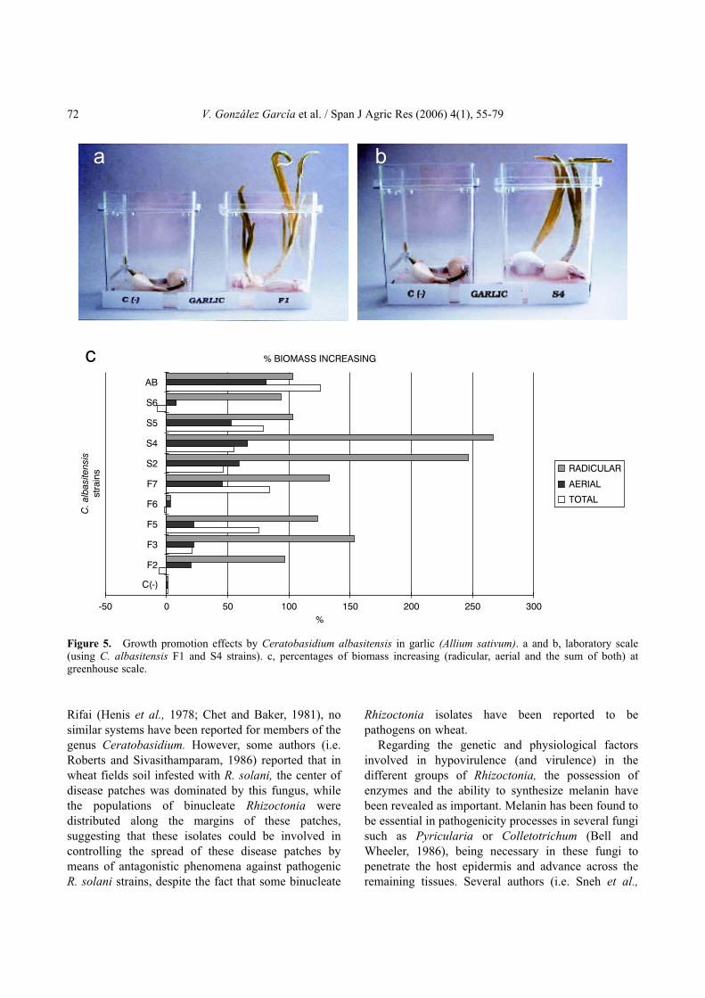
Pseudomonas syringae pv. lachrymans. In addition,Burns and Benson (2000) carried out biocontrolexperiments with binucleate Rhizoctonia againstPythium ultimum and Trichoderma virens in vinca(Catharantus roseus). Furthermore, González et al.(2002) recently reported a new binucleate Rhizoctoniaspecies belonging to the genus Ceratobasidium isolatedfrom Spain (C. albasitensis V. González & V. Rubio),capable of protecting several plant species against R.solani (T. cucumeris) (Fig. 4) and other pathogenicfungal genera such as Fusarium (F. solani), Alternaria(A. alternata) or Penicillium (P. digitatum and P.expansum).
In addition to the protective effects mentionedabove, it has been shown that some binucleateRhizoctonia isolates increased plant growth, even in theabsence of fungal pathogens (Fig. 5). Sneh et al. (1986)reported these effects on the growth of radish, carrot,lettuce, cotton and wheat seedlings and Harris et al.(1993, 1994) found similar phenomena in pepper andsome ornamental species. Finally, Sneh et al. (1986)and Bandy and Tavantzis (1990) observed in potatoseedlings inoculated with hypovirulent isolates of R.solani AG 3, that biomass rates were increased andearly tuberization and flowering occurred compared tountreated controls. These effects on potato plants werealso reported in Spain by González et al. (2000).
The non-pathogenic or hypovirulent Ceratobasidiumspecies constitutes an important part of the group ofnon-sporulating Rhizoctonia-like fungi in soil, and incertain cases these taxa could represent 10-30% of thetotal Rhizoctonia spp. populations (Ichielevich-Austeret al., 1985). Concerning the dynamics of thesepopulations in the soil, there are few studies about thefactors involved in the presence and distribution ofthese isolates in a given agroecosystem and thefrequency of isolation of these strains is stronglydependent on the recovery methods employed. Thus,isolation methods from organic debris, seem to yieldhigh recovery rates of hypovirulent Ceratobasidiumisolates, due to the low selectivity of this methodology(Villajuan-Abgona et al., 1996). The role andsignificance of Ceratobasidium spp. populations in thesoil have not yet been investigated. However, itsprevalence in the soil suggests that these populationscould play an important role in the suppression ofcertain diseases. In spite of the existence of natural orartificial suppressive soils described in the literature,such as those attributed to Trichoderma harzianum
70 V. González García et al. / Span J Agric Res (2006) 4(1), 55-79

Review. Biology and Systematics of the form genus Rhizoctonia 71
Figure 4. Biological protection at laboratory scale with Ceratobasidium albasitensis strains (F1, F2, S2 and S6) in severalplant seedlings, against several R. solani isolates (Me-8.2 and Ralb) at two inoculation moments; both protective and pathogenicstrains simultaneously, and consecutive infection of the pathogen, four days after protective isolates inoculation. a and b, tomato;c and d, alfalfa; e and f, rape; g and h, sugar beet.
a
h
b
e f
g
dc
Rifai (Henis et al., 1978; Chet and Baker, 1981), nosimilar systems have been reported for members of thegenus Ceratobasidium. However, some authors (i.e.Roberts and Sivasithamparam, 1986) reported that inwheat fields soil infested with R. solani, the center ofdisease patches was dominated by this fungus, whilethe populations of binucleate Rhizoctonia weredistributed along the margins of these patches,suggesting that these isolates could be involved incontrolling the spread of these disease patches bymeans of antagonistic phenomena against pathogenicR. solani strains, despite the fact that some binucleate
Rhizoctonia isolates have been reported to bepathogens on wheat.
Regarding the genetic and physiological factorsinvolved in hypovirulence (and virulence) in thedifferent groups of Rhizoctonia, the possession ofenzymes and the ability to synthesize melanin havebeen revealed as important. Melanin has been found tobe essential in pathogenicity processes in several fungisuch as Pyricularia or Colletotrichum (Bell andWheeler, 1986), being necessary in these fungi topenetrate the host epidermis and advance across theremaining tissues. Several authors (i.e. Sneh et al.,
72 V. González García et al. / Span J Agric Res (2006) 4(1), 55-79
Figure 5. Growth promotion effects by Ceratobasidium albasitensis in garlic (Allium sativum). a and b, laboratory scale(using C. albasitensis F1 and S4 strains). c, percentages of biomass increasing (radicular, aerial and the sum of both) atgreenhouse scale.
c % BIOMASS INCREASING
-50 0 50 100 150 200 250 300
C(-)
F2
F3
F5
F6
F7
S2
S4
S5
S6
AB
C.alb
asite
nsis
str
ain
s
%
RADICULAR
AERIAL
TOTAL
a b

1985; Villajuan-Abgona et al., 1993) have pointed outthat hyphae from hypovirulent isolates from the genusCeratobasidium are usually hyaline, while hyphaefrom pathogenic isolates of R. solani are usuallypigmented with brown or grey tones, due to theaccumulation of melanin in their cell walls. No studieshave been designed, to date, to determine a possiblerelationship between the lack of melanin in the cellwalls of some isolates of Ceratobasidium and itshypovirulent behaviour.
It has also been observed that the different isolatesshowed different protective capabilities, suggestingdifferent protection strategies. Moreover, some authorshave shown that in the protective effects of a givenisolate, one or more mechanisms could besimultaneously involved. In spite of the fact that little isstill known about these mechanisms (Sneh, 1996), twomain groups of action have been postulated. A firstgroup could consist of direct interactions, mainlyinvolving horizontal transmission mechanisms ofdsRNA-like mycoviruses, or direct antagonismphenomena (evidenced by competition of nutrients orinfection sites, antibiosis, hyperparasitism, etc.). Asecond type of mechanism proposed corresponds toindirect interactions, such as the existence of physicalbarriers, the production of lytic enzymes or inhibitorysubstances such as phytoalexins or phenols, or theinduction of systemic resistances (SARs) in the planthost (Poromarto et al., 1998). Hence, Hwang et al.(2003) reported expression of systemic resistance inpoinsettia against R. solani stem rot, induced bytreating plants with binucleate Rhizoctonia isolates.
In summary, studies on different species, genera andfamilies that have historically been included under theconcept of the Rhizoctonia species complex, have beentraditionally focused on the description, ecologicalcharacterization or epidemiology of plant diseasesattributed to these organisms. In addition, aclassification system based on hyphal anastomosisreactions has been developed during the last 70 years toclassify isolates from some of the main genera of thecomplex. Conversely, efforts to establish aclassification system for these fungi based on thecomparison of morphometric characters from sexualstages have been hampered due to the low capability ofthese organisms to produce sexual fruitbodies in natureand in the laboratory. As a consequence, much efforthas been paid to developing methods to induceformation of sexual morphs for these fungi in the
laboratory, in order to provide new and reliablemorphometric characters for taxonomical purposes.Together with anatomical features provided byfructifications obtained in vitro, the comparison ofcertain cytological structures (especially septalapparatus) by means of electron microscopy, haspermitted more accurate and phylogenetical groupingto be developed in the form genus. The advent ofmolecular methods to study fungal diversity hasrevealed evolutionary relationships and the molecularbasis of some important ecophysiological processes inseveral Rhizoctonia-like groups, such as recognition,colonization or penetration of plant tissues andfacilitate the development of routine and reliablemethods to identify and characterize isolates of thegroup from complex environmental samples.Furthermore, the discovery of some antagonisticorganisms for several pathogenic Rhizoctonia-likefungi (including reported isolates of hypovirulent ornon-pathogenic members of genera such asThanatephorus or Ceratobasidium), have led to a newway to control diseases caused by members of theRhizoctonia species complex. The success of thesebiocontrol strategies, could reduce the massiveemployment of chemicals and favour the adoption ofIPM (Integrated Pest Management) agriculturalpractices.
In the future, new topics and approaches willchallenge research into the Rhizoctonia complex. Thesewill probably include the development of new methodsto understand evolutionary relationships in geneticallydiverse groups of the complex, the molecular bases ofboth infection and biological protection processes andthe ecological roles played by these fungi in theecosystems where these organisms are usually found.
References
ADAMS G.C., 1988. Thanatephorus cucumeris (Rhizoctoniasolani), a species complex of wide host range. Adv PlantPathol 6, 535-552.
ADAMS G.C., 1996. The genetics of Rhizoctonia species. In:Rhizoctonia species: taxonomy, molecular biology,ecology, pathology and disease control (Sneh B.,Jabaji-Hare S., Neate S., Dijst, G., eds.). KluwerAcademic Publishers. Dordrecht. pp. 101-116.
ADAMS G.C., BUTLER E.E., 1982. A reinterpretation ofthe sexuality of Thanatephorus cucumeris anastomosisgroup four. Mycologia 74, 793-800.
Review. Biology and Systematics of the form genus Rhizoctonia 73

ADAMS G.C., BUTLER E.E., 1983. Environmental factorsinfluencing the formation of basidia and basidiospores inThanatephorus cucumeris. Phytopathology 73, 152-155.
ANDERSEN T.F., 1990. A study of hyphal morphology inthe form genus Rhizoctonia. Mycotaxon 37, 25-46.
ANDERSEN T.F., RASMUSSEN H.N., 1996. Themycorrhizal species of Rhizoctonia. In: Rhizoctoniaspecies: taxonomy, molecular biology, ecology, pathologyand disease control (Sneh B., Jabaji-Hare S., Neate S.,Dijst, G., eds.). Kluwer Academic Publishers. Dordrecht.pp. 379-390.
ANDERSEN T.F., STALPERS J.A., 1994. A check-list ofRhizoctonia epithets. Mycotaxon 51, 437-457.
ANDERSON N.A., 1982. The genetics and pathology ofRhizoctonia solani. Ann Rev Phytopathol 20, 329-347.
BANDY B.P., TAVANTZIS S.M., 1990. Effect ofhypovirulent Rhizoctonia solani on Rhizoctonia disease,growth, and development of potato plants. Am Potato J67, 189-199.
BAKER R., 1990. An overview of current and futurestrategies and models for biological control. In: Biologicalcontrol of soil-borne pathogens (D. Hornby, ed.). CABInternational. Wallingford, UK. pp. 375-388.
BATEMAN D.F., BASHAM H.G. 1976. Degradation ofplant cell walls and membranes by microbial enzymes. In:Encyclopedia Plant Physiology. Vol. 4 (Heitefuss R.,Williams P.H., eds.) Springer-Verlag, New York.pp. 316-355.
BELL A.A., WHEELER M.H., 1986. Biosynthesis andfunction of melanins. Ann Rev Phytopathol 24, 411-451.
BERTAGNOLLI B.L., DAL SOGLIO F.K., SINCLAIR J.B.,1996. Extracellular enzyme profiles of the fungalpathogen Rhizoctonia solani isolate 2B-12 and of twoantagonists, Bacillus megaterium strain B153-2-2 andTrichoderma harzianum isolate Th008.I. Possiblecorrelations with inhibition of growth and biocontrol.Physiol Mol Plant Pathol 48, 145-160.
BHARATHAN N., SASO L., GUDIPATI S., BHARATHANK., WHITED K., ANTHONY K., 2005. Double-strandedRNA: distribution and analysis among isolates ofRhizoctonia solani AG-2 to -13. Plant Pathol 54, 196-203.
BINDER M., HIBBET D.S., LARSSON K., LARSSON E.,LANGER E., LANGER G., 2005. The phylogeneticdistribution of resupinate forms across the major clades ofmushroom-forming fungi (Homobasidiomycetes). SystBiodivers 3, 113-157.
BOIDIN J., MUGNIER J., CANALES R., 1998. Taxonomiemoleculaire des Aphyllophorales. Mycotaxon 66,445-491.
BOYSEN M., BORJA M., DEL MORAL C., SALAZAR O.,RUBIO V., 1996. Identification at strain level ofRhizoctonia solani AG4 isolates by direct sequence ofasymmetric products of the ITS regions. Curr Genet 29,174-181.
BRUNS T.D., WHITE T.J., TAYLOR J.W., 1991. FungalMolecular Systematics. Ann Rev Ecol Syst 22, 525-564.
BURNS J.R., BENSON D.M., 2000. Biocontrol ofdamping-off of Catharanthus roseus caused by Pythium
ultimum with Trichoderma virens and binucleateRhizoctonia fungi. Plant Dis 84, 644-648.
BURPEE L.L., GOULTY L.G., 1984. Suppression of brownpatch disease of creeping bentgrass by isolates ofnonpathogenic Rhizoctonia spp. Phytopathology 74,692-694.
BURPEE L.L., SANDERS P.L., COLE H. Jr, SHERWOODR.T., 1980a. Anastomosis groups among isolates ofCeratobasidium cornigerum and related fungi. Mycologia72, 689-701.
BURPEE L.L., SANDERS P.L., COLE H. Jr., SHERWOODR.T., 1980b. Pathogenicity of Ceratobasidiumcornigerum and related fungi representing fouranastomosis groups. Phytopathology 70, 843-846.
CAPPER A.L., CAMPBELL R., 1986. The effect ofartificially inoculated antagonistic bacteria on theprevalence of take-all disease of wheat in fieldexperiments. J Appl Bacteriol 60, 155-160
CARDOSO J.E., ECHANDI E., 1987. Biological control ofRhizoctonia root rot of snap bean with binucleateRhizoctonia-like fungi. Plant Dis 71, 167-170.
CARLING D.E., 1996. Grouping in Rhizoctonia solani byhyphal anastomosis reaction. In: Rhizoctonia species:taxonomy, molecular biology, ecology, pathology anddisease control (Sneh B., Jabaji-Hare S., Neate S., Dijst,G., eds.). Kluwer Academic Publishers. Dordrecht.pp. 35-47.
CARLING D.E., 2000. Anastomosis groups and subsets ofanastomosis groups of Rhizoctonia solani. In: ThirdInternational Symposium on Rhizoctonia (ISR 2000).Abstracts Book, p. 14.
CARLING D.E., KUNINAGA S., 1990. DNA base sequencehomology in Rhizoctonia solani: inter- and intragrouprelatedness of anastomosis group-9. Phytopathology 80,1362-1364.
CARLING D.E., SUMNER D.R., 1992. Rhizoctonia. In:Methods in research on soilborne phytopathogenic fungi(Singleton, L.L., Mihail, J.D. & Rush, C.M., eds.). APSPress. St. Paul, Minnesota. pp. 157-165.
CARLING D.E., POPE E.J., BRAINARD K.A., CARTER,D.A., 1999. Characterization of mycorrhizal isolates ofRhizoctonia solani from an orchid, including AG-12, anew anastomosis group. Phytopathology 89, 942-946.
CARLING D.E., BAIRD R.E., GITAITIS R.D.,BRAINARD K.A., KUNINAGA S., 2002a.Characterization of AG-13, a newly reported anastomosisgroup of Rhizoctonia solani. Phytopathology 92, 893-899.
CARLING D.E., KUNINAGA S., BRAINARD K.A.,2002b. Hyphal anastomosis reactions, rDNA-internaltranscribed spacer sequences, and virulence levels amongsubsets of Rhizoctonia solani anastomosis group-2(AG-2) and AG-BI. Phytopathology 92, 43-50.
CASTANHO B., BUTLER E.E., 1978. Rhizoctonia decline:Studies on hypovirulence and potential use in biocontrol.Phytopathology 68, 1511-1514.
CERESINI P.C., SHEW H.D., VILGALYS R., CUBETAM.A., 2002. Genetic diversity of Rhizoctonia solani AG-3
74 V. González García et al. / Span J Agric Res (2006) 4(1), 55-79

from potato and tobacco North Carolina. Mycologia,437-449.
CHET I., BAKER R., 1981. Isolation and biocontrolpotential of Trichoderma hamatum from soil naturallysuppressive to R. solani. Phytopathology 71, 286-290.
COOK R.J., ROVIRA A.D., 1976. The role of bacteria in thebiological control Gaeumannomyces graminis bysuppressive soils. Soil Biol Biochem 8, 269-273.
COPES W., RINEHART T., CUBETA M.A., 2005. Genetic,pathogenic and phenotypic characterization ofRhizoctonia spp. isolated from container-grown azalea inthe southern United States. Phytopathology 95, S21(abstr.).
CUBETA M.A., ECHANDI E., 1991. Biological control ofRhizoctonia and Phytium damping-off of cucumber: anintegrated approach. Biol Contr 1, 227-236.
CUBETA M.A., ECHANDI E., ABERNETHY T.,VILGALYS R., 1991. Characterization of anastomosisgroups of binucleate Rhizoctonia species using restrictionanalysis of an amplified ribosomal RNA gene.Phytopathology 81, 1395-1400.
CUBETA M.A., VILGALYS R., 1997. Population biology ofthe Rhizoctonia solani complex. Phytopathology 87,480-484.
CUBETA M.A., VILGALYS R., GONZÁLEZ D., 1996.Molecular analysis of ribosomal RNA genes inRhizoctonia fungi. In: Rhizoctonia species: taxonomy,molecular biology, ecology, pathology and disease control(Sneh B., Jabaji-Hare S., Neate S., Dijst, G., eds.). KluwerAcademic Publishers. Dordrecht. pp. 81-86.
DAMAJ M. JABAJI-HARE S., CHAREST P.M., 1993.Isozyme variation and genetic relatedness in binucleateRhizoctonia species. Phytopathology 83, 864-871.
DE ANDRÉS M.F., GARCÍA-ARENAL F., LÓPEZ M.M.,MELGAREJO P. (coord.), 1998. Patógenos de plantasdescritos en España. Coed. Ministerio de Agricultura,Pesca y Alimentación. Sociedad Española de Fitopatología.Madrid. 526 pp.
DE CANDOLLE A.P., 1815. Mémoire sur les rhizoctones,noveau genre de champignons qui attaque les racines, desplantes et en particulier celle de la luzerne cultivée. MemMus d’Hist Nat 2, 209-216.
DEACON J.W., 1976. Biological control of the take-allfungus Gaeumannomyces graminis, by Phialophora andsimilar fungi. Soil Biol Biochem 8, 275-283.
DOMÍNGUEZ-GARCÍA TEJERO F., 1951. Distribución enEspaña de las plagas y enfermedades de la remolacha.Boletín Patología Vegetal y Entomología Agrícola 18,181-204.
DONK M.A., 1931. Revisie van de NederlandseHeterobasidiomycetae en HomobasodiomycetaeAphyllophoraceae. Deel 1. Mededeelingen van deNederlansche Mycologische Vereenigung 18-20, 67-200.
DONK M.A., 1956. Notes on resupinate fungi II. Thetullasneloid fungi. Reinwardtia 3, 363-379.
DONK M.A., 1958. Notes on resupinate Hymenomycetes V.Fungus 28, 16-36.
DUNCAN S., BARTON J.E., O’BRIEN P., 1993. Analysis ofvariation in isolates of Rhizoctonia solani by randomamplified polymorphic DNA assay. Mycol Res 97,1075-1082.
EL BAKALI M.A., MARTÍN M.P., GARCÍA F., MORET A,NADAL M., MONTÓN C., 2000. First report ofRhizoctonia solani AG-3 in Catalonia (NE Spain). PlantDis 84, 806.
EL BAKALI M.A., NADAL, M., MORET A., MARTÍNM.P., 2002. Incidencia de Rhizoctonia solani en cultivo depatata en Catalunya. Nutri-Fitos 131, 177-179.
EL BAKALI M.A., MARTÍN M.P., MORET A., NADALM., 2003. Identification of Spanish isolates of Rhizoctoniasolani from potato by anastomosis grouping, ITS-RFLPand RAMS-fingerprinting. Phytopathology Med 42,167-176.
ESCANDE A.R., ECHANDI E., 1991. Protection of potatofrom Rhizoctonia canker with binucleate Rhizoctoniafungi. Plant Pathol 40, 197-202.
FLENTJE N.T., 1956. Studies on Pellicularia filamentosa(Pat.) Rogers. I. Formation of the perfect stage. Trans BrMycol Soc 39, 343-356.
FLENTJE N.T., STRETTON H.M., 1964. Mechanisms ofvariation in Thanatephorus cucumeris and T. praticola.Aust J Biol Sci 17, 686-704.
FLENTJE N.T., STRETTON H.M., HAWN E.J., 1963.Nuclear distribution and behaviour throughout the lifecycles of Thanatephorus, Waitea and Ceratobasidiumspecies. Aus J Biol Sci 16, 450-467.
GONZÁLEZ V., PORTAL M.A., ACERO J., RUBIO V.,2000. Biocontrol de patógenos de patata con aisladosprotectores de Ceratobasidium spp. (BNR). In: Libro deActas del Congreso Iberoamericano de Investigación yDesarrollo en Patata. Patata 2000 (Pascualena J. and RitterE., eds). Diputación Foral de Álava. Gasteiz. Spain.
GONZÁLEZ D., CARLING D.E., KUNINAGA S.,VILGALYS R., CUBETA M.A., 2001. Ribosomal DNAsystematics of Ceratobasidium and Thanatephorus withRhizoctonia anamorphs. Mycologia 93, 1138-1150.
GONZÁLEZ V., SALAZAR O., JULIÁN M.C., ACERO J.,PORTAL M.A., MUÑOZ R., LÓPEZ-CÓRCOLES H.,GÓMEZ-ACEBO E., LÓPEZ-FUSTER P., RUBIO V.,2002. Ceratobasidium albasitensis, a new Rhizoctonia-likefungus isolated in Spain. Persoonia 17, 601-614.
GONZÁLEZ D., CUBETA M.A., VILGALYS R., 2003.Molecular systematics of Rhizoctonia phylogeneticallyintegrates anamorphs into Ceratobasidiaceae. InternationalCongress of Plant Pathology, ChristChurh, New Zealand(abstr.) p. 29.
HARRIS A.R., SCHISLER D.A., NEATE S.M., 1993.Culture of Rhizoctonia solani and binucleate Rhizoctoniaspp. on organic substrates for inoculation of seedlings incontainers. Soil Biol Biochem 25, 337-341.
HARRIS A.R., SCHISLER D.A., NEATE S.M., RYDERM.H., 1994. Suppression of damping off caused byRhizoctonia solani and growth promotion, in beddingplants by binucleate Rhizoctonia spp. Soil Biol Biochem26, 263-268.
Review. Biology and Systematics of the form genus Rhizoctonia 75
HASHIBA T., HOMMA Y., HYAKUMACHI M.,MATSUDA J., 1984. Isolation of a DNA plasmid in thefungus Rhizoctonia solani. J Gen Microbiol 130,2067-2070.
HAWN E.J., VANTERPOOL T.C., 1953. Preliminary studyon the sexual stage of Rhizoctonia solani. Can J Bot 6,700-710.
HELGASON T., WATSON I.J., YOUNG J.P.W., 2003.Phylogeny of the Glomerales and Diversisporales (Fungi:Glomeromycota) from actin and elongation factor 1-alphasequences. FEMS Microbiol Lett 229, 127-132.
HENIS Y., GHAFFER A., BAKER R., 1978. Integratedcontrol of Rhizoctonia solani damping-off of radish:Effect of successive plantings, PCNB and Trichodermaharzianum on pathogen and disease. Phytopathology 68,900-909.
HERR L.J., 1988. Mechanisms of biocontrol of Rhizoctoniacrown and root rot of sugar beet by binucleate Rhizoctoniaspp. and Laetisaria arvalis. Ann Appl Biol 113, 107-118.
HERR L.J., 1995. Biological control of Rhizoctonia solaniby binucleate Rhizoctonia spp. and hypovirulent R. solaniagents. Crop Protection 14, 179-186.
HIETALA A.M., KORHONEN K., SEN R., 2003. Anunknown mechanism promotes somatic incompatibility inCeratobasidium bicorne. Mycologia 95, 239-250.
HIETALA A.M., SEN R., LILJA A., 1994. Anamorphic andteleomorphic characteristics of a uninucleate Rhizoctoniasp. isolated from the roots of nursery grown coniferseedlings. Mycol Res 98, 1044-1050.
HWANG J., BENSON D.M., 2003. Expression of inducedsystemic resistance in poinsettia cuttings againstRhizoctonia stem rot by treatment of stock plants withbinucleate Rhizoctonia. Biol Contr 27, 73-80.
ICHIELEVICH-AUSTER M., SNEH B., KOLTIN Y.,BARASH I., 1985. Pathogenicity, host specificity andanastomosis groups of Rhizoctonia spp. isolated fromsoils in Israel. Phytoparasitica 13, 103-112.
JABAJI-HARE S., 1996. Biochemical methods. In:Rhizoctonia species: Taxonomy, molecular biology,ecology, pathology and disease control (Sneh B.,Jabaji-Hare S., Neate S. & G. Dijst, eds.). KluwerAcademic Publishers. Dordrecht. pp. 65-71.
JABAJI-HARE S., BURGER G., FORGET L., LANG B.F.,1994. Extrachromosomal plasmids in the plant pathogenicfungus Rhizoctonia solani. Curr Genet 25, 423-431.
JIAN J., LAKSHMAN D.K., TAVANTZIS S.M., 1998. Avirulence-associated, 6.4-kb, double-stranded RNA fromRhizoctonia solani is phylogenetically related to plantbromoviruses and electron transport enzymes. Mol PlantMicrob Interact 11, 601-609.
JULIÁN M.C., DEBETS F., KEIJER J., 1996. Nuclearbehavior in homokaryotic and heterokaryotic fruiting ofThanatephorus cucumeris (Rhizoctonia solani)anastomosis group 1, subgroup IC. Phytopathology 86,566-574.
JULIÁN M.C., DULLEMANS A.M., SILFHOUT C.,KEIJER J., 1997. Nuclear behavior in homokaryotic andheterokaryotic fruiting of Thanatephorus cucumeris
(Rhizoctonia solani) anastomosis group 1, subgroup IC.Mycologia 89, 361-374.
JULIÁN M.C., ACERO J., SALAZAR O., KEIJER J.,RUBIO V., 1999. Mating type-correlated molecularmarkers and demonstration of heterokaryosis in thephytopathogenic fungus Thanatephorus cucumeris(Rhizoctonia solani) AG1-IC by AFLP DNAfingerprinting analysis. J Biotechnol 67, 49-56.
JÜLICH W., 1981. Higher taxa of Basidiomycetes.Bibliotheca Mycologica, Cramer, Vaduz, 85.
KATSURA K. SUZUKI F., MIYASHITA S., NISHI T.,HIROCHIKA H., HASHIBA T., 1997. The completenucleotide sequence and characterization of the linearDNA plasmid pRS64-2 from the plant pathogenic fungusRhizoctonia solani. Curr Genet 32, 431-435.
KEIJER J., 1996. The initial steps of the infection process inRhizoctonia solani. In: Rhizoctonia species: taxonomy,molecular biology, ecology, pathology and disease control(Sneh B., Jabaji-Hare S., Neate S., Dijst, G., eds.). KluwerAcademic Publishers. Dordrecht. pp. 149-162.
KIRK P.M., CANNON P.F., DAVID J.C., STALPERS J.A.,2001. Ainsworth and Bisby’s dictionary of the fungi. 9th
edition. International Mycological Institute, CABInternational. Wallingford, UK.
KOTILA J., 1929. A study of the biology of a new sporeforming Rhizoctonia, Corticium praticola. Phytopathology19, 1059-1099.
KOTILA J., 1945. Cotton-leaf-spot Rhizoctonia and itsperfect stage on sugar beets. Phytopathology 35, 741-743.
KOTILA J., 1947. Rhizoctonia foliage blight of sugar beets. JAgric Res 74, 289-314.
KUCHAREK T., 2000. Rhizoctonia diseases in abovegroundplant parts of agronomic and vegetable crops. PlantPathology Fact Sheet PP-41. Florida CooperativeExtension Service.
KUNINAGA S., YOKOSAWA R., 1980. A comparison ofDNA base compositions among anastomosis groups inRhizoctonia solani. Ann Phytopathol Soc Japan 46,150-158.
KUNINAGA S., YOKOSAWA R., 1982a. DNA basesequence homology in Rhizoctonia solani I. Geneticrelatedness within anastomosis group 1. Ann PhytopatholSoc Japan 48, 659-667.
KUNINAGA S., YOKOSAWA R., 1982b. DNA basesequence homology in Rhizoctonia solani II. Geneticrelatedness within anastomosis group 2. Ann PhytopatholSoc Japan 48, 668-673.
KUNINAGA S., YOKOSAWA R., 1983. DNA basesequence homology in Rhizoctonia solani III. Geneticrelatedness within AG-3, AG-5, AG-7 and AG-BI. AnnPhytopathol Soc Japan 49, 648-652.
KUNINAGA S., YOKOSAWA R., 1984a. DNA basesequence homology in Rhizoctonia solani IV. Geneticrelatedness within AG-4. Ann Phytopathol Soc Japan 50,322-330.
KUNINAGA S., YOKOSAWA R., 1984b. DNA basesequence homology in Rhizoctonia solani V. Genetic
76 V. González García et al. / Span J Agric Res (2006) 4(1), 55-79

relatedness within AG-6. Ann Phytopathol Soc Japan 50,346-352.
KUNINAGA S., YOKOSAWA R., 1985a. DNA basesequence homology in Rhizoctonia solani VI. Geneticrelatedness among seven anastomosis groups. AnnPhytopathol Soc Japan 51, 127-132.
KUNINAGA S., YOKOSAWA R., 1985b. DNA basesequence homology in Rhizoctonia solani VII. Geneticrelatedness among AG-BI and other anastomosis groups.Ann Phytopathol Soc Japan 51, 133-138.
KUNINAGA S., NATSUAKI T., TAKEUCHI T.,YOKOSAWA R., 1997. Sequence variation in the rDNAITS regions within and between anastomosis groups inRhizoctonia solani. Curr Genet 32, 237-243.
LAKSHMAN D.K., TAVANTZIS S.M., 1994. Spontaneousappearance of genetically distinct double-stranded RNAelements in Rhizoctonia solani. Phytopathology 84,633-639.
LAKSHMAN D.K., TAVANTZIS S.M., 1995. Sequenceanalysis of a hypovirulence-associated double-strandedRNA (dsRNA) from Rhizoctonia solani. Phytopathology85, 1211.
LAKSHMAN D.K., JIAN J., TAVANTZIS S.M., 1988. Anovel double-stranded RNA element from a hypovirulentstrain of Rhizoctonia solani occurs in DNA form, and isgenetically related to the pentafunctional AROM proteinof shikimate pathway. Proc Nat Acad Sci USA 122,311-316.
LANGER G., 1994. Die gattung Botryobasidium.Bibliotheca Micologici 158, 1-459.
LEINER R.H., CARLING D.E., 1994. Characterization ofWaitea circinata (Rhizoctonia) isolated from agriculturalsoils in Alaska. Plant Dis 78, 385-388.
LISTER N., KATAN J., HENIS Y., 1975. Sequentialproduction of polygalacturonase, cellulase and pectinlyase by Rhizoctonia solani. Can J Microbiol 21, 298-304.
LIU Z.L., DOMIER L.L., SINCLAIR J.B., 1993.Subgroup-specific ribosomal DNA polymorphism of theRhizoctonia solani species complex. Mycologia 85,795-800.
LOUVET J., ROXEL F., ALABOUVETTE C., 1976. Miseen évidence de la nature microbiologique d’un sol audéveloppement de la fusariose vasculaire du melon. AnnPhytopathol 8, 425-436.
LUCAS P., SARNIQUET A., 1990. Soil receptivity totake-all: Influence of some cultural practices and soilchemical characteristics. Symbiosis 9, 51-57.
LUCAS P., SMILEY R.W., COLLINS H.P., 1993. Decline ofRhizoctonia root rot on wheat in soils infested withRhizoctonia solani AG-8. Phytopathology 83, 260-265.
MARCUS L., BARASH I., SNEH B., KOLTIN Y.,FINKLER A., 1986. Purification and characterization ofpectinolytic enzimes produced by virulent andhypovirulent isolates of Rhizoctonia solani Kühn. PhysiolMol Plant Pathol 29, 325-336.
MARÍN J.P., SEGARRA J., ALMACELLAS J., 1992.Enfermedades de los cereales en Cataluña en 1988-90.Invest Agr: Prod Prot Veg 7, 261-275.
MARTIN G.W., 1948. New or noteworthy tropical fungi. IV.Lloydia 11, 111-122.
MASUHARA G., NEATE S.M., SCHISLER D.A., 1994.Characteristics of some Rhizoctonia spp. from SouthAustralian plant nurseries. Mycol Res 98, 83-87.
MEHMANN B., BRUNNER I., BRAUS G.H., 1994.Nucleotide sequence variation of chitin synthase genesamong ectomycorrhizal fungi and its potential use intaxonomy. Appl Environ Microbiol 60, 3105-3111.
MELERO-VARA J.M., JIMÉNEZ-DÍAZ R.M., 1990.Etiology, incidence and distribution of cotton seedlingdamping-off in southern Spain. Plant Dis 74, 597-600.
MIYASHITA S., HIROCHIKA H., IKEDA J.E., HASHIBAT., 1990. Linear plasmid DNAs of the plant pathogenicfungus Rhizoctonia solani with unique terminal structures.Mol Gen Genet 220, 165-171.
MOORE R.T., 1978. Taxonomic significance of septalultrastructure with particular reference to the jelly fungi.Mycologia 70, 1007-1024.
MOORE R.T., 1987. The genera of Rhizoctonia-like fungi:Ascorhizoctonia, Ceratorhiza gen. nov., Epulorhiza gen.nov., Moliniopsis, and Rhizoctonia. Mycotaxon 29, 91-99.
MOORE R.T., 1990. Order Platygloeales ord. nov.Mycotaxon 39, 245-248.
MOORE R.T., 1996. The dolipore/parenthesome septum inmodern taxonomy. In: Rhizoctonia species: taxonomy,molecular biology, ecology, pathology and disease control(Sneh B., Jabaji-Hare S., Neate S., Dijst, eds.). KluwerAcademic Publishers. Dordrecht. pp. 13-35.
MULLER K.O., 1923. Über die Bezeihungen zwischenRhizoctonia solani und Hypochnus solani. Del BiolReichsanst F Land u Forstw Arb 13, 198-262.
MURRAY D.I.L., 1982. A modified procedure for fruitingRhizoctonia solani on agar. Trans Br Mycol Soc 79(1),129-135.
MURRAY D.I.L., 1984. Cultural conditions influencingbasidium formation in the Ceratobasidiaceae. Aus J Bot32, 101-108.
NEATE S.M., CRUIKSHANK R.H., ROVIRA A.D., 1988.Pectic enzyme patterns of Rhizoctonia solani isolates fromagricultural soils in South Australia. Trans Br Mycol Soc90, 37-42.
OGOSHI A., 1975. Grouping of Rhizoctonia solani Kühnand their perfect states. Rev Plant Prot Res Japan 8,98-103.
OGOSHI A., ONIKI M., ARAKI T., UI T., 1983a. Studies onthe anastomosis groups of binucleate Rhizoctonia andtheir perfect states. J Fac Agric Hokk Univ 61, 244-260.
OGOSHI A., ONIKI M., ARAKI T., UI T., 1983b.Anastomosis groups of binucleate Rhizoctonia in Japanand North America and their perfect states. Trans MycolSoc Japan 24, 79-87.
ONIKI M., OGOSHI A., ARAKI T., SAKAI R., TANAKAS., 1985. The perfect state of Rhizoctonia oryzae-sativaeand R. zeae, and the anastomosis groups of Waiteacircinata. Trans Mycol Soc Japan 26, 189-198.
PARMETER J.R., WHITNEY H.S., 1970. Taxonomy andnomenclature of the perfect state. In: Rhizoctonia solani:
Review. Biology and Systematics of the form genus Rhizoctonia 77

biology and pathology (Parmeter, J.R., ed.). University ofCalifornia Press. Berkeley. pp. 7-19.
PARMETER J.R., WHITNEY H.S., PLATT W.D., 1967.Affinities of some Rhizoctonia species that resemblemycelium of Thanatephorus cucumeris. Phytopathology57, 218-223.
POPE E.J., CARTER D.A., 2001. Phylogenetic placementand host specificity of mycorrhizal isolates belonging toAG-6 and AG-12 in the Rhizoctonia solani speciescomplex. Mycologia 93, 712-719.
POROMARTO S.H., NELSON B.D., FREEMAN T.P., 1998.Association of binucleate Rhizoctonia with soybean andmechanism of biocontrol of Rhizoctonia solani.Phytopathology 88, 1056-1067.
PRIYATMOJO A., FUKUI H., HYAKUMACHI M., 2005.Characterization of a new group of binucleate Rhizoctoniacausing root and stem rot of cut-flower roses.Phytopathology 95, 784-792.
ROBERTS P., 1999. Rhizoctonia-forming fungi: a taxonomicguide. Royal Botanical Gardens, Kew. 239 pp.
ROBERTS D.L., SIVASITHAMPARAM K., 1986. Identityand pathogenicity of Rhizoctonia spp. associated withbare patch disease of cereals at a field site in WesternAustralia. Neth J Plant Pathol 92, 185-195.
ROGERS D.P., 1935. Notes on the lower Basidiomycetes.Stud Nat Hist Iowa Univ 17, 1-43.
ROSS R.E., KEINATH A.P., CUBETA M.A., 1998.Biological control of wirestem of cabbage with binucleateRhizoctonia. Crop Prot 17, 99-104.
RUBIO V., TAVANTZIS S.M., LAKSHMAN D.K., 1996.Extrachromosomal elements and degree of pathogenicityin Rhizoctonia solani. In: Rhizoctonia species: taxonomy,molecular biology, ecology, pathology and disease control(Sneh B., Jabaji-Hare S., Neate S., Dijst, G., eds.). KluwerAcademic Publishers. Dordrecht. pp. 127-138.
SAKAGUCHI K., 1990. Invertrons, a class of structurallyand functionally related genetic elements that includeslinear DNA plasmids, transposable elements, andgenomes of Adeno-type viruses. Microbiol Rev 54, 66-74.
SAKSENA H.K., 1961. Nuclear phenomena in the basidiumof Ceratobasidium praticola. Can J Bot 39, 717-725.
SALAZAR O., SCHNEIDER J.H.M., JULIÁN M.C.,KEIJER J., RUBIO V., 1999. Phylogenetic subgroupingof Rhizoctonia solani AG 2 isolates based on ribosomalITS sequences. Mycologia 91, 459-467.
SALAZAR O., HYAKUMACHI M., RUBIO V.,, 2000.Phylogenetic grouping of cultural types of Rhizoctoniasolani AG 2-2 based on ribosomal ITS sequences.Mycologia 92, 505-509.
SAMAC D.A., LEONG S.A., 1989. Mitochondrial plasmidsof filamentous fungi: Characteristics and use intransformation vectors. Mol Plant Microb Interact 2,155-159.
SARDIÑA J.R., 1945. Enfermedades de la patata. Pub EstFitopatol Agric La Coruña 5, 111.
SEGAWA K., HARADA Y., 1990. Violet root rot. In:Compendium of apple and pear diseases (Jones A.L. &
Aldwinckle H.S., eds.). American PhytopathologicalSociety Press. St. Paul, Minnesota.
SHAN X.C., LIEW E.C.Y., WEATHERHEAD M.A.,HODGKISS I.J., 2002. Characterization and taxonomicplacement of Rhizoctonia-like endophytes from orchidroots. Mycologia 94, 230-239.
SHERWOOD R.T., 1966. Pectin lyase and polygalacturonaseproduction by Rhizoctonia solani and other fungi.Phytopathology 56, 279-286.
SIMON A., SIVASITHAMPARAM K., 1989. Pathogen-suppression: a case study in biological suppression ofGaeumannomyces graminis var. tritici in soil. Soil BiolBiochem 21, 331-337.
SIMS A.C., 1956. Factors affecting basidiosporedevelopment of Pellicularia filamentosa. Phytopathology46, 413-470.
SINOBAS J., IGLESIAS C., GARCÍA A., 1994.Prospección de las micosis en los invernaderos de Villadel Prado (Madrid) y su incidencia en el cultivo de judía(Phaseolus vulgaris L.). Bol San Veg Plagas 20, 889-898.
SNEH B., 1996. Non pathogenic isolates of Rhizoctonia(np-R) spp. and their role in biological control. In:Rhizoctonia species: taxonomy, molecular biology,ecology, pathology and disease control (Sneh B.,Jabaji-Hare S., Neate S., Dijst, G., eds.). KluwerAcademic Publishers. Dordrecht. pp. 473-483.
SNEH B., 1998. Use of non-pathogenic or hypovirulentfungal strains to protect plants against closely relatedfungal pathogens. Biotechnol Adv 16(1), 1-32.
SNEH B., ICHIELEVICH-AUSTER M., SHOMER I., 1985.Comparative anatomy of colonization of cottonhypocotyls and roots by virulent and hypovirulent isolatesof Rhizoctonia solani. Can J Bot 67, 2142-2149.
SNEH B., ZEIDAN M., ICHIELEVICH-AUSTER M.,BARASH, I., KOLTIN Y., 1986. Increased growthresponses induced by a nonpathogenic isolate ofRhizoctonia solani. Can J Bot 64, 2372-2378.
SNEH B., BURPEE L., OGOSHI A. (eds)., 1991.Identification of Rhizoctonia species. APS Press. St. Paul.
SNEH B., ICHIELEVICH-AUSTER M., 1995. Inducedresistance of cucumber seedlings caused by nonpathogenic Rhizoctonia (np-R) isolates. In: InternationalSymposium on Rhizoctonia, Facts and Challenges inPathology, Taxonomy, Ecology and Disease Control.Abstr. N.º P-8-3. Noordwijkerhout, The Netherlands.
STALPERS J.A., ANDERSEN T.F., 1996. A synopsis of thetaxonomy of teleomorphs connected with Rhizoctonia s.l.In: Rhizoctonia species: taxonomy, molecular biology,ecology, pathology and disease control (Sneh B.,Jabaji-Hare S., Neate S., Dijst G., eds.). Kluwer AcademicPublishers. Dordrecht. pp. 49-63.
STALPERS J.A., ANDERSEN T.F., GAMS W., 1998. Twoproposals to conserve the names Rhizoctonia and R.solani. Taxon 47, 725-726.
STRETTON H.M., MCKENZIE A.R., BAKER K.F.,FLENTJE N.T., 1964. Formation of the basidial stage ofsome isolates of Rhizoctonia. Phytopathology 54,1093-1095.
78 V. González García et al. / Span J Agric Res (2006) 4(1), 55-79

SWEETINGHAM M.W., CRUICKSHANK R.H., WONGD.H., 1986. Pectic zymograms and taxonomy andpathogenicity of the Ceratobasidiaceae. Trans Br MycolSoc 86, 305-311.
TALBOT P.H.B., 1965. Studies on ’Pellicularia’ andassociated genera of Hymenomycetes. Persoonia 3,371-406.
TALBOT P.H.B., KEANE P.J., 1971. Oncobasidium: a newgenus of tulasnelloid fungi. Aus J Bot 19, 203-206.
TAVANTZIS S.M., 2001. dsRNA genetic elements. In:Concepts and applications in agriculture, forestry andmedicine. CRC Press LLC, Boca Ratón, Florida, 304 pp.
TAVANTZIS S.M., BANDY B.P., 1988. Properties of amycovirus from Rhizoctonia solani and itsvirion-associated RNA polymerase. J Gen Virol 69,1465-1477.
TAVANTZIS S.M., PERKINS B.L., BUSHWAY R.J.,BANDY B.P., 1989. Correlation between in vitrosynthesis of phenylacetic acid and virulence inRhizoctonia solani. Phytopathology 79, 1199 (Abst.)
TELLO J.C., LACASA A., MOLINA R., 1985. Notafitopatológica sobre el complejo parasitario del pie de lajudía en la costa de Granada. ITEA Producción Vegetal61, 57-69.
THON M.R., ROYSE D.J., 1999. Partial �-tubulin genesequences for evolutionary studies in the Basidiomycotina.Mycologia 91, 468-474.
TIVOLI B., LEMAIRE J.M., JOUAN B., 1974. Prémunitiondu blé contre Ophiobolus graminis Sacc. par des souchespeu agressives du même parasite. Ann Phytopathol 6,395-406.
TODA T., HYAKUMACHI M., SUGA H., KAGEYAMA K.,TANAKA A., TANI T., 1999. Differentiation ofRhizoctonia AG-D isolates from turfgrass into subgroups Iand II based on rDNA and RAPD analyses. Eur J PlantPathol 105, 835-846.
TODA T., MUSHIKA T., HAYAKAWA T., TANAKA A.,TANI T., HYAKUMACHI M., 2005. Brown ring patch: anew disease on bentgrass caused by Waitea circinata var.circinata. Plant Dis 89, 536-542.
TU C.C., KIMBROUGH J.W., 1975. Morphology,development, and cytochemistry of hyphae and sclerotiaof species in the Rhizoctonia complex. Can J Bot 53,2282-2296.
TU C.C., ROBERTS D.A., KIMBROUGH J.W., 1969.Hyphal fusion, nuclear condition, and perfect stages ofthree species of Rhizoctonia. Mycologia 61, 775-783.
UCHIDA J.Y., ARAGAKI M., YAHATA P.S., 1986.Basidiospore formation by Ceratobasidium sp. on agar.Mycologia 78, 587-592.
VALDER P.G., 1958. The biology of Helicobasidiumpurpureum Pat. Trans Br Mycol Soc 41, 283-308.
VILGALYS R., 1988. Genetic relatedness amonganastomosis groups in Rhizoctonia solani as measured byDNA/DNA hybridization. Phytopathology 78, 698-702.
VILGALYS R., GONZÁLEZ D., 1990. Ribosomal DNArestriction fragment lenght polymorphisms in Rhizoctoniasolani. Phytopathology 80, 151-158.
VILGALYS R., CUBETA M.A., 1994. Molecularsystematics and population biology of Rhizoctonia. AnnRev Pythopathol 32, 135-155.
VILLAJUAN-ABGONA R., KATSUMO N., KAGEYAMAK., HYAKUMACHI M., 1993. Isolation of hypovirulentstrains of Rhizoctonia from soil and their cross protectioneffect. (Abstract). Ann Phytopathol Soc Japan 59, 61.
VILLAJUAN-ABGONA R., KATSUMO N., KAGEYAMAK., HYAKUMACHI M., 1996. Isolation andidentification of hypovirulent Rhizoctonia isolates fromsoil. Plant Pathol 45, 496-904.
WAKO T., ISHIKAWA T., HASHIBA T., 1991. UniqueDNA plasmid pRS64 associated with chromosomalDNAs of the plant pathogenic fungus Rhizoctonia solani.J Gen Microbiol 137, 2817-2821.
WARCUP J.H., 1981. The mycorrhizal relationships ofAustralian orchids. New Phytol 87, 371-381.
WEINHOLD A.R., BOWMAN T., 1974. Repression ofvirulence in Rhizoctonia solani by glucose and 3-o-methylglucose. Phytopathology 64, 985-990.
WEINHOLD A.R., MOTTA J., 1973. Initial host responsesin cotton to infection by Rhizoctonia solani.Phytopathology 63, 157-162.
WEINHOLD A.R., SINCLAIR J.B., 1996. Rhizoctoniasolani: Penetration, colonization and host response. In:Rhizoctonia species: taxonomy, molecular biology,ecology, pathology and disease control (Sneh B.,Jabaji-Hare S., Neate S. & G. Dijst, eds.). KluwerAcademic Publishers. Dordrecht. pp. 163-174.
WHITNEY H.S., PARMETER J.R., 1963. Synthesis ofheterokaryons in Rhizoctonia solani Kühn. Can J Bot 41,879-886.
WONG P.T.W., 1975. Cross-protection against the wheat andoat take-all fungi by Gaeumannomyces graminis var.graminis. Soil Biol Biochem 7, 189-194.
Review. Biology and Systematics of the form genus Rhizoctonia 79