Review Article The Utilization of Imaging Features in the...
10
Review Article The Utilization of Imaging Features in the Management of Intraductal Papillary Mucinous Neoplasms Stefano Palmucci, 1 Claudia Trombatore, 1 Pietro Valerio Foti, 1 Letizia Antonella Mauro, 1 Pietro Milone, 1 Roberto Milazzotto, 2 Rosalia Latino, 2 Giacomo Bonanno, 3 Giuseppe Petrillo, 1 and Antonio Di Cataldo 2 1 Radiodiagnostic and Radiotherapy Unit, University Hospital “Policlinico-Vittorio Emanuele”, Via Santa Sofia 78, 95123 Catania, Italy 2 Department of Surgical Sciences, Organ Transplantation and Advanced Technologies, University Hospital “Policlinico-Vittorio Emanuele”, 95123 Catania, Italy 3 Gastroenterology Unit, University Hospital “Policlinico-Vittorio Emanuele”, 95123 Catania, Italy Correspondence should be addressed to Stefano Palmucci; [email protected] Received 28 March 2014; Revised 19 July 2014; Accepted 24 July 2014; Published 19 August 2014 Academic Editor: Niccola Funel Copyright © 2014 Stefano Palmucci et al. is is an open access article distributed under the Creative Commons Attribution License, which permits unrestricted use, distribution, and reproduction in any medium, provided the original work is properly cited. Intraductal papillary mucinous neoplasms (IPMNs) represent a group of cystic pancreatic neoplasms with large range of clinical behaviours, ranging from low-grade dysplasia or borderline lesions to invasive carcinomas. ey can be grouped into lesions originating from the main pancreatic duct, main duct IPMNs (MD-IPMNs), and lesions which arise from secondary branches of parenchyma, denominated branch-duct IPMNs (BD-IPMNs). Management of these cystic lesions is essentially based on clinical and radiological features. e latter have been very well described in the last fiſteen years, with many studies published in literature showing the main radiological features of IPMNs. Currently, the goal of imaging modalities is to identify “high-risk stigmata” or “worrisome feature” in the evaluation of pancreatic cysts. Marked dilatation of the main duct (>1 cm), large size (3–5 cm), and intramural nodules have been associated with increased risk of degeneration. BD-IPMNs could be observed as microcystic or macrocystic in appearance, with or without communication with main duct. eir imaging features are frequently overlapped with cystic neoplasms. e risk of progression for secondary IPMNs is lower, and subsequently an imaging based follow-up is very oſten proposed for these lesions. 1. Introduction Intraductal papillary mucinous neoplasms (IPMNs) are a subgroup of cystic pancreatic neoplasms, representing an estimated 0.5–9.8% of all pancreatic exocrine tumours [1, 2]. eir incidence has been modified in the last decade, due to the large amount of IPMNs occasionally reported aſter cross-sectional imaging [3, 4]. In the last 15 years also Salvia et al. confirmed the increase in frequency of IPMNs, with an incidence of disease ranging again from 0.5% up to 10% among all exocrine pancreatic tumours [5–8]. Initially, it was difficult to define their nosological entity and, consequently, these mucinous ductal tumours were known variously [1, 9]. Only in 1997 the term intraductal papillary mucinous neoplasms (IPMN) was introduced by the WHO (Word Health Organization) [1, 10]. e term refers to a group of pancreatic neoplasms originating—in papillary form—from the epithelium of the duct system and leading progressively to a dilatation of the duct, which progressively develops a cystic appearance. As with many other cancers, the origin of IPMNs is still unknown. Since they were first reported, they have been associated with chronic pancreatitis. In a recent multicentre control-case study published, some clinical conditions have been associated with the development of IPMN, including diabetes (particularly cases associated with insulin assumption), chronic pancreatitis, and a family history of pancreatic ductal adenocarcinoma [11]. Hindawi Publishing Corporation Gastroenterology Research and Practice Volume 2014, Article ID 765451, 9 pages http://dx.doi.org/10.1155/2014/765451
Transcript of Review Article The Utilization of Imaging Features in the...

Review ArticleThe Utilization of Imaging Features in the Management ofIntraductal Papillary Mucinous Neoplasms
Stefano Palmucci,1 Claudia Trombatore,1 Pietro Valerio Foti,1
Letizia Antonella Mauro,1 Pietro Milone,1 Roberto Milazzotto,2 Rosalia Latino,2
Giacomo Bonanno,3 Giuseppe Petrillo,1 and Antonio Di Cataldo2
1 Radiodiagnostic and Radiotherapy Unit, University Hospital “Policlinico-Vittorio Emanuele”, Via Santa Sofia 78, 95123 Catania, Italy2 Department of Surgical Sciences, Organ Transplantation and Advanced Technologies,University Hospital “Policlinico-Vittorio Emanuele”, 95123 Catania, Italy
3 Gastroenterology Unit, University Hospital “Policlinico-Vittorio Emanuele”, 95123 Catania, Italy
Correspondence should be addressed to Stefano Palmucci; [email protected]
Received 28 March 2014; Revised 19 July 2014; Accepted 24 July 2014; Published 19 August 2014
Academic Editor: Niccola Funel
Copyright © 2014 Stefano Palmucci et al. This is an open access article distributed under the Creative Commons AttributionLicense, which permits unrestricted use, distribution, and reproduction in any medium, provided the original work is properlycited.
Intraductal papillary mucinous neoplasms (IPMNs) represent a group of cystic pancreatic neoplasms with large range of clinicalbehaviours, ranging from low-grade dysplasia or borderline lesions to invasive carcinomas. They can be grouped into lesionsoriginating from the main pancreatic duct, main duct IPMNs (MD-IPMNs), and lesions which arise from secondary branchesof parenchyma, denominated branch-duct IPMNs (BD-IPMNs). Management of these cystic lesions is essentially based on clinicaland radiological features.The latter have been very well described in the last fifteen years, with many studies published in literatureshowing the main radiological features of IPMNs. Currently, the goal of imaging modalities is to identify “high-risk stigmata” or“worrisome feature” in the evaluation of pancreatic cysts. Marked dilatation of the main duct (>1 cm), large size (3–5 cm), andintramural nodules have been associated with increased risk of degeneration. BD-IPMNs could be observed as microcystic ormacrocystic in appearance, with or without communication with main duct.Their imaging features are frequently overlapped withcystic neoplasms.The risk of progression for secondary IPMNs is lower, and subsequently an imaging based follow-up is very oftenproposed for these lesions.
1. Introduction
Intraductal papillary mucinous neoplasms (IPMNs) are asubgroup of cystic pancreatic neoplasms, representing anestimated 0.5–9.8% of all pancreatic exocrine tumours [1,2]. Their incidence has been modified in the last decade,due to the large amount of IPMNs occasionally reportedafter cross-sectional imaging [3, 4]. In the last 15 yearsalso Salvia et al. confirmed the increase in frequency ofIPMNs, with an incidence of disease ranging again from0.5% up to 10% among all exocrine pancreatic tumours[5–8].
Initially, it was difficult to define their nosological entityand, consequently, these mucinous ductal tumours wereknown variously [1, 9]. Only in 1997 the term intraductal
papillary mucinous neoplasms (IPMN) was introduced bytheWHO (WordHealthOrganization) [1, 10].The term refersto a group of pancreatic neoplasms originating—in papillaryform—from the epithelium of the duct system and leadingprogressively to a dilatation of the duct, which progressivelydevelops a cystic appearance.
As with many other cancers, the origin of IPMNs is stillunknown. Since they were first reported, they have beenassociated with chronic pancreatitis.
In a recent multicentre control-case study published,some clinical conditions have been associated with thedevelopment of IPMN, including diabetes (particularly casesassociated with insulin assumption), chronic pancreatitis,and a family history of pancreatic ductal adenocarcinoma[11].
Hindawi Publishing CorporationGastroenterology Research and PracticeVolume 2014, Article ID 765451, 9 pageshttp://dx.doi.org/10.1155/2014/765451

2 Gastroenterology Research and Practice
(a) (b)
Figure 1: CT postcontrast examination in a patient who was suffering from jaundice and abdominal pain: axial images after contrastadministration (a) and curved-MPR images (b). White arrows in (a) and (b) show a marked dilatation of the entire main pancreatic duct,from the head to the tail of the gland, associated with subtotal parenchymal atrophy. No dilatation of secondary branches was observed, andradiological diagnosis of MD-IPMN was formulated. The high degree of main pancreatic duct dilatation (>1 cm) was considered as high-risk stigmata and required surgical treatment. In addition, white arrows show mild wall enhancement. Final diagnosis of invasive cancer(adenocarcinoma) in IPMN was reported.
The natural history of small pancreatic cysts is not yetclearly understood. According to their biological behaviour,the WHO classification system currently separates IPMNsinto
(i) benign (intraductal papillary mucinous adenoma)(ii) borderline (intraductal papillary mucinous tumors
with moderate dysplasia)(iii) malignant (intraductal papillary mucinous carci-
noma, noninvasive or invasive).In fact, IPMNs display a spectrum of cytoarchitectural
atypia, ranging from none to borderline to marked and canalso be associated with invasive carcinoma [12]. Similarly tothe mucinous cystic neoplasms and the pancreatic intraep-ithelial neoplasia (PanIN), IPMNs are currently consideredprecursors and precancerotic lesions of the pancreas [13].Thetransformation from a benign into a malignant histologictype may take several years (approximately 5 years) and thisevent is not observed in all cases [14].
Cystic pancreatic neoplasms include a large spectrumof lesions with different radiological appearance [1, 3, 15–18]. Their diagnosis requires a multidisciplinary approach[19, 20] because a significant overlap of clinical and radio-logical features has been reported among these tumours. Theknowledge of typical imaging features of IPMNs is crucialfor making a correct diagnosis, excluding not only otherpancreatic cystic lesions but also peripancreatic structureswhich could simulate pancreatic disease [21].
Indeed, the aim of this review is to describe the imagingfeatures of IPMNs, emphasizing the most important signsinvolved in the management of these neoplasms.
2. Cross-Sectional Imaging Features
IPMNs can develop at any point in the pancreatic ductal sys-tem. According to their site of origin, they are distinguishedinto [9]
Figure 2: BD-IPMN in a 67-year-old female. MRCP acquisitionclearly shows a cystic lesion centred on the body of pancreaticparenchyma (white arrows). The cyst shows a curved tubular shape.Due to the absence of high-risk-stigmata and worrisome features,lesions were safely managed.
(i) main duct IPMNs (MD-IPMNs)
(ii) branch-duct IPMNs (BD-IPMNs)
(iii) both (mixed type).
Main radiological features of IPMNs have been reportedin a popular pictorial essay by Procacci et al. in 1999 [9].
MD-IPMNs originate from the main pancreatic duct andare also indicated as “Primary IPMNs” (Figure 1). They mayexhibit a diffuse or segmental involvement ofmain pancreaticduct. BD-IPMNs, which develop from secondary branches ofmain pancreatic duct, have been also reported as “SecondaryIPMNs” (Figure 2).
Several studies have documented the different biologicalbehaviour of primary and secondary IPMNs. The possibilityofmalignant degeneration is strongly dependent on the site oforigin because MD-IPMNs show a risk of progression of 60–92%, whereas IPMNs arising from secondary branches havea lower value of degeneration, approximately 6–40% [22, 23].

Gastroenterology Research and Practice 3
(a) (b)
Figure 3: MRCP images (a and b), obtained using 2D FSE sequence and 3D FRFSE technique, respectively. BD-IPMN of about 3 centimeterslocated in the uncinate process of pancreas, with a typical microcystic appearance. No other worrisome features were found by EUS; thepatient was successfully enrolled in a follow-up program.
Mixed type includes a combined pattern of presentation,with involvement of both main pancreatic duct and sec-ondary branches.
A recent “European experts consensus statement on cystictumours of the pancreas” [24] clearly suggests that the mainrole of CT/MR imaging is “to reduce differential diagnoseswhen a cystic pancreatic lesion is revealed by ultrasonography.”Thus,MR andMRCP play an important role in the identifica-tion of the relationship between cystic lesions and pancreaticduct system. In case of connection, a diagnosis of IPMNcouldbe suggested [24], whereas when connection is not identified,alternative diagnoses should include serous cystadenoma ormucinous cystadenoma. These cystic neoplasms are differ-entiated on the basis of their architecture: honeycombingand microcystic appearance are generally associated withserous lesions, whereas oligocystic/macrocystic appearance isfrequently encountered in cases of mucinous cystadenomas[3, 16, 24, 25]. In addition, site of lesion and gender areimportant factors used for differential diagnosis [25].
Currently, cross-sectional imaging modalities have highaccuracy in the diagnosis and assessment of loco-regionalinfiltration of cystic tumours of the pancreas; namely, CT hasaccuracy of 1.2–2.9%, whereas MRI reports higher values —13.5–44.7% [24].
MRI and MRCP clearly distinguish the cystic dilatationof main pancreatic duct due to their high contrast resolu-tion. Two-dimensional single shot fast spin echo (SSFSE)sequences and three-dimensional (3D) fast recovery fast spinecho (FRFSE) sequences are generally able to demonstratethe dilatation of main pancreatic duct or the cystic lesionoriginating from main duct (Figure 2). 3D FRFSE sequencesmay recognize the dilatation of main pancreatic duct alsousing multiplanar reconstruction (MPR) or maximum inten-sity projection (MIP) postprocessing techniques (Figure 3)[24, 26].
MPR images are strongly recommended for the identifi-cation of the communication of secondary IPMNs with mainpancreatic duct. In a recent study by Sahani et al. [27] CTand MRCP were compared in the assessment of BD-IPMNs.For cyst communication, the overall sensitivity values ofmultidetector CT and MRCP were, respectively, 83% and
87%. Due to their high diagnostic performance, MPR/MIPpostprocessing need to be performed simultaneously duringCT and MR/MRCP examinations [24].
The goal of both cross-sectional imaging modalities—CTand MR with MRCP—is to identify some imaging featuresreported as “high-risk stigmata” or “worrisome feature”in the evaluation of pancreatic cysts. “High-risk stigmata”include essentially main pancreatic duct dilatation ≥10mm(Figures 1 and 4) and the presence of solid components show-ing enhancement after contrast administration [28].
“Worrisome features,” reported by IAP, are size of cyst≥3 cm, thickened cyst wall with enhancement after contrastadministration, mural nodules without enhancement aftercontrast, main duct with diameter of 5–9mm, abrupt changein the main pancreatic duct caliber with distal pancreaticatrophy, and lymphadenopathy [28].
2.1. MD-IPMNs. MD-IPMNs are usually located in theproximal portion of the gland (75%), but they can also berecognized in the rest of the pancreatic parenchyma [29].Main pancreatic duct dilatation is the typical radiologicalfeature observed in primary IPMNs, involving the full lengthof the duct; segmental or diffuse dilatation ofmain pancreaticduct should exceed 5mm, even if recent articles report that alower size (5mm) could be also adopted for the diagnosis ofMD-IPMNs [28].
The measurement of main pancreatic duct is a crucialstep in the evaluation of MD-IPMNs: a diameter of 5–9mm is considered a “worrisome feature,” whereasmain ductmeasurement ≥10mm is reported as “high-risk stigmata.”
Both CT and MRI images could demonstrate theincreased size of the duct, as its progressive dilatation couldinduce a parenchymal atrophy (Figure 1). Another typicalfinding observed inMD-IPMNs is the dilatation of the majorpapilla, the minor papilla, or both, with a bulging of the mainpancreatic duct into the duodenal lumen [30]. Moreover, thediffuse main pancreatic duct dilatation is often associatedwith the dilatation of some branch ducts, particularly in theuncinate process and in the tail of the pancreas.
Both diffuse and segmental primary IPMNs have beenassociated with malignancy in the case of mural nodules or
4 Gastroenterology Research and Practice
(a) (b) (c)
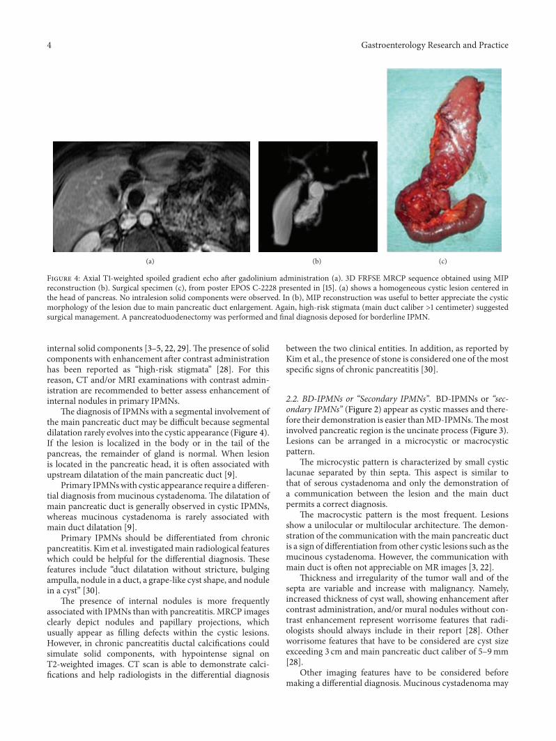
Figure 4: Axial T1-weighted spoiled gradient echo after gadolinium administration (a). 3D FRFSE MRCP sequence obtained using MIPreconstruction (b). Surgical specimen (c), from poster EPOS C-2228 presented in [15]. (a) shows a homogeneous cystic lesion centered inthe head of pancreas. No intralesion solid components were observed. In (b), MIP reconstruction was useful to better appreciate the cysticmorphology of the lesion due to main pancreatic duct enlargement. Again, high-risk stigmata (main duct caliber >1 centimeter) suggestedsurgical management. A pancreatoduodenectomy was performed and final diagnosis deposed for borderline IPMN.
internal solid components [3–5, 22, 29].The presence of solidcomponents with enhancement after contrast administrationhas been reported as “high-risk stigmata” [28]. For thisreason, CT and/or MRI examinations with contrast admin-istration are recommended to better assess enhancement ofinternal nodules in primary IPMNs.
The diagnosis of IPMNs with a segmental involvement ofthe main pancreatic duct may be difficult because segmentaldilatation rarely evolves into the cystic appearance (Figure 4).If the lesion is localized in the body or in the tail of thepancreas, the remainder of gland is normal. When lesionis located in the pancreatic head, it is often associated withupstream dilatation of the main pancreatic duct [9].
Primary IPMNswith cystic appearance require a differen-tial diagnosis frommucinous cystadenoma.The dilatation ofmain pancreatic duct is generally observed in cystic IPMNs,whereas mucinous cystadenoma is rarely associated withmain duct dilatation [9].
Primary IPMNs should be differentiated from chronicpancreatitis. Kim et al. investigatedmain radiological featureswhich could be helpful for the differential diagnosis. Thesefeatures include “duct dilatation without stricture, bulgingampulla, nodule in a duct, a grape-like cyst shape, and nodulein a cyst” [30].
The presence of internal nodules is more frequentlyassociated with IPMNs than with pancreatitis. MRCP imagesclearly depict nodules and papillary projections, whichusually appear as filling defects within the cystic lesions.However, in chronic pancreatitis ductal calcifications couldsimulate solid components, with hypointense signal onT2-weighted images. CT scan is able to demonstrate calci-fications and help radiologists in the differential diagnosis
between the two clinical entities. In addition, as reported byKim et al., the presence of stone is considered one of the mostspecific signs of chronic pancreatitis [30].
2.2. BD-IPMNs or “Secondary IPMNs”. BD-IPMNs or “sec-ondary IPMNs” (Figure 2) appear as cystic masses and there-fore their demonstration is easier thanMD-IPMNs.Themostinvolved pancreatic region is the uncinate process (Figure 3).Lesions can be arranged in a microcystic or macrocysticpattern.
The microcystic pattern is characterized by small cysticlacunae separated by thin septa. This aspect is similar tothat of serous cystadenoma and only the demonstration ofa communication between the lesion and the main ductpermits a correct diagnosis.
The macrocystic pattern is the most frequent. Lesionsshow a unilocular or multilocular architecture. The demon-stration of the communication with the main pancreatic ductis a sign of differentiation fromother cystic lesions such as themucinous cystadenoma. However, the communication withmain duct is often not appreciable on MR images [3, 22].
Thickness and irregularity of the tumor wall and of thesepta are variable and increase with malignancy. Namely,increased thickness of cyst wall, showing enhancement aftercontrast administration, and/or mural nodules without con-trast enhancement represent worrisome features that radi-ologists should always include in their report [28]. Otherworrisome features that have to be considered are cyst sizeexceeding 3 cm and main pancreatic duct caliber of 5–9mm[28].
Other imaging features have to be considered beforemaking a differential diagnosis. Mucinous cystadenoma may

Gastroenterology Research and Practice 5
(a) (b)
Figure 5: Linear EUS image of a MD-IPMN (a): a lobulated anechoic cystic lesion is clearly depicted (white arrow). (b) shows EUS-FNA ofthe same lesions. In this lesion (about 3 cm in size), the absence of mural nodules and positive or suspicious cytology allowed a conservativemanagement.
exhibit peripheral calcifications, which could reproduce an“eggshell” appearance [31]. Also, favourite locations in thepancreatic parenchyma are different for the lesions becausesecondary IPMNs are very often reported in the uncinateprocess [29], whereas mucinous cystadenoma is generallyencountered in the body or in the tail of the pancreas.
IPMNs need to be differentiated from pancreatic pseu-docysts, which develop as a complication of pancreatitis inup to 20–40% of cases [30]. In a recent work, “a grape-like appearance” has been associated with IPMNs in 79%of cases, whereas a unilocular cyst shape was reported in34% of patients affected by chronic pancreatitis. However,unilocular secondary IPMNs are very difficult to differentiatefrom pseudocysts. Careful collection of clinical history isvery important in these cases because pseudocysts generallydevelop as a complication of a severe episode of pancreatitis.
BD-IPMNs could be observed in amultifocal appearance.In this pattern of morphological presentation, IPMNs aredivided into five classes: diffuse, proximal, proximally diffuse,distal, and bridge morphology [22]. The multifocality ofIPMNs is responsible for an increased cumulative risk ofneoplastic degeneration [32]. In this case, patients need tobe followed over time in order to identify early signs ofprogression or degeneration.
2.3. EUS. Endoscopic ultrasonography (EUS) plays animportant role in the diagnostic evaluation of IPMNs dueto the possibility to collect fluid from cystic lesions. It canprovide high resolution contrast images of pancreatic cysticlesions, demonstrating many important details about cysticlesions, such as wall thickness, presence of septa, and muralnodules [33]. In addition, it permits measurement of thepancreatic ducts and provides visualization of communica-tion between cystic lesions and main pancreatic duct. Alsostrictures could be visualized along the course of main duct,contributing to the differential diagnosis between chronicpancreatitis and MD-IPMN [34–36].
In addition, EUS is able to guide fine-needle aspiration(FNA) (Figure 5) [37]. The fluid content could be analysedfor the presence of oncological marker.
It has been well documented that CEA and CA 72.4 levelsin the cystic fluid of the mucinous lesions are much higher(typically over 800 ng/mL) than those of nonmucinous ones
[38]. Moreover, CEA and CA72.4 levels are higher in malig-nant mucinous neoplasms [39–43]. In a work by Brugge etal. a level of 192 ng/mL for CEA has a diagnostic sensitivityof 75%, a specificity of 84%, and an accuracy of 79% indifferential diagnosis of mucinous and nonmucinous cysts[41].
In view of these considerations, several studies haverecently investigated the diagnostic and prognostic valuesof these markers in order to establish the risk of malig-nant degeneration. Also inflammatory mediator proteins(cytokines, chemokines, and growth factors) — contained inpancreatic cyst fluid — could be used as potential diagnos-tic biomarkers able to characterize IPMNs [44]. However,sensitivity and specificity observed are not so high; detectionof K-ras mutation in the pancreatic fluid can indicate thepresence of amalignant cystic lesion, evenwith poor sensitiv-ity (20%) [29]. The reported threshold level of 192 ng/mL forCEA has been evaluated as a predictor value of malignancyfor IPMNs in a recent work by Kucera et al. [45].The authorsfound that the mean level of intracystic CEA increases pro-gressively from low-grade to high-grade of dysplasia (rangingfrom 1.261±1.679 ng/mL to 10.807±36.203 ng/mL). Amonginvasive cancers, the mean level reported was lower thanIPMNs with various degrees of dysplasia.The reported sensi-tivity, specificity, positive predictive value, negative predictivevalue, and accuracy of a cyst fluid CEA concentration greaterthan 200 ng/mL for the diagnosis of malignant IPMN—including lesions with high-grade dysplasia and invasiveIPMNs—were, respectively, 52.4%, 42.3%, 42.3%, 52.4%, and46.8% [45].
On the basis of the mentioned studies, EUS—even withFNA—does not show such high values of sensitivity andspecificity in the diagnosis of IPMNs. In addition, it isan invasive [24], heavily operator-dependent modality thatrequires patient sedation [37]. Recent “European expert con-sensus statement on cystic tumours of the pancreas” remarkedthat EUS is “an invasive diagnostic procedure,” which needsto be performed after cross-sectional imaging (CT/MRI),in a multimodality imaging assessment of cystic pancreaticneoplasms [24].
After CT/MRI examinations, “All cysts with worrisomefeature or cysts exceeding 3 cm in size without worrisomefeature” should be investigated by EUS [28]; identification

6 Gastroenterology Research and Practice
of mural nodules, main duct signs of involvement bydisease, or a cytology suspicion could suggest surgery[28].
Recently, some authors have proposed EUS imaging inthe follow-up evaluation of secondary IPMNs. Kamata, ina recent retrospective study, compared the diagnostic valueof EUS, ultrasonography, CT, and MRI in the assessmentof pancreatic ductal adenocarcinoma arising from MD-IPMNs [46]. The population study included a total of 169patients. All the mentioned imaging modalities followed 102patients having side branch IPMNs without mural nodulesand symptoms. The follow-up was performed in order toverify the incidence of IPMN-derived and/or concomitantpancreatic ductal adenocarcinoma. At the first follow-upexamination, 17 IPMN-derived and 11 concomitant ductaladenocarcinomas were detected by the authors, with EUSoverall sensitivity higher than other imaging modalities. Forthe entire follow-up period of the study, EUS maintainedits better diagnostic accuracy in the detection of concomi-tant duct adenocarcinoma. Other authors have performeda follow-up study through US and MRCP in a large seriesof patients (𝑛 = 109) with BD-IPMNs [29]. In this study,EUS and ERCP were performed only in select cases, whenthe diagnosis was still unclear or doubtful after conventionalcross-sectional imaging modalities.
However, the invasiveness and the variability representlimitations to adopting EUS in the follow-up of MD-IPMNs.
3. Management
Currently, management of IPMNs is one of the most debatedtopics in literature, and it is essentially based on cross-sectional imaging modalities (CT/MR) and EUS. There is nosufficient evidence for pancreatoscopy inmanagement of cys-tic tumours and subsequently for IPMNs [24]. ERCP couldbe useful in selected cases, for example, in the evaluation ofprimary IPMNs with diffuse dilatation of main pancreaticduct, without evidence of mural nodules. In these cases, thediffuse increased caliber of main duct with bulging of majorpapilla promotes the right diagnosis ofMD-IPMNs and couldsuggest the correct surgical approach.
First of all, cross-sectional imaging modalities should beable to clearly distinguish the three radiological patterns ofpresentation. As previously reported, primary IPMNs show aprogression risk higher than secondary forms. In addition,multifocal branch-duct IPMNs have a cumulative risk ofmalignancy degeneration due to the coexistence of manycystic lesions.
High-risk stigmata, represented by dilatation of the mainpancreatic duct equal to or more than 10mm and/or solidcomponents with enhancement after contrast, in view ofits frequent association with malignancy, require surgicaltreatment. In fact, in a study performed by Abdeljawad, theprevalence of malignancy in 52 patients with pure mainduct IPMN was analysed [47]. Among 16 asymptomaticpatients reporting IPMNs, 4 had malignant lesions. In thesymptomatic group (36 out of 52 patients), 25 lesions weremalignant on histology. The size of the main pancreatic
duct was analysed by authors using ROC analysis, and thelargest area under the curve used to distinguish betweenbenign and malignant MD-IPMN was found using a thresh-old level of main pancreatic duct of 8mm (0.83; 95%CI = 0.72–0.94).
Worrisome features—including cyst size ≥3 cm, thick-ened cyst wall with enhancement after contrast adminis-tration, mural nodules without enhancement after contrast,main ductwith diameter of 5–9mm, and abrupt change in themain pancreatic duct caliber with distal pancreatic atrophyand lymphadenopathy—require further investigation [28].As previously reported, EUS plays an important role in themanagement because confirmation of worrisome featurescould require a surgical treatment [28]. If absent, IPMNscould be monitored using MR/MRCP at 3 months and EUSannually for the first 2 years [28].
Regarding the size, cysts exceeding 3 cm, even if con-sidered a worrisome feature, did not show a high value ofcorrelation with malignancy. In a series observed by Sahaniet al., only 5 out of 8 lesions with diameter >3 cm weremalignant at pathological examination. In another series of26 patients with secondary IPMNs reported by Manfrediet al., a significant change in the size of cystic lesions wasobserved. However, this imaging finding does not necessarilycorrelate with malignant transformation or increased suspi-cion of malignancy [4].
Therefore, the presence of nodules is probably themost significant change which needs to be carefully eval-uated because it is strongly suspected as an indicator ofmalignancy.
Salvia has evaluated nonoperative management of sec-ondary branches IPMNs in a prospective study, by per-forming contrast enhanced US and MRCP. Lesions wereless than 3.5 cm in diameter and without nodules or solidcomponents. Their study included a total of 109 patients. Afirst group (20 patients, 18.3%) required immediate surgeryfor the presence of symptoms or clinical and morphologicalfeatures associated with malignancy. Among this group, theauthors found only 2 patients with invasive carcinoma and1 patient with carcinoma in situ. The remainder of thepatients were evaluated with an average follow-up of 32months. After an average follow-up of 18.2 months, Salviaet al. [29] reported only 5 patients with an increase inthe size of the lesion. These patients underwent surgeryand their final diagnosis was branch-duct adenoma in3 cases and borderline lesions in 2 patients [29]. Thus,this study confirms that BD-IPMNs could be managed byimaging.
Finally, secondary IPMNs arranged in a multifocal pat-tern (Figure 6) should be evaluated for their increased riskof degeneration [48]. However, in another study, Salviaexamined a total of 131 patients having multifocal secondaryIPMNs. Here, only 10 patients were surgically managed,whereas the majority was followed for an average period of40 months. 121 patients were conservatively managed, andthey remained asymptomatic, without nodules or increase indiameter of their lesions. As reported by the authors, IPMNsin a multifocality setting could also be managed in a safe andreliable way [49].

Gastroenterology Research and Practice 7
(a) (b)
(c)
Figure 6: Coronal MRCP acquisitions in an asymptomatic 70-year-old female patient with an incidental radiological finding of multiplepancreatic cystic lesions; MRCP exams were performed in 2009 (a), in 2012 (b), and in 2013 (c). Multiple small cystic lesions in the pancreaticparenchyma are clearly depicted in the threeMRCP acquisitions, some of them showing a typical connection to themain pancreatic duct.Thistypical radiological pattern suggests the diagnosis of multifocal BD-IPMNs. Nomain pancreatic duct dilatation is observed. Cystic lesions donot show intraluminal solid components or mural nodules. Over time theMRI monitoring initially showed a mild enlargement of the lesions(from a to b) and then a size-reduction (from b to c). As reported in literature, IPMNs in a multifocal setting could also be managed in a safeand reliable mode.
4. Conclusion
Gastroenterologists, radiologists, and surgeons should beconfident utilizing all imaging features of IPMNs.
On the basis of the diagnostic patterns analysed,
(i) radiologists should distinguish between primary, sec-ondary, and mixed IPMNs; cross-sectional imagingfeatures need to clearly demonstrate the relationshipsbetween IPMNs and pancreatic duct system;
(ii) identifying high-risk stigmata or worrisome featuresis recommended in order to suggest the correctmanagement;
(iii) in case of IPMNs with high-risk stigmata, a surgicalapproach is needed, namely, for lesions with markeddilatation of the main pancreatic duct (≥1 cm) orshowing internal solid enhancing components;
(iv) if worrisome features are depicted on cross-sectionalimaging modalities, EUS investigation is required.Confirmation of these worrisome features requiressurgery. In their absence, a follow-up procedure by
CT/MRI could be safely adopted, monitoring thedevelopment of malignant signs.
Finally, all imaging features should be related to clinicalconditions of patients (age, comorbidities, and performancestatus) for a correct management of the disease.
Conflict of Interests
The authors declare that there is no conflict of interestsregarding the publication of this paper.
References
[1] M. Acar and S. Tatli, “Cystic tumors of the pancreas: a radiolog-ical perspective,” Diagnostic and Interventional Radiology, vol.17, no. 2, pp. 143–149, 2011.
[2] M. Suyama, H. Ariyama, and K. Ogawa, “Clinical diagnosis ofmucin producing pancreatic carcinoma,” Tan to Sui, vol. 7, pp.739–745, 1986.
[3] D. V. Sahani, R. Kadavigere, A. Saokar, C. Fernandez-delCastillo,W.R. Brugge, andP. F.Hahn, “Cystic pancreatic lesions:

8 Gastroenterology Research and Practice
a simple imaging-based classification system for guiding man-agement,” Radiographics, vol. 25, no. 6, pp. 1471–1484, 2005.
[4] R.Manfredi, S.Mehrabi,M.Motton et al., “MR imaging andMRcholangiopancreatography of multifocal intraductal papillarymucinous neoplasms of the side branches: MR pattern and itsevolution,” Radiologia Medica, vol. 113, no. 3, pp. 414–428, 2008.
[5] R. Salvia, S. Crippa, M. Falconi et al., “Branch-duct intraductalpapillarymucinous neoplasms of the pancreas: to operate or notto operate?” Gut, vol. 56, no. 8, pp. 1086–1090, 2007.
[6] M. Falconi, R. Salvia, C. Bassi, G. Zamboni, G. Talamini, andP. Pederzoli, “Clinicopathological features and treatment ofintraductal papillary mucinous tumour of the pancreas,” BritishJournal of Surgery, vol. 88, no. 3, pp. 376–381, 2001.
[7] R. Salvia, C. Fernandez-Del Castillo, C. Bassi et al., “Main-ductintraductal papillarymucinous neoplasms of the pancreas: clin-ical predictors of malignancy and longterm survival followingresection,” Annals of Surgery, vol. 239, no. 5, pp. 678–687, 2004.
[8] T. A. Sohn, C. J. Yeo, J. L. Cameron et al., “Intraductal papillarymucinous neoplasms of the pancreas: an updated experience,”Annals of Surgery, vol. 239, no. 6, pp. 788–799, 2004.
[9] C. Procacci, A. J. Megibow, G. Carbognin et al., “Intraductalpapillary mucinous tumor of the pancreas: a pictorial essay,”Radiographics, vol. 19, no. 6, pp. 1447–1463, 1999.
[10] G. Kloppel, E. Solcia, D. S. Longnecker, C. Capella, and L.H. Sobin, Eds., Histologic Typing of Tumors of the ExocrinePancreas, Springer, Geneva, Switzerland, 1996.
[11] G. Capurso, S. Boccia, R. Salvia et al., “Risk factors for intra-ductal papillary mucinous neoplasm (ipmn) of the pancreas:a multicentre case-control study,” The American Journal ofGastroenterology, vol. 108, no. 6, pp. 1003–1009, 2013.
[12] N. V. Adsay, K. C. Conlon, S. Y. Zee, M. F. Brennan, and D.S. Klimstra, “Intraductal papillary-mucinous neoplasms of thepancreas: an analysis of in situ and invasive carcinomas in 28patients,” Cancer, vol. 94, no. 1, pp. 62–77, 2002.
[13] R. H. Hruban, K. Takaori, D. S. Klimstra et al., “An illustratedconsensus on the classification of pancreatic intraepithelialneoplasia and intraductal papillary mucinous neoplasms,” TheAmerican Journal of Surgical Pathology, vol. 28, no. 8, pp. 977–987, 2004.
[14] P. J. Allen,M. D'Angelica,M. Gonen et al., “A selective approachto the resection of cystic lesions of the pancreas: results from539 consecutive patients,” Annals of Surgery, vol. 244, no. 4, pp.572–582, 2006.
[15] S. Palmucci, L. A. Mauro, G. Failla et al., “ Intraductal PapillaryMucinous Neoplasm (IPMN), serous cystadenoma and muci-nous cystadenoma: imaging findings observed with MagneticResonance (MR) and Magnetic Resonance Cholangiopancre-atography (MRCP),” in Proceedings of the ECR, 2011.
[16] D. Sahani, S. Prasad, S. Saini, and P. Mueller, “Cystic pan-creatic neoplasms evaluation by CT and magnetic resonancecholangiopancreatography,” Gastrointestinal Endoscopy Clinicsof North America, vol. 12, no. 4, pp. 657–672, 2002.
[17] J. S. Su, M. L. Jeong, J. K. Young et al., “Differentiation of intra-ductal papillary mucinous neoplasms from other pancreaticcystic masses: comparison of multirow-detector CT and MRimaging using ROC analysis,” Journal of Magnetic ResonanceImaging, vol. 26, no. 1, pp. 86–93, 2007.
[18] N. I. Sainani, A. Saokar, V. Deshpande, C. Fernandez-DelCastillo, P. Hahn, and D. V. Sahani, “Comparative performanceof MDCT and MRI with MR cholangiopancreatography incharacterizing small pancreatic cysts,”The American Journal ofRoentgenology, vol. 193, no. 3, pp. 722–731, 2009.
[19] D. V. Sahani, D. J. Lin, A. M. Venkatesan et al., “Multidisci-plinary approach to diagnosis and management of intraductalpapillary mucinous neoplasms of the pancreas,” Clinical Gas-troenterology and Hepatology, vol. 7, no. 3, pp. 259–269, 2009.
[20] M. J. Clores, A.Thosani, and J. M. Buscaglia, “Multidisciplinarydiagnostic and therapeutic approaches to pancreatic cysticlesions,” Journal of Multidisciplinary Healthcare, vol. 7, pp. 81–91, 2014.
[21] S. Palmucci, L. A.Mauro, P.Milone et al., “Diagnosis of rupturedsuperior mesenteric artery aneurysm mimicking a pancreaticmass,” World Journal of Gastroenterology, vol. 16, no. 18, pp.2298–2301, 2010.
[22] F. Castelli, D. Bosetti, R. Negrelli et al., “Multifocal branch-duct intraductal papillary mucinous neoplasms (IPMNs) ofthe pancreas: magnetic resonance (MR) imaging pattern andevolution over time,” Radiologia Medica, vol. 118, no. 6, pp. 917–929, 2013.
[23] M. Tanaka, S. Chari, V. Adsay et al., “International consensusguidelines for management of intraductal papillary mucinousneoplasms and mucinous cystic neoplasms of the pancreas,”Pancreatology, vol. 6, no. 1-2, pp. 17–32, 2006.
[24] M. del Chiaro, C. Verbeke, R. Salvia et al., “European expertsconsensus statement on cystic tumours of the pancreas,” Diges-tive and Liver Disease, vol. 45, no. 9, pp. 703–711, 2013.
[25] S. Palmucci, C. Cappello, C. Trombatore et al., “Cystic pan-creatic neoplasms: diagnosis and management emphasizingtheir imaging features,” European Review for Medical andPharmacological Sciences, vol. 18, no. 8, pp. 1259–1268, 2014.
[26] S. Palmucci, L. A. Mauro, M. Coppolino et al., “Evaluation ofthe biliary and pancreatic system with 2D SSFSE, breathhold3D FRFSE and respiratory-triggered 3D FRFSE sequences,”Radiologia Medica, vol. 115, no. 3, pp. 467–482, 2010.
[27] D. V. Sahani, R. Kadavigere, M. Blake, C. Fernandez-DelCastillo, G. Y. Lauwers, and P. F. Hahn, “Intraductal papillarymucinous neoplasm of pancreas: multi-detector row CT with2D curved reformations-correlation with MRCP,” Radiology,vol. 238, no. 2, pp. 560–569, 2006.
[28] M. Tanaka, C. Fernandez-del Castillo, V. Adsay et al., “Interna-tional consensus guidelines 2012 for the management of IPMNandMCNof the pancreas,” Pancreatology, vol. 12, no. 3, pp. 183–197, 2012.
[29] R. Salvia, S. Crippa, S. Partelli et al., “Pancreatic cystic tumours:when to resect, when to observe,” European Review for Medicaland Pharmacological Sciences, vol. 14, no. 4, pp. 395–406, 2010.
[30] J. H. Kim, S. S. Hong, Y. J. Kim, J. K. Kim, and H. W. Eun,“Intraductal papillary mucinous neoplasm of the pancreas: dif-ferentiate from chronic pancreatits by MR imaging,” EuropeanJournal of Radiology, vol. 81, no. 4, pp. 671–676, 2012.
[31] A. Khalid and W. Brugge, “ACG practice guidelines for thediagnosis and management of neoplastic pancreatic cysts,”American Journal of Gastroenterology, vol. 102, no. 10, pp. 2339–2349, 2007.
[32] J. K. Sai,M. Suyama,Y.Kubokawa et al., “Management of branchduct-type intraductal papillarymucinous tumor of the pancreasbased on magnetic resonance imaging,” Abdominal Imaging,vol. 28, no. 5, pp. 694–699, 2003.
[33] K. de Jong, M. J. Bruno, and P. Fockens, “Epidemiology,diagnosis, and management of cystic lesions of the pancreas,”Gastroenterology Research and Practice, vol. 2012, Article ID147465, 8 pages, 2012.
[34] N. Kobayashi, K. Sugimori, T. Shimamura et al., “Endoscopicultrasonographic findings predict the risk of carcinoma in

Gastroenterology Research and Practice 9
branch duct intraductal papillary mucinous neoplasms of thepancreas,” Pancreatology, vol. 12, no. 2, pp. 141–145, 2012.
[35] S. A. Pais, S. Attasaranya, J. K. Leblanc, S. Sherman, C. M.Schmidt, and J. DeWitt, “Role of endoscopic ultrasound in thediagnosis of intraductal papillarymucinous neoplasms: correla-tionwith surgical histopathology,”Clinical Gastroenterology andHepatology, vol. 5, no. 4, pp. 489–495, 2007.
[36] H. Kubo, K. Nakamura, S. Itaba et al., “Differential diagnosis ofcystic tumors of the pancreas by endoscopic ultrasonography,”Endoscopy, vol. 41, no. 8, pp. 684–689, 2009.
[37] S. Palmucci, L. A. Mauro, S. la Scola et al., “Magnetic resonancecholangiopancreatography and contrast-enhanced magneticresonance cholangiopancreatography versus endoscopic ultra-sonography in the diagnosis of extrahepatic biliary pathology,”Radiologia Medica, vol. 115, no. 5, pp. 732–746, 2010.
[38] L. A. van der Waaij, H. M. van Dullemen, and R. J. Porte, “Cystfluid analysis in the differential diagnosis of pancreatic cysticlesions: a pooled analysis,” Gastrointestinal Endoscopy, vol. 62,no. 3, pp. 383–389, 2005.
[39] M. B. Pitman, K. Lewandrowski, J. Shen, D. Sahani, W. Brugge,and C. Fernandez-Del Castillo, “Pancreatic cysts: preoperativediagnosis and clinical management,” Cancer Cytopathology, vol.118, no. 1, pp. 1–13, 2010.
[40] W. R. Brugge, G. Y. Lauwers, D. Sahani, C. Fernandez-DelCastillo, andA. L.Warshaw, “Cystic neoplasms of the pancreas,”New England Journal of Medicine, vol. 351, no. 12, pp. 1218–1269,2004.
[41] W. R. Brugge, K. Lewandrowski, E. Lee-Lewandrowski et al.,“Diagnosis of pancreatic cystic neoplasms: a report of thecooperative pancreatic cyst study,” Gastroenterology, vol. 126,no. 5, pp. 1330–1336, 2004.
[42] C. Sperti, C. Pasquali, S. Pedrazzoli, P. Guolo, and G. Liessi,“Expression of mucin-like carcinoma-associated antigen in thecyst fluid differentiates mucinous from nonmucinous pancre-atic cysts,”TheAmerican Journal of Gastroenterology, vol. 92, no.4, pp. 672–675, 1997.
[43] P. Hammel, H. Voitot, V. Vilgrain, P. Levy, P. Ruszniewski, and P.Bernades, “Diagnostic value of CA 72-4 and carcinoembryonicantigen determination in the fluid of pancreatic cystic lesions,”European Journal of Gastroenterology and Hepatology, vol. 10,no. 4, pp. 345–348, 1998.
[44] L. S. Lee, A. M. Bellizzi, P. A. Banks et al., “Differentiatingbranch duct and mixed IPMN in endoscopically collectedpancreatic cyst fluid via cytokine analysis,” GastroenterologyResearch and Practice, vol. 2012, Article ID 247309, 10 pages,2012.
[45] S. Kucera, B. A. Centeno, G. Springett et al., “Cyst fluid carci-noembryonic antigen level is not predictive of invasive cancerin patients with intraductal papillarymucinous neoplasm of thepancreas,” Journal of the Pancreas, vol. 13, no. 4, pp. 409–413,2012.
[46] K. Kamata, M. Kitano, M. Kudo et al., “Value of EUS in earlydetection of pancreatic ductal adenocarcinomas in patientswithintraductal papillary mucinous neoplasms,” Endoscopy, vol. 46,no. 1, pp. 22–29, 2014.
[47] K. Abdeljawad, K. C. Vemulapalli, C. M. Schmidt et al., “Preva-lence of malignancy in patients with puremain duct intraductalpapillarymucinous neoplasms,”Gastrointestinal Endoscopy, vol.79, no. 4, pp. 623–629, 2014.
[48] R. Salvia, S. Partelli, S. Crippa et al., “Intraductal papillarymuci-nous neoplasms of the pancreas with multifocal involvement of
branch ducts,” The American Journal of Surgery, vol. 198, no. 5,pp. 709–714, 2009.
[49] K. C. Chiang, J. T. Hsu, H. Y. Chen et al., “Multifocal intraductalpapillary mucinous neoplasm of the pancreas: a case report,”World Journal of Gastroenterology, vol. 15, no. 5, pp. 628–632,2009.

Submit your manuscripts athttp://www.hindawi.com
Stem CellsInternational
Hindawi Publishing Corporationhttp://www.hindawi.com Volume 2014
Hindawi Publishing Corporationhttp://www.hindawi.com Volume 2014
MEDIATORSINFLAMMATION
of
Hindawi Publishing Corporationhttp://www.hindawi.com Volume 2014
Behavioural Neurology
EndocrinologyInternational Journal of
Hindawi Publishing Corporationhttp://www.hindawi.com Volume 2014
Hindawi Publishing Corporationhttp://www.hindawi.com Volume 2014
Disease Markers
Hindawi Publishing Corporationhttp://www.hindawi.com Volume 2014
BioMed Research International
OncologyJournal of
Hindawi Publishing Corporationhttp://www.hindawi.com Volume 2014
Hindawi Publishing Corporationhttp://www.hindawi.com Volume 2014
Oxidative Medicine and Cellular Longevity
Hindawi Publishing Corporationhttp://www.hindawi.com Volume 2014
PPAR Research
The Scientific World JournalHindawi Publishing Corporation http://www.hindawi.com Volume 2014
Immunology ResearchHindawi Publishing Corporationhttp://www.hindawi.com Volume 2014
Journal of
ObesityJournal of
Hindawi Publishing Corporationhttp://www.hindawi.com Volume 2014
Hindawi Publishing Corporationhttp://www.hindawi.com Volume 2014
Computational and Mathematical Methods in Medicine
OphthalmologyJournal of
Hindawi Publishing Corporationhttp://www.hindawi.com Volume 2014
Diabetes ResearchJournal of
Hindawi Publishing Corporationhttp://www.hindawi.com Volume 2014
Hindawi Publishing Corporationhttp://www.hindawi.com Volume 2014
Research and TreatmentAIDS
Hindawi Publishing Corporationhttp://www.hindawi.com Volume 2014
Gastroenterology Research and Practice
Hindawi Publishing Corporationhttp://www.hindawi.com Volume 2014
Parkinson’s Disease
Evidence-Based Complementary and Alternative Medicine
Volume 2014Hindawi Publishing Corporationhttp://www.hindawi.com