Review Article The Role of Power Doppler Ultrasonography...
10
Review Article The Role of Power Doppler Ultrasonography as Disease Activity Marker in Rheumatoid Arthritis Shaloo Bhasin 1 and Peter P. Cheung 1,2 1 Division of Rheumatology, National University Hospital, Singapore 2 Yong Loo Lin School of Medicine, National University of Singapore, Singapore Correspondence should be addressed to Peter P. Cheung; peter [email protected] Received 11 January 2015; Accepted 7 April 2015 Academic Editor: Chao Hung Hung Copyright © 2015 S. Bhasin and P. P. Cheung. is is an open access article distributed under the Creative Commons Attribution License, which permits unrestricted use, distribution, and reproduction in any medium, provided the original work is properly cited. Structural damage in rheumatoid arthritis (RA) occurs early if inflammation is not treated promptly. Treatment targeted to reduce inflammation, in particular, that of synovial inflammation in the joints (synovitis), has been recommended as standard treat-to- target recommendations by rheumatologists. e goal is to achieve disease remission (i.e., no disease activity). Several accepted remission criteria have not always equated to the complete absence of true inflammation. Over the last decade, musculoskeletal ultrasonography has been demonstrated to detect subclinical synovitis not appreciated by routine clinical or laboratory assessments, with the Power Doppler modality allowing clinicians to more readily appreciate true inflammation. us, targeting therapy to Power Doppler activity may provide superior outcomes compared with treating to clinical targets alone, making it an attractive marker of disease activity in RA. However, more validation on its true benefits such as its benefits to patients in regard to patient related outcomes and issues with standardized training in acquisition and interpretation of power Doppler findings are required. 1. Introduction Concepts in Monitoring and Treatment of Rheumatoid Arthri- tis. Rheumatoid arthritis (RA) is a chronic inflammatory disease associated with significant functional limitations and disability. Diagnosing RA begins with a thorough medical history of the patient, focusing on the presence, location, and duration of joint pain and stiffness as well as physical assessment of synovitis [1]. Since there is no single test to diagnose RA, clinicians use a number of tests to support the clinical diagnosis. is traditionally includes rheumatoid factor (RF), anticitrullinated peptide (anti-CCP), erythrocyte sedimentation rate (ESR), and/or serum C-reactive protein (CRP) levels as well as imaging using radiographs of the hands, wrists, and feet [2–4]. In addition, imaging with ultra- sonography (US) and magnetic resonance imaging (MRI) has increased the ability to diagnose the disease earlier when the clinical presentation is unclear or when clinical synovitis is equivocal. e need to diagnose RA early and commence disease modifying antirheumatic drugs (DMARDs) has led to an updated RA classification criteria, jointly proposed by the American College of Rheumatology (ACR) and the European League Against Rheumatism (EULAR) [1]. Management of RA such as the determination of treat- ment decisions depends on a number of factors. Although the level of disease activity is of paramount importance, the disease duration, acknowledgement of poor prognostic factors (e.g., seropositivity for RF and or anti-CCP, erosions, and extra-articular disease), and the level of patient’s dis- ability as well as self-reported impact of disease have to be taken into account [5]. ere is no single gold standard for quantifying the level of disease activity. Hence, clinicians would routinely use a number of parameters such as clinical assessment of tender and swollen joints, a global assessment of disease activity, and either an ESR or CRP level, for example, to determine the level of disease activity. A number of validated instruments for RA in the form of composite indices that combine these parameters into a score (some of them weighed) have been routinely used in clinical practice and clinical trials. is allows a standardized way to quantify Hindawi Publishing Corporation Disease Markers Volume 2015, Article ID 325909, 9 pages http://dx.doi.org/10.1155/2015/325909
Transcript of Review Article The Role of Power Doppler Ultrasonography...
Review ArticleThe Role of Power Doppler Ultrasonography as Disease ActivityMarker in Rheumatoid Arthritis
Shaloo Bhasin1 and Peter P. Cheung1,2
1Division of Rheumatology, National University Hospital, Singapore2Yong Loo Lin School of Medicine, National University of Singapore, Singapore
Correspondence should be addressed to Peter P. Cheung; peter [email protected]
Received 11 January 2015; Accepted 7 April 2015
Academic Editor: Chao Hung Hung
Copyright © 2015 S. Bhasin and P. P. Cheung. This is an open access article distributed under the Creative Commons AttributionLicense, which permits unrestricted use, distribution, and reproduction in any medium, provided the original work is properlycited.
Structural damage in rheumatoid arthritis (RA) occurs early if inflammation is not treated promptly. Treatment targeted to reduceinflammation, in particular, that of synovial inflammation in the joints (synovitis), has been recommended as standard treat-to-target recommendations by rheumatologists. The goal is to achieve disease remission (i.e., no disease activity). Several acceptedremission criteria have not always equated to the complete absence of true inflammation. Over the last decade, musculoskeletalultrasonography has been demonstrated to detect subclinical synovitis not appreciated by routine clinical or laboratory assessments,with the PowerDopplermodality allowing clinicians tomore readily appreciate true inflammation.Thus, targeting therapy to PowerDoppler activity may provide superior outcomes compared with treating to clinical targets alone, making it an attractive markerof disease activity in RA. However, more validation on its true benefits such as its benefits to patients in regard to patient relatedoutcomes and issues with standardized training in acquisition and interpretation of power Doppler findings are required.
1. Introduction
Concepts in Monitoring and Treatment of Rheumatoid Arthri-tis. Rheumatoid arthritis (RA) is a chronic inflammatorydisease associated with significant functional limitations anddisability. Diagnosing RA begins with a thorough medicalhistory of the patient, focusing on the presence, location,and duration of joint pain and stiffness as well as physicalassessment of synovitis [1]. Since there is no single test todiagnose RA, clinicians use a number of tests to supportthe clinical diagnosis. This traditionally includes rheumatoidfactor (RF), anticitrullinated peptide (anti-CCP), erythrocytesedimentation rate (ESR), and/or serum C-reactive protein(CRP) levels as well as imaging using radiographs of thehands, wrists, and feet [2–4]. In addition, imaging with ultra-sonography (US) andmagnetic resonance imaging (MRI) hasincreased the ability to diagnose the disease earlier when theclinical presentation is unclear or when clinical synovitis isequivocal. The need to diagnose RA early and commencediseasemodifying antirheumatic drugs (DMARDs) has led to
an updated RA classification criteria, jointly proposed by theAmericanCollege of Rheumatology (ACR) and the EuropeanLeague Against Rheumatism (EULAR) [1].
Management of RA such as the determination of treat-ment decisions depends on a number of factors. Althoughthe level of disease activity is of paramount importance,the disease duration, acknowledgement of poor prognosticfactors (e.g., seropositivity for RF and or anti-CCP, erosions,and extra-articular disease), and the level of patient’s dis-ability as well as self-reported impact of disease have to betaken into account [5]. There is no single gold standard forquantifying the level of disease activity. Hence, clinicianswould routinely use a number of parameters such as clinicalassessment of tender and swollen joints, a global assessmentof disease activity, and either an ESR or CRP level, forexample, to determine the level of disease activity. A numberof validated instruments for RA in the form of compositeindices that combine these parameters into a score (some ofthem weighed) have been routinely used in clinical practiceand clinical trials. This allows a standardized way to quantify
Hindawi Publishing CorporationDisease MarkersVolume 2015, Article ID 325909, 9 pageshttp://dx.doi.org/10.1155/2015/325909
2 Disease Markers
the absolute level of disease activity at any given point in time.Some of these measures including disease activity score in28 joints (DAS28), simplified disease activity index (SDAI),and clinical disease activity index (CDAI), for example, areillustrated in Table 1 [5] with their respective thresholds oflevels of disease activity.
Treating to Target to Achieve Disease Remission Is the Goalin RA. Regular disease activity assessment with treatmentadjustments according to a “treatment target” has now beenuniversally accepted as the best practice in the managementof patients with RA. The objective is to enable earlier aggres-sive treatment, through regular disease activity assessmentsand appropriate modifications of therapy, in order to achievedisease remission [6].
However, definitions of remission by clinical criteria(defined by levels of disease activity score, simplified diseaseactivity index) do not always equate to the complete absenceof inflammation. Even with more stringent criteria such asthe ACR/EULAR remission criteria, a Boolean criteria thatrequire <1 of tender and swollen joint, normal CRP, and <1on visual analogue scale of 0–10 on patient global assessment[7] may not necessarily indicate complete absence of synovialinflammation, since subclinical synovitis can be missed byclinical assessment alone. Synovitis is frequently found byimaging, such as by US or MRI in patients considered tobe in remission, and is associated with adverse clinical andfunctional outcomes [8]. Some have argued that targetingtherapy to PDUS activity may provide better outcomescompared with targeting therapy to clinical targets alone [9].This makes it an attractive and feasible marker of diseaseactivity in RA.
Ultrasonography in Rheumatoid Arthritis. Within the lastdecade, musculoskeletal ultrasonography has played anincreasingly important role in the evaluation andmonitoringof patients with chronic inflammatory arthritis. US canreadily evaluate synovitis, a pathological hallmark of RAat both the anatomic and vascular levels. There are 2 UStechniques that are of use:
(i) B-mode or gray scale US: imaging of anatomic struc-tures, which enables visualization of synovial hyper-trophy and/or effusion,
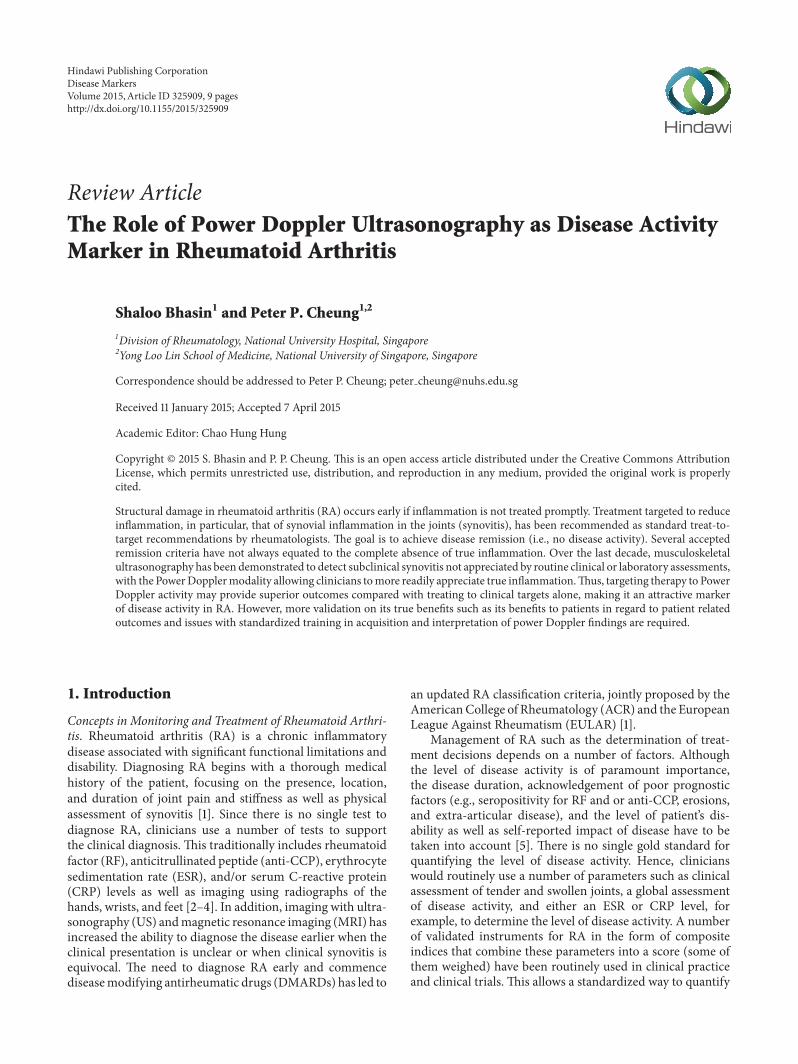
(ii) Power Doppler US (PDUS): blood flow detection,which allows visualization of the movement of bloodvessels, therefore detecting increased microvascularblood flow seen in active synovitis (Figure 1).
Power Doppler Ultrasonography. PDUS is based on Dopplereffect, which consists of the change of frequency of a soundbeam reflected back to the source when it encounters amoving object. Doppler technique detects the movement ofred blood cells in vessels. PDUS technique is more accuratethan conventional Color Doppler. Color Doppler encodesdirection and velocity of blood flow while PDUS displays thetotal integrated Doppler power in color therefore increasingthe sensitivity to detect strength of flow from small vessels
Figure 1: Wrist joint with Power Doppler synovitis in patient withRA.
and low velocity flow which is usually the case in RA patients[10].
PDUS Grading. The most frequently applied method ofgrading severity is by a semiquantitative scoring system inwhich the intensity of the synovial blood flow is graded ina four-step scale [11] in Table 2.
This semiquantitative grading system is considered apractical way to standardize PDUS measurement in RA.Compared to other more sophisticated ways of quantify-ing flow (computer-assisted measurement of color pixels,resistance index, and analysis of Doppler curves, contrast-enhanced Doppler US) [12], it does not involve contrastmedia or further computer-assisted evaluation software. Inaddition, it is validated in the diagnostic and therapeuticoutcome evaluation of patients with RA in various settings.
PDUS Settings. Although using the PDUS setting is usuallypreferred, in newer high-end machines, Color Doppler mayhave better sensitivity than PDUS [13, 14]. In low andintermediate range machines, PDUS always has the highestsensitivity. Therefore, ensuring good standardization of set-tings for the machine is important usually by a technicianfrom the manufacture.
The pulsed repetition frequency (PRF) is Doppler sam-pling frequency of the transducer (how many pulses areemitted per second) and is reported in Hz. This is importantin RA as the goal is to detect as much flow as possible oftenin the small joints such as metacarpal phalangeal (MCP),proximal interphalangeal (PIP), and metatarsal phalangeal(MTP) joints.The settings should therefore be able to achievethe highest sensitivity without noise artifacts [15]. The usualrecommended settings would be adjusting to the lowest pos-sible PRF, wall filter, and persistence. Gain on the threshold tonoise should be achieved with the focus placed on where thehighest sensitivity is required with all priority to color [15].Adjusting and optimizing this correctlywill have great impacton the ability to see inflammatory flow. Adjusting manyparameters of the PDUS is not done at every examination.Fortunately, there is little difference usually from patient topatient and from joint to joint with respect to PDUS settings.An exception is the hip joint because of its deep location.Once the sensitivity of the PDUS has been optimized, the
Disease Markers 3
Table 1: Composite indices measuring disease activity in rheumatoid arthritis.
Instrument Components Thresholds of diseaseActivity levels
Clinical disease activity index (CDAI) (range from 0 to 76.0)
Tender joint count Remission: ≤2.8Swollen joint count Low activity: from >2.8 to 10.0Physician global assessment Moderate: from >10 to 22.0Patient global assessment High: >22.0
Disease activity score in 28 joints (DAS28) (range from 0 to 9.4)
Tender joint count Remission: <2.6Swollen joint count Low activity: from ≥2.6 to <3.2Patient global assessment Moderate: from ≥3.2 to ≤5.1ESR High: >5.1
Simplified disease activity index (SDAI) (range 0 to 86.0)
Tender joint count Remission: ≤3.3Swollen joint count Low activity: from >3.3 to ≤11.0Patient global assessment Moderate: from >11.0 to ≤26.0CRP High: >26.0
Table 2: Semiquantitative grading of severity of Power Dopplersignal in rheumatoid arthritis.
PDUS grading [11]Grade 0: being with no signal visualizedGrade 1: having one single or several vessels visualizedGrade 2: less than 50% of the region of interest having signalGrade 3: being more than 50% of the region of interest havingsignal
settings may be saved as a set-up, which the machine revertsto at every new exam [15].
Using the same set-up with the same machine is rec-ommended to compare treatment response longitudinallyor between patients. The effect of using different machines,PDUS modalities, and settings has a considerable influenceon the quantification of inflammation in RA patients andthis should be taken into account in multicenter studies [16].Much of the variation in the literature concerning detectionbetween hyperemia and normal flow may be attributed todifferences inmachine and settings. To overcome these draw-backs, experts in this field have collaborated to standardizescanning methods, define abnormalities, determine reliabil-ity, and promote education [17–27]. Increasing number oftraining courses is available for rheumatologists to learnhow to use PDUS in their clinical practice [28–34] with ahuge growth in the uptake of US usage over the last 5 yearsespecially in Europe [35].
2. The Role of PDUS in Rheumatoid Arthritis
For a standard measure of disease marker to be endorsedas a valid outcome measure, the measurement should ful-fill a number of metrological properties. In rheumatology,the Outcome Measures in Rheumatology (OMERACT) haddeveloped and recommended some of these principles, dueto the lack of standardized valid outcome measures in
rheumatic diseases. The OMERACT filter has recommendedan outcome measure should be [36]
(i) truthful (construct, content, and criterion validity),(ii) feasible,(iii) discriminatory (sensitive to change and reliable).
Therefore, for PDUS synovitis to be a valid outcome measurein disease activity assessment in RA, it should fulfill theseproperties as well.
2.1. PDUS versus Clinical Assessment of Synovitis. The linkbetween synovial vascularity and ultimate joint damagemakes the differentiation between inactive and activelyinflamed synovium in the rheumatoid joint an importantissue in management of patients with RA [37, 38]. It iswell known that traditional clinical signs such as the tenderand swollen joint count and composite scores of diseaseactivity that includes just clinical measures do not entirelyreflect active inflammation as detected by PDUS [39]. Tenderjoint counts do not correlate with ultrasound-detected jointeffusion, synovitis, or PDUS signal, in contrast with theswollen joint counts, for example [40]. In addition, USdetected subclinical synovitis is not well appreciated byclinical assessment alone. Wakefield et al. [41] reported inearly oligoarthritis that the proportion of patients with US-determined synovial hypertrophy in a “painful only” groupwas much lower (33%) than that in a clinically determinedsynovitis group (79%). US also has the added advantageof being able to differentiate whether the joint is activelyinflamed or not by PDUS [41–44].
Other studies comparing clinical andUS assessment havereported a stronger correlation between US and physicalexamination of joint swelling than joint tenderness [45,46]. Given the close correlation of PDUS data with bothhistological and MRI assessments of synovial inflammation[47–49] and the ability of PDUS to detect increased bloodflow, it may be a suitable bedside tool in routine assessmentof synovitis. Both quantitative and semiquantitative PDUSscores have the possibility to grade the disease activity in
4 Disease Markers
comparison with the standardized joint count, which usuallyallows assessment of presence or absence of swelling or painonly.
2.2. PDUS versus Laboratory Markers of Inflammation. Theconcurrent validity of PDUS is supported by its significantcorrelations with CRP or ESR, which are laboratory markersincluded in several validated composite disease activity inRA [50]. Kawashiri et al. found that PDUS scores not onlycorrelated with composite disease activity indices, but alsopositively correlated with serum biomarkers such as MMP-3,VEGF, and tissue inhibitor of metalloproteinases-1 (TIMP-1).MMP-9 is important for the budding of endothelial cells, andTIMP-1 is an inhibitor of MMP-9; both are elevated in serumand synovial tissues of patients with RA. Since the buddingof endothelial cells is an early step in angiogenesis, MMP-9 may be important in the early phase in the developmentof synovitis in RA [51]. Others have demonstrated PDUScorrelated significantlywith serum levels of IL-6 andVEGF inpatients with early inflammatory arthritis which are furtherimplicated in the pathogenesis of the PDUS signal frominflamed synovial joints [52].
The correlation between PDUS and DAS28 is not sur-prising once the correlations to swollen joint count andCRP were established; both are part of DAS28. Thus, PDUScould perhaps be developed to supplement the joint countsin DAS28. Excellent correlations between DAS28 calculatedwith clinical swollen joint count and swollen joint countderived by PDUS have been demonstrated [53]. As anoutcome measure, US including PDUS is at least as relevantas physical examination but further studies are required toachieve optimal scoring system [54].
2.3. PDUS versus Histopathology. Walther et al. were firstto correlate PDUS findings with synovial histopathology,supporting the value of PDUS. The correlation between thequantitative results of PDUS and the pathologists’ estimationof vascularity was excellent (𝑟 = 0.89, 𝑝 < 0.01) [55].The best correlation was found when a semiquantitative4-point grading scale was used by both the sonographerin assessing PDUS signals and the pathologist in assessingthe degree of vascularity by histopathology. Thus, PDUSprovides a reliable and accurate method for visualizing bloodflow in the synovial tissue [55]. Furthermore, Motomura etal. replicated this and demonstrated significant correlationsbetween PDUS and histopathological findings in RA patientswith active synovitis (𝑟 = 0.54, 𝑝 < 0.01) [56].
2.4. PDUS versusMRI. Although direct comparison of PDUSandMRI is difficult [57], PDUS synovitis appears to correlatewith synovitis detected by MRI [44, 58]. With T1-weighedMRI as the reference standard, PDUS had a sensitivity of 0.70and specificity of 0.78 for detecting inflammation in the smalljoints of the hands [43]. Although US is not as sensitive todetect bone erosions than MRI, US detected erosions have ahigh specificity [43]. In addition, PDUS synovitis may be ableto predict future erosion progression in the joint even thoughit is unable to detect bone marrow abnormalities that MRI is
capable of. However, gadolinium contrast administration isstill required for the assessment of synovitis or tenosynovitisin RA by MRI.
Like MRI synovitis grading, appropriate training is alsorequired for assessment of PDUS synovitis.
2.5. Monitoring of Disease Activity in Rheumatoid ArthritisUsing PDUS. There is an increase in use of PDUS formonitoring joint inflammatory activity in patients with RA[59–61]. This includes routine clinical monitoring with goodcorrelations of change in DAS28 with the change in PDUSscore [62].
The potential role of PDUS in the follow-up of RApatients has been demonstrated more than 10 years ago[63]. A good correlation between the clinical response toinfliximab and decrease in synovial thickness and PDUSsignal was demonstrated, indicating that PDUS could bea feasible and sensitive tool to measure the response totherapy [63]. Subsequently, other groups have demonstratedsimilar findings that PDUS activity reduces significantly withtreatment by other anti-TNF agents [53, 64]. This was alsoconfirmed by another study in RA patients treated withcorticosteroids [65] as well as intra-articular steroids [66].This is important especially in terms of monitoring forresponse in treatment.
Decrease in PDUS synovitis can be seen as early as2 weeks with treatment [66, 67]. Improvements in PDUSsynovitis are at least as sensitive as changes in clinical andlaboratory indices of disease activity [68]. When swollenjoint count in the DAS28 was replaced with that derivedby PDUS, changes in US derived DAS28 were consistentwith and significantly correlated with changes in the originalDAS28 [53]. These results demonstrate the validity of PDUSin longitudinal assessment andmonitoring of disease activityin RA.
2.6. The Role of PDUS Activity in Prognosis in RA. Thereis evidence that RA patients continue to have radiographicprogression despite achieving clinical remission [69–71],which indicates the inadequate sensitivity of conventionalapproaches in detecting active synovitis and predictingstructural damage. PDUS synovitis better reflects pathologicalterations of rheumatoid synovial inflammation in patientsthan that by gray-scale or clinical synovitis assessment [59,72–74].
(i) Patients with Early Rheumatoid Arthritis. Residual PDUSsynovitis is predictive of clinical flare-ups in patients withearly RA (median disease duration of 4 months) treated byconventional DMARDs [74]. Although the predictive validityof PDUS synovitis for structural damage has not been welldescribed in patients with early RA, Kawashiri et al. haddemonstrated patients with early RA with PDUS subclinicalsynovitis were associated with more bone erosions [75].
(ii) Patients with Established RA. In patients with establishedRA, the qualitative importance of subclinical synovitis wasfirst described byBrown et al. [72], by showing that jointswith
Disease Markers 5
PDUS signals may continue to have structural deteriorationirrespective of the achievement of good clinical status. PDUSsynovitismay also be present in long-standing established RApatients even after achieving clinical remission [76].
(iii) Patients on Conventional DMARDs. Saleem et al. showedthat RA patients in clinical remission with residual PDUSsynovitis would develop clinical flare-ups during treatmentwith conventionalDMARDs [77].These data strongly suggestthat RA patients in clinical remission with residual PDUSsynovitis do not achieve “true” remission and are at riskfor subsequent disease flare. In addition, Naredo et al.reported a positive relationship between PDUS synovitis andsubsequent radiographic progression in patients treated withDMARDs [40, 78].
(iv) Patients on Biological DMARDs. PDUS synovitis hasbeen demonstrated to be a useful tool in monitoring patientsunder biologic DMARDs, and its predictive ability forradiographic progression has been validated [53, 79–82]. Inaddition, Hama et al. showed that total PDUS scores were astrong predictor for radiographic progression in RA patientsreceiving tocilizumab [83]. PDUS may also assist with thedetermination of retreatment with rituximab, guided by thepresence of PDUS synovitis that is present before clinicalsigns are present [84].
2.7. Reliability. US, in particular, PDUS, has been known tobe operator dependent and, therefore, like clinical assessmentof synovitis, is liable to interobserver variation. A systematicreview on 35 studies demonstrated that US reliability wasgood in still-image interpretation (both intraobserver andinterobserver), particularly with PDUS mode, especiallyby experienced ultrasonographers. However, PDUS imageacquisition was less reliable. Reliability in semiquantitativeand binary scoring appeared similar, and the knee was themost reliably assessed joint, including image acquisition.Thesmall joints of the hands, which are the most studied in USreliability studies, had good reliability results in still-imageinterpretation, but image acquisition was variable. Results inthe feet were poor and understudied [85]. Recently, Hammeret al. proposed a comprehensive approach to improve synovi-tis scoring, including PDUS which includes training sessionsto achieve consensus on scoring as well as incorporating theuse of reference atlas of representative images of each scorefor all examined joints. This study demonstrated excellentreliability for grey scale and PDUS scoring of a large numberof joints in RA patients [86].
3. Limitations
The lack of standardization of US examinationmethod previ-ously and settings for PDUS can limit the use of this techniquein clinical practice. Although it is generally accepted to use asemiquantitative scoring system, it is not the gold standard.PDUS is operator dependent and liable to reliability prob-lems. It is extremely sensitive to tissue movement, especiallyat low PRF, which can result in flash artifacts [87]. Withoutstrict standardization to determine what is normal and
abnormal, interobserver reliability especially in acquisitionand image interpretation is still a concern [85]. Studies haveattempted to address the question of reliability; however, dataappear conflicting [88–90]. Normal blood flow in the syn-ovium may lead to the presence of hyperaemia being overin-terpreted inmachines with a very sensitive PDUS setting [13].Even when using the samemachine, different examiners mayobtain very different results depending on how the PDUSis adjusted, scanning technique, or the presence of artifacts[15]. Reliability, particularly for acquisition, can have thepotential to improve with standardized teaching programs,development of consensus guidelines, and improvement inmachine quality [19, 27]. Another important factor deter-mining reliability is the experience of the ultrasonographer.In studies where observers had limited knowledge of US,improvement in acquisition reliability after standardizationand training was noted [91].
Feasibility can also be an issue. With the prices of obtain-ing an adequatemachine reducing, accessibility is improving.However, it is not feasible to scan all the joints in the body,as it is time consuming. There is still controversy about theoptimum number of joints to scan for diagnosis, monitoringof disease, and treatment response as well as assessment ofdisease remission. A recent review of studies evaluating theuse of US in RA had included the wrist and MCP jointsof the dominant hand, usually in the dorsal position as theminimum number of joints to scan [92, 93]. Based on theresults of this review, it seemed that it is not necessary toscan large joints when diagnosing RA or evaluating diseaseremission. In general, the more the joints that are scanned,the higher the chance of finding PDUS signs [92]. However,it would be problematic to only scan the small joints ofpatients in cohorts where RA presents with predominantresidual large joint synovitis [94]. A new abbreviated 7-jointultrasound (US7) score had been proposed which combinessoft tissue (synovitis and tenosynovitis/paratenonitis) anddestructive lesions (erosions) in a composite scoring systemfor use in the monitoring of disease activity in RA [95]. Thisis potentially a feasible way to monitor for disease activityalthough further validation in other cohorts is required.
Another limitation is that although PDUS is prognosticof disease and radiographic outcomes, we have limited dataon its relationship with patient reported outcomes. SincePDUS as a disease marker is considered to be more accurateand sensitive, the longitudinal impact of treating to targetaccording to PDUS synovitis and its subsequent effects onpatient reported outcomes are lacking. In addition, we donot know what degree of residual PDUS activity is acceptablefor predicting disease outcomes, such as remission or relapseor even for clinically important radiographic progression.Currently, there is amulticenter randomized controlled studyevaluating this question using ultrasound as the treatmenttarget for RA [9].
4. Conclusion
PDUS correlates significantly with clinical findings and com-mon and novel inflammatory markers along with synovial
6 Disease Markers
histopathology in patients with RA. It has the ability to detectsubclinical synovitis not appreciated by clinical examinationalone. Studies have shown that PDUS is valid, reliable,sensitive to change, and largely feasible. Therefore, it hasa potential role in standard monitoring and follow-up ofpatients for response to treatment as well as prediction offuture structural damage.
However, there are certain limitations including the lackof standardization of PDUS scoring and settings, leading to ahigh level of inter- and intraobserver reliability, controversyin the number of joints to be assessed for diagnosis, andmonitoring of disease as well as its clinical implicationsrelating to patient reported outcomes.These limitations needto be fully addressed before PDUS can be considered as auniversally accepted marker of disease activity in RA.
Conflict of Interests
The authors declare there is no conflict of interests regardingthe publication of this paper.
References
[1] D. Aletaha, T. Neogi, A. J. Silman et al., “Rheumatoid arthritisclassification criteria. An American College of Rheumatol-ogy/European League against Rheumatism collaborative initia-tive,”Arthritis&Rheumatism, vol. 62, no. 9, pp. 2569–2581, 2010.
[2] P. F.Whiting, N. Smidt, J. A. C. Sterne et al., “Systematic review:accuracy of anti-citrullinated peptide antibodies for diagnosingrheumatoid arthritis,” Annals of Internal Medicine, vol. 152, no.7, pp. 456–464, 2010.
[3] L. Klareskog, A. I. Catrina, and S. Paget, “Rheumatoid arthritis,”The Lancet, vol. 373, no. 9664, pp. 659–672, 2009.
[4] E. Farng and J. B. Friedrich, “Laboratory diagnosis of rheuma-toid arthritis,” The Journal of Hand Surgery, vol. 36, no. 5, pp.926–927, 2011.
[5] J. A. Singh, D. E. Furst, A. Bharat et al., “2012 update of the2008AmericanCollege of Rheumatology recommendations forthe use of disease-modifying antirheumatic drugs and biologicagents in the treatment of rheumatoid arthritis,” Arthritis Care& Research, vol. 64, no. 5, pp. 625–639, 2012.
[6] J. S. Smolen, D. Aletaha, J.W. J. Bijlsma et al., “Treating rheuma-toid arthritis to target: recommendations of an internationaltask force,” Annals of the Rheumatic Diseases, vol. 69, pp. 631–637, 2010.
[7] D. T. Felson, J. S. Smolen, G. Wells et al., “American College ofRheumatology/European League Against Rheumatism provi-sional definition of remission in rheumatoid arthritis for clinicaltrials,” Arthritis & Rheumatology, vol. 63, no. 3, pp. 573–586,2011.
[8] A. N. Colebatch, C. J. Edwards, M. Østergaard et al., “EULARrecommendations for the use of imaging of the joints in theclinical management of rheumatoid arthritis,” Annals of theRheumatic Diseases, vol. 72, no. 6, pp. 804–814, 2013.
[9] R. J. Wakefield, M. A. D’Agostino, E. Naredo et al., “Aftertreat-to-target: can a targeted ultrasound initiative improve RAoutcomes?” Annals of the Rheumatic Diseases, vol. 71, no. 6, pp.799–803, 2012.
[10] C.Martinoli, F. Pretolesi, G. Crespi et al., “Power doppler sonog-raphy: clinical applications,” European Journal of Radiology, vol.28, pp. 133–140, 1998.
[11] M. Szkudlarek, M. Court-Payen, S. Jacobsen, M. Klarlund, H.S. Thomsen, and M. Østergaard, “Interobserver agreement inultrasonography of the finger and toe joints in rheumatoidarthritis,”Arthritis and Rheumatism, vol. 48, no. 4, pp. 955–962,2003.
[12] K. Albrecht, U. Muller-Ladner, and J. Strunk, “Quantificationof the synovial perfusion in rheumatoid arthritis using Dopplerultrasonography,”Clinical and Experimental Rheumatology, vol.25, no. 4, pp. 630–638, 2007.
[13] L. Terslev, S. Torp-Pedersen, E. Qvistgaard, P. von der Recke,and H. Bliddal, “Doppler ultrasound findings in healthy wristsand finger joints,” Annals of the Rheumatic Diseases, vol. 63, no.6, pp. 644–648, 2004.
[14] S. Torp-Pedersen, M. Szkudlarek, K. Ellegaard et al., “Colouror Power Doppler—which is more sensitive and do machinesettings matter?” Annals of the Rheumatic Diseases, vol. 71,supplement 3, p. 603, 2012.
[15] S. T. Torp-Pedersen and L. Terslev, “Settings and artefactsrelevant in colour/power Doppler ultrasound in rheumatology,”Annals of the Rheumatic Diseases, vol. 67, no. 2, pp. 143–149,2008.
[16] S. Torp-Pederson, R. Christenson, M. Szkudlarek et al., “Powerand color Doppler ultrasound settings for inflammatory flow:impact on scoring of disease activity in patients with rheuma-toid arthritis,” Arthritis & Rheumatology, vol. 67, no. 2, pp. 386–395, 2015.
[17] R. J. Wakefield, P. V. Balint, M. Szkudlarek et al., “Muscu-loskeletal ultrasound including definitions for ultrasonographicpathology,” The Journal of Rheumatology, vol. 32, no. 12, pp.2485–2487, 2005.
[18] F. Joshua, M. Lassere, G. A. Bruyn et al., “Summary findings ofa systematic review of the ultrasound assessment of synovitis,”Journal of Rheumatology, vol. 34, no. 4, pp. 839–847, 2007.
[19] R. J. Wakefield, M.-A. D’Agostino, A. M. Iagnocco et al., “TheOMERACT Ultrasound Group: status of current activities andresearch directions,” Journal of Rheumatology, vol. 34, no. 4, pp.848–851, 2007.
[20] A. K. Scheel, W. A. Schmidt, K.-G. A. Hermann et al., “Interob-server reliability of rheumatologists performing musculoskele-tal ultrasonography: results from a EULAR ‘train the trainers’course,” Annals of the Rheumatic Diseases, vol. 64, no. 7, pp.1043–1049, 2005.
[21] E. Naredo, I. Moller, C. Moragues et al., “Interobserver reliabil-ity in musculoskeletal ultrasonography: results from a ‘Teachthe Teachers’ rheumatologist course,” Annals of the RheumaticDiseases, vol. 65, no. 1, pp. 14–19, 2006.
[22] J. M. Koski, S. Saarakkala, M. Helle et al., “Assessing theintra- and inter-reader reliability of dynamic ultrasound imagesin power Doppler ultrasonograpny,” Annals of the RheumaticDiseases, vol. 65, no. 12, pp. 1658–1660, 2006.
[23] G. A. W. Bruyn, E. Naredo, I. Moller et al., “Reliability ofultrasonography in detecting shoulder disease in patients withrheumatoid arthritis,”Annals of the Rheumatic Diseases, vol. 68,no. 3, pp. 357–361, 2009.
[24] A. K. Scheel, E. L. Matteson, B. Dasgupta et al., “Reliabilityexercise for the polymyalgia rheumatica classification criteriastudy: the Oranjewoud ultrasound substudy,” InternationalJournal of Rheumatology, vol. 2009, Article ID 738931, 5 pages,2009.
[25] M. A. D’Agostino, P. Aegerter, S. Jousse-Joulin et al., “Howto evaluate and improve the reliability of power Doppler
Disease Markers 7
ultrasonography for assessing enthesitis in spondylarthritis,”Arthritis Care and Research, vol. 61, no. 1, pp. 61–69, 2009.
[26] M. A. D’Agostino, P. G. Conaghan, E. Naredo et al., “TheOMERACTultrasound task force. Advances and priorities,”TheJournal of Rheumatology, vol. 36, pp. 1829–1832, 2009.
[27] E. Naredo, J.W. J. Bijlsma, P. G. Conaghan et al., “Recommenda-tions for the content and conduct of European League AgainstRheumatism (EULAR) musculoskeletal ultrasound courses,”Annals of the Rheumatic Diseases, vol. 67, no. 7, pp. 1017–1022,2008.
[28] R. J. Wakefield, E. Goh, P. G. Conaghan, Z. Karim, and P.Emery, “Musculoskeletal ultrasonography in Europe: results ofa rheumatologist-based survey at a EULARmeeting,” Rheuma-tology, vol. 42, no. 10, pp. 1251–1253, 2003.
[29] A. K. Brown, P. J. O’Connor, T. E. Roberts, R. J. Wakefield, Z.Karim, and P. Emery, “Recommendations for musculoskeletalultrasonography by rheumatologists: setting global standardsfor best practice by expert consensus,” Arthritis & Rheumatol-ogy, vol. 53, no. 1, pp. 83–92, 2005.
[30] A. Taggart, E. Filippucci, G. Wright et al., “Musculoskeletalultrasound training in rheumatology: the Belfast experience,”Rheumatology, vol. 45, no. 1, pp. 102–105, 2006.
[31] A. K. Brown, P. J. O’Connor, T. E. Roberts, R. J. Wakefield, Z.Karim, and P. Emery, “Ultrasonography for rheumatologists:the development of specific competency based educationaloutcomes,” Annals of the Rheumatic Diseases, vol. 65, no. 5, pp.629–636, 2006.
[32] E. Filippucci, G. Meenagh, A. Ciapetti, A. Iagnocco, A. Taggart,and W. Grassi, “E-learning in ultrasonography: a web-basedapproach,” Annals of the Rheumatic Diseases, vol. 66, no. 7, pp.962–965, 2007.
[33] S. A. Wright and A. L. Bell, “Enhancement of undergraduaterheumatology teaching through the use of musculoskeletalultrasound,” Rheumatology, vol. 47, no. 10, pp. 1564–1566, 2008.
[34] M. Backhaus, G.-R. Burmester, T. Gerber et al., “Guidelinesfor musculoskeletal ultrasound in rheumatology,” Annals of theRheumatic Diseases, vol. 60, no. 7, pp. 641–649, 2001.
[35] E. Naredo, M. A. D’Agostino, P. G. Conaghan et al., “Currentstate of musculoskeletal ultrasound training and implementa-tion in Europe: results of a survey of experts and scientificsocieties,” Rheumatology, vol. 49, no. 12, pp. 2438–2443, 2010.
[36] M. Boers, P. Brooks, C. V. Strand, and P. Tugwell, “TheOMERACT filter for outcome measures in rheumatology,”TheJournal of Rheumatology, vol. 25, no. 2, pp. 198–199, 1998.
[37] P. G. Conaghan, P. O’Connor, D. McGonagle et al., “Elucidationof the relationship between synovitis and bone damage: arandomized magnetic resonance imaging study of individualjoints in patients with early rheumatoid arthritis,” Arthritis andRheumatism, vol. 48, no. 1, pp. 64–71, 2003.
[38] S. Ballara, P. C. Taylor, P. Reusch et al., “Raised serum vascularendothelial growth factor levels are associated with destructivechange in inflammatory arthritis,” Arthritis and Rheumatism,vol. 44, no. 9, pp. 2055–2064, 2001.
[39] J. D. Rees, J. Pilcher, C. Heron, and P. D.W. Kiely, “A comparisonof clinical vs ultrasound determined synovitis in rheumatoidarthritis utilizing gray-scale, power Doppler and the intra-venous microbubble contrast agent ‘Sono-Vue’,” Rheumatology,vol. 46, no. 3, pp. 454–459, 2007.
[40] E. Naredo, G. Bonilla, F. Gamero, J. Uson, L. Carmona, andA. Laffon, “Assessment of inflammatory activity in rheuma-toid arthritis: a comparative study of clinical evaluation with
grey scale and power Doppler ultrasonography,” Annals of theRheumatic Diseases, vol. 64, no. 3, pp. 375–381, 2005.
[41] R. J. Wakefield, M. J. Green, H. Marzo-Ortega et al., “Shouldoligoarthritis be reclassified? Ultrasound reveals a high preva-lence of subclinical disease,” Annals of the Rheumatic Diseases,vol. 63, no. 4, pp. 382–385, 2004.
[42] Z. Karim, R. J. Wakefield, P. G. Conaghan et al., “The impact ofultrasonography on diagnosis andmanagement of patients withmusculoskeletal conditions,”Arthritis and Rheumatism, vol. 44,no. 12, pp. 2932–2933, 2001.
[43] M. Szkudlarek, M. Klarlund, E. Narvestad et al., “Ultrasonogra-phy of the metacarpophalangeal and proximal interphalangealjoints in rheumatoid arthritis: a comparison with magneticresonance imaging, conventional radiography and clinicalexamination,” Arthritis Research and Therapy, vol. 8, no. 2,article R52, 2006.
[44] A. K. Brown, M. A. Quinn, Z. Karim et al., “Presence ofsignificant synovitis in rheumatoid arthritis patients withdisease-modifying antirheumatic drug-induced clinical remis-sion: evidence from an imaging study may explain structuralprogression,”Arthritis andRheumatism, vol. 54, no. 12, pp. 3761–3773, 2006.
[45] M. Backhaus, T. Kamradt, D. Sandrock et al., “Arthritis ofthe finger joints: a comprehensive approach comparing con-ventional radiography, scintigraphy, ultrasound, and contrast-enhancedmagnetic resonance imaging,”Arthritis and Rheuma-tism, vol. 42, no. 6, pp. 1232–1245, 1999.
[46] E. Qvistgaard, H. Røgind, S. Torp-Pedersen, L. Terslev, B.Danneskiold-Samsøe, and H. Bliddal, “Quantitative ultra-sonography in rheumatoid arthritis: evaluation of inflammationby Doppler technique,” Annals of the Rheumatic Diseases, vol.60, no. 7, pp. 690–693, 2001.
[47] M. Walther, H. Harms, V. Krenn, T.-P. Faehndrich, and F.Gohlke, “Correlation of power Doppler sonography with vas-cularity of the synovial tissue of the knee joint in patients withosteoarthritis and rheumatoid arthritis,” Arthritis & Rheuma-tism, vol. 44, no. 2, pp. 331–338, 2001.
[48] M. Szkudlarek, M. Court-Payen, C. Strandberg, M. Klarlund, T.Klausen, and M. Østergaard, “Power Doppler ultrasonographyfor assessment of synovitis in themetacarpophalangeal joints ofpatients with rheumatoid arthritis: a comparison with dynamicmagnetic resonance imaging,” Arthritis and Rheumatism, vol.44, no. 9, pp. 2018–2023, 2001.
[49] L. Terslev, S. Torp-Pedersen, A. Savnik et al., “Doppler ultra-sound and magnetic resonance imaging of synovial inflamma-tion of the hand in rheumatoid arthritis: a comparative study,”Arthritis and Rheumatism, vol. 48, no. 9, pp. 2434–2441, 2003.
[50] K. Ellegaard, S. Torp-Pedersen, L. Terslev, B. Danneskiold-Samsøe, M. Henriksen, and H. Bliddal, “Ultrasound colourDoppler measurements in a single joint as measure of diseaseactivity in patients with rheumatoid arthritis—assessment ofconcurrent validity,” Rheumatology, vol. 48, no. 3, pp. 254–257,2009.
[51] S.-Y. Kawashiri, A. Kawakami, N. Iwamoto et al., “The powerDoppler ultrasonography score from 24 synovial sites or 6simplified synovial sites, including the metacarpophalangealjoints, reflects the clinical disease activity and level of serumbiomarkers in patients with rheumatoid arthritis,” Rheumatol-ogy, vol. 50, no. 5, pp. 962–965, 2011.
[52] J. Kitchen and D. Kane, “Correlation of vascular endothelialgrowth factor and interleukin-6 levels with power doppler
8 Disease Markers
ultrasound of synovial joints in early inflammatory arthritis,”Arthritis & Rheumatology, vol. 63, supplement 10, p. 62, 2011.
[53] E. Naredo, I. Moller, A. Cruz, L. Carmona, and J. Garrido,“Power doppler ultrasonographic monitoring of response toanti-tumor necrosis factor therapy in patients with rheumatoidarthritis,” Arthritis and Rheumatism, vol. 58, no. 8, pp. 2248–2256, 2008.
[54] M. Dougados, S. Jousse-Joulin, F. Mistretta et al., “Evaluationof several ultrasonography scoring systems for synovitis andcomparison to clinical examination: results from a prospec-tive multicentre study of rheumatoid arthritis,” Annals of theRheumatic Diseases, vol. 69, no. 5, pp. 828–833, 2010.
[55] M. Walther, H. Harms, V. Krenn, S. Radke, T.-P. Faehndrich,and F. Gohlke, “Correlation of power Doppler sonographywith vascularity of the synovial tissue of the knee joint inpatients with osteoarthritis and rheumatoid arthritis,” Arthritisand Rheumatism, vol. 44, no. 2, pp. 331–338, 2001.
[56] H. Motomura, I. Matsushita, E. Seki et al., “Correlation ofpower doppler ultrasonographic findings with site-matchedhistopathology of the synovial tissue,” Annals of the RheumaticDiseases, vol. 72, supplement 3, article 546, 2013.
[57] Y. K. Tan, M. Østergaard, and P. G. Conaghan, “Imagingtools in rheumatoid arthritis: ultrasound vsmagnetic resonanceimaging,” Rheumatology, vol. 51, supplement 7, pp. vii36–vii42,2012.
[58] J. L. Hoving, R. Buchbinder, S. Hall et al., “A comparison ofmagnetic resonance imaging, sonography, and radiography ofthe hand in patients with early rheumatoid arthritis,” Journal ofRheumatology, vol. 31, no. 4, pp. 663–675, 2004.
[59] J. E. Freeston, R. J.Wakefield, P. G. Conaghan, E.M.Hensor, S. P.Stewart, andP. Emery, “Adiagnostic algorithm for persistence ofvery early inflammatory arthritis: the utility of power Dopplerultrasound when added to conventional assessment tools,”Annals of the Rheumatic Diseases, vol. 69, no. 2, pp. 417–419,2010.
[60] W. A. Schmidt, “Value of sonography in diagnosis of rheuma-toid arthritis,”TheLancet, vol. 357, no. 9262, pp. 1056–1057, 2001.
[61] R. J. Wakefield, A. K. Brown, P. J. O’Connor, and P. Emery,“Power Doppler sonography: improving disease activity assess-ment in inflammatory musculoskeletal disease,” Arthritis andRheumatism, vol. 48, no. 2, pp. 285–288, 2003.
[62] P. Zufferey, A. Scherer, H. R. Ziswiler et al., “Sensitivity tochange of the ultrasound synovitis SONAR score inRApatients:results of the SCQM cohort,” Arthritis & Rheumatism, vol. 64,supplement 10, article 107, 2012.
[63] C. Ribbens, B. Andre, S. Marcelis et al., “Rheumatoid hand jointsynovitis: gray-scale and power Doppler US quantificationsfollowing anti-tumor necrosis factor-alpha treatment: pilotstudy,” Radiology, vol. 229, no. 2, pp. 562–569, 2003.
[64] U. Fiocco, F. Ferro, M. Vezzu et al., “Rheumatoid and psoriaticknee synovitis: clinical, grey scale, and power Doppler ultra-sound assessment of the response to etanercept,” Annals of theRheumatic Diseases, vol. 64, no. 6, pp. 899–905, 2005.
[65] J. Teh, K. Stevens, L. Williamson, J. Leung, and E. G. McNally,“Power Doppler ultrasound of rheumatoid synovitis: quantifi-cation of therapeutic response,”British Journal of Radiology, vol.76, no. 912, pp. 875–879, 2003.
[66] E. Filippucci, A. Farina, M. Carotti, F. Salaffi, and W. Grassi,“Grey scale and power Doppler sonographic chages inducedby intra-articular steroid injection treatment,” Annals of theRheumatic Diseases, vol. 63, no. 6, pp. 740–743, 2004.
[67] T. Kamishima, A. Sagawa, K. Tanimura et al., “Semi-quanti-tative analysis of rheumatoid finger joint synovitis using powerDoppler ultrasonography: when to perform follow-up studyafter treatment consisting mainly of antitumor necrosis factoralpha agent,” Skeletal Radiology, vol. 39, no. 5, pp. 457–465, 2010.
[68] P. C. Taylor, A. Steuer, J. Gruber et al., “Comparison of ultra-sonographic assessment of synovitis and joint vascularity withradiographic evaluation in a randomized, placebo-controlledstudy of infliximab therapy in early rheumatoid arthritis,”Arthritis and Rheumatism, vol. 50, no. 4, pp. 1107–1116, 2004.
[69] D. Mulherin, O. Fitzgerald, and B. Bresnihan, “Clinical im-provement and radiological deterioration in rheumatoid arthri-tis: evidence that the pathogenesis of synovial inflammation andarticular erosion may differ,” British Journal of Rheumatology,vol. 35, no. 12, pp. 1263–1268, 1996.
[70] E. T. H.Molenaar, A. E. Voskuyl, H. J. Dinant, P. D. Bezemer, M.Boers, and B. A. C. Dijkmans, “Progression of radiologic dam-age in patients with rheumatoid arthritis in clinical remission,”Arthritis and Rheumatism, vol. 50, no. 1, pp. 36–42, 2004.
[71] S. Lillegraven, F. H. M. Prince, N. A. Shadick et al., “Remissionand radiographic outcome in rheumatoid arthritis: applicationof the 2011 ACR/EULAR remission criteria in an observationalcohort,”Annals of the Rheumatic Diseases, vol. 71, no. 5, pp. 681–686, 2012.
[72] A. K. Brown, P. G. Conaghan, Z. Karim et al., “An explanationfor the apparent dissociation between clinical remission andcontinued structural deterioration in rheumatoid arthritis,”Arthritis and Rheumatism, vol. 58, no. 10, pp. 2958–2967, 2008.
[73] F. Joshua, J. Edmonds, and M. Lassere, “Power Dopplerultrasound in musculoskeletal disease: a systematic review,”Seminars in Arthritis and Rheumatism, vol. 36, no. 2, pp. 99–108,2006.
[74] C. A. Scire, C. Montecucco, V. Codullo, O. Epis, M. Todoerti,and R. Caporali, “Ultrasonographic evaluation of joint involve-ment in early rheumatoid arthritis in clinical remission: powerDoppler signal predicts short-term relapse,” Rheumatology, vol.48, no. 9, pp. 1092–1097, 2009.
[75] S.-Y. Kawashiri, T. Suzuki, Y. Nakashima et al., “Ultrasono-graphic examination of rheumatoid arthritis patients who arefree of physical synovitis: power Doppler subclinical synovitisis associated with bone erosion,” Rheumatology, vol. 53, no. 3,pp. 562–569, 2014.
[76] B. Saleem, A. K. Brown, H. Keen et al., “Should imagingbe a component of rheumatoid arthritis remission criteria?A comparison between traditional and modified compositeremission scores and imaging assessments,” Annals of theRheumatic Diseases, vol. 70, no. 5, pp. 792–798, 2011.
[77] B. Saleem, A. K. Brown, M. Quinn et al., “Can flare bepredicted in DMARD treated RA patients in remission, and isit important? A cohort study,”Annals of the Rheumatic Diseases,vol. 71, no. 8, pp. 1316–1321, 2012.
[78] E. Naredo, P. Collado, A. Cruz et al., “Longitudinal powerDoppler ultrasonographic assessment of joint inflammatoryactivity in early rheumatoid arthritis: predictive value in dis-ease activity and radiologic progression,” Arthritis Care andResearch, vol. 57, no. 1, pp. 116–124, 2007.
[79] M. Østergaard and C. Wiell, “Ultrasonography in rheumatoidarthritis: a very promising method still needing more valida-tion,” Current Opinion in Rheumatology, vol. 16, no. 3, pp. 223–230, 2004.
[80] A. Iagnocco, C. Perella, E. Naredo et al., “Etanercept in thetreatment of rheumatoid arthritis: clinical follow-up over one
Disease Markers 9
year by ultrasonography,” Clinical Rheumatology, vol. 27, no. 4,pp. 491–496, 2008.
[81] A. Iagnocco, E. Filippucci, C. Perella et al., “Clinical and ultra-sonographic monitoring of response to adalimumab treatmentin rheumatoid arthritis,” The Journal of Rheumatology, vol. 35,no. 1, pp. 35–40, 2008.
[82] L. Terslev, S. Torp-Pedersen, E. Qvistgaard et al., “Effects oftreatment with etanercept (Enbrel, TNRF:Fc) on rheumatoidarthritis evaluated by Doppler ultrasonography,” Annals of theRheumatic Diseases, vol. 62, no. 2, pp. 178–181, 2003.
[83] M. Hama, T. Uehara, K. Takase et al., “Power Doppler ultra-sonography is useful for assessing disease activity and predict-ing joint destruction in rheumatoid arthritis patients receiv-ing tocilizumab-preliminary data,”Rheumatology International,vol. 32, no. 5, pp. 1327–1333, 2012.
[84] B. E. Reiche, S. Ohrndorf, E. Feist, J. Messerschmidt, G. R.Burmester, and M. Backhaus, “Usefulness of power Dopplerultrasound for prediction of re-therapy with rituximab inrheumatoid arthritis: a prospective study of longstandingrheumatoid arthritis patients,” Arthritis Care and Research, vol.66, no. 2, pp. 204–216, 2014.
[85] P. P. Cheung, A. Maxime Dougados, and L. Gossec, “Reliabilityof ultrasonography to detect synovitis in rheumatoid arthritis:a systematic literature review of 35 studies (1,415 patients),”Arthritis Care and Research, vol. 62, no. 3, pp. 323–334, 2010.
[86] H. B. Hammer, P. Bolton-King, V. Bakkeheim et al., “Exami-nation of intra and interrater reliability with a new ultrasono-graphic reference atlas for scoring of synovitis in patients withrheumatoid arthritis,”Annals of the Rheumatic Diseases, vol. 70,no. 11, pp. 1995–1998, 2011.
[87] E. Filippucci, A. Iagnocco, F. Salaffi, A. Cerioni, G. Valesini, andW. Grassi, “Power Doppler sonography monitoring of synovialperfusion at the wrist joints in patients with rheumatoidarthritis treated with adalimumab,” Annals of the RheumaticDiseases, vol. 65, no. 11, pp. 1433–1437, 2006.
[88] M. A. D’Agostino, R. J.Wakefield, E. Filippucci et al., “Intra- andinter-observer reliability of ultrasonography for detecting andscoring synovitis in rheumatoid arthritis: a report of a EULARECSISIT task force,” Annals of the Rheumatic Diseases, vol. 64,supplement 3, p. 62, 2005, 64 (Suppl III):62.
[89] E. Naredo, M. Rodriguez, C. Campos et al., “Validity, repro-ducibility, and responsiveness of a twelve-joint simplified powerdoppler ultrasonographic assessment of joint inflammation inrheumatoid arthritis,” Arthritis Care & Research, vol. 59, no. 4,pp. 515–522, 2008.
[90] F. Salaffi, E. Filippucci, M. Carotti et al., “Inter-observer agree-ment of standard joint counts in early rheumatoid arthritis:a comparison with grey scale ultrasonography—a preliminarystudy,” Rheumatology, vol. 47, no. 1, pp. 54–58, 2008.
[91] M.-A. D’Agostino, J.-F. Maillefert, R. Said-Nahal, M. Breban, P.Ravaud, and M. Dougados, “Detection of small joint synovitisby ultrasonography: the learning curve of rheumatologists,”Annals of the Rheumatic Diseases, vol. 63, no. 10, pp. 1284–1287,2004.
[92] D. F. Ten Cate, J. J. Luime, N. Swen et al., “Role of ultrasonog-raphy in diagnosing early rheumatoid arthritis and remissionof rheumatoid arthritis—a systematic review of the literature,”Arthritis Research andTherapy, vol. 15, no. 1, article R4, 2013.
[93] M.N.Witt, F.Meuller, P.Weinert et al., “Ultrasound of synovitisin rheumatoid arthritis: advantages of the dorsal over thepalmar approach to finger joints,”The Journal of Rheumatology,vol. 41, pp. 422–428, 2014.
[94] Y. X. Guo, M. Lahiri, and P. P. Cheung, “Predominance oflarge joint active synovitis by Power Doppler ultrasonographyis associated with higher disease activity and significant impactof disease in multi-ethnic Asian patients with rheumatoidarthritis,” Arthritis & Rheumatology, vol. 66, supplement, p.S1300, 2014.
[95] S. Ohrndorf, B. Halbauer, P. Martus et al., “Detailed jointregion analysis of the 7-joint ultrasound score: evaluation of anarthritis patient cohort over one year,” International Journal ofRheumatology, vol. 2013, Article ID 493848, 9 pages, 2013.

Submit your manuscripts athttp://www.hindawi.com
Stem CellsInternational
Hindawi Publishing Corporationhttp://www.hindawi.com Volume 2014
Hindawi Publishing Corporationhttp://www.hindawi.com Volume 2014
MEDIATORSINFLAMMATION
of
Hindawi Publishing Corporationhttp://www.hindawi.com Volume 2014
Behavioural Neurology
EndocrinologyInternational Journal of
Hindawi Publishing Corporationhttp://www.hindawi.com Volume 2014
Hindawi Publishing Corporationhttp://www.hindawi.com Volume 2014
Disease Markers
Hindawi Publishing Corporationhttp://www.hindawi.com Volume 2014
BioMed Research International
OncologyJournal of
Hindawi Publishing Corporationhttp://www.hindawi.com Volume 2014
Hindawi Publishing Corporationhttp://www.hindawi.com Volume 2014
Oxidative Medicine and Cellular Longevity
Hindawi Publishing Corporationhttp://www.hindawi.com Volume 2014
PPAR Research
The Scientific World JournalHindawi Publishing Corporation http://www.hindawi.com Volume 2014
Immunology ResearchHindawi Publishing Corporationhttp://www.hindawi.com Volume 2014
Journal of
ObesityJournal of
Hindawi Publishing Corporationhttp://www.hindawi.com Volume 2014
Hindawi Publishing Corporationhttp://www.hindawi.com Volume 2014
Computational and Mathematical Methods in Medicine
OphthalmologyJournal of
Hindawi Publishing Corporationhttp://www.hindawi.com Volume 2014
Diabetes ResearchJournal of
Hindawi Publishing Corporationhttp://www.hindawi.com Volume 2014
Hindawi Publishing Corporationhttp://www.hindawi.com Volume 2014
Research and TreatmentAIDS
Hindawi Publishing Corporationhttp://www.hindawi.com Volume 2014
Gastroenterology Research and Practice
Hindawi Publishing Corporationhttp://www.hindawi.com Volume 2014
Parkinson’s Disease
Evidence-Based Complementary and Alternative Medicine
Volume 2014Hindawi Publishing Corporationhttp://www.hindawi.com