Application of Proteomics in Biomarker Discovery: a Primer ...
Review ArticleProteomic Approaches to Biomarker Discovery inCutaneous T-Cell Lymphoma
Alexandra Ion,1 Iris Maria Popa,2 Laura Maria Lucia Papagheorghe,1 Cristina Lisievici,1
Mihai Lupu,1 Vlad Voiculescu,1 Constantin Caruntu,3,4 and Daniel Boda5,6
1Department of Dermatology and Allergology, Elias Emergency University Hospital, 011461 Bucharest, Romania2Department of Plastic and Reconstructive Surgery, “Bagdasar Arseni” Clinical Emergency Hospital, 041915 Bucharest, Romania3Department of Physiology, “Carol Davila” University of Medicine and Pharmacy, 050474 Bucharest, Romania4Department of Dermatology, “Prof. N. C. Paulescu” National Institute of Diabetes, Nutrition and Metabolic Diseases,020475 Bucharest, Romania5Dermatology Research Laboratory, “Carol Davila” University of Medicine and Pharmacy, 050474 Bucharest, Romania6Department of Dermatology, Carol Medical Center, 020915 Bucharest, Romania
Correspondence should be addressed to Alexandra Ion; ale [email protected]
Received 8 April 2016; Revised 23 August 2016; Accepted 24 August 2016
Academic Editor: Simone Ribero
Copyright © 2016 Alexandra Ion et al. This is an open access article distributed under the Creative Commons Attribution License,which permits unrestricted use, distribution, and reproduction in any medium, provided the original work is properly cited.
Cutaneous T-cell lymphoma (CTCL) is the most frequently encountered type of skin lymphoma in humans. CTCL encompassesmultiple variants, but the most common types are mycosis fungoides (MF) and Sezary syndrome (SS). While most cases of MF runa mild course over a period of many years, other subtypes of CTCL are very aggressive. The rapidly expanding fields of proteomicsand genomics have not only helped increase knowledge concerning the carcinogenesis and tumor biology of CTCL but also led tothe discovery of novel markers for targeted therapy. Althoughmultiple biomarkers linked to CTCL have been known for a relativelylong time (e.g., CD25, CD45, CD45RA, and CD45R0), compared to other cancers (lymphoma, melanoma, colon carcinoma, headand neck cancer, renal cancer, and cutaneous B-cell lymphoma), information about the antigenicity of CTCL remains relativelylimited and no dependable protein marker for CTCL has been discovered. Considering the aggressive nature of some types ofCTCL, it is necessary to identify circulating molecules that can help in the early diagnosis, differentiation from inflammatory skindiseases (psoriasis, nummular eczema), and aid in predicting the prognosis and evolution of this pathology. This review aims tobring together some of the information concerning protein markers linked to CTCL, in an effort to further the understanding ofthe convolute processes involved in this complex pathology.
1. Introduction
Early stages of cutaneous T-cell lymphoma (CTCL) arefrequently diagnosed as an indolent disease, usually with along course of evolution. This type of primary lymphomaof the skin is the most frequent seen in humans. However,despite the evolution of medicine and its therapies, thespecific cure is not easily found in some cases (15–20%) thathave a high relapse rate [1].
The most frequent types of CTCL are mycosis fungoides(MF) and Sezary syndrome (SS). This disease is very com-plex, with a yet unknown pathogenesis. The development
of the disease appears to be tightly connected with thevariety of cytokines/chemokines [2]. Most cases of MFevolve over many years, with very slow progression. Earlylesions of CTCL typically present as limited skin patches orplaques, called mycosis fungoides (MF), which can progressto tumor stage. In the tumor stage, the process may alsoinvolve extracutaneous sites, foremost lymph nodes, and, lessfrequently, bone marrow and visceral sites [3]. Somewhatdifferent from MF, SS runs a much faster evolving clinicalcourse, with malignant T cells present in the peripheralblood (PB). Patients usually present with lymphadenopathy,erythroderma, fever, and leukemic involvement [4].
Hindawi Publishing CorporationDisease MarkersVolume 2016, Article ID 9602472, 8 pageshttp://dx.doi.org/10.1155/2016/9602472
2 Disease Markers
CTCL is a type of skin cancer, which represents asignificant percentage of all malignancies; therefore earlydiagnosis and targeted therapy represent the main directionof modern medicine [5]. CTCL, like other dermatologicaland oncological pathologies, has an important impact onthe quality of life of the patient and his family, whichis why understanding how CTCL develops may be usefulin identifying methods of prevention and perfecting newtherapeutic strategies [6, 7].
There are numerous studies that highlight the importanceof proteomics as a tool for identifying, through noninva-sive/minimally invasive procedures, biomarkers that mayallow a more focused approach and management of patient[8–10]. In recent years, the medical community has given agreat importance to proteomics, which presents numerousadvantages such as the fact that, through it, we can identifymolecular changes that sometimes may occur even beforeany other clinical or laboratory change commonly used fordiagnostic; the biomarkers isolated can be used to establishearly diagnosis, as well as monitoring and customizingtherapy [10–14].
2. Cutaneous T-Cell Lymphoma (CTCL)
The increasing number of studies in proteomics andgenomics has not only led to a better understanding ofthe carcinogenesis and tumor biology of CTCL but alsoled to the discovery of novel markers for targeted therapy.Some tailored target therapies for CTCL are chiefly basedon the blockade/inhibition of certain receptors/proteins (IL-2R/CD25, CD4, CD30, and CD52), whose expression bycancer cells can be identified by techniques as immunohis-tochemistry [15, 16].
Well known diagnostic markers for CTCL include CD2,CD3, CD4, CD5, CD7, CD8, CD14, CD16/56, CD19, CD25,CD45, CD45RA, and CD45R0 [2]; although dissonant withcutaneous B-cell lymphomas and plasma cell disorders, nodependable protein marker for CTCL has been discovered.
Various molecules have been investigated as markersfor CTCL, ranging from those involved in general cellularsignaling processes, regulation of cellular proliferation, andapoptosis (Jun,Myc, c-myb, p53, STATs, bcl-2, Fas/CD95, andSOCS-3) to those contributing to disease immunopathologylike the inhibitory MHC receptors (ILT2/CD85j), NK recep-tors (p140/KIR3DL2), and dendritic cell defects (CD40).As shown for other tumors, abnormal expression of thesemolecules influences disease prognosis [17].
It was noted that patients with CTCL had higher serumconcentrations of sIL-2R, which can be correlated withlymph node size, large-cell transformation in the skin, orlymph node with increased severity. Regarding the large-cell transformation, it is important to mention that thisprocess is responsible for the increasing concentration of sIL-2R in serum, considering that sIL-2R is produced in lowquantities by tissue-based lymphoma cells [18]. Along withsIL-2R, it was demonstrated that patients with Sezary syn-drome have elevated levels of 𝛽
2-microglobulin (𝛽
2-MG) and
neopterin, but only the latter may play a role in determining
the prognosis in patients with nonleukemic CTCL [19].However, studies have shown that these biomarkers have alow specificity for CTCL, as they may be found in elevatedconcentrations in other malignancies [20].
In a treatment response study [21] differentially expressedgeneswere significantly associatedwith treatment-responsiveCTCL when compared to treatment-resistant disease. Inpatients with poor treatment response downregulated geneswere involved in epidermal development, Wnt signalingpathway and frizzled signaling, and extracellularmatrix path-way, while upregulated genes were those involved in immuneresponse, T-cell activation, mitosis, and apoptosis [21].
Regulatory T cells (Tregs) have two subsets: “natural”Tregs (CD4+CD25+ T cells) which are selected in the thymusand “induced” Tregswhich appear in periphery fromCD4+Tcells. Although these two categories are very different inmanyaspects, it is important tomention that they both play a part intumor immunity [22]. Studies have shown that a decrease inCD4+CD25+ T cells levels is correlated with a high immuneresponse to self- and non-self-antigens [23]. In addition, themost important phenotypic features of this specialized helperT cell are the presence of CD25 component, 𝛼-chain of theIL-2 (IL-2R𝛼), and the transcription factor FoxP3 [24]. It wasnoted that increasing the levels of CD4+CD25+ T cells canprevent the development of autoimmune conditions, whilea decrease in CD25+ T cells or loss of expression of FoxP3in Tregs may induce a large number of autoimmune diseases[25, 26]. CD25 component is considered to be a stable onein “natural” Tregs and a transient one in “induced” Tregs,but it is important to mention that all Tregs express thismarker. Once the exact mechanisms of Treg induction anddifferentiation are better understood treatment options canbe improved [27].
Lymphoma, melanoma, colon carcinoma, head and neckcancer, and renal cancer have all benefited from ample studieson serologically defined antigens. Meanwhile, informationabout the antigenicity of CTCL remains relatively limited.Forgber et al. tested the sera of 87 cutaneous lymphomapatients and found 64 cases of serum reactivity againstlymphoma cells, but the responses were relatively weak,restricted to a few antigens in each case, and heterogeneous.Fourteen different antigens were identified of which only one,vimentin, had been reported before [28].
Recently discovered by Krejsgaard et al. and confirmedin a meta-analysis study by Zhang et al. in 2012 [29],most malignant T cells exhibit ectopic expression of the B-lymphoid tyrosine kinase (Blk), a member of the Src kinasefamily.Moreover, gene knockdown experiments revealed thatBlk promoted the proliferation of malignant T cells in CTCLpatients [30] while another study suggested that murineBlk also has tumor-suppressive functions depending on thespecific cellular context [29]. Peterson et al. [31] in a recentstudy of Blk in CTCL provided evidence that the activeform of human Blk is able and sufficient to confer cytokineindependence to cytokine-dependent lymphoid cells whilepromoting tumor growth in vivo and supporting growth oflymphoid cells in vitro.
Another recent study analyzed the importance of sCD26(serum CD26) and TARC (thymus and activation-regulated
Disease Markers 3
chemokine) levels in the diagnosis of CTLC. CD26 is a type IItransmembrane glycoprotein with enzyme activity, expressedon different tissues such as epithelial cells, endothelial cells,natural killer cells, and a subset of T cells, which selectivelyremoves theN-terminal dipeptide frompeptides with prolineor alanine in the penultimate position. The soluble form,sCD26, is thought to have the same functions as the mem-brane form.The sCD26 serum levels are significantly lower inpatients with CTLC and psoriasis, while TARC serum levelsare significantly higher in patients with atopic dermatitisand CTLC. The authors have shown that these two markers,combined, are helpful in the diagnosis of CTLC [32]. Piersonet al. demonstrated that a decreased CD26 expression is auseful diagnostic marker of peripheral blood involvementin SS and MF patients, but they have also emphasized thatreactive T cells in tissue and body fluid specimens often showlow levels of CD26 expression; thus this marker should beused in combination with others, in order to facilitate thediagnosis process [33].
Biopsies obtained fromCTCL pointed out that the angio-genesis increases with the tumor stage and could play animportant part in the pathophysiology and the progressionof CTCL. Besides many other angiogenetic and angiostaticfactors, VEGF is thought to play a central role in the tumor-associated angiogenesis, its expression being detected evenin the early stages. Krejsgaard et al. have demonstrated thatadvanced stages of CTCL correlate with an increased expres-sion of VEGF and with aberrant activation of its promotingpathways, JAK3 and JNKs.Thus, novel therapies based on theinhibition of these pathways or on the neutralization ofVEGFmay have an important role in the future and further studiesshould be conducted in this direction [34].
3. Mycosis Fungoides
Almost half of all primary cutaneous lymphomas, as classifiedby the WHO-EORTC, are represented by mycosis fungoides(MF) [2].Mycosis fungoides displays a polymorphous clinicalpicture, varying from patch, plaque, to tumor-stage diseaseand rarely associates visceral involvement. Clinically discern-able lymph node involvement occurs in about 20% and 50%of patients with plaque and tumor-stage MF, respectively[35], while circulating neoplastic cells can be detected evenin patients presenting with limited disease [36, 37]. Unfor-tunately, early stage MF can prove particularly difficult todiagnose mainly due to its benign clinical aspect [38].
Cowen et al. [39] analyzed the possibility of using serumproteomics to distinguish between patients with tumor-stage(T3) mycosis fungoides, patients with psoriasis, and healthypatients. In their study, serum protein profiling successfullydistinguished between MF, psoriasis, and healthy controlswith acceptable accuracy using the SELDI-TOF CiphergenMS-based data. Mycosis fungoides detection sensitivity inMF versus controls and MF versus psoriasis groups was71.4% and 78.6%, respectively, while the specificity kept above90% in both models. The authors convey the possibility of aunique signature of tumor-stage MF and that perhaps earlierstages of MF would not be as easy to distinguish from other
nonneoplastic inflammatory diseases [39]. Moshkovskii et al.[40] attempted to estimate the probability of a correct MFdiagnosis based on serumprotein profiling using SELDI-TOFMS.The authors found that using their data, differentiation ofMF from psoriasis had only 75% specificity and 65.2% sensi-tivity, an indication of low diagnostic value. However, whencomparing protein peaks from MF versus normal controlsthey found a specificity of 77.7% and a sensitivity of 78.2%which, according to expert scale ofAUC (area under curve), isconsidered to be good [41, 42].The relatively low values foundin MF versus psoriasis patients could be explained by thecapacity of direct MS profiling to detect only major proteins,their modifications, and alterations in their serum levels [40].
Manganese superoxide dismutase (SOD2) is an enzymewhich is encoded by the SOD2 gene on chromosome 6and is a member of the superoxide dismutase (SOD) family,whose function is to transform toxic superoxide anion intohydrogen peroxide [43, 44]. Neoplastic cells have an anaer-obic metabolism which puts them under intrinsic oxidativestress; due to this process, malignant cells rely on antioxidantenzymes aiming to eliminate reactive oxygen species, whichmay explain why in malignant lymphocytes inMF there is anoverexpression of SOD2 (in epidermal keratinocytes as wellas in dermal keratinocytes) [45]. Even though in a mousemodel of T-cell lymphoma, SOD2 has a tumor suppressoreffect, it was demonstrated that in HIV-infected HUT-78cells, overexpression of SOD2 may have a tumor-supportivefunction due to the fact that it increases resistance to heat andradiation [46, 47].
S100A8 is found in the cytoplasm of neutrophils, macro-phages, and endothelial cells as calprotectin (S100A8/A9)[48]. Lately it has been documented as a biomarker that isoverexpressed inmany types of cancers includingMF lesions,where it is limited to the epidermis [45]. Even though thecause and role of its overexpression in MF remain unknown,S100A8 appears to be influenced by the hyperproliferativestate in psoriasis; this process suggests a similar molecularmechanism in MF [49].
The 15-kDa cytosolic protein FABP5 is a member of thefatty acid binding protein family, involved in the uptake,transport andmetabolismof fatty acids, and cellular signalinginfluencing differentiation and the regulation of cellulargrowth [50].The overexpression of FABP5 in the lesional skinof MF patients could be linked to its role in the transportand metabolism of fatty acids in the epidermis, and in turn,the altered lipid metabolism may affect the proliferation anddifferentiation of keratinocytes [45].
Regarding the differential diagnosis, CD26 soluble serumlevels and the expressions of TOX, Tplastin, TWIST, CD158, and nkP46 have been taken in consideration. TOX isa small DNA binding protein which is regulated in thethymus during positive selection. It has an important rolein the CD4+ T-cell development, but after that it is nolonger expressed. Recently, some reports have shown thatin MF, TOX is overexpressed in mature CD4+ lymphocytes.Furthermore, TOX is a direct target of miR-223, which isconsiderably reduced in MF/CTCL, leading to an importantexpression of TOX. Other targets of miR-223 are E2F1 andMEF2C. miRNA are small noncoding RNAs that target
4 Disease Markers
IL2R
IL2
CD40
JAK2
STAT4
MYC c-FOS
HCK
LYN
CD40L
TRAF1
T cellproliferation
TNFR1
TRAF1
TRAF2IAP
JNK activation
TRADDFADD
Caspase 8
Antiapoptosis Apoptosis
Caspase 3
BIRC1/3
Caspase 7
Mitochondrialapoptosispathway
Caspase 9
Cytoplasmatic membrane
TNF𝛼
TNF𝛼
TNF𝛼
NF-𝜅B
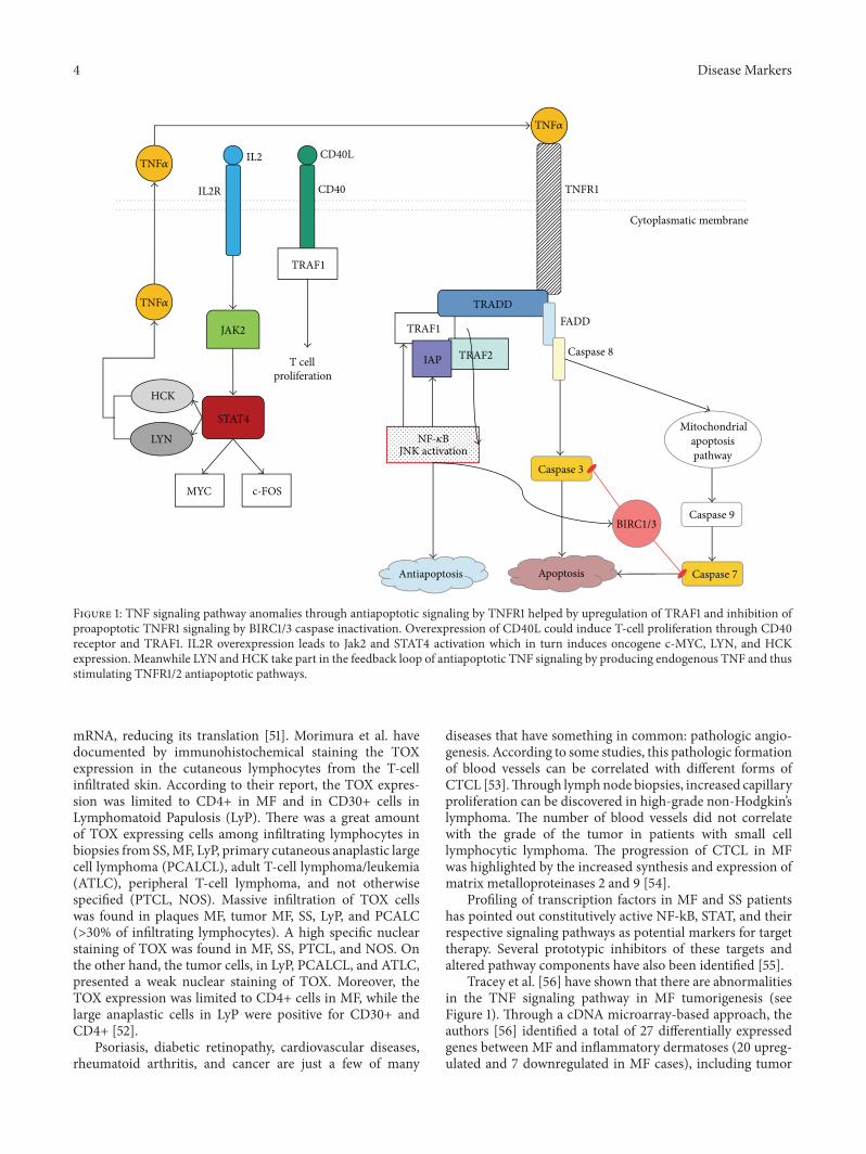
Figure 1: TNF signaling pathway anomalies through antiapoptotic signaling by TNFR1 helped by upregulation of TRAF1 and inhibition ofproapoptotic TNFR1 signaling by BIRC1/3 caspase inactivation. Overexpression of CD40L could induce T-cell proliferation through CD40receptor and TRAF1. IL2R overexpression leads to Jak2 and STAT4 activation which in turn induces oncogene c-MYC, LYN, and HCKexpression. Meanwhile LYN andHCK take part in the feedback loop of antiapoptotic TNF signaling by producing endogenous TNF and thusstimulating TNFR1/2 antiapoptotic pathways.
mRNA, reducing its translation [51]. Morimura et al. havedocumented by immunohistochemical staining the TOXexpression in the cutaneous lymphocytes from the T-cellinfiltrated skin. According to their report, the TOX expres-sion was limited to CD4+ in MF and in CD30+ cells inLymphomatoid Papulosis (LyP). There was a great amountof TOX expressing cells among infiltrating lymphocytes inbiopsies from SS,MF, LyP, primary cutaneous anaplastic largecell lymphoma (PCALCL), adult T-cell lymphoma/leukemia(ATLC), peripheral T-cell lymphoma, and not otherwisespecified (PTCL, NOS). Massive infiltration of TOX cellswas found in plaques MF, tumor MF, SS, LyP, and PCALC(>30% of infiltrating lymphocytes). A high specific nuclearstaining of TOX was found in MF, SS, PTCL, and NOS. Onthe other hand, the tumor cells, in LyP, PCALCL, and ATLC,presented a weak nuclear staining of TOX. Moreover, theTOX expression was limited to CD4+ cells in MF, while thelarge anaplastic cells in LyP were positive for CD30+ andCD4+ [52].
Psoriasis, diabetic retinopathy, cardiovascular diseases,rheumatoid arthritis, and cancer are just a few of many
diseases that have something in common: pathologic angio-genesis. According to some studies, this pathologic formationof blood vessels can be correlated with different forms ofCTCL [53].Through lymphnode biopsies, increased capillaryproliferation can be discovered in high-grade non-Hodgkin’slymphoma. The number of blood vessels did not correlatewith the grade of the tumor in patients with small celllymphocytic lymphoma. The progression of CTCL in MFwas highlighted by the increased synthesis and expression ofmatrix metalloproteinases 2 and 9 [54].
Profiling of transcription factors in MF and SS patientshas pointed out constitutively active NF-kB, STAT, and theirrespective signaling pathways as potential markers for targettherapy. Several prototypic inhibitors of these targets andaltered pathway components have also been identified [55].
Tracey et al. [56] have shown that there are abnormalitiesin the TNF signaling pathway in MF tumorigenesis (seeFigure 1). Through a cDNA microarray-based approach, theauthors [56] identified a total of 27 differentially expressedgenes between MF and inflammatory dermatoses (20 upreg-ulated and 7 downregulated in MF cases), including tumor
Disease Markers 5
necrosis factor receptor (TNFR) and other oncogenes andapoptosis inhibitors. They designed a 6-gene “signature”(FJX1, STAT4, SYNE1, TRAF1, BIRC3, and Hs.127160) pre-dictionmodel that may help to differentiate MF from inflam-matory dermatoses. The model correctly identified 97% ofcases in a blind test performed on 24 MF patients with lowclinical stages [56]. cDNA microarray and quantitative RT-PCR expression analyses of peripheral blood samples, usinga panel of genes (including STAT4, GATA-3, PLS3, CD1D,and TRAIL), have been shown to identify CTCL patients(who have at least 5% circulating tumor cells) with an overallaccuracy of 90% [57, 58].
4. Sezary Syndrome
The metastatic potential of tumor cells is dependent onangiogenesis, which creates the conditions for tumor growthand progression [59, 60]. Angiopoietins represent a familyof ligands for endothelium-specific tyrosine kinase Tie2receptor. This family of proteins consists of 4 structurallysimilar members: Ang-1, Ang-2, Ang-3, and Ang-4 of whichAng-1 and Ang-2 have been identified to have angiogeneticproperties, similar to VEGF [61–63]. Cells from MF skinlesions express Ang-2, but the serum levels of this protein areelevated only in patients with Sezary syndrome (SS), whichmay indicate that serum Ang-2 is produced by circulatingtumor cells in SS [4, 64]. Kawaguchi et al. showed in astudy published in 2014 that serum concentration of Ang-2was increased, even in patients with MF, when the diseaseprogressed, thus demonstrating that Ang-2 could be used asa disease activity marker [65].
TOX staining may also be useful in the differentialdiagnosis between SS and erythrodermic dermatitis.The firstmentioned presented more than 50% nuclear staining of theinfiltrating lymphocytes, while the last ones had a slightlydim nuclear staining of TOX (∼25%). In addition, c-MYCdid not have a significant contribution to the differentialdiagnosis [66]. Tplastin, a member of the fimbrin/plastinfamily expressed by epithelial tissues and nonhematopoieticmesenchymal cells, did not show a significantly higherexpression in SS than in the benign erythrodermic inflam-matory diseases (EID). In contrast, the detection of CD158K/KIR3DL2 (a killer immunoglobulin-like receptor usu-ally expressed by some circulating T CD8+ lymphocytes, NKcells, and recently some reports suggested that it might beexpressed by some subsets of CD4+ T cells) transcripts usingRT-PCRwas significantly overexpressed in SS, in comparisonwith EID, and may be used as a diagnosis marker even in theearly stages of SS [67].
In another study, Michel et al. have demonstrated forthe first time that the combination of five biomarkers (PL53,TWIST, CD158K, KIR3DL2, and NKp46) using PCR hasa high importance in the early diagnosis of SS [68]. Inadvanced MF/SS increased expression of Twist protein (atranscription factor which blocks p53 and inhibits c-MYCinduced apoptosis, believed to promote the progression ofsolid tumors) was found through RT-PCR detection, butfurther studies are needed in order to correlate the high
expression with the stages of these diseases [69]. NK46pbelongs to natural cytotoxicity receptor (NCR) families ofnatural killer (NK) receptors. This receptor is frequentlyexpressed by neoplastic cells in SS and it may be used as adiagnosis marker in the blood and the skin using RT-PCR[70].
5. Conclusions
Given the high prevalence of CTCL, it is imperative to deter-mine specific biomarkers in order to distinguish betweenbenign and aggressive prognostic courses. The diagnosis ofCTCL requires a more holistic approach through whichmolecular findings are to be integrated with clinical, histo-logical, and immunophenotypic data. Thus, future studiesshould be aimed at defining appropriate molecules withhigh sensitivity and specificity for the evaluation of diseasetreatment and prognosis. Moreover at-risk patients wouldbenefit from diagnostic markers in order to prevent diseaseprogression and late diagnosis, when appropriate therapiesare of little efficiency. Establishing accurate protein markerswould also be helpful for identifying target therapies.
Competing Interests
The authors declare that there are no competing interestsregarding the publication of this paper.
Authors’ Contributions
All the authors equally contributed to this work.
Acknowledgments
This paper is partly supported by Grants PN-II-PT-PCCA-2013-4-1407 (Project 190/2014) and PNII-PT-PCCA-2013-4-1386 (Project 185/2014) financed by Executive Agency forHigher Education, Research, Development and Innovation.
References
[1] M. H. Imam, P. J. Shenoy, C. R. Flowers, A. Phillips, and M. J.Lechowicz, “Incidence and survival patterns of cutaneous T-celllymphomas in theUnited States,” Leukemia and Lymphoma, vol.54, no. 4, pp. 752–759, 2013.
[2] R. Willemze, E. S. Jaffe, G. Burg et al., “WHO-EORTC classi-fication for cutaneous lymphomas,” Blood, vol. 105, no. 10, pp.3768–3785, 2005.
[3] E. C. Vonderheid, M. G. Bernengo, G. Burg et al., “Updateon erythrodermic cutaneous T-cell lymphoma: report of theInternational Society for Cutaneous Lymphomas,” Journal of theAmerican Academy of Dermatology, vol. 46, no. 1, pp. 95–106,2002.
[4] X.-S. Wu, A. S. Lonsdorf, and S. T. Hwang, “Cutaneous T-celllymphoma: roles for chemokines and chemokine receptors,”Journal of Investigative Dermatology, vol. 129, no. 5, pp. 1115–1119,2009.
6 Disease Markers
[5] M. Neagu, C. Caruntu, C. Constantin et al., “Chemicallyinduced skin carcinogenesis: updates in experimental models(review),” Oncology Reports, vol. 35, no. 5, pp. 2516–2528, 2016.
[6] L. E. Selman, T. Beynon, E. Radcliffe et al., “‘We’re all carrying aburden that we’re not sharing’: a qualitative study of the impactof cutaneous T-cell lymphoma on the family,” British Journal ofDermatology, vol. 172, no. 6, pp. 1581–1592, 2015.
[7] C. Caruntu, C. Grigore, A. Caruntu, A. Diaconeasa, and D.Boda, “The role of stress in skin diseases,” Internal Medicine-Medicina Interna, vol. 8, no. 3, pp. 73–84, 2011.
[8] V. Voiculescu, B. Calenic, M. A. Ghita et al., “From normal skinto squamous cell carcinoma—a quest for novel biomarkers,”Disease Markers, vol. 2016, Article ID 4517492, 14 pages, 2016.
[9] M. Neagu, C. Constantin, C. Tanase, and D. Boda, “Patentedbiomarker panels in early detection of cancer,” Recent Patentson Biomarkers, vol. 1, no. 1, pp. 10–24, 2011.
[10] A. Bulman, M. Neagu, and C. Constantin, “Immunomics inskin cancer—improvement in diagnosis, prognosis and therapymonitoring,”Current Proteomics, vol. 10, no. 3, pp. 202–217, 2013.
[11] M. Lupu, C. Caruntu, M. A. Ghita et al., “Gene expressionand proteome analysis as sources of biomarkers in basal cellcarcinoma,” Disease Markers, vol. 2016, Article ID 9831237, 9pages, 2016.
[12] C. Caruntu, D. Boda, G. Dumitrascu, C. Constantin, and M.Neagu, “Proteomics focusing on immune markers in psoriaticarthritis,”Biomarkers inMedicine, vol. 9, no. 6, pp. 513–528, 2015.
[13] M. Neagu, C. Constantin, G. R. Dumitrascu et al., “Inflam-mation markers in cutaneous melanoma—edgy biomarkers forprognosis,” Discoveries, vol. 3, no. 1, article e38, pp. 1–13, 2015.
[14] M. Neagu, “The immune system—a hidden treasure forbiomarker discovery in cutaneous melanoma,” Advances inClinical Chemistry, vol. 58, pp. 89–140, 2012.
[15] C.-D. Klemke and S. Goerdt, “Molecular biology and targetedtherapy of cutaneous T-cell lymphomas,” Giornale Italiano diDermatologia e Venereologia, vol. 143, no. 6, pp. 365–374, 2008.
[16] F. Lansigan, J. Choi, and F. M. Foss, “Cutaneous T-cell lym-phoma,”Hematology/Oncology Clinics of North America, vol. 22,no. 5, pp. 979–996, 2008.
[17] C.-D. Klemke, S. Goerdt, D. Schrama, and J. C. Becker, “Newinsights into the molecular biology and targeted therapy ofcutaneous T-cell lymphomas,” Journal of the German Society ofDermatology, vol. 4, no. 5, pp. 395–406, 2006.
[18] E. C. Vonderheid, Q. Zhang, S. R. Lessin et al., “Use of serumsoluble interleukin-2 receptor levels to monitor the progres-sion of cutaneous T-cell lymphoma,” Journal of the AmericanAcademy of Dermatology, vol. 38, no. 2I, pp. 207–220, 1998.
[19] J. C. Hassel, R. Meier, H. Joller-Jemelka, G. Burg, and R.Dummer, “Serological immunomarkers in cutaneous T celllymphoma,” Dermatology, vol. 209, no. 4, pp. 296–300, 2004.
[20] N. Escher, M. Kaatz, C. Melle et al., “Posttranslational modi-fications of transthyretin are serum markers in patients withmycosis fungoides,” Neoplasia, vol. 9, no. 3, pp. 254–259, 2007.
[21] J. Shin, S. Monti, D. J. Aires et al., “Lesional gene expressionprofiling in cutaneous T-cell lymphoma reveals natural clustersassociatedwith disease outcome,”Blood, vol. 110, no. 8, pp. 3015–3027, 2007.
[22] S. Chattopadhyay, N. G. Chakraborty, and B. Mukherji, “Reg-ulatory T cells and tumor immunity,” Cancer Immunology,Immunotherapy, vol. 54, no. 12, pp. 1153–1161, 2005.
[23] S. Sakaguchi, N. Sakaguchi, M. Asano, M. Itoh, and M. Toda,“Immunologic self-tolerance maintained by activated T cellsexpressing IL-2 receptor alpha-chains (CD25). Breakdown of asingle mechanism of self-tolerance causes various autoimmunediseases,” The Journal of Immunology, vol. 155, no. 3, pp. 1151–1164, 1995.
[24] M. Ono, H. Yaguchi, N. Ohkura et al., “Foxp3 controls regula-tory T-cell function by interacting with AML1/Runx1,” Nature,vol. 446, no. 7136, pp. 685–689, 2007.
[25] R. Setoguchi, S. Hori, T. Takahashi, and S. Sakaguchi, “Homeo-staticmaintenance of natural Foxp3+ CD25+ CD4+ regulatory Tcells by interleukin (IL)-2 and induction of autoimmune diseaseby IL-2 neutralization,” The Journal of Experimental Medicine,vol. 201, no. 5, pp. 723–735, 2005.
[26] L.M.Williams and A. Y. Rudensky, “Maintenance of the Foxp3-dependent developmental program inmature regulatory T cellsrequires continued expression of Foxp3,” Nature Immunology,vol. 8, no. 3, pp. 277–284, 2007.
[27] P. L. Vieira, J. R. Christensen, S. Minaee et al., “IL-10-secretingregulatory T cells do not express Foxp3 but have comparableregulatory function to naturally occurring CD4+CD25+ regula-tory T cells,” The Journal of Immunology, vol. 172, no. 10, pp.5986–5993, 2004.
[28] M. Forgber, S. Gellrich, T. Sharav, W. Sterry, and P. Walden,“Proteome-based analysis of serologically defined tumor-associated antigens in cutaneous lymphoma,” PLoS ONE, vol.4, no. 12, Article ID e8376, 2009.
[29] H. Zhang, C. Peng, Y. Hu et al., “The Blk pathway functionsas a tumor suppressor in chronic myeloid leukemia stem cells,”Nature Genetics, vol. 44, no. 8, pp. 861–871, 2012.
[30] T. Krejsgaard, C. S. Vetter-Kauczok, A. Woetmann et al.,“Ectopic expression of B-lymphoid kinase in cutaneous T-celllymphoma,” Blood, vol. 113, no. 23, pp. 5896–5904, 2009.
[31] D. L. Petersen, T. Krejsgaard, J. Berthelsen et al., “B-lymphoidtyrosine kinase (Blk) is an oncogene and a potential target fortherapy with dasatinib in cutaneous T-cell lymphoma (CTCL),”Leukemia, vol. 28, no. 10, pp. 2109–2112, 2014.
[32] T. Miyagaki, M. Sugaya, H. Suga et al., “Serum soluble CD26levels: diagnostic efficiency for atopic dermatitis, cutaneousT-cell lymphoma and psoriasis in combination with serumthymus and activation-regulated chemokine levels,” Journal ofthe European Academy of Dermatology and Venereology, vol. 27,no. 1, pp. 19–24, 2013.
[33] D. M. Pierson, D. Jones, T. Muzzafar et al., “Utility of CD26in flow cytometric immunophenotyping of T-cell lymphomasin tissue and body fluid specimens,” Cytometry Part B: ClinicalCytometry, vol. 74, no. 6, pp. 341–348, 2008.
[34] T. Krejsgaard, C. S. Vetter-Kauczok, A. Woetmann et al.,“Jak3- and JNK-dependent vascular endothelial growth factorexpression in cutaneous T-cell lymphoma,” Leukemia, vol. 20,no. 10, pp. 1759–1766, 2006.
[35] E. C. Vonderheid, L. W. Diamond, S.-M. Lai, F. Au, andM. A. Dellavecchia, “Lymph node histopathologic findings incutaneous T-cell lymphoma. A prognostic classification systembased onmorphologic assessment,”American Journal of ClinicalPathology, vol. 97, no. 1, pp. 121–129, 1992.
[36] E. A. Fraser-Andrews, A. J. Woolford, R. Russell-Jones, P. T.Seed, and S. J. Whittaker, “Detection of a peripheral bloodT cell clone is an independent prognostic marker in mycosisfungoides,” Journal of Investigative Dermatology, vol. 114, no. 1,pp. 117–121, 2000.
Disease Markers 7
[37] J. M. Muche, A. Lukowsky, K. Asadullah, S. Gellrich, and W.Sterry, “Demonstration of frequent occurrence of clonal T cellsin the peripheral blood of patients with primary cutaneous T-cell lymphoma,” Blood, vol. 90, no. 4, pp. 1636–1642, 1997.
[38] H. S. Zackheim and T. H. McCalmont, “Mycosis fungoides: thegreat imitator,” Journal of the American Academy of Dermatol-ogy, vol. 47, no. 6, pp. 914–918, 2002.
[39] E.W.Cowen, C.-W. Liu, S.M. Steinberg et al., “Differentiation oftumour-stage mycosis fungoides, psoriasis vulgaris and normalcontrols in a pilot study using serumproteomic analysis,”BritishJournal of Dermatology, vol. 157, no. 5, pp. 946–953, 2007.
[40] S. A. Moshkovskii, E. E. Sokolova, E. V. Brattseva et al.,“Proteome and cytokine serum profiling to diagnose a mycosisfungoides,” Proteomics—Clinical Applications, vol. 5, no. 7-8, pp.432–439, 2011.
[41] J. A. Hanley and B. J. McNeil, “The meaning and use of thearea under a receiver operating characteristic (ROC) curve,”Radiology, vol. 143, no. 1, pp. 29–36, 1982.
[42] M. H. Zweig and G. Campbell, “Receiver-operating charac-teristic (ROC) plots: a fundamental evaluation tool in clinicalmedicine,” Clinical Chemistry, vol. 39, no. 4, pp. 561–577, 1993.
[43] P. Becuwe, M. Ennen, R. Klotz, C. Barbieux, and S. Grande-mange, “Manganese superoxide dismutase in breast cancer:from molecular mechanisms of gene regulation to biologicaland clinical significance,” Free Radical Biology and Medicine,vol. 77, pp. 139–151, 2014.
[44] L. W. Oberley, “Mechanism of the tumor suppressive effect ofMnSOD overexpression,” Biomedicine and Pharmacotherapy,vol. 59, no. 4, pp. 143–148, 2005.
[45] J. Liu, Y. Zeng, Y. Zhou, D. Ma, and B. Wang, “Proteomicexpression analysis of mycosis fungoides (MF) skin tissues:overexpressions of SOD2, S100A8 and FABP5 in MF,” Journalof Dermatological Science, vol. 60, no. 1, pp. 42–44, 2010.
[46] C. I. van de Wetering, M. C. Coleman, D. R. Spitz, B. J. Smith,and C. M. Knudson, “Manganese superoxide dismutase genedosage affects chromosomal instability and tumor onset in amouse model of T cell lymphoma,” Free Radical Biology andMedicine, vol. 44, no. 8, pp. 1677–1686, 2008.
[47] G. H. W. Wong, T. McHugh, R. Weber, and D. V. Goed-del, “Tumor necrosis factor 𝛼 selectively sensitizes humanimmunodeficiency virus-infected cells to heat and radiation,”Proceedings of the National Academy of Sciences of the UnitedStates of America, vol. 88, no. 10, pp. 4372–4376, 1991.
[48] R. Donato, “Intracellular and extracellular roles of S100 pro-teins,” Microscopy Research and Technique, vol. 60, no. 6, pp.540–551, 2003.
[49] A.-M. Broome, D. Ryan, and R. L. Eckert, “S100 protein sub-cellular localization during epidermal differentiation and pso-riasis,” Journal of Histochemistry & Cytochemistry, vol. 51, no. 5,pp. 675–685, 2003.
[50] C. Hohoff, T. Borchers, B. Rustow, F. Spener, and H. VanTilbeurgh, “Expression, purification, and crystal structuredetermination of recombinant human epidermal-type fatty acidbinding protein,” Biochemistry, vol. 38, no. 38, pp. 12229–12239,1999.
[51] L. Y. McGirt, C. M. Adams, D. A. Baerenwald, J. P. Zwerner, J.A. Zic, and C. M. Eischen, “MiR-223 regulates cell growth andtargets proto-oncogenes inmycosis fungoides/cutaneousT-Celllymphoma,” Journal of Investigative Dermatology, vol. 134, no. 4,pp. 1101–1107, 2014.
[52] S. Morimura, M. Sugaya, H. Suga et al., “TOX expressionin different subtypes of cutaneous lymphoma,” Archives ofDermatological Research, vol. 306, no. 9, pp. 843–849, 2014.
[53] G. Mazur, E. Jaskuła, I. Kryczek et al., “Proinflammatorychemokine gene expression influences survival of patients withnon-Hodgkin’s lymphoma,” FoliaHistochemica et Cytobiologica,vol. 49, no. 2, pp. 240–247, 2011.
[54] B. Ridell and K. Norrby, “Intratumoral microvascular density inmalignant lymphomas of B-cell origin,” APMIS, vol. 109, no. 1,pp. 66–72, 2001.
[55] U. Dobbeling, “Transcription factor profiling shows new waystowards new treatment options of cutaneous T cell lymphomas,”Current Drug Discovery Technologies, vol. 4, no. 1, pp. 24–30,2007.
[56] L. Tracey, R. Villuendas, A. M. Dotor et al., “Mycosis fungoidesshows concurrent deregulation ofmultiple genes involved in theTNF signaling pathway: an expression profile study,” Blood, vol.102, no. 3, pp. 1042–1050, 2003.
[57] L. Kari, A. Loboda, M. Nebozhyn et al., “Classification andprediction of survival in patients with the leukemic phaseof cutaneous T cell lymphoma,” The Journal of ExperimentalMedicine, vol. 197, no. 11, pp. 1477–1488, 2003.
[58] M. Nebozhyn, A. Loboda, L. Kari et al., “Quantitative PCRon 5 genes reliably identifies CTCL patients with 5% to 99%circulating tumor cells with 90% accuracy,” Blood, vol. 107, no.8, pp. 3189–3196, 2006.
[59] J. Folkman andM. Klagsbrun, “Angiogenic factors,” Science, vol.235, no. 4787, pp. 442–447, 1987.
[60] J. Folkman, “Angiogenesis in cancer, vascular, rheumatoid andother disease,” Nature Medicine, vol. 1, no. 1, pp. 27–31, 1995.
[61] T. Asahara, D. Chen, T. Takahashi et al., “Tie2 receptor ligands,angiopoietin-1 and angiopoietin-2, modulate VEGF- inducedpostnatal neovascularization,” Circulation Research, vol. 83, no.3, pp. 233–240, 1998.
[62] G. D. Yancopoulos, S. Davis, N. W. Gale, J. S. Rudge, S. J.Wiegand, and J. Holash, “Vascular-specific growth factors andblood vessel formation,”Nature, vol. 407, no. 6801, pp. 242–248,2000.
[63] G. Thurston, “Complementary actions of VEGF and Angio-poietin-1 on blood vessel growth and leakage,” Journal ofAnatomy, vol. 200, no. 6, pp. 575–580, 2002.
[64] M. Sugaya, “Chemokines and cutaneous lymphoma,” Journal ofDermatological Science, vol. 59, no. 2, pp. 81–85, 2010.
[65] M. Kawaguchi, M. Sugaya, H. Suga et al., “Serum levelsof angiopoietin-2, but not angiopoietin-1, are elevated inpatients with erythrodermic cutaneous T-cell lymphoma,” ActaDermato-Venereologica, vol. 94, no. 1, pp. 9–13, 2014.
[66] S. E. Boonk, F. Cetinozman,M.H. Vermeer, P.M. Jansen, and R.Willemze, “Differential expression of TOX by skin-infiltratingT cells in Sezary syndrome and erythrodermic dermatitis,”Journal of Cutaneous Pathology, vol. 42, no. 9, pp. 604–609, 2015.
[67] N. Ortonne, S. Le Gouvello, H. Mansour et al., “CD158K/KIR3DL2 transcript detection in lesional skin of patients witherythroderma is a tool for the diagnosis of Sezary syndrome,”Journal of Investigative Dermatology, vol. 128, no. 2, pp. 465–472,2008.
[68] L. Michel, F. Jean-Louis, E. Begue, A. Bensussan, and M.Bagot, “Use of PLS3, Twist, CD158k/KIR3DL2, and NKp46gene expression combination for reliable Sezary syndromediagnosis,” Blood, vol. 121, no. 8, pp. 1477–1478, 2013.

8 Disease Markers
[69] M. Goswami, M. Duvic, A. Dougherty, and X. Ni, “IncreasedTwist expression in advanced stage of mycosis fungoides andSezary syndrome,” Journal of Cutaneous Pathology, vol. 39, no.5, pp. 500–507, 2012.
[70] N. Ortonne, S. Le Gouvello, R. Tabak et al., “CD158k/KIR3DL2and NKp46 are frequently expressed in transformed mycosisfungoides,” Experimental Dermatology, vol. 21, no. 6, pp. 461–463, 2012.

Submit your manuscripts athttp://www.hindawi.com
Stem CellsInternational
Hindawi Publishing Corporationhttp://www.hindawi.com Volume 2014
Hindawi Publishing Corporationhttp://www.hindawi.com Volume 2014
MEDIATORSINFLAMMATION
of
Hindawi Publishing Corporationhttp://www.hindawi.com Volume 2014
Behavioural Neurology
EndocrinologyInternational Journal of
Hindawi Publishing Corporationhttp://www.hindawi.com Volume 2014
Hindawi Publishing Corporationhttp://www.hindawi.com Volume 2014
Disease Markers
Hindawi Publishing Corporationhttp://www.hindawi.com Volume 2014
BioMed Research International
OncologyJournal of
Hindawi Publishing Corporationhttp://www.hindawi.com Volume 2014
Hindawi Publishing Corporationhttp://www.hindawi.com Volume 2014
Oxidative Medicine and Cellular Longevity
Hindawi Publishing Corporationhttp://www.hindawi.com Volume 2014
PPAR Research
The Scientific World JournalHindawi Publishing Corporation http://www.hindawi.com Volume 2014
Immunology ResearchHindawi Publishing Corporationhttp://www.hindawi.com Volume 2014
Journal of
ObesityJournal of
Hindawi Publishing Corporationhttp://www.hindawi.com Volume 2014
Hindawi Publishing Corporationhttp://www.hindawi.com Volume 2014
Computational and Mathematical Methods in Medicine
OphthalmologyJournal of
Hindawi Publishing Corporationhttp://www.hindawi.com Volume 2014
Diabetes ResearchJournal of
Hindawi Publishing Corporationhttp://www.hindawi.com Volume 2014
Hindawi Publishing Corporationhttp://www.hindawi.com Volume 2014
Research and TreatmentAIDS
Hindawi Publishing Corporationhttp://www.hindawi.com Volume 2014
Gastroenterology Research and Practice
Hindawi Publishing Corporationhttp://www.hindawi.com Volume 2014
Parkinson’s Disease
Evidence-Based Complementary and Alternative Medicine
Volume 2014Hindawi Publishing Corporationhttp://www.hindawi.com