
Review Article - Hindawi Publishing...
7
Hindawi Publishing Corporation Journal of Thyroid Research Volume 2012, Article ID 736161, 6 pages doi:10.1155/2012/736161 Review Article The Evolving Role of Selenium in the Treatment of Graves’ Disease and Ophthalmopathy Leonidas H. Duntas Endocrine Unit, Evgenidion Hospital, University of Athens, 20 Papadiamantopoulou Street, 11528 Athens, Greece Correspondence should be addressed to Leonidas H. Duntas, [email protected] Received 5 September 2011; Accepted 17 October 2011 Academic Editor: Juan Carlos Galofr´ e Copyright © 2012 Leonidas H. Duntas. This is an open access article distributed under the Creative Commons Attribution License, which permits unrestricted use, distribution, and reproduction in any medium, provided the original work is properly cited. Graves’ disease (GD) and ophthalmopathy (GO) are organ-specific autoimmune-inflammatory disorders characterized by a complex pathogenesis. The inflammatory process is dominated by an imbalance of the antioxidant-oxidant mechanism, increased production of radical oxygen species (ROS), and cytokines which sustain the autoimmune process and perpetuate the disease. Recently, selenium, which is a powerful antioxidant, has been successfully applied in patients with mild GO, slowing the progression of disease, decreasing the clinical activity score, and appreciably improving the quality of life. The mechanisms of selenium action are variable. The aim of this review is to summarize the actions of selenium in GD and GO. Selenium as selenocysteine is incorporated in selenoproteins, such as glutathione peroxidase which catalyzes the degradation of hydrogen peroxide and lipid hydroperoxide that are increasingly produced in hyperthyroidism. Moreover, selenium decreases the formation of proinflammatory cytokines, while it contributes, in synergy with antithyroid drugs, to stabilization of the autoimmune process in GD and alleviation of GO. It is now to be clarified whether enforced nutritional supplementation has the same results and whether prolonging selenium administration may have an impact on the prevention of disease. 1. Introduction Observed and briefly described, though not published, by Parry in the late 1700s, Graves’ disease (GD) was definitively identified and documented by Robert Graves in 1835 and classically described by von Basedow in 1840 [1–3]. GD is an autoimmune disease characterized by the activation of autoantibodies against the TSH receptor (TRAB), leading to excessive thyroid hormone production [4]. GD manifests, interalia, via thyrotoxicosis and extrathyroid involvement often entailing orbitopathy (GO) and, rarely, dermopathy (pretibial myxedema) and acropathy. Moreover, the TRAB, by stimulating cyclic adenosine monophosphate (AMP), cause proliferation and hyperplasia of the thyroid follicular cells resulting in enlargement of the gland, frequently the first sign of the disease, the swelling ranging from slight to marked [5]. Clinically, the thyroid is firm in consistency and tender in patients with a greatly enlarged goiter, while palpation lobulations are also commonly detected which can be mistaken for nodules. No single gene has been pinpointed as causing GD, a disease which is most prevalent in women between the ages of 20 and 50 years. However, it has been associated with certain MHC Class II HLA alleles depending on the racial group, for example, HLA-DR3 in whites [4]. An association of GD with polymorphisms of the cytotoxic T-lymphocyte antigen 4 (CTLA-4) gene has also been established, suggest- ing a functional role of CTLA-4 in autoreactive T cells [4, 5]. A combination of genetic and environmental factors is responsible for the initiation of autoimmunity. Interactions between genetic and environmental factors are underscored by the existing associations linking age at diagnosis, goiter, disease severity, smoking, and family history [6]. In addi- tion, iodine repletion in iodine-deficient areas is usually accompanied by an increased incidence of GD due to the Jod-Basedow phenomenon. Stress is also thought to be a significant factor precipitating GD in susceptible individuals [7], while smoking is well established as being linked to GO but not to GD [8]. Treatment modalities of GD consist of administration of antithyroid drugs, radioiodine therapy, or surgery. Radioio- dine therapy, is favored only in USA, whereas antithyroid drugs, including methimazole, carbimazole, and propylth- iouracil, comprise first choice treatment in the rest of the
Transcript of Review Article - Hindawi Publishing...
Hindawi Publishing CorporationJournal of Thyroid ResearchVolume 2012, Article ID 736161, 6 pagesdoi:10.1155/2012/736161
Review Article
The Evolving Role of Selenium in the Treatment ofGraves’ Disease and Ophthalmopathy
Leonidas H. Duntas
Endocrine Unit, Evgenidion Hospital, University of Athens, 20 Papadiamantopoulou Street, 11528 Athens, Greece
Correspondence should be addressed to Leonidas H. Duntas, [email protected]
Received 5 September 2011; Accepted 17 October 2011
Academic Editor: Juan Carlos Galofre
Copyright © 2012 Leonidas H. Duntas. This is an open access article distributed under the Creative Commons Attribution License,which permits unrestricted use, distribution, and reproduction in any medium, provided the original work is properly cited.
Graves’ disease (GD) and ophthalmopathy (GO) are organ-specific autoimmune-inflammatory disorders characterized by acomplex pathogenesis. The inflammatory process is dominated by an imbalance of the antioxidant-oxidant mechanism, increasedproduction of radical oxygen species (ROS), and cytokines which sustain the autoimmune process and perpetuate the disease.Recently, selenium, which is a powerful antioxidant, has been successfully applied in patients with mild GO, slowing theprogression of disease, decreasing the clinical activity score, and appreciably improving the quality of life. The mechanismsof selenium action are variable. The aim of this review is to summarize the actions of selenium in GD and GO. Selenium asselenocysteine is incorporated in selenoproteins, such as glutathione peroxidase which catalyzes the degradation of hydrogenperoxide and lipid hydroperoxide that are increasingly produced in hyperthyroidism. Moreover, selenium decreases the formationof proinflammatory cytokines, while it contributes, in synergy with antithyroid drugs, to stabilization of the autoimmune processin GD and alleviation of GO. It is now to be clarified whether enforced nutritional supplementation has the same results andwhether prolonging selenium administration may have an impact on the prevention of disease.
1. Introduction
Observed and briefly described, though not published, byParry in the late 1700s, Graves’ disease (GD) was definitivelyidentified and documented by Robert Graves in 1835 andclassically described by von Basedow in 1840 [1–3]. GD isan autoimmune disease characterized by the activation ofautoantibodies against the TSH receptor (TRAB), leading toexcessive thyroid hormone production [4]. GD manifests,interalia, via thyrotoxicosis and extrathyroid involvementoften entailing orbitopathy (GO) and, rarely, dermopathy(pretibial myxedema) and acropathy. Moreover, the TRAB,by stimulating cyclic adenosine monophosphate (AMP),cause proliferation and hyperplasia of the thyroid follicularcells resulting in enlargement of the gland, frequently thefirst sign of the disease, the swelling ranging from slightto marked [5]. Clinically, the thyroid is firm in consistencyand tender in patients with a greatly enlarged goiter, whilepalpation lobulations are also commonly detected which canbe mistaken for nodules.
No single gene has been pinpointed as causing GD, adisease which is most prevalent in women between the ages
of 20 and 50 years. However, it has been associated withcertain MHC Class II HLA alleles depending on the racialgroup, for example, HLA-DR3 in whites [4]. An associationof GD with polymorphisms of the cytotoxic T-lymphocyteantigen 4 (CTLA-4) gene has also been established, suggest-ing a functional role of CTLA-4 in autoreactive T cells [4, 5].
A combination of genetic and environmental factors isresponsible for the initiation of autoimmunity. Interactionsbetween genetic and environmental factors are underscoredby the existing associations linking age at diagnosis, goiter,disease severity, smoking, and family history [6]. In addi-tion, iodine repletion in iodine-deficient areas is usuallyaccompanied by an increased incidence of GD due to theJod-Basedow phenomenon. Stress is also thought to be asignificant factor precipitating GD in susceptible individuals[7], while smoking is well established as being linked to GObut not to GD [8].
Treatment modalities of GD consist of administration ofantithyroid drugs, radioiodine therapy, or surgery. Radioio-dine therapy, is favored only in USA, whereas antithyroiddrugs, including methimazole, carbimazole, and propylth-iouracil, comprise first choice treatment in the rest of the

2 Journal of Thyroid Research
world. Nevertheless, according to a recent study examiningthe frequency of antithyroid drug prescription in USA,methimazole (MMI) has lately become the most frequentlyprescribed antithyroid drug, indicating a clear shift towardspharmacological treatment as the primary treatment optionin GD [9]. Treatment should be planned for a period of atleast 12 months, and patients are usually becoming euthyroidwithin this timeframe; nevertheless, the duration of theremission period is unpredictable, since the disease is markedby cycles of remission and relapse of variable duration [4].
Recently, evidence has emerged indicating that seleniumadministration could be effective and safe in patients withGD and with mild forms of GO [10].
The aim of this paper is to briefly evaluate the currentknowledge concerning the pathogenesis of GD and GO anddiscuss the evolving role of selenium within the context ofits potential as a therapeutic means of intervention in thesedisorders.
2. Pathogenesis of GD and GO
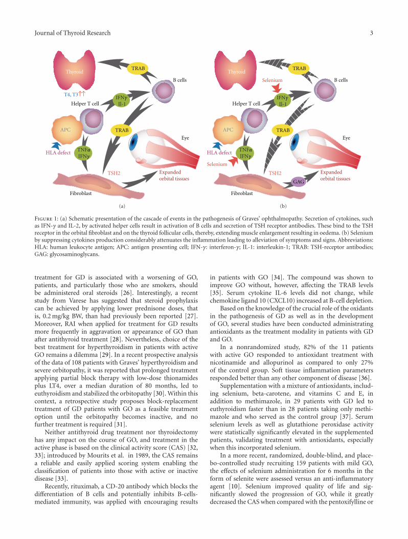
Hyperthyroidism is caused by the binding of TSH-stimu-lating antibodies to the TSH receptor, a G-protein-coupledreceptor. However, the first step in this process is consideredto be precipitation by environmental factors of an HLA-related organ-specific defect in suppressor T-lymphocytefunction [5]. This leads to decreased suppression of thyroid-directed helper T-lymphocytes which, in the presence ofdendritic cells and macrophages, produce the cytokines γ-interferon (IFNγ) and interleukin-1 (IL-1), subsequentlydifferentiating B cells to plasma cells and generating TRAB.Concomitantly, IFNγ enhances the expression of HLA-DRantigens on the surface of thyroid cells (Figure 1). Thus, IFNγmodulates the autoimmune process and, by stimulatingchemokine production by thyroid follicular cells, contributesto the maintenance of the autoimmune process [10]. Thecontribution of dendritic cells and B cells is apparentlycrucial for the initiation of disease since they express thecostimulatory molecules, CD80 and CD86, that are keytriggers for the reaction of T lymphocytes to thyroid cellpresenting antigens [4]. TRAB stimulate the TSHR on thethyroid follicular cells, resulting in increased thyroid hor-mone production, which may further reduce the number andfunction of suppressor T lymphocytes and stimulate helper Tlymphocyte, thus, perpetuating the cyclicity of disease [4, 5].
GO is a complex autoimmune disease. Whereas thecycle of GD consists of two components, immunologicaland hormonal, that perpetuate the process, the progressionof GD to GO, and rarely to dermopathy, is likely to bea positive feedback cycle composed of three interrelatedcomponents: mechanical, immunological, and cellular [5].Comprehensive reviews on the pathophysiology of GOhave recently been published [11–14]. Briefly, the loss oftolerance of T cells to the TSHR, via as yet unknownmechanisms, ignites the autoimmune process. The TSHRis internalized and presented by antigen-presenting cells tohelper T cells. Subsequently, the TRAB, which are secretedby activated B cells, recognize the TSHR on the fibroblasts
of the orbita, where they initiate the ocular changes [12,13]. The fibroblasts have been recognized as target cells inGO. Orbital fibroblasts stimulated by IFNγ, tumor necrosisfactor-α (TNF-α), growth factors and oxygen reactive species(ROS), secrete hyaluronic acid, and prostaglandin E2, knownmediators of inflammation, while a subgroup may differ-entiate into mature adipocytes presenting TSHR [13, 14].The subsequent proliferation of adipocytes and fibroblastsresults in increased synthesis of glycosaminoglycans (GAG),which causes edema of orbital structures, extraocular muscleenlargement, and adipose tissue expansion; these events areconstituting the signs of disease [15].
Concerning the recent enquiry as to whether autoimmu-nity against IGF-1R is primarily involved in the pathogenesisof GO, it is likely that it is not specific but instead constitutesa secondary reaction of the autoimmune process [16]
The mechanisms promoting oxidative stress have alsobeen implicated in the pathogenesis of GO. Hyperthyroidismincreases oxidants and decreases antioxidants leading tooxidative stress, this process is dominated by the productionof ROS which have long been recognized as intermediatesof various essential biological redox reactions [17, 18]. Theadverse effects induced by ROS have been suggested as beingpartly responsible for the tissue injury. Mitochondria area major source of superoxide anion (O2
−) and hydrogenperoxides (H2O2), while a number of intracellular enzymes,xanthine oxidase being the best known, are involved inoxidation reactions in which molecular oxygen (O2) isreduced to O2
− [19].Ongoing autoimmunity may contribute to increased
oxidative stress even in euthyroid GD patients, while patientswho have relapsed present increased markers of oxidativestress [20]. Moreover, the content of 8-hydroxy 2′-deoxy-guanosine (8-OHdG), an important biomarker of oxidativeDNA damage, was found significantly higher in orbitalfibroblasts together with O2
− and H2O2, underscoring themajor role that ROS play in the pathogenesis of GO [21].
Recently, increased 11β-hydroxysteroid dehydrogenase(11β-HSD1) expression, induced by cytokines, was describedin orbital adipose cells, a condition leading to elevated localgeneration of cortisol by 11β-HSD1, which may suppresscytokine synthesis and resolve the inflammation [22]. 11β-HSD1 activates cortisone to cortisol in peripheral andvisceral adipose tissues. According to the authors, sincefailure to produce adequate levels of local glucocorticoids inthe orbita may signify persistence of the disease, 11β-HSD1could provide a new therapeutic target of disease [22].
3. Presentation and TreatmentNovelties of GD and GO
TRAB levels in serum are pathognomonic for GD, predictingthe course of disease and response to antithyroid treatment;they do not, on the other hand, predict the development ofGO [23]. In conjunction with the high levels of TRAB, therisk of relapse is related to young age, male gender, and largegoiter [24]. Tobacco smoking has been consistently linkedto development or deterioration of GO [8, 25]. Since RAI
Journal of Thyroid Research 3
Helper T cell
HLA defect
TSH2
Fibroblast
Eye
Thyroid
Expandedorbital tissues
B cells
IFNγIl-1
TRAB
TNFαIFNγ
TRABAPC
T4, T3
(a)
Helper T cell
HLA defect
Selenium
Selenium
Fibroblast
Eye
Thyroid
Expandedorbital tissues
B cells
APC
TSH2
TRAB
TRAB
GAG
IFNγIl-1
TNFαIFNγ
(b)
Figure 1: (a) Schematic presentation of the cascade of events in the pathogenesis of Graves’ ophthalmopathy. Secretion of cytokines, suchas IFN-γ and IL-2, by activated helper cells result in activation of B cells and secretion of TSH receptor antibodies. These bind to the TSHreceptor in the orbital fibroblast and on the thyroid follicular cells, thereby, extending muscle enlargement resulting in oedema. (b) Seleniumby suppressing cytokines production considerably attenuates the inflammation leading to alleviation of symptoms and signs. Abbreviations:HLA: human leukocyte antigen; APC: antigen presenting cell; IFN-γ: interferon-γ; IL-1: interleukin-1; TRAB: TSH-receptor antibodies;GAG: glycosaminoglycans.
treatment for GD is associated with a worsening of GO,patients, and particularly those who are smokers, shouldbe administered oral steroids [26]. Interestingly, a recentstudy from Varese has suggested that steroid prophylaxiscan be achieved by applying lower prednisone doses, thatis, 0.2 mg/kg BW, than had previously been reported [27].Moreover, RAI when applied for treatment for GD resultsmore frequently in aggravation or appearance of GO thanafter antithyroid treatment [28]. Nevertheless, choice of thebest treatment for hyperthyroidism in patients with activeGO remains a dilemma [29]. In a recent prospective analysisof the data of 108 patients with Graves’ hyperthyroidism andsevere orbitopathy, it was reported that prolonged treatmentapplying partial block therapy with low-dose thionamidesplus LT4, over a median duration of 80 months, led toeuthyroidism and stabilized the orbitopathy [30]. Within thiscontext, a retrospective study proposes block-replacementtreatment of GD patients with GO as a feasible treatmentoption until the orbitopathy becomes inactive, and nofurther treatment is required [31].
Neither antithyroid drug treatment nor thyroidectomyhas any impact on the course of GO, and treatment in theactive phase is based on the clinical activity score (CAS) [32,33]; introduced by Mourits et al. in 1989, the CAS remainsa reliable and easily applied scoring system enabling theclassification of patients into those with active or inactivedisease [33].
Recently, rituximab, a CD-20 antibody which blocks thedifferentiation of B cells and potentially inhibits B-cells-mediated immunity, was applied with encouraging results
in patients with GO [34]. The compound was shown toimprove GO without, however, affecting the TRAB levels[35]. Serum cytokine IL-6 levels did not change, whilechemokine ligand 10 (CXCL10) increased at B-cell depletion.
Based on the knowledge of the crucial role of the oxidantsin the pathogenesis of GD as well as in the developmentof GO, several studies have been conducted administratingantioxidants as the treatment modality in patients with GDand GO.
In a nonrandomized study, 82% of the 11 patientswith active GO responded to antioxidant treatment withnicotinamide and allopurinol as compared to only 27%of the control group. Soft tissue inflammation parametersresponded better than any other component of disease [36].
Supplementation with a mixture of antioxidants, includ-ing selenium, beta-carotene, and vitamins C and E, inaddition to methimazole, in 29 patients with GD led toeuthyroidism faster than in 28 patients taking only methi-mazole and who served as the control group [37]. Serumselenium levels as well as glutathione peroxidase activitywere statistically significantly elevated in the supplementedpatients, validating treatment with antioxidants, especiallywhen this incorporated selenium.
In a more recent, randomized, double-blind, and place-bo-controlled study recruiting 159 patients with mild GO,the effects of selenium administration for 6 months in theform of selenite were assessed versus an anti-inflammatoryagent [10]. Selenium improved quality of life and sig-nificantly slowed the progression of GO, while it greatlydecreased the CAS when compared with the pentoxifylline or
4 Journal of Thyroid Research
placebo group. A 6-month followup confirmed the results ofthe 6-month treatment. The authors hypothesized a reversalof the disturbed antioxidant-oxidant balance in GD and GOalthough the exact mechanisms of selenium action are notelucidated.
In another study assessing the selenium levels in patientsgoing into remission (n = 24) and relapses (n = 59),no statistically significant differences were detected betweenthe two groups. However, patients in remission of GD hadthe highest (>120 μg/L) serum selenium levels, while it isof interest that TRAB levels and selenium were negativelycorrelated [38].
4. Mechanisms of SeleniumAction in GD and GO
Selenium is vital for a wide range of biological processes;hence, the state of “selenostasis” is essential for wellbeing andhuman health [39]. The many biological and clinical benefitsconferred by selenium are achieved by virtue of its remark-able antioxidative effects mediated mainly by the selenopro-teins GPx and TRx reductase. TRx is a stress- and iodine-induced protein, possessing strong redox activities, and it hasbeen postulated that it may be implicated in the regulationof T3 production in GD. It has been reported highlyproduced in GD and expressed in the thyroid follicular cells.Nevertheless, its precise role, though of considerable interestdue to its characteristics, remains as yet unraveled [40].
The hypermetabolic state in acute GD, the intracellularATP, and increased oxygen consumption lead to mitochon-dria dysfunction, which generates ROS and disrupts theoxidant and antioxidant balance, thereby, causing oxidativestress and tissue injury [41]. By activating GPxs, seleniumignites the “second line” of antioxidant defense, behindthe enzymatic “first line” defense system composed ofthe superoxide dismutase (SOD) and catalase (CAT) [42].Thus, SOD and CAT synthesize an efficient antioxidativemechanism capable of neutralizing the biologic effects of freeradicals; when this mechanism is saturated, the “second line,”regulated by selenium availability, is activated. Experimentalstudies in hyperthyroidism have documented an enhancedactivity of the TRx and GPx systems, stimulated by thecalcium phosphatidylinositol cascade which is usually acti-vated in hyperthyroidism, as well as increased levels of SODand of glutathione in erythrocytes [43, 44]. These findingsprovide evidence of an upregulation of the antioxidativeand protective systems in acute GD, depending, however,on the duration and severity of the disease; these system(s)might become saturated, following which supplementationor nutritional intervention is required.
The induced oxidative stress enflames lipid peroxidationand activates various inflammatory pathways. ROS maystimulate the NF-κB pathway, a cornerstone of immuneand inflammatory response, which has been associated withincreased production of TNF-α and IL-6 cytokines [45].Selenium inhibits NF-κB from binding to its gene promotersand consequently diminishes cytokine production and atten-uates the inflammation; by contrast, selenium is likely not to
interfere with the translocation of NF-κB and its subunits tothe nucleus [46]. This could be one of the most importantanti-inflammatory effects of selenium supplementation andthus be of potential benefit for patients suffering from GDand, especially, GO.
In GO, the balance of T helper (Th) 1/Th2 lymphocytesshifts to a prevalence of Th1 type CD4+, which plays a pivotalrole in the development of disease [47]. Consequently,the ratio Th1/Th2 has been proposed as a biomarker ofdisease activity and as a target for specific immune therapyof GO. The subsequent overproduction of cytokines, suchas TNF-α and IFNγ, sustains the inflammatory process.It is of interest that treatment with a mixture containingselenium-suppressed Th1 while upregulating Th2 [48]. Th1predominate in eye muscles (EM) and IFN-γ, TNF-α, IL-1β, and IL-6 mRNA have been abundantly detected in EMin contrast to orbit fat where IL-4 and IL-10 mRNA, withsignificant variations within patients, were more frequentlydetected [49]. Thus, mediated by the suppression of Th1-likecytokines, selenium alleviates the soft tissue inflammationand improves eye motility.
ROS, such as H2O2, may also activate p38 mitogen-activated protein kinase (p38MAPK) and induce expressionof high levels of cyclooxygenase (COX)-2; this reaction isdepending on the severity of GO, in orbital fibroadiposetissues [50]. Recently, it has been shown that seleniumwas able to reduce H2O2-mediated expression of COX-2in vascular endothelial cells by inhibiting the p38 MAPKpathway [51].
In summary, selenium influences the inflammatory pro-cess in GD and GO by inhibiting various pathways thoughits mechanism of action is not completely clarified. It isnonetheless possible that, in synergy with antithyroid drugsor immune modulators, selenium might offer an alternativetherapeutic approach in patients with severe disease. Italso remains to be established whether enforced nutritionalsupplementation has the same effects and whether long-termselenium administration in the form of selenomethionineor as nutritional intervention may have an impact on theincidence of relapse of GD and GO.
Conflict of Interests
The author declares that there is no conflict of interests.
References
[1] C. H. Parry, Collections from the Unpublished Medical Writingsof the Late Caleb Hillier Parry, vol. 2, Underwood, London,UK, 1982.
[2] R. Graves, “Newly observed affection of the thyroid: clinicallectures,” The London Medical and Surgical Journal, vol. 7, pp.516–517, 1835.
[3] K. von Basedow, “Exophthalmos durch Hypertrophie desZellgewebes in der Augenhohle,” Wochenschr Heilkunde, vol.6, pp. 197–204, 1840.
[4] A. P. Weetman, “Graves’ disease,” The New England Journal ofMedicine, vol. 343, no. 17, pp. 1236–1248, 2000.
[5] R. Volpe, “Grave’s disease. Pathogenesis,” in Werner andIngbar’s The Thyroid: A Fundamental and Clinical Text,

Journal of Thyroid Research 5
L. E. Braverman and R. D. Utiger, Eds., pp. 648–657, J. B.Lippincott, Philadelphia, Pa, USA, 6th edition, 1986.
[6] N. Manji, J. D. Carr-Smith, K. Boelaert et al., “Influences ofage, gender, smoking, and family history on autoimmune thy-roid disease phenotype,” The Journal of Clinical Endocrinology& Metabolism, vol. 91, no. 12, pp. 4873–4880, 2006.
[7] L. Chiovato and A. Pinchera, “Stressful life events and Graves’disease,” European Journal of Endocrinology, vol. 134, no. 6, pp.680–682, 1996.
[8] L. Bartalena, F. Bogazzi, M. L. Tanda, L. Manetti, E. Dell’Unto,and E. Martino, “Cigarette smoking and the thyroid,” Euro-pean Journal of Endocrinology, vol. 133, no. 5, pp. 507–512,1995.
[9] A. B. Emiliano, L. Governale, M. Parks, and D. S. Cooper,“Shifts in propylthiouracil and methimazole prescribing prac-tices: antithyroid drug use in the United States from 1991 to2008,” The Journal of Clinical Endocrinology & Metabolism, vol.95, no. 5, pp. 2227–2233, 2010.
[10] C. Marcocci, G. J. Kahaly, G. E. Krassas et al., “Selenium andthe course of mild Graves’ orbitopathy,” The New EnglandJournal of Medicine, vol. 364, no. 20, pp. 1920–1931, 2011.
[11] M. Rotondi, E. Lazzeri, P. Romagnani, and M. Serio, “Rolefor interferon-γ inducible chemokines in endocrine autoim-munity: an expanding field,” Journal of EndocrinologicalInvestigation, vol. 26, no. 2, pp. 177–180, 2003.
[12] B. S. Prabhakar, R. S. Bahn, and T. J. Smith, “Currentperspective on the pathogenesis of Graves’ disease and oph-thalmopathy,” Endocrine Reviews, vol. 24, no. 6, pp. 802–835,2003.
[13] R. S. Bahn, “Pathophysiology of Graves’ ophthalmopathy:the cycle of disease,” The Journal of Clinical Endocrinology &Metabolism, vol. 88, no. 5, pp. 1939–1946, 2003.
[14] T. J. Smith, “Pathogenesis of Graves’ orbitopathy: a 2010update,” Journal of Endocrinological Investigation, vol. 33, no.6, pp. 414–421, 2010.
[15] R.S. Bahn, “Mechanisms of disease-Graves’ ophthalmopathy,”The New England Journal of Medicine, vol. 362, pp. 726–738,2010.
[16] W. M. Wiersinga, “Autoimmunity in Graves’ ophthalmopathy:the result of an unfortunate marriage between TSH receptorsand IGF-1 receptors?” The Journal of Clinical Endocrinology &Metabolism, vol. 96, no. 8, pp. 2386–2394, 2011.
[17] L. Bartalena, M. L. Tanda, E. Piantanida, and A. Lai, “Oxida-tive stress and Graves’ ophthalmopathy: in vitro studies andtherapeutic implications,” BioFactors, vol. 19, no. 3-4, pp. 155–163, 2003.
[18] M. Aslan, N. Cosar, H. Celik et al., “Evaluation of oxidativestatus in patients with hyperthyroidism,” Endocrine, vol. 40,no. 2, pp. 285–289, 2011.
[19] H. Kohler and H. Jenzer, “Interaction of lactoperoxidase withhydrogen peroxide. Formation of enzyme intermediates andgeneration of free radicals,” Free Radical Biology & Medicine,vol. 6, no. 3, pp. 323–339, 1989.
[20] E. Ademoglu, N. Ozbey, Y. Erbil et al., “Determination ofoxidative stress in thyroid tissue and plasma of patients withGraves’ disease,” European Journal of Internal Medicine, vol. 17,no. 8, pp. 545–550, 2006.
[21] C. C. Tsai, S. B. Wu, C. Y. Cheng et al., “Increased oxidativeDNA damage, lipid peroxidation, and reactive oxygen speciesin cultured orbital fibroblasts from patients with Gravesophthalmopathy: evidence that oxidative stress has a role inthis disorder,” Eye, vol. 24, no. 9, pp. 1520–1525, 2010.
[22] J. W. Tomlinson, O. M. Durrani, I. J. Bujalska et al., “Therole of 11β-hydroxysteroid dehydrogenase 1 in adipogenesis
in thyroid-associated ophthalmopathy,” The Journal of ClinicalEndocrinology & Metabolism, vol. 95, no. 1, pp. 398–406, 2010.
[23] A. K. Eckstein, M. Plicht, H. Lax et al., “Thyrotropin receptorautoantibodies are independent risk factors for graves’ oph-thalmopathy and help to predict severity and outcome of thedisease,” The Journal of Clinical Endocrinology & Metabolism,vol. 91, no. 9, pp. 3464–3470, 2006.
[24] J. Orgiazzi and A. M. Madec, “Reduction of the risk of relapseafter withdrawal of medical therapy for Graves’ disease,”Thyroid, vol. 12, no. 10, pp. 849–853, 2002.
[25] M. N. Stan and R. S. Bahn, “Risk factors for developmentor deterioration of Graves’ ophthalmopathy,” Thyroid, vol. 20,no. 7, pp. 777–783, 2010.
[26] L. Bartalena, C. Marcocci, F. Bogazzi, M. Panicucci, A. Lepri,and A. Pinchera, “Use of corticosteroids to prevent progres-sion of Graves’ ophthalmopathy after radioiodine therapy forhyperthyroidism,” The New England Journal of Medicine, vol.321, no. 20, pp. 1349–1352, 1989.
[27] A. Lai, L. Sassi, E. Compri et al., “Lower dose prednisoneprevents radioiodine-associated exacerbation of initially mildor absent Graves’ orbitopathy: a retrospective cohort study,”The Journal of Clinical Endocrinology & Metabolism, vol. 95,no. 3, pp. 1333–1337, 2010.
[28] L. Bartalena, C. Marcocci, F. Bogazzi et al., “Relation betweentherapy for hyperthyroidism and the course of Graves’ oph-thalmopathy,” The New England Journal of Medicine, vol. 338,no. 2, pp. 73–78, 1998.
[29] L. Bartalena, “The dilemma of how to manage Graves’hyperthyroidism in patients with associated orbitopathy,” TheJournal of Clinical Endocrinology & Metabolism, vol. 96, no. 3,pp. 592–599, 2011.
[30] P. Laurberg, D. C. Berman, S. Andersen, and I. B. Pedersen,“Sustained control of graves’ hyperthyroidism during long-term low-dose antithyroid drug therapy of patients with severegraves’ orbitopathy,” Thyroid, vol. 21, no. 9, pp. 951–956, 2011.
[31] L. Elbers, M. Mourits, and W. Wiersinga, “Outcome of verylong-term treatment with antithyroid drugs in Graves’ hyper-thyroidism associated with Graves’ orbitopathy,” Thyroid, vol.21, no. 3, pp. 279–283, 2011.
[32] L. Bartalena, A. Pinchera, and C. Marcocci, “Management ofgraves’ ophthalmopathy: reality and perspectives,” EndocrineReviews, vol. 21, no. 2, pp. 168–199, 2000.
[33] M. P. Mourits, L. Koornneef, W. M. Wiersinga, M. F. Prummel,A. Berghout, and R. van der Gaag, “Clinical criteria for theassessment of disease activity in Graves’ ophthalmology: anovel approach,” British Journal of Ophthalmology, vol. 73, no.8, pp. 639–644, 1989.
[34] G. Vannucchi, I. Campi, M. Bonomi et al., “Rituximab treat-ment in patients with active Graves’ orbitopathy: effects onproinflammatory and humoral immune reactions,” Clinical &Experimental Immunology, vol. 161, no. 3, pp. 436–443, 2010.
[35] M. Salvi, G. Vannucchi, I. Campi, and P. Beck-Peccoz,“Rituximab in the treatment of thyroid eye disease: sciencefiction?” Orbit, vol. 28, no. 4, pp. 251–255, 2009.
[36] E. A. Bouzas, P. Karadimas, G. Mastorakos, and D. A. Koutras,“Antioxidant agents in the treatment of Graves’ ophthalmop-athy,” American Journal of Ophthalmology, vol. 129, no. 5, pp.618–622, 2000.
[37] V. B. Vrca, F. Skreb, I. Cepelak, Z. Romic, and L. Mayer,“Supplementation with antioxidants in the treatment ofGraves’ disease; the effect on glutathione peroxidase activityand concentration of selenium,” Clinica Chimica Acta, vol.341, no. 1-2, pp. 55–63, 2004.

6 Journal of Thyroid Research
[38] T. Wertenbruch, H. S. Willenberg, C. Sagert et al., “Serumselenium levels in patients with remission and relapse ofGraves’ disease,” Medicinal Chemistry, vol. 3, no. 3, pp. 281–284, 2007.
[39] L. H. Duntas, “Selenium and the thyroid: a close-knit connec-tion,” The Journal of Clinical Endocrinology & Metabolism, vol.95, no. 12, pp. 5180–5188, 2010.
[40] M. Kihara, K. Kontani, A. Yamauchi et al., “Expression ofthioredoxin in patients with Graves’ disease,” InternationalJournal of Molecular Medicine, vol. 15, no. 5, pp. 795–799,2005.
[41] P. Venditti, S. Di Meo, and T. De Leo, “Effect of thyroid stateon characteristics determining the susceptibility to oxidativestress of mitochondrial fractions from rat liver,” CellularPhysiology and Biochemistry, vol. 6, no. 5, pp. 283–295, 1996.
[42] M. Abalovich, S. Llesuy, S. Gutierrez, and M. Repetto,“Peripheral parameters of oxidative stress in Graves’ disease:the effects of methimazole and 131 iodine treatments,” ClinicalEndocrinology, vol. 59, no. 3, pp. 321–327, 2003.
[43] L. H. Duntas, “The role of selenium in thyroid autoimmunityand cancer,” Thyroid, vol. 16, no. 5, pp. 455–460, 2006.
[44] A. Seven, O. Seymen, S. Hatemi, H. Hatemi, G. Yigit, andG. Candan, “Antioxidant status in experimental hyperthy-roidism: effect of vitamin E supplementation,” Clinica ChimicaActa, vol. 256, no. 1, pp. 65–74, 1996.
[45] P. J. Barnes and M. Karin, “Nuclear factor-κB—a pivotaltranscription factor in chronic inflammatory diseases,” TheNew England Journal of Medicine, vol. 336, no. 15, pp. 1066–1071, 1997.
[46] F. Zhang, W. Yu, J. L. Hargrove et al., “Inhibition of TNF-α induced ICAM-1, VCAM-1 and E-selectin expression byselenium,” Atherosclerosis, vol. 161, no. 2, pp. 381–386, 2002.
[47] N. Xia, S. Zhou, Y. Liang et al., “CD4+ T cells and theTh1/Th2 imbalance are implicated in the pathogenesis ofGraves’ ophthalmopathy,” International Journal of MolecularMedicine, vol. 17, no. 5, pp. 911–916, 2006.
[48] Y. Chang, S. L. Piao, S. Gao, and D. M. Zheng, “Regulatoryeffects of micronutrient complex on the expression of Th1 andTh2 cytokines in diabetic C57BL mice,” Wei Sheng Yan Jiu, vol.34, no. 1, pp. 64–66, 2005.
[49] Y. Hiromatsu, D. Yang, T. Bednarczuk, I. Miyake, K. Non-aka, and Y. Inoue, “Cytokine profiles in eye muscle tissueand orbital fat tissue from patients with thyroid-associatedophthalmopathy,” The Journal of Clinical Endocrinology &Metabolism, vol. 85, no. 3, pp. 1194–1199, 2000.
[50] Y. B. Li, J. Y. Han, W. Jiang, and J. Wang, “Seleniuminhibits high glucose-induced cyclooxygenase-2 and P-selectinexpression in vascular endothelial cells,” Molecular BiologyReports, vol. 38, no. 4, pp. 2301–2306, 2011.
[51] E. B. Y. Konuk, O. Konuk, M. Misirlioglu, A. Menevse, andM. Unal, “Expression of cyclooxygenase-2 in orbital fibroad-ipose connective tissues of Graves’ ophthalmopathy patients,”European Journal of Endocrinology, vol. 155, no. 5, pp. 681–685, 2006.

Submit your manuscripts athttp://www.hindawi.com
Stem CellsInternational
Hindawi Publishing Corporationhttp://www.hindawi.com Volume 2014
Hindawi Publishing Corporationhttp://www.hindawi.com Volume 2014
MEDIATORSINFLAMMATION
of
Hindawi Publishing Corporationhttp://www.hindawi.com Volume 2014
Behavioural Neurology
EndocrinologyInternational Journal of
Hindawi Publishing Corporationhttp://www.hindawi.com Volume 2014
Hindawi Publishing Corporationhttp://www.hindawi.com Volume 2014
Disease Markers
Hindawi Publishing Corporationhttp://www.hindawi.com Volume 2014
BioMed Research International
OncologyJournal of
Hindawi Publishing Corporationhttp://www.hindawi.com Volume 2014
Hindawi Publishing Corporationhttp://www.hindawi.com Volume 2014
Oxidative Medicine and Cellular Longevity
Hindawi Publishing Corporationhttp://www.hindawi.com Volume 2014
PPAR Research
The Scientific World JournalHindawi Publishing Corporation http://www.hindawi.com Volume 2014
Immunology ResearchHindawi Publishing Corporationhttp://www.hindawi.com Volume 2014
Journal of
ObesityJournal of
Hindawi Publishing Corporationhttp://www.hindawi.com Volume 2014
Hindawi Publishing Corporationhttp://www.hindawi.com Volume 2014
Computational and Mathematical Methods in Medicine
OphthalmologyJournal of
Hindawi Publishing Corporationhttp://www.hindawi.com Volume 2014
Diabetes ResearchJournal of
Hindawi Publishing Corporationhttp://www.hindawi.com Volume 2014
Hindawi Publishing Corporationhttp://www.hindawi.com Volume 2014
Research and TreatmentAIDS
Hindawi Publishing Corporationhttp://www.hindawi.com Volume 2014
Gastroenterology Research and Practice
Hindawi Publishing Corporationhttp://www.hindawi.com Volume 2014
Parkinson’s Disease
Evidence-Based Complementary and Alternative Medicine
Volume 2014Hindawi Publishing Corporationhttp://www.hindawi.com


