REVIEW ARTICLE Colposcopy: The Rising Star in Oral DiagnosisREVIEW ARTICLE Colposcopy: The Rising...
5
IJCMR INTERNATIONAL JOURNAL OF CONTEMPORARY MEDICAL RESEARCH Volume 2 | Issue 4| 903 REVIEW ARTICLE Colposcopy: The Rising Star in Oral Diagnosis Yatidnya N Sippy 1 , Mandavi Waghmare 2 , Sandeep Pagare 3 , Pooja Sachdev Rajpal 4 ABSTRACT Premalignant Lesions and oral cancers undoubtedly have an upward trend all over the globe. The mortality of oral cancer patients can be decreased if timely detections and treatments are implemented at the stage of infancy. Thus the diagno- sis of these lesions cannot be completely left on just clinical findings. Therefore histopathological examination is consid- ered as the gold standard in diagnosing oral lesions. For this, suitable biopsy site is of high importance. This re- search presents a review of the colposcope, its oral applica- tion and its importance in timely diagnosis of the lesions. Thus the direct oral microscopy or Colposcopy aidesand directs the clinician in identifying more representative loca- tions for biopsy as compared to mere clinical examination. The other contributing factors for the usage of colposcopy are its ease of utility, accuracy of usage, flexibility in its us- age,and most important it’s a non-invasive technique. Keywords: Colposcopy, Vascular Patterns, Lugol’s Iodine, Toluidine Blue, Dysplasia How to cite this article: Yatidnya N Sippy, Mandavi Wagh- mare, Sandeep Pagare, Pooja Sachdev Rajpal. Colposcopy: the rising star in oral diagnosis. International Journal of Con- temporary Medical Research 2015;2(4):903-907 1 III Year MDS, 2 Professor and Guide, 3 Professor and HOD, 4 Associate Professor, Dept of OMR, School of Dentistry, D.Y. Patil Dental College and Hospital, Navi Mumbai, India Corresponding author: Dr Mrs Yatidnya Nitin Sippy, 04, Omkar Apartments, Nr Muncippal Garden, Opp Vimla Talav, Uran, Raigad. Source of Support: Nil Conflict of Interest: None INTRODUCTION Globally, Oral Cancer is ranked as the 06 th most com- mon cancer with the incidence highest in India. The mortality of oral cancer patients can be decreased if timely detections and treatments are implemented at the stage of infancy. 1 Carcinoma of buccal mucosa, has an increased inci- dence, attributed to the quid habit in Indian population and being preceded with numerous pre-malignant le- sions and conditions the most common being leuko- plakia and pouch keratosis. Though, biopsy with his- topathological examination is still considered to be the gold standard in the diagnosis of oral cancer and pre-cancerous lesions and conditions,the selection of the site for biopsy is the most important criteria to ar- rive at a correct diagnosis. But, as biopsy site is a sub- jective choice, it is possible that biopsy specimens are taken from unrepresentative sites of the lesion or, be- fore the morphologic changes could be detected in it. 1 Some of the many conventional techniques developed to identify early carcinoma are toluidine blue vital stain- ing, acetic acid staining, exfoliative cytology, brush bi- opsy, auto fluorescence, chemiluminescenseetc. 2 WHY COLPOSCOPY 1) Toluidine blue vital staining has been advocated as a simple, inexpensive and sensitive chairside test but it has shown the risk of affinity of the stain with DNA. It gives false positive staining as high as 30%, the risk of which is attributed to enhanced staining of ulcerations and filiform papillae of the tongue. 2) Exfoliative cytology has a false-negative rate of ap proximately 30%. 3) Other techniques such as autofluorescence, oral brush biopsy, acetic acid staining, Chemiluminescenceetc are however not accurate to es- tablish a final diagnosis. 3 Hence, there is a pressing need for a non-invasive, more accurate diagnostic tool. This is fulfilled by col- poscopy to a great extent. HISTORY “COLPOSCOPY” The word is derived from the An- cient Greek word kolpos means “hollow, womb, va- gina” and skopos means “look at”. This procedure was developed in 1925 by a German physician Hans Hinselmann. He reported the construction of the first
Transcript of REVIEW ARTICLE Colposcopy: The Rising Star in Oral DiagnosisREVIEW ARTICLE Colposcopy: The Rising...

IJCMR
INTERNATIONAL JOURNAL OF CONTEMPORARY MEDICAL RESEARCH Volume 2 | Issue 4|
903
REVIEW ARTICLE
Colposcopy: The Rising Star in Oral DiagnosisYatidnya N Sippy1, Mandavi Waghmare2, Sandeep Pagare3, Pooja Sachdev Rajpal4
ABSTRACT
Premalignant Lesions and oral cancers undoubtedly have an upward trend all over the globe. The mortality of oral cancer patients can be decreased if timely detections and treatments are implemented at the stage of infancy. Thus the diagno-sis of these lesions cannot be completely left on just clinical findings. Therefore histopathological examination is consid-ered as the gold standard in diagnosing oral lesions.For this, suitable biopsy site is of high importance. This re-search presents a review of the colposcope, its oral applica-tion and its importance in timely diagnosis of the lesions. Thus the direct oral microscopy or Colposcopy aidesand directs the clinician in identifying more representative loca-tions for biopsy as compared to mere clinical examination. The other contributing factors for the usage of colposcopy are its ease of utility, accuracy of usage, flexibility in its us-age,and most important it’s a non-invasive technique.
Keywords: Colposcopy, Vascular Patterns, Lugol’s Iodine, Toluidine Blue, Dysplasia
How to cite this article: Yatidnya N Sippy, Mandavi Wagh-mare, Sandeep Pagare, Pooja Sachdev Rajpal. Colposcopy: the rising star in oral diagnosis. International Journal of Con-temporary Medical Research 2015;2(4):903-907
1III Year MDS, 2Professor and Guide, 3Professor and HOD, 4Associate Professor, Dept of OMR, School of Dentistry, D.Y. Patil Dental College and Hospital, Navi Mumbai, India
Corresponding author: Dr Mrs Yatidnya Nitin Sippy, 04, Omkar Apartments, Nr Muncippal Garden, Opp Vimla Talav, Uran, Raigad.
Source of Support: Nil
Conflict of Interest: None
INTRODUCTION
Globally, Oral Cancer is ranked as the 06th most com-mon cancer with the incidence highest in India. The mortality of oral cancer patients can be decreased if timely detections and treatments are implemented at
the stage of infancy.1
Carcinoma of buccal mucosa, has an increased inci-dence, attributed to the quid habit in Indian population and being preceded with numerous pre-malignant le-sions and conditions the most common being leuko-plakia and pouch keratosis. Though, biopsy with his-topathological examination is still considered to be the gold standard in the diagnosis of oral cancer and pre-cancerous lesions and conditions,the selection of the site for biopsy is the most important criteria to ar-rive at a correct diagnosis. But, as biopsy site is a sub-jective choice, it is possible that biopsy specimens are taken from unrepresentative sites of the lesion or, be-fore the morphologic changes could be detected in it.1
Some of the many conventional techniques developed to identify early carcinoma are toluidine blue vital stain-ing, acetic acid staining, exfoliative cytology, brush bi-opsy, auto fluorescence, chemiluminescenseetc.2
WHY COLPOSCOPY
1) Toluidine blue vital staining has been advocated as a simple, inexpensive and sensitive chairside test but it has shown the risk of affinity of the stain with DNA. It gives false positive staining as high as 30%, the risk of which is attributed to enhanced staining of ulcerations and filiform papillae of the tongue.
2) Exfoliative cytology has a false-negative rate of ap proximately 30%.
3) Other techniques such as autofluorescence, oral brush biopsy, acetic acid staining,
Chemiluminescenceetc are however not accurate to es-tablish a final diagnosis.3
Hence, there is a pressing need for a non-invasive, more accurate diagnostic tool. This is fulfilled by col-poscopy to a great extent.
HISTORY
“COLPOSCOPY” The word is derived from the An-cient Greek word kolpos means “hollow, womb, va-gina” and skopos means “look at”. This procedure was developed in 1925 by a German physician Hans Hinselmann. He reported the construction of the first

INTERNATIONAL JOURNAL OF CONTEMPORARY MEDICAL RESEARCH Volume 2 | Issue 4|
904Sippy et al. Colposcopy: The Rising Star in Oral Diagnosis
colposcope. This was then used in detection of cervi-cal tumour.The magnification initially was however only around 7.5 times. It was only in the year 2000, GoranGynther used colposcope for oral lesions.4
Advantages of colposcopy1) High resolution,2) Good magnification, 3) Good illumination, 4) Data can be stored for future use if it is connected
to a storage device.5) Detects lesions at an early stage, 6) Painless, non-invasive, with an accuracy of 80-
90%. 7) Prevents multiple biopsies in patients thus adding
to their convenience and comfort level.
Disadvantages of colposcopy1) The results can vary due to subjective variance.2) Complexity and Cost of the procedure,
COLPOSCOPE
A colposcope is a stereoscopic binocular field micro-scope whichprovides 3-dimensional image of tissue surfaces examined.It is connected to a video monitor that can magnify the area of interest 4-40 times its nor-mal size. It has a long focal length of about 200mm which provides optimal working distance.There is a powerful halogen lamp for illumination connected to a system of lenses via a fibreoptic cable. A digital colpo-scope helps in live viewing and computerized manipu-lation of colposcopic image when required (Fig-1)5
Different aspects of the tissue surface are brought to light by numerous light filters. The use of a green or blue filter improves the examina-tion of vascular changes and colour tone.The unfiltered white or yellow light diminishes the contrast between the terminal vessels and the surrounding tissue. The green filter removes red light thereby enhancing the vascular details by making blood vessels appear dark.3
Mechanism of actionTheimage incolposcopy isdue to the reciprocal rela-tionship between the epithelium and the underlying connective tissue stroma wherein the epithelium acts as a filter through which both the incident and the re-flected light pass. The redness is due to its rich vas-cularity. This intensity represents the ratio of reflected and absorbed light and it is related to the thickness of the epithelium, the optical density, the vascularity and the nature of the underlying stroma.4
STAINING PROCEDURE
Acetic acid and iodine solutions (Lugol’s or Schiller’s) can also be applied to the surface epithelium to im-prove visualization of abnormal areas.
Acetic acid• 4% acetic acid is applied to the mucosa using cot-
ton swabs for about 30 s. • Application of acetic acid causes reversible coagu-
lation / precipitation of cellular proteins and causes swelling of abnormal squamous epithelial cells.
• If the epithelium contains a lot of cellular proteins, acetic acid coagulates these proteins, which may obliterate the colour of the stroma.
• The normal squamous epithelium appears pink. It is the dysplastic cells which are most affected.
• The resulting acetowhitening is seen distinctly as compared with the normal pinkish colour of the surrounding normal squamous epithelium.7
• Acetowhite areas correlate with higher nuclear density.
Lugol SolutionIf after acetic acid application no lesions are visible, Lugol’s solution or Schiller’s solution is used for fur-ther examination of abnormalities. The principle is based on glycogen content of the cytoplasm and the reaction is known as the iodine– starch reaction, visual-ized by a colour change. As there is enhanced glycolysis
Figure-1: Depicting the parts of a Colposcope
INTERNATIONAL JOURNAL OF CONTEMPORARY MEDICAL RESEARCH Volume 2 | Issue 4|
Sippy et al. Colposcopy: The Rising Star in Oral Diagnosis 905
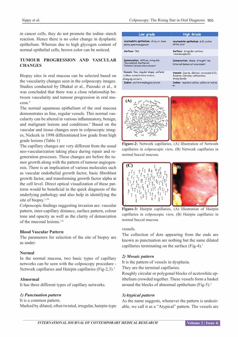
vessels. The collection of dots appearing from the ends are known as punctuation are nothing but the same dilated capillaries terminating on the surface (Fig-4).1
2) Mosaic patternIt is the pattern of vessels in dysplasia. They are the terminal capillaries. Roughly circular or polygonal blocks of acetowhite ep-ithelium crowded together. These vessels form a basket around the blocks of abnormal epithelium (Fig-5).3
3)Atypical patternAs the name suggests, whenever the pattern is undesir-able, we call it as a “Atypical” pattern. The vessels are
in cancer cells, they do not promote the iodine–starch reaction. Hence there is no color change in dysplastic epithelium. Whereas due to high glycogen content of normal epithelial cells, brown color can be noticed.
TUMOUR PROGRESSION AND VASCULAR CHANGES
Biopsy sites in oral mucosa can be selected based on the vascularity changes seen in the colposcopy images.Studies conducted by Dhakal et al., Pazouki et al., it was concluded that there was a close relationship be-tween vascularity and tumour progression in oral mu-cosa.8
The normal squamous epithelium of the oral mucosa demonstrates as fine, regular vessels. This normal vas-cularity can be altered in various inflammatory, benign, and malignant lesions and conditions.9 Based on the vascular and tissue changes seen in colposcopic imag-es, Niekerk in 1998 differentiated low grade from high grade lesions (Table 1)The capillary changes are very different from the usual neo-vascularization taking place during repair and re-generation processes. These changes are before the tu-mor growth along with the pattern of tumour angiogen-esis. There is an implication of various molecules such as vascular endothelial growth factor, basic fibroblast growth factor, and transforming growth factor alpha at the cell level. Direct optical visualization of these pat-terns would be beneficial in the quick diagnosis of the underlying pathology and also help in identifying the site of biopsy.1,10
Colposcopic findings suggesting invasion are: vascular pattern, inter-capillary distance, surface pattern, colour tone and opacity as well as the clarity of demarcation of the mucosal lesions.1,6
Blood Vascular PatternThe parameters for selection of the site of biopsy are as under:
NormalIn the normal mucosa, two basic types of capillary networks can be seen with the colposcopy procedure - Network capillaries and Hairpin capillaries (Fig-2,3).3
AbnormalIt has three different types of capillary networks.
1) Punctuation patternIt is a common pattern. Marked by dilated, often twisted, irregular, hairpin-type
Figure-2: Network capillaries, (A) Illustration of Network capillaries in colposcopic view, (B) Network capillaries in normal buccal mucosa.
Figure-3: Hairpin capillaries, (A) Illustration of Hairpin capillaries in colposcopic view, (B) Hairpin capillaries in normal buccal mucosa.

INTERNATIONAL JOURNAL OF CONTEMPORARY MEDICAL RESEARCH Volume 2 | Issue 4|
906Sippy et al. Colposcopy: The Rising Star in Oral Diagnosis
in the selection of biopsy site for leukoplakia patients while clinical criterion was found to be more appropri-ate for carcinoma buccal mucosa cases.14
FUTURE SCOPE
1. Colposcopy could be further used to compare ef-fects of different treatment modalities, ( radiation and chemotherapy) on the junction between the tu-mour and normal tissue.
2. Patient follow up studies to identify signs of pro-gression for any mucosal lesions can be further done by colposcopy.
3. For improving the patient care with early diagno-sis, colposcopy can be incorporated in our routine practice.
irregular in size and shape and are coarse. Such forma-tion indicates neoplasia. They may be looped, branched, or reticular. Some more characteristics are their sharp turns, dilations, and luminal narrowing (Fig-6).3
VASCULAR CHANGES INDICATING DEGREE OF DYSPLASIA
1) Fine punctuation and mosaicism by narrow low-grade lesions ▬► essels and uniform intercapillary distances
2) A coarse pattern resulting from a wider and high grade abnormalities ▬► more variable vessel di-ameter and spacing
3) The mosaic tiles with central punctuation ▬► car-cinoma in situ.
4) Fracturing of previously intact mosaic and ▬► squamous micro invasion or cancer punctuate pat-terns with the production of predominantly thread like vessels.
With the neoplastic growth, the demand of oxygen and nutrition rises. Thus angiogenesis occurs due to tumour and local tissue production of VEGF, PDGF, EGF, and other cytokines. This further results in the proliferation of blood vessels and neovascularization. The surface epithelium may be lost in these areas leading to irregu-lar surface contour and friability.11 Common to all these vascular patterns is irregular vessel dilatation and in-tercapillary distances greater than the normal distance of 50-200 μm. With the increasing degree of dysplasia, the distance increases so much that the maximum dis-tances may exceed 700 μm.
DISCUSSION
In the year 2000, a research conducted by GoranGny-therportrayed 29 out of 35 of their patients with var-ious premalignant lesions and suspected malignancy showed changes in the vascular picture on microscopy. Authors have concluded that direct oral microscopy of mucosal lesions seem to offer advantage in selecting more representative sites for biopsy.12
In 2013, Abhishek et al conducted a study to assess the role of oral colposcopy for biopsy site selection in cas-es of carcinoma of buccal mucosa. The biopsy samples taken from the clinical presentation of cases showed a sensitivity of 0.7826 (78%) and specificity of 0.5714 (57%). Biopsy samples of colposcopically directed bi-opsy specimens had a sensitivity of 0.6842 (68%) with a specificity of 0.5455 (54%).13
S. Ahmed and Nayyer AS performed a study for clini-cally diagnosed cases of carcinoma buccal mucosa. It concluded that colposcopy was found more significant
Figure-4: (A) Illustration of Punctate capillaries in colpo-scopic view, (B) Punctate capillaries in leukoplakia.
Figure-5: (A) Illustration of Mosaic capillaries in colpo-scopic view, (B) Mosaic capillaries in leukoplakia pointing towards high grade dysplasia.
Figure-6: (A) Illustration of Atypical capillaries in colpo-scopic view, (B) Atypical capillaries in leukoplakia sug-gestive of epithelial dysplasia and ongoing malignant transformation.

INTERNATIONAL JOURNAL OF CONTEMPORARY MEDICAL RESEARCH Volume 2 | Issue 4|
Sippy et al. Colposcopy: The Rising Star in Oral Diagnosis 907
CONCLUSION
In conclusion, Oral Colposcopy is a promising tool in the hands of the oral physician for an objective identifi-cation of the most representative site for biopsy. How-ever future studies need to be carried out to evaluatethe efficacy and accuracy in detecting oral lesions with varying degrees of dysplasia, in comparison with other diagnostic methods.
REFERENCES
1. Mubeen Khan, AbhishekSingh, Gayitri H.C., U.D.Bafna and Siddique Ahmed; Colposcopy- An old technique with a new diagnosticapproach in oral squamous cell carcinoma, review onclinical implications; Universal Journal of Medicine and Dentistry 2012;1:089-097.
2. Silverman S Jr, Dillon WP. Diagnosis oral can-cer, 3rd ed.New York: American Cancer Society 1990:41-60.
3. Ashwini Kumar, Y.Uday Shankar, SathyaPrakash, B.Radhika, Nikhat Fatima; Colposcopy - A Novel Diagnostic Technique for Oral Mucosal Lesions; Journal of Clinical and Diagnostic Research. 2014;8:ZE25-ZE28.
4. Abhishek S. N, Mubeen K, Gayitri HC, Uttam Ku-mar B, Siddique A; Review on Clinical Implica-tions of Colposcopy in Oral Squamous Cell Carci-noma: A Practical Approach; Middle East Journal of Cancer 2013;4:63-72.
5. ShambulingappaPallagatti, Soheyl Sheikh, Nidhi-Puri, Deepak Gupta, Balwinder Singh; Colpos-copy: A new ray in the diagnosis of oral lesions; Indian J Dent Res 2011;22:810-5.
6. Fedele S. Diagnostic aids in the screening of oral cancer Head Neck Oncol.2009;1:5-15.
7. Martin IC, Kerawala CJ, Reed M. The application of toluidine blue as a diagnosticadjunct in the de-tection of epithelial dysplasia. Oral Surg Oral Med Oral PatholOralRadiolEndod. 1998;85:444-46.
8. Pazouki S, Chisholm DM, Adi MM et al. The as-sociation between tumour progression and vascu-larity in the oral mucosa. J Pathol 1997;183:39-43.
9. Smith RA, Cokkinides V, Von EschenbachAC.American Cancer Society guidelines for the ear-lydetection of cancer. CA Cancer J Clin 2002;52:8-22
10. P.L.Estrange, J. Bevenius, L.Williams; Intraoral application of microcolpohysteroscopy: A new technique for clinical examination of oral tissues at high magnification; Oral surgery, oral medicine, oral pathology 1989;67:282–285.
11. Dresang LT. Colposcopy: An Evidence-Based Up-
date. J AmBoardFam Med. 2005;18:383-92.12. Gorangynther, Bjorn Rozell, Amdersheimdahl;Di-
rect oral microscopy and its value in diagnosing mucosal lesions: A pilot study; oral surg, oral med, oral patho, oral radio and endod2000;90:164–170.
13. Abhishek Singh Nayyar, Mubeen Khan, PoojaG-upta;Colposcopy in oral mucosa;International Journal of Pathology; 2012;10: 48-49.
14. S.Ahmed, Nayyer AS; Colposcopy in oral epi-thelial dysplasia: seeing the unseen, a pilot study; Journal of cancer research and therapeutics 2014.