Review Article Cardioprotective Potentials of Plant ...
20
Review Article Cardioprotective Potentials of Plant-Derived Small Molecules against Doxorubicin Associated Cardiotoxicity Shreesh Ojha, 1 Hasan Al Taee, 1 Sameer Goyal, 2 Umesh B. Mahajan, 2 Chandrgouda R. Patil, 2 D. S. Arya, 3 and Mohanraj Rajesh 1 1 Department of Pharmacology and erapeutics, College of Medicine and Health Sciences, United Arab Emirates University, P.O. Box 17666, Al Ain, UAE 2 Department of Pharmacology, R. C. Patel Institute of Pharmaceutical Education and Research, Shirpur, Maharashtra 425405, India 3 Department of Pharmacology, All India Institute of Medical Sciences, New Delhi 110029, India Correspondence should be addressed to Mohanraj Rajesh; [email protected] Received 8 January 2016; Revised 2 April 2016; Accepted 20 April 2016 Academic Editor: Sidhartha D. Ray Copyright © 2016 Shreesh Ojha et al. is is an open access article distributed under the Creative Commons Attribution License, which permits unrestricted use, distribution, and reproduction in any medium, provided the original work is properly cited. Doxorubicin (DOX) is a potent and widely used anthracycline antibiotic for the treatment of several malignancies. Unfortunately, the clinical utility of DOX is oſten restricted due to the elicitation of organ toxicity. Particularly, the increased risk for the development of dilated cardiomyopathy by DOX among the cancer survivors warrants major attention from the physicians as well as researchers to develop adjuvant agents to neutralize the noxious effects of DOX on the healthy myocardium. Despite these pitfalls, the use of traditional cytotoxic drugs continues to be the mainstay treatment for several types of cancer. Recently, phytochemicals have gained attention for their anticancer, chemopreventive, and cardioprotective activities. e ideal cardioprotective agents should not compromise the clinical efficacy of DOX and should be devoid of cumulative or irreversible toxicity on the na¨ ıve tissues. Furthermore, adjuvants possessing synergistic anticancer activity and quelling of chemoresistance would significantly enhance the clinical utility in combating DOX-induced cardiotoxicity. e present review renders an overview of cardioprotective effects of plant-derived small molecules and their purported mechanisms against DOX-induced cardiotoxicity. Phytochemicals serve as the reservoirs of pharmacophore which can be utilized as templates for developing safe and potential novel cardioprotective agents in combating DOX-induced cardiotoxicity. 1. Introduction Doxorubicin (DOX) is a potent and widely used anthracy- cline antibiotic for the treatment of cancers. However, the major impeding issue pertaining to the clinical application of DOX is related to its ability to induce untoward toxicity to the healthy tissues [1]. e occurrence of fatal cardiotoxicity in pediatric as well as in adult patients is characterized by an irreversible cardiomyopathy which compromises the clinical utility of DOX and accounts for the major cause of the chemotherapy related morbidity and mortality [1]. In spite of introducing several less toxic derivatives of DOX, elicitation of cardiotoxicity still remains the major concern [2]. However, with the advent of newer class of monoclonal antibodies revolutionized cancer chemotherapy, still this approach is burdened with myriad adverse effects [3]. us, the use of traditional cytotoxic drugs continues to be a preferred mode for the treatment of cancer. To limit the DOX-induced cardiotoxicity, several molecules, such as beta blockers, angiotensin receptor blockers, amifostine, dexrazoxane, Mesna (2-mercaptoethane sulfonate Na), leucovorin, and erythropoietin, have been evaluated as cardioprotective adjuvants in preclinical studies [4]. Recently, dexrazoxane, when subjected to clinical trial against combating DOX-induced cardiotoxicity, exhibited marked cardioprotection and did not compromise the anticancer activity of DOX [5]. Similarly, carvedilol (beta blocker) has also been demonstrated to confer protection against DOX-induced cardiotoxicity in human subjects [6]. However, large-scale clinical applications of these adjuvants Hindawi Publishing Corporation Oxidative Medicine and Cellular Longevity Volume 2016, Article ID 5724973, 19 pages http://dx.doi.org/10.1155/2016/5724973
Transcript of Review Article Cardioprotective Potentials of Plant ...

Review ArticleCardioprotective Potentials of Plant-Derived Small Moleculesagainst Doxorubicin Associated Cardiotoxicity
Shreesh Ojha,1 Hasan Al Taee,1 Sameer Goyal,2 Umesh B. Mahajan,2
Chandrgouda R. Patil,2 D. S. Arya,3 and Mohanraj Rajesh1
1Department of Pharmacology andTherapeutics, College of Medicine and Health Sciences, United Arab Emirates University,P.O. Box 17666, Al Ain, UAE2Department of Pharmacology, R. C. Patel Institute of Pharmaceutical Education and Research, Shirpur, Maharashtra 425405, India3Department of Pharmacology, All India Institute of Medical Sciences, New Delhi 110029, India
Correspondence should be addressed to Mohanraj Rajesh; [email protected]
Received 8 January 2016; Revised 2 April 2016; Accepted 20 April 2016
Academic Editor: Sidhartha D. Ray
Copyright © 2016 Shreesh Ojha et al. This is an open access article distributed under the Creative Commons Attribution License,which permits unrestricted use, distribution, and reproduction in any medium, provided the original work is properly cited.
Doxorubicin (DOX) is a potent and widely used anthracycline antibiotic for the treatment of several malignancies. Unfortunately,the clinical utility of DOX is often restricted due to the elicitation of organ toxicity. Particularly, the increased risk for thedevelopment of dilated cardiomyopathy by DOX among the cancer survivors warrants major attention from the physicians as wellas researchers to develop adjuvant agents to neutralize the noxious effects of DOXon the healthymyocardium.Despite these pitfalls,the use of traditional cytotoxic drugs continues to be the mainstay treatment for several types of cancer. Recently, phytochemicalshave gained attention for their anticancer, chemopreventive, and cardioprotective activities.The ideal cardioprotective agents shouldnot compromise the clinical efficacy of DOX and should be devoid of cumulative or irreversible toxicity on the naıve tissues.Furthermore, adjuvants possessing synergistic anticancer activity and quelling of chemoresistance would significantly enhancethe clinical utility in combating DOX-induced cardiotoxicity. The present review renders an overview of cardioprotective effects ofplant-derived small molecules and their purported mechanisms against DOX-induced cardiotoxicity. Phytochemicals serve as thereservoirs of pharmacophore which can be utilized as templates for developing safe and potential novel cardioprotective agents incombating DOX-induced cardiotoxicity.
1. Introduction
Doxorubicin (DOX) is a potent and widely used anthracy-cline antibiotic for the treatment of cancers. However, themajor impeding issue pertaining to the clinical application ofDOX is related to its ability to induce untoward toxicity tothe healthy tissues [1]. The occurrence of fatal cardiotoxicityin pediatric as well as in adult patients is characterizedby an irreversible cardiomyopathy which compromises theclinical utility of DOX and accounts for the major cause ofthe chemotherapy related morbidity and mortality [1]. Inspite of introducing several less toxic derivatives of DOX,elicitation of cardiotoxicity still remains the major concern[2]. However, with the advent of newer class of monoclonalantibodies revolutionized cancer chemotherapy, still this
approach is burdened with myriad adverse effects [3]. Thus,the use of traditional cytotoxic drugs continues to be apreferred mode for the treatment of cancer.
To limit the DOX-induced cardiotoxicity, severalmolecules, such as beta blockers, angiotensin receptorblockers, amifostine, dexrazoxane,Mesna (2-mercaptoethanesulfonate Na), leucovorin, and erythropoietin, have beenevaluated as cardioprotective adjuvants in preclinical studies[4]. Recently, dexrazoxane, when subjected to clinical trialagainst combating DOX-induced cardiotoxicity, exhibitedmarked cardioprotection and did not compromise theanticancer activity of DOX [5]. Similarly, carvedilol (betablocker) has also been demonstrated to confer protectionagainst DOX-induced cardiotoxicity in human subjects [6].However, large-scale clinical applications of these adjuvants
Hindawi Publishing CorporationOxidative Medicine and Cellular LongevityVolume 2016, Article ID 5724973, 19 pageshttp://dx.doi.org/10.1155/2016/5724973

2 Oxidative Medicine and Cellular Longevity
−
− −
−
−
−
−
−
−
Doxorubicin
Ca++ overload ↑
MDA ↑
Doxorubicin semiquinone
ONOO− ↑
ROS ↑
p53
Fe2+
Doxorubicin-Fe2+
MM-CK ↑
iNOS ↑
Disruption ofNO regulation
H2O2 ↑
Oxidative stress ↑
JNKApoptosis ↑
NF-𝜅B ↑
Cellular disruptionin mitochondria
mtDNA synthesisinhibition
Caspase-3activation
Caspase cascadeMitochondrialdysfunctionDNA damage
Cardiomyocytesdamage
Cardiac toxicity
Myocardial infarction/cardiachypertrophy
Cytochrome C release ↑
MitochondrialDNA damage ↑
Mitochondriaenergy depletion ↑
Endogenousantioxidants ↓
Posttranscriptionaldysfunction ↑
Apoptosomeformation ↑
Arjunolic acid
Apigenin
Calceolarioside
Curcumin
Eugenol
Frederine
Ginsenosides
Hesperetin
Resveratrol
Thymoquinone
Oleuropein
Anthocyanins
Avicularin
Salvianolic acids
Caffeic acid
Cannabidiol
Chrysoeriol
Eugenol
Gingerol
Kaempferol
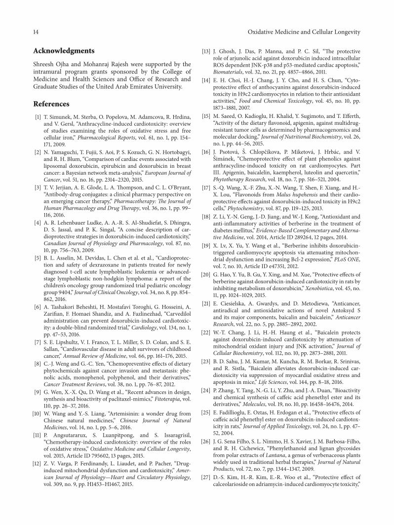
Figure 1: This scheme shows the pathways involved in the elicitation of DOX-induced adverse effects in the myocardium and its attenuationby phytochemicals.
are yet to be established in human subjects. The substantialburden arising from cancer and cardiotoxicity and theirinterrelationship in imposing morbidity and mortality hasemerged as the major driving factor for the academia andpharmaceutical industry to devise and develop strategies thatcan simultaneously provide long-term cardioprotection fromDOX-associated cardiotoxicity without compromising theefficacy of cancer chemotherapy [7]. Since the origin of thehuman civilization, plants and herbs have been traditionallyused in the treatment of various diseases and ailments [8].In this direction, the prospect of harnessing the potentials ofplant-derived small molecules (phytochemicals) appears toprovide rich dividends, since phytochemicals have beenextensively studied in the preclinical studies and shownto possess anti-inflammatory, antioxidant, and anticanceractivities. Phytochemicals are the natural constituents of
herbs and plants. Moreover, anticancer drug paclitaxel andantimalarial agent artemisinin are phytochemicals originallyextracted from plants; however, these agents are nowchemically synthesized [9, 10].
In this review, we have systematically presented theevidence wherein the phytochemicals were investigatedfor cardioprotective effects against DOX and the relevantmechanisms. Several mechanisms have been postulated forthe development of DOX-induced cardiotoxicity. However,oxidative stress driven inflammation, apoptosis, andmyocar-dial remodeling have emerged to be the key players in DOX-induced cardiotoxicity. In-depth description of pathophys-iology of DOX-induced cardiotoxicity has been reviewedelsewhere [11, 12]; however, to provide clarity to our dis-cussion, we have provided a simplified scheme for thepathomechanisms in Figure 1.

Oxidative Medicine and Cellular Longevity 3
2. Phytochemicals LimitingDOX-Induced Cardiotoxicity
In this section, we systematically described the phytochem-icals investigated for their cardioprotective activity againstDOX-induced toxic effects in the myocardium. Phytochem-icals investigated in the animal model of DOX-induced car-diotoxicity are listed in Table 1 and themolecules investigatedin cell culture models (in vitro) are presented in Table 2.
2.1. Arjunolic Acid. Arjunolic acid is a chiral triterpenoidsaponin, isolated from Terminalia arjuna. Arjunolic acidtreatment to adult rat cardiomyocytes in the presence ofDOX attenuated caspase-dependent apoptotic signaling byameliorating proapoptotic p53, p38, and JNK-MAPKs andmitochondrial pathways leading to apoptosis. Furthermore,arjunolic acid when administered to rats significantly inhib-ited DOX-induced myocardial toxicity by mitigating oxida-tive stress and apoptotic pathways [13].
2.2. Anthocyanins. Anthocyanins are a group of polyphenoliccompounds which are abundantly found in common fruitsand vegetables. The effect of six anthocyanidins (cyanidinchloride, delphinidin chloride, malvidin chloride,pelargonidin chloride, peonidin chloride, and petunidinchloride) and seven anthocyanins (cyanidin 3-O-𝛽-galacto-pyranoside chloride, cyanidin 3-O-𝛽-glucopyranosidechloride, delphinidin 3-O-𝛽-glucopyranoside chloride,malvidin 3-O-𝛽-glucopyranoside chloride, pelargonidin3-O-𝛽-glucopyranoside chloride, peonidin 3-O-𝛽-glucopy-ranoside chloride, and petunidin 3-O-𝛽-glucopyranosidechloride) was investigated against DOX-induced cardio-toxicity in H9c2 cardiomyoblasts. All these anthocyanidinsimproved the cell viability via quenching of reactiveoxygen species (ROS) [14]. Delphinidin was found toconfer protection against DOX and etoposide inhibitionof topoisomerase II, thus warranting careful scrutinyagainst the use of these agents in combating DOX-inducedcardiotoxicity.
2.3. Apigenin. Apigenin is a flavone type flavonoid predom-inantly found in flowers of chamomile. It is also present inedible plants such as fruits (oranges, apples, cherries, andgrape fruits) and vegetables (onions, celery, parsley, broccoli,pepper, barley, and tomatoes) [15]. Apigenin treatment toadult rat cardiomyocytes significantly improved cell viabilityin the presence of DOX and the mechanism appeared toinvolve quenching of ROS, mitigation of lipid peroxidation,and myocyte necrosis [16]. However, to date, no studies havedemonstrated apigenin efficacy in vivo.
2.4. Avicularin. Avicularin, chemically a biflavonoid and aquercetin glycoside, is isolated from the leaves of a floweringplant,Malus hupehensis, popularly known as Chinese crabap-ple, Hupeh crab, or tea crabapple [17].The protective effects ofavicularin against DOX-induced cardiotoxicity were demon-strated in H9c2 cells and the mechanism purported was via
its antioxidant property [17]. However, the cardioprotectiveeffects of avicularin are yet to be confirmed in vivo.
2.5. Berberine. Berberine is an alkaloid identified in theroots and bark of the Berberis species [18]. For the firsttime, its protective effects on DOX-induced cardiotoxicity inmice were studied by Lv et al. [19]. Berberine was found toreduce mortality, improve body weight and cardiac function,and restore ECG changes in DOX-treated rats. Furthermore,in neonatal rat cardiomyocytes, berberine inhibited DOX-induced apoptosis via counteracting ROS induced p53 activa-tion, mitochondrial dissipation, executioner caspase activa-tion, and activation of AMPK [19]. Moreover, berberine hasalso been shown to inhibit DOX-induced cardiotoxicity viamitigation of biotransformation of DOX, thereby limiting thebioavailability of doxorubicinol (a major alcohol metaboliteof DOX) in the cytoplasm of rat hearts [20].
2.6. Baicalein. Baicalein is a flavonoid derived from theroots of Scutellaria baicalensis [21]. By using cultured chickcardiomyocytes in vitro, it was shown that baicalein atten-uated DOX-induced ROS activation, proapoptotic MAPK,and apoptosis [22]. Recently, in a murine model of DOX-induced cardiotoxicity, baicalein significantly mitigated car-diac injury via augmenting nuclear factor E2-related factor-2 (Nrf2), antioxidant defense, blunting of nitrative stress,inflammation, and apoptosis [23].
2.7. Caffeic Acid Phenethyl Ester (CAPE). Caffeic acidphenethyl ester is an active component of propolis whichis the major hive product of bees and is known to berich in flavonoids [24]. CAPE has been demonstrated toattenuate DOX-induced cardiotoxicity via attenuation ofROS generation and apoptosis. Furthermore, it also improvedcardiac function as assessed by hemodynamic measurementsand preserved the myocardial structure [25].
2.8. Calceolarioside. Calceolarioside is a phenylpropanoidglycoside isolated from Calceolaria hypericina known topossess antiplatelet and anticancer activities [26]. Calceolar-ioside attenuated DOX-induced cardiotoxicity in H9c2 cellsvia upregulation of antioxidant enzymes and suppression ofapoptosis [27]. However, these effects are yet to be confirmedin animal models.
2.9. Cannabidiol. Cannabidiol (CBD) is a major nonpsy-choactive constituent of the plant Cannabis sativa, popularlyknown as Marijuana and used for recreational as well asmedicinal purposes [28]. In a chronic model of DOX-induced cardiotoxicity in rats, CBD has been demonstratedto suppress myocardial toxicity via attenuating oxidativestress, inflammation, and cell death pathways [29]. Recently,Hao et al. demonstrated that CBD attenuated DOX-inducedcardiotoxicity via augmenting mitochondrial biogenesis andblunting of oxidative and nitrative stress and apoptosis [30].It is also pertinent to note that CBD also exerts severalcardioprotective actions against diabetic cardiovascular com-plications [31] and it has been approved in Canada and

4 Oxidative Medicine and Cellular Longevity
Table 1: Phytochemicals investigated for cardioprotective activity against DOX-induced cardiotoxicity in in vivo studies.
Phytochemical
DOX-inducedcardiomyopathy-animal model
(acute or chronic)
Cardiac functiondetermined(yes/no)
References
Arjunolic acid Chronic Yes [13]Berberine Chronic Yes [20]Berberine Acute Yes [19]Baicalein Chronic No [23]Caffeic acid phenethyl ester Acute Yes [25]Cannabidiol Acute Yes [30]Cannabidiol Chronic No [29]Carotenoids Acute No [34]Eugenol Acute Yes [51]Gingerol Chronic No [56]23-Hydroxybetulinic acid Chronic Yes [60]Hesperetin Chronic No [64]Hesperidin Acute No [62]Isorhamnetin Chronic No [66]Indole-3-carbinol Chronic No [67]Kaempferol Chronic No [69]Lycopene Acute No [70]Lycopene Acute No [71]Lycopene Acute Yes [72]Mangiferin Acute No [79]Mangiferin Acute No [78]Naringenin Acute No [79]Naringenin Acute No [80]Ocotillol Acute No [86]Ocotillol Chronic No [86]Hydroxytyrosol Chronic No [65]Tetrandrine Chronic Yes [140]Periplogenin Chronic No [92]p-coumaric acid Acute No [82]Procyanidins Chronic Yes [100]Robinin Acute No [114]Thymoquinone Acute No [137]Thymoquinone Acute No [138]Silibinin Chronic Yes [126]Sesamin Acute No [124]Sesamol Chronic No [125]Tetrahydroxystilbeneglucoside Acute No [135]
Oleuropein Acute No [87]Oleuropein Acute No [88]Oleuropein Chronic Yes [89]Frederine Acute Yes [52]

Oxidative Medicine and Cellular Longevity 5
Table 1: Continued.
Phytochemical
DOX-inducedcardiomyopathy-animal model
(acute or chronic)
Cardiac functiondetermined(yes/no)
References
Visnagin Acute Yes [146]Visnagin Chronic Yes [146]Schisandrin B Acute Yes [121]Schisandrin B Chronic Yes [122]Salvianolic acid A Acute Yes [117]Tanshinone IIA Chronic Yes [133]Oleuropein Acute No [87]Oleuropein Acute No [88]Oleuropein Chronic Yes [89]Frederine Acute Yes [52]Visnagin Acute Yes [146]Visnagin Chronic Yes [146]Schisandrin B Acute Yes [121]Schisandrin B Chronic Yes [122]Salvianolic acid A Acute Yes [117]Tanshinone IIA Chronic Yes [133]
Europe for the management of pain associated with multiplesclerosis [32].
2.10. Carotenoids. Carotenoids are the organic pigments andconstitute a large group of more than 600 compounds foundin plants, which impart color to the leaves and fruits [33].These are produced from 8 isoprene molecules and contain40 carbon atoms and are also known as tetraterpenoids withpolyisoprenoid structure having a long conjugated doublebond system forming the backbone of the molecule, whichmay be terminated by cyclic end groups that contain oxygen-bearing substitutes. The electron-rich conjugated systemof the polyene is believed to afford antioxidant and freeradical scavenging activity and these pharmacological effectswere attributed to health benefits offered by carotenoids[33]. Recently, the cardioprotective efficacy of carotenoidswas demonstrated in DOX-induced tumor-bearing mice[34]. Specifically, carotenoids were found to improve theantioxidant defense and preserve themyocardialmembranes,reflected as reduced leakage of myocyte injury markerenzymes without compromising DOX activity on the tumorgrowth inhibition [34].
2.11. Chrysin. Chrysin is a flavone class of flavonoid andone of the most important bioactive constituents of differentfruits, vegetables, andmushrooms [35]. Recently, chrysin car-dioprotective effect against DOX-induced acute cardiotox-icity in rats was demonstrated by Mantawy et al. [36].Chrysin was found to improve antioxidant defense, attenuateoxidative/nitrative stress, and suppress the generation ofinflammatory mediators [36].
2.12. Catechins. Epigallocatechin-3-gallate (EGCG) accountsfor 50–80% of catechins in green tea and represents about200–300mg in a brewed cup of green tea. Convincing data areavailable to demonstrate that catechins possess potent antiox-idant, anti-inflammatory, immunomodulatory, cardioprotec-tive, and anticancer activities [37]. Green tea leaf extractsupplementation in cultured rat cardiomyocytes showed itsability to protect the cells against DOX-induced decreasedH9c2 cells viability, via quenching of ROS generation [38].EGCG in vitro has been demonstrated to protect cardiomy-ocytes of neonatal rat hearts from DOX-induced cytotoxicityby attenuating ROS production, apoptosis, and increasingactivities and protein expression of endogenous antioxidantenzymes [39]. In another study, EGCG treatment to ratcardiomyocytes significantly attenuated DOX-induced ROSgeneration and alterations in myocyte contractile dynamicsvia modulation of proteins involved in calcium handlingsystem [40]. In addition, EGCG also elicited cardioprotectiveeffects on a chronicmodel of DOX-induced cardiotoxicity viaattenuation of oxidative stress and apoptosis pathways [41].
It has been reported that supplementation of EGCG inthe diet increases the activities of P-450 family of reductase,augments the bioavailability of DOX, and could predisposethe subject to increased risk of cardiotoxicity [42]. Hence,further studies are warranted to investigate and extendthe cardioprotective benefits of ECGG, without untowardperpetuation of DOX-induced cardiotoxicity.
2.13. Chrysoeriol. Chrysoeriol is a flavone compound isolatedfrom the leaves of Digitalis purpurea, popularly known asFoxglove and well reputed for its cardioprotective actions

6 Oxidative Medicine and Cellular Longevity
Table 2: Phytochemicals exhibiting cytoprotection in the in vitromodels of DOX-induced cardiotoxicity.
PhytochemicalConcentration
of thephytochemical
Cell culturemodel
DOX dose andtime of
incubationReferences
Arjunolic acid 100 𝜇g/mL Neonatal ratcardiomyocytes 1 𝜇M for 12 h [13]
Apigenin 25–100𝜇M Rat heartcardiomyocytes 100 𝜇M for 8 h [16]
Avicularin 10–80 𝜇M H9c2 cells 20𝜇M for 24 h [17]
Berberine 0.06, 0.25, 1.0,and 4.0 𝜇M
Neonatal ratcardiomyocytesand MCF-7 cells
1 𝜇M for 2 h [19]
Baicalein 25𝜇MChick embryocardiomyocytesand MCF-7 cells
1, 10, 50, or100𝜇M for 24 h [22]
Calceolarioside 40𝜇M H9c2 cells 1, 2, or 5 𝜇M for30 h [27]
23-Hydroxybetulinic acid 0.2, 2, and20𝜇M H9c2 cells 5 𝜇M for 18 h [60]
Isorhamnetin 3.125 to25 𝜇g/mL
MCF-7, HepG2,and Hep2 cells 1 𝜇M for 36 h [66]
Kaempferol 5 to 50 𝜇M H9c2 cells 1 𝜇M for 24 h [69]
Morin hydrate 0.17mM ECV304 andHepG2 cells 6mM for 12 h [77]
Naringenin-7-O-glucoside 10–80 𝜇M H9c2 cells 10 𝜇M for 24 h [83]
Osthole 10–40 𝜇M Neonatal ratcardiomyocytes 1 𝜇mol for 24 h [90]
Luteolin-7-O-𝛽-D-glucopyranoside 5, 10, and 20𝜇M H9c2 cells 20 𝜇M for 24 h [75]Luteolin-7-O-𝛽-D-glucopyranoside 5–80𝜇M H9c2 cells 10 𝜇M for 24 h [76]
Vincristine 10–30 𝜇M Adult mousecardiomyocytes
15 and 20 𝜇g/mLfor 24 h [144]
Sulforaphane 2.5 𝜇M H9c2 cells 5 𝜇g/mL for16–18 h [129]
C-Phycocyanin 10 𝜇M Adult ratcardiomyocytes
10𝜇M for 4, 24,and 48 h [94]
Plantainoside D 1–20 𝜇g/mL H9c2 cells 1, 2, and 4 𝜇Mfor 30 h [93]
Sesamol 12.5–50 𝜇M H9c2 cells 1 𝜇M for 30min [125]
Tetrahydroxystilbene glucoside 3–300 𝜇M Neonatal ratcardiomyocytes
1 𝜇mol/L for24 h [143]
Chrysoeriol 20𝜇g/mL H9c2 cells 1𝜇Mfor 24 h [43]
Visnagin 20𝜇M
Neonatal rat andzebrafish
cardiomyocytes,cardiac HL-1
cells
100 𝜇M for 48 h [146]
Z-Guggulsterone 1–30 𝜇M H9C2 cells 1 𝜇M for 24 h [143]
Tanshinone IIA 0.1, 0.3, 1, and3 𝜇M
Neonatal ratcardiomyocytes 1 𝜇M for 24 h [134]
Tanshinone IIA 1.6–40 𝜇M H9c2 cells 1 𝜇M for 24 h [133]
Tanshinone IIA 0.5, 1, and2 𝜇mol/L
Neonatal ratcardiomyocytes
1 𝜇mol/L for24 h [132]
Sodium tanshinone IIA sulphonate 0.05–0.5mM Mice heartmitochondria
0.2mmol for10min [131]
Anthocyanidins and anthocyanins 0–100 𝜇M H9c2 cells andMCF-7 cells 1 𝜇M for 24 h [14]

Oxidative Medicine and Cellular Longevity 7
Table 2: Continued.
PhytochemicalConcentration
of thephytochemical
Cell culturemodel
DOX dose andtime of
incubationReferences
Caffeic, chlorogenic, and rosmarinicacid 100 and 200 𝜇M
Rat heartmicrosomes andmitochondria
100 𝜇M for 8 h [116]
ECV304 cells: human umbilical vein endothelial cells; HepG2 cells: human hepatocellular carcinoma cells; MCF-7 cells: human breast carcinoma; H9c2 cells:rat ventricular cardiomyoblast cells.
[43]. Chrysoeriol has been found to reduce cell death andattenuate ROS generated oxidative stress and lipid peroxida-tion inDOX-induced cardiotoxicity inH9c2 cardiomyoblastswithout affecting antitumor activity of DOX [43]. For anevidence based approach, additional studies on its cardiopro-tective efficacies are warranted.
2.14. Curcumin. Curcumin is a phenolic yellow pigmentconstituent found in the rhizomatous parts of Curcumalonga (turmeric) [44]. Several in vitro and in vivo stud-ies have demonstrated curcumin cardioprotective actionsagainst DOX-induced myocardial toxicity. The key mech-anisms postulated for curcumin cardioprotective activityinclude diminution of oxidative stress, inflammation, andassociated cell death pathways [45–48]. Although curcuminhas been reported to elicit several beneficial effects in variouspreclinical studies, the bioavailability is yet to be establishedin human subjects. Therefore, derivatives of curcumin arebeing perused to increase its bioavailability and, in thisdirection, a recent report suggests that the nanoparticleof curcumin could ameliorate DOX-induced cardiotoxicity[49].
2.15. Eugenol. Eugenol is the active component of essentialoil isolated from Syzygium aromaticum, popularly knownas clove, which is one of the common ingredients of spicemixtures [50]. Eugenol treatment was shown to significantlyimprove antioxidant defense mechanisms, decrease lipidperoxidation, and attenuate abnormal Ca2+ transients inthe cardiomyocytes along with inhibition of apoptosis inrat hearts following acute DOX administration. Eugenolalso preserved the myocardium and restored hemodynamicsalong with preserved histology [51].
2.16. Frederine. 7-Monohydroxyethylrutoside (monoHER2,frederine) is a synthetic flavonoid, and it significantly inhib-ited DOX-induced myocardial toxicity, via suppression ofoxidative stress and apoptosis in a chronic murine model[52, 53]. In addition, monoHER2 did not interfere with DOXanticancer activity in vitro and in vivo [54]. Considering theseobservations, monoHER2 could be further developed for thisclinical application as cardioprotective adjuvant.
2.17. Gingerol. Gingerol is the pungent phenolic constituentof Zingiber officinalis (ginger) [55]. In a chronic model of
DOX-induced cardiomyopathy, gingerol inhibited DOX-inducedmyocardial ROS generation, inflammation via atten-uation of NF-𝜅B activation, and downregulation of solublereceptor for advanced glycation end products (sRAGE). Inaddition, gingerol also inhibited myocardial apoptosis viamitigating caspase-3 activities [56].
2.18. Ginsenosides. Ginsenosides are the saponin constituentsof Panax notoginseng, popularly known as ginseng in tra-ditional Chinese medicine [57]. Ginsenosides have beenclassified into protopanaxatriols (Rg1, Rh1, and PPT) andprotopanaxadiols (Rg3, Rh2, and PPD). Of the ginsenosides,protopanaxadiols such as ginsenoside Rb1, ginsenoside Rh2,and compound K have been shown to exhibit anticancer andanti-inflammatory activities [57].
Ginsenoside Rh2 (Rh2) has been shown to elicit car-dioprotective effects against DOX-induced cardiotoxicity inH9c2 cell line, as well as in vivo in an acute mouse andchronic rat model of DOX-induced cardiomyopathy. Rh2enhanced cell viability of H9c2 cells and ameliorated DOX-induced release of the CK-MB, LDH. Furthermore, Rh2ameliorated DOX-induced myocardial toxicity in mouse andrats via suppressing oxidative stress and improved the indicesof cardiac function as determined by ECG [58].
2.19. Diosgenin. Diosgenin is a steroid saponin found abun-dantly in several plants including Solanum and Dioscoreaspecies and Costus speciosus. In a chronic model of DOX-induced cardiomyopathy, diosgenin elicited cardioprotectiveeffects, via activation of prosurvival kinase, protein kinaseA (PKA), diminution of p38-MAPK, caspase-3 activities,and generation of free radicals along with attenuation ofinflammatory mediators. Mechanistically, it was found toimprove myocardial fibrosis and increase the cardiac levels ofcGMP via modulation of phosphodiesterase-5 activity [59].
2.20. Hydroxybetulinic Acid. 23-Hydroxybetulinic acid is iso-lated from Pulsatilla chinensis. In a chronic murine modelof DOX-induced cardiomyopathy, 23-hydroxybetulinic acidsignificantly improved the survival of the animals and inhib-ited apoptosis mainly via inhibition of DOX metabolism inthe mitochondria. Similar results were also obtained in H9c2cells [60].
2.21. Hesperidin. Hesperidin is a bioflavonoid abundantlyfound in vegetables and citrus fruits such as oranges, lemons,

8 Oxidative Medicine and Cellular Longevity
and grapefruits [61]. Citrus flavonoids have been shownto reduce risk of cardiovascular diseases prominently dueto their antioxidant and anti-inflammatory effects involvingnumerous cell signaling pathways [61]. It has been found toimprove antioxidant status, inhibit lipid peroxidation, andreducemyocardial enzyme leakage by salvagingmyocardiumin DOX-induced cardiotoxicity in acute toxicity model con-ducted rats [62]. Additionally, it sensitized cancer cells toDOX-induced apoptosis and showed synergism in inhibitingP-gp and multidrug resistance, thus appearing to be effectiveas an adjunct to enhance the efficacy and attenuate theresistance to DOX during chemotherapy [63]. Thus, thepotential of citrus flavonoids as cochemotherapeutic andcardioprotective agents is encouraging but further studiesare warranted for conclusive evidence in cancer as well aschemotherapy associated cardiotoxicity. In addition, hes-peretin (aglycone) derivative of hesperidin also amelioratedchronic DOX treatment associated cardiotoxicity in rats, viaattenuation of p38-MAPK, caspase-3, and NF-𝜅B activationand oxidative stress in the myocardial tissues [64].
2.22. Hydroxytyrosol. Hydroxytyrosol is a polyphenolic con-stituent inOlea europaea, popularly known as olive oil, whichis widely used in food and medicine [65]. Hydroxytyrosolhas been shown to improve cardiac function by maintaininghomeostasis at mitochondrial level, by preserving mitochon-drial electron transport chain complexes I–IV and inhibitingapoptosis-inducing factor, and oxidative stress markers inchronic DOX-induced cardiotoxicity in rats harboring breastcancer [65]. Furthermore, hydroxytyrosol did not compro-mise theDOXantitumor activity against the implanted tumorcells and also improved the survival of the animals [65].
2.23. Isorhamnetin. Isorhamnetin is a flavonol aglyconeabundantly found in several medicinal plants, such as Hip-pophae rhamnoides (sea buckthorn) [66]. Isorhamnetin sig-nificantly conferred cardioprotection in a chronic model ofDOX-induced cardiotoxicity. The mechanism of cardiopro-tection involves suppression of oxidative stress and activationof mitochondrial apoptotic pathway and mitogen-activatedprotein kinase pathways, suggesting antioxidant mediatedcardioprotectivemechanism. Similar results were obtained inH9c2 cells [66]. Furthermore, it also synergistically improvedthe DOX anticancer activity in tumor cell lines [66].
2.24. Indole-3-carbinol. Indole-3-carbinol is a natural indolecompound predominantly found in cruciferous vegetables[67]. In a chronic DOX-infusion associated cardiotoxicitymurine model, indole-3-carbinol was found to reduce solidEhrlich tumor size and volume, augment antioxidant defense,and inhibit lipid peroxidation, leading to stabilization of cellmembrane and reduced leakage of myocyte injury markerenzymes. It was also found to decrease sphingosine kinase1 (SphK1) activity and inflammatory mediators along withmitigating histological perturbations and modulating celldeath mediators [67].
2.25. Kaempferol. Kaempferol is one of the most commondietary flavonoids and it is well studied for its antiapoptotic,cardioprotective, antioxidative, anti-inflammatory, chemo-preventive, and anticancer properties as well as modula-tion of chemoresistance [68]. The cardioprotective effectsof kaempferol against DOX-induced cardiotoxicity in ratswere demonstrated using a chronic model. Kaempferolcounteracted cardiotoxicity by inhibiting p53 expressionin mitochondrion-dependent apoptotic signaling and ERK-dependent mitogen-activated protein kinase pathway follow-ing binding to the promoter region of the Bax proapoptoticgene. It also effectively suppressed DOX-induced extracellu-lar signal-regulated kinase (ERK1/2) activation but had noeffect on p38 and JNK [69].
2.26. Lycopene. Lycopene is a carotenoid and nonprovitaminA found abundantly in Lycopersicum esculentum (tomatoes)and known to impart color to tomatoes [33]. Karimi et al., in avery early study, demonstrated the protective effect of tomatoextract and lycopene on acute DOX-induced cardiotoxicityin mice [70]. Tomato extract and lycopene prevented risein myocyte injury marker enzyme, CK-MB, in serum andameliorated cardiomyocytes injury evidenced by histopatho-logical examination. Furthermore, Yilmaz et al. studied theprotective role of lycopene inDOX-induced heart and kidneytoxicities using biochemical and histopathological assess-ments and reported that lycopene has potential to inhibit lipidperoxidation and improve antioxidants evidenced by reducedlipid peroxides and improved GSH in both the heart andthe kidneys. The protective effect was further substantiatedby histopathological changes. The authors concluded thattreatment with lycopene might prevent cardiac and renaltoxicities in rats [71]. However, the results were not furthersubstantiated by functional improvement as lycopene didnot prevent left ventricular systolic dysfunction induced byDOX [72]. However, it suppressed DOX-induced myocytedamage without preventing interstitial collagen accumula-tion increase [72].
Lycopene supplementation also increased lycopeneabsorption in heart, liver, and plasma and, in another study,the same group of authors showed that there was no depletionof lycopene from myocardium of lycopene-supplementedrats treated with DOX and that higher antioxidant capacityin myocardium and less oxidative cleavage of lycopene inintestinal mucosa of DOX-treated rats suggest an antioxidantrole of DOX rather than acting as a prooxidant [73]. Theauthors further showed that tomato-oleoresin enhances thechemotherapeutic effect of DOX. It maintained lycopenelevels in heart and protected against cardiac oxidativeDNA damage induced by DOX in rats [73]. The lycopeneprotected the heart against DOX associated cardiotoxicityby several mechanisms including the quenching of singletoxygen, peroxy radicals, reaction with free radicals, restoringlevels of vitamin E and vitamin C, reducing DNA damage,restoring cellular antioxidants, and preventing depletionof glutathione. Several studies showed the potential role oflycopene in the prevention of side effects of antineoplasticdrugs in cell culture and animal models [74]. These results

Oxidative Medicine and Cellular Longevity 9
suggest that tomato extract and lycopene inhibit DOXcardiotoxicity and collectively might serve as a novelcombination chemotherapeutic agent with DOX to limit freeradical-mediated organ injury. However, further studies arerequired to investigate the role of lycopene in mitigating theside effects of chemotherapy in human subjects.
2.27. Luteolin-7-O-𝛽-D-glucopyranoside. Luteolin-7-O-𝛽-D-glucopyranoside is a flavonoid isolated from the plantDracocephalum tanguticum that is widely used in Chineseand Tibetan traditional medicine [75]. The cytoprotectiveactivities were demonstrated on DOX-induced cytotoxic-ity in H9c2 cardiomyocytes. Among several isolated com-pounds, luteolin-7-O-𝛽-D-glucopyranoside was found toshow antioxidant effect and a potent cytoprotective activityagainst DOX-induced toxicity as evidenced by decreaseddeath ofH9c2 cells, reducedmyocyte injurymarker enzymes,and reduced intracellular concentration of ROS and Ca2+[75]. Recently, Yao et al. also demonstrated protective effectsof luteolin-7-O-glucoside on DOX cytotoxicity in H9c2 cells[76]. It was found to improve cell viability and ameliorate ROSgeneration and mitochondrial depolarization. Furthermore,it enhanced the expression of prosurvival kinases and dimin-ished ROS generation [76].
2.28. Morin Hydrate. Morin hydrate is a biflavonoid com-monly found in fruits such as guava, fig, almond, grapes,and apple and vegetables such as onion, seed weeds, andseveral other members of Moraceae family [77]. It has beenobserved to enhance antioxidant defense against oxidativestress in human umbilical vein endothelial cells (ECV304)and HepG2 cells and minimize DOX toxicity in ECV304and primary mouse cardiomyocytes [77]. However, in vivostudies regarding morin efficacy in combating DOX-inducedcardiotoxicity are still lacking.
2.29. Mangiferin. Mangiferin is a xanthonoid structure withC-glucosyl linkage and polyhydroxy component found inmany plant species; however, Mangifera indica (mango tree)is the major source [78]. Using a chronic model of car-diomyopathy in rats, mangiferin has been shown to exertcardioprotective action against DOX-induced cardiotoxicityby inhibition of proinflammatory mediators and proapop-totic genes and regulating calcium homeostasis modulatingproteins [79].
2.30. Naringin. Naringin is a flavanone glycoside abundantlyfound in citrus fruits such as lemon, oranges, and grape-fruits and in tomatoes. It has been documented to possessnumerous biological properties such as antioxidant, anti-inflammatory, and antiapoptotic activities [80]. In an acutemodel of DOX-induced cardiotoxicity, naringin treatmentimproved antioxidant defense and inhibited lipid peroxi-dation along with histopathological preservation, therebyreducing leakage of myocyte marker enzymes. It alsodecreased the levels of inflammatory mediators and restoredthe mitochondrial complexes (I–IV) activities in the hearttissues along with histopathological salvage [80]. The studies
indicate cardioprotective effects; however, further clinicalresearch is required to provide significant insights into themechanisms underlying the effects of naringin on humansubjects.
2.31. Naringenin and Its Derivatives. Naringenin is a fla-vanone commonly found in citrus fruits such as grapefruit,orange, and lemon [80]. Arafa et al. demonstrated thatnaringenin elicited antioxidant mediated protection againstDOX-induced cardiac toxicity in Swiss albino rats [81].Recently, the combination of p-coumaric acid and naringeninwas found to be superior in exerting antioxidant mediatedcardioprotection against DOX-induced cardiotoxicity in rats[82]. Additionally, naringenin enhanced antitumor effect ofDOX by selectively modulating drug efflux pathways; thatis, it inhibited the activity of multidrug resistance-associatedprotein and did not affect the in vivo pharmacokinetics ofintravenously administered DOX [80].
Naringenin-7-O-glucoside is a flavanone glycoside iso-lated fromDracocephalum rupestre. It has been demonstratedto protect against DOX-induced cardiotoxicity in H9c2 cells[83]. Particularly, naringenin-7-O-glucoside improved cellviability, prevented the release of myocyte injury markerenzymes LDH and CK, and augmented antioxidant defense[84]. Furthermore, naringenin-7-O-glucoside was observedto enhance NAD(P)H: quinone oxidoreductase (NQO1) andERK activation and Nrf2 protein levels in DOX stressedH9c2 cells. These phenotypic changes brought about bynaringenin-7-O-glucoside are attributed to the induction ofantioxidant defense and attenuation of cell death pathways[85]. However, in vivo studies are yet to be performed.
2.32. Ocotillol. Ocotillol is an aglycone derivative ofpseudoginsenoside-F11, which is devoid of sugar moietyand is found in American ginseng, Panax quinquefolius.Fu et al. reported that ocotillol enhances survival rateof animals in both acute and chronic models of DOX-induced cardiotoxicity. Ocotillol prevented depletion ofglutathione and lipid peroxidation along with restorationof myocyte injury marker enzymes following preservationof cardiomyocytes cell membrane. Furthermore, ocotillolalso improved cardiac function and hence was suggested asan adjuvant for counteracting DOX-induced cardiotoxicity[86].
2.33. Oleuropein. Oleuropein is a phenolic constituent ofOlea europaea (olive oil) [87]. Andreadou et al. have shownthe protective effect of oleuropein against DOX-inducedacute cardiotoxicity in rats. Oleuropein was found to restorethe myocardial necrosis marker enzyme levels and atten-uation of oxidative stress and apoptosis [88]. Moreover,the same group of authors in a separate study demon-strated that oleuropein treatment aids the compensationof distressed energy metabolic pathways mechanistically byrestoration of metabolites to the normal levels as DOXgenerated free radicals nonenzymatically convert pyruvateto acetate and alpha-ketoglutarate to succinate [87]. Thecardioprotective role of oleuropein in chronic DOX-induced

10 Oxidative Medicine and Cellular Longevity
cardiomyopathy has also been demonstrated [89]. Particu-larly, oleuropein treatment significantly suppressed DOX-induced oxidative/nitrative stress, augmented prosurvivalkinases, and improved the cardiac functions [89].
2.34. Osthole. Osthole is a coumarin compound found inseveralmedicinal plants such asCnidiummonnieri andAngel-ica pubescens [90]. In rat neonatal cardiomyocytes, ostholesignificantly improved the survival of the cells by abrogatingapoptosis, wherein the mechanism appeared to involve thesuppression of mitochondrial pathway of apoptosis triggeredby DOX [90]. However, in vivo studies have not beenconducted, and it is warranted to recapitulate the in vitrocardioprotective actions of osthole.
2.35. p-coumaric Acid. p-coumaric acid is a phenolic acidthat serves as a precursor of other phenolic compounds andis found in plants such as peanut, tea, and coffee [82]. p-coumaric acid has been shown to attenuate oxidative stressand inhibit myocyte injury in DOX-induced myocardialinjury in rats [91]. In a separate study, p-coumaric acid incombination with naringenin showed cardioprotection byaugmentation of antioxidant defense against DOX-inducedcardiotoxicity in rats [82].
2.36. Periplogenin. Periplogenin is a cardenolide isolatedfrom Aegle marmelos, commonly known as Bael in thetraditional Indian system of medicine [92]. Periplogeninhas been shown to decrease lipid peroxide levels, improveantioxidant defense, and salvage cardiomyocytes in a chronicmodel of DOX-induced cardiotoxicity in rats [92].
2.37. Plantainoside D. Plantainoside D is an iridoid gluco-side isolated from Picrorhiza scrophulariiflora (Picrorhiza).The chemopreventive and antioxidant activities encouragedevaluating the cardioprotective effect of plantainoside Dagainst DOX-induced apoptosis in H9c2 cells. PlantainosideD was found to inhibit oxidative stress and proinflammatorycytokines expression and attenuate apoptosis in H9c2 car-diomyoblasts [93].
2.38. Phycocyanin. C-Phycocyanin is a biliprotein foundin Spirulina platensis, blue-green algae. Phycocyanin hasbeen found to protect against DOX-induced oxidative stressand apoptosis in adult rat cardiomyocytes as evidenced byreduced ROS formation, DNA fragmentation, and attenua-tion of Bax as well as release of cytochrome C and increase inthe activity of caspase-3 [94]. However, these in vitro findingsare yet to be confirmed in rodent model of DOX-inducedcardiomyopathy.
2.39. Proanthocyanidin and Derivatives. Proanthocyanidinsare a mixture of structurally and functionally diverse chem-icals which are predominantly found in grape seed andshow high bioavailability and protect the organs from toxicchemicals used to induce diseases in in vitro and in vivostudies [95, 96]. Ray et al. reported the bioavailabilityand protective property of grape seed proanthocyanidin
and a novel IH636 grape seed proanthocyanidin extractagainst DOX-induced cardiotoxicity as well as multiorganprotection in mice [97]. Bagchi et al. further reported thatIH636 proanthocyanidin extract afforded protection wassuperior to vitamin C, vitamin E, and beta-carotene anddemonstrated significant cytotoxicity towards human breast,lung, and gastric adenocarcinoma cells, while enhancingthe growth and viability of normal cells in both in vitroand in vivo studies [98]. In another study, proanthocyani-dins enhanced DOX-induced antitumor effect and reverseddrug resistance and mechanisms attributed partially to thepromotion of DOX-induced apoptosis through elevationof intracellular DOX, Ca2+, and Mg2+ concentration andreduction of pH value and mitochondrial membrane poten-tial in DOX-resistant K562/DOX cells [99]. Furthermore,proanthocyanidin strongly enhanced the antitumor activityof DOX and ameliorated chronic DOX-induced myocardialoxidative stress and immunosuppression in tumor-bearingmice [99]. In addition, grape seed proanthocyanidin alsoshowed antioxidant mediated cardioprotection against bothhigh and low dose DOX-induced cumulative chronic car-diotoxicities in rats [100].
2.40. Resveratrol. Resveratrol, a natural phytoalexin, is com-monly found in Vitis vinifera (grapes) [101]. Resveratrolinduced antioxidants and phase 2 enzymes in the H9c2cells, accompanied by increased resistance to oxidative andelectrophilic cell injury [102]. Additionally, there was nosignificant effect of resveratrol on NADPH-cytochrome P-450 reductase (P-450 reductase), which plays an importantrole in the metabolism of many endogenous compounds andxenobiotics including DOX. The enzyme P-450 reductaseactivates them to their more toxic metabolites via one elec-tron reduction which triggers free radical cascade. In somecases, however, such transformation is essential to producetherapeutic effect of anticancer drugs [42].
In DOX-induced cardiotoxicity in rats, resveratrol hasbeen shown to ameliorate the severity of cardiac dysfunctionand prevent oxidant stress responses [103, 104]. Furthermore,resveratrol was found to confer cardioprotection and reducecardiac fibrosis in acute as well as chronic in vivo modelsof DOX-induced cardiomyopathy in rats. Mechanistically,resveratrol has been demonstrated to protect against DOX-induced oxidative stress through changes in mitochondrialfunction, specifically the Sirt1 pathway, leading to cardiaccell survival [105]. Resveratrol attenuated DOX-inducedcardiomyocyte apoptosis in mice via upregulation of Sirt1-mediated p53 deacetylation and activation of Sirt1, a NAD+-dependent deacetylase, resulting in improved mitochondrialfunction, which culminates in activation of the transcriptionfactors which coordinate expression of key antioxidant pro-teins by binding to the antioxidant response elements thatregulate cell survival [106].The overexpression of Sirt1 inhib-ited cell apoptosis by suppression of p38-MAPK phospho-rylation and caspase-3 activation along with amelioration ofROS generation and prevented DOX-induced functional lossin DOX-induced cardiomyocyte injury [106]. DOX inducesautophagy in cardiomyocytes which is a degradation system

Oxidative Medicine and Cellular Longevity 11
for eukaryotic cells to turn over organelles and long-livedproteins, thereby maintaining cellular homeostasis. Thus,aberrant autophagy activity impairs basal cardiac structureand function, making animals more sensitive to stress-induced heart failure. The ability of resveratrol to inhibitautophagy is mediated by inhibition of p70S6 kinase 1 (S6K1)that is essential for resveratrol to suppress DOX-inducedautophagy and cytotoxic effects [107].
DOX inhibits AMP-activated protein kinase (AMPK),resulting in Sirt1 dysfunction and p53 accumulation inmouseembryonic fibroblasts, and pharmacological activation ofAMPK by resveratrol has been shown to alleviate the sideeffects of DOX in H9c2 cells [108]. Furthermore, resvera-trol has been shown to confer cardioprotection in DOX-induced cardiomyocyte apoptosis in nude mice by inductionof heme oxygenase-1 (HO-1) mediated mechanisms [109,110]. Resveratrol was also reported to aid the differentiationof adipose-derived mesenchymal stem cells to cardiomy-ocytes and protected against noxious effects of DOX tothe myocardium [111]. Furthermore, resveratrol supplementalong with exercise training was found to be more effectivein preventing DOX-induced LV remodeling associated withthe reduction of DOX-induced oxidative stress [112]. In spiteof these advances made in preclinical studies, resveratrolbioavailability is seldom established in human subjects andthis warrants further approaches to extend its beneficialeffects to mankind [113].
2.41. Robinin. Robinin is a flavonoid glycoside isolated fromleaves ofVigna unguiculata, a dietary plant used in traditionalcuisine in India [114]. Treatment with robinin was found toimprove endogenous antioxidants, reduce ROS genera-tion, and inhibit lipid peroxidation and proinflammatorymediators such as cyclooxygenase (COX2) and lipoxyge-nase (LOX15) along with restoring myocyte injury markerenzymes. The improvement in the level of transforminggrowth factor-𝛽1 (TGF-𝛽1), Smad2, murine double minute(Mdm2), Smad3, cyclin-dependent kinase inhibitor 2A,Smad4, and Smad7 in addition to favorable modulation ofp53, Bcl-2, and Bax revealed the cardioprotective mechanismof robinin in combating DOX-induced cardiotoxicity [114].
2.42. Rosmarinic Acid. Rosmarinic acid is an ester of caffeicacid abundantly found in numerous plants, being mostcommon in Boraginaceae and Lamiaceae families [115]. Ros-marinic acid showed remarkable cytoprotection against DOXtoxicity in neonatal rat cardiomyocytes and DOX-inducedlipid peroxidation of heart membranes, mitochondria, andmicrosomes and effects were found to be comparable todexrazoxane [116]. Furthermore, it inhibited DOX-inducedoxidative stress and apoptosis in H9c2 cardiomyoblasts byimproving cell viability, inhibiting the production of ROS,and activation of prosurvival kinases [115]. However, in vivocardioprotective actions against DOX-induced cardiotoxicityare hitherto unknown.
2.43. Salvianolic Acids. Salvianolic acids especially salviano-lic acid A and salvianolic acid B are themost abundant water-soluble compounds extracted from Salvia miltiorrhiza (Dan-shen or red sage) [117]. Salvianolic acid A has been shown toprotect against DOX-induced mitochondrial toxicity in vitroin rat cardiomyocytes due to its antioxidant action, withoutantagonizing effect on the antitumor activity of DOX [118].The protective effects of salvianolic acid were reconfirmedin vivo in another study, against DOX cardiotoxicity in mice[117], via abrogation of oxidative stress and inflammation.
2.44. Schisandrin B. Schisandrin B, a dibenzocyclooctadienelignin, is isolated from the fruit of Schisandra chinensis. Ithas been shown to salvage cardiomyocytes, via conferringantioxidant defense by restoring glutathione flux in an acuteanimal model of DOX-induced cardiotoxicity [119]. Fur-thermore, it also mitigated DOX-induced cardiotoxicity inrabbits [120]. Recently, cardioprotective effects of schisandrinB against DOX-induced cardiotoxicity were reconfirmed andthe mechanism of protection was evidenced by ameliora-tion of proinflammatory cytokines, lipid peroxidation, DNAdamage, apoptosis, and MAPK activation in the myocardialtissues [121, 122].
2.45. Salidroside. Salidroside, a phenylethanoid glycoside,has been isolated from the roots ofRhodiola rosea (Roseroot).Wang et al. demonstrated that treatment of salidrosideto either H9c2 cells or mice stressed with acute DOXadministration conferred cardioprotective effects.Themech-anism was defined to involve antioxidant and suppression ofproapoptotic mediators [123]. The cardioprotective effects ofsalidroside were reconfirmed in a placebo controlled clinicaltrial wherein sixty patients with breast cancer receivingepirubicin were given salidroside (600mg/day) or placebostarting 1 week before chemotherapy and assessed at baselineand 7 days after each new epirubicin dose of 100mg/m2.Decline in strain rate peak was observed at an epirubicindose of 200mg/m2, with no significant differences betweensalidroside and placebo. At increasing cumulative doses ofepirubicin, the strain rate normalized only with salidroside,showing a significant difference in comparison with placeboat epirubicin doses of 300mg/m2.The authors concluded thatsalidroside may provide protection against chemotherapy-induced early left ventricular regional systolic dysfunction inpatients with breast cancer. Based on preclinical and clinicaldata, salidroside needs to be investigated further in a largerpopulation for advocating salidroside as an adjuvant to thwartthe DOX-induced cardiotoxicity.
2.46. Sesamin. Sesamin is amajor lignin obtained from seedsof Sesamum indicum. Sesamin was observed to increase theendogenous antioxidant enzymes and prevent mitochondrialdamage via activation of Sirt1 in an acute model of DOX-induced cardiotoxicity [124].
2.47. Sesamol. Sesamol is a phenolic constituent of oilobtained from seeds of Sesamum indicum and is usedcommonly as an edible oil. The cardioprotective effects of

12 Oxidative Medicine and Cellular Longevity
sesamol were confirmed in an in vivo study, wherein sesamolmitigated cumulative DOX-induced cardiomyopathy in rats[125]. Sesamol improved antioxidant defense status, reducedmyocyte injury marker enzymes released from cardiomy-ocytes, and inhibited lipid hydroperoxide. The salvage oftissues evidenced by biochemical and histopathological stud-ies demonstrated cardioprotective effects of sesamol [125].However, further mechanistic studies should be carried outinvestigating the effect onDOX efficacy and pharmacokineticinteraction.
2.48. Silibinin. Silibinin, a flavonolignan, is an active compo-nent of Silybummarianum (milk thistle), popularly known assilymarin and known to constitute 50–70% of the silymarinextract. The cardioprotective effect exerted by silymarin,silibinin, dehydrosilibinin, and silychristin was comparableto that of dexrazoxane, while silydianin exerted the bestprotective effect [126].
2.49. Sulforaphane. Sulforaphane is an organosulfur com-pound found in a significant amount in cruciferous veg-etables, especially in broccoli (Brassica oleracea) [127]. Thecardioprotective effects of sulforaphane were first demon-strated in H9c2 rat myoblasts as evidenced by reducednumber of apoptotic cells along with decreased expres-sion of proapoptotic proteins such as Bax, caspase-3, andcytochrome C [128]. It also reduced ROS generation andrestoredmitochondrial membrane potential [128]. Moreover,the cardioprotective effects of sulforaphane were found to bemediated by the activation of the Kelch-like ECH-associatedprotein 1 (Keap1)/NF-E2-related factor-2 (Nrf2)/antioxidant-responsive element (ARE) pathway, which in turn mediatesthe induction of HO-1 [129].
2.50. Tanshinone IIA and Derivatives. Tanshinones are thegroup of bioactive compounds isolated from Salvia milti-orrhiza (Danshen), a Chinese medicinal plant reputed forthe management of cardiovascular diseases in particular,angina pectoris, atherosclerosis, myocardial infarction, andischemic-reperfusion injury [130]. Sodium tanshinone IIAsulfonate, a water-soluble derivative of tanshinone IIA, wasdemonstrated to be beneficial in reducing DOX-inducedcardiotoxicity in mice hearts and in cultured cardiomyocytes[131]. Treatment with sodium tanshinone IIA sulfonate pre-vented decrease in body weight and reduced myocardiallipid peroxidation in mice along with improved activities ofendogenous antioxidant enzymes. In addition, the antioxida-tive mechanism was also supported by in vitro experimentsshowing that sodium tanshinone IIA sulphonate scavengedDOX semiquinone free radical in heart homogenate andinhibited DOX-induced mitochondrial lipid peroxidationand swelling [131].
Furthermore, another study demonstrated the benefi-cial effect of tanshinone IIA on decreasing DOX-inducedapoptosis in neonatal rat cardiomyocytes and underlyingmolecular mechanisms [132]. Tanshinone IIA amelioratedapoptosis and ROS generation induced by DOX in a dose-dependentmanner. It was further supported by the inhibition
of DOX-mediated reduction of the ratio of Bcl-2/Bax [132].Furthermore, a separate study also recapitulated that tanshi-none IIA significantly inhibited DOX-induced toxic effectsin H9c2 cells as well as in animal models of cardiotoxicity[133]. In this study, tanshinone IIA was shown to improvecell viability and ameliorate apoptosis of DOX-inducedcytotoxicity in H9c2 cells. Furthermore, the cardioprotectiveeffects of tanshinone IIA sodium sulfonate were confirmedby decreased ST interval and QRS interval in ECG; improvedhistological appearance of myocardium; increased myocar-dial tensile strength; and decreased fibrosis [133]. Recently,Hong et al. evaluated the protective effect of tanshinone IIAon DOX-induced cardiomyocyte apoptosis and explored itsintracellular mechanisms using primary cultured neonatalrat cardiomyocytes. Tanshinone IIA was found to inhibitDOX-induced reactive oxygen species generation, reduce theexpression of caspase-3 and cytochrome C, and increaseBcL-x(L) expression, resulting in protecting cardiomyocytesfrom DOX-induced apoptosis. In addition, tanshinone IIAalso enhanced Akt phosphorylation in cardiomyocytes andinhibited apoptosis [134].
2.51. Tetrahydroxystilbene Glucoside. Tetrahydroxystilbeneglucoside is one of the active components extracted fromPolygonum multiflorum (knot grass). For the first time,Zhang et al. demonstrated its protective effect on neonaterat cardiomyocytes and on an acute mouse model of DOX-induced cardiotoxicity [135]. In the mouse model, it wasshown to inhibit lipid peroxidation accompanying improvedglutathione, reduced animal mortality, preserved histopatho-logical changes, and restored levels of myocyte injury markerenzymes. In the in vitro study, it preventedDOX-induced lossof mitochondrial membrane potential, caspase-3 activation,and upregulation of Bax protein expression along with upreg-ulation of Bcl-2 and inhibited ROS generation. It was alsoobserved to inhibit DOX-induced increases in intracellularCa2+ and apoptosis of cardiomyocytes in a concentration-dependent manner [135].
2.52. Thymoquinone. Thymoquinone is the main active con-stituent of the volatile oil of Nigella sativa Linn., popularlyknown as black seed, used for culinary and medicinalpurposes [136]. Thymoquinone suppressed DOX-inducedcardiotoxicity in an acute murine model of cardiomyopathy,without compromising antitumor activity of DOX [137]. Fur-thermore, thymoquinone also circumvented DOX-mediatedcardiotoxicity in acute model, wherein the key mechanismwas postulated to involve antioxidant pathways [138]. Finally,thymoquinone synergistically increased DOX activity inseveral cancer cell lines and prevented DOX-induced toxicityin H9c2 cells [139].
2.53. Tetrandrine. Tetrandrine is a bisbenzylisoquinolinealkaloid isolated from the dried root of Stephania tetran-dra. In a chronic model of DOX-induced cardiomyopathy,tetrandrine significantly inhibited myocardial apoptosis viaquenching of ROS and restoration of mitochondrial capacity.These beneficial effects were corroborated with improved

Oxidative Medicine and Cellular Longevity 13
indices of cardiac function [140]. It is pertinent to note thattetrandrine had a negligible effect in DOX pharmacokineticsproperties in rodents, suggesting that tetrandrine might bea suitable candidate to be developed as cardioprotectiveadjuvant [141].
2.54. Z-Guggulsterone. Guggulsterone is a major active com-ponent of Commiphora mukul, popularly known as Gugguland reputed for its antihyperlipidemic and cardioprotectiveeffects inAyurvedicmedicine [142].Wang et al. demonstratedthe protective activity of guggulsterone againstDOX-inducedcytotoxicity in H9c2 cells. It was found to improve cell via-bility, morphology, and cytotoxicity and cellular antioxidantsalong with inhibition of apoptosis by altering activity ofPARP, caspase-3, cytochrome C release, and Bcl-2 proteinsand reducing the activation of NF-𝜅B [143].
2.55. Vincristine. Vincristine is an alkaloidal constituentisolated from Catharanthus roseus (Madagascar periwinkle),also known as Vinca rosea. Recently, its potential to preventDOX-induced cardiomyocyte death and related mechanismshas been reported in adult mouse cardiac myocytes [144].Vincristine treatment to cardiomyocytes in the presenceof DOX increased the cell viability. This was concordantwith decreased PARP and caspase-3 activities and increasedactivation of prosurvival kinase Akt and diminished MAPKpathways [144]. However, the precise cardioprotective effectsin vivo are yet to be demonstrated.
2.56. Visnagin. Visnagin is an active constituent isolatedfrom fruit extracts ofAmmi visnaga known as toothpickweedand used in traditional Chinese medicine for cardiovasculardiseases [145]. Visnagin was recently shown to be cardiopro-tective in a zebrafishmodel ofDOX-induced cardiomyopathythat recapitulates the cardiomyocyte apoptosis and contrac-tility similar to those observed in humans [146]. Visnaginwas found to rescue the cardiac performance and circulatorydefects caused by DOX in zebrafish. It also attenuatedDOX-induced apoptosis in cultured cardiomyocytes and invivo in zebrafish and mouse hearts along with improvedcardiac contractility inDOX-treatedmice. Additionally, it didnot interfere with DOX efficacy in several cultured tumorlines or in zebrafish and mouse xenograft models. Visnaginwas observed to bind mitochondrial malate dehydrogenase(MDH2), a key enzyme in the tricarboxylic acid cycle thatcontributed to cardioprotection [146].
3. Concluding Remarks andFuture Perspectives
From the analysis of the literature, it is evident that severalphytochemicals exhibited cardioprotective effects in vitroand in vivo against DOX-induced cardiotoxicity. The keypathways modulated by phytochemicals in cardiomyocytesinclude oxidative stress, inflammation, and cell death path-ways, as demonstrated in Figure 1. Majority of the phy-tochemicals were demonstrated to elicit cardioprotectiveactivity in preclinical studies. However, they have not been
translated for clinical utility in human subjects. The majorimpediment to the development of phytochemical basedcardioprotective adjuvants pertains to their negligible phar-macokinetic actions in human subjects. Particularly, thepoor or lack of bioavailability in human subjects retardsthe enthusiasm for further pharmaceutical development [113,147].
In order to improve the bioavailability of phytochemi-cals, various synthetic derivatives have been pursued [44].Although significant strides have been taken in delineatingthe pathomechanisms for DOX-induced cardiotoxicity, stillwe do not have bona fide clinical biomarker to predict earlychanges in the myocardium of patients who received DOXtreatment [148]. Therefore, it is of paramount significanceto devise a biomarker to predict the DOX-induced car-diotoxicity, because most patients (cancer survivors) exhibitdilated cardiomyopathy several years after exposure to DOX[148]. Furthermore, from Table 1, it is evident that thereis discrepancy regarding the employment of appropriatemodels in studying phytochemicals protective effects in nul-lifying DOX-induced cardiotoxicity. Therefore, future stud-ies addressing the phytochemicals protective effects againstDOX cardiotoxicity should utilize the physiologically rel-evant cumulative (chronic) dosage regimen in rodents. Inaddition, future studies should obligatorily investigate thenoninterference of phytochemicals against DOX anticanceractivities in orthotropic tumor-bearing mouse models.
In sum, to exploit the true potentials of plant-derivedcompounds for drug development, significant intellectualand financial contributions are warranted from academia andpharmaceutical industry. Unfortunately, the major imped-iment in this direction is the lack of proper intellectualproperty rights protection that could protect the financialviability of the drug development projects based on phyto-chemicals for the treatment of cardiomyopathy. This caveatcoupledwith other pharmacodynamics and pharmacokineticlapses pertaining to the phytochemicals precludes the atten-tion of major pharmaceutical companies in their portfolioinvestments toward the drug development. However, aca-demic research should be directed to develop phytochemicalsderived small molecules with significant bioavailability inhuman subjects. Perhaps this approach could be envisagedfor translational application in combating DOX-inducedcardiotoxicity.
Competing Interests
The authors disclose no competing interests.
Authors’ Contributions
Shreesh Ojha, Hasan Al Taee, Sameer Goyal, and Umesh B.Mahajan researched the literature. Shreesh Ojha, SameerGoyal, and Umesh B. Mahajan drafted the paper. Chandr-gouda R. Patil and D. S. Arya edited the paper. MohanrajRajesh researched the literature and wrote and edited thepaper. All authors read the contents of the paper andapproved the same.
14 Oxidative Medicine and Cellular Longevity
Acknowledgments
Shreesh Ojha and Mohanraj Rajesh were supported by theintramural program grants sponsored by the College ofMedicine and Health Sciences and Office of Research andGraduate Studies of the United Arab Emirates University.
References
[1] T. Simunek, M. Sterba, O. Popelova, M. Adamcova, R. Hrdina,and V. Gersl, “Anthracycline-induced cardiotoxicity: overviewof studies examining the roles of oxidative stress and freecellular iron,” Pharmacological Reports, vol. 61, no. 1, pp. 154–171, 2009.
[2] N. Yamaguchi, T. Fujii, S. Aoi, P. S. Kozuch, G. N. Hortobagyi,and R. H. Blum, “Comparison of cardiac events associated withliposomal doxorubicin, epirubicin and doxorubicin in breastcancer: a Bayesian network meta-analysis,” European Journal ofCancer, vol. 51, no. 16, pp. 2314–2320, 2015.
[3] T. V. Jerjian, A. E. Glode, L. A. Thompson, and C. L. O’Bryant,“Antibody-drug conjugates: a clinical pharmacy perspective onan emerging cancer therapy,” Pharmacotherapy: The Journal ofHuman Pharmacology and Drug Therapy, vol. 36, no. 1, pp. 99–116, 2016.
[4] A. R. Lehenbauer Ludke, A. A.-R. S. Al-Shudiefat, S. Dhingra,D. S. Jassal, and P. K. Singal, “A concise description of car-dioprotective strategies in doxorubicin-induced cardiotoxicity,”Canadian Journal of Physiology and Pharmacology, vol. 87, no.10, pp. 756–763, 2009.
[5] B. L. Asselin, M. Devidas, L. Chen et al. et al., “Cardioprotec-tion and safety of dexrazoxane in patients treated for newlydiagnosed t-cell acute lymphoblastic leukemia or advanced-stage lymphoblastic non-hodgkin lymphoma: a report of thechildren’s oncology group randomized trial pediatric oncologygroup 9404,” Journal of Clinical Oncology, vol. 34, no. 8, pp. 854–862, 2016.
[6] A. Tashakori Beheshti, H. Mostafavi Toroghi, G. Hosseini, A.Zarifian, F. Homaei Shandiz, and A. Fazlinezhad, “Carvediloladministration can prevent doxorubicin-induced cardiotoxic-ity: a double-blind randomized trial,” Cardiology, vol. 134, no. 1,pp. 47–53, 2016.
[7] S. E. Lipshultz, V. I. Franco, T. L. Miller, S. D. Colan, and S. E.Sallan, “Cardiovascular disease in adult survivors of childhoodcancer,” Annual Review of Medicine, vol. 66, pp. 161–176, 2015.
[8] C.-J. Weng and G.-C. Yen, “Chemopreventive effects of dietaryphytochemicals against cancer invasion and metastasis: phe-nolic acids, monophenol, polyphenol, and their derivatives,”Cancer Treatment Reviews, vol. 38, no. 1, pp. 76–87, 2012.
[9] G. Wen, X.-X. Qu, D. Wang et al., “Recent advances in design,synthesis and bioactivity of paclitaxel-mimics,” Fitoterapia, vol.110, pp. 26–37, 2016.
[10] W. Wang and Y.-S. Liang, “Artemisinin: a wonder drug fromChinese natural medicines,” Chinese Journal of NaturalMedicines, vol. 14, no. 1, pp. 5–6, 2016.
[11] P. Angsutararux, S. Luanpitpong, and S. Issaragrisil,“Chemotherapy-induced cardiotoxicity: overview of the rolesof oxidative stress,” Oxidative Medicine and Cellular Longevity,vol. 2015, Article ID 795602, 13 pages, 2015.
[12] Z. V. Varga, P. Ferdinandy, L. Liaudet, and P. Pacher, “Drug-induced mitochondrial dysfunction and cardiotoxicity,” Amer-ican Journal of Physiology—Heart and Circulatory Physiology,vol. 309, no. 9, pp. H1453–H1467, 2015.
[13] J. Ghosh, J. Das, P. Manna, and P. C. Sil, “The protectiverole of arjunolic acid against doxorubicin induced intracellularROS dependent JNK-p38 and p53-mediated cardiac apoptosis,”Biomaterials, vol. 32, no. 21, pp. 4857–4866, 2011.
[14] E. H. Choi, H.-J. Chang, J. Y. Cho, and H. S. Chun, “Cyto-protective effect of anthocyanins against doxorubicin-inducedtoxicity in H9c2 cardiomyocytes in relation to their antioxidantactivities,” Food and Chemical Toxicology, vol. 45, no. 10, pp.1873–1881, 2007.
[15] M. Saeed, O. Kadioglu, H. Khalid, Y. Sugimoto, and T. Efferth,“Activity of the dietary flavonoid, apigenin, against multidrug-resistant tumor cells as determined by pharmacogenomics andmolecular docking,” Journal of Nutritional Biochemistry, vol. 26,no. 1, pp. 44–56, 2015.
[16] J. Psotova, S. Chlopcıkova, P. Miketova, J. Hrbac, and V.Simanek, “Chemoprotective effect of plant phenolics againstanthracycline-induced toxicity on rat cardiomyocytes. PartIII. Apigenin, baicalelin, kaempherol, luteolin and quercetin,”Phytotherapy Research, vol. 18, no. 7, pp. 516–521, 2004.
[17] S.-Q. Wang, X.-F. Zhu, X.-N. Wang, T. Shen, F. Xiang, and H.-X. Lou, “Flavonoids from Malus hupehensis and their cardio-protective effects against doxorubicin-induced toxicity in H9c2cells,” Phytochemistry, vol. 87, pp. 119–125, 2013.
[18] Z. Li, Y.-N. Geng, J.-D. Jiang, andW.-J. Kong, “Antioxidant andanti-inflammatory activities of berberine in the treatment ofdiabetesmellitus,” Evidence-Based Complementary andAlterna-tive Medicine, vol. 2014, Article ID 289264, 12 pages, 2014.
[19] X. Lv, X. Yu, Y. Wang et al., “Berberine inhibits doxorubicin-triggered cardiomyocyte apoptosis via attenuating mitochon-drial dysfunction and increasing Bcl-2 expression,” PLoS ONE,vol. 7, no. 10, Article ID e47351, 2012.
[20] G. Hao, Y. Yu, B. Gu, Y. Xing, and M. Xue, “Protective effects ofberberine against doxorubicin-induced cardiotoxicity in rats byinhibiting metabolism of doxorubicin,” Xenobiotica, vol. 45, no.11, pp. 1024–1029, 2015.
[21] E. Ciesielska, A. Gwardys, and D. Metodiewa, “Anticancer,antiradical and antioxidative actions of novel Antoksyd Sand its major components, baicalin and baicalein,” AnticancerResearch, vol. 22, no. 5, pp. 2885–2892, 2002.
[22] W.-T. Chang, J. Li, H.-H. Haung et al., “Baicalein protectsagainst doxorubicin-induced cardiotoxicity by attenuation ofmitochondrial oxidant injury and JNK activation,” Journal ofCellular Biochemistry, vol. 112, no. 10, pp. 2873–2881, 2011.
[23] B. D. Sahu, J. M. Kumar, M. Kuncha, R. M. Borkar, R. Srinivas,and R. Sistla, “Baicalein alleviates doxorubicin-induced car-diotoxicity via suppression of myocardial oxidative stress andapoptosis in mice,” Life Sciences, vol. 144, pp. 8–18, 2016.
[24] P. Zhang, Y. Tang, N.-G. Li, Y. Zhu, and J.-A. Duan, “Bioactivityand chemical synthesis of caffeic acid phenethyl ester and itsderivatives,”Molecules, vol. 19, no. 10, pp. 16458–16476, 2014.
[25] E. Fadillioglu, E. Oztas, H. Erdogan et al., “Protective effects ofcaffeic acid phenethyl ester on doxorubicin-induced cardiotox-icity in rats,” Journal of Applied Toxicology, vol. 24, no. 1, pp. 47–52, 2004.
[26] J. G. Sena Filho, S. L. Nimmo, H. S. Xavier, J. M. Barbosa-Filho,and R. H. Cichewicz, “Phenylethanoid and lignan glycosidesfrom polar extracts of Lantana, a genus of verbenaceous plantswidely used in traditional herbal therapies,” Journal of NaturalProducts, vol. 72, no. 7, pp. 1344–1347, 2009.
[27] D.-S. Kim, H.-R. Kim, E.-R. Woo et al., “Protective effect ofcalceolarioside on adriamycin-induced cardiomyocyte toxicity,”

Oxidative Medicine and Cellular Longevity 15
European Journal of Pharmacology, vol. 541, no. 1-2, pp. 24–32,2006.
[28] R. Durst, H. Danenberg, R. Gallily et al., “Cannabidiol, anonpsychoactive Cannabis constituent, protects againstmyocardial ischemic reperfusion injury,” American Journal ofPhysiology—Heart and Circulatory Physiology, vol. 293, no. 6,pp. H3602–H3607, 2007.
[29] A. A. Fouad, W. H. Albuali, A. S. Al-Mulhim, and I. Jresat,“Cardioprotective effect of cannabidiol in rats exposed to dox-orubicin toxicity,” Environmental Toxicology and Pharmacology,vol. 36, no. 2, pp. 347–357, 2013.
[30] E. Hao, P. Mukhopadhyay, Z. Cao et al., “Cannabidiol protectsagainst doxorubicin-induced cardiomyopathy by modulatingmitochondrial function and biogenesis,” Molecular Medicine,vol. 21, pp. 38–45, 2015.
[31] M. Rajesh, P. Mukhopadhyay, S. Btkai et al., “Cannabidiolattenuates cardiac dysfunction, oxidative stress, fibrosis, andinflammatory and cell death signaling pathways in diabeticcardiomyopathy,” Journal of the American College of Cardiology,vol. 56, no. 25, pp. 2115–2125, 2010.
[32] B. Horvth, P. Mukhopadhyay, G. Hask, and P. Pacher, “Theendocannabinoid system and plant-derived cannabinoids indiabetes and diabetic complications,” American Journal ofPathology, vol. 180, no. 2, pp. 432–442, 2012.
[33] M. Pennant, M. Steur, C. Moore, A. Butterworth, and L.Johnson, “Comparative validity of vitamin C and carotenoids asindicators of fruit and vegetable intake: a systematic review andmeta-analysis of randomised controlled trials,” British Journalof Nutrition, vol. 114, no. 9, pp. 1331–1340, 2015.
[34] R. Indu, T. S. Azhar, A. Nair, and C. K. K. Nair, “Amelio-ration of doxorubicin induced cardio-and hepato-toxicity bycarotenoids,” Journal of Cancer Research and Therapeutics, vol.10, no. 1, pp. 62–67, 2014.
[35] S. F. Nabavi, N. Braidy, S. Habtemariam et al., “Neuroprotectiveeffects of chrysin: from chemistry to medicine,”NeurochemistryInternational, vol. 90, pp. 224–231, 2015.
[36] E.M.Mantawy,W.M. El-Bakly, A. Esmat, A.M. Badr, and E. El-Demerdash, “Chrysin alleviates acute doxorubicin cardiotoxic-ity in rats via suppression of oxidative stress, inflammation andapoptosis,” European Journal of Pharmacology, vol. 728, no. 1,pp. 107–118, 2014.
[37] H.-S. Kim, M. J. Quon, and J.-A. Kim, “New insights intothe mechanisms of polyphenols beyond antioxidant properties;lessons from the green tea polyphenol, epigallocatechin 3-gallate,” Redox Biology, vol. 2, pp. 187–195, 2014.
[38] S. Hrelia, A. Bordoni, C. Angeloni et al., “Green tea extracts cancounteract the modification of fatty acid composition inducedby doxorubicin in cultured cardiomyocytes,” ProstaglandinsLeukotrienes and Essential Fatty Acids, vol. 66, no. 5-6, pp. 519–524, 2002.
[39] W. Li, S. Nie, M. Xie, Y. Chen, C. Li, and H. Zhang, “A majorgreen tea component, (−)-epigallocatechin-3-gallate, amelio-rates doxorubicin-mediated cardiotoxicity in cardiomyocytes ofneonatal rats,” Journal of Agricultural and Food Chemistry, vol.58, no. 16, pp. 8977–8982, 2010.
[40] J. Zheng, H. C. M. Lee, M. M. bin Sattar, Y. Huang, and J.-S. Bian, “Cardioprotective effects of epigallocatechin-3-gallateagainst doxorubicin-induced cardiomyocyte injury,” EuropeanJournal of Pharmacology, vol. 652, no. 1–3, pp. 82–88, 2011.
[41] N. M. Saeed, R. N. El-Naga, W. M. El-Bakly, H. M.Abdel-Rahman, R. A. Salah Eldin, and E. El-Demerdash,
“Epigallocatechin-3-gallate pretreatment attenuatesdoxorubicin-induced cardiotoxicity in rats: a mechanisticstudy,” Biochemical Pharmacology, vol. 95, no. 3, pp. 145–155,2015.
[42] J. Dudka, J. Jodynis-Liebert, E. Korobowicz et al., “Activityof NADPH-cytochrome P-450 reductase of the human heart,liver and lungs in the presence of (-)-epigallocatechin gallate,quercetin and resveratrol: an in vitro study,” Basic & ClinicalPharmacology & Toxicology, vol. 97, no. 2, pp. 74–79, 2005.
[43] Z. Liu, X.-D. Song, Y. Xin et al., “Protective effect of chrysoeriolagainst doxorubicin-induced cardiotoxicity in vitro,” ChineseMedical Journal, vol. 122, no. 21, pp. 2652–2656, 2009.
[44] S. Prasad, A. K. Tyagi, and B. B. Aggarwal, “Recent develop-ments in delivery, bioavailability, absorption and metabolismof curcumin: the golden pigment from golden spice,” CancerResearch and Treatment, vol. 46, no. 1, pp. 2–18, 2014.
[45] N. Venkatesan, “Curcumin attenuation of acute adriamycinmyocardial toxicity in rats,”British Journal of Pharmacology, vol.124, no. 3, pp. 425–427, 1998.
[46] A. Dayton, K. Selvendiran, S. Meduru et al., “Ameliora-tion of doxorubicin-induced cardiotoxicity by an anticancer-antioxidant dual-function compound, HO-3867,” Journal ofPharmacology and ExperimentalTherapeutics, vol. 339, no. 2, pp.350–357, 2011.
[47] L. Hosseinzadeh, J. Behravan, F. Mosaffa, G. Bahrami, A.Bahrami, and G. Karimi, “Curcumin potentiates doxorubicin-induced apoptosis in H9c2 cardiac muscle cells through gener-ation of reactive oxygen species,” Food and Chemical Toxicology,vol. 49, no. 5, pp. 1102–1109, 2011.
[48] A. V. Swamy, S. Gulliaya, A. Thippeswamy, B. C. Koti, andD. V. Manjula, “Cardioprotective effect of curcumin againstdoxorubicin-induced myocardial toxicity in albino rats,” IndianJournal of Pharmacology, vol. 44, no. 1, pp. 73–77, 2012.
[49] D. Pramanik, N. R. Campbell, S. Das et al., “A composite poly-mer nanoparticle overcomes multidrug resistance and amelio-rates doxorubicin-associated cardiomyopathy,” Oncotarget, vol.3, no. 6, pp. 640–650, 2012.
[50] K. Pramod, S. H. Ansari, and J. Ali, “Eugenol: a naturalcompound with versatile pharmacological actions,” NaturalProduct Communications, vol. 5, no. 12, pp. 1999–2006, 2010.
[51] A. A. Fouad and M. T. Yacoubi, “Mechanisms underlying theprotective effect of eugenol in rats with acute doxorubicincardiotoxicity,” Archives of Pharmacal Research, vol. 34, no. 5,pp. 821–828, 2011.
[52] F. A. A. van Acker, E. Boven, K. Kramer, G. R.M.M.Haenen, A.Bast, andW. J. F. van der Vijgh, “Frederine, a new and promisingprotector against doxorubicin-induced cardiotoxicity,” ClinicalCancer Research, vol. 7, no. 5, pp. 1378–1384, 2001.
[53] F. A. A. van Acker, S. A. B. E. van Acker, K. Kramer, G. R. M.M.Haenen, A. Bast, and W. J. F. van der Vijgh, “7-Monohydrox-yethylrutoside protects against chronic doxorubicin-inducedcardiotoxicity when administered only once per week,” ClinicalCancer Research, vol. 6, no. 4, pp. 1337–1341, 2000.
[54] S. A. B. E. van Acker, E. Boven, K. Kuiper et al., “Monohydrox-yethylrutoside, a dose-dependent cardioprotective agent, doesnot affect the antitumor activity of doxorubicin,”Clinical CancerResearch, vol. 3, no. 10, pp. 1747–1754, 1997.
[55] A. A. Oyagbemi, A. B. Saba, and O. I. Azeez, “Molecular targetsof [6]-gingerol: its potential roles in cancer chemoprevention,”Biofactors, vol. 36, no. 3, pp. 169–178, 2010.

16 Oxidative Medicine and Cellular Longevity
[56] W. M. El-Bakly, M. L. Louka, A. M. El-Halawany, and M. F.Schaalan, “6-Gingerol ameliorated doxorubicin-induced car-diotoxicity: role of nuclear factor kappa B and protein glyca-tion,” Cancer Chemotherapy and Pharmacology, vol. 70, no. 6,pp. 833–841, 2012.
[57] S. F. Nabavi, A. Sureda, S. Habtemariam, and S. M. Nabavi,“Ginsenoside Rd and ischemic stroke; a short review of liter-atures,” Journal of Ginseng Research, vol. 39, no. 4, pp. 299–303,2015.
[58] H.Wang, P. Yu, H. Gou et al., “Cardioprotective effects of 20(S)-ginsenoside Rh2 against doxorubicin-induced cardiotoxicity invitro and in vivo,” Evidence-Based Complementary and Alterna-tive Medicine, vol. 2012, Article ID 506214, 8 pages, 2012.
[59] C.-T. Chen, Z.-H. Wang, C.-C. Hsu, H.-H. Lin, and J.-H. Chen,“In vivo protective effects of diosgenin against doxorubicin-induced cardiotoxicity,” Nutrients, vol. 7, no. 6, pp. 4938–4954,2015.
[60] F. Zhou, G. Hao, J. Zhang et al., “Protective effect of 23-hydroxybetulinic acid on doxorubicin-induced cardiotoxicity: acorrelation with the inhibition of carbonyl reductase-mediatedmetabolism,” British Journal of Pharmacology, vol. 172, no. 23,pp. 5690–5703, 2015.
[61] A. Roohbakhsh, H. Parhiz, F. Soltani, R. Rezaee, and M.Iranshahi, “Molecularmechanisms behind the biological effectsof hesperidin and hesperetin for the prevention of cancer andcardiovascular diseases,” Life Sciences, vol. 124, pp. 64–74, 2015.
[62] I. T. Abdel-Raheem and A. A. Abdel-Ghany, “Hesperidinalleviates doxorubicin-induced cardiotoxicity in rats,” Journal ofthe EgyptianNational Cancer Institute, vol. 21, no. 2, pp. 175–184,2009.
[63] N. F. Khedr and R. M. Khalil, “Effect of hesperidin on micebearing Ehrlich solid carcinoma maintained on doxorubicin,”Tumor Biology, vol. 36, no. 12, pp. 9267–9275, 2015.
[64] P. P. Trivedi, S. Kushwaha, D. N. Tripathi, and G. B. Jena,“Cardioprotective effects of hesperetin against doxorubicin-induced oxidative stress and DNA damage in rat,” Cardiovas-cular Toxicology, vol. 11, no. 3, pp. 215–225, 2011.
[65] S. Granados-Principal, N. El-Azem, R. Pamplona et al.,“Hydroxytyrosol ameliorates oxidative stress and mitochon-drial dysfunction in doxorubicin-induced cardiotoxicity in ratswith breast cancer,”Biochemical Pharmacology, vol. 90, no. 1, pp.25–33, 2014.
[66] J. Sun, G. Sun, X. Meng et al., “Isorhamnetin protects againstdoxorubicin-induced cardiotoxicity in vivo and in vitro,” PLoSONE, vol. 8, no. 5, Article ID e64526, 2013.
[67] A. A. Adwas, A. A. Elkhoely, A. M. Kabel, M. N. Abdel-Rahman, and A. A. Eissa, “Anti-cancer and cardioprotectiveeffects of indol-3-carbinol in doxorubicin-treatedmice,” Journalof Infection and Chemotherapy, vol. 22, no. 1, pp. 36–43, 2016.
[68] P. Soucek, E. Kondrova, J. Hermanek et al., “New modelsystem for testing effects of flavonoids on doxorubicin-relatedformation of hydroxyl radicals,” Anti-Cancer Drugs, vol. 22, no.2, pp. 176–184, 2011.
[69] J. Xiao, G.-B. Sun, B. Sun et al., “Kaempferol protects againstdoxorubicin-induced cardiotoxicity in vivo and in vitro,” Toxi-cology, vol. 292, no. 1, pp. 53–62, 2012.
[70] G. Karimi, M. Ramezani, and A. Abdi, “Protective effectsof lycopene and tomato extract against doxorubicin-inducedcardiotoxicity,” Phytotherapy Research, vol. 19, no. 10, pp. 912–914, 2005.
[71] S. Yilmaz, A. Atessahin, E. Sahna, I. Karahan, and S. Ozer, “Pro-tective effect of lycopene on adriamycin-induced cardiotoxicity
and nephrotoxicity,” Toxicology, vol. 218, no. 2-3, pp. 164–171,2006.
[72] A. L. A. Ferreira, D. M. F. Salvadori, M. C. M. O. Nascimentoet al., “Tomato-oleoresin supplement prevents doxorubicin-induced cardiacmyocyte oxidative DNAdamage in rats,”Muta-tion Research/Genetic Toxicology and Environmental Mutagene-sis, vol. 631, no. 1, pp. 26–35, 2007.
[73] A. L. A. Ferreira, K.-J. Yeum, L. S. Matsubara et al., “Doxoru-bicin as an antioxidant: maintenance of myocardial levels oflycopene under doxorubicin treatment,” Free Radical Biologyand Medicine, vol. 43, no. 5, pp. 740–751, 2007.
[74] K. Sahin, N. Sahin, andO.Kucuk, “Lycopene and chemotherapytoxicity,”Nutrition and Cancer, vol. 62, no. 7, pp. 988–995, 2010.
[75] S.-Q. Wang, X.-Z. Han, X. Li, D.-M. Ren, X.-N. Wang, and H.-X. Lou, “Flavonoids from Dracocephalum tanguticum and theircardioprotective effects against doxorubicin-induced toxicity inH9c2 cells,” Bioorganic & Medicinal Chemistry Letters, vol. 20,no. 22, pp. 6411–6415, 2010.
[76] H. Yao, Z. Shang, P. Wang et al., “Protection of luteolin-7-O-glucoside against doxorubicin-induced injury throughPTEN/Akt and ERK pathway in H9c2 cells,” CardiovascularToxicology, vol. 16, no. 2, pp. 101–110, 2016.
[77] L.D. S. Kok, Y. P.Wong, T.W.Wu,H.C.Chan, T. T.Kwok, andK.P. Fung, “Morin hydrate: a potential antioxidant in minimizingthe free-radicals- mediated damage to cardiovascular cells byanti-tumor drugs,” Life Sciences, vol. 67, no. 1, pp. 91–99, 2000.
[78] W. Arozal, F. D. Suyatna, V. Juniantito et al., “The effectsof mangiferin (Mangifera indica L) in doxorubicin-inducedcardiotoxicity in rats,” Drug Research, vol. 65, no. 11, pp. 574–580, 2015.
[79] F. D. Agustini, W. Arozal, M. Louisa et al., “Cardioprotectionmechanism of mangiferin on doxorubicin-induced rats: focuson intracellular calcium regulation,” Pharmaceutical Biology, Inpress.
[80] H.-S. Park, J. U.-H. Oh, J. H. Lee, and Y.-J. Lee, “Minor effectsof the citrus flavonoids naringin, naringenin and quercetin, onthe pharmacokinetics of doxorubicin in rats,” Pharmazie, vol.66, no. 6, pp. 424–429, 2011.
[81] H. M. Arafa, M. F. Abd-Ellah, and H. F. Hafez, “Abatementby naringenin of doxorubicin-induced cardiac toxicity in rats,”Journal of the Egyptian National Cancer Institute, vol. 17, no. 4,pp. 291–300, 2005.
[82] S. S. Shiromwar and V. R. Chidrawar, “Combined effects ofp-coumaric acid and naringenin against doxorubicin-inducedcardiotoxicity in rats,” Pharmacognosy Research, vol. 3, no. 3, pp.214–219, 2011.
[83] X. Han, J. Pan, D. Ren, Y. Cheng, P. Fan, and H. Lou,“Naringenin-7-O-glucoside protects against doxorubicin-induced toxicity in H9c2 cardiomyocytes by inductionof endogenous antioxidant enzymes,” Food and ChemicalToxicology, vol. 46, no. 9, pp. 3140–3146, 2008.
[84] X.Han,D. Ren, P. Fan, T. Shen, andH. Lou, “Protective effects ofnaringenin-7-O-glucoside on doxorubicin-induced apoptosisin H9C2 cells,” European Journal of Pharmacology, vol. 581, no.1-2, pp. 47–53, 2008.
[85] X. Han, S. Gao, Y. Cheng et al., “Protective effect of naringenin-7-O-glucoside against oxidative stress induced by doxorubicininH9c2 cardiomyocytes,” BioScience Trends, vol. 6, no. 1, pp. 19–25, 2012.
[86] X. Fu, L. Kong, M. Tang et al., “Protective effect of ocotillolagainst doxorubicin-induced acute and chronic cardiac injury,”Molecular Medicine Reports, vol. 9, no. 1, pp. 360–364, 2014.

Oxidative Medicine and Cellular Longevity 17
[87] I. Andreadou, M. Papaefthimiou, A. Zira et al., “Metabonomicidentification of novel biomarkers in doxorubicin cardiotoxicityand protective effect of the natural antioxidant oleuropein,”NMR in Biomedicine, vol. 22, no. 6, pp. 585–592, 2009.
[88] I. Andreadou, F. Sigala, E. K. Iliodromitis et al., “Acute doxoru-bicin cardiotoxicity is successfully treated with the phytochemi-cal oleuropein through suppression of oxidative and nitrosativestress,” Journal of Molecular and Cellular Cardiology, vol. 42, no.3, pp. 549–558, 2007.
[89] I. Andreadou, E. Mikros, K. Ioannidis et al., “Oleuropein pre-vents doxorubicin-induced cardiomyopathy interfering withsignaling molecules and cardiomyocyte metabolism,” Journal ofMolecular and Cellular Cardiology, vol. 69, pp. 4–16, 2014.
[90] H. Xu, Y. Han, M. Zhang, M. Yan, and C. Gao, “Protective roleof Osthole onmyocardial cell apoptosis induced by doxorubicinin rats,” International Journal of Clinical and ExperimentalPathology, vol. 8, no. 9, pp. 10816–10823, 2015.
[91] M. H. Abdel-Wahab, M. A. El-Mahdy, M. F. Abd-Ellah, G.K. Helal, F. Khalifa, and F. M. A. Hamada, “Influence of p-coumaric acid on doxorubicin-induced oxidative stress in rat’sheart,” Pharmacological Research, vol. 48, no. 5, pp. 461–465,2003.
[92] S. Panda and A. Kar, “Periplogenin-3-O- -D-Glucopyranosyl -(1 → 6)- -D-glucopyaranosyl- -(1 → 4) -D-cymaropyranoside,isolated from Aegle marmelos protects doxorubicin inducedcardiovascular problems and hepatotoxicity in rats,” Cardiovas-cular Therapeutics, vol. 27, no. 2, pp. 108–116, 2009.
[93] D.-S. Kim, E.-R. Woo, S.-W. Chae et al., “Plantainoside Dprotects adriamycin-induced apoptosis in H9c2 cardiac musclecells via the inhibition of ROS generation and NF-𝜅B activa-tion,” Life Sciences, vol. 80, no. 4, pp. 314–323, 2007.
[94] M. Khan, S. Varadharaj, J. C. Shobha et al., “C-phycocyaninameliorates doxorubicin-induced oxidative stress and apoptosisin adult rat cardiomyocytes,” Journal of Cardiovascular Pharma-cology, vol. 47, no. 1, pp. 9–20, 2006.
[95] A.Maimoona, I. Naeem, Z. Saddiqe, andK. Jameel, “A reviewonbiological, nutraceutical and clinical aspects of Frenchmaritimepine bark extract,” Journal of Ethnopharmacology, vol. 133, no. 2,pp. 261–277, 2011.
[96] N. A. E. Boghdady, “Antioxidant and antiapoptotic effectsof proanthocyanidin and ginkgo biloba extract againstdoxorubicin-induced cardiac injury in rats,” Cell Biochemistryand Function, vol. 31, no. 4, pp. 344–351, 2013.
[97] S. D. Ray, D. Patel, V. Wong, and D. Bagchi, “In vivo protectionof DNA damage associated apoptotic and necrotic cell deathsduring acetaminophen-induced nephrotoxicity, amiodarone-induced lung toxicity and doxorubicin-induced cardiotoxic-ity by a novel IH636 grape seed proanthocyanidin extract,”Research Communications in Molecular Pathology and Pharma-cology, vol. 107, no. 1-2, pp. 137–166, 2000.
[98] D. Bagchi, S. D. Ray, M. Bagchi, H. G. Preuss, and S. J. Stohs,“Mechanistic pathways of antioxidant cytoprotection by a novelIH636 grape seed proanthocyanidin extract,” Indian Journal ofExperimental Biology, vol. 40, no. 6, pp. 717–726, 2002.
[99] X.-Y. Zhang,D.-C. Bai, Y.-J.Wu,W.-G. Li, andN.-F. Liu, “Proan-thocyanidin from grape seeds enhances anti-tumor effect ofdoxorubicin both in vitro and in vivo,” Pharmazie, vol. 60, no.7, pp. 533–538, 2005.
[100] E. Demirkaya, A. Avci, V. Kesik et al., “Cardioprotective roles ofaged garlic extract, grape seed proanthocyanidin, and hazelnuton doxorubicin-induced cardiotoxicity,” Canadian Journal ofPhysiology and Pharmacology, vol. 87, no. 8, pp. 633–640, 2009.
[101] C. Cal, H. Garban, A. Jazirehi, C. Yeh, Y. Mizutani, and B.Bonavida, “Resveratrol and cancer: chemoprevention, apopto-sis, and chemo-immunosensitizing activities,” Current Medic-inal Chemistry—Anti-Cancer Agents, vol. 3, no. 2, pp. 77–93,2003.
[102] Z. Cao and Y. Li, “Potent induction of cellular antioxidants andphase 2 enzymes by resveratrol in cardiomyocytes: protectionagainst oxidative and electrophilic injury,” European Journal ofPharmacology, vol. 489, no. 1-2, pp. 39–48, 2004.
[103] E. Tatlidede, O. Sehirli, A. Velioglu-Ogun et al., “Resveratroltreatment protects against doxorubicin-induced cardiotoxicityby alleviating oxidative damage,” Free Radical Research, vol. 43,no. 3, pp. 195–205, 2009.
[104] G. Y. Wang, Y. M. Wang, L. N. Zhang et al., “Effect ofresveratrol on heart function of rats with adriamycin-inducedheart failure,” Zhongguo Zhong Yao Za Zhi, vol. 32, no. 15, pp.1563–1565, 2007.
[105] E. D. Brookins Danz, J. Skramsted, N. Henry, J. A. Bennett, andR. S. Keller, “Resveratrol prevents doxorubicin cardiotoxicitythroughmitochondrial stabilization and the Sirt1 pathway,” FreeRadical Biology and Medicine, vol. 46, no. 12, pp. 1589–1597,2009.
[106] C. Zhang, Y. Feng, S. Qu et al., “Resveratrol attenuatesdoxorubicin-induced cardiomyocyte apoptosis inmice throughSIRT1-mediated deacetylation of p53,” Cardiovascular Research,vol. 90, no. 3, pp. 538–545, 2011.
[107] X. Xu, K. Chen, S. Kobayashi, D. Timm, and Q. Liang, “Resver-atrol attenuates doxorubicin-induced cardiomyocyte death viainhibition of p70 S6 kinase 1-mediated autophagy,” Journal ofPharmacology and ExperimentalTherapeutics, vol. 341, no. 1, pp.183–195, 2012.
[108] S.Wang, P. Song, andM.-H. Zou, “Inhibition of AMP-activatedprotein kinase 𝛼 (AMPK𝛼) by doxorubicin accentuates geno-toxic stress and cell death in mouse embryonic fibroblasts andcardiomyocytes: role of p53 and SIRT1,”The Journal of BiologicalChemistry, vol. 287, no. 11, pp. 8001–8012, 2012.
[109] C.-Y. Chen, J.-H. Jang, M.-H. Li, and Y.-J. Surh, “Resveratrolupregulates heme oxygenase-1 expression via activation of NF-E2-related factor 2 in PC12 cells,” Biochemical and BiophysicalResearch Communications, vol. 331, no. 4, pp. 993–1000, 2005.
[110] J. Gu, Z.-P. Song, D.-M. Gui, W. Hu, Y.-G. Chen, and D.-D.Zhang, “Resveratrol attenuates doxorubicin-induced cardiomy-ocyte apoptosis in lymphoma nude mice by heme oxygenase-1 induction,” Cardiovascular Toxicology, vol. 12, no. 4, pp. 341–349, 2012.
[111] F. A. Pinarli, N. N. Turan, F. G. Pinarli et al., “Resveratrol andadipose-derived mesenchymal stem cells are effective in theprevention and treatment of doxorubicin cardiotoxicity in rats,”Pediatric Hematology and Oncology, vol. 30, no. 3, pp. 226–238,2013.
[112] V. W. Dolinsky, K. J. Rogan, M. M. Sung et al., “Bothaerobic exercise and resveratrol supplementation attenuatedoxorubicin-induced cardiac injury in mice,” American Journalof Physiology—Endocrinology and Metabolism, vol. 305, no. 2,pp. E243–E253, 2013.
[113] M. G. Novelle, D.Wahl, C. Dieguez,M. Bernier, and R. de Cabo,“Resveratrol supplementation: where are we now and whereshould we go?” Ageing Research Reviews, vol. 21, 15 pages, 2015.
[114] P. A. Janeesh and A. Abraham, “Robinin modulatesdoxorubicin-induced cardiac apoptosis by TGF-I21 signalingpathway in Sprague Dawley rats,” Biomedicine &Pharmacotherapy, vol. 68, no. 8, pp. 989–998, 2014.

18 Oxidative Medicine and Cellular Longevity
[115] D.-S. Kim,H.-R. Kim, E.-R.Woo, S.-T.Hong,H.-J. Chae, and S.-W. Chae, “Inhibitory effects of rosmarinic acid on adriamycin-induced apoptosis in H9c2 cardiac muscle cells by inhibitingreactive oxygen species and the activations of c-Jun N-terminalkinase and extracellular signal-regulated kinase,” BiochemicalPharmacology, vol. 70, no. 7, pp. 1066–1078, 2005.
[116] S. Chlopcıkova, J. Psotova, P.Miketova, J. Sousek,V. Lichnovsky,and V. Simanek, “Chemoprotective effect of plant phenolicsagainst anthracycline-induced toxicity on rat cardiomyocytespart II. Caffeic, chlorogenic and rosmarinic acids,” PhytotherapyResearch, vol. 18, no. 5, pp. 408–413, 2004.
[117] B. Jiang, L. Zhang, M. Li et al., “Salvianolic acids preventacute doxorubicin cardiotoxicity in mice through suppressionof oxidative stress,” Food and Chemical Toxicology, vol. 46, no.5, pp. 1510–1515, 2008.
[118] T.-J. Lin, G.-T. Liu, Y. Liu, and G.-Z. Xu, “Protection bysalvianolic acid A against adriamycin toxicity on rat heartmitochondria,” Free Radical Biology and Medicine, vol. 12, no.5, pp. 347–351, 1992.
[119] L. Li, Q. Pan, W. Han, Z. Liu, L. Li, and X. Hu, “SchisandrinB prevents doxorubicin-induced cardiotoxicity via enhancingglutathione redox cycling,” Clinical Cancer Research, vol. 13, no.22, pp. 6753–6760, 2007.
[120] F. Che, Y. Liu, and C. Xu, “Prevention and treatmentof doxorubicin-induced cardiotoxicity by dexrazoxane andschisandrin B in rabbits,” International Journal of Toxicology,vol. 30, no. 6, pp. 681–689, 2011.
[121] R. A. Thandavarayan, V. V. Giridharan, S. Arumugam et al.,“Schisandrin b prevents doxorubicin induced cardiac dysfunc-tion by modulation of DNA damage, oxidative stress andinflammation through inhibition of mapk/p53 signaling,” PLoSONE, vol. 10, no. 3, Article ID e0119214, 2015.
[122] Y. Xu, Z. Liu, J. Sun et al., “Schisandrin B prevents doxorubicin-induced chronic cardiotoxicity and enhances its anticanceractivity in vivo,” PLoS ONE, vol. 6, no. 12, Article ID e28335,2011.
[123] X.-L. Wang, X. Wang, L.-L. Xiong et al., “Salidroside improvesdoxorubicin-induced cardiac dysfunction by suppression ofexcessive oxidative stress and cardiomyocyte apoptosis,” Journalof Cardiovascular Pharmacology, vol. 62, no. 6, pp. 512–523,2013.
[124] S. Su, Q. Li, Y. liu et al., “Sesamin ameliorates doxorubicin-induced cardiotoxicity: involvement of Sirt1 and Mn-SODpathway,” Toxicology Letters, vol. 224, no. 2, pp. 257–263, 2014.
[125] A. Chennuru and M. T. S. Saleem, “Antioxidant, lipid low-ering, and membrane stabilization effect of sesamol againstdoxorubicin-induced cardiomyopathy in experimental rats,”BioMed Research International, vol. 2013, Article ID 934239, 5pages, 2013.
[126] A. Raskovic, N. Stilinovic, J. Kolarovic, V. Vasovic, S. Vuk-mirovic, and M. Mikov, “The protective effects of silymarinagainst doxorubicin-induced cardiotoxicity and hepatotoxicityin rats,”Molecules, vol. 16, no. 10, pp. 8601–8613, 2011.
[127] C. A. Houghton, R. G. Fassett, and J. S. Coombes, “Sul-foraphane: translational research from laboratory bench toclinic,” Nutrition Reviews, vol. 71, no. 11, pp. 709–726, 2013.
[128] B. Li, D. S. Kim, R. K. Yadav, H. R. Kim, and H. J. Chae,“Sulforaphane prevents doxorubicin-induced oxidative stressand cell death in rat H9c2 cells,” International Journal ofMolecular Medicine, vol. 36, no. 1, pp. 53–64, 2015.
[129] P. Singh, R. Sharma, K. McElhanon, C. D. Allen, J. K.Megyesi, and H. Bene, “Sulforaphane protects the heart
from doxorubicin-induced toxicity,” Free Radical Biology andMedicine, vol. 86, pp. 90–101, 2015.
[130] H.-L. Tian, T. Yu, N.-N. Xu et al., “A novel compound modifiedfrom tanshinone inhibits tumor growth in vivo via activation ofthe intrinsic apoptotic pathway,” Cancer Letters, vol. 297, no. 1,pp. 18–30, 2010.
[131] G.-Y. Zhou, B.-L. Zhao, J.-W. Hou, G.-E. Ma, and W.-J. Xin,“Protective effects of sodium tanshinone IIA sulphonate againstadriamycin-induced lipid peroxidation in mice hearts in vivoand in vitro,” Pharmacological Research, vol. 40, no. 6, pp. 487–491, 1999.
[132] J. Gao, G. Yang, R. Pi et al., “Tanshinone IIA protects neona-tal rat cardiomyocytes from adriamycin-induced apoptosis,”Translational Research, vol. 151, no. 2, pp. 79–87, 2008.
[133] B. Jiang, L. Zhang, Y. Wang et al., “Tanshinone IIA sodium sul-fonate protects against cardiotoxicity induced by doxorubicin invitro and in vivo,” Food and Chemical Toxicology, vol. 47, no. 7,pp. 1538–1544, 2009.
[134] H.-J. Hong, J.-C. Liu, P.-Y. Chen, J.-J. Chen, P. Chan, andT.-H. Cheng, “Tanshinone IIA prevents doxorubicin-inducedcardiomyocyte apoptosis through Akt-dependent pathway,”International Journal of Cardiology, vol. 157, no. 2, pp. 174–179,2012.
[135] S.-H. Zhang, W.-Q. Wang, and J.-L. Wang, “Protective effectof tetrahydroxystilbene glucoside on cardiotoxicity induced bydoxorubicin in vitro and in vivo,” Acta Pharmacologica Sinica,vol. 30, no. 11, pp. 1479–1487, 2009.
[136] C. C. Woo, S. Y. Loo, V. Gee et al., “Anticancer activity ofthymoquinone in breast cancer cells: possible involvement ofPPAR-𝛾 pathway,” Biochemical Pharmacology, vol. 82, no. 5, pp.464–475, 2011.
[137] O. A. Al-Shabanah, O. A. Badary, M. N. Nagi, N. M. Al-Gharably, A. C. Al-Rikabi, and A. M. Al-Bekairi, “Thymo-quinone protects against doxorubicin-induced cardiotoxicitywithout compromising its antitumor activity,” Journal of Exper-imental and Clinical Cancer Research, vol. 17, no. 2, pp. 193–198,1998.
[138] M. N. Nagi and M. A. Mansour, “Protective effect of thymo-quinone against doxorubicin-induced cardiotoxicity in rats: apossible mechanism of protection,” Pharmacological Research,vol. 41, no. 3, pp. 283–289, 2000.
[139] R. K. Brown, G.Wilson,M. A. Tucci, andH. A. Benghuzzi, “Theeffects of thymoquinone and Doxorubicin on leukemia andcardiomyocyte cell lines,” Biomedical Sciences Instrumentation,vol. 50, pp. 391–396, 2014.
[140] M. Xu, L. Sheng, X. Zhu, S. Zeng, D. Chi, and G.-J. Zhang,“Protective effect of tetrandrine on doxorubicin-induced car-diotoxicity in rats,” Tumori, vol. 96, no. 3, pp. 460–464, 2010.
[141] C.-L. Dai, H.-Y. Xiong, L.-F. Tang et al., “Tetrandrine achievedplasma concentrations capable of reversing MDR in vitro andhad no apparent effect on doxorubicin pharmacokinetics inmice,” Cancer Chemotherapy and Pharmacology, vol. 60, no. 5,pp. 741–750, 2007.
[142] R. Deng, “Therapeutic effects of guggul and its constituentguggulsterone: cardiovascular benefits,” Cardiovascular DrugReviews, vol. 25, no. 4, pp. 375–390, 2007.
[143] W.-C. Wang, Y.-H. Uen, M.-L. Chang et al., “Protective effectof guggulsterone against cardiomyocyte injury induced bydoxorubicin in vitro,” BMC Complementary and AlternativeMedicine, vol. 12, no. 1, article 138, 10 pages, 2012.
[144] K. Chatterjee, J. Zhang, R. Tao, N. Honbo, and J. S. Karliner,“Vincristine attenuates doxorubicin cardiotoxicity,”Biochemical

Oxidative Medicine and Cellular Longevity 19
and Biophysical Research Communications, vol. 373, no. 4, pp.555–560, 2008.
[145] L. Xi, “Visnagin—a new protectant against doxorubicin car-diotoxicity? Inhibition of mitochondrial malate dehydrogenase2 (MDH2) and beyond,” Annals of Translational Medicine, vol.4, no. 4, p. 65, 2016.
[146] Y. Liu, A. Asnani, L. Zou et al., “Visnagin protects againstdoxorubicin-induced cardiomyopathy through modulation ofmitochondrial malate dehydrogenase,” Science TranslationalMedicine, vol. 6, no. 266, Article ID 266ra170, 2014.
[147] P.Anand,A. B.Kunnumakkara, R.A.Newman, andB. B.Aggar-wal, “Bioavailability of curcumin: problems and promises,”Molecular Pharmaceutics, vol. 4, no. 6, pp. 807–818, 2007.
[148] M. Airoldi, D. Amadori, S. Barni et al., “Clinical activity andcardiac tolerability of non-pegylated liposomal doxorubicin inbreast cancer: a synthetic review,”Tumori, vol. 97, no. 6, pp. 690–692, 2011.

Submit your manuscripts athttp://www.hindawi.com
Stem CellsInternational
Hindawi Publishing Corporationhttp://www.hindawi.com Volume 2014
Hindawi Publishing Corporationhttp://www.hindawi.com Volume 2014
MEDIATORSINFLAMMATION
of
Hindawi Publishing Corporationhttp://www.hindawi.com Volume 2014
Behavioural Neurology
EndocrinologyInternational Journal of
Hindawi Publishing Corporationhttp://www.hindawi.com Volume 2014
Hindawi Publishing Corporationhttp://www.hindawi.com Volume 2014
Disease Markers
Hindawi Publishing Corporationhttp://www.hindawi.com Volume 2014
BioMed Research International
OncologyJournal of
Hindawi Publishing Corporationhttp://www.hindawi.com Volume 2014
Hindawi Publishing Corporationhttp://www.hindawi.com Volume 2014
Oxidative Medicine and Cellular Longevity
Hindawi Publishing Corporationhttp://www.hindawi.com Volume 2014
PPAR Research
The Scientific World JournalHindawi Publishing Corporation http://www.hindawi.com Volume 2014
Immunology ResearchHindawi Publishing Corporationhttp://www.hindawi.com Volume 2014
Journal of
ObesityJournal of
Hindawi Publishing Corporationhttp://www.hindawi.com Volume 2014
Hindawi Publishing Corporationhttp://www.hindawi.com Volume 2014
Computational and Mathematical Methods in Medicine
OphthalmologyJournal of
Hindawi Publishing Corporationhttp://www.hindawi.com Volume 2014
Diabetes ResearchJournal of
Hindawi Publishing Corporationhttp://www.hindawi.com Volume 2014
Hindawi Publishing Corporationhttp://www.hindawi.com Volume 2014
Research and TreatmentAIDS
Hindawi Publishing Corporationhttp://www.hindawi.com Volume 2014
Gastroenterology Research and Practice
Hindawi Publishing Corporationhttp://www.hindawi.com Volume 2014
Parkinson’s Disease
Evidence-Based Complementary and Alternative Medicine
Volume 2014Hindawi Publishing Corporationhttp://www.hindawi.com


![Methane Potentials from Grape Marc by a Laboratory … · Methane Potentials from Grape Marc by a Laboratory Scale Plant ... (Ribéreau-Gayon et al., ... (vs) [wt%] 24.7 3.2](https://static.fdocuments.net/doc/165x107/5ac08f037f8b9aca388c084e/methane-potentials-from-grape-marc-by-a-laboratory-potentials-from-grape-marc.jpg)