Review Article Applications of Nanoparticles for MRI ...
13
Hindawi Publishing Corporation Journal of Nanomaterials Volume 2013, Article ID 148578, 12 pages http://dx.doi.org/10.1155/2013/148578 Review Article Applications of Nanoparticles for MRI Cancer Diagnosis and Therapy Barbara Blasiak, 1,2 Frank C. J. M. van Veggel, 3 and Boguslaw Tomanek 1,2,4,5 1 Institute of Nuclear Physics, Polish Academy of Sciences, 31-342 Krakow, Poland 2 Department of Clinical Neurosciences, University of Calgary, AB, Canada T2N 4N1 3 Department of Chemistry, University of Victoria, Victoria, BC, Canada V8W 3V6 4 under Bay Regional Research Institute, under Bay, ON, Canada P7B 6V4 5 Department of Oncology, University of Alberta, AB, Canada T6G 1Z2 Correspondence should be addressed to Barbara Blasiak; [email protected] Received 21 June 2013; Accepted 5 August 2013 Academic Editor: Tifeng Jiao Copyright © 2013 Barbara Blasiak et al. is is an open access article distributed under the Creative Commons Attribution License, which permits unrestricted use, distribution, and reproduction in any medium, provided the original work is properly cited. Recent technological advances in nanotechnology, molecular biology, and imaging technology allow the application of nanomaterials for early and specific cancer detection and therapy. As early detection is a prerequisite for successful treatment, this area of research has been rapidly growing. is paper provides an overview of recent advances in production, functionalization, toxicity reduction, and application of nanoparticles to cancer diagnosis, treatment, and treatment monitoring. is review focuses on superparamagnetic nanoparticles used as targeted contrast agents in MRI, but it also describes nanoparticles applied as contrasts in CT and PET. A very recent development of core/shell nanoparticles that promises to provide positive contrast in MRI of cancer is provided. e authors concluded that despite unenviable obstacles, the progress in the area will lead to rapidly approaching applications of nanotechnology to medicine enabling patient-specific diagnosis and treatment. 1. Introduction Despite many efforts, cancer is among the top three causes of death in modern society [1], demanding improved treatment, that currently includes surgery, chemotherapy, and various types of radiation therapy. Although there is a substantial progress in effective cancer treatment and many forms of cancer are treatable, the therapies are not always effective and oſten have undesired side-effects [1]. As early diagnosis is essential for successful therapy, both new diagnosis and treat- ment methods need to be developed. Nanotechnology, com- bined with other disciplines such as molecular biology and imaging technology, provides unique capabilities and enables innovative diagnosis and therapy. Furthermore, it also allows individualized treatment and treatment monitoring, taking into account patients’ variability and thus their response to treatment, ensuring optimal efficacy of the applied therapy. While this technology is currently mostly applied to various types of cancer, it could soon find applications to other diseases. 2. Nanomaterials in Cancer Diagnosis As early diagnosis is associated with positive outcome, using any type of therapy, there are many incentives for developing technologies that can detect cancer at its earliest stages. In most cases, detection of stage 1 cancers is associated with a higher than 90% 5-year survival rate [2, 3] due to availability of curative treatment. Currently, cancer is detected using various medical tests such as blood, urine, or imaging techniques followed by biopsy. Conventional anatomical imaging techniques typi- cally detect cancers when they are few millimeters (e.g., MRI) or centimetres (e.g., PET) in diameter, at which time they already consist of more than a million cells. Recently pro- posed molecular imaging aims at rectifying this disadvantage. e development of this new imaging modality became pos- sible due to the recent progress in nanotechnology, molecular and cell biology, and imaging technologies. While molecular imaging applies to various imaging techniques such as Pos- itron Emission Tomography (PET), computed tomography
Transcript of Review Article Applications of Nanoparticles for MRI ...

Hindawi Publishing CorporationJournal of NanomaterialsVolume 2013, Article ID 148578, 12 pageshttp://dx.doi.org/10.1155/2013/148578
Review ArticleApplications of Nanoparticles for MRI CancerDiagnosis and Therapy
Barbara Blasiak,1,2 Frank C. J. M. van Veggel,3 and Boguslaw Tomanek1,2,4,5
1 Institute of Nuclear Physics, Polish Academy of Sciences, 31-342 Krakow, Poland2Department of Clinical Neurosciences, University of Calgary, AB, Canada T2N 4N13Department of Chemistry, University of Victoria, Victoria, BC, Canada V8W 3V64Thunder Bay Regional Research Institute, Thunder Bay, ON, Canada P7B 6V45Department of Oncology, University of Alberta, AB, Canada T6G 1Z2
Correspondence should be addressed to Barbara Blasiak; [email protected]
Received 21 June 2013; Accepted 5 August 2013
Academic Editor: Tifeng Jiao
Copyright © 2013 Barbara Blasiak et al.This is an open access article distributed under the Creative Commons Attribution License,which permits unrestricted use, distribution, and reproduction in any medium, provided the original work is properly cited.
Recent technological advances in nanotechnology, molecular biology, and imaging technology allow the application ofnanomaterials for early and specific cancer detection and therapy. As early detection is a prerequisite for successful treatment, thisarea of research has been rapidly growing. This paper provides an overview of recent advances in production, functionalization,toxicity reduction, and application of nanoparticles to cancer diagnosis, treatment, and treatment monitoring. This review focuseson superparamagnetic nanoparticles used as targeted contrast agents inMRI, but it also describes nanoparticles applied as contrastsin CT and PET. A very recent development of core/shell nanoparticles that promises to provide positive contrast in MRI of canceris provided. The authors concluded that despite unenviable obstacles, the progress in the area will lead to rapidly approachingapplications of nanotechnology to medicine enabling patient-specific diagnosis and treatment.
1. Introduction
Despite many efforts, cancer is among the top three causes ofdeath inmodern society [1], demanding improved treatment,that currently includes surgery, chemotherapy, and varioustypes of radiation therapy. Although there is a substantialprogress in effective cancer treatment and many forms ofcancer are treatable, the therapies are not always effective andoften have undesired side-effects [1]. As early diagnosis isessential for successful therapy, both new diagnosis and treat-ment methods need to be developed. Nanotechnology, com-bined with other disciplines such as molecular biology andimaging technology, provides unique capabilities and enablesinnovative diagnosis and therapy. Furthermore, it also allowsindividualized treatment and treatment monitoring, takinginto account patients’ variability and thus their response totreatment, ensuring optimal efficacy of the applied therapy.While this technology is currently mostly applied to varioustypes of cancer, it could soon find applications to otherdiseases.
2. Nanomaterials in Cancer Diagnosis
As early diagnosis is associated with positive outcome, usingany type of therapy, there are many incentives for developingtechnologies that can detect cancer at its earliest stages. Inmost cases, detection of stage 1 cancers is associated with ahigher than 90% 5-year survival rate [2, 3] due to availabilityof curative treatment.
Currently, cancer is detected using various medical testssuch as blood, urine, or imaging techniques followed bybiopsy. Conventional anatomical imaging techniques typi-cally detect cancers when they are fewmillimeters (e.g., MRI)or centimetres (e.g., PET) in diameter, at which time theyalready consist of more than a million cells. Recently pro-posedmolecular imaging aims at rectifying this disadvantage.The development of this new imaging modality became pos-sible due to the recent progress in nanotechnology, molecularand cell biology, and imaging technologies. While molecularimaging applies to various imaging techniques such as Pos-itron Emission Tomography (PET), computed tomography

2 Journal of Nanomaterials
(CT), or ultrasound, of particular interest is magnetic reso-nance imaging (MRI) that provides the best spatial resolutionwhen compared to other techniques and is noninvasive orat least minimally invasive. Unfortunately, MRI has not beenapplied to its full potential for the diagnosis of cancer mostlybecause of its low specificity (false-positive rate of 10% forbreast cancer) [4–8]. The lack of MRI specificity can be,however, rectified using cell markers and unique propertiesof paramagnetic and superparamagnetic nanoparticles (NP),which can be utilized to be detected with MRI in smallquantities. Super(paramagnetic) nanoparticles when placedin the magnetic field disturb the field causing faster waterproton relaxation, thus enabling detection with MRI.
Nanoparticles, typically smaller than 100 nm, have beenapplied to medicine [9, 10] due to their unique magneticproperties and sizes, comparable to the largest biologicalmol-ecules, such as enzymes, receptors, or antibodies, that enablediagnostic, therapy as well as combined therapy and diag-nostic (known as theranostics) [11, 12]. Nanoparticles withpotential MRI-related medical applications comprise variousmaterials, such as metals (gold, silver, and cobalt) or metaloxides (Fe
3O4, TiO2, and SiO
2).
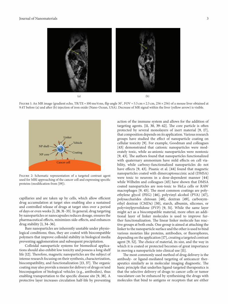
A passive or active method can be used to deliver nano-particles to the specific site. An example of passive applicationof iron-based nanoparticles is liver cancer that lacks an effi-cient method of early diagnosis. Current techniques, includ-ing ultrasound, CT, and MRI, detect liver tumors only whenthey have grown to about 5 centimeters in diameter. By thattime, the cancer is especially aggressive, resisting chemother-apy, and difficult to remove surgically. Application of iron-based nanoparticles improved MRI sensitivity due to accu-mulation of iron in the liver caused by selective action of thehepatobiliary system (Figure 1).This type of contrast deliverydoes not apply, however, to most of the cancers thus targeted,and active delivery is used.
From the point of view ofMRI technique, to increaseMRIsensitivity, two types of contrast agents, providing positive ornegative image contrast, are used. Contrast agents comprisinggadolinium (Gd) or manganese (Mn) provide hyperintenseT1-weighted tumor images [13–16], while superparamagnetic
nanoparticles reduce T2and 𝑇
2
∗ of surrounding water mol-ecules, thus decreasing MR signal in T
2- and 𝑇
2
∗-weightedMRI (negative image contrast) in the areas corresponding tothe location of the disease [17]. Gadolinium (III), with its highelectron magnetic moment, is the most common T
1contrast
agent, that provides nonspecific positive T1contrast.
Free Gd3+ is toxic (LD50= 0.2mmol kg−1 in mice); there-
fore, it is administered in the form of stable chelate complexesthat prevent the release of themetal ion in vivo [18]. Followingintravascular injection, nonspecific Gd-based compoundsdistribute rapidly between plasma and interstitial spaces andare ultimately eliminated through the renal route with half-lives of about 1.6 h [18]. Polyaminocarboxylate ligands, whichincorporate nitrogen and oxygen donor atoms, are used tocoordinate the Gd center. The Gd-based contrast agents areprovided commercially by various suppliers. Accumulationof these contrast agents is solely based on differences in thevasculature between tumor and normal tissues; thus, MRIrecognition of specific tumor types is not achieved.Molecular
MR imaging rectifies this drawback by taking advantage ofthe distinctive cell properties (such as a unique pattern ofprotein expression) of the tumor and combines them withsuperparamagnetic nanoparticles enabling both sensitiveand specific detection of molecular targets associated withearly events in carcinogenesis [2]. To enable MR specificity,nanoparticles may be conjugated with various organic vehi-cles (Figure 2), for example, with single domain antibodies(sdAb) that are specific for proteins that are overexpressed onthe surface of the tumor cells, in the tumor microenviron-ment (e.g., the extracellular matrix (ECM)), or by the tumorvasculature.
There are various corresponding receptors such as epider-mal growth factor receptor (EGFR), a cell surface receptorknown to be overexpressed, for example, in the triple negative(TN) breast cancers or secreted clusterin (sCLU), and a pro-tein that is secreted into the microenvironment and that hasbeen shown to be associated with the progression of primaryto metastatic carcinoma. Insulin Growth Factor BindingProtein 7 (IGFBP-7) has been shown to be specifically over-expressed by the tumor vasculature; it can also be used asa vascular target [19, 20]. The agents against these selectedtargets can be developed using single domain antibodies thathave been shown to specifically bind to these targets. Sucha probe allows localization of the disease in vivo, and poten-tially gives insight into biological processes (e.g., angiogenesisand metastasis) which are critical to tumor development andcan, therefore, be used to monitor the response of a tumor toindividualized therapy. This way, treatment may be appliedat a curable stage and adjusted if needed. Furthermore, MRI,in particular when combined with application of nanopar-ticles, has a capability in cancer staging, following up theprogress of treatment, and accurate detection of lymph nodesinvolvement in disease [21] as showed in the recently reporteddetection of small and otherwise undetectable lymph nodemetastases in patients with prostate cancer [22, 23].
3. Therapeutic Applications of Nanoparticles
While diagnostic is a common medical application of nano-particles, they can also be used for therapy [9, 24, 25]. Theirproperties offer unique interactions with biomolecules bothon the surface and inside the cells, enabling significantimprovement in cancer diagnosis and treatment [26]. There-fore, nanoparticles have been recently utilized by biologists,pharmacologists, and physicists, physicians as well as thepharmaceutical industry [27].
There are about 20 clinically approved nanomedicinesused for treatment. An example is Abraxane, an albumin-bound form of paclitaxel with Cobalt of mean particle size ofapproximately 130 nm that is used to treat breast cancer [28].Doxil, also based on Cobalt, is used for the treatment ofrefractory ovarian cancer and AIDS-related Kaposi’s sarcomaand it consists of nanoparticles with a polyethylene glycol(PEG) coating [11, 29, 30].
A primary attribute of nanoparticles delivery systems istheir potential to enhance the accumulation of anticanceragents in tumor cells as some nanoparticles passively accu-mulate in tumors after their intravenous administration[1, 28, 31–33]. Nanoparticles can penetrate through small
Journal of Nanomaterials 3
(a) (b)
Figure 1: An MR image (gradient echo, TR/TE= 100ms/4ms, flip angle 30∘, FOV= 5.5 cm× 2.5 cm, 256× 256) of a mouse liver obtained at9.4T before (a) and after (b) injection of iron oxide (Nano-Ocean, USA). Decrease of MR signal within the liver (yellow arrow) is visible.
NP core Coating
VehicleNP shell
Linker
Cancer cell
Figure 2: Schematic representation of a targeted contrast agentused for MRI approaching of the cancer cell and expressing specificproteins (modification from [19]).
capillaries and are taken up by cells, which allow efficientdrug accumulation at target sites enabling also a sustainedand controlled release of drugs at target sites over a periodof days or even weeks [1, 28, 31–33]. In general, drug targetingby nanoparticles or nanocapsules reduces dosage, ensures thepharmaceutical effects, minimizes side-effects, and enhancesdrug stability [1, 34–36].
Bare nanoparticles are inherently unstable under physio-logical conditions; thus, they are coated with biocompatiblepolymers that improve colloidal stability in biological mediapreventing agglomeration and subsequent precipitation.
Colloidal nanoparticle systems for biomedical applica-tions should also exhibit low toxicity and possess a long shelflife [12]. Therefore, magnetic nanoparticles are the subject ofintense research focusing on their synthesis, characterization,biocompatibility, and functionalization [13, 37]. The organiccoatingmay also provide ameans for delivery of drugs or/andbioconjugation of biological vehicles (e.g., antibodies), thusenabling transportation to the specific disease site [9, 38]. Aprotective layer increases circulation half-life by preventing
action of the immune system and allows for the addition oftargeting agents. [11, 30, 39–42]. The core particle is oftenprotected by several monolayers of inert material [9, 17],that composition depends on its application. Various researchgroups have studied the effect of nanoparticle coating oncellular toxicity [9]. For example, Goodman and colleagues[43] demonstrated that cationic nanoparticles were mod-erately toxic, while as-anionic nanoparticles were nontoxic[9, 43]. The authors found that nanoparticles functionalizedwith quaternary ammonium have mild effects on cell via-bility, while carboxy-functionalized nanoparticles do nothave effects [9, 43]. Pisanic et al. [44] found that magneticnanoparticles coated with dimercaptosuccinic acid (DMSA)were toxic to neurons in a dose-dependent manner [44]while Wilhelm and colleagues [45] have shown that DMSAcoated nanoparticles are non-toxic to HeLa cells or RAWmacrophages [9, 45]. The most common coatings are poly-ethylene glycol (PEG) [46], polyvinyl alcohol (PVA) [47],polysaccharides chitosan [48], dextran [49], carboxym-ethyl dextran (CMDx) [50], starch, albumin, silicones, orpolyvinylpyrrolidone (PVP) [9, 51]. While the same layermight act as a biocompatible material, more often an addi-tional layer of linker molecules is used to improve fur-ther functionalization. The linear linker molecule has reac-tive groups at both ends. One group is aimed at attaching thelinker to the nanoparticle surface and the other is used to bindvarious moieties like proteins, antibodies, or fluorophores,depending on the application [17], creating a targeted contrastagent [9, 52]. The choice of material, its size, and the way inwhich it is coated or protected becomes of great importancein moving a nanoparticle into clinical use [11].
The most commonly used method of drug delivery is theantibody- or ligand-mediated targeting of anticancer ther-apeutics similarly as in molecular imaging diagnostic. Thebasic principle that underlies ligand-targeted therapeutics isthat the selective delivery of drugs to cancer cells or tumorvasculature can be enhanced by synthesising the drugs withmolecules that bind to antigens or receptors that are either

4 Journal of Nanomaterials
uniquely expressed or over-expressed on target cells [1, 53–56]. The use of new synthesis techniques, such as condensa-tion reactions, allowed the incorporation of various targetingligands to the nanoparticle shell, including EGF-related tar-gets [11, 57, 58], transferrin [11, 59, 60], lactoferrin [11, 61],transactivating transcriptional activator [11, 62], aptamers [11,63], and numerous other peptides such as chlorotoxin [11, 64–68]. For example, the use of the peptide sequence known asAngiopep has recently become important for the targeting ofbrain cancer [11, 69] as both the BBB and gliomas are knownto overexpress the corresponding receptors [11, 69]. Manyresearchers have recently utilized various coatings to improvethe drug delivery. For example, Veiseh et al. [66, 70] foundthat the incorporation of chlorotoxin onto functionalizedFe3O4nanoparticles resulted in a significant increase in
the total uptake within the brain tumors of mice after invivo injection when compared with untargeted particles;Kim and coworkers found that hydrophobic drugs couldbe incorporated into monolayers of polyelectrolyte-coatedgold nanoparticles for cellular delivery [11, 71]. Liu et al.utilized polymer-coatedmagnetic nanoparticles to deliver theanticancer drug epirubicin and to provide an MRI contrastagent for brain cancer [11, 72].
An example of an organic-based delivery vehicle isliposomes, which are spherical in shape and consist of aphospholipid shell that can be used to encapsulate and deliverboth hydrophobic and hydrophilic drugs [11]. They are onaverage 100 nm in diameter [41, 73–75]. Doxorubicin was thefirst drug to be delivered by liposomes to brain tumors [11,41, 74, 75]. High-density lipoprotein (HDL) nanoparticles areclosely related to liposomal nanocarriers, having the stabilityand monodispersity of inorganic nanoparticles combinedwith the shielding ability of liposomes that improve circula-tion half-lives of therapeutics [76–78].
Nanoparticles with controllable sizes ranging from afew nanometers up to tens of nanometers are of particularinterest. They are thousands of times smaller than cells andcomparable with viruses, proteins, and genes.Therefore, theyare able to cross biological membranes, interact closely withbiomolecules enabling access to intra- and extracellular spa-ces thus inducing various responses in biological systems [79]and improving cancer therapy and/or diagnosis. Nanoparti-cles provide a means to increase transport across the BBBand/or blood-brain-tumor barrier (BBTB) and for this reasonhave been exploited in the treatment of brain cancer [11, 80–86]. For example, nanoparticles are promising in gliomatreatment. This brain cancer is particularly difficult to treat[11, 87, 88] as neurosurgery is ineffective, while chemotherapysuffers from the inability of therapeutics to cross the blood-brain barrier (BBB). Several different types of nanoparticleshave been employed as imaging and delivery agents for braincancer treatment, including Fe
3O4nanoparticles [11, 42, 66,
70, 89–93], gadolinium [81, 94–96], gold [97], semiconductorquantum [QDs] [11, 58, 98], and organic-based (dendrimer,hydrogel, and polymer) nanoparticles [11, 64, 67, 68, 73, 99–101].
While nanoparticles can function as delivery vehicleswith variable sizes, shapes, and surfaces that serve to increasebioavailability and specificity of cancer therapeutics, they
can also allow loading of additional drugs for simultaneousmultidrug delivery. The addition of imaging probes may beutilized for simultaneous diagnosis, therapy, andmonitoring.Finally, toxicity of nanoparticles could also be potentiallyutilized to destroy the cancer cells [11, 41, 102–104].
Although not yet fully developed, methods of activationof nanoparticles after reaching the target are being investi-gated. An example is the use of metallic nanoparticles thatcan be heated with light, radiofrequency, or magnetic fieldsfor thermal ablation of tumors [17, 43–46, 105–107].The oscil-lating magnetic field can be applied after the particles reachthe tissue of interest, as determined, for example, byMRI.Thedrug release is induced by the temperature increase gener-ated by the magnetic nanoparticles subject to an oscillatingmagnetic field. This temperature increase is then utilizedto stimulate a thermoresponsive polymer synthesised to thenanoparticle surface.
4. Iron-Based Nanoparticles
The inorganic nanoparticles that have been applied clinicallyare mainly nanoparticles based on iron oxide, Fe
3O4, with
diameters around 50 nm as these nanoparticles have beenrelatively well-tolerated.
The most common and the first to be applied in MRInanoparticle is the so-called small and ultrasmall superpara-magnetic iron oxide (SPIO and USPIO, resp.). SPIONs aretypically monocrystalline composed of magnetite (Fe
3O4) or
maghemite (𝛾-Fe2O4) [22]. Because iron oxide has a relatively
low saturation magnetization, it requires the use of large par-ticles to achieve sufficient MRI contrast [4]. Iron oxide nano-particles vary in size and may have different types of surfacecoating, which significantly affect their blood half-life, bio-distribution, and uptake. The synthesis method utilized toproduce SPIO nanoparticles determines the size and polydis-persity of the particle population [5, 6].
Magnetic iron oxide particles have been used clinicallysince 1987, when they were applied for the detection of focalliver and spleen lesions withMRI. SPIOs, with hydrodynamicdiameter larger than 30 nm, tend to have a short blood half-life as they are taken up by mononuclear phagocytosingsystem (MPS) in liver and spleen, leading to a significant MRsignal loss in these tissues in 𝑇
2-weighted MR images [22,
108]. Focal liver lesions without an MPS or without an intactMPS do not show this accumulation and maintain their pre-contrast signal intensity [22]. Thus, SPIO-enhanced MRIshows an increase of liver-to-tumor contrast with respect tothe precontrast images, allowing differential diagnosis ofmalignant versus benign liver lesions or metastases [14, 15,22]. The USPIOs (<30 nm diameter) can escape the initialuptake by liver and spleen; thus, they can reach other targetsthat can be then indirectly detectable with MRI and thus areused as targeted contrast agents after their bioconjugation.
There are several commercially available compounds con-taining superparamagnetic iron oxide such as Feridex (Ber-lex, USA), Endorem (Guerbet, EU), and Resovist (Schering,EU, Japan). They are mostly used for liver and spleen tumorsdiagnosis [22]. These particles are of medium size and arecoated with dextran (Feridex, Endorem) or an alkali-treated

Journal of Nanomaterials 5
lowmolecular weight carboxydextran (Resovist).Their relax-ivity (𝑟
2= 1/𝑇
2) varies [109]: 186mM−1s−1 (Resovist,
4.0 nm core, hydrodynamic diameter 60 nm), 120mM−1s−1(Feridex, 4.96 nm core, hydrodynamic diameter 160 nm), and65mM−1s−1 (Combinex, 5.85 nm core, hydrodynamic diam-eter 30 nm) at 1.5 T.
Although iron oxides have been the most widely used,biomedical applications of magnetic ferrites are currentlybeing intensely investigated. In particular, substituted mag-netic spinel ferrites of the general formula MFe
2O4(where
M=Zn2+, Mn2+, Co2+, Ni2+, and Mg2+) offer the opportu-nity to fine-tune the magnetic properties of the inorganicnanoparticle core as a function of the kind of divalent ion [16].Large magnetic moments, observed in these nanoparticles,are preferred for most applications, as they reduce theamount of nanoparticles needed to detect them with MRI.However, their toxic effects are often considerable and needto be reduced. Therefore, a balance between larger magneticmoments, nanoparticles concentration, and their biocompat-ibility is the goal of the researchers involved in the synthesisof clinically relevant nanoparticles [16].
5. Other Nanoparticles
As mentioned above, other than iron oxide based nanoparti-cles with potential clinical application in MRI and/or CT arecobalt (Co), gold (Au@Fe), and platinum (Pt@Fe). As theyhave much higher saturation magnetization value than thatof the iron, they have much larger effect on proton relaxation(𝑟1= 7.4mM−1s−1, 𝑟
2= 88mM−1s−1 for copolymer at
1.5 T, 3.9 nm core diameter, 28 nm particle diameter [110])providing better MR contrast than iron oxide in the sameconcentration and allowing smaller particle cores to be usedwithout compromisingMR sensitivity [4]. Probably the mostfrequently used is cobalt. While cobalt toxicity is an issue, theundesired effects of cobalt in man are difficult to evaluate,as they are also dependent on nutritional factors [111]. Manypatients have taken up to 50mg cobalt per day as treatmentof refractory anemia for long periods with little or no toxicity[4]. Most cobalt drugs also contain ferrous sulfate, whichmay affect the amount of cobalt absorbed, since cobalt andiron share a common absorption pathway. In contrast, 10mgcobalt/day taken by heavy beer drinkers in the 1960smayhaveresulted in cardiomyopathy [111], as the effect of inadequateprotein intake, thiamine intake, zinc depletion, and alcoholmay render the heart more sensitive to Co2+ toxicity [4].
As Au@Fe magnetic moment is high and it has limitedreactivity, it can also be used as an MR contrast agent. Thereare many subtypes of gold-based nanoparticles depending ontheir size, shape, and physical properties. The earliest studiedgold-based nanoparticles were gold nanospheres (althoughnot exactly spherical in a strict sense). Subsequently, goldnanorods, nanoshells, and nanocages have been investigated[26]. With continued development in the synthesis tech-niques over the last two decades, most of these gold nano-particles can now be produced with well-controlled sizedistribution.
Gold nanoparticles have recently been investigated indelivering therapeutics to the brain cancer [86, 112–114].
These nanoparticles have the advantages of relatively straight-forward synthesis, easy surface functionalization, small sizes,ability to be excreted by the body and remain relativelynontoxic [11, 57, 82]. Because gold is an excellent absorber ofX-rays, it was used for improved cancer therapy. The tumorscould be loaded with contrast agents containing gold increas-ing the radiation dose within the tumor and thus reducingunwanted radiation of normal tissue [115]. Qian et al. [116]applied gold nanoparticles for in vivo tumor targeting anddetection based on pegylated gold nanoparticles and surface-enhanced Raman scattering (SERS). Colloidal gold has beenfound to amplify the efficiency of Raman scattering by 14-15orders of magnitude [116]. A gold colloid was encoded withRaman reporter molecules and covered with a layer of thiol-PEG. Approximately 1.4-1.5 × 10⁴ reporter molecules wereadsorbed on each 60 nm colloid gold [116].
One of the most interesting and promising biomedicalapplications of Au-based nanoparticles is their application forintracellular delivery vectors for drugs and genes [117, 118].Yan et al. [119] proposed one-pot-synthesized polypeptide-conjugated Au nanoparticles for gene delivery and efficienttransfection. In their approach, positively charged polypep-tides were used to serve as capping agents as well as reduc-tants eliminating the need for an external reducing agent.The resulting positively charged polypeptide-conjugated goldnanoparticles were applied for gene delivery due to prolonged(almost two weeks) and gradual intracellular uptake andtransfection [119].
In addition to providingMRI contrast, gold nanoparticlesmay provide a suitable bimodal, CT, andMRI contrast [11, 42,83, 120, 121]. It is worth to mention that gold nanoparticleshave been examined by the USA National Institute of Stan-dards and Technology as a potential standard for researchbased on nanosized particles [86, 97, 113, 114].
6. Core-Shell Nanoparticles
The very recent development in nanotechnology enabled theproduction of complex particles consisting of the core andshell, each made of different atoms, such as FePt@Au [109].In principle, there are two types of core/shell nanoparticlesused in imaging applications: inorganic/organic and inor-ganic/inorganic [122]. The most common organic shell is sil-ica (SiO
2), while inorganicmaterial comprises variousmetals.
Many inorganic core-shell nanoparticles have been con-structed, includingAu@Ag [123], Au@Co [124], Au@Pt [125],Au@TiO
2[126], Au@Fe
2O3[127], Ni@Ag [128], Fe@Ag [129],
Ni@Pt [130], Co@Au [131], Fe@Pt [132], LaF3@Eu [133] or 𝛽-
NaYF4: Yb3+, and Er3+/𝛽-NaYF
4[134].
This development allowed new applications of nanopar-ticles, for example, as targeted contrast agents generatingpositive contrast inMRI. Standard contrast agents shortening𝑇2have been developed, yet efficient targeted contrast agents
shortening both 𝑇1and 𝑇
2are still an area of research as the
core/shell nanoparticles could provide improved tumordelineation and hyperintense tumor MRI due to shorteningboth 𝑇
1and 𝑇
2, unlike standard iron-based nanoparticles
that shorten mostly 𝑇2[43, 135–137]. These core-shell nano-
particles can be stabilized by an organic coating that can be

6 Journal of Nanomaterials
pegylated for the reduction of nonspecific binding andfurther chemically modified for subsequent bioconjugationof the biological vehicles such as antibodies, for example,against IGFBP7 used for glioma detection [19].
7. Production of Nanoparticles
A commonly used method of magnetite synthesis is thecoprecipitation of iron salts in aqueous media at room tem-perature under basic, inert conditions [7, 8]. This relativelystraightforward method results in the formation of largeamounts of magnetic core clusters of about 36 nm composedof single particles around 10 nm; however, the generatedclusters are very polydisperse. Difficult control of aggregationand particle size distribution are the disadvantages of thecoprecipitation method. An alternative to coprecipitation isthe thermal decomposition method [24, 25, 34, 38, 51, 52].In this method, an iron oleate precursor is prepared which isthen decomposed into an iron oxide at high temperature in anorganic solvent.The resulting nanoparticles have narrow sizedistributions but unfortunately are coatedwith a hydrophobiclayer of oleic acid. In order to obtain stable aqueous disper-sions of these particles in water, OA on the surface of theparticles is exchanged for another ligand [35] which not onlystabilizes the particle in suspension but can also serve to cova-lently attach other molecules to the surface of the particle [8].
Themost common synthesis methods of core/shell nano-particles are chemical vapor deposition, laser-induced assem-bly, self-assembly, and colloidal aggregation [138, 139].
In themicroemulsionmethod [140], surfactants allow thehomogenization of all types of reactants, and the particlesformed are capped by the surfactant molecules [141]. Thus,the size of the nanoparticles can be controlled varying a con-centration of surfactant [142].Mandal et al. [141] used glucoseto control shell growth of gold or silver onto Fe
3O4particles
upon heating of the mixture of Fe3O4particles. To cover
Fe3O4nanoparticles with gold or silver, a modified micro-
emulsion method has been used [141]. This method allowsshell thickness of the core-shell particles to be tunable andallowed production of structures of size from 18 to 30 nmwithvarying proportion of Fe
3O4to the noble metal precursor
salts [141].Very recently a very promising method of production of
3D colloidal spheres containing various nanoparticles wasproposed [143]. These multifunctional nanoparticles may beused for different applications such as multimodal imaging,remotely controlled release, targeted drug delivery, or simul-taneous diagnosis and therapy [144].This so-called template-assisted fabrication process uses porous calcium (CaCO
3)
microspheres as a sacrificial template. This method allowseasy control of the size of the spheres, flexible tuning of theirbiochemical and physical properties, and encapsulation ofvarious nanoparticles. The process comprises adsorption ofnanoparticles into the porous CaCO
3sphere, encapsulation
of polyelectrolytes, and removal of the template by cross-linking. The end product is a colloidal sphere. Using thismethod, Au nanoparticles and cross-linked poly-L-lysine(PLL) (P-AuNPs) [143], citrate-stabilized gold nanoparti-cles (C-AuNPs) [145], cetyl trimethylammonium bromide
(CTAB) capped gold nanorods (GNRs) [146], and magneticnanoparticles (𝛾-Fe
2O3) were used to create 3D hybrid
colloidal spheres [147].
8. Nanoparticles for Multimodal Imaging
While various imaging techniques, such asMRI, CT, PositronEmission Tomography (PET), and infrared (IR) imaging,have been used for diagnosis and treatment monitoring, eachone delivers different information on disease and its location.There is no perfect imaging method, as each technique hasits advantages and disadvantages. MRI provides the best softtissue contrast but its sensitivity is low; PET is more sensitivethan MRI but its spatial resolution is low; CT is fast butsoft tissue contrast is low; and finally infrared imaging is fastand very sensitive but the depth of penetration is very low.Nanotechnology allowing production ofmultimodal contrastagents (“all in one”) takes advantages of all these modalities.
Of particular interest is the recent development of rareearth upconversion nanophosphors (RE-UCNPs) [148–152]as potential contrast agents because of their optical and bio-chemical properties, such as sharp emission lines, long life-times, and nonphotoblinking. In particular, Yb3+ and Tm3+codoped RE-UCNPs emitting at 800 nm have been used for awhole-body small-animal near-infrared imaging [153]. Thistechnique allowed detection of only 50 cells in a whole-bodymouse imaging [154]. Unfortunately, photoluminescentimaging has a low light penetration depth, but this limitationcould be rectified by simultaneous application of MRI or/andCT with a contrast agent suitable for all these techniques.Therefore, Gd3+ was synthesised with RE-UCNPs creat-ing magnetic-luminescent RE-UCNPs contrast agent forbimodal imaging, allowing 𝑇
1-enhanced MRI and upcon-
version luminescence imaging (UCL) [155]. Furthermore, toenable CT, MRI, and luminescence imaging using the samecontrast, superparamagnetic nanoparticles have been synthe-sized with RE-UCNPs using a crosslinker anchoring method[156]. An example isNaYF
4: Yb, Er@Fe
3O4@Au,which could
be used for MRI, optical, and CT imaging [157]. NaYF4: Yb,
Tm@Fe𝑥O𝑦
core-shell nanostructure was used for 𝑇2-
weightedMRI andUCL bimodal lymphatic imaging [158]. Ofparticular interest for multimodal contrasts may be NaLuF
4
because RE-UCNPs based on the NaLuF4have high UCL
quantum yield [159] and high X-ray absorption coefficient.Another example of multimodal application of nanoparticlesis their simultaneous utilization in high-resolution MRI andhigh-sensitivity PET formore accurate disease detection.ThePET marker (e.g., Cu64) can be added to an MR marker, cre-ating a MRI/PET contrast agent. Furthermore, radionuclideattachment can be achieved via chelating agents.
9. Conclusions
Recent developments in nanomaterials, molecular and cellu-lar biology, and imaging technology enabled to enhance ourdiagnostic and therapeutic capabilities, improving detectionlimits from the tissue down to the cell and even to the molec-ular level. We can now combine atom and biomolecular

Journal of Nanomaterials 7
manipulation applying quantum physics, molecular chem-istry, biology, and genetics to fabricate minute syntheticstructures [1, 160] and to apply them along with high-resolu-tion noninvasive imaging technologies for diagnosis, therapy,and treatment monitoring. Current investigation of nanoma-terials in animal models has offered less invasive diagnosisand induced fewer side-effects due to improved targeting, yetup to date their clinical applications have been limited. Themajor obstacle seems to be the long time needed for clinicaltrials and associated costs. Despite that nanomaterials willlikely have a significant impact on patient care in the future.
References
[1] Y. Liu, H. Miyoshi, andM. Nakamura, “Nanomedicine for drugdelivery and imaging: a promising avenue for cancer therapyand diagnosis using targeted functional nanoparticles,” Interna-tional Journal of Cancer, vol. 120, no. 12, pp. 2527–2537, 2007.
[2] R. Weissleder, “Molecular imaging in cancer,” Science, vol. 312,no. 5777, pp. 1168–1171, 2006.
[3] R. Etzioni, N. Urban, S. Ramsey et al., “The case for early detec-tion,” Nature Reviews Cancer, vol. 3, no. 4, pp. 243–252, 2003.
[4] L.M. Parkes, R.Hodgson, L. T. Lu et al., “Cobalt nanoparticles asa novel magnetic resonance contrast agent—relaxivities at 1.5and 3 Tesla,” Contrast Media and Molecular Imaging, vol. 3, no.4, pp. 150–156, 2008.
[5] J. D. G. Duran, J. L. Arias, V. Gallardo, and A. V. Delgado,“Magnetic colloids as drug vehicles,” Journal of PharmaceuticalSciences, vol. 97, no. 8, pp. 2948–2983, 2008.
[6] A. K. Gupta and M. Gupta, “Synthesis and surface engineeringof iron oxide nanoparticles for biomedical applications,”Bioma-terials, vol. 26, no. 18, pp. 3995–4021, 2005.
[7] S. Lefebure, E. Dubois, V. Cabuil, S. Neveu, and R. Massart,“Monodisperse magnetic nanoparticles: preparation and dis-persion in water and oils,” Journal of Materials Research, vol. 13,no. 10, pp. 2975–2981, 1998.
[8] A.-H. Lu, E. L. Salabas, and F. Schuth, “Magnetic nanoparticles:synthesis, protection, functionalization, and application,”Ange-wandte Chemie, vol. 46, no. 8, pp. 1222–1244, 2007.
[9] M. Latorre and C. Rinaldi, “Applications of magnetic nanopar-ticles in medicine: magnetic fluid hyperthermia,” Puerto RicoHealth Sciences Journal, vol. 28, no. 3, pp. 227–238, 2009.
[10] M. C. Roco, “Nanoscale science and engineering: unifying andtransforming tools,” AIChE Journal, vol. 50, no. 5, pp. 890–897,2004.
[11] J. D. Meyers, T. Doane, C. Burda, and J. P. Basilion, “Nanoparti-cles for imaging and treating brain cancer,” Nanomedicine, vol.8, no. 1, pp. 123–143, 2013.
[12] S. S. Kelkar and T. M. Reineke, “Theranostics: combining imag-ing and therapy,” Bioconjugate Chemistry, vol. 22, no. 10, pp.1879–1903, 2011.
[13] C. N. Ramchand, P. Pande, P. Kopcansky, and R. V. Mehta,“Application ofmagnetic fluids inmedicine and biotechnology,”Indian Journal of Pure and Applied Physics, vol. 39, no. 10, pp.683–686, 2001.
[14] Y.-X. J. Wang, S. M. Hussain, and G. P. Krestin, “Superparam-agnetic iron oxide contrast agents: physicochemical character-istics and applications inMR imaging,”EuropeanRadiology, vol.11, no. 11, pp. 2319–2331, 2001.
[15] M. Taupitz, S. Schmitz, and B.Hamm, “Superparamagnetic ironoxide particles: current state and future development,” RoFoFortschritte auf dem Gebiet der Rontgenstrahlen und der Bildge-benden Verfahren, vol. 175, no. 6, pp. 752–765, 2003.
[16] M. Colombo, S. Carregal-Romero, and M. F. Casula, “Biolog-ical applications of magnetic nanoparticles,” Chemical SocietyReviews, vol. 41, pp. 4306–4334, 2012.
[17] O. V. Salata, “Applications of nanoparticles in biology andmedi-cine,” Journal of Nanobiotechnology, vol. 2, article 3, 2004.
[18] E. J. Werner, A. Datta, C. J. Jocher, and K. N. Raymond, “High-relaxivity MRI contrast agents: where coordination chemistrymeetsmedical imaging,”Angewandte Chemie, vol. 47, no. 45, pp.8568–8580, 2008.
[19] B. Tomanek, U. Iqbal, B. Blasiak et al., “Evaluation of braintumor vessels specific contrast agents for glioblastoma imag-ing,” Neuro-Oncology, vol. 14, no. 1, pp. 53–63, 2012.
[20] U. Iqbal, U. Trojahn, H. Albaghdadi et al., “Kinetic analysis ofnovel mono- and multivalent VHH-fragments and their appli-cation for molecular imaging of brain tumours,” British Journalof Pharmacology, vol. 160, no. 4, pp. 1016–1028, 2010.
[21] K. Firouznia, S. Amirmohseni, M. Guiti et al., “MR relaxivitymeasurement of iron oxide nano-particles for MR lymphogra-phy applications,” Pakistan Journal of Biological Sciences, vol. 11,no. 4, pp. 607–612, 2008.
[22] R. Lawaczeck, M. Menzel, and H. Pietsch, “Superparamagneticiron oxide particles: contrast media for magnetic resonanceimaging,” Applied Organometallic Chemistry, vol. 18, no. 10, pp.506–513, 2004.
[23] M. G. Harisinghani, J. Barentsz, P. F. Hahn et al., “Noninvasivedetection of clinically occult lymph-nodemetastases in prostatecancer,” New England Journal of Medicine, vol. 348, no. 25, pp.2491–2499, 2003.
[24] M.H. F.Meyer,M. Stehr, S. Bhuju et al., “Magnetic biosensor forthe detection ofYersinia pestis,” Journal ofMicrobiologicalMeth-ods, vol. 68, no. 2, pp. 218–224, 2007.
[25] J. E. Kirsch, “Basic principles of magnetic resonance contrastagents,” Topics in Magnetic Resonance Imaging, vol. 3, no. 2, pp.1–18, 1991.
[26] W. Cai, T. Gao, H. Hong, and J. Sun, “Applications of gold nano-particles in cancer,” Nanotechnology, Science and Applications,vol. 1, pp. 17–32, 2008.
[27] E. Duguet, S. Vasseur, S. Mornet, and J.-M. Devoisselle,“Magnetic nanoparticles and their applications in medicine,”Nanomedicine, vol. 1, no. 2, pp. 157–168, 2006.
[28] C. J. Sunderland, M. Steiert, J. E. Talmadge, A. M. Derfus, andS. E. Barry, “Targeted nanoparticles for detecting and treatingcancer,” Drug Development Research, vol. 67, no. 1, pp. 70–93,2006.
[29] V.Wagner, A. Dullaart, A.-K. Bock, and A. Zweck, “The emerg-ing nanomedicine landscape,”Nature Biotechnology, vol. 24, no.10, pp. 1211–1217, 2006.
[30] S. K. Nune, P. Gunda, P. K. Thallapally, Y.-Y. Lin, M. Laird For-rest, and C. J. Berkland, “Nanoparticles for biomedical imag-ing,” Expert Opinion on Drug Delivery, vol. 6, no. 11, pp. 1175–1194, 2009.
[31] G. Orive, A. R. Gascon, R. M. Hernandez, A. Domınguez-Gil,and J. L. Pedraz, “Techniques: new approaches to the deliveryof biopharmaceuticals,” Trends in Pharmacological Sciences, vol.25, no. 7, pp. 382–387, 2004.

8 Journal of Nanomaterials
[32] M. C. Roco, “Nanotechnology: convergence with modern biol-ogy and medicine,” Current Opinion in Biotechnology, vol. 14,no. 3, pp. 337–346, 2003.
[33] I. Brigger, C. Dubernet, and P. Couvreur, “Nanoparticles in can-cer therapy and diagnosis,” Advanced Drug Delivery Reviews,vol. 54, no. 5, pp. 631–651, 2002.
[34] T. M. Fahmy, P. M. Fong, A. Goyal, and W. M. Saltzman, “Tar-geted for drug delivery,”Materials Today, vol. 8, no. 8, pp. 18–26,2005.
[35] T. M. Fahmy, R. M. Samstein, C. C. Harness, and W. M. Saltz-man, “Surface modification of biodegradable polyesters withfatty acid conjugates for improved drug targeting,”Biomaterials,vol. 26, no. 28, pp. 5727–5736, 2005.
[36] P. Couvreur, G. Barratt, E. Fattal, P. Legrand, and C. Vauthier,“Nanocapsule technology: a review,” Critical Reviews in Thera-peutic Drug Carrier Systems, vol. 19, no. 2, pp. 99–134, 2002.
[37] J. Roger, J. N. Pons, R. Massart, A. Halbreich, and J. C. Bacri,“Some biomedical applications of ferrofluids,” The EuropeanPhysical Journal Applied Physics, vol. 5, no. 3, pp. 321–325, 1999.
[38] F. Alexis, J.-W. Rhee, J. P. Richie, A. F. Radovic-Moreno, R.Langer, andO. C. Farokhzad, “New frontiers in nanotechnologyfor cancer treatment,” Urologic Oncology, vol. 26, no. 1, pp. 74–85, 2008.
[39] L. Zhang, F. X. Gu, J. M. Chan, A. Z. Wang, R. S. Langer, and O.C. Farokhzad, “Nanoparticles in medicine: therapeutic applica-tions and developments,” Clinical Pharmacology andTherapeu-tics, vol. 83, no. 5, pp. 761–769, 2008.
[40] M. Ferrari, “Cancer nanotechnology: opportunities and chal-lenges,” Nature Reviews Cancer, vol. 5, no. 3, pp. 161–171, 2005.
[41] D. A. Orringer, Y. E. Koo, T. Chen, R. Kopelman, O. Sagher,and M. A. Philbert, “Small solutions for big problems: theapplication of nanoparticles to brain tumor diagnosis andtherapy,” Clinical Pharmacology andTherapeutics, vol. 85, no. 5,pp. 531–535, 2009.
[42] S. A. Anderson, J. Glod, A. S. Arbab et al., “Noninvasive MRimaging of magnetically labeled stem cells to directly identifyneovasculature in a glioma model,” Blood, vol. 105, no. 1, pp.420–425, 2005.
[43] C. M. Goodman, C. D. McCusker, T. Yilmaz, and V. M. Rotello,“Toxicity of gold nanoparticles functionalized with cationic andanionic side chains,” Bioconjugate Chemistry, vol. 15, no. 4, pp.897–900, 2004.
[44] T. R. Pisanic II, J. D. Blackwell, V. I. Shubayev, R. R. Finones, andS. Jin, “Nanotoxicity of iron oxide nanoparticle internalizationin growing neurons,”Biomaterials, vol. 28, no. 16, pp. 2572–2581,2007.
[45] C. Wilhelm, C. Billotey, J. Roger, J. N. Pons, J.-C. Bacri, and F.Gazeau, “Intracellular uptake of anionic superparamagneticnanoparticles as a function of their surface coating,” Biomate-rials, vol. 24, no. 6, pp. 1001–1011, 2003.
[46] Y. Zhang and J. Zhang, “Surface modification of monodispersemagnetite nanoparticles for improved intracellular uptake tobreast cancer cells,” Journal of Colloid and Interface Science, vol.283, no. 2, pp. 352–357, 2005.
[47] J.-S. Kim, T.-J. Yoon, K.-N. Yu et al., “Cellular uptake of mag-netic nanoparticle is mediated through energy-dependentendocytosis in A549 cells,” Journal of Veterinary Science, vol. 7,no. 4, pp. 321–326, 2006.
[48] S. R. Bhattarai, B. K. Remant, S. Y. Kim et al., “N-hexanoyl chi-tosan stabilizedmagnetic nanoparticles: implication for cellular
labeling and magnetic resonance imaging,” Journal of Nano-biotechnology, vol. 6, article 1, 2008.
[49] R. Trehin, J.-L. Figueiredo, M. J. Pittet, R.Weissleder, L. Joseph-son, and U. Mahmood, “Fluorescent nanoparticle uptake forbrain tumor visualization,” Neoplasia, vol. 8, no. 4, pp. 302–311,2006.
[50] J. H. Clement, M. Schwalbe, N. Buske et al., “Differentialinteraction of magnetic nanoparticles with tumor cells andperipheral blood cells,” Journal of Cancer Research and ClinicalOncology, vol. 132, no. 5, pp. 287–292, 2006.
[51] H.-Y. Lee, S.-H. Lee, C. Xu et al., “Synthesis and characterizationof PVP-coated large core iron oxide nanoparticles as an MRIcontrast agent,”Nanotechnology, vol. 19, no. 16, Article ID 165101,2008.
[52] P. Phanapavudhikul, S. Shen, W. K. Ng, and R. B. H. Tan, “For-mulation of Fe
3O4/acrylate co-polymer nanocomposites as
potential drug carriers,” Drug Delivery, vol. 15, no. 3, pp. 177–183, 2008.
[53] W. Arap, R. Pasqualini, and E. Ruoslahti, “Cancer treatment bytargeted drug delivery to tumor vasculature in a mouse model,”Science, vol. 279, no. 5349, pp. 377–380, 1998.
[54] S. Dagar, M. Sekosan, B. S. Lee, I. Rubinstein, and H. Onyuksel,“VIP receptors as molecular targets of breast cancer: implica-tions for targeted imaging and drug delivery,” Journal of Con-trolled Release, vol. 74, no. 1–3, pp. 129–134, 2001.
[55] B. P. Eliceiri and D. A. Cheresh, “Adhesion events in angiogene-sis,” Current Opinion in Cell Biology, vol. 13, no. 5, pp. 563–568,2001.
[56] C. Ehrhardt, C. Kneuer, and U. Bakowsky, “Selectins—anemerging target for drug delivery,” Advanced Drug DeliveryReviews, vol. 56, no. 4, pp. 527–549, 2004.
[57] Y. Cheng, J. D. Meyers, A.-M. Broome, M. E. Kenney, J. P. Basil-ion, andC. Burda, “Deep penetration of a PDTdrug into tumorsby noncovalent drug-gold nanoparticle conjugates,” Journal ofthe American Chemical Society, vol. 133, no. 8, pp. 2583–2591,2011.
[58] H. Jackson, O. Muhammad, H. Daneshvar et al., “Quantumdots are phagocytized by macrophages and colocalize withexperimental gliomas,”Neurosurgery, vol. 60, no. 3, pp. 524–529,2007.
[59] W.-H. Ren, J. Chang, C.-H. Yan et al., “Development of trans-ferrin functionalized poly(ethylene glycol)/poly(lactic acid)amphiphilic block copolymeric micelles as a potential deliverysystem targeting brain glioma,” Journal of Materials Science, vol.21, no. 9, pp. 2673–2681, 2010.
[60] J. Chang, A. Paillard, C. Passirani et al., “Transferrin adsorp-tion onto PLGA nanoparticles governs their interaction withbiological systems from blood circulation to brain cancer cells,”Pharmaceutical Research, vol. 29, pp. 1495–1505, 2012.
[61] H. Xie, Y. Zhu, W. Jiang et al., “Lactoferrin-conjugated super-paramagnetic iron oxide nanoparticles as a specific MRI con-trast agent for detection of brain glioma in vivo,” Biomaterials,vol. 32, no. 2, pp. 495–502, 2011.
[62] L. Han, A. Zhang, H.Wang, P. Pu, C. Kang, and J. Chang, “Con-struction of novel brain-targeting gene delivery system by natu-ral magnetic nanoparticles,” Journal of Applied Polymer Science,vol. 121, no. 6, pp. 3446–3454, 2011.
[63] H. Gao, J. Qian, S. Cao et al., “Precise glioma targeting of andpenetration by aptamer and peptide dual-functioned nanopar-ticles,” Biomaterials, vol. 33, no. 20, pp. 5115–5123, 2012.

Journal of Nanomaterials 9
[64] O. Veiseh, F. M. Kievit, J. W. Gunn, B. D. Ratner, andM. Zhang,“A ligand-mediated nanovector for targeted gene delivery andtransfection in cancer cells,”Biomaterials, vol. 30, no. 4, pp. 649–657, 2009.
[65] C. Sun, O. Veiseh, J. Gunn et al., “In vivo MRI detection ofgliomas by chlorotoxin-conjugated superparamagnetic nano-probes,” Small, vol. 4, no. 3, pp. 372–379, 2008.
[66] O. Veiseh, C. Sun, C. Fang et al., “Specific targeting of braintumors with an optical/magnetic resonance imaging nanoprobeacross the blood-brain barrier,” Cancer Research, vol. 69, no. 15,pp. 6200–6207, 2009.
[67] R. Kopelman, Y.-E. Lee Koo,M. Philbert et al., “Multifunctionalnanoparticle platforms for in vivo MRI enhancement and pho-todynamic therapy of a rat brain cancer,” Journal of Magnetismand Magnetic Materials, vol. 293, no. 1, pp. 404–410, 2005.
[68] G. R. Reddy, M. S. Bhojani, P. McConville et al., “Vascular tar-geted nanoparticles for imaging and treatment of brain tumors,”Clinical Cancer Research, vol. 12, no. 22, pp. 6677–6686, 2006.
[69] H. Xin, X. Jiang, J. Gu et al., “Angiopep-conjugated poly(ethyl-ene glycol)-co-poly(𝜀-caprolactone) nanoparticles as dual-tar-geting drug delivery system for brain glioma,” Biomaterials, vol.32, no. 18, pp. 4293–4305, 2011.
[70] O. Veiseh, C. Sun, J. Gunn et al., “Optical and MRI multifunc-tional nanoprobe for targeting gliomas,”Nano Letters, vol. 5, no.6, pp. 1003–1008, 2005.
[71] C. K. Kim, P. Ghosh, C. Pagliuca, Z.-J. Zhu, S.Menichetti, andV.M. Rotello, “Entrapment of hydrophobic drugs in nanoparticlemonolayerswith efficient release into cancer cells,” Journal of theAmerican Chemical Society, vol. 131, no. 4, pp. 1360–1361, 2009.
[72] H.-L. Liu, M.-Y. Hua, H.-W. Yang et al., “Magnetic resonancemonitoring of focused ultrasound/magnetic nanoparticle tar-geting delivery of therapeutic agents to the brain,” Proceedings ofthe National Academy of Sciences of the United States of America,vol. 107, no. 34, pp. 15205–15210, 2010.
[73] C.O.Noble,M. T. Krauze, D. C.Drummond et al., “Novel nano-liposomal CPT-11 infused by convection-enhanced deliveryin intracranial tumors: pharmacology and efficacy,” CancerResearch, vol. 66, no. 5, pp. 2801–2806, 2006.
[74] T. Siegal, A. Horowitz, and A. Gabizon, “Doxorubicin encap-sulated in sterically stabilized liposomes for the treatment ofa brain tumor model: biodistribution and therapeutic efficacy,”Journal of Neurosurgery, vol. 83, no. 6, pp. 1029–1037, 1995.
[75] K. Fabel, J. Dietrich, P. Hau et al., “Long-term stabilization inpatients with malignant glioma after treatment with liposomaldoxorubicin,” Cancer, vol. 92, no. 7, pp. 1936–1942, 2001.
[76] Z. Zhang, J. Chen, L. Ding et al., “HDL-mimicking peptide-lipidnanoparticles with improved tumor targeting,” Small, vol. 6, no.3, pp. 430–437, 2010.
[77] I. R. Corbin, J. Chen,W. Cao,H. Li, S. Lund-Katz, andG. Zheng,“Enhanced cancer-targeted delivery using engineered high-density lipoprotein-based nanocarriers,” Journal of BiomedicalNanotechnology, vol. 3, no. 4, pp. 367–376, 2007.
[78] W. Chen, P. A. Jarzyna, G. A. F. van Tilborg et al., “RGD pep-tide functionalized and reconstituted high-density lipoproteinnanoparticles as a versatile and multimodal tumor targetingmolecular imaging probe,” FASEB Journal, vol. 24, no. 6, pp.1689–1699, 2010.
[79] I. Lynch, K. A. Dawson, and S. Linse, “Detecting cryptic epit-opes created by nanoparticles,” Science’s STKE, vol. 2006, no.327, article pe14, 2006.
[80] W. M. Pardridge, “CNS drug design based on principles ofblood-brain barrier transport,” Journal of Neurochemistry, vol.70, no. 5, pp. 1781–1792, 1998.
[81] M. Kumar, Z. Medarova, P. Pantazopoulos, G. Dai, and A.Moore, “Novel membrane-permeable contrast agent for braintumor detection by MRI,”Magnetic Resonance in Medicine, vol.63, no. 3, pp. 617–624, 2010.
[82] M. F. Kircher, A. de la Zerda, J. V. Jokerst et al., “A brain tumormolecular imaging strategy using a new triple-modality MRI-photoacoustic-Raman nanoparticle,” Nature Medicine, vol. 18,no. 5, pp. 829–834, 2012.
[83] G. Nie, H. J. Hah, G. Kim et al., “Hydrogel nanoparticles withcovalently linked coomassie blue for brain tumor delineationvisible to the surgeon,” Small, vol. 8, no. 6, pp. 884–891, 2012.
[84] W. M. Pardridge, “Re-engineering biopharmaceuticals fordelivery to brain with molecular Trojan horses,” BioconjugateChemistry, vol. 19, no. 7, pp. 1327–1338, 2008.
[85] R. Gabathuler, “Approaches to transport therapeutic drugsacross the blood-brain barrier to treat brain diseases,” Neuro-biology of Disease, vol. 37, no. 1, pp. 48–57, 2010.
[86] F. Sousa, S. Mandal, C. Garrovo et al., “Functionalized goldnanoparticles: a detailed in vivo multimodal microscopic braindistribution study,” Nanoscale, vol. 2, no. 12, pp. 2826–2834,2010.
[87] P. Y. Wen and S. Kesari, “Malignant gliomas in adults,” NewEngland Journal of Medicine, vol. 359, no. 5, pp. 492–507, 2008.
[88] N. J. Ullrich and S. L. Pomeroy, “Pediatric brain tumors,” Neu-rologic Clinics, vol. 21, no. 4, pp. 897–913, 2003.
[89] N. Y. Hernandez-Pedro, E. Rangel-Lopez, R. Magana-Maldon-ado et al., “Application of nanoparticles on diagnosis and ther-apy in gliomas,” BioMed Research International, vol. 2013, Arti-cle ID 351031, 20 pages, 2013.
[90] C. G. Hadjipanayis, R. Machaidze, M. Kaluzova et al.,“EGFRvIII antibody-conjugated iron oxide nanoparticles formagnetic resonance imaging-guided convection-enhanceddelivery and targeted therapy of glioblastoma,”Cancer Research,vol. 70, no. 15, pp. 6303–6312, 2010.
[91] M.-Y. Hua, H.-L. Liu, H.-W. Yang et al., “The effectiveness of amagnetic nanoparticle-based delivery system for BCNU in thetreatment of gliomas,” Biomaterials, vol. 32, no. 2, pp. 516–527,2011.
[92] P. M. Costa, A. L. Cardoso, L. S. Mendonca, and A. Serani,“Tumor-targeted chlorotoxin-coupled nanoparticles for nucleicacid delivery to glioblastoma cells: a promising system forglioblastoma treatment,” Molecular Therapy Nucleic Acids, vol.2, pp. 1–20, 2013.
[93] R. Mejıas, S. Perez-Yague, A. G. Roca et al., “Liver and brainimaging through dimercaptosuccinic acid-coated iron oxidenanoparticles,” Nanomedicine, vol. 5, no. 3, pp. 397–408, 2010.
[94] R.C.Mehta,G. B. Pike, S. P.Haros, andD.R. Enzmann, “Centralnervous system tumor, infection, and infarction: detection withgadolinium-enhanced magnetization transfer MR imaging,”Radiology, vol. 195, no. 1, pp. 41–46, 1995.
[95] F. M. Kievit, O. Veiseh, C. Fang et al., “Chlorotoxin labeledmagnetic nanovectors for targeted gene delivery to glioma,”ACSNano, vol. 4, no. 8, pp. 4587–4594, 2010.
[96] J. Y. Park, M. J. Baek, E. S. Choi et al., “Paramagnetic ultrasmallgadolinium oxide nanoparticles as advanced T1 MRI contrastagent: account for large longitudinal relaxivity, optimal particlediameter, and in Vivo T1 MR images,” ACS Nano, vol. 3, no. 11,pp. 3663–3669, 2009.

10 Journal of Nanomaterials
[97] Y. Cheng, J. D. Meyers, R. S. Agnes et al., “Addressing braintumors with targeted gold nanoparticles: a new gold standardfor hydrophobic drug delivery?” Small, vol. 7, no. 16, pp. 2301–2306, 2011.
[98] J. Jung, A. Solanki, K. A. Memoli et al., “Selective inhibition ofhuman brain tumor cells through multifunctional quantum-dot-based siRNA delivery,” Angewandte Chemie, vol. 49, no. 1,pp. 103–107, 2010.
[99] S. C. J. Steiniger, J. Kreuter, A. S. Khalansky et al., “Chemother-apy of glioblastoma in rats using doxorubicin-loaded nanopar-ticles,” International Journal of Cancer, vol. 109, no. 5, pp. 759–767, 2004.
[100] J. M. Koziara, J. J. Oh, W. S. Akers, S. P. Ferraris, and R. J.Mumper, “Blood compatibility of cetyl alcohol/polysorbate-based nanoparticles,” Pharmaceutical Research, vol. 22, no. 11,pp. 1821–1828, 2005.
[101] W. Lu, Q. Sun, J. Wan, Z. She, and X.-G. Jiang, “Cationic albu-min-conjugated pegylated nanoparticles allow gene deliveryinto brain tumors via intravenous administration,” CancerResearch, vol. 66, no. 24, pp. 11878–11887, 2006.
[102] H. Sarin, A. S. Kanevsky, H. Wu et al., “Effective transvasculardelivery of nanoparticles across the blood-brain tumor barrierinto malignant glioma cells,” Journal of Translational Medicine,vol. 6, article 80, 2008.
[103] R. Langer, “Drug delivery and targeting,” Nature, vol. 392, no.6679, pp. S5–S10, 1998.
[104] V. P. Torchilin, “Multifunctional nanocarriers,” Advanced DrugDelivery Reviews, vol. 58, no. 14, pp. 1532–1555, 2006.
[105] G. Terentyuk, G. Akchurin, I. Maksimova, G. Maslyakova, N.Khlebtsov, and V. Tuchin, “Cancer laser thermotherapy medi-ated by plasmonic nanoparticles,” in Handbook of Photonics forBiomedical Science, pp. 763–797, 2010.
[106] K. Maier-Hauff, R. Rothe, R. Scholz et al., “Intracranial ther-motherapy using magnetic nanoparticles combined with exter-nal beam radiotherapy: results of a feasibility study on patientswith glioblastoma multiforme,” Journal of Neuro-Oncology, vol.81, no. 1, pp. 53–60, 2007.
[107] A. Jordan, R. Scholz, K. Maier-Hauff et al., “The effect ofthermotherapy using magnetic nanoparticles on rat malignantglioma,” Journal of Neuro-Oncology, vol. 78, no. 1, pp. 7–14, 2006.
[108] A. Barrefelt, M. Saghafian, R. Kuiper R et al., “Biodistribution,kinetics, and biological fate of SPION microbubbles in the rat,”International Journal of Nanomedicine, vol. 8, pp. 3241–3254,2013.
[109] H. B. Na, I. C. Song, and T. Hyeon, “Inorganic nanoparticles forMRI contrast agents,” Advanced Materials, vol. 21, no. 21, pp.2133–2148, 2009.
[110] V. B. Bregar, J. Lojk, V. Sustar, P. Veranic, andM. Pavlin, “Visual-ization of internalization of functionalized cobalt ferrite nano-particles and their intracellular fate,” International Journal ofNanomedicine, vol. 8, pp. 919–931, 2013.
[111] C. S. Alexander, “Cobalt-beer cardiomyopathy. A clinical andpathologic study of twenty-eight cases,” The American Journalof Medicine, vol. 53, no. 4, pp. 395–417, 1972.
[112] N. Khlebtsov and L. Dykman, “Biodistribution and toxicity ofengineered gold nanoparticles: a review of in vitro and in vivostudies,” Chemical Society Reviews, vol. 40, no. 3, pp. 1647–1671,2011.
[113] W.H.De Jong,W. I. Hagens, P. Krystek,M. C. Burger, A. J. A.M.Sips, and R. E. Geertsma, “Particle size-dependent organ distri-bution of gold nanoparticles after intravenous administration,”Biomaterials, vol. 29, no. 12, pp. 1912–1919, 2008.
[114] G. Sonavane, K. Tomoda, and K. Makino, “Biodistribution ofcolloidal gold nanoparticles after intravenous administration:effect of particle size,” Colloids and Surfaces B, vol. 66, no. 2, pp.274–280, 2008.
[115] J. F. Hainfeld, F. A. Dilmanian, D. N. Slatkin, andH.M. Smilow-itz, “Radiotherapy enhancement with gold nanoparticles,” Jour-nal of Pharmacy and Pharmacology, vol. 60, no. 8, pp. 977–985,2008.
[116] X. Qian, X.-H. Peng, D. O. Ansari et al., “In vivo tumor target-ing and spectroscopic detection with surface-enhanced Ramannanoparticle tags,” Nature Biotechnology, vol. 26, no. 1, pp. 83–90, 2008.
[117] P. Ghosh, G. Han, M. De, C. K. Kim, and V. M. Rotello, “Goldnanoparticles in delivery applications,”Advanced Drug DeliveryReviews, vol. 60, no. 11, pp. 1307–1315, 2008.
[118] D. A. Giljohann, D. S. Seferos,W. L. Daniel, M. D.Massich, P. C.Patel, and C. A. Mirkin, “Gold nanoparticles for biology andmedicine,” Angewandte Chemie, vol. 49, no. 19, pp. 3280–3294,2010.
[119] X. Yan, J. Blacklock, J. Li, and H. Mohwald, “One-pot synthesisof polypeptide-gold nanoconjugates for in vitro gene transfec-tion,” ACS Nano, vol. 6, no. 1, pp. 111–117, 2012.
[120] R. Popovtzer, A. Agrawal, N. A. Kotov et al., “Targeted goldnanoparticles enable molecular CT imaging of cancer,” NanoLetters, vol. 8, no. 12, pp. 4593–4596, 2008.
[121] L. Faucher, A.-A. Guay-Begin, J. Lagueux, M.-F. Cote, E. Petit-clerc, and M.-A. Fortin, “Ultra-small gadolinium oxide nano-particles to image brain cancer cells in vivo withMRI,” ContrastMedia and Molecular Imaging, vol. 6, no. 4, pp. 209–218, 2011.
[122] R. Ghosh Chaudhuri and S. Paria, “Core/shell nanoparticles:classes, properties, synthesismechanisms, characterization, andapplications,” Chemical Reviews, vol. 112, no. 4, pp. 2373–2433,2012.
[123] R. Guzel, Z. Ustundag, H. Eksi et al., “Effect of Au and Au@Agcore-shell nanoparticles on the SERS of bridging organicmolecules,” Journal of Colloid and Interface Science, vol. 351, no.1, pp. 35–42, 2010.
[124] F. Bao, J. F. Li, B. Ren, J. Yao, R. Gu, and Z. Tian, “Synthesis andcharacterization ofAu@Co andAu@Ni core-shell nanoparticlesand their applications in surface-enhanced Raman spectro-scopy,” Journal of Physical Chemistry C, vol. 112, pp. 345–350,2008.
[125] S. Kumar and S. Zou, “Electrooxidation of carbon monoxideand methanol on platinum-overlayer- coated gold nanoparti-cles: effects of film thickness,” Langmuir, vol. 23, no. 13, pp.7365–7371, 2007.
[126] Y. Chen, B. Zhu, M. Yao, S. Wang, and S. Zhang, “The prepara-tion and characterization of Au@TiO
2nanoparticles and their
catalytic activity for CO oxidation,” Catalysis Communications,vol. 11, no. 12, pp. 1003–1007, 2010.
[127] H. Yin, Z. Ma,M. Chi, and S. Dai, “The preparation and charac-terization of Au@TiO
2nanoparticles and their catalytic activity
for CO oxidation,” Catalysis Today, vol. 160, pp. 87–95, 2011.[128] L. Xia, X. Hu, X. Kang, H. Zhao, M. Sun, and X. Cihen, “A one-
step facile synthesis of Ag-Ni core-shell nanoparticles in water-in-oilmicroemulsions,”Colloids and Surfaces A, vol. 367, no. 1–3,pp. 96–101, 2010.

Journal of Nanomaterials 11
[129] L. Lu, W. Zhang, D. Wang, X. Xu, J. Miao, and Y. Jiang,“Fe@Ag core-shell nanoparticles with both sensitive plasmonicproperties and tunable magnetism,” Materials Letters, vol. 64,pp. 1732–1734, 2010.
[130] G. Wang, H. Wu, D. Wexler, H. Liu, and O. Savadogo, “Ni@Ptcore-shell nanoparticles with enhanced catalytic activity foroxygen reduction reaction,” Journal of Alloys and Compounds,vol. 503, no. 1, pp. L1–L14, 2010.
[131] W. R. Lee,M. Kim, and J. Choi, “Redox-transmetalation processas a generalized synthetic strategy for core-shell magneticnanoparticles,” Journal of Analytical and Applied Chemistry, vol.127, pp. 16090–16097, 2005.
[132] X.-B. Zhang, J.-M. Yan, S. Han, H. Shioyama, and Q. Xu, “Mag-netically recyclable Fe@Pt core-shell nanoparticles and theiruse aselectrocatalysts for ammonia borane oxidation: the roleof crystallinity of thecore,” Journal of the American ChemicalSociety, vol. 131, no. 8, pp. 2778–2779, 2009.
[133] J. W. Stouwdam and F. C. J. M. van Veggel, “Improvement inthe luminescence properties and processability of LaF
3/Ln and
LaPO4/Ln nanoparticles by surface modification,” Langmuir,
vol. 20, no. 26, pp. 11763–11771, 2004.[134] Y.Wang, L. Tu, J. Zhao, Y. Sun, X. Kong, andH. Zhang, “Upcon-
version luminescence of 𝛽-NaYF4: Yb3+, Er3+𝛽-NaYF
4core/
shell nanoparticles: excitation power density and surface depen-dence,” Journal of Physical Chemistry C, vol. 113, no. 17, pp. 7164–7169, 2009.
[135] N. J. J. Johnson,W. Oakden, G. J. Stanisz, R. Scott Prosser, and F.C. J. M. van Veggel, “Size-tunable, ultrasmall NaGdF
4nanopar-
ticles: insights into their T1MRI contrast enhancement,”Chem-
istry of Materials, vol. 23, no. 16, pp. 3714–3722, 2011.[136] G. K. Das, N. J. J. Johnson, J. Cramen et al., “NaDyF
4nanoparti-
cles as T2contrast agents for ultrahigh fieldmagnetic resonance
imaging,” Journal of Physical Chemistry Letters, vol. 3, no. 4, pp.524–529, 2012.
[137] C. Dong, A. Korinek, B. Blasiak, B. Tomanek, and F. C. J.M. van Veggel, “Cation exchange: a facile method to makeNaYF
4:Yb,Tm-NaGdF
4core-shell nanoparticles with a thin,
tunable, and uniform shell,” Chemistry of Materials, vol. 27, no.4, pp. 1297–1305, 2012.
[138] Y. Y. Wang, K. F. Cai, and X. Yao, “Facile synthesis of PbTenanoparticles and thin films in alkaline aqueous solution atroom temperature,” Journal of Solid State Chemistry, vol. 182, no.12, pp. 3383–3386, 2009.
[139] S.-H. Yoo, L. Liu, and S. Park, “Nanoparticle films as a conduct-ing layer for anodic aluminum oxide template-assisted nanorodsynthesis,” Journal of Colloid and Interface Science, vol. 339, no.1, pp. 183–186, 2009.
[140] M. A. Lopez-Quintela and J. Rivas, “Chemical reactions inmicroemulsions: a powerful method to obtain ultrafine parti-cles,” Journal of Colloid and Interface Science, vol. 158, no. 2, pp.446–451, 1993.
[141] M. Mandal, S. Kundu, S. K. Ghosh et al., “Magnetite nanopar-ticles with tunable gold or silver shell,” Journal of Colloid andInterface Science, vol. 286, no. 1, pp. 187–194, 2005.
[142] M. Mandal, S. Kundu, T. K. Sau, S. M. Yusuf, and T. Pal, “Syn-thesis and characterization of superparamagnetic Ni-Pt nanoal-loy,” Chemistry of Materials, vol. 15, no. 19, pp. 3710–3715, 2003.
[143] X. Yan, J. Li, and H. Mohwald, “Templating assembly of multi-functional hybrid colloidal spheres,” Advanced Materials, vol.24, pp. 2663–2667, 2012.
[144] X. Yan, P. Zhu, J. Fei, and J. Li, “Self-assembly of peptide-inorganic hybrid spheres for adaptive encapsulation of guests,”Advanced Materials, vol. 22, no. 11, pp. 1283–1287, 2010.
[145] S. Link and M. A. El-Sayed, “Size and temperature dependenceof the plasmon absorption of colloidal gold nanoparticles,”Journal of Physical Chemistry B, vol. 103, no. 21, pp. 4212–4217,1999.
[146] B. Nikoobakht and M. A. El-Sayed, “Preparation and growthmechanism of gold nanorods (NRs) using seed-mediatedgrowthmethod,”Chemistry ofMaterials, vol. 15, no. 10, pp. 1957–1962, 2003.
[147] J. Johnson and F. C. J. M. van Veggel, “Sodium lanthanide fluo-ride core-shell nanocrystals: a general perspective on epitaxialshell growth,” Nano Research, 2013.
[148] J. Zhou, Z. Liu, and F. Li, “Upconversion nanophosphors forsmall-animal imaging,” Chemical Society Reviews, vol. 41, no. 3,pp. 1323–1349, 2012.
[149] J. Shen, L.-D. Sun, and C.-H. Yan, “Luminescent rare earthnanomaterials for bioprobe applications,” Dalton Transactions,no. 42, pp. 5687–5697, 2008.
[150] X. Zhu, J. Zhou,M. Chen,M. Shi,W. Feng, and F. Li, “Core-shellFe3O4@NaLuF
4: Yb,Er/Tm nanostructure for MRI, CT and
upconversion luminescence tri-modality imaging,” Biomateri-als, vol. 33, no. 18, pp. 4618–4627, 2012.
[151] F. Wang, Y. Han, C. S. Lim et al., “Simultaneous phase and sizecontrol of upconversion nanocrystals through lanthanide dop-ing,” Nature, vol. 463, no. 7284, pp. 1061–1065, 2010.
[152] S. Wu, G. Han, D. J. Milliron et al., “Non-blinking and photo-stable upconverted luminescence from single lanthanide-dopednanocrystals,” Proceedings of the National Academy of Sciencesof the United States of America, vol. 106, no. 27, pp. 10917–10921,2009.
[153] M. Nyk, R. Kumar, T. Y. Ohulchanskyy, E. J. Bergey, and P. N.Prasad, “High contrast in vitro and in vivo photoluminescencebioimaging using near infrared to near infrared up-conversioninTm3+ andYb3+ dopedfluoride nanophosphors,”NanoLetters,vol. 8, no. 11, pp. 3834–3838, 2008.
[154] Q. Liu, Y. Sun, T. Yang, W. Feng, C. Li, and F. Li, “Sub-10 nmhexagonal lanthanide-doped NaLuF
4upconversion nanocrys-
tals for sensitive bioimaging in vivo,” Journal of the AmericanChemical Society, vol. 133, no. 43, pp. 17122–17125, 2011.
[155] J. Zhou, Y. Sun, X. Du, L. Xiong, H. Hu, and F. Li, “Dual-modal-ity in vivo imaging using rare-earth nanocrystals with near-infrared to near-infrared (NIR-to-NIR) upconversion lumines-cence andmagnetic resonance properties,” Biomaterials, vol. 31,no. 12, pp. 3287–3295, 2010.
[156] J. Shen, L.-D. Sun, Y.-W. Zhang, and C.-H. Yan, “Superparam-agnetic and upconversion emitting Fe
3O4/NaYF
4:Yb,Er hetero-
nanoparticles via a crosslinker anchoring strategy,” ChemicalCommunications, vol. 46, no. 31, pp. 5731–5733, 2010.
[157] L. Cheng, K. Yang, Y. Li et al., “Facile preparation of multifunc-tional upconversion nanoprobes for multimodal imaging anddual-targeted photothermal therapy,” Angewandte Chemie, vol.50, no. 32, pp. 7385–7390, 2011.
[158] A. Xia, Y. Gao, J. Zhou et al., “Core-shell NaYF4:Yb3+,Tm3+
@FexOynanocrystals for dual-modality T2-enhancedmagnetic
resonance and NIR-to-NIR upconversion luminescent imagingof small-animal lymphatic node,” Biomaterials, vol. 32, no. 29,pp. 7200–7208, 2011.

12 Journal of Nanomaterials
[159] T. Yang, Y. Sun, Q. Liu, W. Feng, P. Yang, and F. Li, “Cubic sub-20 nm NaLuF
4-based upconversion nanophosphors for high-
contrast bioimaging in different animal species,” Biomaterials,vol. 33, no. 14, pp. 3733–3742, 2012.
[160] S. E.McNeil, “Nanotechnology for the biologist,” Journal of Leu-kocyte Biology, vol. 78, no. 3, pp. 585–594, 2005.

Submit your manuscripts athttp://www.hindawi.com
ScientificaHindawi Publishing Corporationhttp://www.hindawi.com Volume 2014
CorrosionInternational Journal of
Hindawi Publishing Corporationhttp://www.hindawi.com Volume 2014
Polymer ScienceInternational Journal of
Hindawi Publishing Corporationhttp://www.hindawi.com Volume 2014
Hindawi Publishing Corporationhttp://www.hindawi.com Volume 2014
CeramicsJournal of
Hindawi Publishing Corporationhttp://www.hindawi.com Volume 2014
CompositesJournal of
NanoparticlesJournal of
Hindawi Publishing Corporationhttp://www.hindawi.com Volume 2014
Hindawi Publishing Corporationhttp://www.hindawi.com Volume 2014
International Journal of
Biomaterials
Hindawi Publishing Corporationhttp://www.hindawi.com Volume 2014
NanoscienceJournal of
TextilesHindawi Publishing Corporation http://www.hindawi.com Volume 2014
Journal of
NanotechnologyHindawi Publishing Corporationhttp://www.hindawi.com Volume 2014
Journal of
CrystallographyJournal of
Hindawi Publishing Corporationhttp://www.hindawi.com Volume 2014
The Scientific World JournalHindawi Publishing Corporation http://www.hindawi.com Volume 2014
Hindawi Publishing Corporationhttp://www.hindawi.com Volume 2014
CoatingsJournal of
Advances in
Materials Science and EngineeringHindawi Publishing Corporationhttp://www.hindawi.com Volume 2014
Smart Materials Research
Hindawi Publishing Corporationhttp://www.hindawi.com Volume 2014
Hindawi Publishing Corporationhttp://www.hindawi.com Volume 2014
MetallurgyJournal of
Hindawi Publishing Corporationhttp://www.hindawi.com Volume 2014
BioMed Research International
MaterialsJournal of
Hindawi Publishing Corporationhttp://www.hindawi.com Volume 2014
Nano
materials
Hindawi Publishing Corporationhttp://www.hindawi.com Volume 2014
Journal ofNanomaterials