
Review and Guidelines of Care for the Postoperative … · #1 Diagnosis for Transplant = HCV...
39
Review and Guidelines of Care for the Postoperative Orthotopic Liver Transplant Recipient in the Intensive Care Unit Billy Cameron, MSN, ACNP-BC, BA Assistant Director of Advanced Practice Surgical Patient Care Center Acute Care Nurse Practitioner Assistant in Surgery, Division of Surgery Surgical Intensive Care Unit Vanderbilt University Medical Center
Transcript of Review and Guidelines of Care for the Postoperative … · #1 Diagnosis for Transplant = HCV...

Review and Guidelines of Care for the
Postoperative Orthotopic Liver Transplant
Recipient in the
Intensive Care Unit
Billy Cameron, MSN, ACNP-BC, BA
Assistant Director of Advanced Practice
Surgical Patient Care Center
Acute Care Nurse Practitioner
Assistant in Surgery, Division of Surgery
Surgical Intensive Care Unit
Vanderbilt University Medical Center

Objectives
• Brief Review Hepatobiliary Anatomy and Physiology
• Understand Hepatobiliary Diseases Leading to Transplant
• Discuss Criteria for listing with UNOS
• Discuss Mortality Risks
• Discuss Recent Literature and Guidelines for Issues Specific to Patient Population Postoperative Care
• Discharge Considerations
• Case Study

Anatomy Review
• Right Upper Quadrant of Abdomen
• Connections:- Vena Cava- Venous System- Arterial System- Biliary System- GI Tract (cystic duct;
gall bladder connections)
https://www.boundless.com/physiology/textbooks/boundless-anatomy-and-physiology-
textbook/the-digestive-system-23/the-small-intestine-and-associated-structures-222/anatomy-of-
the-liver-and-gallbladder-1087-5228/images/the-billiary-tree/

Hepatic Segmented Anatomy
http://www.livertransplantsindia.com/liver-liver-diseases.php

Intrahepatic Anatomy
• Functional unit = lobule 50K – 100K
• Kupffer cells = resident macrophages
• Hepatocyte = functional cells within lobular zones and ascini (plural of acinus) with large amount of smooth endoplasmic reticulum
VERY
VASCULAR
ORGAN!

Arteriovenous Hepatic Flow
http://www.daviddarling.info/images/blood_circulation_and_liver.jpg

Hepatic Biliary Flow• Bile duct is fed
by bile canaliculi and excreted through GIT
• Bile contains metabolized product
• Jaundice occurs when heme byproducts are not “conjugated”with albumin
http://natural-health-happiness.blogspot.com/2010/05/bitterness-structure-and-function.html

5 Major Hepatic Functions1) Maintenance of Acid/Base Balance
• Increase of lactate inhibits ATP creation in aerobic phase anaerobic metabolism metabolic derangement/liver dysfunction
2) Detoxification• Hepatically metabolized medications; ammonia to urea encephalopathy
3) Protein Synthesis• Clotting factors (except Factor VII/vonWillebrands), thrombins, albumin. Inability to
produce/regulate large proteins DIC, intravascular volume depletion ascites
4) Phagocyte Clearance of Organisms• RBC debris, risk for infection (bacteremia, SBP, PNA)
5) Glucose and Lipid Metabolism• Glycogen = stored glucose
• Glucagon = initiation of glucose release
• Gluconeogenesis = Creation of glucose

Disease Processes Leading to Transplant
Acute Hepatic Failure
- Underlying chronic conditions (discussed later; HCV)
- Drug overdose (tylenol, dilantin, mushrooms, herbs)
- Infection
- Coagulopathies
- Portal Vein Thrombosis
- Heat Stroke
- Profound sepsis
- Traumatic injury
- Profound metabolic acidosis (surgically induced)

Disease Processes Leading to Transplant
Chronic Hepatic Disease:
• Hepatocellular Diseases
Alcoholic Cirrhosis
Chronic Hepatitis with cirrhosis (HBV, HCV)
Cryptogenic Cirrhosis
(unknown etiology)
NASH (Nonalcoholic steatohepatitis aka: fatty liver disease): Rich environment for HCC
Autoimmune Hepatitis

Chronic Hepatic Failure Continued
• Genetic Disorders (Hepatic dysmetabolism)
Alpha 1 Antitrypsin Deficiency (macro nodular cirrhosis)
Wilson’s Disease (impaired copper excretion)
Hemochromatosis (altered absorption of iron)
• Chemically Induced
Over use/abuse of phenytoin, statins, acetaminophen, isoniazid, ketoconazole, tegretol, wild mushroom ingestion, variety of herbs

Chronic Hepatic Failure Continued
• Cholestatic Diseases
Primary Sclerosing Cholangitis (inflammatory disease of intra/extrahepatic bile ducts)
Primary Biliary Cirrhosis (fibrosis of small intrahepatic bile ductules)
Secondary Biliary Cirrhosis (chronic obstruction of bile ducts…gallstones, pancreatitis, PSC
• Vascular Diseases
Budd-Chiari (hepatic vein occlusions)

Hepatocellular Carcinoma
• Primary Liver Cancers
Hepatocellular Carcinoma (Milan criteria: no more than 3 tumors <2cm; one tumor <5cm)
Can be treated with TACE (transarterial chemoembolization) if outside of Milan
Can be treated with RFA (radiofrequency ablation) if outside of Milan
Surgical Resection if multiple tumors and if tumor location will allow
• Secondary Liver Cancers
Hepatocellular carcinoma metastatic from primary colon cancer

Liver Images
https://www.healthtap.com/#topics/can-liver-cirrhosis-cause-hematuria

Getting to Transplant: United Network of Organ Sharing
• UNOS: United Network of Organ Sharing Richmond, VA
Lists ALL organs for transplant
Lists liver transplants according to ABO blood type and MELD score (surgeons make other determinations at time of harvest ie; size, quality, age)
Dynamic list: changes based on severity of disease
Exceptions for HCC, Hyper/hyponatremia, FHF, graft failure, hepatic artery thrombosis
#1 Diagnosis for Transplant = HCV Cirrhosis

UNOS Regions
http://transplant.emory.edu/ETCNewsletter/ETCnews_0914.htm

MELD
• Model for End Stage Liver Disease– Ranges from 6 – 40; the higher the number, the sicker the patient and less
time on the list
– Calculates risk of dying within 3 months of scoring
– Utilizes 3 lab results: SCr, INR, Bilirubin
(3.8 x log(e) (bilirubin mg/dL) + 11.2 x log(e) (INR) + 9.6 log(e) (creatinine mg/dL) = MELD
– Much easier to use MELD calculator at UNOS.org or Mayo Clinic website
– Renews every year <10;
• 3 months if 11-18
• every month 18-24
• every 7 days >25 or Status 1A (emergent listings ie; HAT, FHF, imminent
death without transplant)

Indicators for Increased Mortality• Acute Kidney Injury (decrease RBC transfusions in OR, treat
hepatorenal syndrome complications prior to transplant…tee them up). May take up to 3 days for adequate renal flow/metabolism to occur post-transplant…be patient and don’t rush to treat poor UOP
• Hyperglycemia (untreated)
• High MELD (can be in MICU prior to transplant…again, tee them up).
• Can correct INR with FFP prior to transplant
• Can administer CRRT if HRS is present
• Risks for infection (immunosuppression, SBP, comorbidities).
– Can use probiotics/prebiotics (simple as yogurt if on antibiotics >3 days)

Intraoperative Management
Hepatectomy
Donor liver off ice solution
Anastomoses
Reperfusion
• Trouble can occur
• Thromboses
• MI
• Rebound HTN
• Hyperglycemia
• AKI
http://medimania.blogspot.com/2011/07/what-is-liver-transplantation.html

Intraoperative RisksTransfusions of Red Blood Cells:
• Trend is to use more cellsaver for volume and cell replacement
High volume RBC transfusions can lead to:
AKI (>10 RBC units) can lead to CRRT increased mortality risk
Lung injury (TRALI, TACO, hemothorax)
Transfusion related reactions/injuries
Increased risk for infections
Hemodynamic instability in the postoperative phase
• Solutions
Assessment of the field during surgery for field-directed volume resuscitation
Cellsaver
Some support for Albumin to increase post-op cardiovascular function and total protein concentration. No significant differences in fluid balance, LOS, ICU mortality, or one year mortality
Prolonged time in OR after abdominal closure to assess for bleeding; prevents take backs to OR.

Intraoperative RisksHepatovascular Reconstructions:
• Anatomical requirements
• Size mismatch (may require stents)
• Recipient vasculature doesn’t match (length/diameter)
• Donor vasculature mismatch
• Intraoperative improvisation
• Can lead to increased bleeding/leakage
• JP drainage surveillance
• Can lead to thromboses
• If artery complication; be on the alert for signs/symptoms of graft failure
• LFT flare (>900/1000)…should trend down over 3 days postop
• Can diagnose with abdominal doppler with arterial/venous flow assessment specific to liver
• Accessory vessels view

Angiogram: Hepatic Artery Thrombosis
http://cvandir.blogspot.com/2012_11_01_archive.html

Hyperglycemia
• Surgical Stress
• Steroids
• Blood transfusions
• Catecholamines
• Immunosuppression medication– BG can typically be >200 with extreme BG >300 during neohepatic phase
– Persistent neohepatic hyperglycemia without decline can be indicator for early graft dysfunction
Treatment: Guidelines state <180mg/dL. Will often require insulin infusion until BG under control careful transition to oral antiglycemics (simultaneous transition). Patient may go home on regular insulin. Prednisone and Prograf can continue to contribute to hyperglycemia.
Intraoperative Risks
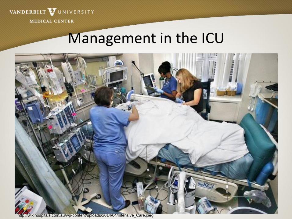
Management in the ICU
http://wikihospitals.com.au/wp-content/uploads/2014/04/Intensive_Care.jpeg

Care in the ICU: The Must Haves• Assess thoroughly on admission to note changes later
• Ecchymosis
• Edema
• Vent settings
• Hemodynamics/lines
• PAC less common; assess fluid balance clinically• IVC ultrasound
• Furosemide (careful with renal function…
may need to involve Nephrology)
• CVP
• Edema
• Bleeding/oozing
https://media.lanecc.edu/users/driscolln/RT116/softchalk/Oxygenation_Assessment/pitting%20edema.jpg

Care in the ICU: The Must Haves
• Immediate labs• CBC
• Coags with INR
• BMP with Mg, Phos (ATP cycle)
• Hepatic panel: AlkP, Tbili, AST, ALT, LDH
• Document JP drain volumes• Note fluid type: bilious blood bilious (where is
the leak?)
• Every 4 hours NO LOW WALL SUCTION leads to unwarranted bleeding
AlkP + Tbili = Biliary
AST + ALT = Cellular
LDH = Graft
Function

Jackson Pratt Drains
• JP#1: RUQ, exits most laterally on the right; lateral to the right lobe of the liver, drains retroperitoneum (aka: “gutter drain”)
• JP#2: Placed below the porta hepatis (opening for major blood vessels to enter and leave the liver), near common bile duct anastomosis, common area where vessels enter and exit the liver
• JP#3: LUQ, beneath left lobe of liver, drains retrohepatic cava
ww.medscape.com/viewarticle/451209_12

Care in the ICU: The Must Haves
• Order tacrolimus (prograf, FK5) drug level for 0530 following admission (nursing education!!!) for 0600 dose administration. Therapeutic range dependent on level.
• Be diligent the first 24 hours…assess with every patient encounter• small changes can lead to BIG events:
– bleeding
– graft failure
– VAE
– lab vigilance (stay on top of these q6 hours x24 hours)
• Communication with HBS team Imperative!

Discharge Planning
• Medication education (education with primary caregiver)
• Staying local x90 days
• Importance of environmental safety– Face masks in public places
– Sit in back of room/mass transit
– No gardening
– No fresh fruits
– No grapefruit-containing products
• Immunosuppression and titration of other drugs (insulin, prednisone, antibiotics, antivirals, MMF).
• Tacrolimus for life (nephrotoxic)

CASE STUDYMr. B is a 60 yr old male who is post op day 2 from OLT due to cryptogenic cirrhosis. He had no prior comorbidities other than encephalopathy related to his cirrhosis. On your morning exam, he is doing well other than some mild conversational dyspnea, which is new, and some heavy ecchymosis on his right chest and flank. He slept on his right side overnight. His nurse confirms that this is a new finding from his assessment yesterday. Mr. B’s SpO2 has drifted down from 98% on RA to 92% requiring 2L NC. His cough is nonproductive. All other vital signs are within normal limits. His CVP is 6. His LFTs are trending down nicely. He still has 4+/4 pitting edema in his lower extremities. You know that he received 80 units of RBCs in the OR due to bleeding during transplant.

Case Study
What are your differential diagnoses?:
a) Hemothorax
b) Transfusion Related Acute Lung Injury
c) Hypervolemia
d) Pneumonia
e) All of the above

Case Study
Later in the day, the RN calls you to the bedside as Mr. B’s pulmonary status is declining. He has increased his NC to 8L and reports breaths per minute at 40. Mr. B’s cognitive status is also declining with short lapses in memory and situational awareness. You examine Mr. B and discover that the ecchymosis you witnessed this morning has spread over his entire R chest and flank. You are concerned and order a chest xray.

Case Study
The chest x-ray shows a Right hemothorax. What caused this?
a) High volume resuscitation in the OR
b) Intravascular depletion
c) High volume transfusion of RBCs in the OR
d) Lying on his right side all night
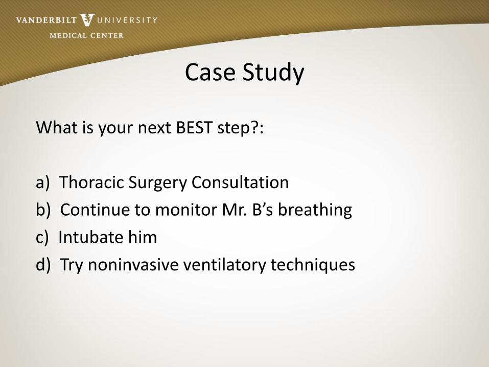
Case Study
What is your next BEST step?:
a) Thoracic Surgery Consultation
b) Continue to monitor Mr. B’s breathing
c) Intubate him
d) Try noninvasive ventilatory techniques
Case Study
Thoracic Surgery and Hepatobiliary Surgery decide a chest tube is required to drain the blood from Mr. B’s chest. During the thoracotomy, his breathing status sharply declines, experiences loss of consciousness, and requires intubation. He continues to develop an ARDs picture and remains on the ventilator for the next 3 days.

Case Study
Knowing what you know about this patient, what puts him at very high risk for a ventilator associated event leading to a ventilator associated complication (infection)?
a) Intensive care
b) Loss of consciousness during thoracotomy
c) Multimodal pharmaceutical immunosuppression
d) Age

Case Study
Mr. B experiences a full recovery and is discharged to a local inpatient rehab facility. What is your biggest take home from his case?
a) High volume RBC transfusion in OLT can lead to acute lung injury
b) Maintain high alert on physical assessment changes
c) Listen to your RN
d) Liver transplant patients can change in moments; you must know their history, both pretransplant and intraoperatively.
e) All of the above

References• Agarwal, B. et al. (2011). Critical care issues in patients after liver transplantation. American Association for the Study of
Liver Diseases, 17, 511-527
• Bernardi, E. et al. (2005). Intensive care complications in liver and multivisceral transplantion. Transplantation Proceedings, 37, 2618-2621
• Chiarandini, L., et al. (2010). Liver transplant quality and safety plan in anesthesia and intensive care medicine. Transplant Proceedings. 42, 2229-2232
• Eghtesad, B., Fung, J., Miller, C. (2012). Post-liver transplantation management. Cleveland Clinic Center for Continuing Education. www.clevelandclinicmeded.com. Retrieved from www 11/15/12.
• Ertmer, C., et al. (2014). Impact of human albumin infusion on organ function in orthotopic liver transplantation – a retrospective match-pair analysis. Clinical Transplantation. 29: 67-75.
• Geevarghese, Sunil. Personal conversations and physician reviewed content. Hepatobiliary and Liver Transplant Surgeon, Vanderbilt University Medical Center. July-August, 2015.
• Liver Transplant Patients. www.uphs.upenn.edu/surgery/Education/trauma/SCCS/protocols/Resident_Pathway.pdf. Retrieved from www 7/15/14
• Martini, J., Wheeler, A. (2006). Critical Care Medicine 3rd Ed. pp 507-513
• Oclese, V., et al. (2013). Medscape reference article. http://emedicine.medscape.com/article/431573
• Papdopoulos, S., et al. (2014). Causes and incidence of renal replacement therapy application in orthotopic liver tranplantation patients: our experience. Transplantation Proceedings. 46: 3228-3231.
• Park, C. (2015). Predictive roles of intraoperative blood glucose for post-transplant outcomes in liver transplantation. World Journal of Gastroenterology. Vol 21, Issue 22, 6835-6841
• Sawas, T., et al. (2015). Patients receiving prebiotics and probiotics before liver transplantation develop fewer infectionsthan controls: a systematic review and meta-analysis. Clinical Gastroenterology and Hepatology. Accepted May 26, 2015.
• Yu, J., et al. (2015). Predictors of early hospitalization after deceased donor liver transplantation. Digest of DigestiveScience. June 30, 2015.