Retrospective study on medical vs. surgical management of ... · Retrospective study on medical vs....
33
Retrospective study on medical vs. surgical management of right dorsal displacements in the horse Malla Muhonen Supervisor: Michael Hewetson Director: Riitta-Mari Tulamo Department of equine and small animal medicine Faculty of veterinary medicine University of Helsinki Year of completion: 2016
Transcript of Retrospective study on medical vs. surgical management of ... · Retrospective study on medical vs....
Retrospective study on medical vs. surgical management of right dorsal displacements in
the horse
Malla Muhonen Supervisor: Michael Hewetson Director: Riitta-Mari Tulamo Department of equine and small animal medicine Faculty of veterinary medicine University of Helsinki Year of completion: 2016

0
1.! Introduction,.................................................................................................................................,2!1.1,Left,dorsal,displacement,.....................................................................................................................,2!1.2,Large,colon,volvulus,.............................................................................................................................,4!1.3,Retroflexion,of,the,pelvic,flexure,.....................................................................................................,4!1.4,Right,dorsal,displacement,..................................................................................................................,4!
2.! Pathogenesis,of,Right,dorsal,displacement,.......................................................................,5!
3.! Prevalence,of,Right,dorsal,displacement,...........................................................................,7!
4.! Diagnosis,of,right,dorsal,displacement,...............................................................................,7!4.2,Clinical,signs,............................................................................................................................................,7!4.2,Rectal,examination,...............................................................................................................................,8!4.3,Laboratory,findings,..............................................................................................................................,8!4.4,Ultrasonography,....................................................................................................................................,9!
5.! Defining,whether,a,RDD,should,be,treated,surgically,or,medically,......................,10!5.1,Pain,assessment,...................................................................................................................................,10!5.2,Status,.......................................................................................................................................................,11!5.3,Rectal,examination,.............................................................................................................................,11!5.4,Abdominal,distension,and,gas,accumulation,............................................................................,11!5.5,Laboratory,findings,............................................................................................................................,11!5.6,Other,factors,.........................................................................................................................................,12!
6.! Medical,treatment,of,RDD,.....................................................................................................,12!6.1,Medical,treatment,...............................................................................................................................,12!6.2,Treating,pain,.........................................................................................................................................,13!6.3,Alterations,in,intestinal,motility,....................................................................................................,14!6.4,Laxatives,..................................................................................................................................................,14!6.5,Fluid,therapy,.........................................................................................................................................,15!
7.! Surgical,treatment,of,colic,....................................................................................................,15!
8.! Prognosis,....................................................................................................................................,16!
9.! Materials,and,methods,..........................................................................................................,16!Statistical,analysis,......................................................................................................................................,18!
10.! Results,.......................................................................................................................................,19!
11.! Discussion,................................................................................................................................,22!

1
12.! BIBLIOGRAPHY,......................................................................................................................,27!
!
Tiedekunta - Fakultet - Faculty Eläinlääketieteellinen tiedekunta
Osasto - Avdelning – Department Kliinisen hevos- ja pieneläinlääketieteen osasto
Tekijä - Författare - Author Malla Muhonen Työn nimi - Arbetets titel - Title Retrospective study on medical vs. surgical management of right dorsal displacements in the horse Oppiaine - Läroämne - Subject Työn laji - Arbetets art - Level Tutkimusosan sisältävä lisensiaatintutkielma
Aika - Datum - Month and year 2.2.2016
Sivumäärä - Sidoantal - Number of pages 30
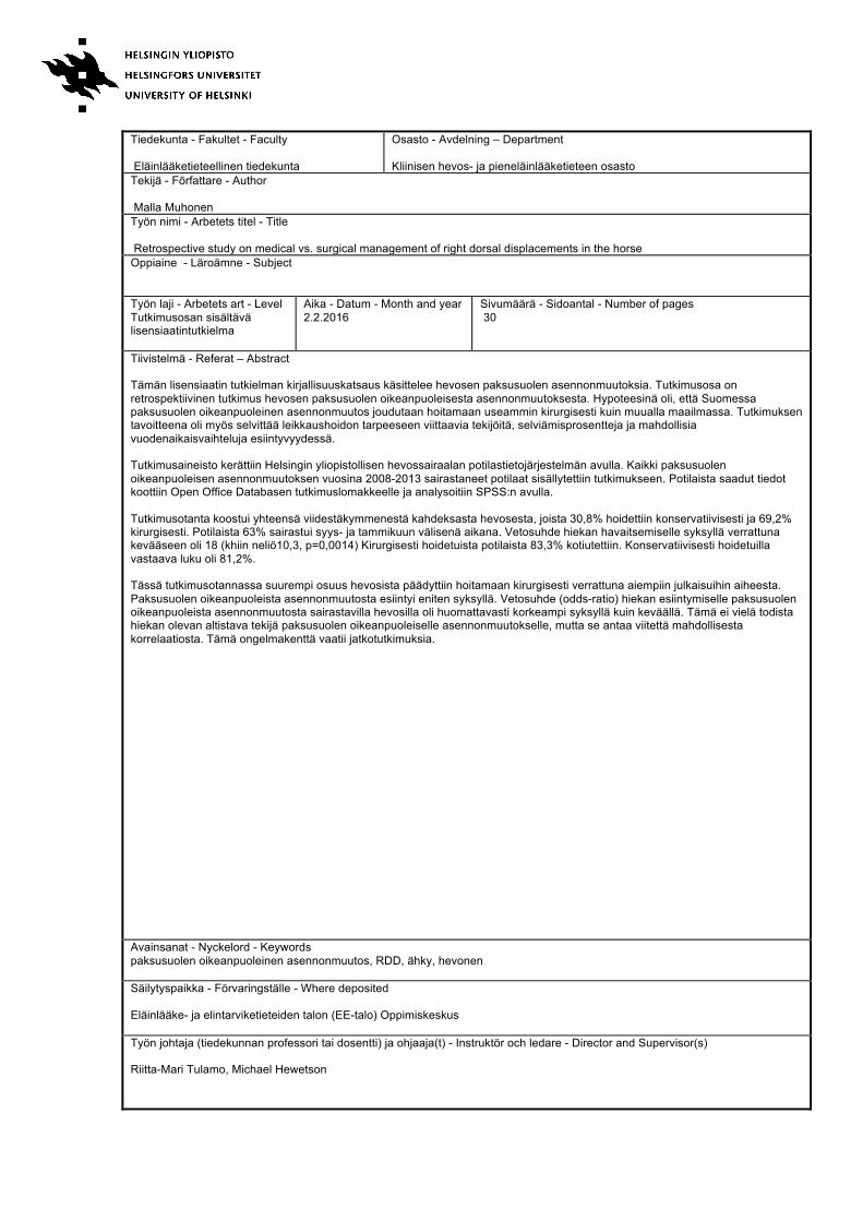
Tiivistelmä - Referat – Abstract Tämän lisensiaatin tutkielman kirjallisuuskatsaus käsittelee hevosen paksusuolen asennonmuutoksia. Tutkimusosa on retrospektiivinen tutkimus hevosen paksusuolen oikeanpuoleisesta asennonmuutoksesta. Hypoteesinä oli, että Suomessa paksusuolen oikeanpuoleinen asennonmuutos joudutaan hoitamaan useammin kirurgisesti kuin muualla maailmassa. Tutkimuksen tavoitteena oli myös selvittää leikkaushoidon tarpeeseen viittaavia tekijöitä, selviämisprosentteja ja mahdollisia vuodenaikaisvaihteluja esiintyvyydessä. Tutkimusaineisto kerättiin Helsingin yliopistollisen hevossairaalan potilastietojärjestelmän avulla. Kaikki paksusuolen oikeanpuoleisen asennonmuutoksen vuosina 2008-2013 sairastaneet potilaat sisällytettiin tutkimukseen. Potilaista saadut tiedot koottiin Open Office Databasen tutkimuslomakkeelle ja analysoitiin SPSS:n avulla. Tutkimusotanta koostui yhteensä viidestäkymmenestä kahdeksasta hevosesta, joista 30,8% hoidettiin konservatiivisesti ja 69,2% kirurgisesti. Potilaista 63% sairastui syys- ja tammikuun välisenä aikana. Vetosuhde hiekan havaitsemiselle syksyllä verrattuna kevääseen oli 18 (khiin neliö10,3, p=0,0014) Kirurgisesti hoidetuista potilaista 83,3% kotiutettiin. Konservatiivisesti hoidetuilla vastaava luku oli 81,2%. Tässä tutkimusotannassa suurempi osuus hevosista päädyttiin hoitamaan kirurgisesti verrattuna aiempiin julkaisuihin aiheesta. Paksusuolen oikeanpuoleista asennonmuutosta esiintyi eniten syksyllä. Vetosuhde (odds-ratio) hiekan esiintymiselle paksusuolen oikeanpuoleista asennonmuutosta sairastavilla hevosilla oli huomattavasti korkeampi syksyllä kuin keväällä. Tämä ei vielä todista hiekan olevan altistava tekijä paksusuolen oikeanpuoleiselle asennonmuutokselle, mutta se antaa viitettä mahdollisesta korrelaatiosta. Tämä ongelmakenttä vaatii jatkotutkimuksia.
Avainsanat - Nyckelord - Keywords paksusuolen oikeanpuoleinen asennonmuutos, RDD, ähky, hevonen Säilytyspaikka - Förvaringställe - Where deposited Eläinlääke- ja elintarviketieteiden talon (EE-talo) Oppimiskeskus Työn johtaja (tiedekunnan professori tai dosentti) ja ohjaaja(t) - Instruktör och ledare - Director and Supervisor(s) Riitta-Mari Tulamo, Michael Hewetson
2
1. INTRODUCTION#
Displacement of the large colon into an abnormal topographical position is a
common cause of colic in the horse. Displacement causes an extra-mural
obstruction, leading to accumulation of ingesta and gas in the colon lumen, and
in some cases vascular occlusion. Medical treatment is often successful, but
depending upon the type of displacement, surgical intervention may be required.
This is especially important if there is evidence of vascular occlusion. There are
currently four types of displacements reported in the literature and each will be
reviewed briefly:
1.1#Left#dorsal#displacement#
In left dorsal displacement (LDD), the left dorsal and ventral colons move
dorsolaterally over the spleen to lie on the nephrosplenic ligament (between the
spleen and the left kidney). In LDD, the colon is trapped dorsally by the
abdominal wall, medially by the left kidney, ventrally by the nephrosplenic
ligament and laterally by the spleen. The colon’s sternal and diaphragmatic
flexures can stay at their normal positions ventrally to the stomach or they can
dislocate dorsally to the stomach (Brown et al 2007). Of all colic patients
requiring surgery, only 2,7% (8/300) are LDD patients (Mair & Smith 2005)
LDD is typically diagnosed via rectal examination and ultrasonography findings.
On rectal examination, the left ventral colon is situated dorsal to the left dorsal
colon and one is able to follow the colon up to the nephrosplenic space using the
colon taenial bands as a reference (Rakestraw & Hardy 2012). LDD can also be
diagnosed by ultrasonography. On ultrasonography of a normal horse, the left
kidney should be visible adjacent to the spleen in intercostal spaces 15-17. In the
case of a LDD, there is gas-filled large colon behind the spleen and the left
kidney is therefore not ultrasonographically visible (Rakestraw & Hardy 2012).
Typical medical treatments for LDD are withholding of food, fluid therapy,
exercise, analgesics, rolling under general anesthesia and the judicial use of
phenylephrine (Hardy et al 2000). Phenylephrine causes vasoconstriction of the
3
spleen, hence reducing it’s size (van Harreveld et al 1999). As the spleen shrinks,
it gives the large colon room to escape the nephrosplenic space and thus helps the
LDD resolve. Phenylephrine and rolling the horse under general anaesthesia are
considered more effective compared to horses treated with phenylephrine and
exercise (Fultz et al 2013). The success rate of horses rolled under general
anaesthesia was 84% in comparison to exercise, where the success rate was
63,2% (Fultz et al 2013).
Medical treatment might not work if there is excessive gas accumulation in the
trapped large colon. In these cases the gas may be evacuated by using a trocar in
the left paralumbar fossa (Hardy et al 2000).
If medical treatment is not effective or if the horse is severely painful or bloated,
abdominal surgery should be performed and the colon displacement should be
manually corrected. Surgical treatment can be performed via standing flank
laparotomy or ventral midline celiotomy (Hardy et al 2000). The advantages of a
standing flank laparotomy are that no general anaesthesia is needed, the recovery
time is shorter and the surgeon has direct access to the nephrosplenic space
(Rakestraw & Hardy 2012). The advantages of the ventral midline celiotomy are
that if the original diagnosis is incorrect and there is some other type of
displacement, it is still possible to correct the problem, which would not be
possible when performing a standing flank laparotomy (Rakestraw & Hardy
2012). Also, if the sternal and diaphragmatic loops of the colon are situated
dorsally to the stomach, it is possible to correct that too using a ventral midline
celiotomy approach (Rakestraw & Hardy 2012). Due to these reasons, the
ventral midline celiotomy is a more common approach compared to standing
flank laparotomy (Hardy et al 2000).
Suturing the dorsal splenic margin to the nephrosplenic ligament can prevent re-
occurance of LDD (Röcken et al 2005). This method prevents the colon from
rising to the nephrosplenic space, but the colon can still dislocate between the
spleen and the abdominal wall (Röcken et al 2005).

4
1.2#Large#colon#volvulus#
In large colon volvulus, a loop of large colon rotates along its long axis. The
cecum can also be involved in this type of displacement. Large colon volvulus
causes occlusion of the mesenteric blood supply to the colon, leading to
infarction of the affected segment of intestine, and is a life-threatening condition
(Brown et al 2007). Compared to other types of large colon displacements, large
colon volvulus usually causes the most intense pain reaction. Predisposing
factors for large colon volvulus are recent parturition, recent dietary changes,
access to lush pasture, medication during the previous 7 days, increase in stabling
hours in the previous 14 days and previous recurrent colic episodes (Suthers et al
2013, Rakestraw & Hardy 2012). The only effective treatment for large colon
volvulus is surgical reposistioning of the colon (Rakestraw & Hardy 2012, Fiege
et al 2014). Preoperative measurements, like fluid therapy and relief of pressure
in the abdominal cavity should be performed in order to increase the amount of
circulating blood (Rakestraw & Hardy). Large colon volvulus is one of the most
typical reasons for colic horses ending up in surgery (17,0% of all types of
colics) (Mair & Smith 2005). The short-term survival rate of large colon volvulus
is 65-70% (Mair & Smith 2005, Fiege et al 2014).
1.3#Retroflexion#of#the#pelvic#flexure#
There is very little published data on retroflexion of the pelvic flexure.
Mair&Smith (2005) describe an overall prevalence of 2,3% of all colic surgeries
presented at a large referral centre and a short-term survival rate of 85,7%. In
comparison, a study performed on the Norwegian horse population reported a
prevalence of 3,4% (Wormstrand et al 2014). Interestingly, horses that have had
retroflexion of the pelvic flexure are more predisposed to recurrent colic episodes
when compared to horses that have had other types of colic surgeries (Smith &
Mair 2010). The reason for this is unclear.
1.4#Right#dorsal#displacement#
Right dorsal displacement (RDD) is a type of large colon displacement in which
the left dorsal and ventral colons migrate to the right side of the abdominal cavity
and lie between the cecum and the abdominal wall (Brown et al 2007). RDD is a
non-strangulating obstruction and causes mild to moderate colic signs in horses.

5
Patients with RDD can either be treated medically or surgically. Medical
treatment consists of intravenous fluids and analgesics. In surgery, a ventral
midline celiotomy is performed in order to correct the displacement and, if
needed, evacuate impacted feed or gas from the large intestine.
While much has been written about the treatment of LDD and large colon
volvulus, very little information on RDD is available in the literature, despite the
fact that it is a common cause of colic in the horse. In particular, predisposing
factors, clinical information used to determine the need for medical vs. surgical
treatment of RDD and short term prognosis for medical vs. surgical treatment of
RDD have not been well described before. Furthermore, in our study population,
it has been hypothesized that that sand accumulation may predispose horses to
RDD.
Therefore, the aims of this study were to analyse retrospective data from a large
group of horses with RDD, and determine if 1) there are any predisposing actors
that can be indentified and in particular, determine if there is an association
between sand accumulation in the colon and RDD; 2) determine if there are any
specific clinical or clinicopathological factors at presentation that may help
differentiate between horses that require medical vs. surgical treatment, and 3),
compare the short term survival for horses with RDD treated medically vs.
surgically.
2. PATHOGENESIS+OF+RIGHT"DORSAL"DISPLACEMENT#
The large colon of a horse is only attached to the abdominal wall at 3 sites: the
ceocolic fold, the dorsal attachment of the right dorsal colon and the
duodenocolic fold. Otherwise the entire large colon is unattached and therefore
prone to displacements. Normally the left colon shortens and relaxes lengthwise
in the tempo given by the pacemaker cells located in the pelvic flexure
(Rakestraw & Hardy 2012). If the pacemaker cells become disturbed for some
reason, the pelvic flexure can start moving towards the diaphragm. In the case of
RDD, the pelvic flexure (which normally lies in the left caudal part of the
abdominal cavity) migrates cranially towards the diaphragm. Then it turns to the
right caudal quadrant of the abdominal cavity. Finally it moves between the
6
cecum and the right abdominal wall. This is called a counterclockwise rotation
(when viewed caudoventrally). Another migratory route for the pelvic flexure is
a clockwise rotation. In this scenario, the pelvic flexure will end up lying next to
the diaphragm and the left colon will be situated between the cecum and the right
abdominal wall. This is a far less common presentation for a RDD. Right dorsal
displacement is a non-strangulating displacement where gas and ingesta are not
able to pass through the alimentary tract. The displacement can also be partial i.e.
the pelvic flexure only moves next to the diaphragm but does not migrate
between the caecum and right abdominal wall. In this scenario, the horse will
often continue to pass small amounts of faeces (Rakestraw & Hardy 2012).
There is very little published data on the predisposing factors for RDD, but it has
been suggested that excessive amounts of carbohydrates could expose horses to
right dorsal displacements (Blikslager 2010). Carbohydrates are fermented in the
large colon and if a horse ingests excessive amounts of carbohydrates it can
result in the production of excessive gas that predisposes to displacements
(Rakestraw & Hardy 2012). There is also some evidence that large colon
displacements might be seasonal, with peak occurrence in spring and fall (Archer
et al 2006, Hillyer et al 2001). In contrast, a study in Sweden reported that colic
was more likely to occur during the winter months from October to March
(Egenvall et al 2008). The authors hypothesized that the incidence of colic
increased during wintertime due to different exercise and feeding routines when
compared to summertime. As a whole, colic (including colon displacement) is
more likely to occur at times when the horses were managed intensively (Archer
et al 2006).
In one study looking at simple colon obstruction and distension, it was reported
that travelling in the previous 24 hours increased the risk of colic (Hillyer et al
2002). In the same study, it was found that horses that wind sucked or cribbed
were 70 times more likely to get colic compared to horses that didn’t have these
habits (Hillyer et al 2002). Other significant predisposing factors in this study
were a change in exercise routines, hours spent in the stable, long duration
between routine dental care, anthelmintic admission during the past 7 days and
previous colic episodes (Hillyer et al 2002).

7
Sand accumulation in the large colon is also thought to predispose horses to
RDD but as of yet, there is no data to support this.
3. PREVALENCE)OF)RIGHT)DORSAL'DISPLACEMENT#
There are only two studies available where large colon displacements are
separated to their own groups and not counted as a single group. In this study,
RDD was the second most common reason for surgical treatment of colic (14%).
The most common reason was large colon volvulus with 17% (Mair & Smith
2005). Another study reported that 11,1% of horses that went to surgery had a
RDD and it was the second most common reason for surgery (large colon
volvulus being the most common reason) (Wormstrand et al 2014).
4. DIAGNOSIS#OF#RIGHT#DORSAL#DISPLACEMENT#
There is no single diagnostic test with which one would be able to diagnose a
RDD. The most important diagnostic methods are evaluation of clinical signs,
rectal examination, ultrasonography and laboratory data. A right dorsal
displacement can only be confirmed definitively during an exploratory
laparotomy or at autopsy.
4.2#Clinical#signs#
Horses with RDD have been reported to have mild to moderate abdominal pain.
Horses may kick at their ventral abdomen, try to lie down, roll, or they can just
be lethargic and anorexic. The abdominal discomfort usually decreases with
analgesics, but often recurs soon thereafter. The amount of discomfort is often
progressive (Gardner et al 2005). Horses can continue to pass small amounts of
faeces despite a right dorsal displacement (Rakestraw & Hardy 2013). In some
cases, bloating and a “ping” sound on percussion may occur (McGovern et al
2011). Horses with right dorsal displacement usually have some gastrointestinal
sounds audible (McGovern et al 2011). The heart rate in horses with RDD is
usually elevated and typically horses with RDD will have no signs of
endotoxemia or circulatory shock (McGovern et al 2011).

8
4.2#Rectal#examination#
Rectal examination is one of the most important diagnostic methods when
diagnosing right dorsal displacement. In a counter-clockwise RDD the pelvic
flexure is palpable on the right hand side and in a clockwise rotation the pelvic
flexure is not palpable (Rakestraw & Hardy). In addition to the lack of pelvic
flexure, the examiner is able to feel a large distended, gas-filled colon that is
orientated horizontally across the abdominal wall (McGovern et al 2011). In the
early stages of a RDD, one might still be able to feel the pelvic flexure, but the
right dorsal colon is distended with gas.
4.3#Laboratory#findings#
In one study it has been reported that 18 of 37 horses (49%) with RDD had
elevated gamma glutamyl transferase (GGT) values (Gardner et al 2005). In
comparison, only 1 of 48 (2%) of horses with LDD had increased GGT values.
GGT is a hepatic enzyme which is associated with hepatic injury. Probably the
elevation in GGT values during RDD is because the right dorsal colon is attached
to the visceral surface of the liver by a fibrous sheet. This proportion of the
mesoduodenum blends with the hepatoduodenal ligament for the last 5 cm before
attaching to the liver. The bile duct is situated within this 5 cm part. When a
RDD occurs, the right dorsal colon moves caudally and laterally causing tension
on the fibrous sheet. The portion of the hepatoduodenal ligament closest to the
liver also becomes tense, causing obstruction of the bile duct. This increases
GGT activity. As the diasease progresses, the liver is not able to pass the
conjugated bilirubin into the bile ducts, thus conjugated bilirubin values in blood
will increase. This increase was reported in 33% of RDD patients (Gardner et al
2005). The GGT values normalized after surgical treatment of RDD in 9 of 11
horses (Gardner et al 2005).
Other bloodwork and peritoneal fluid is usually within the normal range, but can
become abnormal as time passes (Grenager & Durham 2011).
9
4.4#Ultrasonography#
Blood is supplied to the right dorsal colon via the right colic artery and to the
right ventral colon via the ileocolic artery. Both of these vessels are situated on
the medial aspect of the mesocolon, thus they should not be visible on ultrasound
of a normal horse. When a RDD occurs, the left dorsal and ventral colons rotate
to the right side of abdominal cavity between the cecum and the abdominal wall.
At the same time, the medial surface of the left dorsal and ventral colons will be
situated laterally. So, as the medial surface of the mesocolon now lies adjacent to
the abdominal wall, it is possible to detect the branches of the ileocolic and right
colic artery on ultrasonography in a horse with RDD. Similar vessels can also
been seen in horses with a 180° colon volvulus. (Ness et al 2012)
At first the caecal vessels are identified and they are followed axially and
cranially (Grenager & Durham 2011). The abnormal vascularisation can be seen
in the intercostal spaces approximately at the costochondral junction on the right
side of the horse (usually from 12th to 17th intercostal space) (Ness et al 2012). In
order to make a diagnosis, one should see the vessels in at least 2 intercostal
spaces (Grenager & Durham 2011). In order to rule out other causes of colic, the
rest of the abdomen is also scanned routinely. One should evaluate the thickness
of the large colon wall, determine the location of colonic sacculations, evaluate
the small intestine motility, distension and wall thickness, evaluate the amount of
peritoneal fluid, evaluate the gastric location and size and check the
nephrosplenic space (Grenager & Durham 2011).
In two studies, 57-70% of horses with RDD showed abnormal vascularization on
ultrasonography (Grenager & Durham 2011 and Ness et al 2012). All the horses
with abnormal vascularization detectable on the ultrasonography were diagnosed
with RDD during an exploratory laparotomy (Grenager & Durham 2011).
Sand accumulation that may be associated with RDD can also be investigated
with ultrasonography. Ultrasound has been reported to have a specificity and
sensitivity of 87,5%. Ultrasonography is not very good in defining the quantity
of sand accumulation however (Korolainen & Ruohoniemi 2002).
10
5. DEFINING'WHETHER'A'RDD!SHOULD'BE'TREATED'
SURGICALLY*OR*MEDICALLY#
5.1#Pain#assessment#
Pain is the most important factor when defining whether a horse should be
treated surgically or medically (Toefner et al 2003). Pain should be evaluated
when the horse is not restrained, preferably in a box. A horse is showing pain
when it is pawing the bedding, looking at the flanks, kicking the abdomen, lying
down and rolling (Rakestraw & Hardy 2012).
The most severe colic patients, whom require surgical intervention immediately,
are generally the most painful ones. RDD patients are not typically severely
painful, instead they show mild to moderate signs of pain. Still, in some cases,
horses with RDD can show intensive signs of pain. In this situation the horse
won’t respond to analgesics or at least the pain will re-occur shortly after
medication (Rakestraw & Hardy 2012). On the other hand, non-strangulating
colics will respond to analgesics and don’t need immediate abdominal surgery.
The degree of pain can be determined by the horse’s response to non-steroidal
anti-inflammatory drugs. Mild pain typically responds to NSAID’s for 8 to 12
hours. Moderate pain decreases for a limited time and requires several
administrations. Horses with severe pain show violent behaviour and the pain
will not be controlled with analgesics. Horses with severe pain require immediate
abdominal surgery. (Rakestraw & Hardy 2012)
A study by Toefner et al (2003) shows that the most important variables
suggesting surgical intervention were severe abdominal pain (OR=57,17),
distended small intestine (OR= 31,42), cyanotic mucous membranes
(OR=17,88), peritoneal fluid hemolysis (OR 15,14) and absent abdominal sounds
(OR= 11,97). Lethargic horses were also reported to have a high odds ratio (OR=
36,75) compared to horses not showing any signs of pain. Here we of course
have to take to account, that this study was done on all types of colics and colic
surgeries, not only RDDs, so the importance of pain might vary in the case of
RDD, being a non-strangulating displacement and typically not causing severe
signs of pain.

11
The problem with horses with severe pain is that they would need immediate
surgery but the owner can’t transport the horse to an equine hospital if it is acting
violently. Therefore it is necessary for a vet to come to the stables and give the
horse a sufficient amount of analgesics prior to the transport.
5.2#Status#
Capillary refill time (CRT) indicates the circulatory function of the horse. If CRT
is prolonged in a horse with RDD, it indicates that the RDD has caused a
circulatory dysfunction. A horse with prolonged CRT is 1,70 times more likely
to end up in surgery (Toefner et al 2003).
5.3#Rectal#examination#
A horse with intestinal impaction detectable on rectal palpation is 2,51 times
prone to surgical intervention compared to a horse with no abnormal rectal
findings (Toefner et al 2003). Horizontal distension, which is the most common
type of rectal finding in RDD patients, was not reported as it’s own group in this
study. It was probably grouped into the category of “other findings”. Horses that
had “other findings” in rectal examination had an odds ratio of 2,85 compared to
horses with no rectal abnormalities.
5.4#Abdominal#distension#and#gas#accumulation#
An article by Ducharme et al (1989) reports that abdominal distension is the most
important factor discriminating whether a horse should be treated medically or
surgically. Ninety percent of horses with moderate or severe abdominal
distension were surgical candidates (Ducharme et al 1989).
5.5#Laboratory#findings#
Blood lactate concentration reflects the amount of ischemic injury and may
therefore be used to determine the prognosis in colic horses (Rakestraw & Hardy
2012). Lactate can also be determined from peritoneal fluid. A high lactate
concentration in peritoneal fluid is a more sensitive indicator for intestinal
ischaemia than plasma lactate values (Latson et al 2005). This is controversial, as
another study demonstrated that peritoneal fluids with high lactate value had a
lower odds ratio for going to surgery compared to plasma lactate concentration
(15,50 versus 18,55) (Toefner et al 2003). As RDD is typically a non-

12
strangulating displacement, it might be that neither plasma nor peritoneal lactate
concentration increases in horses with RDD. If the RDD is severe and starts
obstructing blood vessels, lactate values may begin to elevate.
5.6#Other#factors#
One study has reported increased rectal temperature decreased the need for
surgery in horses with severe pain (Toefner et al 2003). The rise of rectal
temperature was thought to result from increased activity due to pain,
inflammation of the rectal area or cardiovascular dysfunction. Increased body
temperature is often associated with peritonitis, enteritis or colitis, which do not
require surgical intervention (Toefner et al 2003).
The colour of the peritoneal fluid is also a factor that can be used to discriminate
surgical and non-surgical patients from one another (Ducharme et al 1989). If the
colour of the peritoneal fluid is normal or cloudy, the horse typically doesn’t
require surgical intervention (Ducharme et al 1989). If the abdominal fluid is
serosanguineus, the horse is more likely to require surgical intervention
(Ducharme et al 1989)
In summary, it should be remembered that delayed surgical intervention
predisposes the horse to visceral rupture. Therefore it is recommended to take the
colic horse to surgery promptly after admission. This way we are able to
maximize the probability of a successful outcome (Rakestraw & Hardy 2012).
6. MEDICAL(TREATMENT(OF!RDD#
6.1#Medical#treatment#
Horses with RDD are treated with intravenous fluids, analgesics and forced
exercise (McGovern et al 2011). Exercise can be, for example 10-15 minutes
lunge reining every 1-2 hours (McGovern et al 2011)
Horses treated medically should be reassessed regularly. PCV should be taken
and a rectal examination performed once every 2 hours. If there are any signs of
endotoxemia or cardiovascular compromise (e.g. increased body temperature or

13
heart rate, abnormally coloured mucus membranes etc), the need for surgical
intervention should be reassessed. (McGovern et al 2011)
The main goals of medical treatment of colic are to relieve pain, restore normal
gut motility, restore and maintain hydration and electrolyte/acid-base balance
and, if needed, treat endotoxaemia (Edwards 2013). When relieving pain it must
be taken into account that the pain medication should not depress the gut activity,
predispose to hypovolemic shock or mask the signs of endotoxemia (Edwards
2013)
6.2#Treating#pain#
Nonsteroidal anti-inflammatory drugs are used to relieve visceral pain. They
function through inhibition of cyclo-oxygenase enzyme-mediated biosynthesis of
prostaglandins (Moilanen & Kankaanranta 2013).
Sedatives can also be used for achieving analgesia and sedation. Examples of
sedatives used for colic are xylazine, detomidine, romifidine and acepromazine
(Edwards 2013).
The ability of achieving analgesia and sedation varies among different sedatives.
Fourty mg/kg of detomidine has been reported to have a satisfactory sedative
effect on 84,2 % of colic horses. In comparison, 20mg/kg of detomidine had a
satisfactory effect in only 71,5% of colic horses (Jochle et al 1989). The
analgesic effect was rated satisfactory or highly satisfactory in 100% of the
horses receiving 40 mg/kg detomidine. None of the horses receiving 0,1mg/kg
butorphanol were reported to have satisfactory or highly satisfactory effect on
pain. Also no differences in side effects were reported between the detomidine
doses or the butorphanol dose (Jochle et al 1989). So one could say that
detomidine is the most effective analgesic and sedative agent compared to
flunixin, butorphanol and xylazine. Problem in detomidine is that it has more
cardiovascular effects compared to, for example, xylazine (Yamashita et al
2000). Xylazine has also been reported to have less effect on duodenal motility
compared to detomidine and a combination of xylazine and butorphanol (Merritt
et al 1998). Xylazine has also been reported to have a longer analgesic effect
than butorphanol (90 minutes versus 60 minutes) (Muir & Robertson 1985).

14
Narcotic agents, such as morphine can also be used, but it can cause excitement
in horses and is thus combined with some other drug, like xylazine (Edwards
2013).
6.3#Alterations#in#intestinal#motility#
If the horse has increased intestinal motility, spasmolytics can be used. Horses
with large colon displacements usually have decreased intestinal motility and so
spasmolytics are not indicated in most cases.
6.4#Laxatives#
Laxatives are used to resolve impaction masses by increasing the amount of fluid
in the large colon and hence increasing gastrointestinal motility. Magnesium
sulphate, sodium sulphate and mineral oils have been reported to have a laxative
effect (Hotwagner & Iben 2008, Lopes et al 2004). Laxatives should always be
administered with either enteral or iv-fluids to avoid dehydration. Enteral fluids
combined with laxatives have been reported more effective compared to iv-fluids
(Lopes et al 2002). Balanced electrolyte solution containing sodium chloride,
potassium chloride and sodium bicarbonate is the safest enteral fluid, while
compared to water it does not predispose to hyponatremia (Lopes et al 2004).
Sodium sulphate hydrates the right dorsal colon better than magnesium sulphate
(Lopes et al 2004). The problem in using sodium sulphate as a laxative is that it
can cause severe hypocalcemia and hypernatremia (Lopes et al 2004).
Magnesium sulphate did not have similar injurious effects.
Mineral oil given orally via a nasogastric tube is considered a safe but not very
effective laxative. In the case of sand enteropathy, psyllium increases the outflow
of sand from the intestine when combined with mineral oils (Hotwagner & Iben
2008). Psyllium is especially useful for horses with sand enteropathy. Although
this crossover study proves that psyllium increases the outflow of sand
artificially fed to horses, another study shows that feeding psyllium does not
always resolve the sand enteropathy (Ruohoniemi et al 2001). In this study, 14
horses with large colon sand accumulation were fed psyllium and the results
were monitored with abdominal x-rays. In this study, the sand accumulation
resolved with only psyllium administration in 2 foals and 4 adult horses. Other 3
horses required laxatives, like mineral oil or magnesium sulphate in order to
15
resolve the sand accumulation. In one horse, no laxatives were effective, but the
sand accumulation resolved on pasture.
6.5#Fluid#therapy#
Fluid therapy consists of 3 different kinds of goals: replacing the deficits,
maintaining the achieved hydration status and overhydration. Overhydration is
commonly used in horses with colonic impaction (Edwards 2013).
7. SURGICAL)TREATMENT)OF"COLIC#
Surgical treatment of RDD is typically done using a ventral midline celiotomy
approach. There are some reports that a standing flank laparotomy would be
another way to manage RDD (Rakestraw & Hardy 2012). This is contradictory to
other articles that claim that a standing flank laparotomy is only useful uterine
torsions, ovariectomies and small intestine problems (Kummer 2012). For ventral
midline celiotomy, the horse is in general anaesthesia and placed in dorsal
recumbency. The incisision is made through the linea alba because it is a fibrous
structure which is easy to close and the amount of haemorrhage is minimal. In
ventral midline celiotomy, one has a good visibility to the abdomen and more
parts of the intestine are possible to exteriorize (Kummer 2012). At surgery, the
pelvic flexure is identified, the large colon is exteriorized and the displacement is
corrected (Rakestraw & Hardy 2012). If there is a large impaction mass present
in the large colon, an enterotomy can be performed. If there is just accumulation
of gas in the large colon, it can be evacuated with a needle.
The most typical reason for deaths during colic surgeries have been reported to
be cardiac arrest (13 of 635 colic surgery patients) (Mee et al 1998). Anaesthetic
complications occur more frequently in emergency colic surgeries compared to
elected anaesthesia (Dukti & White 2008). Abdominal bloating is associated with
ventilation difficulties causing hypoxia (Dukti & White 2008). This is why it is
important to evacuate gas from to intestine as quickly as possible during surgery.
Possible complications requiring euthanasia during RDD surgery are intestinal
rupture, inaccessible large colon and haemorrhage (Mee et al 1998). If bleeding
and haemorrhage occur during surgery, the source of the blood should be located
and sutured. The mesenteric vessels of the large colon are especially prone to

16
bleeding (Dukti & White 2008). Mesenteric vessels should be ligated with both
staples and sutures (Dukti & White 2008). The large colon is especially
predisposed to ruptures if it is gas-filled or otherwise distended. If a rupture
occurs, it is important to prevent the intestinal contents contaminating the
abdominal cavity. Gastrointestinal ruptures can occur spontaneously or they can
be iatrogenic. Gastrointestinal ruptures predispose to patient to incisional
infections and peritonitis (Dukti & White 2008), and are in most cases fatal.
8. PROGNOSIS#
Horses with RDD going to surgery have a short-term survival rate (= to
discharge) of 93% (Mair & Smith 2005). This is relatively high compared to the
total short-term survival rate of horses going to colic surgery, which was only
70,3% (Mair & Smith 2005). The survival rate is also better compared to all
types of large colon colics, which was 89,9% (Mair & Smith 2005). Only one of
the 42 RDD patients included in this study was euthanized during anaesthesia.
The reason for the euthanasia was an intestinal rupture (Mair & Smith 2005).
Another study presented less optimistic short-term survival rates for large colon
colic horses. A study done in Norway by Wormstrand et al (2014) reported a
short-term survival rate for RDD patients of 81,8% and for all types of large
colon colics a short-term survival rate of 66,7%. So, in both studies, we are able
to see that right dorsal displacement has a better short-term survival rate
compared to other types of large colon colic surgeries.
Horses with RDD are likely to have recurrent colic episodes after RDD surgery (Smith & Mair
2010). Fourty one percent of horses with a RDD episode had recurrent colic. Typically this
recurrent colic was also RDD. Horses with RDD are more likely to develop postoperative colic
compared to horses with LDD or nonstrangulating volvulus (Smith & Mait 2010). Horses with
retroflexion of the pelvic flexure were as likely to develop post-operative colic as were horses
with RDD (Smith & Mair 2010). The reason for RDD patients to have postoperative recurrent
colics is unclear.
9. MATERIALS)AND)METHODS#

17
The data was collected from Helsinki University Equine hospitals patient
information system from the January 2008 to December 2013. The Provet®
patient information system was searched with terms “Right dorsal displacement”,
“RDD”, or in finnish “large colon displacement”. All patient reports containing
these search terms were reviewed. Patients that had sufficient admission status,
laboratory results and were diagnosed with RDD either by rectal,
ultrasonography, surgical or pathological findings were included in this study.
Horses that were euthanized during the first 24 hours without an attempt of
surgery were categorized as non-treated horses because the reason for euthanasia
was either financial or poor prognosis. Horses that went to surgery were
categorized as surgically treated patients and horses that did not go to surgery but
were at the hospital for longer than 24 hours were categorized as medically
treated patients. A pre-surgical diagnosis of RDD was made on rectal
examination by identifying horizontal distension of the large colon.
Ultrasonography was also used to rule out other possible colics and, in some
cases, to identify a turtle sign suggesting a RDD. In some of the horses, the
diagnosis was confirmed at surgery or autopsy.
Information was collected with OpenOffice database datasheet. Information
retrieved included the age, gender and insurance status of the horses. Duration of
colic symptoms and whether the horse was referred or not was also recorded. Of
the basic status, heart rate, respiratory rate, degree of abdominal distension,
borborygmi, nasogastric reflux, mucus membranes and capillary refill time was
recorded. borborygmi were typically recorded from 4 locations (right flank side
up and down, left hand side up and down). The borborygmi were recorded with
zeroes and plus-signs. 0 means no borborygmi audible, one plus-sign means
decreased, but audible borborygmi , two plusses mean normal borborygmi and
three means increased borborygmi . In order to get an overall understanding of
the amount of borborygmi present in RDD patients, we changed the plus signs
into numbers (1 referring to decreased borborygmi and number 3 referring to
increased borborygmi and counted the average of all 4 sites. From the rectal
examination, findings on impaction, gas distension and horizontal distension was
recorded. From the ultrasound, signs of large colon edema, distended small
intestine, excessive amounts of abdominal fluid and signs of turtle formation
18
were recorded. Also, if available, white blood cell count and total protein of
abdominocentesis was recorded. From the laboratory findings packed cell
volume, total protein, lactate, creatinine, white blood cell count, fibrinogen, pH,
sodium, calcium, bicarbonate, chloride and GGT was recorded.
The presence of sand in the large colon was diagnosed by either sand x-ray or at
surgery. Many of the horses had sand x-rays taken but only a few of the patient
records had the results of the x-ray available. The sand x-rays were re-reviewed
for this thesis in order to get the information of sand impaction in horses
presented at the hospital with RDD. In horses that went to surgery, sand
accumulation was marked positive only if there was a specific remark of sand in
the surgery report.
The outcome (discharged or euthanized), bill and time spent at the hospital were
also recorded. No follow-up after discharge was performed.
Statistical#analysis#
The information retrospectively collected from Provet® patient database were
first screened for typing errors and consistency by assessing the extreme values
in Excel® spreadsheet. In cases, where the suspicious entries could not be
corrected with acceptable certainty (lab value order of magnitude, wrong
sequence of time stamps etc.) the original record in Provet® was re-reviewed.
The clean database was locked before statistical analysis with SPSS® (version
21). In addition to SPSS some basic illustrations of the data were performed with
Wizard for Mac. Wizard is a statistics program that provides basically the same
information as SPSS but the user interface is more straightforward and the view
is more illustrative.
No formal power calculations were performed. The number of included patients
was dependent on the cases in the Provet system of Equine Hospital of Helsinki
University.
The statistical analyses are of a descriptive nature. Means or medians are
presented depending on which is appropriate. Either quantiles, standard
19
deviations, or 95% confidence intervals are given to describe the observed
distributions.
Since no formal H0 hypothesis has been established, the p-values presented
should be interpreted only as an alternative way of presenting confidence
intervals, not as an indicator of statistical significance.
10. RESULTS#
Fifty-eight horses were included in this study. Six of these horses were
euthanized due to poor prognosis or financial reasons during the first 24 hours at
the hospital. These patients were evaluated as their own, non-treated group.
Those horses that went to surgery were counted on their own, as the surgically
treated group. The horses that were not treated surgically were counted as the
medically treated group. There were 36 surgical and 16 medical patients. So, of
the treated patients, 30,8% of the patients were treated medically and 69,2%
surgically. The study population consisted of 31 geldings, 21 mares and 6
stallions.
The median age of horses presented was 10,4 (SD 5,88). The median age of non-
treated was 16,5 (SD 8,16) years, medically managed 9,5 years (SD 4,6) and
surgical 10,4 years (SD 5,6).
Seasonal distribution of RDD patients is shown in figure 1. Notably 37/58 (64%)
of the horses were diagnosed during the fall (September to January).

20
Figure 1: Seasonal distribution of RDD patients.
Figure 2: Seasonal distribution of sand enteropathy
The seasonal distribution of sand observed is visible in Figure 2.The information
of the sand status of the horse was available in 43/58 (74%) of the horses
included in the study. Of these horses, 14/43 (33%) had sand and 29/43 (67%)
did not. Of horses that went to surgery, 11/36 (31%) were reported to have a sand
impaction in their large colon. Of the medically treated horses 3/7 (43%) of the
horses had a sand impaction based on X-ray. All except one of the RDD patients
0!2!4!6!8!10!12!14!
Seasonal,distribution,of,RDD,patients,
0!
2!
4!
6!
8!
10!
12!
Horses!with!no!sand!Horses!with!sand!

21
with a sand impaction in their large colon where presented between September
and January.
Thirty nine out of fifty eight (67,2%) of the horses included in this study were
insured. Only 2/6 (33,3%) of the non-treated horses had insurance. Of the
surgical treated horses, 27/36 (75%) had insurance. Of the medically treated
patients, 10/16 (62,5%) had insurance.
The median time at the hospital for medically treated patients was 3,8 days (SD
5,0) and for the surgically treated patients 7,5 days (SD 4,7).
The bill of horses going to surgery ranged from 1951-8401€ (mean 5023€). The
medical patients’ bills ranged from 480-1984€ (mean € 1295). The bill of the
non-treated patients ranged from 603-1275€ (mean € 914).
Horses that were not treated had a shorter duration of symptoms before
admission compared to the two other groups (non-treated median 0,33 days
(range from 0,08-1,48 days), medical median 0,75 days (range from 0,21-2,42
days) and surgical patients median 0,42 days (range from 0,08-10,29 days))
The non-treated horses had the highest heartrate (median 60,67 bpm). There was
not much difference between medical and surgical patients (median 44 bpm and
48 bpm, respectively).
The mean borborygmi were slightly decreased in all three groups (mean 0,81 CI
±0,17). The surgically treated horses had the poorest borborygmi (mean 0,69 CI
±0,24). The mean mean borborygmi of medically treated patients was 1,13 (CI
±0,25) (p=0,032).
In the medically treated horses there was the fewest amount of gas accumulation
in the intestines on rectal examination (9/16, 56%). Of the surgically treated
patients, 25/36 (69%) had gas in rectal examination. Five out of six (83%) of the
non-treated patients had gas on rectal examination.
The total protein was lower in surgically treated patients compared to patients
that were treated medically (surgical: 60,68 g/l (CI ±2,79) and medical: 65,82 g/l
22
(CI ±4,35 (p=0,063)) The non-treated horses had the highest total protein
concentrations (70,5 g/l ±9,43).
The blood ionized calcium concentration varied between medically and
surgically treated patients. The medically treated patients had a mean calcium
concentration of 1,43 mmol/l (CI ±0,05) and the surgically treated had a mean
calcium concentration of 1,36 mmol/l (CI ±0,04) (p=0,019).
Medically treated patients had higher plasma potassium levels compared to
surgical patients (3,52 mmol/l CI ±0,25 and 3,26 mmol/l CI ±0,14 (p=0,074)).
GGT was measured from 45 of the RDD patients. Only 9 (18,2%) of these had
increased GGT-values (over 50 U/l). There was no significant difference in GGT
between the three different groups.
The pH value also varied among different groups. The medically treated patients
had the mean pH of 7,421 (CI ±0,025), the surgically treated 7,463 (CI ±0,016)
and the euthanised horses 7,443 ±0,034 (p=0,196).
Of the horses admitted during fall (from September to January) 13 horses had
sand and 13 did not have sand. During the rest of the year, only 1 horse was
diagnosed with sand impaction in its’ large colon and 18 were diagnosed
negative for sand. Odds ratio for sand observed during fall was 18, Chi-square
10.3, p=0,0014.
Of the medical patients 13/16 (81,2%) survived to discharge and 30/36 (83,3%)
of the surgically treated patients survived to discharge.
11. DISCUSSION#
There is one retrospective article on medical and surgical management of RDD
(McGovern et al 2011). So knowledge of factors indicating whether a horse
should be treated medically or surgically are not thoroughly illustrated. The aim
of this study was to illustrate some of these factors.
Twelve of fifty-eight (20%) of the horses were admitted in November and 37/58
(64%) of the horses were admitted at the hospital between September and

23
January. In Finland the horses are taken off pasture typically in the end of August
or beginning of September. Through the winter horses are usually kept in sand
paddocks. We believe that one reason for the increase of RDD cases during
autumn is the feeding change (from pasture to dry hay) and that when moved to a
sand paddock, horses start eating sand. Most of the sand accumulations were
diagnosed between October and November, which coincides well with the time
when pasture season ends in Finland. In Southern Finland there is a permanent
snow cover from beginning of January to the beginning of March (FMI 2012).
This could be one reason for the decrease of diagnosed RDD patients between
February and May.
Horses diagnosed with RDD during fall had a significantly higher odds ratio for
observed sand compared to horses that were diagnosed at other time of the year.
Of all the patients presented at the equine hospital (excluding the ones that were
categorized as non-treated) only 25% were treated successfully medically and
69,2% went to surgery. It seems that we have a higher rate of surgically treated
RDD patients compared to previous reports (64% treated successfully
conservatively) (McGovern et al 2011). One suspected reason is that in Finland
horses spend more time in sand paddocks and less time on pasture compared to
horses in the United Kingdom, where the previous study was done. Also there is
not so much sandy soil in the southeast United Kingdom. In the previous study,
sand was not confirmed as a factor predisposing to RDD. It is likely that sand
accumulation causes large intestine hypermotility, which predisposes the horse to
RDD. Sand can also accumulate to the left dorsal colon. Sand is heavier than
normal intestinal content and therefore pulls the dorsal intestine ventrally. In the
case of a clockwise right dorsal displacement, sand accumulated in the right
dorsal colon is more prone to flip and situate itself ventrally to the right ventral
colon. In our case, 14/45 (31%) of the horses had sand in their large intestine.
This could be one reason why we have more RDDs that require surgical
intervention.
The non-treated horses had higher heart rates compared to the other two groups.
Probably this is because these horses were the most painful ones and they were
24
evaluated to have a poor prognosis and were therefore elected for euthanasia
without treatment.
The surgically treated horses had poorest GI-sounds. This is probably because
patients going to surgery are usually the severest and most painful ones and
therefore their borborygmi is also most depressed.
On rectal examination, surgical and the non-treated patients were more often
reported to have gas accumulation in their intestines compared to the medically
treated patients. Gas accumulation causes abdominal distension and abdominal
distension causes visceral pain. Horses showing acute pain are the most common
ones to be elected for surgery.
There were only 9 horses that had a GGT value over the reference value. This is
only 18,4% of the horses from which GGT was measured. In a previous study it
was reported that 49% of horses that went for surgery because of right dorsal
displacement had high GGT values. In our study, this percentage is much lower.
Differences to this previous study are that not all of our horses went to surgery
and the GGT sample was taken either post or preoperatively. It is possible that
some of our surgically treated patients had elevated GGT values preoperatively
but when the displacement was corrected and the tension in the hepatoduodenal
ligament eased and the biliary duct opened again, allowing bile to flow the
intestine and so reduce the plasma GGT level before the GGT sample was taken.
Also, in the previous study all the horses were opted for surgery. Of the
medically treated patients, only 10% had plasma GGT above the reference range.
In contrast, 28,1% of the operated patients had elevated plasma GGT value. So it
could be interpreted that the more severe displacements, that required surgical
intervention, were also the ones obstructing the bile duct and therefore these
patients had higher plasma GGT values.
There was a significant difference in blood calcium levels in surgically and
medically treated patients. The surgically treated patients had lower calcium
levels compared to the medically treated. It has been reported that colic horses
have a slightly lower calcium concentration compared to healthy controls, but
this was not a significant difference (Latson et al 2005). The reason for this can

25
be anorexia or that calcium is lost into the intestinal lumen (Hesselkilde et al
2014).
The survival rate of surgical patients (83,3%) is well in line with the previous
study made in Norway (81,8%) by Wormstrand et al (2014). The present study’s
survival rate was lower compared to the study performed by Mair & Smith in
2005. No clear explanation for this difference could be presented. Possibly there
are some differences in the surgical techniques between Helsinki equine hospital
and Bell Equine Veterinary Clinic. Many of the surgical RDD patients at
Helsinki Equine hospital had also colonic impaction masses that required
enterotomy. Enterotomy predisposes to post-operative complications and might
be an explanation to the lower survival rate.
The medically treated patients spent less time at the hospital compared to the
surgically treated horses. Medically treated horses were re-fed and discharged as
soon as the displacement had resolved. Surgically treated patients were more
prone to get complications (wound infections, peritonitis etc.) and required
longer hospitalizing compared to medically treated patients. The time spent at the
hospital influences also the size of the bill. Also the cost of surgery is higher than
the cost of intravenous fluids and pain medication. Therefore surgically treated
patients had larger bills compared to medically treated.
81% (47/58) of the RDD patients admitted at the hospital were referred by a
colleague in the field. In most cases, the referring vet had given the horse some
pain medication, so probably the heart rate and respiratory rate were lower at the
hospital than what they would had actually been without the pain medication.
Because Helsinki equine hospital is a referral hospital, we most likely get the
most severe cases that have already been assessed by the referring vet to be
surgical patients. Probably some of the mildest RDD patients that do not require
surgical intervention are treated at home medically and therefore do not end up in
our records. So probably our data is not fully representative when assessing how
many of the right dorsal displacements need surgical intervention and how many
can be treated medically.

26
There were very few horses that showed the turtle sign in ultrasonography. This
is probably because the first research articles on turtle sign have been published
in 2011 and it hasn’t been looked for before. All three cases in which turtle sign
has been reported have been during year 2013. So it is probable that there has
been more horses presenting with the turtle sign in ultrasonography but it has not
been looked for.
Of the insured horses, 27/37 (69%) went to surgery compared with horses that
did not have insurance where only 6/15 (47%) went to surgery. It is probable that
the insured horses were more valuable and the owners opted for surgery straight
away when it was recommended, because they did not have to consider the costs.
On the other hand, those owners that did not have their horses insured were
concerned about the bill and wanted to try medical treatment first. So it might be
that some of the horses that went to surgery could have recovered with medical
treatment but went ahead with surgical correction to be on the safe side.
It is also possible that the surgeons at Helsinki university equine hospital tend to
send horses with milder symptoms to surgery than the surgeons at Donnington
Grove. In the previous study by McGovern et al (2011) the horses were treated
medically if there was no sign of endotoxemia, severe pain, seroanguious fluid
on abdominocentesis or severe distension of the large colon. In the case of our
study, in some cases it might have been that the horse was opted for surgery
without any of these symptoms but only with a diagnosis of right dorsal
displacement.
This study shows that there is a seasonal distribution in RDD cases diagnosed at
Helsinki University Equine Hospital. It also shows that odds ratio for sand in
horses diagnosed with RDD is higher during fall months compared to other times
of the year. This does not prove that sand in the large intestine is a predisposing
factor for RDD, but it shows that there is a possible correlation. Further research
should be done on this field.
27
12. BIBLIOGRAPHY#
1) Archer DC, Pinchbeck GL, Proudman CJ, Clough HE. Is equine colic seasonal?
Novel application of a model based approach. BMC Vet Res 2006, 2: 27.
2) Blikslager AT. Obstructive disorders in the gastrointestinal tract. In Reed M,
Bayly WM, Sellon DC (ed) Equine internal medicine. Saunders Elsevier 2010.
p.882-892.
3) Brown CC, Baker DC, Barker IK. Volume 2, chapter 1, Alimentary system in
(ed.) Maxie MG: Pathology of domestic animals. Fifth edition, W.B. Saunders
2007: 1-297.
4) Christophersen MT, Dupont N, Berg-Sorensen KS, Konnerup C, Pihl TH,
Andersen PH. Short-term survival and mortality rates in a retrospective study of
colic in 1588 Danish horses. Acta Vet Scand 2014, 56: 20-0147-56-20.
5) Ducharme NG, Pascoe PJ, Lumsden JH, Ducharme GR. A computer-derived
protocol to aid in selecting medical versus surgical treatment of horses with
abdominal pain. Equine Vet J 1989, 21: 447-450.
6) Dukti S, White N. Surgical Complications of Colic Surgery. Veterinary Clinics of
North America: Equine Practice 2008, 24: 515-534.
7) Edwards GB. Chapter 2 - Gastroenterology 1. Colic. In: Mair,TS, Love,S,
Schumacher,J, Smith,RK, Frazer,G (ed.) Equine Medicine, Surgery and
Reproduction (Second Edition). W.B. Saunders, Oxford 2012: 21-47.
8) Egenvall A, Penell J, Bonnett BN, Blix J, Pringle J. Demographics and Costs of
Colic in Swedish Horses. Journal of Veterinary Internal Medicine 2008, 22: 1029-
1037.
9) Fiege JK, Hackett ES, Rao S, Gillette SC, Southwood LL. Current Treatment of
Ascending Colon Volvulus in Horses: A Survey of ACVS Diplomates. Veterinary
Surgery 2014, n/a-n/a.
10) FMI, Finnish meteorological institute, snow statistics,
http://en.ilmatieteenlaitos.fi/snow-statistics, accessed July 20th 2014, last updated
22.2.2012.
11) Fultz LE, Peloso JG, Giguere S, Adams AR. Comparison of phenylephrine
administration and exercise versus phenylephrine administration and a rolling

28
procedure for the correction of nephrosplenic entrapment of the large colon in
horses: 88 cases (2004-2010). J Am Vet Med Assoc 2013, 242: 1146-1151.
12) Gardner RB, Nydam DV, Mohammed HO, Ducharme NG, Divers TJ. Serum
Gamma Glutamyl Transferase Activity in Horses with Right or Left Dorsal
Displacements of the Large Colon. Journal of Veterinary Internal Medicine 2005,
19: 761-764.
13) Grenager NS, Durham MG. Ultrasonographic evidence of colonic mesenteric
vessels as an indicator of right dorsal displacement of the large colon in 13 horses.
Equine Vet J 2011, 43: 153-155.
14) Hallowell GD. Retrospective study assessing efficacy of treatment of large
colonic impactions. Equine Vet J 2008, 40: 411-413.
15) Hardy J, Minton M, Robertson JT, Beard WL, Beard LA. Nephrosplenic
entrapment in the horse: A retrospective study of 174 cases. Equine Vet J 2000,
32: 95-97.
16) van P.D. Harreveld , E.M. Gaughan, L.W. Valentino (1999) A retrospective
analysis of left dorsal displacement of the large colon treated with phenylephrine
hydrochloride and exercise in 12 horses (1996–98), New Zealand Veterinary
Journal, 47:3, 109-111
17) Hesselkilde EZ, Almind ME, Petersen J, Flethoj M, Praestegaard KF, Buhl R.
Cardiac arrhythmias and electrolyte disturbances in colic horses. Acta Vet Scand
2014, 56: 58.
18) Hillyer MH, Taylor FGR, French NP. A cross-sectional study of colic in horses on
Thoroughbred training premises in the British Isles in 1997. Equine Vet J 2001,
33: 380-385.
19) Hillyer MH, Taylor FGR, Proudman CJ, Edwards GB, Smith, French NP. Case
control study to identify risk factors for simple colonic obstruction and distension
colic in horses. Equine Vet J 2002, 34: 455-463.
20) Hotwagner K, Iben C. Evacuation of sand from the equine intestine with mineral
oil, with and without psyllium. J Anim Physiol Anim Nutr 2008, 92: 86-91.
21) Jochle W, Moore JN, Brown J, Baker GJ, Lowe JE, Fubini S, Reeves MJ,
Watkins JP, White NA. Comparison of detomidine, butorphanol, flunixin
meglumine and xylazine in clinical cases of equine colic. Equine Vet J 1989, 21:
111-116.

29
22) Korolainen R, Ruohoniemi M. Reliability of ultrasonography compared to
radiography in revealing intestinal sand accumulations in horses. Equine Vet J
2002, 34: 499-504.
23) Kummer MR. Chapter 34 - Surgical Approaches to the Abdomen. In: Auer,JA,
Stick,JA (ed.) Equine Surgery (Fourth Edition). W.B. Saunders, Saint Louis 2012:
407-410.
24) Latson KM, Nieto JE, Beldomenico PM, Snyder JR. Evaluation of peritoneal fluid
lactate as a marker of intestinal ischaemia in equine colic. Equine Vet J 2005, 37:
342-346.
25) Lopes MA, Walker BL, White NA,2nd, Ward DL. Treatments to promote colonic
hydration: enteral fluid therapy versus intravenous fluid therapy and magnesium
sulphate. Equine Vet J 2002, 34: 505-509.
26) Mair TS, Smith LJ. Survival and complication rates in 300 horses undergoing
surgical treatment of colic. Part 1: Short-term survival following a single
laparotomy. Equine Vet J 2005, 37: 296-302.
27) McGovern KF, Bladon BM, Fraser BSL, Boston RC. Attempted Medical
Management of Suspected Ascending Colon Displacement in Horses. Veterinary
Surgery 2012, 41: 399-403.
28) Mee AM, Cripps PJ, Jones RS. A retrospective study of mortality associated with
general anaesthesia in horses: emergency procedures. Vet Rec 1998, 142: 307-
309.
29) Merritt A, Burrow J, Hartless C. Effect of xylazine, detomidine, and a
combination of xylazine and butorphanol on equine duodenal motility.:. 1998, 59:
619-23.
30) Moilanen E, Kankaanranta H. Chapter: Eikosanoidit ja tulehduskipulääkkeet. In:
Koulu,M, Mervaala,E (ed.) Farmakologia ja toksikologia. 9th edition.
Kustannusosakeyhtiö Medicina, 2013: 307-342.
31) Muir WW, FAU - Robertson JT, Robertson JT. Visceral analgesia: effects of
xylazine, butorphanol, meperidine, and pentazocine in horses. - Am J Vet
Res.1985 Oct;46(10):2081-4.
32) Ness SL, Bain FT, Zantingh AJ, Gaughan EM, Story MR, Nydam DV, Divers TJ.
Ultrasonographic visualization of colonic mesenteric vasculature as an indicator

30
of large colon right dorsal displacement or 180 degrees volvulus (or both) in
horses. Can Vet J 2012, 53: 378-382.
33) Rakestraw PC, Hardy J. Chapter 37 - Large Intestine. In: Auer,JA, Stick,JA (ed.)
Equine Surgery (Fourth Edition). W.B. Saunders, Saint Louis 2012: 454-494.
34) Röcken M, Schubert C, Mosel G, Litzke LF. Indications, Surgical Technique, and
Long-term Experience with Laparoscopic Closure of the Nephrosplenic Space in
Standing Horses. Veterinary Surgery 2005, 34: 637-641.
35) Ruohoniemi M, Kaikkonen R, Raekallio M, Luukkanen L. Abdominal
radiography in monitoring the resolution of sand accumulations from the large
colon of horses treated medically. Equine Vet J 2001, 33: 59-64.
36) Smith LJ, Mair TS. Are horses that undergo an exploratory laparotomy for
correction of a right dorsal displacement of the large colon predisposed to post
operative colic, compared to other forms of large colon displacement? Equine Vet
J 2010, 42: 44-46.
37) Suthers JM, Pinchbeck GL, Proudman CJ, Archer DC. Risk factors for large colon
volvulus in the UK. Equine Vet J 2013, 45: 558-563.
38) Wormstrand BH, Ihler CF, Diesen R, Krontveit RI. Surgical treatment of equine
colic - a retrospective study of 297 surgeries in Norway 2005-2011. Acta Vet
Scand 2014, 56: 38.
39) Yamashita K, Tsubakishita S, Futaok S, Ueda I, Hamaguchi H, Seno T, Katoh S,
Izumisawa Y, Kotani T, Muir WW. Cardiovascular effects of medetomidine,
detomidine and xylazine in horses. J Vet Med Sci 2000, 62: 1025-1032.