Retroperitoneal abscess after transanal minimally invasive ... · Retroperitoneal abscess after...
5
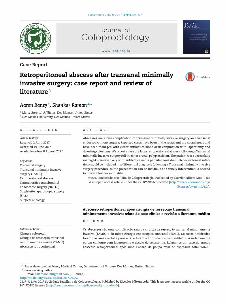
j coloproctol (rio j). 2 0 1 7; 3 7(4) :323–327 www.jcol.org.br Journal of Coloproctology Case Report Retroperitoneal abscess after transanal minimally invasive surgery: case report and review of literature Aaron Raney b , Shankar Raman a,∗ a Mercy Surgical Affiliates, Des Moines, United States b Des Moines University, Des Moines, United States a r t i c l e i n f o Article history: Received 2 April 2017 Accepted 19 June 2017 Available online 8 August 2017 Keywords: Colorectal surgery Transanal minimally invasive surgery (TAMIS) Retroperitoneal abscess Natural orifice transluminal endoscopic surgery (NOTES) Single-site laparoscopic surgery (SILS) Surgical oncology a b s t r a c t Abscesses are a rare complication of transanal minimally invasive surgery and transanal endoscopic micro surgery. Reported cases have been in the rectal and pre-sacral areas and have been managed with either antibiotics alone or in conjunction with laparotomy and diverting colostomy. We report a case of a large retroperitoneal abscess following a Transanal minimally invasive surgery full thickness rectal polyp excision. The patient was successfully managed conservatively with antibiotics and a percutaneous drain. Retroperitoneal infec- tion should be included in a differential diagnosis following a Transanal minimally invasive surgery procedure as the presentation can be insidious and timely intervention is needed to prevent further morbidity. © 2017 Sociedade Brasileira de Coloproctologia. Published by Elsevier Editora Ltda. This is an open access article under the CC BY-NC-ND license (http://creativecommons.org/ licenses/by-nc-nd/4.0/). Abscesso retroperitoneal após cirurgia de ressecc ¸ão transanal minimamente invasiva: relato de caso clínico e revisão a literatura médica Palavras-chave: Cirurgia colorretal Cirurgia de ressecc ¸ão transanal minimamente invasiva (TAMIS) Abscesso retroperitoneal r e s u m o Os abscessos são uma complicac ¸ão rara da cirurgia de ressecc ¸ão transanal minimamente invasiva (TAMIS) e da micro cirurgia endoscópica transanal (TEMS). Os casos notificados foram nas áreas rectal e pré-sacral e foram administrados com antibióticos isoladamente ou em conjunto com laparotomia e desvio de colostomia. Relatamos um caso de grande abscesso retroperitoneal após uma excisão de pólipo retal de espessura total TAMIS. Paper developed at Mercy Medical Center, Department of Surgery, Des Moines, United States. ∗ Corresponding author. E-mail: [email protected] (S. Raman). http://dx.doi.org/10.1016/j.jcol.2017.06.007 2237-9363/© 2017 Sociedade Brasileira de Coloproctologia. Published by Elsevier Editora Ltda. This is an open access article under the CC BY-NC-ND license (http://creativecommons.org/licenses/by-nc-nd/4.0/).
Transcript of Retroperitoneal abscess after transanal minimally invasive ... · Retroperitoneal abscess after...
j coloproctol (rio j). 2 0 1 7;3 7(4):323–327
Journal ofColoproctology
C
Ril
Aa
b
a
A
R
A
A
K
C
T
s
R
N
e
S
(
S
P
C
C
m
A
h2B
www.jco l .org .br
ase Report
etroperitoneal abscess after transanal minimallynvasive surgery: case report and review ofiterature�
aron Raneyb, Shankar Ramana,∗
Mercy Surgical Affiliates, Des Moines, United StatesDes Moines University, Des Moines, United States
r t i c l e i n f o
rticle history:
eceived 2 April 2017
ccepted 19 June 2017
vailable online 8 August 2017
eywords:
olorectal surgery
ransanal minimally invasive
urgery (TAMIS)
etroperitoneal abscess
atural orifice transluminal
ndoscopic surgery (NOTES)
ingle-site laparoscopic surgery
SILS)
urgical oncology
a b s t r a c t
Abscesses are a rare complication of transanal minimally invasive surgery and transanal
endoscopic micro surgery. Reported cases have been in the rectal and pre-sacral areas and
have been managed with either antibiotics alone or in conjunction with laparotomy and
diverting colostomy. We report a case of a large retroperitoneal abscess following a Transanal
minimally invasive surgery full thickness rectal polyp excision. The patient was successfully
managed conservatively with antibiotics and a percutaneous drain. Retroperitoneal infec-
tion should be included in a differential diagnosis following a Transanal minimally invasive
surgery procedure as the presentation can be insidious and timely intervention is needed
to prevent further morbidity.
© 2017 Sociedade Brasileira de Coloproctologia. Published by Elsevier Editora Ltda. This
is an open access article under the CC BY-NC-ND license (http://creativecommons.org/
licenses/by-nc-nd/4.0/).
Abscesso retroperitoneal após cirurgia de resseccão transanalminimamente invasiva: relato de caso clínico e revisão a literatura médica
alavras-chave:
r e s u m o
Os abscessos são uma complicacão rara da cirurgia de resseccão transanal minimamente
irurgia colorretalirurgia de resseccão transanal
inimamente invasiva (TAMIS)
bscesso retroperitoneal
invasiva (TAMIS) e da micro cirurgia endoscópica transanal (TEMS). Os casos notificados
foram nas áreas rectal e pré-sacral e foram administrados com antibióticos isoladamente
ou em conjunto com laparotomia e desvio de colostomia. Relatamos um caso de grande
abscesso retroperitoneal após uma excisão de pólipo retal de espessura total TAMIS.
� Paper developed at Mercy Medical Center, Department of Surgery, Des Moines, United States.∗ Corresponding author.
E-mail: [email protected] (S. Raman).ttp://dx.doi.org/10.1016/j.jcol.2017.06.007237-9363/© 2017 Sociedade Brasileira de Coloproctologia. Published by Elsevier Editora Ltda. This is an open access article under the CCY-NC-ND license (http://creativecommons.org/licenses/by-nc-nd/4.0/).
324 j coloproctol (rio j). 2 0 1 7;3 7(4):323–327
Cirurgia endoscópica
transluminal de orifício natural
(NOTES)
Cirurgia laparoscópica de único
local (SILS)
Oncologia cirúrgica
O paciente foi tratado com sucesso com a administracão de antibióticos e drenagem per-
cutânea. Para prevenir mais morbidade é necessária incluir a infeccão retroperitoneal no
diagnostico diferencial após um procedimento TAMIS onde a apresentacão pode ser insid-
iosa e a intervencão atempada.
© 2017 Sociedade Brasileira de Coloproctologia. Publicado por Elsevier Editora Ltda. Este
e um artigo Open Access sob uma licenca CC BY-NC-ND (http://creativecommons.org/
Introduction
Transanal minimally invasive surgery (TAMIS) is a recentlydeveloped surgical approach first described in 2010 by Atallahet al.1 It is a form of natural orifice transluminal endoscopicsurgery (NOTES) that is an alternative to the previously devel-oped transanal endoscopic micro surgery (TEMS) technology.In contrast to TEMS, TAMIS utilizes ordinary laparoscopicinstruments instead of TEMS specific instruments. Thoughthere have been no extensive comparative studies, TAMIS plat-form has been quickly adopted due to decreased upfront cost,readily accessible instruments, and faster learning curve.1,2
A single-incision laparoscopic surgery (SILS) port, adaptedfor transanal use (SILSTM Port, Covidien, Dublin, Ireland)or a specific transanal platform (GelPOINT© Path TransanalAccess Platform, Applied Medical, Rancho Santa Margarita,CA, USA), is placed and secured in the anus.1 Standard laparo-scopic instruments are then placed through this port andallow for conventional laparoscopic dissection.2 Pneumorec-tum allows for adequate visualization and the shorter TAMISplatform provides advantageous working angles within therectum as well as circumferential dissection without patientre-positioning.2,3 Furthermore, the soft platform has the the-oretical advantage of better functional outcomes due to lesstraction on the anal wall.2
TAMIS has proven to be an effective platform for R0local resection of benign neoplasia and early rectal cancer.2
Reported procedure specific complications have been pri-marily bleeding, peritoneal entry, and rectal abscess.3–5 Thetrue incidence of post-TAMIS abscesses is not well knownand there has not been a large retroperitoneal abscess doc-umented. We report a case of an extensive retroperitonealabscess formation following a TAMIS procedure withoutperitoneal entry. This was managed nonoperatively withantibiotics, and percutaneous drainage.
Case presentation
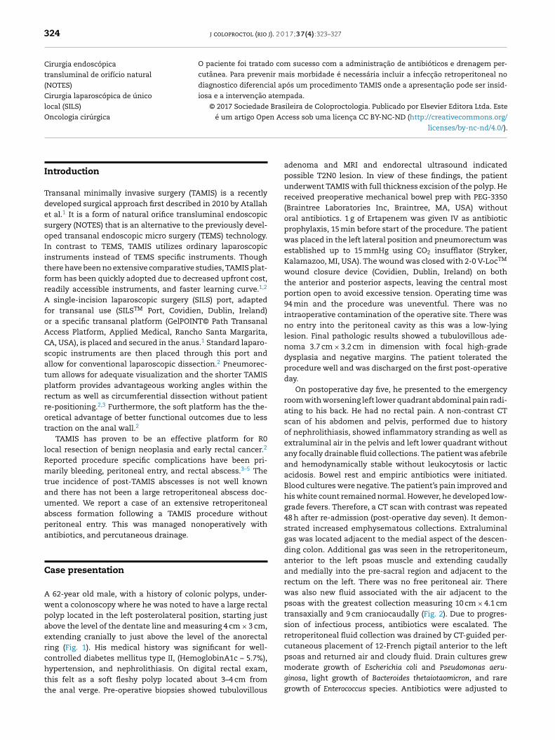
A 62-year old male, with a history of colonic polyps, under-went a colonoscopy where he was noted to have a large rectalpolyp located in the left posterolateral position, starting justabove the level of the dentate line and measuring 4 cm × 3 cm,extending cranially to just above the level of the anorectalring (Fig. 1). His medical history was significant for well-
controlled diabetes mellitus type II, (HemoglobinA1c – 5.7%),hypertension, and nephrolithiasis. On digital rectal exam,this felt as a soft fleshy polyp located about 3–4 cm fromthe anal verge. Pre-operative biopsies showed tubulovillouslicenses/by-nc-nd/4.0/).
adenoma and MRI and endorectal ultrasound indicatedpossible T2N0 lesion. In view of these findings, the patientunderwent TAMIS with full thickness excision of the polyp. Hereceived preoperative mechanical bowel prep with PEG-3350(Braintree Laboratories Inc, Braintree, MA, USA) withoutoral antibiotics. 1 g of Ertapenem was given IV as antibioticprophylaxis, 15 min before start of the procedure. The patientwas placed in the left lateral position and pneumorectum wasestablished up to 15 mmHg using CO2 insufflator (Stryker,Kalamazoo, MI, USA). The wound was closed with 2-0 V-LocTM
wound closure device (Covidien, Dublin, Ireland) on boththe anterior and posterior aspects, leaving the central mostportion open to avoid excessive tension. Operating time was94 min and the procedure was uneventful. There was nointraoperative contamination of the operative site. There wasno entry into the peritoneal cavity as this was a low-lyinglesion. Final pathologic results showed a tubulovillous ade-noma 3.7 cm × 3.2 cm in dimension with focal high-gradedysplasia and negative margins. The patient tolerated theprocedure well and was discharged on the first post-operativeday.
On postoperative day five, he presented to the emergencyroom with worsening left lower quadrant abdominal pain radi-ating to his back. He had no rectal pain. A non-contrast CTscan of his abdomen and pelvis, performed due to historyof nephrolithiasis, showed inflammatory stranding as well asextraluminal air in the pelvis and left lower quadrant withoutany focally drainable fluid collections. The patient was afebrileand hemodynamically stable without leukocytosis or lacticacidosis. Bowel rest and empiric antibiotics were initiated.Blood cultures were negative. The patient’s pain improved andhis white count remained normal. However, he developed low-grade fevers. Therefore, a CT scan with contrast was repeated48 h after re-admission (post-operative day seven). It demon-strated increased emphysematous collections. Extraluminalgas was located adjacent to the medial aspect of the descen-ding colon. Additional gas was seen in the retroperitoneum,anterior to the left psoas muscle and extending caudallyand medially into the pre-sacral region and adjacent to therectum on the left. There was no free peritoneal air. Therewas also new fluid associated with the air adjacent to thepsoas with the greatest collection measuring 10 cm × 4.1 cmtransaxially and 9 cm craniocaudally (Fig. 2). Due to progres-sion of infectious process, antibiotics were escalated. Theretroperitoneal fluid collection was drained by CT-guided per-cutaneous placement of 12-French pigtail anterior to the leftpsoas and returned air and cloudy fluid. Drain cultures grew
moderate growth of Escherichia coli and Pseudomonas aeru-ginosa, light growth of Bacteroides thetaiotaomicron, and raregrowth of Enterococcus species. Antibiotics were adjusted to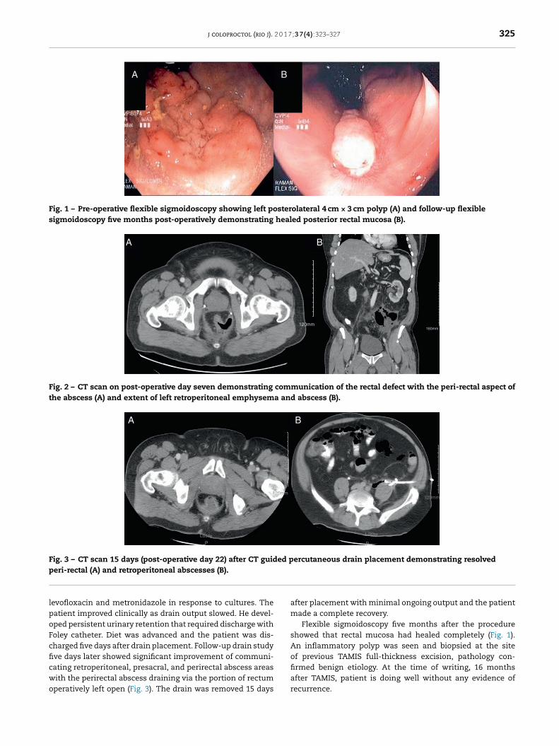
j coloproctol (rio j). 2 0 1 7;3 7(4):323–327 325
A B
Fig. 1 – Pre-operative flexible sigmoidoscopy showing left posterolateral 4 cm × 3 cm polyp (A) and follow-up flexiblesigmoidoscopy five months post-operatively demonstrating healed posterior rectal mucosa (B).
BA
120mm160mm
Fig. 2 – CT scan on post-operative day seven demonstrating communication of the rectal defect with the peri-rectal aspect ofthe abscess (A) and extent of left retroperitoneal emphysema and abscess (B).
A B
Lossy
P
Lossy
P
120mm120mm
Fig. 3 – CT scan 15 days (post-operative day 22) after CT guided percutaneous drain placement demonstrating resolvedp
lpoFcficwo
eri-rectal (A) and retroperitoneal abscesses (B).
evofloxacin and metronidazole in response to cultures. Theatient improved clinically as drain output slowed. He devel-ped persistent urinary retention that required discharge witholey catheter. Diet was advanced and the patient was dis-harged five days after drain placement. Follow-up drain studyve days later showed significant improvement of communi-
ating retroperitoneal, presacral, and perirectal abscess areasith the perirectal abscess draining via the portion of rectumperatively left open (Fig. 3). The drain was removed 15 daysafter placement with minimal ongoing output and the patientmade a complete recovery.
Flexible sigmoidoscopy five months after the procedureshowed that rectal mucosa had healed completely (Fig. 1).An inflammatory polyp was seen and biopsied at the siteof previous TAMIS full-thickness excision, pathology con-
firmed benign etiology. At the time of writing, 16 monthsafter TAMIS, patient is doing well without any evidence ofrecurrence.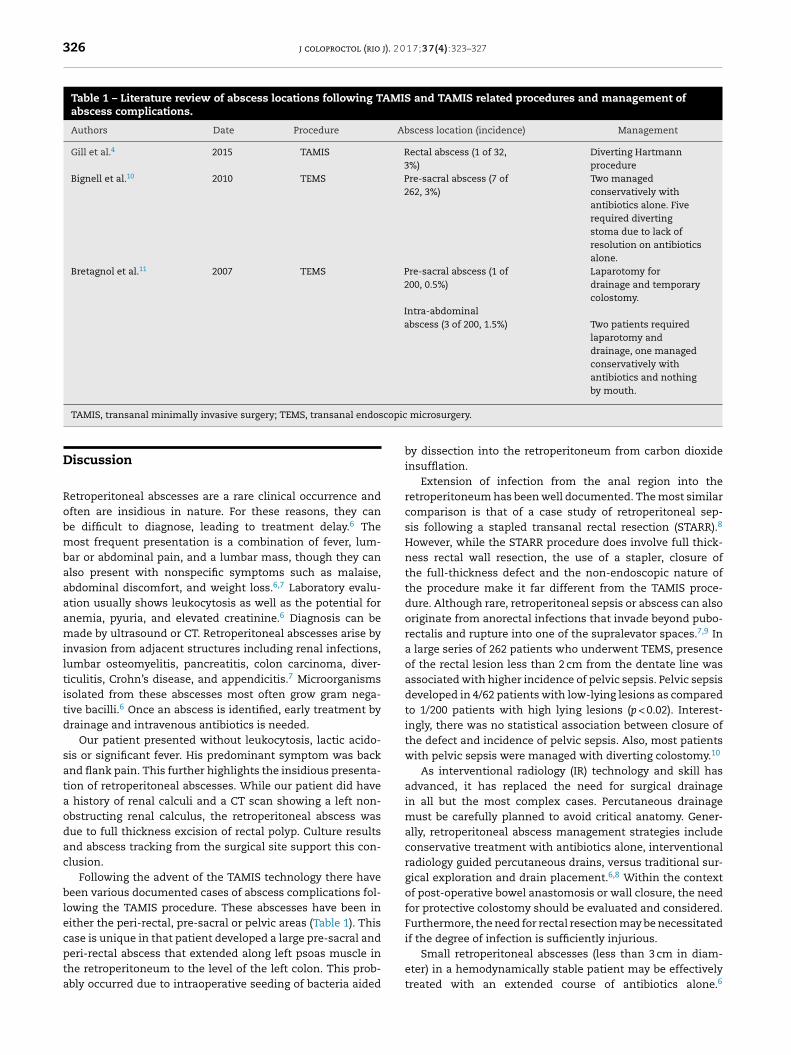
326 j coloproctol (rio j). 2 0 1 7;3 7(4):323–327
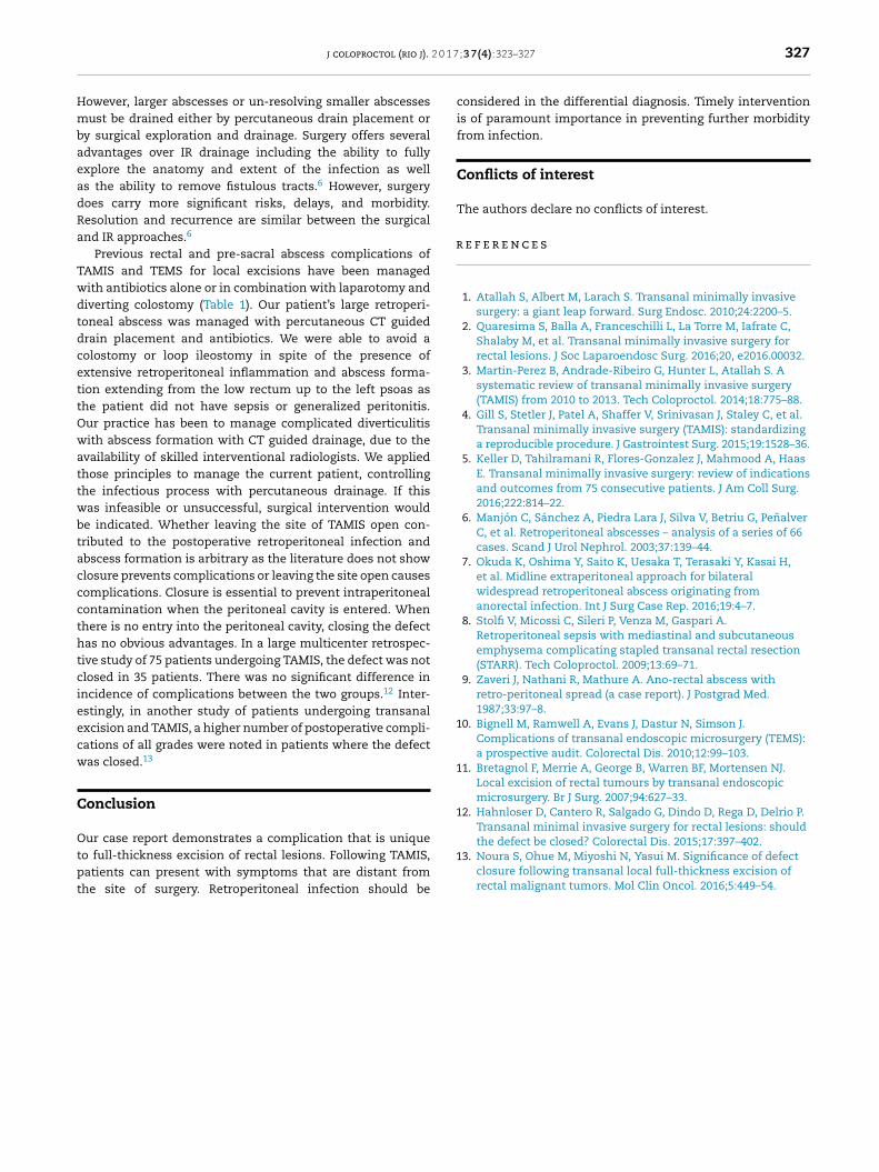
Table 1 – Literature review of abscess locations following TAMIS and TAMIS related procedures and management ofabscess complications.
Authors Date Procedure Abscess location (incidence) Management
Gill et al.4 2015 TAMIS Rectal abscess (1 of 32,3%)
Diverting Hartmannprocedure
Bignell et al.10 2010 TEMS Pre-sacral abscess (7 of262, 3%)
Two managedconservatively withantibiotics alone. Fiverequired divertingstoma due to lack ofresolution on antibioticsalone.
Bretagnol et al.11 2007 TEMS Pre-sacral abscess (1 of200, 0.5%)
Intra-abdominalabscess (3 of 200, 1.5%)
Laparotomy fordrainage and temporarycolostomy.
Two patients requiredlaparotomy anddrainage, one managedconservatively withantibiotics and nothingby mouth.
copic
TAMIS, transanal minimally invasive surgery; TEMS, transanal endosDiscussion
Retroperitoneal abscesses are a rare clinical occurrence andoften are insidious in nature. For these reasons, they canbe difficult to diagnose, leading to treatment delay.6 Themost frequent presentation is a combination of fever, lum-bar or abdominal pain, and a lumbar mass, though they canalso present with nonspecific symptoms such as malaise,abdominal discomfort, and weight loss.6,7 Laboratory evalu-ation usually shows leukocytosis as well as the potential foranemia, pyuria, and elevated creatinine.6 Diagnosis can bemade by ultrasound or CT. Retroperitoneal abscesses arise byinvasion from adjacent structures including renal infections,lumbar osteomyelitis, pancreatitis, colon carcinoma, diver-ticulitis, Crohn’s disease, and appendicitis.7 Microorganismsisolated from these abscesses most often grow gram nega-tive bacilli.6 Once an abscess is identified, early treatment bydrainage and intravenous antibiotics is needed.
Our patient presented without leukocytosis, lactic acido-sis or significant fever. His predominant symptom was backand flank pain. This further highlights the insidious presenta-tion of retroperitoneal abscesses. While our patient did havea history of renal calculi and a CT scan showing a left non-obstructing renal calculus, the retroperitoneal abscess wasdue to full thickness excision of rectal polyp. Culture resultsand abscess tracking from the surgical site support this con-clusion.
Following the advent of the TAMIS technology there havebeen various documented cases of abscess complications fol-lowing the TAMIS procedure. These abscesses have been ineither the peri-rectal, pre-sacral or pelvic areas (Table 1). Thiscase is unique in that patient developed a large pre-sacral and
peri-rectal abscess that extended along left psoas muscle inthe retroperitoneum to the level of the left colon. This prob-ably occurred due to intraoperative seeding of bacteria aidedmicrosurgery.
by dissection into the retroperitoneum from carbon dioxideinsufflation.
Extension of infection from the anal region into theretroperitoneum has been well documented. The most similarcomparison is that of a case study of retroperitoneal sep-sis following a stapled transanal rectal resection (STARR).8
However, while the STARR procedure does involve full thick-ness rectal wall resection, the use of a stapler, closure ofthe full-thickness defect and the non-endoscopic nature ofthe procedure make it far different from the TAMIS proce-dure. Although rare, retroperitoneal sepsis or abscess can alsooriginate from anorectal infections that invade beyond pubo-rectalis and rupture into one of the supralevator spaces.7,9 Ina large series of 262 patients who underwent TEMS, presenceof the rectal lesion less than 2 cm from the dentate line wasassociated with higher incidence of pelvic sepsis. Pelvic sepsisdeveloped in 4/62 patients with low-lying lesions as comparedto 1/200 patients with high lying lesions (p < 0.02). Interest-ingly, there was no statistical association between closure ofthe defect and incidence of pelvic sepsis. Also, most patientswith pelvic sepsis were managed with diverting colostomy.10
As interventional radiology (IR) technology and skill hasadvanced, it has replaced the need for surgical drainagein all but the most complex cases. Percutaneous drainagemust be carefully planned to avoid critical anatomy. Gener-ally, retroperitoneal abscess management strategies includeconservative treatment with antibiotics alone, interventionalradiology guided percutaneous drains, versus traditional sur-gical exploration and drain placement.6,8 Within the contextof post-operative bowel anastomosis or wall closure, the needfor protective colostomy should be evaluated and considered.Furthermore, the need for rectal resection may be necessitatedif the degree of infection is sufficiently injurious.
Small retroperitoneal abscesses (less than 3 cm in diam-eter) in a hemodynamically stable patient may be effectivelytreated with an extended course of antibiotics alone.6
2 0 1 7
HmbaeadRa
TwdtdcettOwattwbtacccthtcieecw
C
Otpt
r
1
1
1
j coloproctol (rio j).
owever, larger abscesses or un-resolving smaller abscessesust be drained either by percutaneous drain placement or
y surgical exploration and drainage. Surgery offers severaldvantages over IR drainage including the ability to fullyxplore the anatomy and extent of the infection as wells the ability to remove fistulous tracts.6 However, surgeryoes carry more significant risks, delays, and morbidity.esolution and recurrence are similar between the surgicalnd IR approaches.6
Previous rectal and pre-sacral abscess complications ofAMIS and TEMS for local excisions have been managedith antibiotics alone or in combination with laparotomy andiverting colostomy (Table 1). Our patient’s large retroperi-oneal abscess was managed with percutaneous CT guidedrain placement and antibiotics. We were able to avoid aolostomy or loop ileostomy in spite of the presence ofxtensive retroperitoneal inflammation and abscess forma-ion extending from the low rectum up to the left psoas ashe patient did not have sepsis or generalized peritonitis.ur practice has been to manage complicated diverticulitisith abscess formation with CT guided drainage, due to the
vailability of skilled interventional radiologists. We appliedhose principles to manage the current patient, controllinghe infectious process with percutaneous drainage. If thisas infeasible or unsuccessful, surgical intervention woulde indicated. Whether leaving the site of TAMIS open con-ributed to the postoperative retroperitoneal infection andbscess formation is arbitrary as the literature does not showlosure prevents complications or leaving the site open causesomplications. Closure is essential to prevent intraperitonealontamination when the peritoneal cavity is entered. Whenhere is no entry into the peritoneal cavity, closing the defectas no obvious advantages. In a large multicenter retrospec-
ive study of 75 patients undergoing TAMIS, the defect was notlosed in 35 patients. There was no significant difference inncidence of complications between the two groups.12 Inter-stingly, in another study of patients undergoing transanalxcision and TAMIS, a higher number of postoperative compli-ations of all grades were noted in patients where the defectas closed.13
onclusion
ur case report demonstrates a complication that is uniqueo full-thickness excision of rectal lesions. Following TAMIS,atients can present with symptoms that are distant fromhe site of surgery. Retroperitoneal infection should be
1
;3 7(4):323–327 327
considered in the differential diagnosis. Timely interventionis of paramount importance in preventing further morbidityfrom infection.
Conflicts of interest
The authors declare no conflicts of interest.
e f e r e n c e s
1. Atallah S, Albert M, Larach S. Transanal minimally invasivesurgery: a giant leap forward. Surg Endosc. 2010;24:2200–5.
2. Quaresima S, Balla A, Franceschilli L, La Torre M, Iafrate C,Shalaby M, et al. Transanal minimally invasive surgery forrectal lesions. J Soc Laparoendosc Surg. 2016;20, e2016.00032.
3. Martin-Perez B, Andrade-Ribeiro G, Hunter L, Atallah S. Asystematic review of transanal minimally invasive surgery(TAMIS) from 2010 to 2013. Tech Coloproctol. 2014;18:775–88.
4. Gill S, Stetler J, Patel A, Shaffer V, Srinivasan J, Staley C, et al.Transanal minimally invasive surgery (TAMIS): standardizinga reproducible procedure. J Gastrointest Surg. 2015;19:1528–36.
5. Keller D, Tahilramani R, Flores-Gonzalez J, Mahmood A, HaasE. Transanal minimally invasive surgery: review of indicationsand outcomes from 75 consecutive patients. J Am Coll Surg.2016;222:814–22.
6. Manjón C, Sánchez A, Piedra Lara J, Silva V, Betriu G, PenalverC, et al. Retroperitoneal abscesses – analysis of a series of 66cases. Scand J Urol Nephrol. 2003;37:139–44.
7. Okuda K, Oshima Y, Saito K, Uesaka T, Terasaki Y, Kasai H,et al. Midline extraperitoneal approach for bilateralwidespread retroperitoneal abscess originating fromanorectal infection. Int J Surg Case Rep. 2016;19:4–7.
8. Stolfi V, Micossi C, Sileri P, Venza M, Gaspari A.Retroperitoneal sepsis with mediastinal and subcutaneousemphysema complicating stapled transanal rectal resection(STARR). Tech Coloproctol. 2009;13:69–71.
9. Zaveri J, Nathani R, Mathure A. Ano-rectal abscess withretro-peritoneal spread (a case report). J Postgrad Med.1987;33:97–8.
0. Bignell M, Ramwell A, Evans J, Dastur N, Simson J.Complications of transanal endoscopic microsurgery (TEMS):a prospective audit. Colorectal Dis. 2010;12:99–103.
1. Bretagnol F, Merrie A, George B, Warren BF, Mortensen NJ.Local excision of rectal tumours by transanal endoscopicmicrosurgery. Br J Surg. 2007;94:627–33.
2. Hahnloser D, Cantero R, Salgado G, Dindo D, Rega D, Delrio P.Transanal minimal invasive surgery for rectal lesions: should
the defect be closed? Colorectal Dis. 2015;17:397–402.3. Noura S, Ohue M, Miyoshi N, Yasui M. Significance of defectclosure following transanal local full-thickness excision ofrectal malignant tumors. Mol Clin Oncol. 2016;5:449–54.