Retinal Imaging - University of Arizona · Retinal Imaging “The eyes are the ... is binocular, so...
24
1 Retinal Imaging “The eyes are the window to the soul” 16 th century proverb The retina gives an unobstructed view of the human vascular. Also, retinal imaging is important for diagnosing retina diseases. Retinal Physiology
Transcript of Retinal Imaging - University of Arizona · Retinal Imaging “The eyes are the ... is binocular, so...

1
Retinal Imaging
“The eyes are the window to the soul”16th century proverb
The retina gives an unobstructed viewof the human vascular. Also, retinalimaging is important for diagnosingretina diseases.
Retinal Physiology

2
Rods and Cones
Rod and Cone Density

3
Rod and Cone Mosaic
Direct Ophthalmoscopy
Patient Observer
Observer adjusts power of lens wheel so that the patient’sfar point is imaged to the Observer’s far point.

4
Indirect Ophthalmoscopy
Patient Observer
The Observer holds a high power lens in front of the Patient and views the intermediate image formed by the lens. The system is binocular, so a 3-D feel for structures with the eye is obtained.
Fundus CameraA fundus camera is a device for photographing the retina. It is basedon the indirect ophthalmoscopy principle, where the Observer’s eye is replaced with a camera.

5
1°5°
Angular Size
30°
5° x 7°
Dimensions
0.25 mm 1.25 mm
7.5 mm
1.5 x 2 mm

6
Red-Free ImagesFundus images are sometimes takenwith a green filter to enhance the contrast of the blood vessels.
Fluorescein AngiographySodium Fluorescein dye is injectedinto a vein in the patient. As thedye circulates, it passes throughthe vessels of the retina. Blue light is used to illuminate the retinaand fundus photographs are takento capture the green fluorescenceof the dye. This technique aids invisualizing the vessels of the retina.

7
Indocyanine Green (ICG) Angiography
Indocyanine Green dye is injectedinto a vein in the patient. As thedye circulates, it passes throughthe vessels of the retina. Digital fundus photographs are taken to capture the infrared fluorescenceof the dye. This technique aids invisualizing the vessels of the choroid.
Retinal Mosaic

8
Stereo Pairs• Two images of the retina can be taken from slightly
different angles to form a stereo pair.• Useful for visualize cupping of the optic nerve head in
glaucoma and edema (swelling) that occurs from some pathologies.
Red/Blue AnaglyphsTake the red channel from the left hand image and the blueAnd green channels from the right hand image.

9
Red/Blue AnaglyphsMerge images to create and anaglyph
Cup to Disk Ratio• Diameter of the cupped region of the optic nerve head
divided by the diameter of the optic nerve head.• Normal is ~0.3-0.5.• Abnormal values are higher and are associated with
glaucoma
C/D = 0.6

10
Portable Fundus Camera
Portable Fundus Camera

11
Normal Retina
Shaken Baby Syndrome
• Severe head injury of baby from being shaken.• Cause is usually child abuse.• Poor prognosis, typically severe disabilities in survivors.• Injuries and symptons may include:
– Bruising, swelling and bleeding in the brain.– Lethargy, loss of consciousness, convulsions.– Hemorrhages in the retina.– Possible broken or dislocated bones.

12
Shaken Baby Syndrome
• SBS and Retinal Hemorrhages– 50-80% of SBS have retinal hemorrhages– 60-90% are bilateral– Retinal Hemorrhages is strong confirmatory evidence
of abuse.
Shaken Baby Syndrome• Victims are usually in a pediatric intensive care unit and cannot be
easily moved to ophthalmology departments for conventional fundusphotographs.
• Victims usually cannot sit upright, so conventional fundus cameras cannot be used.
• Emergency room and intensive care personnel may not be well versed in traditional retinal examination, so an easy to use camera may aid in identifying retinal hemorrhages.
• Inexpensive cameras can be placed in multiple hospitals.• Digital photographs can easily be transmitted for consultation by off-
site ophthalmologists.• Child Protective Services and the Police can use the photographs as
evidence against abusers, instead of solely relying on the expert verbal testimony of an ophthalmologist.

13
Shaken Baby Syndrome
Child Abuse

14
Veterinary Use
Scanning Laser Ophthalmoscopy
Scanning confocalmicroscope

15
Adaptive Optics
Nerve Fibers Blood Vessels Photoreceptors
Adaptive Optics

16
Retinal Densitometry
Scanning Laser Polarimeter
17
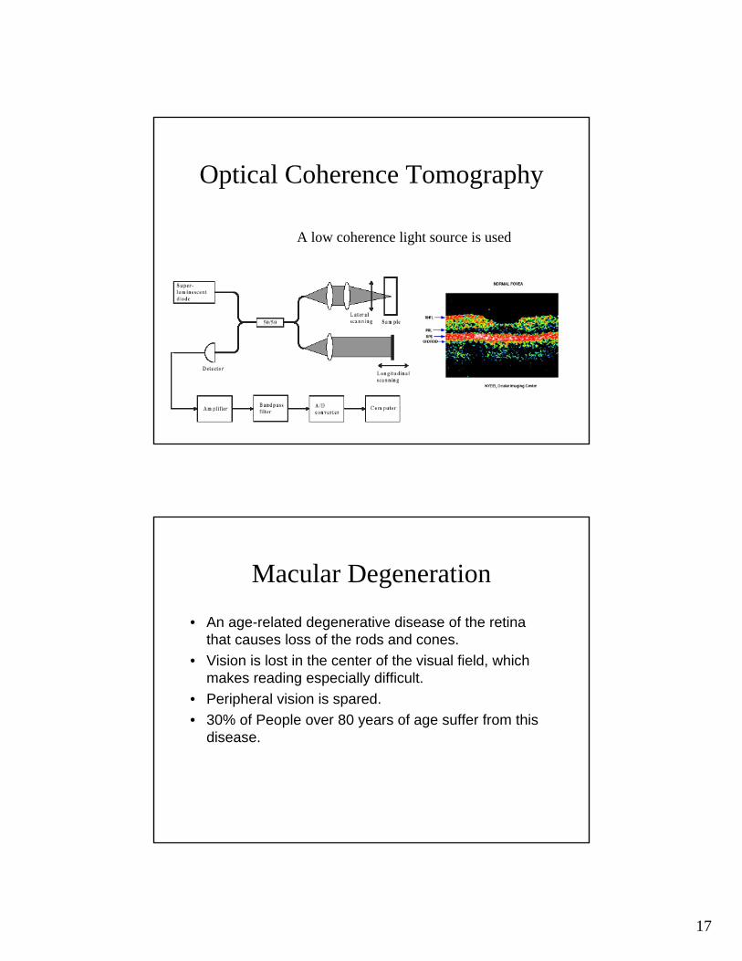
Optical Coherence Tomography
A low coherence light source is used
Macular Degeneration
• An age-related degenerative disease of the retina that causes loss of the rods and cones.
• Vision is lost in the center of the visual field, which makes reading especially difficult.
• Peripheral vision is spared.• 30% of People over 80 years of age suffer from this
disease.

18
Macular Degeneration
Macular Degeneration

19
Macular Degeneration
Macular Degeneration

20
Retinitis Pigmentosa
• Hereditary Disease• Slowly destroys photoreceptors• Usually progresses earlier in life• Starts in mid-periphery and works towards central and
peripheral vision.
Retinitis Pigmentosa

21
Background
• In 1967 GS Brindley applied an electrical stimulation to the retina of a completely blind nurse.
• The nurse reported seeing some very distorted images and light.
• This means the wiring is ok, but the sensors are bad.
Subretinal Implant

22
FDA Trials
• Implant surgery was performed in the first three recipients in June 2000, and three others received the implant in July 2001.
• The patients range in age from 46 to 76 years and baseline vision ranged from count fingers to almost no light perception.
• Surgery was performed unilaterally with the fellow eye serving as a control

23
FDA Trials
• All patients have noted varying degrees of improvement in their vision, with some gaining perception of light and shapes.
• Two patients showed dramatic improvements in visual acuity following treatment: one from none at baseline to 20 to 25 letters and another from 20 at baseline to 40 letters.
Epiretinal Implant

24
Hurdles For Retinal Implants
• Materials – Body must not reject the sensor – Silicone, Platinum & Iridium
• Lifetime – Sensor must function for years or decades• Power – Energy must be indirectly supplied to the implant
– Solar powered batteries.