Retina Ocular Hemorrhages in Neonatal Porcine Eyes …injury/Publications/Coats_etal_2010.pdfOcular...
6
Ocular Hemorrhages in Neonatal Porcine Eyes from Single, Rapid Rotational Events Brittany Coats, 1,2 Gil Binenbaum, 2,3,4 Robert L. Peiffer, 4,5 Brian J. Forbes, 3,4 and Susan S. Margulies 1 PURPOSE. To characterize ocular hemorrhages from single, rapid head rotations in the neonatal pig. METHODS. Three- to 5-day-old anesthetized piglets (n 51) underwent a single, rapid (117-266 rad/s) head rotation in the sagittal (n 13), coronal (n 7), or axial (n 31) planes. Six hours after injury, the animals were euthanatized and perfusion fixed, and the brain and eyes were harvested for gross and histopathologic examination by masked neuro- and ocular pa- thologists. RESULTS. Ocular hemorrhage was found in 73% of animals (51% bilateral). Intraocular hemorrhage was primarily located near the vitreous base (70% of injured animals had ciliary body hemorrhage, and 11% had peripheral retinal hemorrhage). Hemorrhages were also found in the anterior chamber (11%), vitreous (5%), and optic nerve (disc, 8%; nerve sheath, 57%). Rapid axial head rotations resulted in a higher incidence of intraocular hemorrhage than coronal or sagittal head rotations, but the difference did not reach statistical significance (P 0.06). Control eyes had no injuries. CONCLUSIONS. Optic nerve sheath and ciliary body hemorrhages were common in piglets that experienced a single, rapid head rotation. Retinal hemorrhage was present in a smaller number of animals. Most intraocular hemorrhages were located in re- gions of strong vitreous attachment, suggesting that this animal model will be useful in investigating the effect of vitreoretinal adhesion on ocular hemorrhage caused by inertial head rota- tions. Extrapolation of this model to the human infant should not be made until the effect of anatomic differences between the human and pig on the occurrence and patterns of ocular injuries is further investigated. (Invest Ophthalmol Vis Sci. 2010;51:4792– 4797) DOI:10.1167/iovs.10-5211 A busive head trauma (AHT) is the leading cause of death and disability from child abuse 1,2 and is characterized by intracranial hemorrhage and/or intraocular hemorrhage (pri- marily retinal), with or without additional systemic injuries. In cases of AHT with less severe signs and symptoms, AHT is often misdiagnosed. 3 An increased understanding of the mech- anisms of the injuries associated with AHT may assist in the evaluation of difficult cases. Careful in vitro and animal stud- ies 4 –10 have aided in elucidating the mechanisms of pediatric traumatic brain injury and have provided clinicians with em- pirical data to help identify the inconsistent histories associ- ated with brain injury. However, few controlled experimental studies to date have investigated mechanisms of retinal hem- orrhages in pediatric head trauma, and an animal model has yet to be clearly established. There is a lack of agreement about the mechanisms by which retinal hemorrhages develop in AHT. Theories include vitreous traction, acute increases in retinal arterial or venous pressure from raised intracranial or intrathoracic pressure, and tracking of intracranial blood. A viable animal model must be developed to experimentally test mechanistic theories and investigate the effect of repetitive back-and-forth head rota- tions (often reported in AHT) on ocular hemorrhages. The neonatal piglet is an established large animal model for pedi- atric brain injury, 4,9,11 and several similarities of the piglet retina to the human retina make it a promising model for studying traumatic retinal hemorrhages. To evaluate the potential of the piglet as an immature animal model for retinal hemorrhages and other ocular injuries resulting from inertial head trauma, we retrospectively exam- ined the eyes of 3- to 5-day-old piglets involved in multiple studies of traumatic brain injury. We sought to characterize ocular hemorrhages in animals undergoing single, rapid head rotation. METHODS Inertial Head Rotation In a protocol approved by the Animal Care and Use Committee at the University of Pennsylvania and in accordance with the animal research guidelines set forth in the ARVO Statement for the Use of Animals in Ophthalmic and Vision Research, 51 neonatal (3–5-day-old) piglets were anesthetized via an endotracheal tube with 2% to 4% isofluorane. Once the piglet was fully anesthetized, its head was secured by a padded snout clamp to the linkage assembly of a pneumatic actuator (HYGE device; Bendix Corp., South Bend, IN). The device did not hinder or compress the chest cavity of the animal. The actuator and linkage assembly created a purely inertial, nonimpact rotation of the head around the C3 to C5 segment of the spine. The rotational velocity of the head was measured with an angular rate transducer (ARS-06; ATA Sensors, Albuquerque, NM) and acquired at a 10-kHz sampling rate onto a computer. Each animal underwent a single, rapid unidirec- tional head rotation in one of three planes: sagittal (n 13), coronal (n 7), or axial (n 31). Heart rate, respiratory rate, body temper- ature, end-tidal CO 2 , and oxygen saturation were recorded before the injury and at 30-minute intervals after the injury. Five uninjured control From the 1 Department of Bioengineering and the 4 Department of Ophthalmology, Scheie Eye Institute, University of Pennsylvania, Phil- adelphia, Pennsylvania; the 3 Division of Ophthalmology, Children’s Hospital of Philadelphia, Philadelphia, Pennsylvania; and 5 Merck & Co, Inc., West Point, Pennsylvania. 2 These authors contributed equally to the work presented here and should therefore be regarded as equivalent authors. Supported by National Institute of Neurological Disorders and Stroke (NINDS) Grant R01-NS39679 (SSM) and National Eye Institute Grant K12-EY01539 (GB), from the National Institutes of Health, Be- thesda, MD. Submitted for publication January 14, 2010; revised March 12 and 29, 2010; accepted March 31, 2010. Disclosure: B. Coats, None; G. Binenbaum, None; R.L. Peiffer, Merck & Co, Inc. (E); B.J. Forbes, None; S.S. Margulies, None Corresponding author: Brittany Coats, Department of Bioengineer- ing, University of Pennsylvania, 240 Skirkanich Hall, 210 S. 33rd Street, Philadelphia, PA 19104; [email protected]. Retina Investigative Ophthalmology & Visual Science, September 2010, Vol. 51, No. 9 4792 Copyright © Association for Research in Vision and Ophthalmology
Transcript of Retina Ocular Hemorrhages in Neonatal Porcine Eyes …injury/Publications/Coats_etal_2010.pdfOcular...

Ocular Hemorrhages in Neonatal Porcine Eyes fromSingle, Rapid Rotational Events
Brittany Coats,1,2 Gil Binenbaum,2,3,4 Robert L. Peiffer,4,5 Brian J. Forbes,3,4
and Susan S. Margulies1
PURPOSE. To characterize ocular hemorrhages from single,rapid head rotations in the neonatal pig.
METHODS. Three- to 5-day-old anesthetized piglets (n � 51)underwent a single, rapid (117-266 rad/s) head rotation in thesagittal (n � 13), coronal (n � 7), or axial (n � 31) planes. Sixhours after injury, the animals were euthanatized and perfusionfixed, and the brain and eyes were harvested for gross andhistopathologic examination by masked neuro- and ocular pa-thologists.
RESULTS. Ocular hemorrhage was found in 73% of animals (51%bilateral). Intraocular hemorrhage was primarily located nearthe vitreous base (70% of injured animals had ciliary bodyhemorrhage, and 11% had peripheral retinal hemorrhage).Hemorrhages were also found in the anterior chamber (11%),vitreous (5%), and optic nerve (disc, 8%; nerve sheath, 57%).Rapid axial head rotations resulted in a higher incidence ofintraocular hemorrhage than coronal or sagittal head rotations,but the difference did not reach statistical significance (P �0.06). Control eyes had no injuries.
CONCLUSIONS. Optic nerve sheath and ciliary body hemorrhageswere common in piglets that experienced a single, rapid headrotation. Retinal hemorrhage was present in a smaller numberof animals. Most intraocular hemorrhages were located in re-gions of strong vitreous attachment, suggesting that this animalmodel will be useful in investigating the effect of vitreoretinaladhesion on ocular hemorrhage caused by inertial head rota-tions. Extrapolation of this model to the human infant shouldnot be made until the effect of anatomic differences betweenthe human and pig on the occurrence and patterns of ocularinjuries is further investigated. (Invest Ophthalmol Vis Sci.2010;51:4792–4797) DOI:10.1167/iovs.10-5211
Abusive head trauma (AHT) is the leading cause of deathand disability from child abuse1,2 and is characterized by
intracranial hemorrhage and/or intraocular hemorrhage (pri-
marily retinal), with or without additional systemic injuries. Incases of AHT with less severe signs and symptoms, AHT isoften misdiagnosed.3 An increased understanding of the mech-anisms of the injuries associated with AHT may assist in theevaluation of difficult cases. Careful in vitro and animal stud-ies4–10 have aided in elucidating the mechanisms of pediatrictraumatic brain injury and have provided clinicians with em-pirical data to help identify the inconsistent histories associ-ated with brain injury. However, few controlled experimentalstudies to date have investigated mechanisms of retinal hem-orrhages in pediatric head trauma, and an animal model has yetto be clearly established.
There is a lack of agreement about the mechanisms bywhich retinal hemorrhages develop in AHT. Theories includevitreous traction, acute increases in retinal arterial or venouspressure from raised intracranial or intrathoracic pressure, andtracking of intracranial blood. A viable animal model must bedeveloped to experimentally test mechanistic theories andinvestigate the effect of repetitive back-and-forth head rota-tions (often reported in AHT) on ocular hemorrhages. Theneonatal piglet is an established large animal model for pedi-atric brain injury,4,9,11 and several similarities of the pigletretina to the human retina make it a promising model forstudying traumatic retinal hemorrhages.
To evaluate the potential of the piglet as an immatureanimal model for retinal hemorrhages and other ocular injuriesresulting from inertial head trauma, we retrospectively exam-ined the eyes of 3- to 5-day-old piglets involved in multiplestudies of traumatic brain injury. We sought to characterizeocular hemorrhages in animals undergoing single, rapid headrotation.
METHODS
Inertial Head Rotation
In a protocol approved by the Animal Care and Use Committee at theUniversity of Pennsylvania and in accordance with the animal researchguidelines set forth in the ARVO Statement for the Use of Animals inOphthalmic and Vision Research, 51 neonatal (3–5-day-old) pigletswere anesthetized via an endotracheal tube with 2% to 4% isofluorane.Once the piglet was fully anesthetized, its head was secured by apadded snout clamp to the linkage assembly of a pneumatic actuator(HYGE device; Bendix Corp., South Bend, IN). The device did nothinder or compress the chest cavity of the animal. The actuator andlinkage assembly created a purely inertial, nonimpact rotation of thehead around the C3 to C5 segment of the spine. The rotational velocityof the head was measured with an angular rate transducer (ARS-06;ATA Sensors, Albuquerque, NM) and acquired at a 10-kHz samplingrate onto a computer. Each animal underwent a single, rapid unidirec-tional head rotation in one of three planes: sagittal (n � 13), coronal(n � 7), or axial (n � 31). Heart rate, respiratory rate, body temper-ature, end-tidal CO2, and oxygen saturation were recorded before theinjury and at 30-minute intervals after the injury. Five uninjured control
From the 1Department of Bioengineering and the 4Department ofOphthalmology, Scheie Eye Institute, University of Pennsylvania, Phil-adelphia, Pennsylvania; the 3Division of Ophthalmology, Children’sHospital of Philadelphia, Philadelphia, Pennsylvania; and 5Merck & Co,Inc., West Point, Pennsylvania.
2These authors contributed equally to the work presented hereand should therefore be regarded as equivalent authors.
Supported by National Institute of Neurological Disorders andStroke (NINDS) Grant R01-NS39679 (SSM) and National Eye InstituteGrant K12-EY01539 (GB), from the National Institutes of Health, Be-thesda, MD.
Submitted for publication January 14, 2010; revised March 12 and29, 2010; accepted March 31, 2010.
Disclosure: B. Coats, None; G. Binenbaum, None; R.L. Peiffer,Merck & Co, Inc. (E); B.J. Forbes, None; S.S. Margulies, None
Corresponding author: Brittany Coats, Department of Bioengineer-ing, University of Pennsylvania, 240 Skirkanich Hall, 210 S. 33rd Street,Philadelphia, PA 19104; [email protected].
Retina
Investigative Ophthalmology & Visual Science, September 2010, Vol. 51, No. 94792 Copyright © Association for Research in Vision and Ophthalmology

animals underwent identical procedures, but no head rotation wasinduced.
Ocular Examination and Histology
In a subset of animals (subset 1: 10 injured, 2 control), a pediatricophthalmologist (BJF) performed a dilated ophthalmic fundus exami-nation with indirect ophthalmoscopy before injury, immediately afterinjury, and between 4 and 6 hours after injury. All animals wereeuthanatized and perfusion fixed 6 hours after injury. Both eyes in eachanimal were harvested by removing the orbital roof, transecting theoptic nerve just anterior to the optic chiasm, and removing the eyeanteriorly. The brain and eyes of each animal were stored in 10%formalin in preparation for histologic analysis. Each eye was sectionedwith one cross-sectional cut through the transected end of the opticnerve, one cut through the pupil–optic nerve–macular plane, and onecut through a plane either superior or inferior to the optic nerve. Allslices were stained with hematoxylin and eosin (H&E).
In a second subset of animals (subset 2: 41 injured, 3 control), noophthalmic examination was performed, but eyes were extracted andstored in 10% formalin in the same manner as those in subset 1. In lieuof the ophthalmic examination, the eyes were examined grossly beforehistologic analysis, and three additional cuts were made for histology.Briefly, the eyes were removed en bloc, and the extraocular musclesand surrounding soft tissues were examined for hemorrhages or ab-normalities and photographed in color. The muscles and surroundingsoft tissue were subsequently removed, and the anatomic dimensionswere measured, including horizontal, vertical, and anterior–posteriordimensions of the globe; horizontal and vertical dimensions of thecornea; and the diameter of the pupil. The optic nerve was examinedfor hemorrhages and the length recorded. After external inspection, ahorizontal cut was made in each eye starting at a point just above thesuperior corneoscleral limbus and passing through a point just abovethe superior optic disc margin. The resulting superior and inferiorcalottes were examined in 70% alcohol under a dissecting microscopeand photographed in color. Five thin (5 �m) sections of each eye (onesection through the pupil–optic nerve–head plane, two sectionsslightly superior and inferior to that plane, and two sections throughthe retinal periphery superior and inferior to this plane) were stainedwith H&E (Fig. 1).
H&E slides from both animal subsets were examined microscopi-cally by an ocular pathologist (RLP). The pathologist, who was maskedto the mechanical loading experienced by the animal, evaluated eacheye for the presence and location of ocular abnormalities, includingretinal hemorrhages, optic nerve head swelling, optic nerve sheath
hemorrhage, and other ocular hemorrhage (e.g., ciliary body, hy-phema, subconjunctival, or extraocular muscle) or injury (e.g., retinaldetachment). Brain pathology for both subsets was performed by aneuropathologist. Pearson’s �2 test was used to assess the effect ofhead rotation direction (sagittal, axial, or coronal) on the incidence ofocular hemorrhage, bilateral ocular hemorrhage, optic nerve sheathhemorrhage, retinal hemorrhage, and ciliary body hemorrhage. P �0.05 was considered significant (JMP Statistical Software; SAS Institute,Cary, NC).
RESULTS
The average ocular dimensions (�SD) after removal of extraoc-ular tissues from 44 piglets (3–5 days old) were 16.6 � 0.6,15.0 � 0.1, and 14.1 � 0.8 mm for the horizontal, vertical, andanterior–posterior axes, respectively. Average corneal widthand height were 10.8 � 0.6 and 9.1 � 1.0 mm, respectively;pupil diameter was 7.2 � 0.6 mm; and mass (measured in 6 ofthe 44 animals) was 2.1 � 0.1 g. The ranges of measuredangular velocities and accelerations of the head rotations were117 to 266 rad/s and 30.6–101 krad/s2. By design, sagittal headrotations occurred at slightly lower loads (mean � SD, 185 �17 rad/s) compared with axial and coronal head rotations(207 � 31 and 208 � 11 rad/s, respectively) because pigletshave limited range of motion in the neck in this direction.
Ocular hemorrhage was found in 73% of all animals under-going a single, rapid head rotation and was bilateral in 51% ofthe animals with ocular hemorrhage. Intraocular hemorrhagewas primarily located near the vitreous base (70% of the ani-mals with eye injury had ciliary body hemorrhage, 11% hadperipheral retinal hemorrhage), but hemorrhages were alsofound in the anterior chamber (11%) and optic nerve (disc 8%,nerve sheath 57%).
Overall, animals that experienced a rapid head rotation inthe axial direction had a higher incidence of ocular hemor-rhage (81%) than did those with a rapid head rotation in thesagittal (62%) or coronal plane (57%), but these differences didnot reach statistical significance (P � 0.06; Fig. 2), possiblybecause of limited statistical power (� � 0.54). In addition,there was no significant effect of head rotation direction on theincidence of bilateral injury (P � 0.32), optic nerve disc orsheath hemorrhage (P � 0.21), ciliary body hemorrhage (P �0.12), and retinal hemorrhage (P � 0.53). Three animals diedat 1 to 2 hours after injury due to cardiac or respiratorycomplications, but the ocular findings in these animals werenot unique from those in the remaining 48 animals surviving 6hours.
Subset 1
In the first subset of eyes (n � 10), no signs of ocular injurywere found during indirect ophthalmic examination. However,histologic staining revealed three (30%) animals with opticnerve sheath hemorrhage (subdural or intradural), five (50%)with hemorrhage located in the stroma of the ciliary body, two(20%) with hyphema, and one (10%) with a unilateral prereti-nal hemorrhage (Fig. 3) located near the ora ciliaris retinae(akin to the ora serrata in humans). It is possible that thisretinal hemorrhage was not seen on ophthalmic examinationbecause of the inherent difficulty in systematically examiningthe ora in the neonatal piglet.
Subset 2
Gross examination in the second subset of animals revealed six(15%) with optic nerve sheath hemorrhages confined to focalregions on the optic nerve and eight (20%) with extraocularhemorrhage in either the muscle or fat tissue. H&E stainingrevealed 11 additional animals with optic nerve sheath hemor-
FIGURE 1. Five microsections were made in each eye from thesecond animal subset. Three sections were cut through the pupil–optic nerve plane (1, 2, 3). Two sections were cut inferior (I) andsuperior (S) to that plane. All sections were stained with H&E formicroscopic analysis.
IOVS, September 2010, Vol. 51, No. 9 Ocular Hemorrhages from Single, Rapid Rotational Events 4793

rhage, increasing the total occurrence of optic nerve sheathhemorrhages in this subset to 17 (41%) animals. Optic nervesheath hemorrhages were mostly subdural (Fig. 4A) or intra-dural (Fig. 4B). Twenty-three (56%) animals had other signs ofocular trauma, including hemorrhage in the stroma of theciliary body (56%), hyphema (5%), persistent hyaloid vesselhemorrhage (2%), vitreous hemorrhage (5%), optic disc hem-orrhage located either over the disc or in the parenchyma (7%),and three animals had subretinal (5%) or preretinal (7%) hem-orrhage (Fig. 5). All these hemorrhages (with the exception ofthe hyaloid vessel hemorrhage, hyphema, and optic disc hem-orrhage) were located near the ora. In total, 28 (68%) animalsin the second subset had some form of ocular hemorrhage,which was bilateral in 14 animals. Pathology of the five controlanimals revealed no ocular injuries.
Brain Pathology
Gross brain examination was completed in 46 of the 51 animalsin the experimental group. Thirty-eight (83%) of these animalshad bilateral subdural hemorrhages, five (11%) had unilateralsubdural hemorrhages, and the remaining three (6%) had littleor no blood found on examination. Microscopic evaluationwas performed on the brains of 31 experimental animals.H&E staining confirmed the distribution of subdural andsubarachnoid hemorrhage, as identified on gross examina-
tion, and further revealed 15 (48%) animals with whitematter hemorrhage. Staining with NF-68 (n � 10) or �APP(n � 21) revealed 23 (74%) animals with axonal injury,which was diffuse in 15 and focal in 8. Sagittal and axialhead rotations resulted more often in bilateral subduralhemorrhage, white matter hemorrhage, and diffuse axonalinjury than did coronal head rotations (Table 1).
Twenty-six (68%) of the animals with bilateral subduralhemorrhages had ocular hemorrhages, but only one of theanimals with unilateral subdural hemorrhage had ocular injury(ciliary body hemorrhage). Conversely, all animals with ocularhemorrhages had associated brain injury, except two that hadsmall ciliary body hemorrhages and no intracranial findings.
Gross examination and microscopic pathology were per-formed on all control brain specimens, and no abnormalitieswere reported.
DISCUSSION
For better differentiation between patterns of ocular hemor-rhage from accidental and AHT in children, it is important tounderstand the mechanisms and loading conditions that cancause these injuries. We sought to evaluate an immature animalmodel for its potential to study mechanisms of traumatic hem-orrhage associated with head accelerations. We found thatapproximately three-fourths of the animals in this preliminarymodel developed ocular hemorrhage as the result of a single,high-velocity head rotation, even though the limited number ofhistologic cuts may have led to an underestimation of findings.Seventy percent of hemorrhages were located in a region ofstrong vitreoretinal attachment in the pig. Thus, this animalmodel will be useful for evaluating the role of the vitreoretinalinterface in the development of hemorrhage from rotationalhead accelerations. However, to assess the clinical relevance ofthis model, one must consider the pattern and severity ofocular injuries, the similarities and differences between pig andhuman ocular and orbital anatomy, and the nature and magni-tude of the loads applied.
Retinal hemorrhages have been reported to occur in 0% to20% of cases of pediatric accidental head trauma, depending onthe cause of injury. The higher reported incidences are fromcase series investigating crush head injuries or fatal motorvehicle accidents. When present, the retinal hemorrhages inyoung children with accidental head trauma are typically in-
FIGURE 2. The incidence of ocularinjuries observed from single, rapidhead rotations, grouped by the planeof head rotation. The numbers at thetop of the bars are the number ofanimals. In subsets 1 and 2, combin-ing axial head rotations resulted in aslightly higher occurrence of ocularinjury than with the sagittal or coro-nal rotations, but this finding was notstatistically significant with a Pear-son’s �2 test (P � 0.06). The inci-dence of various types of ocular hem-orrhage (i.e., optic nerve, ciliarybody, and retinal) was also not signif-icantly different among the groups.
FIGURE 3. Peripheral preretinal and ciliary body hemorrhage in a 3- to5-day-old piglet after a single high-velocity rotational head acceleration.The hemorrhage was not detected during indirect ophthalmic exami-nation. H&E. Magnification, �10.
4794 Coats et al. IOVS, September 2010, Vol. 51, No. 9

traretinal, few in number, and confined to the posterior pole,even in complex falls with multiple impacts.12–16 Rarely, incases of severe trauma, such as fatal motor vehicle accidents orcrush head injuries, more extensive hemorrhages can beseen.17 In contrast, retinal hemorrhages in children with AHTcan range from a few, isolated intraretinal hemorrhages, asseen in accidental trauma, to numerous hemorrhages that maybe multilayered and extend into and throughout the retinalperiphery and can include macular folds and hemorrhagicmacular cysts.18–22 The distribution and types of hemorrhagesreported in abusive and accidental head trauma may be infor-mative with regard to mechanistic etiology. The overlap infindings at the mild end of the injury spectrum and occasionallyat the severe end of the injury spectrum (e.g., fatal car acci-dents) suggests that there may be some common mecha-nism(s) underlying the retinal hemorrhages in accidental andAHT. However, the additional patterns of injury in AHT (e.g.,macular cysts) suggest that additional mechanisms may beinvolved in those cases.
A favored theory in the literature is that acceleration–decel-eration forces cause the vitreous to pull on the retina, possibly
damaging retinal vessels, with subsequent hemorrhaging, orcausing changes in vascular autoregulation.23 In our study,most of the intraocular hemorrhages were located near thevitreous base in the form of ciliary body hemorrhage or lessfrequently as peripheral retinal hemorrhage. It is not clear howthis pattern of injury relates clinically to pediatric head trauma,as the vitreous base is not typically visualized on clinical ex-amination in the awake child without scleral depression, andophthalmologists are unlikely to perform scleral depression inawake children in the absence of posterior pole findings. How-ever, the location of the hemorrhages is coincident with areasof strong vitreoretinal attachment in the pig,24 suggesting apossible role for traction in the model. In fact, ciliary body andoptic nerve sheath hemorrhages have been described on post-mortem examination in AHT.20,21
Another mechanistic theory involves the direct trackingof blood from the brain along the optic nerve and into theeye. Most of the animals in our study had an optic nervesheath subdural hemorrhage and a few animals had micro-scopic optic disc hemorrhages. However, the nerve sheathhemorrhages were focal rather than spread over the lengthof the nerve, and no retinal hemorrhage was observed pos-
FIGURE 4. Histology of eyes from 3-to 5-day-old piglets undergoing a sin-gle rapid angular head acceleration.Of all the animals, 64% had opticnerve sheath hemorrhage. Opticnerve hemorrhages were subdural(A), intradural (B), and epidural (notshown). H&E. Magnification: (A)�20; (B) �10.
FIGURE 5. Histology of eyes from 3-to 5-day-old piglets undergoing a sin-gle rapid angular head accelerationfound ocular hemorrhages in 84% ofthe eyes. Nonoptic nerve sheathhemorrhages included (A) ciliarybody hemorrhage, (B) hyphema, (C)hemorrhage from a persistent hya-loid vessel, and (D) subretinal andpreretinal hemorrhages. H&E. Magni-fication: (A) �20; (B–D) �10.
IOVS, September 2010, Vol. 51, No. 9 Ocular Hemorrhages from Single, Rapid Rotational Events 4795
teriorly in the eye; on the contrary, most of the intraocularhemorrhages were found anteriorly.
A third theory involves increased intracranial or intravenouspressure. The pattern of ocular hemorrhage observed in thispreliminary model matches neither the peripapillary hemor-rhages associated with papilledema nor the distinctive retinalhemorrhage pattern of retinal venous occlusion, but we didnot measure intracranial or intravenous pressure in this study.
We selected the immature piglet as a potential model forthe study of retinal hemorrhages, as the porcine retina is moresimilar to the human retina than other domestic animals.25,26
Specifically, the porcine retina does not have a tapetum andcontains a well-developed vascular arcade, with the majorretinal vessels lying within the nerve fiber layer and capillariespresent throughout multiple layers of the retina. The vitreousbase of the pig is comparable to the human vitreous base andstraddles the ora. However, the piglet eye does not contain afovea or a macula, and there is debate in the literature overwhether it has a macula-like region devoid of major retinalvessels.25,26 We found one persistent hyaloid vessel hemor-rhage in our study, but piglet hyaloid vessels typically regressbefore birth, with hyaloid remnants persisting for 1 week afterbirth.27 Since completion of this study, we have examined theeyes of several additional 3- to 5-day-old piglets and noted manywith clear hyaloid stalks, but none with hyaloid arteries con-taining blood.
Vitreoretinal attachments in the pig have been reported tooccur throughout the retina, but one plasmin-assisted vitrectomystudy demonstrated that the attachments at the vitreous base arestronger compared to other regions.24 In humans, vitreoretinalattachments occur across the entire retina, as they do in the pig,but locations of stronger attachment are at the vitreous base, opticnerve head, macula, and along major retinal vessels.28,29 Shouldvitreoretinal traction play a significant role in the development ofretinal hemorrhages, these ocular anatomic differences may influ-ence the patterns of hemorrhage seen experimentally.
Contrasts in human and porcine orbital anatomy may alsoaffect the patterns of observed injury. The pig has an openorbit, and instead of a bony closure, a strong fibrous liga-ment stretches from the frontal bone to the zygomatic bone,effectively enclosing the orbit but with slightly less rigiditythan in humans.25 Although this relative laxity may allowmore freedom of movement for the orbital contents, thepig’s extraocular muscles may inhibit the globe’s motion.The annulus of Zinn is absent, and the origins of the musclesare on bone and are characterized as extremely strong.25 Inaddition, the pig, like most domestic animals, has an additionalmuscle called the retractor bulbi, which inserts circumferentiallyon the globe and retracts it into the orbit, allowing the nictitatingmembrane, or third eyelid, of the animal to close and furtherinhibit movement of the globe within the orbit.25 Future studiesare necessary to evaluate whether movement within the orbit isan important factor in the pathogenesis of ocular hemorrhage inpediatric head trauma.
A final anatomic consideration is the orientation of theporcine eye. We observed that animals with axial headrotations appeared to have more ocular hemorrhages thandid animals with sagittal or coronal rotations, although thedifferences were not statistically significant. The visual axisof the pig is oriented approximately 30° outward from thatof the human. This 30° offset causes a 50% decrease in theforce being applied along the optic nerve during sagittalhead rotation and an 87% increase in the force along theoptic nerve during coronal head rotation. The offset wouldnot cause any difference in the forces applied to the eyeduring axial head rotation. Assuming that there is a relation-ship between the inertial load applied along the optic nerveand the development of ocular hemorrhage, these calcula-tions suggest that for a given load, our porcine animalstudies may underestimate the incidence of ocular hemor-rhage from sagittal rotations and overestimate the incidencefrom coronal rotations in comparison with that expected ina human.
It is important to recognize that the load applied in thisstudy was a single, high-velocity rotation of the head and nota low-velocity, repetitive, back-and-forth motion often re-ported in cases of AHT. We selected a single, high-velocityhead rotation to establish the ability of the model to produceocular hemorrhages from angular head acceleration and toserve as a baseline for future studies investigating the effectof cyclic back-and-forth head rotations on eye injury. Todate, the only controlled animal experiment investigatingretinal hemorrhages from repetitive, cyclic head rotationshas been in mice.30 The mouse eye is extremely small inmass compared with the human infant eye, and to achievean equivalent scaled load, a shaking frequency five timesgreater than that found physically possible in investigationsof loads from shaking infant surrogate dolls (2–3 Hz) isnecessary.31 Investigations of cyclic loading that can betterapproximate real-world forces and are more similar to hu-man retinal and vasculature structure would be of greatervalue in a large animal model such as the pig.
The single, high-velocity head rotations applied in this ani-mal model were of sufficient magnitude to result in severebrain injury in almost all the animals. When scaled to the brainmass of a human infant (420 g), the applied head rotations aregreater than those measured for low-height falls (1–2 feet) ontoconcrete,32 but less than would occur during an inflicted im-pact onto a hard surface.31,33 Because the mass of the imma-ture porcine eye is similar to the mass of the human infant eye(2.29 g),34 scaling is less important. The axial length of thepiglet eye measured postmortem is smaller (14 mm) than theaxial length measured in vivo in the human infant (17 mm).35
Postmortem shrinkage may account for some shortening, but itis unclear whether the remaining difference is primarily attrib-utable to vitreous chamber size or anterior chamber depth orhow changes in axial length may affect ocular hemorrhagefrom rapid, nonimpact rotations of the head. Clinical conclu-
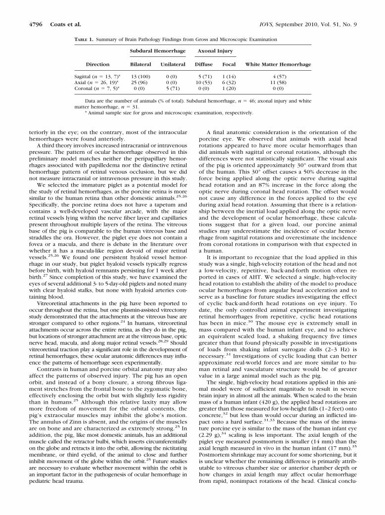
TABLE 1. Summary of Brain Pathology Findings from Gross and Microscopic Examination
Direction
Subdural Hemorrhage Axonal Injury
Bilateral Unilateral Diffuse Focal White Matter Hemorrhage
Sagittal (n � 13, 7)* 13 (100) 0 (0) 5 (71) 1 (14) 4 (57)Axial (n � 26, 19)* 25 (96) 0 (0) 10 (53) 6 (32) 11 (58)Coronal (n � 7, 5)* 0 (0) 5 (71) 0 (0) 1 (20) 0 (0)
Data are the number of animals (% of total). Subdural hemorrhage, n � 46; axonal injury and whitematter hemorrhage, n � 31.
* Animal sample size for gross and microscopic examination, respectively.
4796 Coats et al. IOVS, September 2010, Vol. 51, No. 9
sions regarding ocular injuries cannot be made from theseanimal studies until we know how anatomic differences be-tween the human and porcine orbit affect the way mechanicalloads applied to the head are translated to mechanical loadsexperienced by the eye.
To date, there is no experimental model that can be used toinvestigate the mechanisms and loading conditions that causeretinal hemorrhages in pediatric head injuries. The develop-ment of such a model may have important clinical, legal, andsocial implications related to accurately identifying and pro-tecting children who are suffering child abuse, while correctlydifferentiating accidental injuries. We investigated the poten-tial of the neonatal piglet as such a model. Optic nerve sheathand ciliary body hemorrhages were common in piglets thatexperienced a single, nonimpact head rotation, with someretinal hemorrhage, which was less common but all of whichwas located near the ciliary body at the vitreous base. Althoughthis pattern differs from the severe, diffuse retinal hemorrhagesoften seen in cases of AHT, we are encouraged by the highpercentage of animals demonstrating a rather consistent pat-tern of ocular injuries, the localization of injuries to regions ofstrong vitreoretinal attachment in the pig, and the commonfinding of optic sheath nerve hemorrhages. Future studies willinclude evaluation of lower velocity cyclic loading conditionsand manipulations of the vitreoretinal interface and extraocularmuscle anatomy. Anatomic differences between the speciesmay affect the observed injury patterns in the model, and suchdifferences must be investigated before experimental resultscan be safely extrapolated to head injuries in infants.
Acknowledgments
The authors thank Colin Smith for completing and interpreting theneuropathology data.
References
1. Frasier LD. Abusive head trauma in infants and young children: aunique contributor to developmental disabilities. Pediatr Clin NAm. 2008;55:1269–1285.
2. Overpeck MD, Brenner RA, Trumble AC, Trifiletti LB, BerendesHW. Risk factors for infant homicide in the United States. N EnglJ Med. 1998;339:1211–1216.
3. Jenny C, Hymal K, Ritzen A, Reinert S, Hay T. Analysis of missedcases of abusive head trauma. JAMA. 1999;281:621–626.
4. Armstead W, Kurth C. Different cerebral hemodynamic responsesfollowing fluid percussion brain injury in the newborn and juve-nile pig. J Neurotrauma. 1994;11:487–497.
5. Finnie J, Blumbergs P, Manavis J. Multi-focal cerebellar granularlayer necrosis in traumatically head-injured lambs. Vet Pathol.1999;36:256–258.
6. Margulies SS, Meaney DF Smith D, Chen X-H, Miller R, RaghupathiR. A Comparison of Diffuse Brain Injury in the Newborn andAdult Pig. Barcelona, Spain: International Research Committee onthe Biomechanics of Impact. 1999.
7. Prange M, Margulies S. Anisotropy and Inhomogeneity of theMechanical Properties of Brain Tissue at Large Deformation.Presented at Prevention Through Biomechanics. Novi, MI; 1999.
8. Prange M, Margulies S. Tissue Strain Thresholds for Axonal Injuryin the Infant Brain. In: Kamm R, ed. Bioengineering Conference,Snowbird, UT. New York: The American Society of MechanicalEngineers; 2001:833–834.
9. Raghupathi R, Margulies SS. Traumatic axonal injury after closedhead injury in the neonatal pig. J Neurotrauma. 2002;19:843–853.
10. Shaver E, Duhaime AC, Curtis M, Gennarelli L, Barrett R. Experi-mental acute subdural hematoma in infant piglets. Pediatr Neuro-surg. 1996;25:123–129.
11. Duhaime AC, Margulies SS, Durham SR, et al. Maturation-depen-dent response of the piglet brain to scaled cortical impact. J Neu-rosurg. 2000;93:455–462.
12. Christian C, Taylor A, Hertle R, Duhaime A. Retinal hemorrhages causedby accidental household trauma. J Pediatrics. 1999;135:125–127.
13. Johnson D, Braun D, Friendly D. Accidental head trauma andretinal hemorrhage. Neurosurgery. 1993;33:231–235.
14. Sturm V, Knecht PB, Landau K, Menke MN. Rare retinal haemor-rhages in translational accidental head trauma in children. Eye(Lond). 2009;23(7):1535–1541.
15. Trenchs V, Curcoy A, Morales M, Serra A, Navarro R, Pou J. Retinalhemorrhages in head trauma resulting from falls: differential diag-nosis with non-accidental trauma in patients younger than 2 yearsof age. Child Nervous Syst. 2008;24:815–820.
16. Duhaime A, Alario A, Lewander W, et al. Head injury in very youngchildren: mechanisms, injury types, and ophthalmologic findingsin 100 hospitalized patients under two years of age. Pediatrics.1992;90:179–185.
17. Kivlin J, Currie M, Greenbaum V, Simons K, Jentzen J. Retinalhemorrhages in children following fatal motor vehicle crashes: acase series. Arch Ophthalmology. 2008;126:800–804.
18. Budenz D, Farber M, Mirchandani H, Park H, Rorke L. Ocular andoptic nerve hemorrhages in abused infants with intracranial inju-ries. Ophthalmology. 1994;101:559–565.
19. Elner SG, Elner VM, Arnall M, Albert DM. Ocular and associatedsystematic findings in suspected child abuse: a necropsy study.Arch Ophthalmol. 1990;108:1094–1101.
20. Emerson MV, Jakobs E, Green WR. Ocular autopsy and histopatho-logic features of child abuse. Ophthalmology. 2007;1384–1394.
21. Marshall DH, Brownstein S, Dorey MW, Addison DJ, Carpenter B.The spectrum of postmortem ocular findings in victims of shakenbaby syndrome. Can J Ophthalmol. 2001;36:377–383.
22. Riffenburgh R, Sathyavagiswaran L. Ocular findings at autopsy ofchild abuse victims. Ophthalmology. 1991;98:1519–1524.
23. Wygnanski-Jaffe T, Levin A, Shafiq A, et al. Postmortem orbital findings inshaken baby syndrome. Am J Ophthalmol. 2006;142:233–240.
24. Gandorfer A, Putz E, Welge-Lussen U, Gruterich M, Ulbig M,Kampik A. Ultrastructure of the vitreoretinal interface followingplasmin assisted vitrectomy. Br J Ophthalmol. 2001;85:6–10.
25. Prince J, Diesem C, Eglitis I, Ruskell G. Anatomy and Histology ofthe Eye and Orbit in Domestic Animals. Springfield: Charles C.Thomas; 1960:307.
26. Rootman J. Vascular system of the optic nerve head and retina inthe pig. Br J Ophthalmol. 1971;808–819.
27. De Schaepdrijver L, Simoens P, Lauwers H, De Geest J, CharlierG. The hyaloid vascular system of the pig. Anat Embryol.1989;180:549 –554.
28. Sebag J. Anatomy and pathology of the vitreoretinal interface. Eye.1992;6:541–552.
29. Sebag J, Balazs EA. Morphology and ultrastructure of human vitre-ous fibers. Invest Ophthalmol Vis Sci. 1989;30:1867–1871.
30. Bonnier C, Mesples B, Carpentier S, Henin D, Gressens P. Delayedwhite matter injury in a murine model of shaken baby syndrome.Brain Pathol. 2002;12:320–328.
31. Prange M, Coats B, Duhaime AC, Margulies S. Anthropomorphicsimulations of falls, shakes, and inflicted impacts in infants. J Neu-rosurg. 2003;99:143–150.
32. Coats B, Margulies SS. Potential for head injuries in infants fromlow-height falls. J Neurosurg Pediatr. 2008;2:1–10.
33. Duhaime A, Gennarelli T, Thibault L, Bruce D, Margulies S, WiserR. The shaken baby syndrome: a clinical, pathological, and biome-chanical study. J Neurosurg. 1987;66:409–415.
34. Bron A, ed. Wolf’s Anatomy of the Eye and Orbit. 8th ed. NewYork: Chapman & Hall Medical; 1997:736.
35. Fledelius H. Pre-term delivery and the growth of the eye. anoculometric study of eye size around term-time. Acta Ophthalmol.1992;204(suppl):10–15.
IOVS, September 2010, Vol. 51, No. 9 Ocular Hemorrhages from Single, Rapid Rotational Events 4797