
Restriction of mitochondrial calcium overload by mcu ... · The loss of dopaminergic neurons (DA)...
12
RESEARCH ARTICLE Restriction of mitochondrial calcium overload by mcu inactivation renders a neuroprotective effect in zebrafish models of Parkinson’s disease Smijin K. Soman 1, * ,§ , Michal Bazala 1 , Marcus Keatinge 2,3, ‡ , Oliver Bandmann 2,3 and Jacek Kuznicki 1,§ ABSTRACT The loss of dopaminergic neurons (DA) is a pathological hallmark of sporadic and familial forms of Parkinson’s disease (PD). We have previously shown that inhibiting mitochondrial calcium uniporter (mcu) using morpholinos can rescue DA neurons in the PTEN-induced putative kinase 1 ( pink1) −/− zebrafish model of PD. In this article, we show results from our studies in mcu knockout zebrafish, which was generated using the CRISPR/Cas9 system. Functional assays confirmed impaired mitochondrial calcium influx in mcu −/− zebrafish. We also used in vivo calcium imaging and fluorescent assays in purified mitochondria to investigate mitochondrial calcium dynamics in a pink1 −/− zebrafish model of PD. Mitochondrial morphology was evaluated in DA neurons and muscle fibers using immunolabeling and transgenic lines, respectively. We observed diminished mitochondrial area in DA neurons of pink1 −/− zebrafish, while deletion of mcu restored mitochondrial area. In contrast, the mitochondrial volume in muscle fibers was not restored after inactivation of mcu in pink1 −/− zebrafish. Mitochondrial calcium overload coupled with depolarization of mitochondrial membrane potential leads to mitochondrial dysfunction in the pink1 −/− zebrafish model of PD. We used in situ hybridization and immunohistochemical labeling of DA neurons to evaluate the effect of mcu deletion on DA neuronal clusters in the ventral telencephalon of zebrafish brain. We show that DA neurons are rescued after deletion of mcu in pink1 −/− and the 1-methyl- 4-phenyl-1,2,3,6-tetrahydropyridine (MPTP) zebrafish model of PD. Thus, inactivation of mcu is protective in both genetic and chemical models of PD. Our data reveal that regulating mcu function could be an effective therapeutic target in PD pathology. KEY WORDS: Mitochondria, mcu, Parkinson’s disease, Zebrafish, CRISPR/Cas9, Neuroprotection INTRODUCTION Parkinson’s disease (PD) is the second most common neurodegenerative disease affecting 1% of the population above 60 years of age (Kalia and Lang, 2016). The classical pathology includes progressive loss of dopaminergic (DA) neurons in the substantia nigra pars compacta (SNpc), resulting in reduced dopamine levels in the striatum, leading to motor and non-motor symptoms. Unlike sporadic forms of PD which account for 95% of PD cases, familial forms of PD are mainly caused by mutations in synuclein alpha (SNCA), parkin, PTEN-induced putative kinase 1 (PINK1), DJ-1 and leucine-rich repeat kinase 2 (LRRK2) (Klein and Westenberger, 2012). Many of the gene mutations leading to familial forms of PD in humans are associated with mitochondrial dysfunction (Exner et al., 2012). PINK1 is a serine-threonine kinase and is an activator of Parkin-mediated ubiquitylation; this quality control process maintains a healthy pool of mitochondria in the cellular system (Valente et al., 2004; Kondapalli et al., 2012). The mechanism behind the selective loss of DA neurons in the SNpc of PD patients is still poorly understood. DA neurons in SNpc are distinct, as they are autonomously active and employ L-type calcium (Ca 2+ ) channels instead of conventional sodium channels to maintain the pace-making activity (Guzman et al., 2015). The pace- making activity of the DA neurons makes the mitochondria susceptible to Ca 2+ overload. Shortening and simplification of the dendritic arbor observed in neurodegenerative diseases occur through a process of excitatory mitochondrial toxicity, which triggers mitophagy and pre-synaptic mitochondrial depletion, mechanisms that are distinct from classic excitotoxicity (Dagda et al., 2011; Verma et al., 2018). Mitochondrial Ca 2+ overload leads to neuronal loss in neurodegenerative diseases and ischemia models (Kruman and Mattson, 1999; Kostic et al., 2015; Matthew et al., 2018). The post- mortem tissue samples of the SNpc from sporadic PD patients have a substantial decrease in complex I activity, asserting the role of mitochondrial dysfunction in PD pathology (Schapira et al., 1990). Excessive Ca 2+ release from endoplasmic reticulum (ER) causes persistent mitochondrial Ca 2+ overload leading to mitochondrial dysfunction that triggers apoptosis cascade in DA neurons (Lee et al., 2018). Ca 2+ is transported into mitochondria through mitochondrial calcium uniporter (mcu), and mitochondrial Na/Ca 2+ exchanger (NCLX) is responsible for moving Ca 2+ out of mitochondria (Baughman et al., 2011). Impaired NCLX function has been implicated to be responsible for mitochondrial Ca 2+ overload during pink1 deficiency (Gandhi et al., 2009; Kostic et al., 2015). However, further studies are required to evaluate the contribution of mitochondrial Ca 2+ influx channels and their regulation in mitochondrial calcium overload. mcu is part of a multi-subunit Ca 2+ channel capable of several states of mcu activity. mcu is a 40 kDa protein that consists of two coiled-coil domains, two transmembrane domains and a short motif of amino acids between the two Received 24 April 2019; Accepted 13 September 2019 1 Laboratory of Neurodegeneration, International Institute of Molecular and Cell Biology, Ksie ̨ cia Trojdena 4, 02-109, Warsaw, Poland. 2 Medical Research Council Centre for Developmental and Biomedical Genetics, University of Sheffield, Firth Court, Western Bank, Sheffield S10 2TN, UK. 3 Sheffield Institute for Translational Neuroscience (SITraN), University of Sheffield, 385a Glossop Road, Sheffield, S10 2HQ, UK. *Present address: Department of Pharmacology, University of Nevada, Reno, Nevada, USA. ‡ Present address: Department of Pharmacology, University of Nevada, Reno 89557, Nevada, USA. § Authors for correspondence ( [email protected]; [email protected]) S.K.S., 0000-0002-3891-118X; M.B., 0000-0002-2237-2206; M.K., 0000-0003- 2055-1187; J.K., 0000-0001-6486-0657 This is an Open Access article distributed under the terms of the Creative Commons Attribution License (https://creativecommons.org/licenses/by/4.0), which permits unrestricted use, distribution and reproduction in any medium provided that the original work is properly attributed. 1 © 2019. Published by The Company of Biologists Ltd | Biology Open (2019) 8, bio044347. doi:10.1242/bio.044347 Biology Open by guest on June 29, 2020 http://bio.biologists.org/ Downloaded from
Transcript of Restriction of mitochondrial calcium overload by mcu ... · The loss of dopaminergic neurons (DA)...

RESEARCH ARTICLE
Restriction of mitochondrial calcium overload by mcu inactivationrenders a neuroprotective effect in zebrafish models ofParkinson’s diseaseSmijin K. Soman1,*,§, Michal Bazała1, Marcus Keatinge2,3,‡, Oliver Bandmann2,3 and Jacek Kuznicki1,§
ABSTRACTThe loss of dopaminergic neurons (DA) is a pathological hallmark ofsporadic and familial forms of Parkinson’s disease (PD). We havepreviously shown that inhibiting mitochondrial calcium uniporter (mcu)using morpholinos can rescue DA neurons in the PTEN-inducedputative kinase 1 (pink1)−/− zebrafish model of PD. In this article, weshow results from our studies in mcu knockout zebrafish, which wasgenerated using the CRISPR/Cas9 system. Functional assaysconfirmed impaired mitochondrial calcium influx in mcu−/− zebrafish.We also used in vivo calcium imaging and fluorescent assays in purifiedmitochondria to investigate mitochondrial calcium dynamics in apink1−/− zebrafish model of PD. Mitochondrial morphology wasevaluated in DA neurons and muscle fibers using immunolabelingand transgenic lines, respectively. We observed diminishedmitochondrial area in DA neurons of pink1−/− zebrafish, while deletionof mcu restored mitochondrial area. In contrast, the mitochondrialvolume in muscle fibers was not restored after inactivation of mcu inpink1−/− zebrafish. Mitochondrial calcium overload coupled withdepolarization of mitochondrial membrane potential leads tomitochondrial dysfunction in the pink1−/− zebrafish model of PD. Weused in situ hybridization and immunohistochemical labeling of DAneurons to evaluate the effect ofmcu deletion on DA neuronal clustersin the ventral telencephalon of zebrafish brain. We show that DAneurons are rescued after deletion ofmcu in pink1−/− and the 1-methyl-4-phenyl-1,2,3,6-tetrahydropyridine (MPTP) zebrafish model of PD.Thus, inactivation of mcu is protective in both genetic and chemicalmodels of PD. Our data reveal that regulatingmcu function could be aneffective therapeutic target in PD pathology.
KEY WORDS: Mitochondria, mcu, Parkinson’s disease, Zebrafish,CRISPR/Cas9, Neuroprotection
INTRODUCTIONParkinson’s disease (PD) is the second most commonneurodegenerative disease affecting 1% of the population above60 years of age (Kalia and Lang, 2016). The classical pathologyincludes progressive loss of dopaminergic (DA) neurons in thesubstantia nigra pars compacta (SNpc), resulting in reduceddopamine levels in the striatum, leading to motor and non-motorsymptoms. Unlike sporadic forms of PD which account for 95% ofPD cases, familial forms of PD are mainly caused by mutations insynuclein alpha (SNCA), parkin, PTEN-induced putative kinase 1(PINK1), DJ-1 and leucine-rich repeat kinase 2 (LRRK2) (Kleinand Westenberger, 2012). Many of the gene mutations leading tofamilial forms of PD in humans are associated with mitochondrialdysfunction (Exner et al., 2012). PINK1 is a serine-threonine kinaseand is an activator of Parkin-mediated ubiquitylation; this qualitycontrol process maintains a healthy pool of mitochondria in thecellular system (Valente et al., 2004; Kondapalli et al., 2012).
The mechanism behind the selective loss of DA neurons in theSNpc of PD patients is still poorly understood. DA neurons in SNpcare distinct, as they are autonomously active and employ L-typecalcium (Ca2+) channels instead of conventional sodium channels tomaintain the pace-making activity (Guzman et al., 2015). The pace-making activity of the DA neurons makes the mitochondriasusceptible to Ca2+ overload. Shortening and simplification of thedendritic arbor observed in neurodegenerative diseases occur througha process of excitatory mitochondrial toxicity, which triggersmitophagy and pre-synaptic mitochondrial depletion, mechanismsthat are distinct from classic excitotoxicity (Dagda et al., 2011; Vermaet al., 2018). Mitochondrial Ca2+ overload leads to neuronal loss inneurodegenerative diseases and ischemia models (Kruman andMattson, 1999; Kostic et al., 2015; Matthew et al., 2018). The post-mortem tissue samples of the SNpc from sporadic PD patients have asubstantial decrease in complex I activity, asserting the role ofmitochondrial dysfunction in PD pathology (Schapira et al., 1990).Excessive Ca2+ release from endoplasmic reticulum (ER) causespersistent mitochondrial Ca2+ overload leading to mitochondrialdysfunction that triggers apoptosis cascade in DA neurons (Lee et al.,2018). Ca2+ is transported into mitochondria through mitochondrialcalcium uniporter (mcu), and mitochondrial Na/Ca2+ exchanger(NCLX) is responsible for moving Ca2+ out of mitochondria(Baughman et al., 2011). Impaired NCLX function has beenimplicated to be responsible for mitochondrial Ca2+ overload duringpink1 deficiency (Gandhi et al., 2009; Kostic et al., 2015). However,further studies are required to evaluate the contribution ofmitochondrial Ca2+ influx channels and their regulation inmitochondrial calcium overload. mcu is part of a multi-subunit Ca2+
channel capable of several states of mcu activity. mcu is a 40 kDaprotein that consists of two coiled-coil domains, two transmembranedomains and a short motif of amino acids between the twoReceived 24 April 2019; Accepted 13 September 2019
1Laboratory of Neurodegeneration, International Institute of Molecular and CellBiology, Ksiecia Trojdena 4, 02-109, Warsaw, Poland. 2Medical Research CouncilCentre for Developmental and Biomedical Genetics, University of Sheffield, FirthCourt, Western Bank, Sheffield S10 2TN, UK. 3Sheffield Institute for TranslationalNeuroscience (SITraN), University of Sheffield, 385a Glossop Road, Sheffield,S10 2HQ, UK.*Present address: Department of Pharmacology, University of Nevada, Reno,Nevada, USA. ‡Present address: Department of Pharmacology, University ofNevada, Reno 89557, Nevada, USA.
§Authors for correspondence ( [email protected];[email protected])
S.K.S., 0000-0002-3891-118X; M.B., 0000-0002-2237-2206; M.K., 0000-0003-2055-1187; J.K., 0000-0001-6486-0657
This is an Open Access article distributed under the terms of the Creative Commons AttributionLicense (https://creativecommons.org/licenses/by/4.0), which permits unrestricted use,distribution and reproduction in any medium provided that the original work is properly attributed.
1
© 2019. Published by The Company of Biologists Ltd | Biology Open (2019) 8, bio044347. doi:10.1242/bio.044347
BiologyOpen
by guest on June 29, 2020http://bio.biologists.org/Downloaded from

transmembrane domains critical for Ca2+ transport. Overexpression ofmcu increases the rate of mitochondrial Ca2+ influx and mutation inthe highly conserved DIME motif ablates mcu activity (Baughmanet al., 2011; Stefani et al., 2013).Zebrafish are an ideal model for studying human diseases, as it is a
vertebrate with high fecundity and short generation times.Additionally, drugs can be administered to zebrafish embryosthrough the aqueous environment. PD is the most studied movementdisorder in zebrafish (Vaz et al., 2018). Zebrafish have a well-characterized dopaminergic neuronal system and contain orthologs forapproximately 82% of all human disease genes (Howe et al., 2013).The pink1−/− zebrafishmodel of PD exhibits classical pathologies seenin human PD cases, such as loss of DA neurons and complex 1inhibition (Flinn et al., 2013). We have previously reported thatinhibition of mcu using morpholino and ruthenium red rescues DAneurons in the pink1−/− zebrafish model of PD (Soman et al., 2017). Inthis study, we generated anmcu null zebrafish line to further determinethe underlying mechanisms of the observed rescue effect and inparticular, whether DA neuronal loss can be rescued in PD models ofzebrafish by eliminating mitochondrial Ca2+ overload. In addition tothe pink1−/− zebrafish model of PD, we also used 1-methyl-4-phenyl-1,2,3,6-tetrahydropyridine (MPTP)-treated zebrafish embryos as amodel of PD. MPTP is a drug that is often used to induce PD byinducing specific loss of dopaminergic neurons, a decrease ofdopamine and motility impairments in zebrafish embryos(Anichtchik et al., 2004).
RESULTSGeneration of mcu knockout zebrafish using the CRISPR/Cas9 systemA frameshift mutation into mcu was generated by CRISPR/Cas9mutagenesis targeting exon 3. The resultant allele from the initialfounder screen yielded a 70 bp insertion and an 18 bp deletion(position 13:4750214), confirmed by direct sequencing. F1embryos from the founders were raised and in-crossed to generateheterozygote, homozygote and wild-type (wt) lines. The PCRresults from wt, heterozygous and homozygous larvae are shown inFig. 1C. Compared with wt larvae, heterozygous larvae exhibiteddouble bands at the site of mutation. The indel mutation resulted in aframeshift mutation and a premature stop codon, leading to aputative truncated protein lacking the transmembrane region. Thehomozygous mcu mutant zebrafish (mcush214=mcu−/−) line wasviable, fertile and no abnormalities in morphology, development orswimming behavior were observed. We performed qPCR to assaymcu mRNA level. 70% reduction in mcu gene expression wasobserved at 1 day post fertilization (dpf) in mcu−/− when comparedto the wt (Fig. 1D) zebrafish. Whole-mount in situ hybridization(WISH) revealed that mcu expression found in brain, head and liverwas significantly diminished in 3 dpf mcu−/− zebrafish (Fig. 1E).
Mitochondrial calcium influx is abolished in mcu−/− zebrafishMitochondrial calcium influx was measured in purifiedmitochondria of mcu−/− zebrafish for functional confirmation ofmcu deletion. Mitochondria from wt and mcu−/− zebrafish wereincubated in a lowCa2+ buffer with Ca2+-sensitive fluorescent probe(Oregon Green). Ca2+ was subsequently added to the medium todetect possible Ca2+ uptake by mitochondria. A rapid drop influorescence was observed as a result of mitochondrial Ca2+ uptakeby mitochondria purified from wt zebrafish (Fig. 1F, green line).Then, an increase in fluorescence was observed, indicating Ca2+
efflux after the opening of mitochondrial permeability transitionpore (mPTP). A similar effect was induced by the ionophore
carbonyl cyanide 3-chlorophenylhydrazon (CCCP) (yellow line),which caused mitochondrial membrane-potential loss and efflux ofcalcium. There was no drop in the fluorescence intensity of purifiedmitochondria from mcu−/− zebrafish (blue line), indicating a non-functional calcium influx system after deletion of mcu. Thefluorescence curve of mcu−/− zebrafish was similar to the oneobserved in Ruthenium Red-treated wt zebrafish (magenta line).Ruthenium Red blocks mitochondrial Ca2+ uptake by inhibition ofmcu. In both cases, the constant level of fluorescence signalindicated no Ca2+ uptake and thereby suggests a loss-of-functionmutation in mcu−/− zebrafish.
Mitochondrial Ca2+ homeostasis is disturbed in pink1−/−
zebrafishWe analyzed in vivo neuronal activity in pink1−/− zebrafish andCa2+ levels in purified mitochondria. In vivo experiments wereperformed in transgenic zebrafish lines expressing geneticallyencoded fluorescent Ca2+ indicator (GCaMP5G) in neurons. Toevoke Ca2+ efflux from mitochondrial stores, zebrafish were treatedwith CCCP.We focused our observations on the area postrema (AP)in the hindbrain region (Fig. 2A,B), since it showed maximumsignal strength and encompasses high density of dopaminergicreceptors (Tay et al., 2011). As observed in vivo by light sheetfluorescence microscopy (LSFM), CCCP induced an increase ofGCaMP5G fluorescence in AP neurons of pink1−/− zebrafishcompared to the wt. The increase in fluorescence intensity of theGCaMP5G probe, which is localized in the cytosol, was a result ofmitochondrial Ca2+ efflux (Brocard et al. 2001) (Fig. 2C1,C2). Theratio of fluorescent intensity at basal conditions (Fmin) to CCCPtreatment (Fmax) was calculated and is denoted as Fmax/Fmin. Therewas a significant increase of Fmax/Fmin in 3 dpf pink1−/− zebrafishlarvae [3.582 (s.e.m.±0.073; N=134)] when compared to 3 dpf wtzebrafish larvae [3.104 (s.e.m.±0.070; N=117)] (Fig. 2D).Comparable results were observed when we averaged Fmax/Fmin
from nine measured neurons calculated from every fish ( pink1−/−
3589; s.e.m.±0,17; N=15 and wt 3104; s.e.m.±0,15; N=13)(Fig. 2E). This indicates that pink1 deficient condition leads toaltered Ca2+ homeostasis.
Mitochondrial calcium overload is accounted for in PDpathogenesis, especially in the pink1-deficient model of PD(Gandhi et al., 2009; Kostic et al., 2015). Thereby, it was essentialto perform a mitochondrial Ca2+ influx assay to check whetherpink1−/− zebrafish are also exposed to mitochondrial calciumoverload.We observed altered dynamics of Ca2+ influx in pink1−/−
compared to wt. Specifically, Ca2+ influx was less efficient andlasted relatively shorter in the mitochondria of pink1−/− zebrafish.This is plotted as a ratio of maximum fluorescence after Ca2+
addition (Fig. 2F, max) to the minimum fluorescence during Ca2+
influx (Fig. 2F, min). Increased value of the ratio representsefficient mitochondrial Ca2+ influx. For wt, it was 1.18(s.e.m.±0.03; N=11) and for pink1−/−, 1.09 (s.e.m.±0.02; N=13)with P=0.025, indicating that mitochondrial Ca2+ stores inpink1−/− zebrafish are overloaded.
mcu inactivation enables rescue of dopaminergic neuronsin pink1−/− zebrafishWe next crossed mcu−/− zebrafish with pink1−/−zebrafish to generateviable and fertile homozygous double-mutant zebrafish (pink1;mcu)−/−. To analyze the effect of mcu inactivation on dopaminergicneuronal viability, WISH and fluorescent in situ hybridization usingtyrosine hydroxylase (TH) riboprobewere performed in the followingexperimental groups of 3 dpf zebrafish: wt, pink1−/−, mcu−/− and
2
RESEARCH ARTICLE Biology Open (2019) 8, bio044347. doi:10.1242/bio.044347
BiologyOpen
by guest on June 29, 2020http://bio.biologists.org/Downloaded from

Fig. 1. Generation, screening and validation of mcu−/− zebrafish. (A) CRISPR/Cas9-based gene editing was used to generate mcu−/− zebrafish.Schematics showing the targeted genomic sequence for the introduction of the indel mutation in the exon 3 containing the mcu coding gene. The identified18 base pair deletion (shown in faded font) and 70 bp insertion (shown in highlighted font) is denoted in the cDNA sequence. (B) The indel mutation leads toa frameshift mutation with a premature stop codon. The predicted protein product for mcu mutant allele is shown in the lower panel. (C) RT-PCR datashowing wt mcu allele in the first column; the second column shows the wt and mutant allele in heterozygous mutants and the third column shows the mcumutant allele in mcu homozygous zebrafish. (D) q-PCR data show significant downregulation (***P<0.0001) of mcu gene expression in mcu−/− zebrafishwhen compared to wt, indicating mutation-induced RNA decay. Statistical analysis with t-test of three different experiments with n=3. (E) Representativeimages showing in situ hybridization using mcu riboprobe. mcu expression is abolished in mcu−/− 3 dpf zebrafish. (F) Impaired mitochondrial calcium ionsinflux/efflux in isolated mitochondria from 24 hpf larvae in mcu−/− zebrafish. Control mitochondria (green line) had an uptake in calcium added to solution,which created a drop in the fluorescence. After ∼5 min, we observed calcium leakage (rise in fluorescence). Blank control (black line) was mitochondriawithout added calcium. Ruthenium Red (pink line) prevented calcium uptake. CCCP (yellow line) caused leakage of calcium, observed as risingfluorescence. Uptake in mcu−/− (blue line) was completely blocked in given circumstances, being like the dynamics of Ruthenium Red treatment.
3
RESEARCH ARTICLE Biology Open (2019) 8, bio044347. doi:10.1242/bio.044347
BiologyOpen
by guest on June 29, 2020http://bio.biologists.org/Downloaded from

(pink1; mcu)−/− (Fig. 3A–H). Alternative labeling of dopaminergicneurons in the same four groups of fish was performed byimmunohistochemistry (IHC) with anti-TH antibody (Fig. 3 I–L).The analysis was focused on TH-labeled neurons concentrated on 1,2, 4 and 5 subgroups of dopaminergic neurons within thediencephalon (Rink and Wullimann, 2001). It is considered tocontain ascending dopaminergic neurons analogous to those in themammalian SNpc (Rink and Wullimann, 2002). The number ofdopaminergic neurons in pink1−/− 3 dpf zebrafish was reduced byapproximately 20% as earlier described (Flinn et al., 2013; Somanet al., 2017) (Fig. 3B,F,J), while in mcu−/− mutant no loss ofdopaminergic neurons was observed (Fig. 3C,G.K). The absence ofmcu in pink1−/− 3 dpf zebrafish resulted in the rescue of dopaminergicneurons (Fig. 3D,H,L,). The quantification of labeled neuronsshowed rescue of DA neurons in (pink1; mcu)−/− double-knockout
zebrafish, calculated separately for WISH and IHC (Fig. 3M–N).These results indicate that absence of mcu protects dopaminergicneurons during pink1 deficient condition.
Mitochondrial membrane potential is partly restored aftermcu deletionMitochondrial dysfunction is closely associated with PD, and therebywe checked the functional state of mitochondrial membrane potentialand mitochondrial respiration. We assayed membrane potential inisolated mitochondria using JC-1 dye. It exhibits membrane potential-dependent accumulation in mitochondria, indicated by thefluorescence emission shift from green (Ex 485 nm/Em 516 nm) tored (Ex 579 nm/Em 599 nm). Mitochondria isolated from wt showedideal uptake of JC-1 dye and subsequent aggregation leading to a shiftin fluorescence from green to red during the first 15 min ofmeasurements. In mitochondria isolated from pink1−/−, reducedJC-1 aggregation was observed, indicating low mitochondrialmembrane potential. Strikingly, in the mitochondria isolated from( pink1; mcu)−/− double-mutant, there was an inclination towardsJC-1 aggregation, indicating higher mitochondrial membranepotential (Fig. 4).
Structural alterations in mitochondriaMitochondrial dynamics in the form of mitochondrial fission andfusion are a cellular response to mitochondrial stress. In this study,mitochondrial area was investigated in the dopaminergic neuronsacross the experimental groups. Dopaminergic neurons were markedwith TH primary antibody and mitochondrial structures were markedwith Tom20 antibody; DAPI staining was added for contrastingbackground (Fig. 5A–L). There was a significant reduction inmitochondrial area observed in pink1−/− zebrafish (Fig. 5F,P) whencompared to wt (Fig. 5B,P) and mcu deletion partially restoredmitochondrial area (Fig. 5J,P). Alternatively, mitochondrial volumewas studied in muscle fibers using transgenic zebrafish expressingmitochondria-localized GFP [Tg(Xla.Eef1a1:mlsEGFP)] andimaging was performed using LSFM (Fig. 5M–O). There was asignificant change of mitochondrial volume in muscle fibers(Fig. 5Q), however mcu deletion did not restore mitochondrialvolume. Conversely, mitochondrial structure was altered in the formof increased sphericity in pink1−/− zebrafish when compared to wt;mcu deletion restored mitochondrial sphericity (Fig. 5R).
Fig. 2. Mitochondrial calcium homeostasis is altered in pink1−/−
zebrafish. (A–C) Tg(HuC:GCaMP5G) line with AP localization (A,B) andcalcium efflux (C). (A) Lateral view of zebrafish head. Confocal image fromGCaMP5G is merged with bright field view image. Scale bar: 100 μm.(B) Dorsal view of AP localization. In A and B, AP is highlighted with amagenta box. Scale bar: 200 μm. (C) The process of calcium efflux in APneurons before (C1) and after (C2) CCCP treatment. Brighter colorrepresents higher calcium concentration. Scale bar: 10 μm.(D) Fluorescence ratio of calcium released from mitochondria to thecytoplasm to the basal calcium in the cytoplasm, presented as individualsingle-measured neurons. (E) The same ratios, but presented as individualfish (average from nine neurons). The statistical significance (P-value) of twogroups of values was calculated using a two-tailed, two-sample unequalvariance t-test calculated in GraphPad Prism 5. *P<0.05, ****P<0.001.Horizontal bars are means with s.e.m. (wt: n=13 fish, n=117 neurons;pink1−/−: n=15 fish, n=134 neurons). (F) Calcium ions influx/efflux in isolatedmitochondria from 24 hpf larvae. On the timeline we can compare thedynamics of calcium influx/efflux in wt, pink1−/− and (pink1; mcu)−/−
mutants. Uptake in pink1−/− was lower than in wt, whereas in (pink1; mcu)−/−
was completely blocked in given circumstances. Results are mean withs.e.m. [wt had 11, pink1−/− had 13, mcu−/− had three and (pink1; mcu)−/−
had four independent replications] gathered during six independentexperiments. Every replication was done on 200 larvae per variant.
4
RESEARCH ARTICLE Biology Open (2019) 8, bio044347. doi:10.1242/bio.044347
BiologyOpen
by guest on June 29, 2020http://bio.biologists.org/Downloaded from

Fig. 3. Dopaminergic neurons are rescued after deleting mcu in pink1−/− zebrafish. (A–D) Representative images of wt (A), pink1−/− (B), mcu−/− (C)and (pink1; mcu)−/− (D) 3 dpf larvae after WISH using TH-specific riboprobe. (E–F) Representative images of wt (E), pink1−/− (F), mcu−/− (G) and (pink1;mcu)−/− (H) 3 dpf larvae after whole-mount FISH using TH-specific riboprobe and TSA/Cy3-based signal amplification. There was a significant decrease(P<0.001) in number of dopaminergic neurons in pink1−/− zebrafish when compared to wt. In (pink1; mcu)−/− zebrafish there was a significant increase(P<0.05) in number of dopaminergic neurons when compared to pink1−/− zebrafish. (I–L) Representative images of wt (I), pink1−/− (J), mcu−/− (K) and(pink1; mcu)−/− (L) 3 dpf larvae after immunohistochemistry using TH-specific antibody. Arrowheads show absence of dopaminergic neurons. (M) Graphicalrepresentation of chromogenic WISH. There was a significant decrease (**P<0.01) in number of dopaminergic neurons in pink1−/− zebrafish when comparedto wt. In (pink1; mcu)−/− zebrafish, there was a significant increase (***P<0.001) in number of dopaminergic neurons when compared to pink1−/− zebrafish.(N) Graphical representation of immunohistochemistry. There was a significant decrease (***P<0.001) in number of dopaminergic neurons in pink1−/−
zebrafish when compared to wt. In (pink1; mcu)−/− zebrafish there was a significant increase (***P<0.001) in number dopaminergic neurons when comparedto pink1−/− zebrafish. The mean number of diencephalic dopaminergic neurons for wt, pink1−/−, mcu−/− and (pink1; mcu)−/− was calculated over threeindependent experiments (n=10 embryos per genotype and experiment). Scale bars: 100 μm.
5
RESEARCH ARTICLE Biology Open (2019) 8, bio044347. doi:10.1242/bio.044347
BiologyOpen
by guest on June 29, 2020http://bio.biologists.org/Downloaded from

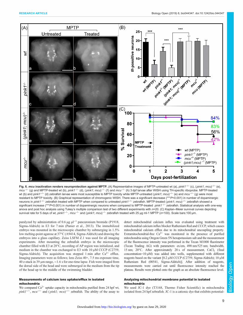
mcu inactivation lends resistance to MPTP toxicityWe next checked if lack of mcu can protect dopaminergic neuronsagainst toxins such as MPTP, known to induce PD in human andused to generate PD animal models. MPTP is a precursor to MPP+,which is a neurotoxin that acts by inhibiting oxidativephosphorylation causing dopaminergic neuronal death. Followingexperimental groups of 3 dpf zebrafish wt, pink1−/−, mcu−/− and( pink1; mcu)−/− were treated with 25 µM MPTP. 24 h postfertilization (hpf) embryos were monitored for survival aftertreatment for 5 days and results were plotted as Kaplan–Meiersurvival curves (Fig. 6C). After treatment with MPTP, there were56% and 42% survival rates in the wt and pink1−/− zebrafish,respectively. However, survival of bothmcu−/− and ( pink1; mcu)−/−
double mutants was much higher; 83% and 84%, respectively.These data show a protective effect of mcu deletion on pink1−/−
zebrafish against the Parkinsonian neurotoxin MPTP. Thereby, weinvestigated the effects of MPTP on the different dopaminergicneural clusters using in situ hybridization. The neuronal populationsmost severely affected by MPTP were the olfactory bulb, pretectaland diencephalic populations. In wt, DA neurons were found to bereduced to a small number of neurons in the locus coeruleus(Fig. 6A), while in the pink1−/− line treated withMPTP, DA neuronswere scarcely present. However, despite MPTP treatment in mcu−/−
and in ( pink1; mcu)−/− zebrafish, the majority of dopaminergicneurons were present (Fig. 6B). This indicates that lack of Mcuprevents the deleterious effects of the neurotoxin on the embryo’sdopaminergic system.
DISCUSSIONWe previously reported that mcu inhibition using morpholino isprotective to dopaminergic neurons in a pink1−/− zebrafish model ofPD (Soman et al., 2017). Thereby, it is imperative to evaluate theneuroprotective role ofmcu inactivation in a null mutant. In this report,we provide data on the characterization ofmcu−/− zebrafish, generatedusing a CRISPR/Cas9 approach, and the effects ofmcu inactivation on
mechanisms linked to PD pathogenesis in the pink1−/− zebrafishmodel. We show that loss of mcu expression results in abatedmitochondrial calcium influx inmcu−/− zebrafish. Our results confirmthe role of Mcu in the trafficking of Ca2+ into mitochondria ofzebrafish, thus providing evidence of mcu conservation duringevolution. Previous reports have shown that mcu−/− mice havenormal basal metabolism, however under strenuous work conditionsmcu−/− mice succumb to compromised energy production (Pan et al.,2013). Following rodent models andmcumorphant zebrafish,mcu−/−
zebrafish are viable, fertile and lack gross morphological aberrations.The ability of vertebrate organisms to thrive irrespective of mcuexpression is intriguing and could be due to additional Ca2+ traffickingchannels present in the mitochondria for maintaining basal levels ofcalcium in the mitochondrial matrix (Hamilton et al., 2018).
There is compelling evidence supporting the role of alteredmitochondrial calcium handling in the pathogenesis of PD (Calì et al.,2012; Lieberman et al., 2017; Tabata et al., 2018). Our resultsin pink1−/− zebrafish show enhanced CCCP triggered mitochondrialcalcium efflux compared to wt zebrafish. Mitochondrialuncoupling by CCCP primes mPTP opening and release of innermitochondrial calcium stores (Vaur et al., 2000). The increasedrelease of mitochondrial calcium after CCCP treatment indicatesmitochondrial calcium overload in pink1−/− zebrafish. In line withour findings, a recent study showed that GSK3βSer9-mediatedblocking of the mPTP leads to increased CCCP-induced mitochondrialcalcium release in pink1 knockdown M17 cells (Parrado-Fernándezet al., 2018). pink1 knockout models have mitochondrial Ca2+
accumulation, possibly as a result of mitochondrial Na+/Ca2+
exchanger dysfunction or altered ER-mitochondria contact sites (Leeet al., 2018). The absence of viable transgenic ( pink1; mcu)−/−
double mutants expressing GCaMP5G redirected us to studycalcium influx in purified mitochondria. In a definite trend withour calcium efflux results, extra-mitochondrial calcium influx isincreased in purified mitochondria of pink1−/− zebrafish comparedwith wt. The double mutants ( pink1; mcu)−/− showed restoredcalcium levels. The results from mitochondrial calcium efflux andinflux experiments support the notion that mitochondrial calciumoverload is prevalent in pink1 deficiency.
Mitochondrial calcium stimulates adenosine triphosphate (ATP)synthesis, which involves simultaneous generation and salvaging ofreactive oxygen species (ROS) (McCormack et al., 1990; Brookeset al., 2004). However, mitochondrial calcium overload canaugment ROS generation, leading to inhibition of respiratorychain complex and loss of mitochondrial membrane potential(ΔΨm) (Rizzuto et al., 2012). We had previously shown thatinhibiting mcu function does not negate mitochondrial respirationand restores complex 1 function in pink1 deficient condition. Wehere show that ΔΨm is reduced in purified mitochondria of pink1−/−
zebrafish and mcu deletion restores ΔΨm. Previous reports in cell-based models have suggested that a loss-of-function mutation inPink1 causes a partial decrease in ΔΨm (Abramov et al., 2011;Gandhi et al., 2009). It is intriguing to understand whetherdecreased ΔΨm is caused by complex I inhibition or oxidativestress during pink1 deficient condition. In an earlier report, it wasshown that loss of ΔΨm could take place independent ofmitochondrial respiratory complex inhibitors (Petersén et al.,2000). Thereby it is likely that during pink1-deficient condition,altered mitochondrial calcium handling could lead to oxidativestress and thereby loss of ΔΨm.
Mitochondrial structural dynamics are altered as a result ofimpaired pink1 mediated mitophagy (Poole et al., 2008; Tsai et al.,2018). Electron microscopy studies in pink1−/− zebrafish have
Fig. 4. Mitochondrial membrane potential partly restored after mcudeletion. The graph represents ratio of JC-1 dye emission spectrum(590 nm to 530 nm). wt mitochondria showed a slight uptake of JC-1, whichturned into leakage after ∼18 min after the start of the experiment, whenmitochondria began to lose membrane integrity in an in vitro environment.pink1−/− mitochondria did not show uptake, instead we observed JC-1leakage starting at ∼6 min after experiment initialization. Both variantsstarted and ended at similar levels. Results are mean with s.e.m. [wt hadtwo, pink1−/− had two and (pink1; mcu)−/− had one independentreplication(s)] gathered during two independent experiments. Everyreplication was done on 200 larvae per variant.
6
RESEARCH ARTICLE Biology Open (2019) 8, bio044347. doi:10.1242/bio.044347
BiologyOpen
by guest on June 29, 2020http://bio.biologists.org/Downloaded from

Fig. 5. See next page for legend.
7
RESEARCH ARTICLE Biology Open (2019) 8, bio044347. doi:10.1242/bio.044347
BiologyOpen
by guest on June 29, 2020http://bio.biologists.org/Downloaded from

shown enlarged mitochondrial structures in muscle fibers (Flinnet al., 2013). We see a loss of DA neuronal specific mitochondrialstructures in pink1−/− zebrafish when compared to wt. In contrast,muscle fiber mitochondrial morphology is altered in the form ofelongated structures. Previous reports have shown evidence of inter-tissue and intracellular heterogeneity in mitochondrial populations(Kuznetsov and Margreiter, 2009). Additionally, SNpc DA neuronshave more reduced mitochondrial volume than SNpc non-DAneurons, making DA neurons more vulnerable to mitochondrialdysfunction (Liang et al., 2007).The paramount pathology seen in patients with PD is the loss of DA
neurons in SNpc. The employment of calcium channels in DAneurons to maintain pace-keeping activity predispose them tomitochondrial calcium overload. Mitochondrial calcium overload ispathological in a diverse array of neurodegenerative diseases includingPD and modulation of mitochondrial calcium influx or effluxmechanisms are a plausible strategy for therapeutic intervention in PD.
MATERIALS AND METHODSAnimal maintenanceZebrafish were maintained under standard conditions followinginternational and national ethical and animal welfare guidelines (LocalCommission for the Ethics of Animal Experimentation, Warsaw, licensenumber: 657/2015). Mutant lines were generated in the AB background.
Generation of mcu−/− zebrafish using the CRISPR/Cas9 systemA DNA ultramer was used to generate the gRNA as previously described(Hruscha et al., 2013). The gRNAwas injected into one-cell stage embryosalongside Cas9 protein (NEB). The CRISPR/Cas9 target site containedrestriction sites against Mwo1. PCR products prepared from genomic DNA(GenElute™ 96 Well Tissue Genomic DNA Purification Kit, Sigma-Aldrich) extracted from individual embryos at 24 hpf were primarilyscreened using restriction digestion. Mwo1 sites (GCNNNNNNNNGC)were disrupted in target sites where CRISPR/Cas9-mediated disruption tookplace. Further screening was done using DNA sequencing. Uponconfirmation of indel mutations, the remaining embryos were grown in28°C till 5 dpf and transferred to aquarium tanks.
Zebrafish linesThis study was performed in wt AB, pink1 mutant ( pink1−/−) (Flinn et al.,2013), mcu mutant (mcu−/−) and double mutant for pink1 and mcu ( pink1;mcu)−/− zebrafish lines. For calcium imaging experiments, the followingzebrafish lines were used: Tg(HuC: GCaMP5G) line (Ahrens et al., 2013)and pink1−/− zebrafish lines were out-crossed, and the 3 dpf embryos were
screened for the fluorescent signal. At maturity, Tg(HuC: GCaMP5G+/−);pink1+/− zebrafish embryos were in-crossed to generate Tg(HuC:GCaMP5G+/−); pink1 (used as a control) and Tg(HuC: GCaMP5G+/−);pink1−/−. The offspring from in-cross of Tg(HuC: GCaMP5G+/−); pink1+/+
and in-cross of Tg(HuC: GCaMP5G+/−); pink1−/− were used forexperiments. All zebrafish in the calcium imaging experiments were ofmitfa−/− (nacre) background.
Chemical treatmentZebrafish embryos were collected and incubated in E3 medium for 24 h inPetri dishes. After 24 hpf, embryos were segregated into six-well plates with10 embryos in each. Stock solutions of MPTP (10 mg ml−1) (Sigma-Aldrich) were made by adding sterile water directly to the bottle with aneedle and syringe. All manipulations with MPTP were performed under achemical hood. MPTP was diluted in E3 solution to achieve finalconcentrations of 25 µg ml−1. The embryos were kept in an incubatormaintained at 28°C. The mortality was noted every day until 5 dpf. On the5th day, the embryos were washed with fresh E3 solution and fixed in 4%paraformaldehyde (PFA) for 2 h and further processed for in situhybridization.
RT-PCRThe primers were designed against flanking exons of the target site. RNAwas isolated from a pool of 20 embryos using Tri-reagent (Sigma-Aldrich),and cDNAwas generated using Verso cDNA synthesis kit (Thermo FisherScientific). PCR was performed using a specific primer, using BioMix™Red (Bioline) master mix.
qPCRRNAwas extracted from a pool of 20 embryos at 3 dpf using Tri-reagent andcDNA was generated using Verso cDNA synthesis kit (Thermo FisherScientific). qPCR analysis was conducted with gene-specific primers (mcu:forward, 5′-AGACTGTCAGGAGAGCACAC-3′; reverse, 5′-GACGTAC-AGAAATCACCGGC-3′), which were optimized for annealingtemperature, concentration and efficiency. SYBR Green master mix(Roche) was utilized for the enzyme reaction, which was incubated andanalyzed by Light Cycler 96 RT- PCR (Roche). The fold change inexpression was quantified by normalizing the threshold cycle (CT) values ofthe target mRNAs to the CT values of the internal control EF1α in the samesamples (ΔCT=CTtarget−CTEF1α). It was further normalized with the wtcontrol (ΔΔCT=ΔCT−CTcontrol). The fold change in expression was thenobtained (2–ΔΔCT).
Isolation of mitochondria from 24 hpf zebrafishMitochondrial isolation from zebrafish embryos was done using themodified method of Prudent (Prudent et al., 2013). 24 hpf embryos weresegregated to groups of 200 per variant, washed with Ringer’s buffer anddechorionated with pronase solution. Embryos were rewashed with Ringer’suntil all chorions were removed. Embryos were then deyolked in a loose-fitting homogenizer (Glass-Teflon), filled with 1 ml of 0.5× Ginzburgbuffer, with 0.5 mM PMSF and MS-222 (15–50 mg/l) as an anesthetic.After final centrifugation, the pellet was resuspended with 1 ml ofmitochondria isolation buffer. Cells were disrupted with a 1 ml syringeand a 26 G×2/3 needle by 50 uptakes. The lysate was centrifuged twice at1500 g for 10 min to eliminate nuclei. Then the supernatant was transferredto a pre-chilled 1.5 ml centrifuge tube. It was spun at 10,600 g for 10 min topellet mitochondria. Mitochondrial pellet was then suspended in 1 ml ofcold KCl medium and centrifuged for 10 min at 10,600 g at 4°C. Proteinamount was established by Bradford protein assay, and samples wereequalized to 0.1 µg µl−1. Pellet was gently resuspended with 0.15 μl perembryo of KCl medium (125 mM KCl, 2 mM K2HPO4, 1 mM MgCl2 and20 mM HEPES, pH 7).
Imaging the AP regionNeurons of AP in the hindbrain region were selected for the experiment andimage analysis. They have a relatively large diameter of about 7 μm andconvenient localization near the skin (Fig. 2A,B). At 5 dpf, zebrafish were
Fig. 5. Mitochondrial dynamics is altered in pink1−/− zebrafish.(A–L) Representative images of immunohistochemistry performed on wt,pink1−/−, mcu−/− and (pink1; mcu)−/− dissected 4 dpf zebrafish larval brain.DA neurons (red) are marked with anti-TH antibody, mitochondrial structures(green) are marked with anti-Tom20 antibody and DAPI (blue) is shown asnuclear stain. Arrowheads show mitochondrial morphology in muscle fibers.(M–O) Representative images of mitochondrial volumetric analysis in musclefibers of transgenic wt, pink1−/− and (pink1; mcu)−/− 3 dpf zebrafishexpressing mitochondria-localized GFP. Arrowheads point out mitochondrialstructures in somites. (P) Graphical representation and statistical analysis ofmitochondrial area with one-way ANOVA and post-hoc analysis usingTukey’s multiple comparison test of two different experiments with n=15.There was a significant (*P>0.05) decrease in DA neuronal mitochondrialarea in pink1−/− zebrafish when compared to wt; mitochondria area wasrestored (**P>0.01) in (pink1; mcu)−/− zebrafish. (Q,R) Graphicalrepresentation and statistical analyses of muscle mitochondrial volume andsphericity with one-way ANOVA and post-hoc analysis using Tukey’smultiple comparison test of three different experiments with n=30. There wasa slight decrease in mitochondrial volume in pink1−/− zebrafish whencompared to wt. The cumulative sphericity index is significantly (***P>0.001)reduced in pink1−/− zebrafish, unlike wt and (pink1; mcu)−/− zebrafish.
8
RESEARCH ARTICLE Biology Open (2019) 8, bio044347. doi:10.1242/bio.044347
BiologyOpen
by guest on June 29, 2020http://bio.biologists.org/Downloaded from
paralyzed by administration of 0.6 µg µl−1 pancuronium bromide (P1918,Sigma-Aldrich) in E3 for 7 min (Panier et al., 2013). The immobilizedembryo was mounted in the microscopic chamber by submerging in 1.5%lowmelting-point agarose at 37°C (A9414, Sigma-Aldrich) and drawing theembryos into a glass capillary. Zeiss LSFM Z.1 was used for all imagingexperiments. After mounting the zebrafish embryo in the microscopicchamber filled with E3 at 28°C, recording of AP region was initialized, andmedium in the chamber was exchanged to E3 with 10 μM CCCP (C2759,Sigma-Aldrich). The acquisition was stopped 1 min after Ca2+ efflux.Imaging parameters were as follows; lens Zeiss 40×, 7.5 ms exposure time,40 z-stack in 39 μm range,∼1.6 s for one-time lapse. Fish were imaged fromthe dorsal side of the head and were submerged in the medium from the tipof the head up to the middle of the swimming bladder.
Measurements of calcium ions uptake/efflux in isolatedmitochondriaWe compared Ca2+ uptake capacity in mitochondria purified from 24 hpf wt,pink1−/−, mcu−/− and (pink1; mcu)−/− zebrafish. The ability of the assay to
detect mitochondrial calcium influx was evaluated using treatment withmitochondrial calcium influx blocker RutheniumRed and CCCP, which causesmitochondrial calcium efflux due to its mitochondrial uncoupling property.Extramitochondrial-free Ca2+ was monitored in the presence of purifiedmitochondria using Oregon Green 5N hexapotassium salt and the measurementof the fluorescence intensity was performed in the Tecan M1000 fluorimeter(Tecan Trading AG) with parameters: ex/em, 490 nm/525 nm; bandwidth,15 nm; 20°C. After approximately 20 s of measurement, CaCl2 (finalconcentration=10 µM) was added into wells, supplemented with differentreagents based on the variant [0.2 μM CCCP (C2759, Sigma-Aldrich); 10 μMRuthenium Red (00541, Sigma-Aldrich)]. After addition of reagents,measurements were carried out until fluorescence intensity reached theplateau. Results were plotted onto the graph as an absolute fluorescence level.
Analyzing mitochondrial membrane potential in isolatedmitochondriaWe used JC-1 dye (T3168, Thermo Fisher Scientific) in mitochondriaisolated from 24 hpf zebrafish. JC-1 is a cationic dye that exhibits potential-
Fig. 6. mcu inactivation renders neuroprotection against MPTP. (A) Representative images of MPTP-untreated wt (a), pink1−/− (c), (pink1; mcu)−/− (e),mcu−/− (g) and MPTP-treated wt (b), pink1−/− (d), (pink1; mcu)−/− (f ) and mcu−/− (h) 3 dpf larvae after WISH using TH-specific riboprobe. MPTP-treatedwt (b) and pink1−/− (d) zebrafish larvae were most susceptible to MPTP toxicity while MPTP-untreated (pink1; mcu)−/− (e) and mcu−/− (g) were mostresistant to MPTP toxicity. (B) Graphical representation of chromogenic WISH. There was a significant decrease (***P<0.001) in number of dopaminergicneurons in pink1−/− zebrafish treated with MPTP when compared to untreated pink1−/− zebrafish. MPTP-treated (pink1; mcu)−/− zebrafish showed asignificant increase (***P<0.001) in number of dopaminergic neurons when compared to MPTP-treated pink1−/− zebrafish. Statistical analysis with one-wayanova and post hoc analysis using Tukey’s multiple comparison test of two different experiments with n=20. (C) Kaplan–Meier survival curves depictingsurvival rate for 5 days of wt, pink1−/−, mcu−/− and (pink1; mcu)−/− zebrafish treated with 25 µg ml-1 MPTP (n=100). Scale bars:100 µm.
RESEARCH ARTICLE Biology Open (2019) 8, bio044347. doi:10.1242/bio.044347
9
BiologyOpen
by guest on June 29, 2020http://bio.biologists.org/Downloaded from

dependent accumulation inmitochondria, indicated by a fluorescence emissionshift from green (∼525 nm) to red (∼590 nm). We presented results as a ratioof spectra. Isolated mitochondria from 24 hpf zebrafish embryos weretransferred to a 96-well non-transparent plate with a flat bottom (GreinerBio-One), with 80 μl per well (16 μg). Measurement of the fluorescenceintensity was performed in the Tecan M1000 fluorimeter (Tecan Trading AG)with excitation 490 nm and emission in two channels: 530 and 590 nm at20°C. Experiments were conducted until fluorescence showed plateau lines.
In situ hybridizationWISH and fluorescent in situ hybridization (FISH) were performed aspreviously described (Welten et al., 2006; Jowett and Lettice, 1994; Thisseand Thisse, 2008). Briefly, probes for the gene of interest were obtained byPCR using specific primers [th (probe length,756 bp): forward 5′AATTAAC-CCTCACTAAAGGGAGAATGCCGAATTCAAGCAGCTCCAC-3′, reverse5′-TAATACGACTCACTATAGGGAGAAGCGTGCCGTATGTACTG-TGTGC-3; mcu ( probe length,780 bp): forward 5′-TAATACGACTC-ACTATAGGGGCTGAGTAAGAAAGCCGAGC-3′, reverse 5′GATT-TAGGTACTATAGGCACCACATCCCGAAATCTC-3] and in vitrotranscription using T7 Polymerase (Roche) with digoxigenin (DIG)-labeled UTP. Embryos were then fixed using 4% paraformaldehyde andstored at −20°C in methanol. After rehydration, permeabilization in10 µg µl−1 Proteinase K, the probe against target mRNA was added toembryos in hybridization buffer (Hyb) at 65°C overnight, then washedby 75% Hyb, 50% Hyb, 25% Hyb in 2x sodium citrate buffer (SSC) and0.2x SSC. After blocking with 1% bovine serum albumin in the maleicacid buffer, an alkaline phosphatase-conjugated anti-DIG antibody wasadded and proceeded to react with nitro-blue tetrazolium chloride/5-bromo-4-chloro-3-indolyl-phosphate substrate (Roche, 11681451001).With FISH experiments, anti-DIG-POD (Roche) was used instead. Signalamplification was performed using TSA/Cy3 reagent (Perkin Elmer)according to manufacturer’s protocol. Embryos were embedded in 3%methylcellulose and Gelvatol and subsequently, imaging was performed in abright-field microscope and a fluorescent microscope, respectively. The meannumber of these diencephalic dopaminergic neurons for wt, pink1−/−,mcu−/−
and (pink1; mcu)−/− was calculated over three independent experiments(n=10 embryos per genotype and experiment). In order to avoid introducingunintended bias, all embryos were counted with the investigator blindedto the genotype.
IHCWhole-mount antibody staining of 5 dpf zebrafish larvae was performed aspreviously described, with some modifications (Wilson et al., 1990). Inbrief, embryos were fixed in 4% sucrose/4% PFA at room temperature (RT)for 2 h. Following fixation, embryos were washed in PBS, dissected,dehydrated sequentially in methanol-PBT (PBS+0.5% Triton X-100) andstored in 100% methanol at −20°C at least overnight. Embryos wererehydrated sequentially, washed in PBT, digested with Proteinase K(10 µg µl−1 at 120 hpf, for 30 min), and post-fixed in 4% PFA for 20 min atRT. Embryos were blocked for at least 1 h at RT in 10% normal sheepserum/1% DMSO/0.5% Triton X-100 in PBS and incubated in primaryantibodies overnight at 4°C. Embryos were washed between four and sixtimes for at least 30 min in PBT at RT and incubated in secondary antibodiesfor 2 h at RT or overnight at 4°C. Embryos were then washed between fourand six times for at least 30 min at RT or overnight at 4°C in PBT andmounted for imaging in prolong gold (Thermo Fisher Scientific, P10144).The following primary antibodies were used: TH [mouse monoclonal IgG1,#ABIN617906, (Antibodies Online, 1:1000)], tom20 [Rabbit monoclonal,#MA5–32148, (Thermo Fisher Scientific, 1:500)]. The following AlexaFlour secondary antibodies were used: Alexa Fluor® 488 [goat anti-mouseIgG (H+L) cross-adsorbed secondary antibody, #A-11001 (Thermo FisherScientific, 1:200)], Alexa Fluor® 647 [goat anti-rabbit IgG (H+L) cross-adsorbed secondary antibody, #A-21244 (Thermo Fisher Scientific,1:200)]. The mean number of these diencephalic dopaminergic neuronsfor wt, pink1−/−, mcu−/− and (pink1; mcu)−/− was calculated over threeindependent experiments (n=10 embryos per genotype and experiment). Inorder to avoid introducing unintended bias, all embryos were counted withthe investigator blinded to the genotype.
Images post-processing, Fmax/Fmin factor calculations and dataanalysisTime-lapsed images were deconvoluted in ZEN software (Zeiss) using thenearest neighbor method (Castleman, 1979), and converted to BitplaneImaris 8.3 format. SPOTS function (7 μm estimated diameter, withbackground subtraction, without region growing option) with objecttracking (autoregressive motion) was used in Imaris 8.3 (Bitplane) toautomatically find and segregate brightest regions in the 3D region duringthe time lapse. Specifically, for AP, SPOTS were most frequently found inthe large region of the neuron where the axon was attached. This region wasrich in the cytoplasm, and thus the fluorescent signal was also high due tothe increased level of GCaMP5G localized in the cytoplasm. Regiontracking was necessary to overcome muscle contraction movements anddrift. Average fluorescent activity plot from every neuron was exported toMicrosoft Excel. From the whole region of interest in a single fish,approximately nine neurons were perfectly tracked by the Imaris and thoseneurons with the highest fluorescent signal in their maximal peak were takenfor further calculations and statistics. For each neuron, its average lowbaseline and its average high peak were calculated from five minimal andfive maximal measurements respectively. The mean numbers of highfluorescent peaks and low baselines were calculated from nine neurons perzebrafish. As a determinant of the amount of Ca2+ that was released fromcompartments affected by CCCP, we created the Fmax/Fmin factor, calculatedby dividing the average maximum peak fluorescence by the averageminimal baseline fluorescence. We hypothesized that constant efflux ofCa2+ from mitochondria triggered by CCCP activity leads to maximumGCaMP5G saturation [Kd=460 nM (Akerboom et al., 2013)]. In practice,few micromolar Ca2+ concentrations saturate GCaMPG5 signal (Baduraet al., 2014). Thereby, the longer the saturation lasts, and the higher the Fmax/Fmin ratio achieved, the more Ca2+ was released. The statistical significance(P-value) of two groups of values was calculated using a two-tailed, two-sample unequal variance t-test calculated in Graph Pad Prism 5. Results aremeans with s.e.m. (wt: n=13 fish, n=117 neurons; pink1−/−: n=15 fish,n=134 neurons) gathered during five independent experiments.
Statistical analysisAll the experiments were performed in triplicate unless specifically statedotherwise. Data represent the mean±s.e.m. A minimum of 10 embryos wereused per genotype for each replicate experiment. One-way ANOVA andt-tests of significance were used unless stated otherwise as measures ofsignificance (Prism, version 7.0; GraphPad Software).
AcknowledgementsThe authors would like to thank the zebrafish facility in IIMCB and University ofSheffield for excellent support. We thank Dr Michael Orger (The ChampalimaudCentre for the Unknown, Lisbon, Portugal) for providing the GCaMP5G transgeniczebrafish and excellent discussion. We thank Prof. Seok-Yong Choi (Department ofBiomedical Sciences, Chonnam National University Medical School, Gwangju,South Korea) for providing the Tg(Xla.Eef1a1:mlsEGFP) zebrafish line.
Competing interestsThe authors declare no competing or financial interests.
Author contributionsConceptualization: S.K.S., O.B., J.K.; Methodology: S.K.S., M.B., M.K.; Validation:S.K.S., M.B.; Formal analysis: S.K.S., M.B., M.K.; Investigation: S.K.S., M.B., M.K.;Resources: J.K.; Data curation: M.B.; Writing - original draft: S.K.S., M.B., J.K.;Writing - review & editing: S.K.S., M.B., M.K., O.B., J.K.; Visualization: S.K.S.;Supervision: S.K.S., O.B., J.K.; Project administration: S.K.S., J.K.; Fundingacquisition: S.K.S., J.K.
FundingThis project has received funding from the National Science Centre, Poland (2014/15/D/NZ3/05176) to Smijin K. Soman; European Union’s Seventh FrameworkProgramme for research, technological development and demonstration under grantagreement no. 316125; and from Parkinson’s UK [G1304, G1404].
ReferencesAbramov, A. Y., Gegg, M., Grunewald, A., Wood, N. W., Klein, C. and Schapira,
A. H. V. (2011). Bioenergetic consequences of PINK1 mutations in parkinsondisease. PLoS ONE 6, e25622. doi:10.1371/journal.pone.0025622
RESEARCH ARTICLE Biology Open (2019) 8, bio044347. doi:10.1242/bio.044347
10
BiologyOpen
by guest on June 29, 2020http://bio.biologists.org/Downloaded from

Ahrens, M. B., Orger, M. B., Robson, D. N., Li, J. M. and Keller, P. J. (2013).Whole-brain functional imaging at cellular resolution using light-sheet microscopy.Nature Methods 10, 413-420. doi:10.1038/nmeth.2434
Akerboom, J., Carreras Calderon, N., Tian, L., Wabnig, S., Prigge, M., Tolo, J.,Gordus, A., Orger, M. B., Severi, K. E., Macklin, J. J. et al. (2013). Geneticallyencoded calcium indicators for multi-color neural activity imaging and combinationwith optogenetics. Front. Mol. Neurosci. 6, 1-29. doi:10.3389/fnmol.2013.00002
Anichtchik, O. V., Kaslin, J., Peitsaro, N., Scheinin, M. and Panula, P. (2004).Neurochemical and behavioural changes in zebrafish Danio rerio after systemicadministration of 6-hydroxydopamine and 1-methyl-4-phenyl-1,2,3,6-tetrahydropyridine. J. Neurochem. 88, 443-453. doi:10.1111/j.1471-4159.2004.02190.x
Baughman, J. M., Perocchi, F., Girgis, H. S., Plovanich, M., Belcher-Timme,C. A., Sancak, Y., Bao, X. R., Strittmatter, L., Goldberger, O., Bogorad, R. L.et al. (2011). Integrative genomics identifies mcu as an essential component ofthe mitochondrial calcium uniporter. Nature 476, 341-345. doi:10.1038/nature10234
Badura, A., Sun, X. R., Giovannucci, A., Lynch, L. A. and Wang, S. S.-H. (2014).Fast calcium sensor proteins for monitoring neural activity. Neurophotonics 1,025008. doi:10.1117/1.NPh.1.2.025008
Brookes, P. S., Yoon, Y., Robotham, J. L., Anders, M. W. and Sheu, S.-S. (2004).Calcium, ATP, and ROS: a mitochondrial love-hate triangle. Am. J. Physiol. CellPhysiol. 287, C817-C833. doi:10.1152/ajpcell.00139.2004
Brocard, J. B., Tassetto, M. and Reynolds, I. J. (2001). Quantitative evaluation ofmitochondrial calcium content in rat cortical neurones following a glutamatestimulus. J. Physiol. 531, 793-805. doi:10.1111/j.1469-7793.2001.0793h.x
Calì, T., Ottolini, D., Negro, A. and Brini, M. (2012). α-Synuclein controlsmitochondrial calcium homeostasis by enhancing endoplasmic reticulum-mitochondria interactions. J. Biol. Chem. 287, 17914-17929. doi:10.1074/jbc.M111.302794
Castleman, K. R. 1979. Digital Image Processing. Prentice-Hall, Englewood Cliffs,NJ.
Dagda, R. K., Gusdon, A.M., Pien, I., Strack, S., Green, S., Li, C., VanHouten, B.,Cherra, S. J. and Chu, C. T. (2011). Mitochondrially localized PKA reversesmitochondrial pathology and dysfunction in a cellular model of Parkinson’sdisease. Cell Death Differ. 18, 1914-1923. doi:10.1038/cdd.2011.74
Exner, N., Lutz, A. K., Haass, C. and Winklhofer, K. F. (2012). Mitochondrialdysfunction in Parkinson’s disease: molecular mechanisms andpathophysiological consequences. EMBO J. 31, 3038-3062. doi:10.1038/emboj.2012.170
Flinn, L. J., Keatinge, M., Bretaud, S., Mortiboys, H., Matsui, H., De Felice, E.,Woodroof, H. I., Brown, L., Mctighe, A., Soellner, R. et al. (2013). TigarBcauses mitochondrial dysfunction and neuronal loss in PINK1 deficiency. Ann.Neurol. 74, 837-847. doi:10.1002/ana.23999
Gandhi, S., Wood-Kaczmar, A., Yao, Z., Plun-Favreau, H., Deas, E., Klupsch, K.,Downward, J., Latchman, D. S., Tabrizi, S. J., Wood, N.W. et al. (2009). PINK1-associated Parkinson’s disease is caused by neuronal vulnerability to calcium-induced cell Death. Mol. Cell 33, 627-638. doi:10.1016/j.molcel.2009.02.013
Guzman, J. N., Sanchez-Padilla, J., Wokosin, D., Kondapalli, J., Ilijic, E.,Schumacker, P. T. and Surmeier, D. J. (2015). Oxidant stress evoked bypacemaking in dopaminergic neurons is attenuated by DJ-1. Nature 468,696-700. doi:10.1038/nature09536
Hamilton, J., Brustovetsky, T., Rysted, J. E., Lin, Z., Usachev, Y. M. andBrustovetsky, N. (2018). Deletion of mitochondrial calcium uniporterincompletely inhibits calcium uptake and induction of the permeability transitionpore in brain mitochondria. J. Biol. Chem. 293, 15652-15663. doi:10.1074/jbc.RA118.002926
Howe, K., Clark, M. D., Torroja, C. F., Torrance, J., Berthelot, C., Muffato, M.,Collins, J. E., Humphray, S., McLaren, K., Matthews, L. et al. (2013). Thezebrafish reference genome sequence and its relationship to the human genomeKerstin. Nature 58, 154-155. doi:10.1038/nature12111
Hruscha, A., Krawitz, P., Rechenberg, A., Heinrich, V., Hecht, J., Hass, C. andSchmid, B. (2013). Efficient CRISPR/Cas9 genome editing with low off-targeteffects in zebrafish. Development 140, 4982-4987. doi:10.1242/dev.099085
Jowett, T. and Lettice, L. (1994). Whole-mount in situ hybridizationon zebrafishembryos using a mixture of digoxigenin- and fluorescein-labelled probes. TrendsGenet. 10, 73-74. doi:10.1016/0168-9525(94)90220-8
Kalia, L. V. and Lang, A. E. (2016). Parkinson disease in 2015: evolving basic,pathological and clinical concepts in PD.Nat. Rev. Neurol. 12, 65-66. doi:10.1038/nrneurol.2015.249
Klein, C. and Westenberger, A. (2012). PEGylation of antibody fragments greatlyincreases their local residence time following delivery to the respiratory tract.J. Control. Release 2, 91-100. doi:10.1016/j.jconrel.2014.05.021
Kondapalli, C., Kazlauskaite, A., Zhang, N., Woodroof, H. I., Campbell, D. G.,Gourlay, R., Burchell, L., Walden, H., Macartney, T. J., Deak, M. et al. (2012).PINK1 is activated by mitochondrial membrane potential depolarization andstimulates Parkin E3 ligase activity by phosphorylating Serine 65. Open Biol. 2,1-17. doi:10.1098/rsob.120080
Kostic, M., Ludtmann, M. H. R., Bading, H., Hershfinkel, M., Steer, E., Chu, C. T.,Abramov, A. Y. and Sekler, I. (2015). PKA phosphorylation of NCLX reverses
mitochondrial calcium overload and depolarization, promoting survival of PINK1-deficient dopaminergic neurons. Cell Reports 13, 376-386. doi:10.1016/j.celrep.2015.08.079
Kruman, I. I. and Mattson, M. P. (1999). Pivotal role of mitochondrial calciumuptake in neural cell apoptosis and necrosis. J. Neurochem. 72, 529-540. doi:10.1046/j.1471-4159.1999.0720529.x
Kuznetsov, A. V. and Margreiter, R. (2009). Heterogeneity of mitochondria andmitochondrial function within cells as another level of mitochondrial complexity.Int. J. Mol. Sci. 10, 1911-1929. doi:10.3390/ijms10041911
Lee, K.-S., Huh, S., Lee, S., Wu, Z., Kim, A.-K., Kang, H.-Y. and Lu, B. (2018).Altered ER–mitochondria contact impacts mitochondria calcium homeostasis andcontributes to neurodegeneration in vivo in disease models. Proc. Natl Acad. Sci.USA 115, E8844-E8853. doi:10.1073/pnas.1721136115
Liang, C.-L., Wang, T. T., Luby-Phelps, K. and German, D. C. (2007).Mitochondria mass is low in mouse substantia nigra dopamine neurons:implications for Parkinson’s disease. Exp. Neurol. 203, 370-380. doi:10.1016/j.expneurol.2006.08.015
Lieberman, O. J., Choi, S. J., Kanter, E., Saverchenko, A., Frier, M. D., Fiore,G. M., Wu, M., Kondapalli, J., Zampese, E., Surmeier, D. J. et al. (2017). α-Synuclein-dependent calcium entry underlies differential sensitivity of cultured SNand VTA dopaminergic neurons to a parkinsonian neurotoxin. Eneuro 4,ENEURO.0167–17.2017. doi:10.1523/ENEURO.0167–17.2017
Matthew, N., Pavlov, E. V. and Robertson, G. S. (2018). Tamoxifen-inducedknockdown of the mitochondrial calcium uniporter in Thy1-expressing neuronsprotects mice from hypoxic/ischemic brain injury. Cell Death Dis. 9, 606. doi:10.1038/s41419-018-0607-9
Mccormack, J. G., Halestrap, A. P. and Denton, R. M. (1990). Role of calcium ionsin regulation of mammalian intramitochondrial metabolism. Physiol. Rev. 70,391-425. doi:10.1152/physrev.1990.70.2.391
Pan, X., Liu, J., Nguyen, T., Liu, C., Sun, J., Teng, Y., Fergusson, M. M., Rovira,I. I., Allen, M., Springer, D. A. et al. (2013). The physiological role ofmitochondrial calcium revealed by mice lacking the mitochondrial calciumuniporter (mcu). Nat. Cell Biol. 15, 1464-1472. doi:10.1038/ncb2868
Panier, T., Romano, S. A., Olive, R., Pietri, T., Sumbre, G., Candelier, R. andDebregeas, G. (2013). Fast functional imaging of multiple brain regions in intactzebrafish larvae using selective plane illumination microscopy. Front. NeuralCircuits 7, 65. doi:10.3389/fncir.2013.00065
Parrado-Fernandez, C., Schreiner, B., Ankarcrona, M., Conti, M. M., Cookson,M. R., Kivipelto, M., Cedazo-Mınguez, Ã. and Sandebring-Matton, A. (2018).Reduction of PINK1 or DJ-1 impair mitochondrial motility in neurites and alter ER-mitochondria contacts. J. Cell. Mol. Med. 22, 5439-5449. doi:10.1111/jcmm.13815
Petersen, A., Castilho, R. F., Hansson, O., Wieloch, T. and Brundin, P. (2000).Oxidative stress, mitochondrial permeability transition and activation of caspasesin calcium ionophore A23187-induced death of cultured striatal neurons. BrainRes. 857, 20-29. doi:10.1016/S0006-8993(99)02320-3
Poole, A. C., Thomas, R. E., Andrews, L. A., McBride, H. M.,Whitworth, A. J. andPallanck, L. J. (2008). The PINK1/Parkin pathway regulates mitochondrialmorphology. Proc. Natl Acad. Sci. USA 105, 1638-1643. doi:10.1073/pnas.0709336105
Prudent, J., Popgeorgiev, N., Bonneau, B., Thibaut, J., Gadet, R., Lopez, J.,Gonzalo, P., Rimokh, R., Manon, S., Houart, C. et al. (2013). Bcl-wav and themitochondrial calcium uniporter drive gastrula morphogenesis in zebrafish. Nat.Commun. 4, 2330. doi:10.1038/ncomms3330
Rink, E. and Wullimann, M. F. (2001). The teleostean (zebrafish) dopaminergicsystem ascending to the subpallium (striatum) is located in the basaldiencephalon (posterior q tuberculum)*. Brain Res. 889, 316-330. doi:10.1016/S0006-8993(00)03174-7
Rink, E. and Wullimann, M. F. (2002). Development of the catecholaminergicsystem in the early zebrafish brain: an immunohistochemical study. Dev. BrainRes. 137, 89-100. doi:10.1016/S0165-3806(02)00354-1
Rizzuto, R., De Stefani, D., Raffaello, A. andMammucari, C. (2012). Mitochondriaas sensors and regulators of calcium signalling. Nat. Rev. Mol. Cell Biol. 13,566-578. doi:10.1038/nrm3412
Schapira, A., Cooper, J., Dexter, D., Clark, J., Jenner, P. andMarsden, C. (1990).Mitochondria1 Complex I Deficiency in Parkinson's Disease. J. Neurochem. 94,362-369. doi:10.1080/15572536.2003.11833242
Scheitlin, C. G., Julian, J. A., Shanmughapriya, S., Madesh, M., Tsoukias, N. M.and Alevriadou, B. R. (2016). Endothelial mitochondria regulate the intracellularCa 2+ response to fluid shear stress.Am. J. Physiol. Cell Physiol. 310, C479-C490.doi:10.1152/ajpcell.00171.2015
Soman, S., Keatinge, M., Moein, M., Da Costa, M., Mortiboys, H., Skupin, A.,Sugunan, S., Bazala, M., Kuznicki, J. and Bandmann, O. (2017). Inhibition ofthe mitochondrial calcium uniporter rescues dopaminergic neurons in pink1−/−zebrafish. Eur. J. Neurosci. 45, 528-535. doi:10.1111/ejn.13473
Stefani, D. D., Teardo, E., Szabo , I. and Rizzuto, R. (2013). A 40 kDa protein of theinner membrane is the mitochondrial calcium uniporter. Nature 476, 336-340.doi:10.1038/nature10230
Tabata, Y., Imaizumi, Y., Sugawara, M., Andoh-Noda, T., Banno, S., Chai, M. C.,Sone, T., Yamazaki, K., Ito, M., Tsukahara, K. et al. (2018). T-type calciumchannels determine the vulnerability of dopaminergic neurons to mitochondrial
RESEARCH ARTICLE Biology Open (2019) 8, bio044347. doi:10.1242/bio.044347
11
BiologyOpen
by guest on June 29, 2020http://bio.biologists.org/Downloaded from

stress in familial Parkinson disease. Stem Cell Reports 11, 1171-1184. doi:10.1016/j.stemcr.2018.09.006
Tay, T. L., Ronneberger, O., Ryu, S., Nitschke, R. and Driever, W. (2011).Comprehensive catecholaminergic projectome analysis reveals single-neuronintegration of zebrafish ascending and descending dopaminergic systems. Nat.Commun. 2, 112-171. doi:10.1038/ncomms1171
Thisse, C. and Thisse, B. (2008). High-resolution in situ hybridization to whole-mount zebrafish embryos. Nat. Protoc. 3, 59-69. doi:10.1038/nprot.2007.514
Tsai, P. I., Lin, C.-H., Hsieh, C.-H., Papakyrikos, A. M., Kim, M. J., Napolioni, V.,Schoor, C., Couthouis, J., Wu, R.-M., Wszolek, Z. K. et al. (2018). PINK1phosphorylates MIC60/mitofilin to control structural plasticity of mitochondrialcrista junctions. Mol. Cell 69, 744-756.e6. doi:10.1016/j.molcel.2018.01.026
Valente, E. M., Abou-Sleiman, P. M., Caputo, V., Muqit, M. M., Harvey, K.,Gispert, S., Ali, Z., Del Turco, D., Bentivoglio, A. R., Healy, D. G. et al. (2004).Hereditary early-onset parkinson’s disease caused by mutations in PINK1.Science 304, 1158-1160. doi:10.1126/science.1096284
Vaur, S., Sartor, P. and Dufy-Barbe, L. (2000). Calcium store depletion induced bymitochondrial uncoupling in prostatic cells. Gen. Physiol. Biophys. 19, 265-278.
Vaz, R. L., Outeiro, T. F. and Ferreira, J. J. (2018). Zebrafish as an animal model fordrug discovery in Parkinson’s disease and other movement disorders: asystematic review. Front. Neurol. 9, 347. doi:10.3389/fneur.2018.00347
Verma, M., Wills, Z. and Chu, C. T. (2018). Excitatory dendritic mitochondrialcalcium toxicity: Implications for Parkinson’s and other neurodegenerativediseases. Front. Neurosci. 12, 523. doi:10.3389/fnins.2018.00523
Welten, M. C. M., De Haan, S. B., Van Den Boogert, N., Noordermeer, J. N.,Lamers, G. E. M., Spaink, H. P., Meijer, A. H. and Verbeek, F. J. (2006).ZebraFISH: fluorescent in situ hybridization protocol and three-dimensionalimaging of gene expression patterns. Zebrafish 3, 465-476. doi:10.1089/zeb.2006.3.465
Wilson, S. W., Ross, L. S., Parrett, T. and Easter, S. S. Jr. (1990). Thedevelopment of a simple scaffold of axon tracts in the brain of the embryoniczebrafish, Brachydanio rerio. Development (Cambridge, England) 108, 121-145.
RESEARCH ARTICLE Biology Open (2019) 8, bio044347. doi:10.1242/bio.044347
12
BiologyOpen
by guest on June 29, 2020http://bio.biologists.org/Downloaded from