
3d Printed Prosthetic Hand - Washington University in St ...

Corrections
NEUROSCIENCECorrection for “Restoring the sense of touch with a prosthetichand through a brain interface,” by Gregg A. Tabot, John F.Dammann, Joshua A. Berg, Francesco V. Tenore, Jessica L.Boback, R. Jacob Vogelstein, and Sliman J. Bensmaia, whichappeared in issue 45, November 5, 2013, of Proc Natl Acad SciUSA (110:18279–18284; first published October 14, 2013; 10.1073/pnas.1221113110).The authors note, “For the ‘hybrid’ location discrimination
task, we report data obtained from 27 electrodes, 16 of whichwere in area 1; the 11 electrodes in area 3b were divided evenlyacross the two animals (6 and 5). We had previously tested allof the electrodes, including those in area 3b, in the detectionand discrimination tasks (as shown in Fig. 3) and found them allto yield approximately equivalent performance (see Fig 3A).We noticed in the hybrid location discrimination task, however,that one of the animals performed much more poorly based onstimulation of area 3b than it did based on stimulation of area 1(while the other animal performed better based on stimulationof area 1). Having no reason to question any of the arrays, weattributed this discrepancy to differences across animals andarrived at the conclusion, based on pooled data from bothanimals, that stimulation of the two areas yields equivalentperformance in the ‘hybrid location discrimination’ task. Theoverall conclusion, then, was that stimulation of neurons inarea 3b and 1 evokes percepts that are equally localized onthe skin.“Shortly after publication of the paper, we repeated detection
experiments across the arrays and found that the animal could nolonger detect stimulation through the array in area 3b that hadyielded poor performance in the hybrid location discrimina-tion task. It is therefore likely that this array had failed betweenthe time we conducted the initial detection and discriminationexperiments and the time we conducted the hybrid locationdiscrimination task (which required 2–3 months of retraining).If this is the case, and we eliminate data from that bad array,then the median performance on hybrid trials is 83% (up fromthe 80% that was originally reported), which is still statisticallypoorer than that on the location-matched mechanical trials[median difference between performance on mechanical andhybrid trials was 3.3% rather than 5.6%, t(119) = 6.1, P < 0.001](see the corrected Fig. 2). Thus, we probably underestimatedoverall performance on hybrid trials, and thus the degree to whichartificial percepts are localized, in the original publication. Im-portantly, however, performance on hybrid trials based on stim-ulation of area 3b was significantly better than performance basedon stimulation of area 1 [median Δp = 0.028 and 0.054 for areas3b and 1, respectively; t test: t(76) = 2.8, P < 0.01]. Thus, basedon the data obtained from only one animal, it seems as thoughstimulation of area 3b elicits more localized percepts than doesstimulation of area 1, as might be expected given that neuronsin area 3b tend to have smaller receptive fields than theircounterparts in area 1 (1, 2).”
As a result of this error, Fig. 2 and its legend appeared in-correctly. The corrected figure and its corresponding legendappear below.
1. Sur M, Garraghty PE, Bruce CJ (1985) Somatosensory cortex in macaque monkeys:laminar differences in receptive field size in areas 3b and 1. Brain Res 342(2):391–395.
2. Sripati AP, Yoshioka T, Denchev P, Hsiao SS, Johnson KO (2006) Spatiotemporal re-ceptive fields of peripheral afferents and cortical area 3b and 1 neurons in the primatesomatosensory system. J Neurosci 26(7):2101–2114.
www.pnas.org/cgi/doi/10.1073/pnas.1322627111
A
B
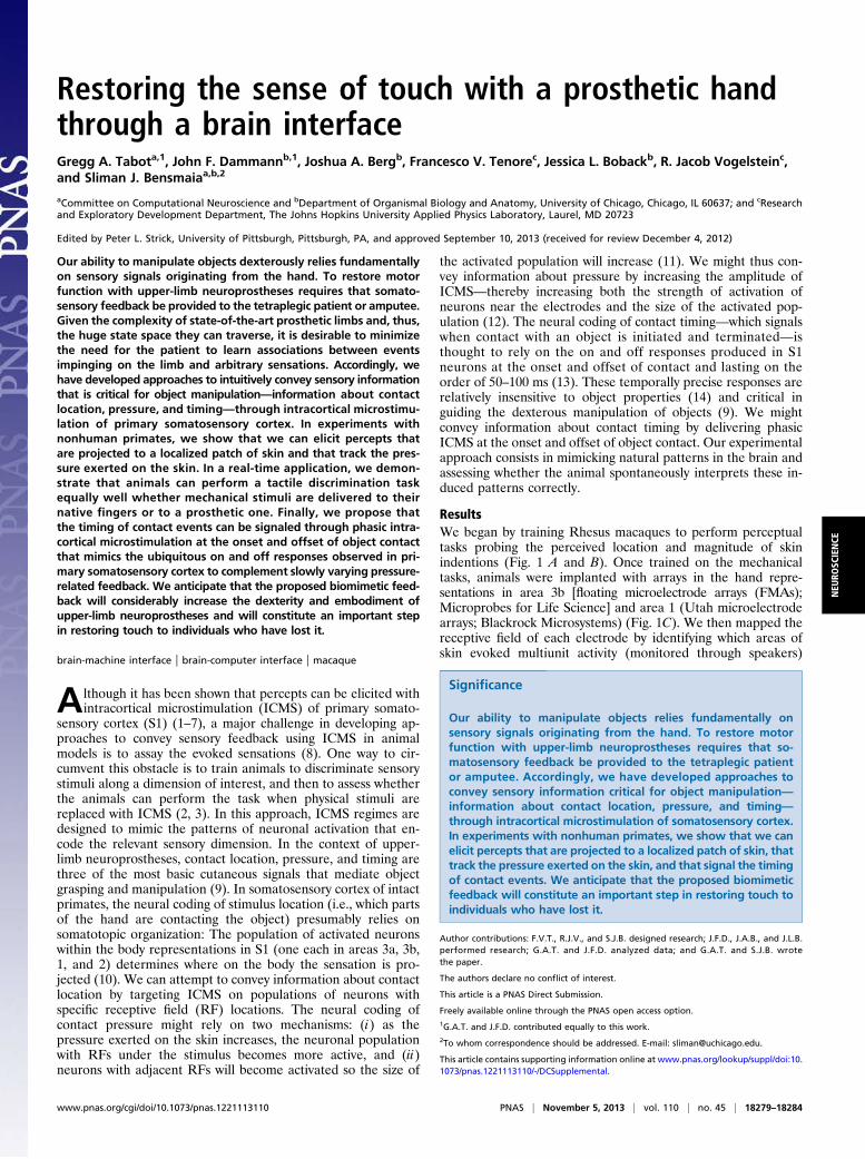
Fig. 2. Localization performance was similar with mechanical touch andICMS. (A) On both mechanical and hybrid trials, the relative locations ofstimuli applied to widely spaced digits were more accurately discriminatedthan were the relative locations of stimuli applied to adjacent digits. Mea-sured from one animal, mechanical performance was based on 1,160 and1,031 trials, respectively (green and gold); hybrid performance on 246 and196 trials, respectively. To compare performance on hybrid trials and per-formance on mechanical trials matched for hand location, we computed thedifference between the two: Δp = pmech(correct) − phybrid(correct). (B) Per-formance on mechanical and hybrid trials was nearly equivalent. Shown isthe distribution of Δp for the two animals tested on this task (88 stimuluspairs, 21 different electrodes, 16 of which are UEAs). Across electrodes,performance was significantly above chance, demonstrating that ICMS yieldsspatially localized percepts. Performance on hybrid trials was somewhatlower than on mechanical location discrimination trials (median Δp = 0.033),suggesting that the elicited percepts may be somewhat more diffuse thannatural ones. There was no significant difference in performance based onstimulation of areas 3b or 1, so data from these two areas are pooled.
www.pnas.org PNAS | January 14, 2014 | vol. 111 | no. 2 | 875–876
CORR
ECTIONS

NEUROSCIENCECorrection for “AMPA receptor exchange underlies transientmemory destabilization on retrieval,” by Ingie Hong, JeongyeonKim, Jihye Kim, Sukwon Lee, Hyoung-Gon Ko, Karim Nader,Bong-Kiun Kaang, Richard W. Tsien, and Sukwoo Choi, whichappeared in issue 20, May 14, 2013, of Proc Natl Acad SciUSA (110:8218–8223; first published April 29, 2013; 10.1073/pnas.1305235110).The authors note that the following statement should be
added to the Acknowledgments: “This work was also supportedby National Research Foundation of Korea Grant 2011-0018209funded by the Ministry of Education, Science and Technology.”
www.pnas.org/cgi/doi/10.1073/pnas.1323623111
SYSTEMS BIOLOGY, CHEMISTRYCorrection for “Heterogeneity in protein expression inducesmetabolic variability in a modeled Escherichia coli population,”by Piyush Labhsetwar, John Andrew Cole, Elijah Roberts,Nathan D. Price, and Zaida A. Luthey-Schulten, which appearedin issue 34, August 20, 2013, of Proc Natl Acad Sci USA (110:14006–14011; first published August 1, 2013; 10.1073/pnas.1222569110).The authors note that the following grant should be added to
the Acknowledgments: National Science Foundation (Center forthe Physics of Living Cells) Contract/Grant PHY-0822613.
www.pnas.org/cgi/doi/10.1073/pnas.1323512111
876 | www.pnas.org
Restoring the sense of touch with a prosthetic handthrough a brain interfaceGregg A. Tabota,1, John F. Dammannb,1, Joshua A. Bergb, Francesco V. Tenorec, Jessica L. Bobackb, R. Jacob Vogelsteinc,and Sliman J. Bensmaiaa,b,2
aCommittee on Computational Neuroscience and bDepartment of Organismal Biology and Anatomy, University of Chicago, Chicago, IL 60637; and cResearchand Exploratory Development Department, The Johns Hopkins University Applied Physics Laboratory, Laurel, MD 20723
Edited by Peter L. Strick, University of Pittsburgh, Pittsburgh, PA, and approved September 10, 2013 (received for review December 4, 2012)
Our ability to manipulate objects dexterously relies fundamentallyon sensory signals originating from the hand. To restore motorfunction with upper-limb neuroprostheses requires that somato-sensory feedback be provided to the tetraplegic patient or amputee.Given the complexity of state-of-the-art prosthetic limbs and, thus,the huge state space they can traverse, it is desirable to minimizethe need for the patient to learn associations between eventsimpinging on the limb and arbitrary sensations. Accordingly, wehave developed approaches to intuitively convey sensory informationthat is critical for object manipulation—information about contactlocation, pressure, and timing—through intracortical microstimu-lation of primary somatosensory cortex. In experiments withnonhuman primates, we show that we can elicit percepts thatare projected to a localized patch of skin and that track the pres-sure exerted on the skin. In a real-time application, we demon-strate that animals can perform a tactile discrimination taskequally well whether mechanical stimuli are delivered to theirnative fingers or to a prosthetic one. Finally, we propose thatthe timing of contact events can be signaled through phasic intra-cortical microstimulation at the onset and offset of object contactthat mimics the ubiquitous on and off responses observed in pri-mary somatosensory cortex to complement slowly varying pressure-related feedback. We anticipate that the proposed biomimetic feed-back will considerably increase the dexterity and embodiment ofupper-limb neuroprostheses and will constitute an important stepin restoring touch to individuals who have lost it.
brain-machine interface | brain-computer interface | macaque
Although it has been shown that percepts can be elicited withintracortical microstimulation (ICMS) of primary somato-
sensory cortex (S1) (1–7), a major challenge in developing ap-proaches to convey sensory feedback using ICMS in animalmodels is to assay the evoked sensations (8). One way to cir-cumvent this obstacle is to train animals to discriminate sensorystimuli along a dimension of interest, and then to assess whetherthe animals can perform the task when physical stimuli arereplaced with ICMS (2, 3). In this approach, ICMS regimes aredesigned to mimic the patterns of neuronal activation that en-code the relevant sensory dimension. In the context of upper-limb neuroprostheses, contact location, pressure, and timing arethree of the most basic cutaneous signals that mediate objectgrasping and manipulation (9). In somatosensory cortex of intactprimates, the neural coding of stimulus location (i.e., which partsof the hand are contacting the object) presumably relies onsomatotopic organization: The population of activated neuronswithin the body representations in S1 (one each in areas 3a, 3b,1, and 2) determines where on the body the sensation is pro-jected (10). We can attempt to convey information about contactlocation by targeting ICMS on populations of neurons withspecific receptive field (RF) locations. The neural coding ofcontact pressure might rely on two mechanisms: (i) as thepressure exerted on the skin increases, the neuronal populationwith RFs under the stimulus becomes more active, and (ii)neurons with adjacent RFs will become activated so the size of
the activated population will increase (11). We might thus con-vey information about pressure by increasing the amplitude ofICMS—thereby increasing both the strength of activation ofneurons near the electrodes and the size of the activated pop-ulation (12). The neural coding of contact timing—which signalswhen contact with an object is initiated and terminated—isthought to rely on the on and off responses produced in S1neurons at the onset and offset of contact and lasting on theorder of 50–100 ms (13). These temporally precise responses arerelatively insensitive to object properties (14) and critical inguiding the dexterous manipulation of objects (9). We mightconvey information about contact timing by delivering phasicICMS at the onset and offset of object contact. Our experimentalapproach consists in mimicking natural patterns in the brain andassessing whether the animal spontaneously interprets these in-duced patterns correctly.
ResultsWe began by training Rhesus macaques to perform perceptualtasks probing the perceived location and magnitude of skinindentions (Fig. 1 A and B). Once trained on the mechanicaltasks, animals were implanted with arrays in the hand repre-sentations in area 3b [floating microelectrode arrays (FMAs);Microprobes for Life Science] and area 1 (Utah microelectrodearrays; Blackrock Microsystems) (Fig. 1C). We then mapped thereceptive field of each electrode by identifying which areas ofskin evoked multiunit activity (monitored through speakers)
Significance
Our ability to manipulate objects relies fundamentally onsensory signals originating from the hand. To restore motorfunction with upper-limb neuroprostheses requires that so-matosensory feedback be provided to the tetraplegic patientor amputee. Accordingly, we have developed approaches toconvey sensory information critical for object manipulation—information about contact location, pressure, and timing—through intracortical microstimulation of somatosensory cortex.In experiments with nonhuman primates, we show that we canelicit percepts that are projected to a localized patch of skin, thattrack the pressure exerted on the skin, and that signal the timingof contact events. We anticipate that the proposed biomimeticfeedback will constitute an important step in restoring touch toindividuals who have lost it.
Author contributions: F.V.T., R.J.V., and S.J.B. designed research; J.F.D., J.A.B., and J.L.B.performed research; G.A.T. and J.F.D. analyzed data; and G.A.T. and S.J.B. wrotethe paper.
The authors declare no conflict of interest.
This article is a PNAS Direct Submission.
Freely available online through the PNAS open access option.1G.A.T. and J.F.D. contributed equally to this work.2To whom correspondence should be addressed. E-mail: [email protected].
This article contains supporting information online at www.pnas.org/lookup/suppl/doi:10.1073/pnas.1221113110/-/DCSupplemental.
www.pnas.org/cgi/doi/10.1073/pnas.1221113110 PNAS | November 5, 2013 | vol. 110 | no. 45 | 18279–18284
NEU
ROSC
IENCE

(Fig. 1D). RF mapping was repeated periodically throughout thestudy to verify that maps were consistent.We assessed the extent to which the animals could perform
these same tasks based on targeted stimulation of neuronal popu-lations in S1. Importantly, ICMS trials were interleaved with me-chanical trials, and individual experimental blocks comprised manydifferent stimulus pairings (hand locations, stimulus amplitudes,etc.), which changed from block to block, so animals never had anopportunity to learn arbitrary stimulus-response contingencies onICMS trials.
Signaling Contact Location. First, we sought to determine whetherwe could elicit percepts that are localized to a predeterminedpatch of skin (see refs. 15 and 16 for visual analogs). To this end,we sequentially delivered indentations to two different skinlocations and had animals judge whether the second stimulus wasmedial or lateral to the first in a two-alternative forced-choicetask (Figs. 1A and 2A). Once performance on the task leveledoff, we replaced, on a subset of trials, one of the two mechanicalstimuli with an ICMS train (Fig. 1D, Inset) delivered to a neu-ronal population whose RFs coincided with that of the replacedstimulus. We assessed whether the animal behaved as if an in-dentation had been delivered to that location. For example, ifthe index finger was indented in the first stimulus interval andneurons whose RFs are located on the small finger were stimu-lated in the second, the correct response was medial (and theanimal saccaded to the right). These hybrid trials, in whicha mechanical stimulus was paired with ICMS, were interleaved
with mechanical trials and multiple hand locations and electro-des were used in each experimental block. We found that per-formance on hybrid trials was significantly above chance [medianperformance 80% correct; t test: t(131) = 9.4, P < 0.001], butgenerally poorer than on the location-matched mechanical trials[median difference between performance on mechanical andhybrid trials was 5.6%, paired t test: t(131) = 7.4, P < 0.001] (Fig.2B). Thus, the projection fields of the artificial percepts seem tobe somewhat more diffuse than are the sensations evoked bypunctate indentations, at least for a subset of electrodes. Per-formance on hybrid trials based on stimulation of area 3b wasnot significantly different from that based on stimulation of area1 [t test: t(130) = 0.28. P > 0.5]. Importantly, performance on thehybrid trials was high and significantly above chance even on thevery first block [81% and 72% correct performance on 150 trials,χ2 test: χ2 (1) = 56.4 and 29.0, P < 0.001], further bolstering theargument that the animal did not perform this task based onlearned (and arbitrary) stimulus-response contingencies. Weconclude that stimulation of a spatially restricted neuronal pop-ulation elicits a percept that is spatially localized, with a projectionfield around its RF.
Signaling Contact Pressure.Next, we sought to develop approachesto convey information about the pressure applied on the pros-thetic limb. We wished to elicit percepts whose magnitudespanned the range of natural tactile experience, ranging from justdetectable to moderately intense. To this end, we first character-ized sensitivity to both mechanical and electrical stimulation.
A
B
D
C
Fig. 1. Experimental design. (A, Upper) Trial structure for all of the behavioral tasks: The cross is a fixation target or a response target, and the yellow circlesindicate the two stimulus intervals. (A, Lower) One example trial each for the location discrimination and the pressure discrimination task. The size of the crossis proportional to the depth of indentation. (B) Depiction of the triaxial indenting stimulator. (Upper Inset) Trajectory of the tactile stimuli, which consisted of1-s-long trapezoidal indentations into the skin. (Lower Inset) Structure of ICMS, which consisted of 300-Hz trains of symmetric biphasic pulses (phase duration =200 μs, interphase duration = 53 μs) (38) lasting 1 s unless otherwise specified. (C) Chronic electrode implants in one of the three animals, showing the UEA,impinging on area 1, flanked by two FMAs, impinging on area 3b. We used FMAs to target area 3b, because the digit representation of area 3b is located deep inthe posterior bank of the central sulcus and cannot be accessed with the 1.5-mm-long UEA electrodes. The UEA and the lateral-anterior FMA impinged on thehand representation; the medial-posterior FMA impinged on the arm representation in all three animals and so it was not used in the experiments. (D) RF map ofthe UEA and the lateral-anterior FMA. The UEA in this animal had RFs on the palm and digits 3–5; the FMA had RFs primarily on digit 2 (index). A red X denotesa reference electrode.
18280 | www.pnas.org/cgi/doi/10.1073/pnas.1221113110 Tabot et al.
Specifically, animals performed a two-alternative forced-choicedetection task, in which a skin indentation was delivered in oneof two consecutive stimulus intervals. The animal indicatedwhether the stimulus was present in the first or second interval bysaccading to the left or right, respectively. Once trained, theanimals performed the detection task with ICMS pulse trainsrather than with mechanical indentations; ICMS blocks wereinterleaved with mechanical blocks (Fig. 3A). We found mostthresholds (defined as 75% correct performance on the de-tection task) ranged from 20 to 40 μA, with no differences acrossareas [two-way ANOVA: F(1,55) = 0.45, P = 0.5] or animals[F(2,55) = 1.89, P = 0.16]. From psychometric functions obtainedin the mechanical and electrical conditions, we developed psy-chometric equivalence functions (PEFs), which relate electricaland mechanical stimuli of equal detectability (Fig. 3B) (SI Ex-perimental Procedures). PEFs adopted a canonical form that waswell approximated by a power function with exponents rangingfrom 0.3 to 0.5 (R2 = 0.995 ± 0.006, mean ± SEM).To achieve a dynamic range of pressure-related sensations
requires that regimes of ICMS extend beyond the periliminalrange. Accordingly, we measured and compared the discrimi-nability of supraliminal mechanical stimuli to that of electricalstimuli. Specifically, we had animals perform a two-alternativeforced-choice pressure discrimination task in which they weresequentially presented with two indentations at different pres-sures and judged which of the two was stronger (Fig. 1A). To
ensure the animal had to attend to both stimulus intervals, twodifferent standard stimuli (150 and 2,000 μm) were each paired,in every experimental block, with five comparison stimuli,ranging in amplitude from 150 to 2,000 μm. Once trained, theanimals performed the same task but judged which of two ICMSpulse trains was more intense. To assess whether PEFs extrap-olate to higher intensities, we used them to convert discrimi-nation thresholds computed from ICMS trials to equivalentmechanical thresholds. We found that PEFs derived from(mechanical and electrical) detection data tended to over-estimate the discrimination thresholds and adjusted the PEFparameters accordingly (SI Experimental Procedures, Fig. S1).To confirm that the adjusted PEFs provide an accurate map-ping between mechanical and electrical stimuli across therange of stimulus intensities tested, we recomputed the me-chanical equivalents of ICMS detection and discriminationthresholds (Fig. S2) and found that they were not significantlydifferent from their actual mechanical counterparts (Fig. S3)[paired t tests: t(30) = 1.3, 0.7, and 0.5 for detection thresholds anddiscrimination thresholds with the two standards, respectively, P >0.2]. These adjusted PEFs thus constitute an accurate mappingbetween mechanical and electrical stimuli of equivalent sensorymagnitude.Next, we wished to test the PEFs in the context of a real-time
somatosensory neuroprosthesis. In these experiments, we hadanimals perform the detection and discrimination tasks based onmechanical stimulation of a prosthetic finger (from the ModularProsthetic Limb, The Johns Hopkins Applied Physics Labora-tory, Laurel, MD). Specifically, we delivered to the prostheticfinger the same stimuli used in the mechanical detection anddiscrimination experiments with the native finger. On eachtrial, the time-varying output of the pressure sensor on theprosthesis was converted into ICMS pulse trains by using thePEFs (see ref. 17 for a description of the hardware imple-mentation). We found the animals’ performance on experi-mental blocks with the prosthetic finger to be equivalent to thaton experimental blocks with their native finger, which validatesthe PEFs (Fig. S4 and Fig. 3C). Finally, we verified that theanimals were making analogous judgments in the mechanicaland electrical stimulation conditions by showing that they couldjudge the relative intensity of paired electrical and mechanicalstimuli (Fig. 4A). Thus, although we cannot make any claims asto the quality of the sensations evoked, we can make specificpredictions as to the range of discriminable sensations that canbe evoked through ICMS.
Signaling Contact Timing. The pressure signal produced duringnormal object manipulation evolves too slowly to providetemporally precise information about initiation or terminationof object contact (18). Because contact with an object signalsthe end of the reach phase in natural reach and grasp (9),information about the timing of contact events must be pre-cise. Thus, the slowly varying pressure-related feedback de-scribed above can be complemented by phasic ICMS pulsetrains at the onset and offset of contact to signal the timing ofcontact events, thereby mimicking the natural on and off responsesof S1 neurons (13). To be efficacious, however, these contactsignals must also be clearly perceptible. Accordingly, we mea-sured the effect of varying stimulus duration on the detectabilityof ICMS by having animals perform a detection task with pulsetrains that varied in amplitude and duration. We found thatdetection functions were largely equivalent for durations of100 ms or longer (Fig. 4B). Thus, an 80-μA, 100-ms pulse train(chosen because it is reliably supraliminal), which correspondsapproximately to the duration of on and off responses in so-matosensory cortex—can be used to signal contact events, whereasthe pressure exerted on the object is signaled through an ICMS
A
B
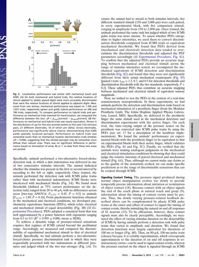
Fig. 2. Localization performance was similar with mechanical touch andICMS. (A) On both mechanical and hybrid trials, the relative locations ofstimuli applied to widely spaced digits were more accurately discriminatedthan were the relative locations of stimuli applied to adjacent digits. Mea-sured from one animal, mechanical performance was based on 1,160 and1,031 trials, respectively (green and gold); hybrid performance on 246 and196 trials, respectively. To compare performance on hybrid trials and per-formance on mechanical trials matched for hand location, we computed thedifference between the two: ΔP = pmech(correct) − phybrid(correct). (B) Per-formance on mechanical and hybrid trials was nearly equivalent. Shown isthe distribution of Δp for the two animals tested on this task (132 stimuluspairs, 27 different electrodes, 16 of which are UEAs). Across electrodes,performance was significantly above chance, demonstrating that ICMSyields spatially localized percepts. Performance on hybrid trials wassomewhat lower than on mechanical location discrimination trials (medianΔP = 0.056), suggesting that the elicited percepts may be somewhat morediffuse than natural ones. There was no significant difference in perfor-mance based on stimulation of areas 3b or 1, so data from these two areasare pooled.
Tabot et al. PNAS | November 5, 2013 | vol. 110 | no. 45 | 18281
NEU
ROSC
IENCE

signal that is modulated according to the pressure exerted on theobject throughout contact.
DiscussionSomatosensory feedback plays a critical role in the dexterousmanipulation of objects (9). Indeed, signals from mechanore-ceptive afferents in the skin convey information about the loca-tion of contact (19, 20) and about the forces exerted on the skinwhen an object is grasped (21–25). Cutaneous afferents alsosignal when our grip on an object is slipping (26). This criticalinformation is often unavailable visually and, when available, isgenerally inadequate to guide motor behavior. Without somato-sensory input, then, we would routinely crush or drop grasped
objects. In addition, the sense of touch confers to our limbsembodiment, making them feel a part of us (27–29). Finally,touch plays an important role in communicating emotions and isa fundamental component of sexual behavior and experience.Given the importance of somatosensation, upper-limb neuro-prostheses will not be clinically relevant until they provide forsomatosensory inputs. Although the need for a highly invasivesurgery sets the bar high for efficacy and reliability (30), ICMShas the potential to achieve sufficient sensory restoration tojustify the risk, particularly in spinal cord injury patients, forwhom many less-invasive options are not available.The present findings provide a blueprint to convert the output
of sensors on a prosthetic limb into patterns of ICMS that elicit
A
C
B
Fig. 3. Information about contact pressure was conveyed by varying ICMS amplitude. (A) Detection of ICMS in areas 3b and 1 followed a sigmoidal relationship toamplitude, shown here for one animal (area 3b: 19,184 trials, 7 electrodes; area 1: 29,498 trials, 27 electrodes). The horizontal dashed line indicates the threshold cri-terion. (Inset) Distribution of detection thresholds (75% detection) for all three animals (area 3b: 19 electrodes; area 1: 35 electrodes). There were no significant dif-ferences in sensitivity to ICMS across animals or anatomical areas. (B) ICMS amplitudewas a power function ofmechanical amplitudematched in perceivedmagnitude.Shown are PEFs derived from all of the electrodes for which there were both detection and discrimination data. Mechanical data from the electrode’s RF was used togenerate the function. The two colors correspond to two differentmonkeyswith 4 and 12 electrodes (the third did not perform the discrimination task so did not yieldPEFs). The darker traces show the pooled PEFs for each monkey. The equations are for the power functions fit to the pooled PEFs for the twomonkeys are shown. (C)Discriminability of stimulus amplitude is equivalentwhenmechanical indentations are applied to the animal’s ownfinger (blue) or to a prostheticfinger and convertedto ICMS (red) (two animals with 240 and 360 trials with the prosthetic finger and 1,120 trials with the native finger). The mapping between time-varying pressure andtime-varying ICMS amplitude was achieved by using the PEF. See Fig. S4 for analogous results in a detection task.
A B
Fig. 4. (A) Animals are able to compare mechanical indentations to ICMS pulse trains scaled by using PEFs. The monkey compared a standard mechanicalstimulus of fixed amplitude to a comparison electrical stimulus of variable amplitude (ranging from 20 to 80 μA) (performance pooled over 4 electrodes, 2 UEAs,and 2 FMAs for a total of 4,114 trials). The amplitude of the standard was matched in subjective magnitude with an electrical stimulus of amplitude 50 μA basedon the PEF of each electrode tested (mean amplitude = 440 μm, range 200–750 μm). The animal judged which of the two stimuli was stronger, demonstratingthat it could compare mechanical and electrical stimuli along a single perceptual dimension (magnitude). Error bars denote the SEM. (B) Sensitivity to ICMSincreases with duration up to ∼100 ms. Thresholds decrease as duration increases from 50 to 100 ms then level off. Thus, a 100-ms pulse at 80 μA will be clearlyperceptible and can be used to signal the onset and offset of contact, mimicking the onset and offset responses observed in the somatosensory cortex of intactindividuals. Error bars denote the SEM. These functions show the mean performance across four electrodes in area 3b in one animal.
18282 | www.pnas.org/cgi/doi/10.1073/pnas.1221113110 Tabot et al.

somatosensory percepts that can then be used to guide the ma-nipulation of objects.Our approach consists of exploiting existing neural repre-
sentations in somatosensory cortex to convey tactile informationimportant for object grasping and manipulation. First, we showthat ICMS elicits spatially localized percepts, a phenomenon thatrelies on the somatotopic organization of S1 and can be used toconvey information about contact location. Although the degreeto which the artificial percepts are localized remains to be elu-cidated, our results suggest that the projections fields may bemore diffuse than are sensations elicited by a punctate in-dentation, at least for some electrodes. The diffuseness of thesensations is not surprising given that ICMS has been shown toevoke sparse, spatially distributed neuronal activity (31). Second,we show that the magnitude of the artificial percepts is gradedaccording to the ICMS amplitude, a phenomenon that can beused to convey information about contact pressure. To ensurethat artificial percepts operate over the same dynamic range asnatural ones, we create mapping between the sensory magnitudeof artificial and natural percepts (PEFs). The question remainswhether the quality of the percept changes as ICMS amplitudeincreases, a question that can be addressed only in experimentswith human subjects (32, 33). Third, we measure the effect ofpulse train duration on detectability to identify the shortest de-tectable ICMS pulse train. We propose that a phasic pulse traincan be used to mimic the cortical signature of contact events,namely a phasic burst mediated at the periphery by rapidlyadapting mechanoreceptive afferents (13). This phasic pulsetrain can then be used to precisely signal the timing of the onsetand offset of contact with objects.To instrument a tetraplegic or amputated patient with a neu-
roprosthesis, the somatotopic organization of the array can bemapped by delivering ICMS pulses through each electrode andhaving the patient report the projected location of the sensationon the hand or phantom hand (34). Then, the pattern of stim-ulation delivered through each electrode can be determined inreal time based on the output of sensors on the correspondinglocation of the prosthesis. Contact with an object would be sig-naled by a phasic ICMS pulse train of fixed amplitude followedby a tonic pulse train, the time-varying amplitude of which tracksthe time-varying pressure exerted on the sensor according toa PEF (calibrated based on the sensitivity of that electrode toelectrical stimulation). Somatosensory feedback can be deliveredwith a delay matching that associated with signal transmissionfrom periphery to cortex with an intact limb so that feedbacksignals can be naturally integrated with ongoing motor planningand execution. The extent to which the proposed approaches willneed to be modified for patients whose somatosensory cortex hasbeen deafferented (through amputation or spinal cord injury)remains to be tested. However, we anticipate that the proposedbiomimetic feedback will considerably increase the dexterity andembodiment of upper-limb neuroprostheses (such as that de-scribed in ref. 35) without extensive training on the patient’s partand will constitute an important step in restoring touch to indi-viduals who have lost it.
Experimental ProceduresAnimal care and handling conformed to the procedures approved by theUniversity of Chicago Animal Care and Use Committee.
Animals. Three Rhesus macaques (two males, one female) were used in thisstudy; all three were 6 y of age and ranged in weight from 6.5 to 12 kg.
Implants. Each of three animals was implanted with one Utah electrode array(UEA; Blackrock Microsystems) in the hand representation of area 1 in theright hemisphere. The UEA consists of 96 1.5-mm-long electrodes, spaced400 μm apart, and spanning a 4 × 4-mm area. Two FMAs (Microprobes forLife Science) were implanted flanking the UEA and impinged on area 3b. EachFMA consists of 16 3-mm-long electrodes spanning a 2.5 × 1.95-mm area.
Only the FMA that impinged on the hand representation was used in thestimulation experiments (the other, more medial and posterior one, im-pinged on the arm representation in all three animals). In experiments withelectrode drives, it has been shown that the distal digit representations inarea 1 are at the surface, whereas the distal digit representations in area 3bare ∼3 mm deep (36). We had specified electrode lengths of 3 mm based onour previous experience that the distal digit representation in area 3b lies atthat depth. That our receptive fields on the FMAs were exclusively cutaneousand located at or near the tip of the finger indicates that these electrodeswere impinging on area 3b.
RF Mapping. We mapped the receptive field of the neuronal populationssurrounding each electrode (in awake animals) by identifying which areas ofskin evokedmultiunit activity (monitored through speakers). RFmappingwasrepeated periodically throughout the study to verify that maps were con-sistent. All three animals yielded maps consistent with previous studies, witha progression from D5 (small finger) to D1 (thumb) proceeding laterally andanteriorly along the central sulcus (37).
Stimulation. The monkey’s arms were placed in padded arm holders andloosely secured in place by Velcro straps. The hand to be stimulated wasplaced palmar side up onto an acrylic mold of that animal’s hand. A drop ofethyl cyanoacrylate (Loctite 401; Henkel) was placed on each aluminumfinger cup fixed within the mold. Each fingernail was then pressed into itsrespective finger cup and held for ∼10 s until the fingers were fixed in place.Animals were trained to hold their hand in position with the palm facing up;the glue was used to assist the animal in keeping its hand in position andwas not strong enough to prevent it from freeing its finger(s). The experi-menter carefully monitored the animal’s hand throughout each experi-mental block to ensure that the hand remained in position and that thetactile stimulator indented the skin as intended. The animal’s view of itshand and of the stimulating apparatus was obstructed.
Mechanical stimuli consisted of trapezoidal indentations delivered byusing a custom-designed and built triaxial indenting stimulator (TIS). The TISconsists of a high-precision low-profile Z-stage (MX80L; Parker Hannifin)mounted on an XY stage (PRO115; Aerotech). The stage allowed us to po-sition the Z stage anywhere on the hand with micometer precision, whereasthe linear motor allowed us to indent the skin with a punctate probe witha diameter of 1 mm. In the location discrimination task, the 3D structure ofthe hand was first mapped by using a high-precision rangefinder (Accurange200–25; Acuity Lasers), mounted on the XY stage, so that the depth of in-dentation could be controlled, with ∼10-μm precision, relative to the heightof the skin surface at each stimulated location. On each trial, the TISindented one location, then the next, with a short interstimulus interval. Inthe detection and pressure discrimination tasks, the stimulator was pre-indented into the skin by 500 μm. Any auditory cues from the TIS weremasked by presenting white noise through speakers.
ICMS trains, lasting 1 s unless otherwise specified, consisted of symmetricbiphasic pulses delivered at 300 Hz using a CereStim 96 (BlackRockMicrosystems). The phase duration was 200 μs, the interphase durationwas 53 μs, and amplitudes ranged from 10 to 100 μA (2–20 nC per phase).We verified that ICMS did not trigger short latency muscle activation(SI Experimental Procedures, Fig. S5).
Psychophysical Tasks. All of the tasks were two alternative forced choice taskswhose sequence and timing are shown in Fig. 1A. The design was counter-balanced so that correct responses were as often “left” as they were “right”to eliminate any possible confounding effect of response bias. Correctresponses were rewarded with juice or water. Performance was computed asthe proportion correct in each stimulation condition. Because the motivationof the animals fluctuates somewhat from day to day, we eliminated blocksin which the animal performed poorly (did not reach 85% correct on theeasiest condition, on which the animals typically reached near perfect per-formance). Importantly, we applied the same exclusion criterion to themechanical and electrical trials. Performance as a function of comparisonamplitude was then fit by using a standard sigmoid.Signaling contact location. Two mechanical stimuli (duration = 1 s) were pre-sented on each trial, one in each stimulus interval (separated by a 1.5-s in-terstimulus interval), at two locations that were displaced from one anotheralong the mediolateral axis. For example, one stimulus might be presentedto the index fingertip, and the second might be presented to the smallfingertip (both on the same hand). The animal’s task was to indicatewhether the second stimulus was medial or lateral to the first by saccadingto the appropriate target (in this example, to the right). The amplitude ofthe stimulus varied pseudorandomly from trial to trial, and ranged from
Tabot et al. PNAS | November 5, 2013 | vol. 110 | no. 45 | 18283
NEU
ROSC
IENCE
1,000 to 2,000 μm, so the animal could not use any intensive cues to performthe task. On hybrid trials, one of the two mechanical stimuli was replacedwith an electrical stimulus delivered through an electrode whose RF locationmatched the location of the replaced mechanical stimulus. In these experi-ments, the intensity of the electrical stimulus was 80 μA to ensure it wassuprathreshold. The animal was rewarded if it responded (that is, produceda saccade) as if a mechanical stimulus had been delivered to that RF location.Hybrid trials were always interleaved with mechanical trials (with twoindentations). Furthermore, multiple hand locations, spanning the palmarsurface of the hand, and corresponding electrodes were interleaved topreclude the animal from learning stimulus-response contingencies (11.2 ± 6.0stimulus pairs per block, mean ± SD). In a subset of measurements, theamplitude of the ICMS pulses was varied on hybrid trials, yielding identicalresults. We report stimulus conditions with at least 20 responses (76 ± 28 and56 ± 26 mean ± SD for mechanical and hybrid trials, respectively).Signaling contact pressure. Detection. One of the two stimulus intervals con-tained a mechanical or electrical stimulus and the other was empty. Theanimal’s task was to indicate whether the stimulus was presented in the firstor the second interval by saccading to the left or right target, respectively.Mechanical indentations varied in amplitude from 50 to 1,000 μm; ICMSamplitude varied from 10 to 50 μA. In detection and discrimination experi-ments, the animal had to perform at least 100 trials on any given experi-mental block for the data to be reported.
Pressure/intensity discrimination. Two mechanical or two ICMS pulse trainswere presented: One of the two stimuli was a standard stimulus at one of twoamplitudes and the otherwas a comparison stimulus, whose amplitude variedover a range. The animal’s task was to indicate whether the second stimuluswas smaller or larger in amplitude than the first by saccading to the left orright target, respectively. In the pressure discrimination task, the amplitudeof the standard stimulus was 150 or 2,000 μm, and was paired with a com-parison stimulus, whose amplitude ranged from 150 to 2,000 μm (excluding
the amplitude of the standard). In the electrical stimulation condition, thestandard amplitude was 30 or 100 μA, and comparisons ranged from 30 to100 μA. The two standard stimuli were (approximately) matched in sensorymagnitude with their mechanical counterparts (based on PEFs derived fromdetection performance). Standard stimuli were interleaved from trial to trialto ensure that the animals attended to both stimulus intervals. Furthermore,mechanical blocks were interleaved with electrical blocks to minimize theanimal’s ability to learn arbitrary stimulus-response pairings on ICMS blocks.
Detection and discrimination task based on stimulation of the prosthetic finger.The details of the implementation are described (17). The time varyingoutput of the sensor was converted into an equivalent indentation depth(based on an empirically established relationship), which was then convertedinto an electrical pulse train by using the PEF.Signaling contact timing. The objective of this experiment was to determine theminimum ICMS duration that elicits a detectable percept to be triggered atthe onset and offset of contact. One of the two stimulus intervals containedamechanical or electrical stimulus and the otherwas empty. The animal’s taskwas to indicate whether the stimulus was presented in the first or the secondinterval by saccading to the left or right target, respectively. ICMS amplitudevaried from 10 to 50 μA and duration varied from 50 to 500 ms.
ACKNOWLEDGMENTS. We thank Louise Manfredi, Melanie Peterson, andThierri Callier for assistance with the data collection; Adam Davidson forassistance with our first floating microelectrode array implants; Hannes Saaland Jeffrey Yau for helpful comments on a previous version of this manuscript;Matthew Johannes and Kapil Katyal for their assistance with the prostheticfinger; and Lee Miller for help with the electromyographic recordings. Thismaterial is based on work supported by the Defense Advanced ResearchProjects Agency under Contract N66001-10-C-4056. G.A.T. was supported byNational Science Foundation Grant DGE-0903637 and S.J.B. by NationalInstitutes of Health Grants R01 NS18787 and NS082865.
1. Richer F, Martinez M, Robert M, Bouvier G, Saint-Hilaire JM (1993) Stimulation ofhuman somatosensory cortex: Tactile and body displacement perceptions in medialregions. Exp Brain Res 93(1):173–176.
2. Romo R, Hernández A, Zainos A, Salinas E (1998) Somatosensory discrimination basedon cortical microstimulation. Nature 392(6674):387–390.
3. Romo R, Hernández A, Zainos A, Brody CD, Lemus L (2000) Sensing without touching:Psychophysical performance based on cortical microstimulation. Neuron 26(1):273–278.
4. Butovas S, Schwarz C (2007) Detection psychophysics of intracortical microstimulationin rat primary somatosensory cortex. Eur J Neurosci 25(7):2161–2169.
5. London BM, Jordan LR, Jackson CR, Miller LE (2008) Electrical stimulation of theproprioceptive cortex (area 3a) used to instruct a behaving monkey. IEEE Trans NeuralSyst Rehabil Eng 16(1):32–36.
6. O’Doherty JE, Lebedev MA, Hanson TL, Fitzsimmons NA, Nicolelis MA (2009) A brain-machine interface instructed by direct intracortical microstimulation. Front IntegrNeurosci 3(20):1–10.
7. O’Doherty JE, et al. (2011) Active tactile exploration using a brain-machine-brain in-terface. Nature 479(7372):228–231.
8. Schiller PH, Slocum WM, Kwak MC, Kendall GL, Tehovnik EJ (2011) New methodsdevised specify the size and color of the spots monkeys see when striate cortex (areaV1) is electrically stimulated. Proc Natl Acad Sci USA 108(43):17809–17814.
9. Johansson RS, Flanagan JR (2009) Coding and use of tactile signals from the fingertipsin object manipulation tasks. Nat Rev Neurosci 10(5):345–359.
10. Rasmussen T, Penfield W (1947) The human sensorimotor cortex as studied by elec-trical stimulation. Fed Proc 6(1 Pt 2):184.
11. Simons SB, et al. (2005) Amplitude-dependency of response of SI cortex to flutterstimulation. BMC Neurosci 6:43.
12. Tehovnik EJ, Tolias AS, Sultan F, SlocumWM, Logothetis NK (2006) Direct and indirectactivation of cortical neurons by electrical microstimulation. J Neurophysiol 96(2):512–521.
13. Pei YC, Denchev PV, Hsiao SS, Craig JC, Bensmaia SJ (2009) Convergence of sub-modality-specific input onto neurons in primary somatosensory cortex. J Neurophysiol102(3):1843–1853.
14. Bensmaia SJ, Denchev PV, Dammann JF, 3rd, Craig JC, Hsiao SS (2008) The repre-sentation of stimulus orientation in the early stages of somatosensory processing. JNeurosci 28(3):776–786.
15. Tehovnik EJ, Slocum WM, Carvey CE, Schiller PH (2005) Phosphene induction and thegeneration of saccadic eye movements by striate cortex. J Neurophysiol 93(1):1–19.
16. Bradley DC, et al. (2005) Visuotopic mapping through a multichannel stimulatingimplant in primate V1. J Neurophysiol 93(3):1659–1670.
17. Berg JA, et al. (2012) Behavioral demonstration of a somatosensory prosthesis. IEEETrans Neural Syst Rehabil Eng 21(3):500–507.
18. Sripati AP, Vogelstein RJ, Armiger RS, Russell AF, Bensmaia SJ; Sung Soo Kim (2009)Conveying tactile feedback in sensorized hand neuroprostheses using a biofidelicmodel of mechanotransduction. IEEE Trans Biomed Circuits Syst 3(6):398–404.
19. Ochoa J, Torebjörk E (1983) Sensations evoked by intraneural microstimulation ofsingle mechanoreceptor units innervating the human hand. J Physiol 342:633–654.
20. Wheat HE, Goodwin AW, Browning AS (1995) Tactile resolution: Peripheral neuralmechanisms underlying the human capacity to determine positions of objects con-tacting the fingerpad. J Neurosci 15(8):5582–5595.
21. Knibestöl M (1973) Stimulus-response functions of rapidly adapting mechanor-eceptors in human glabrous skin area. J Physiol 232(3):427–452.
22. Knibestöl M (1975) Stimulus-response functions of slowly adapting mechanoreceptorsin the human glabrous skin area. J Physiol 245(1):63–80.
23. Macefield VG, Hager-Ross C, Johansson RS (1996) Control of grip force during re-straint of an object held between finger and thumb: Responses of cutaneous affer-ents from the digits. Exp Brain Res 108(1):155–171.
24. Goodwin AW, Wheat HE (2004) Sensory signals in neural populations underlyingtactile perception and manipulation. Annu Rev Neurosci 27:53–77.
25. Muniak MA, Ray S, Hsiao SS, Dammann JF, Bensmaia SJ (2007) The neural coding ofstimulus intensity: Linking the population response of mechanoreceptive afferentswith psychophysical behavior. J Neurosci 27(43):11687–11699.
26. Johansson RS, Westling G (1984) Roles of glabrous skin receptors and sensorimotormemory in automatic control of precision grip when lifting rougher or more slipperyobjects. Exp Brain Res 56(3):550–564.
27. Botvinick M, Cohen J (1998) Rubber hands ‘feel’ touch that eyes see. Nature391(6669):756.
28. Armel KC, Ramachandran VS (2003) Projecting sensations to external objects: Evi-dence from skin conductance response. Proc Biol Sci 270(1523):1499–1506.
29. Marasco PD, Kim K, Colgate JE, Peshkin MA, Kuiken TA (2011) Robotic touch shiftsperception of embodiment to a prosthesis in targeted reinnervation amputees. Brain134(Pt 3):747–758.
30. Blabe C, et al. (2012) Assessing brain-machine interface priorities from the perspectiveof spinal cord injury participants. Society for Neuroscience. Soc Neurosci, in press.
31. Histed MH, Bonin V, Reid RC (2009) Direct activation of sparse, distributed pop-ulations of cortical neurons by electrical microstimulation. Neuron 63(4):508–522.
32. Bak M, et al. (1990) Visual sensations produced by intracortical microstimulation ofthe human occipital cortex. Med Biol Eng Comput 28(3):257–259.
33. Schmidt EM, et al. (1996) Feasibility of a visual prosthesis for the blind based on in-tracortical microstimulation of the visual cortex. Brain 119(Pt 2):507–522.
34. Ramachandran VS, Hirstein W (1998) The perception of phantom limbs. The D. O.Hebb lecture. Brain 121(Pt 9):1603–1630.
35. Johannes MS, et al. (2011) An overview of the developmental process for the ModularProsthetic Limb. The Johns Hopkins University Applied Physics Laboratory TechnicalDigest 30(3):207–216.
36. Pei YC, Hsiao SS, Craig JC, Bensmaia SJ (2010) Shape invariant coding of motion di-rection in somatosensory cortex. PLoS Biol 8(2):e1000305.
37. Pons TP, Garraghty PE, Cusick CG, Kaas JH (1985) A sequential representation of theocciput, arm, forearm and hand across the rostrocaudal dimension of areas 1, 2 and 5in macaque monkeys. Brain Res 335(2):350–353.
38. Koivuniemi AS, Otto KJ (2011) Asymmetric versus symmetric pulses for cortical mi-crostimulation. IEEE Trans Neural Syst Rehabil Eng 19(5):468–476.
18284 | www.pnas.org/cgi/doi/10.1073/pnas.1221113110 Tabot et al.


