Restoring GABAergic inhibition rescues memory deficits in ...
9
Restoring GABAergic inhibition rescues memory deficits in a Huntington’s disease mouse model Zahra Dargaei a , Jee Yoon Bang b , Vivek Mahadevan a,1 , C. Sahara Khademullah a , Simon Bedard a , Gustavo Morrone Parfitt a,b , Jun Chul Kim b , and Melanie A. Woodin a,2 a Department of Cell and Systems Biology, University of Toronto, Toronto, ON M5S 3G5, Canada; and b Department of Psychology, University of Toronto, Toronto, ON M5S 3G3, Canada Edited by Mu-ming Poo, Chinese Academy of Sciences, Shanghai, China, and approved January 5, 2018 (received for review September 25, 2017) Huntington’s disease (HD) is classically characterized as a move- ment disorder, however cognitive impairments precede the motor symptoms by ∼15 y. Based on proteomic and bioinformatic data linking the Huntingtin protein (Htt) and KCC2, which is required for hyperpolarizing GABAergic inhibition, and the important role of inhibition in learning and memory, we hypothesized that aber- rant KCC2 function contributes to the hippocampal-associated learning and memory deficits in HD. We discovered that Htt and KCC2 interact in the hippocampi of wild-type and R6/2-HD mice, with a decrease in KCC2 expression in the hippocampus of R6/2 and YAC128 mice. The reduced expression of the Cl − -extruding cotrans- porter KCC2 is accompanied by an increase in the Cl − -importing cotransporter NKCC1, which together result in excitatory GABA in the hippocampi of HD mice. NKCC1 inhibition by the FDA-approved NKCC1 inhibitor bumetanide abolished the excitatory action of GABA and rescued the performance of R6/2 mice on hippocampal- associated behavioral tests. Huntington’s disease | synaptic inhibition | GABA | chloride | learning H untington’s disease (HD) is primarily characterized by pro- gressive motor incoordination and involuntary movements that result from neurodegeneration of the striatum (1). However, cognitive and behavioral impairments involving the cortex and hippocampus emerge in the early stages of the disease and precede the motor impairments by more than a decade. HD patients display hippocampal-dependent learning and memory deficits (2–4), and mouse models of HD have alterations in ex- citatory synaptic plasticity in the hippocampus and impaired spatial cognition (5–10). However, the learning and memory functions of the hippocampus do not rely solely on synaptic plasticity of glutamatergic synapses. It has recently become clear that inhibitory GABA-releasing interneurons are required for hippocampal-dependent learning and memory tasks (11). For example, inactivating somatostatin-positive interneurons in the hippocampus prevents contextual fear conditioning (12), while ablation of parvalbumin-positive CA1 interneurons impairs spatial working memory (13). The tight link between inhibition and learning and memory is also clear in neurodegenerative disease, where reduced inhibition underlies neuronal network dysfunction in an Alzheimer’s disease mouse model (14) and restoring inhibition rescues the associated memory deficits (15, 16). Despite the requirement of inhibition for learning and memory and the deficits in hippocampal-dependent learning and memory in HD, inhibitory synaptic transmission in the hippo- campus has not been characterized in the HD brain. Synaptic inhibition in the brain is largely mediated by GABA acting on Cl − -permeable GABA A receptors (17). The polarity of GABA A receptor signaling depends on the precise regulation of two cation-chloride cotransporters, KCC2 (K + ·Cl − ) and NKCC1 (Na + ·K + ·2Cl − ) (18, 19). NKCC1-mediated Cl − accumulation into neurons leads to excitatory GABA early in development, whereas KCC2-mediated Cl − extrusion leads to inhibitory GABA in mature neurons (18, 19). When KCC2 and/or NKCC1 expression is disrupted, the neuronal Cl − gradient can collapse, resulting in a profound disruption of inhibition, which contributes to numerous neurological disorders including epileptic seizures (20), Down syn- drome (21), and autism spectrum disorder (ASD) (22). Restoring the neuronal Cl − gradient can rescue GABAergic inhibition deficits and behavioral phenotypes in animal models in several neurological disorders including ASD (22) and Down syndrome (21). Further- more, KCC2-mediated Cl − regulation directly controls synapse specificity of long-term potentiation at CA1 synapses in the hippo- campus of mature animals (23). A prominent group of Htt interactors are proteins involved in synaptic transmission, and their altered interaction with mutant Htt (mHtt) is suggested to contribute to abnormal synaptic transmission in HD (24). Recent proteomic studies revealed that the KCC2 encoding gene, Slc12A5, is highly enriched in Htt proteome (25, 26), but despite the strong correlation, this in- teraction has not been validated. Furthermore, a bioinformatic analysis of the unfolded protein response (UPR)-regulated genes in HD reveals an increase in NKCC1 mRNA and a decrease in KCC2 mRNA (27), which is significant because UPR is impli- cated in numerous neurodegenerative diseases including HD (28–30). Based on the previous data linking (m)Htt and Cl − transporters (25, 26) and the important role of inhibition in learning and memory, we hypothesized that KCC2 function is dysregulated in the HD brain, resulting in weakened inhibitory GABAergic transmission, which contributes to the hippocampal- dependent learning and memory deficits that emerge early in HD. Significance Huntington’s disease (HD) is a fatal neurodegenerative disor- der that currently has no cure. Although HD is classically con- sidered a motor disorder, HD patients experience learning and memory deficits years before the onset of motor symptoms, and these deficits resemble those observed in HD mouse models. In this work, using transgenic mouse models of HD, we demon- strate that the action of the neurotransmitter GABA has switched from inhibitory to excitatory. By treating HD mice with a clinically used diuretic (bumetanide), which restores inhibitory GABA, we rescued the learning and memory deficits. Our data suggest a potential therapeutic approach for the treatment of the cognitive deficits in early HD that can improve patient quality of life and reduce caregiver burden. Author contributions: Z.D., J.C.K., and M.A.W. designed research; Z.D., J.Y.B., V.M., C.S.K., S.B., and G.M.P. performed research; Z.D., J.Y.B., V.M., and C.S.K. analyzed data; and Z.D. and M.A.W. wrote the paper. The authors declare no conflict of interest. This article is a PNAS Direct Submission. This open access article is distributed under Creative Commons Attribution-NonCommercial- NoDerivatives License 4.0 (CC BY-NC-ND). 1 Present address: Section on Cellular and Synaptic Physiology, National Institute of Child Health and Human Development, Bethesda, MD 20892. 2 To whom correspondence should be addressed. Email: [email protected]. This article contains supporting information online at www.pnas.org/lookup/suppl/doi:10. 1073/pnas.1716871115/-/DCSupplemental. E1618–E1626 | PNAS | Published online January 30, 2018 www.pnas.org/cgi/doi/10.1073/pnas.1716871115 Downloaded by guest on November 25, 2021
Transcript of Restoring GABAergic inhibition rescues memory deficits in ...

Restoring GABAergic inhibition rescues memorydeficits in a Huntington’s disease mouse modelZahra Dargaeia, Jee Yoon Bangb, Vivek Mahadevana,1, C. Sahara Khademullaha, Simon Bedarda,Gustavo Morrone Parfitta,b, Jun Chul Kimb, and Melanie A. Woodina,2
aDepartment of Cell and Systems Biology, University of Toronto, Toronto, ON M5S 3G5, Canada; and bDepartment of Psychology, University of Toronto,Toronto, ON M5S 3G3, Canada
Edited by Mu-ming Poo, Chinese Academy of Sciences, Shanghai, China, and approved January 5, 2018 (received for review September 25, 2017)
Huntington’s disease (HD) is classically characterized as a move-ment disorder, however cognitive impairments precede the motorsymptoms by ∼15 y. Based on proteomic and bioinformatic datalinking the Huntingtin protein (Htt) and KCC2, which is requiredfor hyperpolarizing GABAergic inhibition, and the important roleof inhibition in learning and memory, we hypothesized that aber-rant KCC2 function contributes to the hippocampal-associatedlearning and memory deficits in HD. We discovered that Htt andKCC2 interact in the hippocampi of wild-type and R6/2-HD mice,with a decrease in KCC2 expression in the hippocampus of R6/2 andYAC128 mice. The reduced expression of the Cl−-extruding cotrans-porter KCC2 is accompanied by an increase in the Cl−-importingcotransporter NKCC1, which together result in excitatory GABA inthe hippocampi of HD mice. NKCC1 inhibition by the FDA-approvedNKCC1 inhibitor bumetanide abolished the excitatory action ofGABA and rescued the performance of R6/2 mice on hippocampal-associated behavioral tests.
Huntington’s disease | synaptic inhibition | GABA | chloride | learning
Huntington’s disease (HD) is primarily characterized by pro-gressive motor incoordination and involuntary movements
that result from neurodegeneration of the striatum (1). However,cognitive and behavioral impairments involving the cortex andhippocampus emerge in the early stages of the disease andprecede the motor impairments by more than a decade. HDpatients display hippocampal-dependent learning and memorydeficits (2–4), and mouse models of HD have alterations in ex-citatory synaptic plasticity in the hippocampus and impairedspatial cognition (5–10). However, the learning and memoryfunctions of the hippocampus do not rely solely on synapticplasticity of glutamatergic synapses. It has recently become clearthat inhibitory GABA-releasing interneurons are required forhippocampal-dependent learning and memory tasks (11). Forexample, inactivating somatostatin-positive interneurons in thehippocampus prevents contextual fear conditioning (12), whileablation of parvalbumin-positive CA1 interneurons impairsspatial working memory (13). The tight link between inhibitionand learning and memory is also clear in neurodegenerativedisease, where reduced inhibition underlies neuronal networkdysfunction in an Alzheimer’s disease mouse model (14) andrestoring inhibition rescues the associated memory deficits (15,16). Despite the requirement of inhibition for learning andmemory and the deficits in hippocampal-dependent learning andmemory in HD, inhibitory synaptic transmission in the hippo-campus has not been characterized in the HD brain.Synaptic inhibition in the brain is largely mediated by GABA
acting on Cl−-permeable GABAA receptors (17). The polarity ofGABAA receptor signaling depends on the precise regulation oftwo cation-chloride cotransporters, KCC2 (K+·Cl−) and NKCC1(Na+·K+·2Cl−) (18, 19). NKCC1-mediated Cl− accumulationinto neurons leads to excitatory GABA early in development,whereas KCC2-mediated Cl− extrusion leads to inhibitory GABA inmature neurons (18, 19). When KCC2 and/or NKCC1 expression isdisrupted, the neuronal Cl− gradient can collapse, resulting in a
profound disruption of inhibition, which contributes to numerousneurological disorders including epileptic seizures (20), Down syn-drome (21), and autism spectrum disorder (ASD) (22). Restoringthe neuronal Cl− gradient can rescue GABAergic inhibition deficitsand behavioral phenotypes in animal models in several neurologicaldisorders including ASD (22) and Down syndrome (21). Further-more, KCC2-mediated Cl− regulation directly controls synapsespecificity of long-term potentiation at CA1 synapses in the hippo-campus of mature animals (23).A prominent group of Htt interactors are proteins involved in
synaptic transmission, and their altered interaction with mutantHtt (mHtt) is suggested to contribute to abnormal synaptictransmission in HD (24). Recent proteomic studies revealed thatthe KCC2 encoding gene, Slc12A5, is highly enriched in Httproteome (25, 26), but despite the strong correlation, this in-teraction has not been validated. Furthermore, a bioinformaticanalysis of the unfolded protein response (UPR)-regulated genesin HD reveals an increase in NKCC1 mRNA and a decrease inKCC2 mRNA (27), which is significant because UPR is impli-cated in numerous neurodegenerative diseases including HD(28–30). Based on the previous data linking (m)Htt and Cl−
transporters (25, 26) and the important role of inhibition inlearning and memory, we hypothesized that KCC2 function isdysregulated in the HD brain, resulting in weakened inhibitoryGABAergic transmission, which contributes to the hippocampal-dependent learning and memory deficits that emerge early in HD.
Significance
Huntington’s disease (HD) is a fatal neurodegenerative disor-der that currently has no cure. Although HD is classically con-sidered a motor disorder, HD patients experience learning andmemory deficits years before the onset of motor symptoms,and these deficits resemble those observed in HD mouse models.In this work, using transgenic mouse models of HD, we demon-strate that the action of the neurotransmitter GABA has switchedfrom inhibitory to excitatory. By treating HD mice with a clinicallyused diuretic (bumetanide), which restores inhibitory GABA, werescued the learning and memory deficits. Our data suggest apotential therapeutic approach for the treatment of the cognitivedeficits in early HD that can improve patient quality of life andreduce caregiver burden.
Author contributions: Z.D., J.C.K., and M.A.W. designed research; Z.D., J.Y.B., V.M., C.S.K.,S.B., and G.M.P. performed research; Z.D., J.Y.B., V.M., and C.S.K. analyzed data; and Z.D.and M.A.W. wrote the paper.
The authors declare no conflict of interest.
This article is a PNAS Direct Submission.
This open access article is distributed under Creative Commons Attribution-NonCommercial-NoDerivatives License 4.0 (CC BY-NC-ND).1Present address: Section on Cellular and Synaptic Physiology, National Institute of ChildHealth and Human Development, Bethesda, MD 20892.
2To whom correspondence should be addressed. Email: [email protected].
This article contains supporting information online at www.pnas.org/lookup/suppl/doi:10.1073/pnas.1716871115/-/DCSupplemental.
E1618–E1626 | PNAS | Published online January 30, 2018 www.pnas.org/cgi/doi/10.1073/pnas.1716871115
Dow
nloa
ded
by g
uest
on
Nov
embe
r 25
, 202
1

We determined that Htt and KCC2 interact in the hippo-campus of both wild-type (WT) and the R6/2 transgenic mousemodel of HD. We also discovered that decreased KCC2 andincreased NKCC1 expression in the hippocampi HD mice resultin a depolarizing shift in the reversal potential for GABAA
receptor-mediated Cl− currents (EGABA), which renders GABAA
receptor signaling excitatory in R6/2 mice. The treatment of R6/2 micewith the FDA-approved NKCC1 inhibitor bumetanide restored EGABA
to the value seen in WT mice, abolished the excitatory action ofGABA, and rescued the hippocampal-associated memory defi-cits in R6/2 mice.
ResultsHtt Interacts with KCC2 in the Hippocampus of WT and R6/2 Mice.Based on the bioinformatic and proteomic studies linking KCC2and Htt (25, 26), we first determined biochemically whetherKCC2 and Htt interact. We performed coimmunoprecipitation(co-IP) assays from hippocampal brain lysates prepared from WTand the R6/2 mouse model of HD. R6/2 mice express the exon1 human Htt with 120 CAG repeats (31) and develop a relativelyfast progressing neurological phenotype similar to HD. At birth,R6/2 mice are indistinguishable from their littermate controls anddevelop normally until ∼8 wk of age (31). Because we hypothe-sized that KCC2 dysfunction and weakened synaptic inhibitioncontribute to the learning and memory deficits in early stages ofthe disease, we performed the co-IP on hippocampal brain lysatescollected from 7-wk-old WT and R6/2 mice. Using anti-Htt anti-bodies, we found that Htt precipitates KCC2, indicating the ex-istence of a KCC2–Htt complex in vivo (Fig. 1A and SI Appendix,Fig. S1 A–D). KCC2 exists as both monomers (∼140 kDa) andoligomers (>250 kDa), with the oligomeric form believed to be thefunctional form of the transporter in the mature brain (32, 33). Wefound that Htt interacts with both monomeric and oligomericKCC2. As a control, we probed for NKCC1, which is not present inthe Htt interactome (26), and found no interaction with Htt (Fig.1A and SI Appendix, Fig. S1E). To determine whether KCC2 caninteract with mHtt, we performed co-IP assays in COS-7 cellstransfected with KCC2 and the normal or expanded polyglutaminetract of Htt (Htt-FL-15Q-HA and Htt-FL-128Q-HA, respectively).We found that both normal and mutant forms of Htt could pre-cipitate KCC2 (Fig. 1B and SI Appendix, Fig. S2).
KCC2 and NKCC1 Protein Expression Are Altered in the Hippocampusof HD Mice. mHtt interacts with proteins involved in synaptictransmission and can alter the function, cellular distribution, andtotal expression of these interacting proteins (34–36). To de-termine if the KCC2–mHtt interaction alters KCC2 protein ex-pression, we performed Western blot analysis from hippocampalbrain lysates and found a significant decrease in total KCC2 protein(oligomer + monomer) at 7 wk in R6/2 mice relative to WT (Fig.2A and SI Appendix, Fig. S3 A–C; WT, 0.59 ± 0.08; R6/2, 0.33 ±0.04; P = 0.0104). We also observed a significant decrease in fluo-rescence intensity of KCC2 at the surface membrane in the hip-pocampus of R6/2 mice compared with WT controls (Fig. 2Eand SI Appendix, Fig. S4; WT, 69.07 ± 2.09; R6/2, 46.27 ± 2.03;P < 0.0001). To determine if the altered KCC2 and NKCC1expression in the R6/2 hippocampus is common in HD, we repeatedour protein expression experiments in the YAC128 mouse model ofHD, which expresses full-length human Htt with 128 CAG repeats(37). This model has a later onset of motor symptoms (comparedwith R6/2) and closely recapitulates the motor dysfunction andneuropathology observed in human HD (37). We performedWestern blots on hippocampal brain lysates collected from 9-wk-oldWT and YAC128 mice. Consistent with our findings in R6/2, wealso observed a significant decrease in KCC2 expressionin YAC128 mice (0.80 ± 0.07) compared with WT (1.16 ± 0.12;P = 0.030) (Fig. 2B and SI Appendix, Fig. S3 G and H). We alsoprobed for NKCC1, because in other neurological disorderswhere KCC2 is decreased, increases in NKCC1 have beenreported (38, 39) and are thought to represent a reversion to animmature GABAergic phenotype (39, 40). Interestingly, al-though NKCC1 is not in the Htt interactome (25, 26) and doesnot co-IP with Htt (Fig. 1A), we found a significant increase inNKCC1 protein expression at 7 wk in R6/2 mice (Fig. 2C and SIAppendix, Fig. S3 D–F; WT, 0.53 ± 0.09; R6/2, 2.02 ± 0.37; P =0.0104) but not YAC128 mice (Fig. 2D and SI Appendix, Fig. S5 Iand J; WT, 1.30 ± 0.14; YAC128, 1.53 ± 0.21; P = 0.368).To determine if gene transcription underlies the alterations in
protein expression, we performed real-time qPCR on samplesfrom R6/2 and WT mice using isoform-specific amplification ofSlc12A5 (KCC2) and Slc12A2 (NKCC1). We used previouslyidentified stable control genes for R6/2 mice (Rpl13a, ATP5b,Ubc, Canx) (41) and found a significant decrease in KCC2 mRNAfrom R6/2 mice (7 wk old) compared with WT, consistent withbioinformatic predictions (27) (Fig. 2F and SI Appendix, Fig. S5
15Q-FL-Htt-HA128Q-FL-Htt-HA
FL-KCC2-Myc
HA
KCC2
Input
250250
100
Input
250
100
100
KCC2250
WTHtt
NKCC1
M
O
-----
++-
- +-+-+
-+-
+
-----
++-
- +-+-+
-+-
+
IP
A
IP
IgGR6/2
HttIgGHtt
WT R6/2kDa
B
kDa
Fig. 1. KCC2 and Htt interact in the hippocampus of WT and R6/2 mice. (A) Native Htt complexes from C12E9-solubilized hippocampal brain lysatesimmunoprecipitated with anti-Htt antibodies and immunoblotted with the antibodies indicated at right (Htt, KCC2, NKCC1). Shown is a representativeexample of five independent biological replicates (full blots presented in SI Appendix, Fig. S1). Input, input fraction (2% of IP); IP, immunoprecipitation; M,monomer; O, oligomer. (B) Co-IP experiments performed in COS-7 cells transfected with 15Q-Htt-HA, or 128Q-Htt-HA and KCC2-Myc solubilized in RIPAbuffer, immunoprecipitated with anti-HA antibodies, and immunoblotted with the antibodies indicated at right (KCC2, HA). Shown is a representative ex-ample of five independent biological replicates (full blots presented in SI Appendix, Fig. S2).
Dargaei et al. PNAS | Published online January 30, 2018 | E1619
NEU
ROSC
IENCE
PNASPL
US
Dow
nloa
ded
by g
uest
on
Nov
embe
r 25
, 202
1

A–C). The same set of control genes, however, showed variabilitybetween WT and YAC128 hippocampus and showed no statisticaldifferences in the relative expression of KCC2 mRNA expression(SI Appendix, Fig. S5D). Pan-NKCC1 transcript, but not NKCC1a/b,was also significantly increased in R6/2 hippocampal samples butonly compared with one of the control genes, Rpl13a (Fig. 2G andSI Appendix, Fig. S5 A–C).
EGABA Is Depolarized in CA1 Neurons from HD Hippocampi. In matureneurons, relatively high expression of KCC2 results in low levelsof intracellular Cl− and a hyperpolarized reversal potential forGABA (EGABA) (18, 19). When KCC2 is reduced and/orNKCC1 is increased, EGABA depolarizes and can even renderGABA excitatory (21, 42, 43). To address whether the alteredprotein levels of KCC2 and NKCC1 in the hippocampus of R6/2mice disrupt the polarity of GABA (hyperpolarizing vs. depola-rizing), we recorded EGABA from CA1 pyramidal neurons inhippocampal slices using whole-cell patch-clamp recordings. Wefound that EGABA from R6/2 mice was significantly depolarized(−58.05 ± 1.68 mV) compared with WT mice (−67.45 ±1.47 mV; P = 0.0009; Fig. 3 A and B), with no significant dif-ferences in the resting membrane potential (WT, −65.88 ±2.12 mV; R6/2, −67.82 ± 1.23 mV; P = 0.41; Fig. 3D) or synapticconductance (WT, 5.20 ± 0.98 pS; R6/2, 5.23 ± 0.75 pS; P = 0.97;Fig. 3E). During whole-cell patch-clamp recordings, the contentsof the intracellular pipette dialyze with cytoplasm, which canalter the Cl− gradient and EGABA. To preserve the intracellularCl− concentration, we performed gramicidin-perforated patch-clamp recordings and again found that the EGABA was signifi-cantly depolarized in R6/2 hippocampal neurons compared withWT mice (WT, −71.37 ± 1.70 mV; R6/2, −62.07 ± 3.095 mV;
P = 0.02; Fig. 3C). KCC2 function is optimally tested in thepresence of a Cl− load, which simulates the physiological contextduring inhibition (44). To examine KCC2 function in the pres-ence of a Cl− load to drive transporter function, we loaded theintracellular compartment with Cl− through the whole-cell patchpipette (44) (30 mM Cl−). Again, we found a significant depo-larizing shift in EGABA in R6/2 hippocampal neurons comparedwith WT (WT, −50.73 ± 3.59 mV; R6/2, −39.13 ± 2.32 mV; P =0.01; Fig. 3C). To determine if EGABA was also depolarized inYAC128 hippocampal neurons (9 wk old), we repeated ourwhole-cell patch-clamp recordings and again found a significantdepolarization of EGABA compared with age-matched controls(WT, −70.93 ± 1.61 mV; YAC128, −62.18 ± 1.72 mV; P = 0.004;Fig. 3 F and G) with no significant change in resting membranepotential (WT, −66.71 ± 2.25 mV; YAC128, −66.62 ± 1.79; P =0.97; Fig. 3H).Lastly, we investigated whether altered KCC2 and NKCC1
expression and depolarized EGABA also occur in other brainregions. We performed Western blot analysis and whole-cellpatch-clamp recording in the cortex of R6/2 and WT mice. Wefound an increase in the expression of NKCC1 and a decrease inthe expression of KCC2 in the cortices of R6/2 mice relative tothat in WT mice (SI Appendix, Fig. S6). EGABA was also depo-larized in the somatosensory area L4/5 of the cortex in R6/2 micecompared with WT (SI Appendix, Fig. S7).
GABAergic Inhibition Is Converted into Excitation in R6/2 Mice. Adepolarization of EGABA can result in loss of inhibitory drive andincrease neuronal excitability. To directly test this, we made cell-attached patch-clamp recordings from CA1 pyramidal neuronsand recorded the baseline spontaneous spiking activity in WT and
Fig. 2. KCC2 and NKCC1 protein expression is altered in the hippocampi of HD mice. (A, Top) Representative immunoblot images for KCC2 in protein extractsfrom samples of hippocampus lysate from WT and R6/2. (A, Bottom) Quantification of total KCC2 (oligomer + monomer) in WT (n = 11) and R6/2 (n = 11)normalized to β-Tubulin (P = 0.0104; Student’s unpaired t test). (B) Similar to A but for WT (n = 8) and YAC128 (n = 8) (P = 0.030; Student’s unpaired t test). (C,Top) Representative immunoblot images for NKCC1 in protein extracts from samples of hippocampus lysates from WT and R6/2. (C, Bottom) Quantification ofNKCC1 in WT (n = 11) and R6/2 (n = 11) normalized to β-Tubulin (P = 0.0104; Mann–Whitney test). (D) Similar to C but for WT (n = 8) and YAC128 (n = 8) (P =0.368; Student’s unpaired t test). (Full blots are presented in SI Appendix, Fig. S3.) For all panels, circles indicate values from individual animals. (E) Repre-sentative confocal images of hippocampal sections from 7-wk-old WT (Left) and R6/2 (Right) mice double labeled for KCC2 (red) and NeuN (green). [Scale bars,100 μm (Left), 25 μm (Right), and 5 μm (Insets).] See SI Appendix, Fig. S4 for quantifications. (F) Relative fold difference of pan-KCC2 (P = 0.011), KCC2a (P =0.033), KCC2b (P = 0.046), and (G) pan-NKCC1 (P = 0.547), NKCC1a (P = 0.822), NKCC1b (P = 0.852) abundance in R6/2 (n = 5) hippocampi normalized to WT(n = 5) using Student’s unpaired t test. See SI Appendix, Fig. S5 for NKCC1 or KCC2 mRNA quantifications normalized to the geometric means of housekeepinggenes. All summary figures represent mean ± SEM. *P < 0.05.
E1620 | www.pnas.org/cgi/doi/10.1073/pnas.1716871115 Dargaei et al.
Dow
nloa
ded
by g
uest
on
Nov
embe
r 25
, 202
1

R6/2 mice. Our results showed that the baseline spiking activitywas significantly higher in R6/2 (0.25 ± 0.04 Hz) compared with WTmice (0.07 ± 0.01 Hz) (P = 0.001), which was abolished by applica-tion of the GABAA receptor antagonist bicuculline (Fig. 4 A and B).The depolarization of EGABA also suggests that GABAergic
transmission may be excitatory. To determine if GABA is ex-citatory in the hippocampus of R6/2 mice, we tested the effect ofGABA application on spike frequency. Specifically, we madecell-attached patch-clamp recordings from CA1 pyramidal neu-rons during bath application of increasing concentrations of ex-ogenous GABA. GABA application at increasing concentrationsin WT neurons did not result in spiking, consistent with theknown inhibitory action of GABA in the adult brain. However,GABA application at the same concentrations elicited a strongincrease in spike frequency in neurons from R6/2 mice (Fig. 4 Cand D), which was abolished by application of the GABAA re-ceptor antagonist bicuculline (SI Appendix, Fig. S8).
NKCC1 Is Responsible for Depolarized EGABA in R6/2 HippocampalNeurons. We next asked whether the depolarizing shift of theGABA reversal potential is due primarily to an increase inNKCC1 and/or a decrease in KCC2 expression. To answer thisquestion, we first determined the effect of the FDA-approvedNKCC1 inhibitor bumetanide on EGABA. In WT CA1 pyramidalneurons, bumetanide did not significantly change EGABA (Fig. 5A–C; EGABA change: −1.34 ± 1.40 mV; P = 0.37), consistent with
the known low expression of NKCC1 in mature neurons (45, 46).In contrast, bumetanide produced a significant hyperpolarizationof EGABA in neurons from R6/2 mice (Fig. 5 A–C; EGABAchange: −6.22 ± 0.76 mV; P = 0.01), consistent with the in-creased expression of NKCC1 we observed in Fig. 1C. To isolatethe contribution of KCC2 to EGABA, we performed an occlusionexperiment using furosemide (1 mM), which is an NKCC1 andKCC2 inhibitor, together with the NKCC1 inhibitor bumetanide.As expected, EGABA in WT neurons depolarized after KCC2 in-hibition with furosemide (in the presence of bumetanide) (Fig. 5A–C; EGABA change: 10.46 ± 1.70 mV; P = 0.0009), while EGABAdid not significantly change in R6/2 neurons (Fig. 5 A–C; EGABAchange: 1.10 ± 1.05 mV; P = 0.33). This suggests that relativelyhigh expression of NKCC1 accounts for the relatively depolarizedEGABA in hippocampal pyramidal neurons of R6/2 mice. We alsoasked whether inhibitory synapses in R6/2 following treatmentwith bumetanide are functionally similar to WT animals [in arti-ficial cerebrospinal fluid (aCSF)]. We answered this question bycomparing inhibitory synaptic conductance between the twogroups and found no significant difference in conductance be-tween WT (aCSF) and R6/2 treated with bumetanide (P = 0.70)(Fig. 5D), suggesting that inhibitory synapses are not significantlyaltered presymptomatically in the R6/2 hippocampus.Having determined that bumetanide can hyperpolarize EGABA
in CA1 pyramidal neurons from R6/2 mice, we next askedwhether bumetanide can abolish the excitatory action of GABA
WT R6/2
EG
AB
A (m
V)
WT R6/2
Con
duct
ance
(pS
)IPS
C A
mpl
itude
(pA
)
HP (mV)
-80-75-70-65-60-55-50-45
RM
P (m
V)
-80
-75
-70
-65
-60
-55
-50
0
2
4
6
8
10
WT R6/2
A B
D E-80
-60
-40
-20
EG
AB
A (m
V)
- Cl loading gramicidin
WTR6/2
C*** *
*
-80 -70-60
-50 -40
-200
-150
-100
-50
0
50
100
150
200
250 WT R6/2
WT YAC128
EG
AB
A (m
V)
-80-75-70-65-60-55-50-45
G
WT YAC128
-80 -70 -60 -50 -40
-120
-80
-40
0
40
80
120
HP (mV)
IPS
C A
mpl
itude
(pA
)
F
WT YAC128
RM
P (m
V)
-80-75-70-65-60-55-50-45
H**
Fig. 3. EGABA is depolarized in hippocampal CA1 pyramidal neurons of HD mice. (A and F) Example current–voltage curves of inhibitory postsynaptic current(IPSC) recorded at different holding potentials from −80 to −40 mV in whole-cell patch-clamp configuration in CA1 hippocampal neurons fromWT (black) andR6/2 (green) or YAC128 (orange) mice. Insets show sample traces of the corresponding current induced by electrical stimulation in the presence of theglutamate blocker DNQX (20 μM). (Scale bars: 40 pA and 10 ms for A; 100 pA and 10 ms for F.) EGABA is shown at the arrow. (B) Summary of individual EGABArecordings obtained from all IV curves in WT (n = 8) and R6/2 (n = 11) (P = 0.0009; Student’s unpaired t test). (C) Similar to B, but for EGABA using Cl− loadingwhole-cell patch-clamp recording between WT (n = 9) and R6/2 (n = 13) (P = 0.010; Student’s unpaired t test); gramicidin perforated recording between WT(n = 6) and R6/2 (n = 7) (P = 0.029; Student’s unpaired t test). (D) Similar to B but for resting membrane potential (RMP) from WT (n = 8) and R6/2 (n = 11) (P =0.412; Student’s unpaired t test). (E) Similar to B but for synaptic conductance from WT (n = 8) and R6/2 (n = 11) (P = 0.977; Student’s unpaired t test).(G) Similar to B but for EGABA fromWT (n = 7) and YAC128 (n = 13) (P = 0.004; Student’s unpaired t test). (H) Similar to B but for RMP in WT (n = 7) and YAC128(n = 13) (P = 0.973; Student’s unpaired t test). For all panels, circles indicate values from single recordings across a minimum of three mice, and all summaryfigures represent mean ± SEM. *P < 0.05, **P < 0.01, ***P < 0.001.
Dargaei et al. PNAS | Published online January 30, 2018 | E1621
NEU
ROSC
IENCE
PNASPL
US
Dow
nloa
ded
by g
uest
on
Nov
embe
r 25
, 202
1
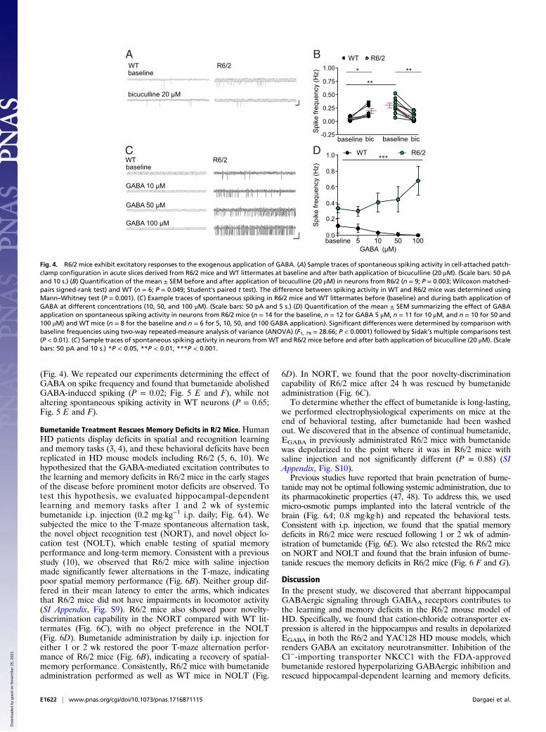
(Fig. 4). We repeated our experiments determining the effect ofGABA on spike frequency and found that bumetanide abolishedGABA-induced spiking (P = 0.02; Fig. 5 E and F), while notaltering spontaneous spiking activity in WT neurons (P = 0.65;Fig. 5 E and F).
Bumetanide Treatment Rescues Memory Deficits in R/2 Mice. HumanHD patients display deficits in spatial and recognition learningand memory tasks (3, 4), and these behavioral deficits have beenreplicated in HD mouse models including R6/2 (5, 6, 10). Wehypothesized that the GABA-mediated excitation contributes tothe learning and memory deficits in R6/2 mice in the early stagesof the disease before prominent motor deficits are observed. Totest this hypothesis, we evaluated hippocampal-dependentlearning and memory tasks after 1 and 2 wk of systemicbumetanide i.p. injection (0.2 mg·kg−1 i.p. daily; Fig. 6A). Wesubjected the mice to the T-maze spontaneous alternation task,the novel object recognition test (NORT), and novel object lo-cation test (NOLT), which enable testing of spatial memoryperformance and long-term memory. Consistent with a previousstudy (10), we observed that R6/2 mice with saline injectionmade significantly fewer alternations in the T-maze, indicatingpoor spatial memory performance (Fig. 6B). Neither group dif-fered in their mean latency to enter the arms, which indicatesthat R6/2 mice did not have impairments in locomotor activity(SI Appendix, Fig. S9). R6/2 mice also showed poor novelty-discrimination capability in the NORT compared with WT lit-termates (Fig. 6C), with no object preference in the NOLT(Fig. 6D). Bumetanide administration by daily i.p. injection foreither 1 or 2 wk restored the poor T-maze alternation perfor-mance of R6/2 mice (Fig. 6B), indicating a recovery of spatial-memory performance. Consistently, R6/2 mice with bumetanideadministration performed as well as WT mice in NOLT (Fig.
6D). In NORT, we found that the poor novelty-discriminationcapability of R6/2 mice after 24 h was rescued by bumetanideadministration (Fig. 6C).To determine whether the effect of bumetanide is long-lasting,
we performed electrophysiological experiments on mice at theend of behavioral testing, after bumetanide had been washedout. We discovered that in the absence of continual bumetanide,EGABA in previously administrated R6/2 mice with bumetanidewas depolarized to the point where it was in R6/2 mice withsaline injection and not significantly different (P = 0.88) (SIAppendix, Fig. S10).Previous studies have reported that brain penetration of bume-
tanide may not be optimal following systemic administration, due toits pharmacokinetic properties (47, 48). To address this, we usedmicro-osmotic pumps implanted into the lateral ventricle of thebrain (Fig. 6A; 0.8 mg·kg·h) and repeated the behavioral tests.Consistent with i.p. injection, we found that the spatial memorydeficits in R6/2 mice were rescued following 1 or 2 wk of admin-istration of bumetanide (Fig. 6E). We also retested the R6/2 miceon NORT and NOLT and found that the brain infusion of bume-tanide rescues the memory deficits in R6/2 mice (Fig. 6 F and G).
DiscussionIn the present study, we discovered that aberrant hippocampalGABAergic signaling through GABAA receptors contributes tothe learning and memory deficits in the R6/2 mouse model ofHD. Specifically, we found that cation-chloride cotransporter ex-pression is altered in the hippocampus and results in depolarizedEGABA in both the R6/2 and YAC128 HD mouse models, whichrenders GABA an excitatory neurotransmitter. Inhibition of theCl−-importing transporter NKCC1 with the FDA-approvedbumetanide restored hyperpolarizing GABAergic inhibition andrescued hippocampal-dependent learning and memory deficits.
baseline
GABA 10 µM
GABA 50 µM
GABA 100 µM
baseline 5 10 50 100
Spi
ke fr
eque
ncy
(Hz)
WT R6/2
0.0
0.2
0.4
0.6
0.8
1.0
GABA (µM)
***C D WT R6/2
Spi
ke fr
eque
ncy
(Hz)
baseline bic baseline bic
WT R6/2WT R6/2A B
baseline
bicuculline 20 µM
**
0.00
0.50
1.00 ***
-0.25
0.25
0.75
Fig. 4. R6/2 mice exhibit excitatory responses to the exogenous application of GABA. (A) Sample traces of spontaneous spiking activity in cell-attached patch-clamp configuration in acute slices derived from R6/2 mice and WT littermates at baseline and after bath application of bicuculline (20 μM). (Scale bars: 50 pAand 10 s.) (B) Quantification of the mean ± SEM before and after application of bicuculline (20 μM) in neurons from R6/2 (n = 9; P = 0.003; Wilcoxon matched-pairs signed-rank test) and WT (n = 6; P = 0.049; Student’s paired t test). The difference between spiking activity in WT and R6/2 mice was determined usingMann–Whitney test (P = 0.001). (C) Example traces of spontaneous spiking in R6/2 mice and WT littermates before (baseline) and during bath application ofGABA at different concentrations (10, 50, and 100 μM). (Scale bars: 50 pA and 5 s.) (D) Quantification of the mean ± SEM summarizing the effect of GABAapplication on spontaneous spiking activity in neurons from R6/2 mice (n = 14 for the baseline, n = 12 for GABA 5 μM, n = 11 for 10 μM, and n = 10 for 50 and100 μM) and WT mice (n = 8 for the baseline and n = 6 for 5, 10, 50, and 100 GABA application). Significant differences were determined by comparison withbaseline frequencies using two-way repeated-measure analysis of variance (ANOVA) (F1, 79 = 28.66; P < 0.0001) followed by Sidak’s multiple comparisons test(P < 0.01). (C) Sample traces of spontaneous spiking activity in neurons from WT and R6/2 mice before and after bath application of bicuculline (20 μM). (Scalebars: 50 pA and 10 s.) *P < 0.05, **P < 0.01, ***P < 0.001.
E1622 | www.pnas.org/cgi/doi/10.1073/pnas.1716871115 Dargaei et al.
Dow
nloa
ded
by g
uest
on
Nov
embe
r 25
, 202
1

Taken together, these findings suggest bumetanide as a potentialtherapy for the treatment of early cognitive deficits in humanHD patients.Recent proteomic and bioinformatic data linked (m)Htt and
KCC2 (25, 26), and in the present study, we provide the bio-chemical validation of this protein interaction. We found thatKCC2 interacts with both Htt and mHtt and KCC2 protein ex-pression is decreased in the hippocampus of two predominantmouse models of HD (R6/2 and YAC128), consistent with arecent report of decreased KCC2 protein expression in thecortex and striatum of R6/2 mice (49). While a decrease inSlc12A5 mRNA accounts for some of the reduction in KCC2protein expression, there is still KCC2 protein present in hip-pocampal neurons of HD mouse models, and thus additionalmechanisms likely also regulate KCC2 protein expression. Theexpanded polyglutamine repeat in mHtt is toxic and can cause
aberrant protein–protein interactions, which interfere with thefunction and expression of diverse cellular proteins (50). mHttaggregates intracellularly, and thus KCC2 may be sequesteredinto the protein aggregates. However, it is also possible that themHtt effect on KCC2 is indirect and mediated by additionalprotein interactions. For example, Htt interacts with brain-typecreatine kinase (CKB), an enzyme involved in energy homeo-stasis (26, 49, 51). Reduced expression of CKB in neuronsexpressing mHtt is a key event in the pathogenesis of HD andcontributes to the neuronal dysfunction associated with HD (51).CKB also interacts, phosphorylates, and activates KCC2 trans-porter function (52). Therefore, the decreased KCC2 expressionin the R6/2 hippocampus may result from the reduced CKB-mediated phosphorylation and activation of KCC2.In addition to a decrease in KCC2 expression, we also found
a significant increase in NKCC1 protein in R6/2 mice, despite
Spi
ke fr
eque
ncy
(Hz)
GABA 100 µM
GABA 100 µM +bumetanide 10 µM
WT R6/2 WTR6/2
E F
EG
AB
A (m
V)
B
-80
-75
-70
-65
-60
-55
-50
0.0
0.5
1.0
1.5
2.0 *
GABA GABA+bum
bumetanide
Cha
nge
in E
GA
BA (m
V)
**
C
-15
-10
-5
0
5
10
15
R6/2
WTWT R6/2
bumetanide+furosemide
***
aCSF bum bum+fur
A
IPS
C A
mpl
itude
(pA
)-80 -70 -60 -50 -40
-100
-50
0
50
100
150 WT
-80 -70 -60 -50 -40
-100
-50
0
50
100
HP (mV)HP (mV)
*** *
IPS
C A
mpl
itude
(pA
)
R6/2bumaCSF
bum+furaCSF
bum bum+fur
aCSF
bumbum+fur
aCSF
bum bum+fur
aCSF bum bum+fur-80
-75
-70
-65
-60
-55
-50WT R6/2
0
2
4
6
8
Con
duct
ance
(pS
)
WT-aCSF
R6/2-bum
D
-0.5GABA GABA+
bum
***
Fig. 5. Inhibition of NKCC1 with bumetanide hyperpolarizes EGABA and reverses the excitatory action of GABA in R6/2 hippocampal neurons. (A) Examplecurrent–voltage curves of inhibitory postsynaptic current (IPSC) recorded in whole-cell patch-clamp recording configuration showing EGABA at differentholding potentials from WT mice before, 20 min after perfusion with the NKCC1 inhibitor bumetanide (10 μM), and 20 min after application of bumetanideand the KCC2 and NKCC1 blocker, furosemide (1 mM). Insets show the sample traces of the corresponding current for WT and R6/2. (Scale bars: 100 pA, and10 ms.) (B) Summary of EGABA before and after application of bumetanide and furosemide in WT (n = 7; bum, P = 0.373; bum + fur, P = 0.0009; Student’spaired t test) and R6/2 (n = 7; bum, P = 0.015; bum + fur, P = 0.468; Wilcoxon matched-pairs signed rank test). Bum, bumetanide; fur, furosemide. (C) Summarygraph showing the changes in EGABA in the presence of bumetanide alone (P = 0.009; Student’s unpaired t test) and bumetanide and furosemide together (P =0.0005; Student’s unpaired t test). (D) Summary of individual synaptic conductance obtained from the slope of the IV curves in WT-aCSF (n = 7) and R6/2 treated with bumetanide (n = 7) (P = 0.7038; Student’s unpaired t test). (E) Sample traces of spontaneous spiking activity in WT (Left) and R6/2 (Right) fromCA1 neurons in the presence of GABA 100 μMwith and without bumetanide (10 μM). (Scale bars: 50 pA and 20 s.) (F) Quantification of spike frequency beforeand after application of bumetanide in WT (n = 5; P = 0.625; Student’s paired t test) and R6/2 (n = 9; P = 0.0294; Student’s paired t test). The differencebetween spiking activity between WT and R6/2 mice was determined using Mann–Whitney test (P = 0.001). *P < 0.05, **P < 0.01, ***P < 0.001.
Dargaei et al. PNAS | Published online January 30, 2018 | E1623
NEU
ROSC
IENCE
PNASPL
US
Dow
nloa
ded
by g
uest
on
Nov
embe
r 25
, 202
1

the fact that NKCC1 does not interact with Htt. Increases inNKCC1 have been previously reported when disruptions inKCC2 are observed and are thought to represent a reversion toan immature GABAergic phenotype (38, 53). However, it is alsopossible that the increase in NKCC1 results from the secondaryeffects of toxic mHtt. For example, mHtt impairs brain-derived
neurotrophic factor (BDNF) gene transcription and reduces theexpression of the BDNF in HD patients and mouse models ofHD (54). BDNF has been reported to regulate NKCC1 proteinexpression. NKCC1 was increased in the rat hippocampus ofpilocarpine-induced temporal lobe epilepsy, which was de-creased by BDNF application (55). Dysregulation of BDNF in
4.5 weeks old WT/R6/2
bumetanide IP injection/micro-osmotic pump
5.5 weeks oldTesting
sample delay30 s
testing delay24 h
training testing
A
B
WT-saline WT-bumetanide R6/2-bumetanideR6/2-saline
Alte
rnat
ion
Suc
cess
Rat
e (%
)
40
50
60
70
80
90
100
Week 1 Week 2
6.5 weeks oldTesting
Alte
rnat
ion
Suc
cess
Rat
e (%
)
40
50
60
70
80
90
100
Week 1 Week 2
Week 1 Week 2
Dis
crim
inat
ion
Inde
x
-0.5
0.0
0.5
1.0
-0.5
0.0
0.5
1.0
delay24 h
training testing
Week 1 Week 2
Dis
crim
inat
ion
Inde
x
C D
E
-0.5
0.0
0.5
1.0
Week 1 Week 2
Dis
crim
inat
ion
Inde
x
Week 1 Week 2-0.5
0.0
0.5
1.0D
iscr
imin
atio
n In
dex
* **
*****
*****
****
*
***********
********
****
*
****
*
*****
****
*
****
*
****
*
F G
Fig. 6. Treatment with bumetanide restores memory function in behavioral tasks in R6/2 mice. (A) A schematic representation of the experimental protocol.(B, Top) A schematic representation of T-maze spontaneous alternation test. (B, Bottom) Quantification of alternation success rate in mice treated withvehicle or bumetanide using i.p. injection (n = 10 for all groups). (C, Top) A schematic representation of NORT. Quantification of NORT results in mice treatedwith vehicle or bumetanide using i.p. injection (n = 10 for all groups). (D, Top) A schematic representation of NOLT. Quantification of NOLT results in micetreated with vehicle or bumetanide using i.p. injection (n = 10 for all groups). (E) Quantification of alternation success in mice treated with vehicle orbumetanide using micro-osmotic pumps (n = 4–5 for all groups). (F) Quantification of NORT results in mice treated with vehicle or bumetanide using micro-osmotic pumps (n = 4 or 5 for all groups). (G) Quantification of NOLT results in mice treated with vehicle or bumetanide using micro-osmotic pumps (n = 4 or5 for all groups). Two-way ANOVA used followed by Tukey’s multiple comparison test (P < 0.05) was used for all of the graphs. All statistical details are in SI Appendix,Tables S2 and S3. Circles indicate values from single animals, and all summary figures represent mean ± SEM. *P < 0.05, **P < 0.01, ***P < 0.001, ****P < 0.0001.
E1624 | www.pnas.org/cgi/doi/10.1073/pnas.1716871115 Dargaei et al.
Dow
nloa
ded
by g
uest
on
Nov
embe
r 25
, 202
1

HD could be a reason for altered NKCC1 protein we observed inthis study. While we did not observe an increase in NKCC1 pro-tein in YAC128 mice, this may be due to the delay in onset ofbehavioral symptoms in full-length transgenic HD models (56).The mechanism underlying the increase in NKCC1 protein ex-pression in the R6/2 hippocampus is not clear. While our real-timeqPCR results do not indicate an increase in Slc12A2, subtle geneexpression difference is challenging to detect. This is especiallytrue in HD, where transcriptional dysregulation is a centralpathogenic mechanism underlying the disease.Presymptomatic HD patients show deficits in attention,
working memory, verbal learning, verbal long-term memory, andlearning of random associations, and these deficits are the ear-liest cognitive manifestations in HD-gene carriers (3). Thesetasks, at least in part, are regulated by the hippocampus (57–60).Hippocampal-dependent behaviors depend on changes in syn-aptic function and plasticity (61), and mouse models of HD havealterations in hippocampal excitatory synaptic plasticity (5, 7–10,62–64). Electrophysiological assessment of hippocampal func-tion has shown that basal neurotransmission at hippocampalsynapses (CA3–CA1 field excitatory postsynaptic potentials)appears normal, whereas long-term potentiation (LTP) is re-duced in transgenic (5) and knock-in (6, 7) mouse models of HD.Our study provides direct evidence that reduced inhibition con-tributes to the learning and memory deficits in the early stages ofHD. KCC2-mediated Cl− regulation directly controls synapsespecificity of LTP at CA1 synapses in mature animals (23); thus,it is possible that the reduction in inhibition we observe in thehippocampus of R6/2 mice leads to a reduction of synapse-specificLTP, which in turn is responsible for the reduced performance of R6/2 mice on hippocampal-dependent learning and memory tests.The alterations in cation-chloride cotransporters in R6/2
hippocampal neurons resulted in a depolarization of EGABAthat was significant enough to render GABA excitatory. Byperforming a pharmacological occlusion experiment, we de-termined that excitatory GABA primarily resulted from theNKCC1-mediated transport of Cl− into the neuron. Consis-tently, bumetanide treatment decreased spontaneous and ex-ogenous GABA-induced spiking in R6/2 neurons and rescuedhippocampal-associated learning and memory test perfor-mance. This is line with previous literature showing thatbumetanide reverses excitatory GABA and improves behavioraloutcomes in animal models of Down syndrome (21), ASD (22),epilepsy (38), and seizure (65).Bumetanide has been used previously to improve behavioral
phenotypes in patients with various neurological disorders whereintracellular Cl− is high and inhibition is disrupted, includingASD (66), schizophrenia (67), and Parkinson’s disease (68).Although bumetanide effectiveness in humans may be further
improved with regard to target specificity and blood–brain bar-rier penetration (47), our behavioral results suggest that a sys-temic i.p. injection is sufficient to improve learning and memorydeficits seen in R6/2 mice. HD is classically considered a motordisorder, however the cognitive and behavioral impairmentsemerge in the early stages of the disease and precede the motorimpairments by ∼15 y, producing a significant burden on care-givers (4). Our findings describe a safe pharmacological ap-proach to reduce cognitive dysfunction in HD that can improvepatient quality of life and reduce caregiver burden.
Materials and MethodsMore detailed information on materials and methods is provided in SI Ap-pendix, SI Materials and Methods.
Animals. All animal procedures were approved by the University of TorontoAnimal Care Committee in accordance with the Canadian Council for AnimalCare guidelines. All efforts were made to minimize animal suffering and toreduce the number of animals used. Two HD mouse models were used in thisstudy: transgenic R6/2 mice containing the mutated Htt gene expressing exon1 of the human Htt gene carrying ∼120 ± 5 CAG repeat expansions (31) andYAC128 mice containing full-length human Htt with 128 CAG repeats (37).Both males and females were used for biochemistry, imaging, electrophys-iology, and behavioral experiments.
Biochemistry and Imaging. Antibodies used in this study have been describedpreviously, as have the methods for immunoblotting, imaging, andimmunoprecipitation (69).
Electrophysiology. To estimate GABA reversal potential (EGABA), we performedwhole-cell and perforated patch-clamp recordings. To record GABA spikingactivity, we used cell-attached voltage-clamp configuration and per-fused the slices with aCSF and GABA at increasing concentration (21).
Behavioral Testing. For behavioral experiments, R6/2 andWT littermates wererandomly assigned to bumetanide (0.2 mg/kg body weight or 2% DMSO insaline; Sigma) for daily i.p. injection or (6 mg/mL bumetanide in 50% DMSO/15% EtOH or 50% DMSO/15% EtOH in ddH2O) for micro-osmotic infusionpumps. On the day of behavioral testing, i.p. injections were given at least 1 hbefore the beginning of the task. For micro-osmotic pump implantations, wetargeted the lateral ventricle to deliver bumetanide via ALZET brain infusionkit cannula (#0008851; ALZET). The pumps were then surgically implanteds.c. on the animal’s back.
ACKNOWLEDGMENTS. We thank Michael Hayden (University of BritishColumbia) for Htt constructs and Thanh Nguyen for technical assistance.This work was supported by the following funding sources: a CanadianInstitutes of Health Research (CIHR) grant (to M.A.W.); Natural Sciences andEngineering Research Council of Canada (NSERC) Discovery Grant MOP491009 and CIHR Grant MOP 496401 (to J.C.K.); an Ontario Graduate Schol-arship (OGS) (Z.D.); and a Brazilian National Council for Scientific and Tech-nological Development (CNPq, Conselho Nacional de DesenvolvimentoCientífico e Tecnológico) postdoctoral fellowship (G.M.P.).
1. Zuccato C, Valenza M, Cattaneo E (2010) Molecular mechanisms and potential ther-apeutical targets in Huntington’s disease. Physiol Rev 90:905–981.
2. Begeti F, Schwab LC, Mason SL, Barker RA (2016) Hippocampal dysfunction definesdisease onset in Huntington’s disease. J Neurol Neurosurg Psychiatry 87:975–981.
3. Lemiere J, Decruyenaere M, Evers-Kiebooms G, Vandenbussche E, Dom R (2004)Cognitive changes in patients with Huntington’s disease (HD) and asymptomaticcarriers of the HD mutation–A longitudinal follow-up study. J Neurol 251:935–942.
4. Paulsen JS (2011) Cognitive impairment in Huntington disease: Diagnosis and treat-ment. Curr Neurol Neurosci Rep 11:474–483.
5. Murphy KP, et al. (2000) Abnormal synaptic plasticity and impaired spatial cognition in micetransgenic for exon 1 of the human Huntington’s disease mutation. J Neurosci 20:5115–5123.
6. Usdin MT, Shelbourne PF, Myers RM, Madison DV (1999) Impaired synaptic plasticityin mice carrying the Huntington’s disease mutation. Hum Mol Genet 8:839–846.
7. Lynch G, et al. (2007) Brain-derived neurotrophic factor restores synaptic plasticity in aknock-in mouse model of Huntington’s disease. J Neurosci 27:4424–4434.
8. Simmons DA, et al. (2009) Up-regulating BDNF with an ampakine rescues synapticplasticity and memory in Huntington’s disease knockin mice. Proc Natl Acad Sci USA106:4906–4911.
9. Kolodziejczyk K, Parsons MP, Southwell AL, Hayden MR, Raymond LA (2014) Striatalsynaptic dysfunction and hippocampal plasticity deficits in the Hu97/18 mouse modelof Huntington disease. PLoS One 9:e94562.
10. Lione LA, et al. (1999) Selective discrimination learning impairments in mice ex-pressing the human Huntington’s disease mutation. J Neurosci 19:10428–10437.
11. Donato F, Rompani SB, Caroni P (2013) Parvalbumin-expressing basket-cell networkplasticity induced by experience regulates adult learning. Nature 504:272–276.
12. Lovett-Barron MM, et al. (2014) Dendritic inhibition in the hippocampus supports fear
learning. Science 343:857–863.13. Murray AJ, et al. (2011) Parvalbumin-positive CA1 interneurons are required for
spatial working but not for reference memory. Nat Neurosci 14:297–299.14. Palop JJ, et al. (2007) Aberrant excitatory neuronal activity and compensatory re-
modeling of inhibitory hippocampal circuits in mouse models of Alzheimer’s disease.Neuron 55:697–711.
15. Verret L, et al. (2012) Inhibitory interneuron deficit links altered network activity andcognitive dysfunction in Alzheimer model. Cell 149:708–721.
16. Schmid LCC, et al. (2016) Dysfunction of somatostatin-positive interneurons associ-ated with memory deficits in an Alzheimer’s disease model. Neuron 92:114–125.
17. Farrant M, Kaila K (2007) The cellular, molecular and ionic basis of GABA(A) receptorsignalling. Prog Brain Res 160:59–87.
18. Rivera C, et al. (1999) The K+/Cl- co-transporter KCC2 renders GABA hyperpolarizing
during neuronal maturation. Nature 397:251–255.19. Kaila K, Price TJ, Payne JA, Puskarjov M, Voipio J (2014) Cation-chloride cotransporters
in neuronal development, plasticity and disease. Nat Rev Neurosci 15:637–654.
Dargaei et al. PNAS | Published online January 30, 2018 | E1625
NEU
ROSC
IENCE
PNASPL
US
Dow
nloa
ded
by g
uest
on
Nov
embe
r 25
, 202
1

20. Kahle KT, et al. (2014) Genetically encoded impairment of neuronal KCC2 cotransporterfunction in human idiopathic generalized epilepsy. EMBO Rep 15:766–774.
21. Deidda G, et al. (2015) Reversing excitatory GABAAR signaling restores synapticplasticity and memory in a mouse model of Down syndrome. Nat Med 21:318–326.
22. Tyzio R, et al. (2014) Oxytocin-mediated GABA inhibition during delivery attenuatesautism pathogenesis in rodent offspring. Science 343:675–679.
23. Ferando I, Faas GC, Mody I (2016) Diminished KCC2 confounds synapse specificity ofLTP during senescence. Nat Neurosci 19:1197–1200.
24. Li SH, Li XJ (2004) Huntingtin-protein interactions and the pathogenesis of Hun-tington’s disease. Trends Genet 20:146–154.
25. Culver BP, et al. (2012) Proteomic analysis of wild-type and mutant huntingtin-associated proteins in mouse brains identifies unique interactions and involvementin protein synthesis. J Biol Chem 287:21599–21614.
26. Shirasaki DI, et al. (2012) Network organization of the huntingtin proteomic inter-actome in mammalian brain. Neuron 75:41–57.
27. Kalathur RKR, et al. (2015) The unfolded protein response and its potential role inHuntington’s disease elucidated by a systems biology approach. F1000 Res 4:103.
28. Matus S, Glimcher LH, Hetz C (2011) Protein folding stress in neurodegenerativediseases: A glimpse into the ER. Curr Opin Cell Biol 23:239–252.
29. Vidal R, Caballero B, Couve A, Hetz C (2011) Converging pathways in the occurrenceof endoplasmic reticulum (ER) stress in Huntington’s disease. Curr Mol Med 11:1–12.
30. Forman MS, Lee VMY, Trojanowski JQ (2003) ‘Unfolding’ pathways in neurodegen-erative disease. Trends Neurosci 26:407–410.
31. Mangiarini L, et al. (1996) Exon 1 of the HD gene with an expanded CAG repeat issufficient to cause a progressive neurological phenotype in transgenic mice. Cell 87:493–506.
32. Blaesse P, et al. (2006) Oligomerization of KCC2 correlates with development of in-hibitory neurotransmission. J Neurosci 26:10407–10419.
33. Uvarov P, et al. (2009) Coexpression and heteromerization of two neuronal K-Cl co-transporter isoforms in neonatal brain. J Biol Chem 284:13696–13704.
34. Modregger J, DiProspero NA, Charles V, Tagle DA, Plomann M (2002) PACSIN 1 in-teracts with huntingtin and is absent from synaptic varicosities in presymptomaticHuntington’s disease brains. Hum Mol Genet 11:2547–2558.
35. Pérez-Otaño I, et al. (2006) Endocytosis and synaptic removal of NR3A-containingNMDA receptors by PACSIN1/syndapin1. Nat Neurosci 9:611–621.
36. Goehler H, et al. (2004) A protein interaction network links GIT1, an enhancer ofhuntingtin aggregation, to Huntington’s disease. Mol Cell 15:853–865, and erratum(2005) 19:287.
37. Slow EJ, et al. (2003) Selective striatal neuronal loss in a YAC128 mouse model ofHuntington disease. Hum Mol Genet 12:1555–1567.
38. Palma E, et al. (2006) Anomalous levels of Cl- transporters in the hippocampal sub-iculum from temporal lobe epilepsy patients make GABA excitatory. Proc Natl AcadSci USA 103:8465–8468.
39. Kahle KT, et al. (2008) Roles of the cation-chloride cotransporters in neurologicaldisease. Nat Clin Pract Neurol 4:490–503.
40. Ben-Ari Y (2002) Excitatory actions of gaba during development: The nature of thenurture. Nat Rev Neurosci 3:728–739.
41. Benn CL, Fox H, Bates GP (2008) Optimisation of region-specific reference gene se-lection and relative gene expression analysis methods for pre-clinical trials of Hun-tington’s disease. Mol Neurodegener 3:17.
42. Dzhala VI, et al. (2005) NKCC1 transporter facilitates seizures in the developing brain.Nat Med 11:1205–1213.
43. Hewitt SA, Wamsteeker JI, Kurz EU, Bains JS (2009) Altered chloride homeostasisremoves synaptic inhibitory constraint of the stress axis. Nat Neurosci 12:438–443.
44. Doyon N, Vinay L, Prescott SA, De Koninck Y (2016) Chloride regulation: A dynamicequilibrium crucial for synaptic inhibition. Neuron 89:1157–1172.
45. Payne JA, Rivera C, Voipio J, Kaila K (2003) Cation-chloride co-transporters in neu-ronal communication, development and trauma. Trends Neurosci 26:199–206.
46. Blaesse P, Airaksinen MS, Rivera C, Kaila K (2009) Cation-chloride cotransporters andneuronal function. Neuron 61:820–838.
47. Puskarjov M, Kahle KT, Ruusuvuori E, Kaila K (2014) Pharmacotherapeutic targetingof cation-chloride cotransporters in neonatal seizures. Epilepsia 55:806–818.
48. Löscher W, Puskarjov M, Kaila K (2013) Cation-chloride cotransporters NKCC1 andKCC2 as potential targets for novel antiepileptic and antiepileptogenic treatments.Neuropharmacology 69:62–74.
49. Hsu Y-T, et al. (2017) Altered behavioral responses to gamma-aminobutyric acidpharmacological agents in a mouse model of Huntington’s disease. Mov Disord 32:1600–1609.
50. Cattaneo E, et al. (2001) Loss of normal huntingtin function: New developments inHuntington’s disease research. Trends Neurosci 24:182–188.
51. Lin YS, Cheng TH, Chang CP, Chen HM, Chern Y (2013) Enhancement of brain-typecreatine kinase activity ameliorates neuronal deficits in Huntington’s disease. BiochimBiophys Acta 1832:742–753.
52. Inoue K, Yamada J, Ueno S, Fukuda A (2006) Brain-type creatine kinase activatesneuron-specific K+-Cl- co-transporter KCC2. J Neurochem 96:598–608.
53. Huberfeld G, et al. (2007) Perturbed chloride homeostasis and GABAergic signaling inhuman temporal lobe epilepsy. J Neurosci 27:9866–9873.
54. Giralt A, et al. (2009) Brain-derived neurotrophic factor modulates the severity of cog-nitive alterations induced by mutant huntingtin: Involvement of phospholipaseCgammaactivity and glutamate receptor expression. Neuroscience 158:1234–1250.
55. Eftekhari S, et al. (2014) BDNF modifies hippocampal KCC2 and NKCC1 expression in atemporal lobe epilepsy model. Acta Neurobiol Exp (Warsz) 74:276–287.
56. Pouladi MA, Morton AJ, Hayden MR (2013) Choosing an animal model for the studyof Huntington’s disease. Nat Rev Neurosci 14:708–721.
57. Kessels RPC, de Haan EHF, Kappelle LJ, Postma A (2001) Varieties of human spatialmemory: A meta-analysis on the effects of hippocampal lesions. Brain Res Brain ResRev 35:295–303.
58. Clarke JR, Cammarota M, Gruart A, Izquierdo I, Delgado-García JM (2010) Plasticmodifications induced by object recognition memory processing. Proc Natl Acad SciUSA 107:2652–2657.
59. Burgess N, Maguire EA, O’Keefe J (2002) The human hippocampus and spatial andepisodic memory. Neuron 35:625–641.
60. Montaldi D, Mayes AR (2010) The role of recollection and familiarity in the functionaldifferentiation of the medial temporal lobes. Hippocampus 20:1291–1314.
61. Bliss TV, Collingridge GL (1993) A synaptic model of memory: Long-term potentiationin the hippocampus. Nature 361:31–39.
62. Giralt A, et al. (2011) Increased PKA signaling disrupts recognition memory andspatial memory: Role in Huntington’s disease. Hum Mol Genet 20:4232–4247.
63. Brito V, et al. (2014) Neurotrophin receptor p75(NTR) mediates Huntington’s disease-associated synaptic and memory dysfunction. J Clin Invest 124:4411–4428.
64. Hodgson JG, et al. (1999) A YAC mouse model for Huntington’s disease with full-lengthmutant huntingtin, cytoplasmic toxicity, and selective striatal neurodegeneration.Neuron 23:181–192.
65. Dzhala VI, et al. (2010) Progressive NKCC1-dependent neuronal chloride accumulationduring neonatal seizures. J Neurosci 30:11745–11761.
66. Lemonnier E, et al. (2013) Treating Fragile X syndrome with the diuretic bumetanide:A case report. Acta Paediatr 102:e288–e290.
67. Lemonnier E, Lazartigues A, Ben-Ari Y (2016) Treating schizophrenia with the diureticbumetanide: A case report. Clin Neuropharmacol 39:115–117.
68. Damier P, Hammond C, Ben-Ari Y (2016) Bumetanide to treat Parkinson disease: Areport of 4 cases. Clin Neuropharmacol 39:57–59.
69. Mahadevan V, et al. (2014) Kainate receptors coexist in a functional complex withKCC2 and regulate chloride homeostasis in hippocampal neurons. Cell Rep 7:1762–1770.
E1626 | www.pnas.org/cgi/doi/10.1073/pnas.1716871115 Dargaei et al.
Dow
nloa
ded
by g
uest
on
Nov
embe
r 25
, 202
1