Restoration of Missing Upper Anterior Teeth using Dental ... · Restoration of Missing Upper...
8
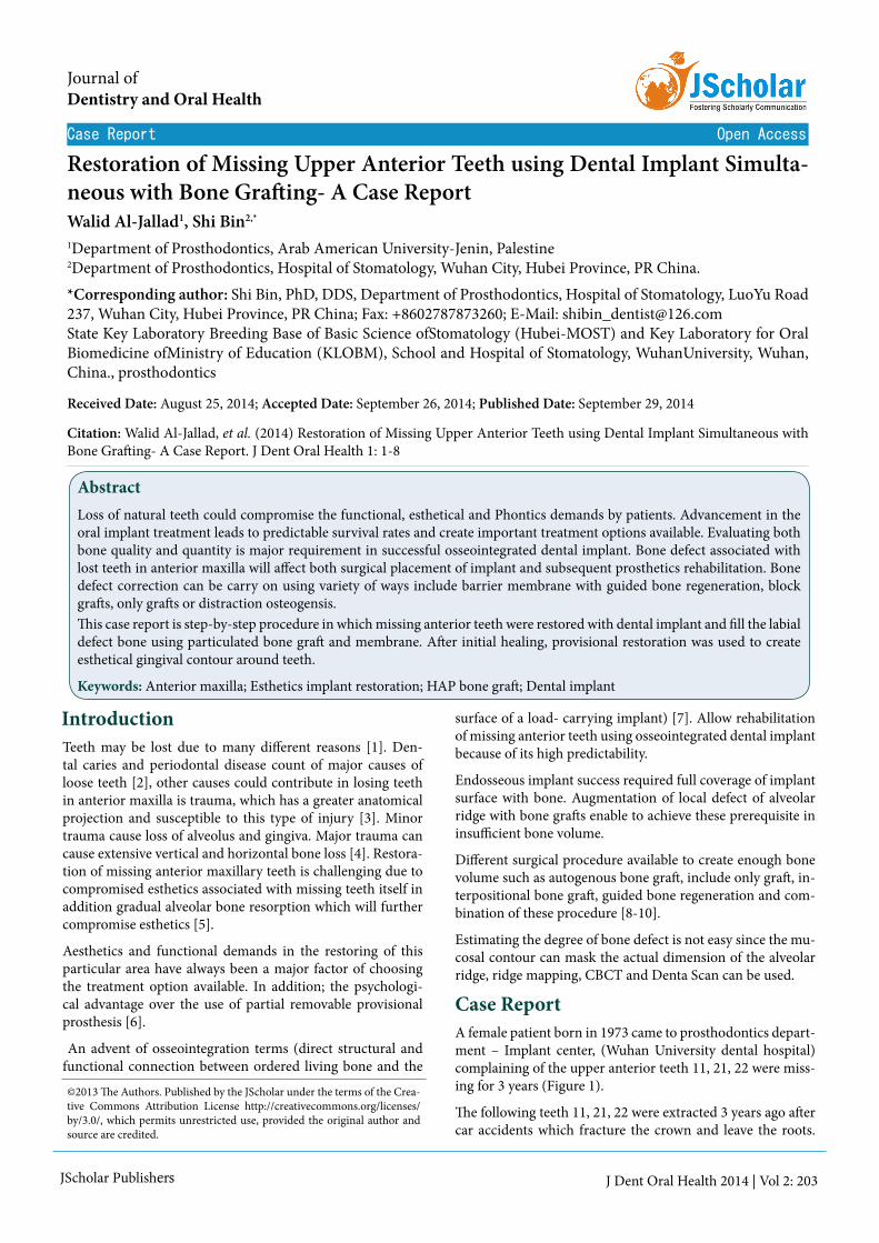
JScholar Publishers Restoration of Missing Upper Anterior Teeth using Dental Implant Simulta- neous with Bone Graſting- A Case Report Walid Al-Jallad 1 , Shi Bin 2,* 1 Department of Prosthodontics, Arab American University-Jenin, Palestine 2 Department of Prosthodontics, Hospital of Stomatology, Wuhan City, Hubei Province, PR China. Case Report Open Access *Corresponding author: Shi Bin, PhD, DDS, Department of Prosthodontics, Hospital of Stomatology, LuoYu Road 237, Wuhan City, Hubei Province, PR China; Fax: +8602787873260; E-Mail: [email protected] State Key Laboratory Breeding Base of Basic Science ofStomatology (Hubei-MOST) and Key Laboratory for Oral Biomedicine ofMinistry of Education (KLOBM), School and Hospital of Stomatology, WuhanUniversity, Wuhan, China., prosthodontics ©2013 e Authors. Published by the JScholar under the terms of the Crea- tive Commons Attribution License http://creativecommons.org/licenses/ by/3.0/, which permits unrestricted use, provided the original author and source are credited. J Dent Oral Health 2014 | Vol 2: 203 Journal of Dentistry and Oral Health Received Date: August 25, 2014; Accepted Date: September 26, 2014; Published Date: September 29, 2014 Citation: Walid Al-Jallad, et al. (2014) Restoration of Missing Upper Anterior Teeth using Dental Implant Simultaneous with Bone Graſting- A Case Report. J Dent Oral Health 1: 1-8 Abstract Loss of natural teeth could compromise the functional, esthetical and Phontics demands by patients. Advancement in the oral implant treatment leads to predictable survival rates and create important treatment options available. Evaluating both bone quality and quantity is major requirement in successful osseointegrated dental implant. Bone defect associated with lost teeth in anterior maxilla will affect both surgical placement of implant and subsequent prosthetics rehabilitation. Bone defect correction can be carry on using variety of ways include barrier membrane with guided bone regeneration, block graſts, only graſts or distraction osteogensis. is case report is step-by-step procedure in which missing anterior teeth were restored with dental implant and fill the labial defect bone using particulated bone graſt and membrane. Aſter initial healing, provisional restoration was used to create esthetical gingival contour around teeth. Keywords: Anterior maxilla; Esthetics implant restoration; HAP bone graſt; Dental implant Introduction Case Report Teeth may be lost due to many different reasons [1]. Den- tal caries and periodontal disease count of major causes of loose teeth [2], other causes could contribute in losing teeth in anterior maxilla is trauma, which has a greater anatomical projection and susceptible to this type of injury [3]. Minor trauma cause loss of alveolus and gingiva. Major trauma can cause extensive vertical and horizontal bone loss [4]. Restora- tion of missing anterior maxillary teeth is challenging due to compromised esthetics associated with missing teeth itself in addition gradual alveolar bone resorption which will further compromise esthetics [5]. Aesthetics and functional demands in the restoring of this particular area have always been a major factor of choosing the treatment option available. In addition; the psychologi- cal advantage over the use of partial removable provisional prosthesis [6]. An advent of osseointegration terms (direct structural and functional connection between ordered living bone and the surface of a load- carrying implant) [7]. Allow rehabilitation of missing anterior teeth using osseointegrated dental implant because of its high predictability. Endosseous implant success required full coverage of implant surface with bone. Augmentation of local defect of alveolar ridge with bone graſts enable to achieve these prerequisite in insufficient bone volume. Different surgical procedure available to create enough bone volume such as autogenous bone graſt, include only graſt, in- terpositional bone graſt, guided bone regeneration and com- bination of these procedure [8-10]. Estimating the degree of bone defect is not easy since the mu- cosal contour can mask the actual dimension of the alveolar ridge, ridge mapping, CBCT and Denta Scan can be used. A female patient born in 1973 came to prosthodontics depart- ment – Implant center, (Wuhan University dental hospital) complaining of the upper anterior teeth 11, 21, 22 were miss- ing for 3 years (Figure 1). e following teeth 11, 21, 22 were extracted 3 years ago aſter car accidents which fracture the crown and leave the roots.
Transcript of Restoration of Missing Upper Anterior Teeth using Dental ... · Restoration of Missing Upper...
JScholar Publishers
Restoration of Missing Upper Anterior Teeth using Dental Implant Simulta-neous with Bone Grafting- A Case ReportWalid Al-Jallad1, Shi Bin2,*
1Department of Prosthodontics, Arab American University-Jenin, Palestine2Department of Prosthodontics, Hospital of Stomatology, Wuhan City, Hubei Province, PR China.
Case Report Open Access
*Corresponding author: Shi Bin, PhD, DDS, Department of Prosthodontics, Hospital of Stomatology, LuoYu Road 237, Wuhan City, Hubei Province, PR China; Fax: +8602787873260; E-Mail: [email protected] State Key Laboratory Breeding Base of Basic Science ofStomatology (Hubei-MOST) and Key Laboratory for Oral Biomedicine ofMinistry of Education (KLOBM), School and Hospital of Stomatology, WuhanUniversity, Wuhan, China., prosthodontics
©2013 The Authors. Published by the JScholar under the terms of the Crea-tive Commons Attribution License http://creativecommons.org/licenses/by/3.0/, which permits unrestricted use, provided the original author and source are credited.
J Dent Oral Health 2014 | Vol 2: 203
Journal of Dentistry and Oral Health
Received Date: August 25, 2014; Accepted Date: September 26, 2014; Published Date: September 29, 2014
Citation: Walid Al-Jallad, et al. (2014) Restoration of Missing Upper Anterior Teeth using Dental Implant Simultaneous with Bone Grafting- A Case Report. J Dent Oral Health 1: 1-8
AbstractLoss of natural teeth could compromise the functional, esthetical and Phontics demands by patients. Advancement in the oral implant treatment leads to predictable survival rates and create important treatment options available. Evaluating both bone quality and quantity is major requirement in successful osseointegrated dental implant. Bone defect associated with lost teeth in anterior maxilla will affect both surgical placement of implant and subsequent prosthetics rehabilitation. Bone defect correction can be carry on using variety of ways include barrier membrane with guided bone regeneration, block grafts, only grafts or distraction osteogensis.This case report is step-by-step procedure in which missing anterior teeth were restored with dental implant and fill the labial defect bone using particulated bone graft and membrane. After initial healing, provisional restoration was used to create esthetical gingival contour around teeth.
Keywords: Anterior maxilla; Esthetics implant restoration; HAP bone graft; Dental implant
Introduction
Case Report
Teeth may be lost due to many different reasons [1]. Den-tal caries and periodontal disease count of major causes of loose teeth [2], other causes could contribute in losing teeth in anterior maxilla is trauma, which has a greater anatomical projection and susceptible to this type of injury [3]. Minor trauma cause loss of alveolus and gingiva. Major trauma can cause extensive vertical and horizontal bone loss [4]. Restora-tion of missing anterior maxillary teeth is challenging due to compromised esthetics associated with missing teeth itself in addition gradual alveolar bone resorption which will further compromise esthetics [5].
Aesthetics and functional demands in the restoring of this particular area have always been a major factor of choosing the treatment option available. In addition; the psychologi-cal advantage over the use of partial removable provisional prosthesis [6].
An advent of osseointegration terms (direct structural and functional connection between ordered living bone and the
surface of a load- carrying implant) [7]. Allow rehabilitation of missing anterior teeth using osseointegrated dental implant because of its high predictability.
Endosseous implant success required full coverage of implant surface with bone. Augmentation of local defect of alveolar ridge with bone grafts enable to achieve these prerequisite in insufficient bone volume.
Different surgical procedure available to create enough bone volume such as autogenous bone graft, include only graft, in-terpositional bone graft, guided bone regeneration and com-bination of these procedure [8-10].
Estimating the degree of bone defect is not easy since the mu-cosal contour can mask the actual dimension of the alveolar ridge, ridge mapping, CBCT and Denta Scan can be used.
A female patient born in 1973 came to prosthodontics depart-ment – Implant center, (Wuhan University dental hospital) complaining of the upper anterior teeth 11, 21, 22 were miss-ing for 3 years (Figure 1).
The following teeth 11, 21, 22 were extracted 3 years ago after car accidents which fracture the crown and leave the roots.

2
JScholar Publishers J Dent Oral Health 2014 | Vol 2: 203
Now she asked for the urgent restoration to improve the func-tion and esthetics.
Conducting a thorough medical history, we found the patient had a good physical condition and denied any systematic dis-eases or any allergic diseases that were relevant to dental treat-ment. And she had no mucosal lesions, no bleeding disorder, no tobacco smoking, and alcohol and drug abuse.
Dental historyQuestioning the patients reported that teeth 11, 21, and 22 were extracted in another hospital 3 years ago. And since then no any further treatment try to solve the problem due to eco-nomical issues.
Clinical ExaminationClinical examination was conducted, and through deep exam-ination of both extra and intra oral examination of reported the following:
Extra-oral: No gross facial asymmetry or swelling was detect-ed. No clicking of TMJ or limitation and deviation of opening.
Intra-oral: Hard tissue examination revels that Teeth 11, 21, 22 were missing. Evaluation of edentulous space shows that the quantity and quality of edentulous area (alveolar bone) was not good. There were obvious bone defect and labial ridge in form of concavity. (Labial dehiscence) The inter-alveolar spac-es were enough for restoration.
Soft tissue examinations show. The thickness of the soft tissue of the edentulous ridge was assessed at different point of the edentulous space and it’s about 2 mm measured by a gradu-ated periodontal probe. The color and texture of mucosa was normal.
No obvious mucosal lesions, fibrous connective tissue dis-placement and other abnormities were detected.
Periodontal status: Oral hygiene was good. No marginal gin-gival inflammation was detected. No periodontal pockets were detected. No other tooth mobility was found.
Figure 1: Preoperative labial viewOcclusion: The overbite and over-jet were normal. The occlu-sion was stable.
Radiographic evaluation showed an excellent condition of the remaining teeth, and there was adequate bone height of
15 mm. the remaining bone was of moderate quality. Alveolar bone had bone defect in missing teeth number 22 (Figure 2). Labial bone dehiscence required bone augmentation in order to achieve long term success [11].
Figure 2: Preoperative peri-apical radiograph
Treatment Option The treatment options were discuss with patients and where summarized as following , implant supported crown of each missing tooth , or two implant supported bridge, FPD and RPD were presented to the patient, and the time, expenditure, advantages, and disadvantages of each plan were introduced. The patient wanted the best way for restoration, and she didn’t care about the time consume. So she chose the implant denture finally.
Details of TreatmentStep 1. Preoperative radiograph examination
Before start implant surgery and as a part of treatment plan process, the patient must have initial radiographic screening. Which is in this case was peri-apical X-ray accomplished by intraoral long cone paralleling technique.
Step 2. Oral hygiene instruction and scaling
The patient understanding of the financial, time, and mainte-nance requirements was crucial, and these obligations must be made clear to the patient initially and during subsequent ap-pointments. Before the surgery, all the supragingival and sub-gingival calculus must be removed and blood screening test were performed.
Step 3. Onlay guided bone regeneration
The patient was informed about the common complications of the implant surgery that could happen, and then he signed a consent form. Because of the lack of enough bone, Onlay guided bone regeneration technique was used after implant

3
JScholar Publishers J Dent Oral Health 2014 | Vol 2: 203
placement. Particulated bone and bone graft from the Bone-TrapTM collected during preparation of the implant site along with Bio-Oss granules for covering the implant [9].
Step 4. Sugical procedure
After local anesthesia injection, surgical stent was positioned and supported by adjacent teeth (Figure 3).A small, sharp-tipped guiding drill was used to create a pre-cise, minimally invasive initial penetration through the muco-sa and into bone to locate the exact position of planed implant (Figure 4).
Figure 3: Surgical stent in position
Figure 4: Small, initial penetration through the mucosa and into bone.
Figure 5: Reflection of full muco-periostal flap
Then full muco-periostal flap were reflected exposing the un-derplaying bone (Figure 5). Position the surgical stent and prepare the implant site with correct angulations and depth for implant fixture (Figure 6). A 2mm diameter twist drill was used to drill to the desired depth. The site was 13mm in length. The direction indicator was used to check orientation.
The drill of 3.5mm in diameter was used to open out the corti-cal bone to the ¢3.5mm.Then the site was enlarged with the ¢4.3mm twist drill. Parallel pin was check in prepared site to evaluate parallelism of prepared implant site for optimal result (Figure 7).
Then screw the site. The Nobel® Replace Select tapered im-plant(¢4.3X13mm) and tapered implant(¢4.3X13mm) were inserted in the site of 11 and 22 respectively initially with an adapter attached to a special contra-angle at slow revolutions, and then by hands using tap wrench and ratchet wrench. The implant was placed with its head just below the crest of the bone, the implant internal triangle tip direct to labial. The in-sert torque was 45 Ncm (Figures 8&9).
The implant was placed with its head at crest bone level, and then put the healing screw on the abutment connection area. Bio-Oss and collagen membrane filled in spacing. The wound was sutured (Figures 10,11&12). The patient was informed to maintain the oral hygiene. 7-10 days later, she must come back
Figure 6: Surgical stent in position after flap reflection (supported by adjacent teeth).
Figure 7: Guide Pin in osteomy prepared site (check for parallelism and angulations)

4
JScholar Publishers J Dent Oral Health 2014 | Vol 2: 203
Figure 8: Placement of implant fixture
Figure 9: Both Implant fixtures in position
Figure 10: Inlay guided bone regeneration
Figure 11: GBR membrane in position
Figure 12: Flap Closer (interrupted suture)
Figure 13: Peri-apical X-ray after first surgery
Figure14: Healing abutment after second surgery
for the removal of the sutures and took a new radiographic for history information (Figure 13).
Step 5. Stage II surgery procedure After 6 months later, stage II surgery was taken. Providing transmucosal healing abutment for two weeks. The fabrication
5
JScholar Publishers J Dent Oral Health 2014 | Vol 2: 203
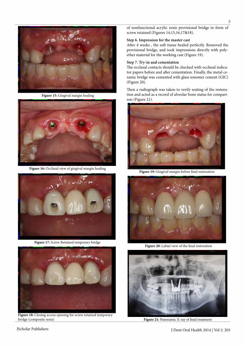
Figure 15: Gingival margin healing
Figure 16: Occlusal view of gingival margin healing
Figure 17: Screw Retained temporary bridge
Figure 18: Closing access opening for screw retained temporary bridge (composite resin)
Figure 19: Gingival margin before final restoration
Figure 20: Labial view of the final restoration
Figure 21: Panoramic X-ray of final treatment
of nonfunctional acrylic resin provisional bridge in form of screw retained (Figures 14,15,16,17&18).
Step 6. Impression for the master castAfter 4 weeks , the soft tissue healed perfectly. Removed the provisional bridge, and took impressions directly with poly-ether material for the working cast (Figure 19).
Step 7. Try-in and cementationThe occlusal contacts should be checked with occlusal indica-tor papers before and after cementation. Finally, the metal ce-ramic bridge was cemented with glass ionomer cement (GIC) (Figure 20).
Then a radiograph was taken to verify seating of the restora-tion and acted as a record of alveolar bone status for compari-son (Figure 21).

6
JScholar Publishers J Dent Oral Health 2014 | Vol 2: 203
Step 8. Periodic recall
The patient was informed some knowledge about maintain the oral hygiene and using of the implant restoration. Recall the patient periodically. The patient was evaluated clinically and radiographically at 1 week, 1 months, 3 months, 6 months, and one year. The examinations should include mobility of implant, soundness and integrity of provisional crowns, peri-odontal status, and marginal bone resorption and so on.
Discussion and conclusionEndosseous dental implants are a predictable modality of tooth replacement that can improve the dental health and quality of life for many people.
Different study conducted by many researches has reported that, the success and survival rate of dental implant placed in anterior maxilla are almost same to other segment of jaw [12]. However, there is often inadequate bone to receive and sup-port implants. This can be the result of trauma, periodontal disease, endodontic infection, post-extraction ridge defects, disuse atrophy, etc. [13].
Successful implant placement in planed site required enough bone volume of sufficient density to enable an implant of the appropriate size to be placed in a desirable position and ori-entation.
Placement of bone grafts in conjunction with endosseous den-tal implant shortens the treatment time without influencing the success rate or increase the complication [14].
The interaction between the graft and the surrounding host bone is very important and is the subject of much research [15].
The degree of bone grafting required for implant placement varies from localized deficiencies to cases where there is a need to change the entire arch form and/or jaw relationship.
Augmentation to create enough bone volume is done using either (GBR) Guided bone regeneration [15,16], autogenous bone grafts, including onlay grafts [17,18] and interposi-tional bone grafts [19], and combinations of these proce-dures. These grafting materials can be used in different clinical situation which can be summarized and following [20-23]:
• Regeneration of periodontal bone and furcation defects.• Osseous defect regeneration.• Regeneration of extraction sockets.• Regeneration of gaps around block grafts.• Horizontal alveolar crest augmentation.• Sinus augmentation.
Using guide bone regeneration in construction of bone defects in predictable methods for regeneration of adequate bone vol-ume for proper placement of dental implant. These can be si-multaneously with implant placemat or staged approach [24].Using of particular dental implant of specific body design and surface characteristic will influence the treatment success [25]. Tapered dental implant which mimicking the shape of natural root will have high initial stability [26]. The surface of dental implant using here is TiUnite surface, which enhanced osse-
ointegration and anchorage in surrounding bone. In the long term, it maintains marginal bone and soft tissue levels, maxi-mizing functional endurance and esthetics [27-29].
Here a grafting with particulated hydroxyapatite bone graft to cover implant threaded exposed and to establish good labial bone contour to improve esthetic.
Another pre-requesting of this case to achieve optimal result is soft tissue management. Successful dental implant restoration in issue framing esthetic zone required a healthy and correctly contoured soft tissue framing, which is defined as the gingival contour that surrounds the prosthesis [30,31]. Preservation of interdental papilla and gingival margin which is symmetrical with gingival architecture of adjacent gingival [32,33]. Achiev-ing aesthetics interdental papilla which is completely fill the space between teeth or implants required interproximal bone crest of 5 mm. of estimated contact point in planned restora-tion [34,35].
Three dimensional position of implant required to achieve optimal emergence profile [36]. Mesiodistally positioning of implant required 1.5 mm space between implant and adjacent teeth or between implant / implant [32,36].
Labio-Palatel positioning is also important, too far labillay re-sult in overcontouring of the crown and can cause recession due to decrease thickness of buccal bone [35]. While palatal positiong produce ridge lap which limiting the type of design and increasing the difficulty for maintenance [37-39]. The third position is apicoronal positiong , which if the implant is too far apically positiong they will be bone resorption and gingival recision. On other hand if there is coronal placement the esthetics may be comprmise due to visibility of implant shoulder [40]. The implant should be placed 1.5 mm to 3.0 mm below the CEJ for optimal implant esthetics [41].
Tissue training help to develop a proper emergence profile and natural tooth appearance, help in re-establish normal gingival tissue contours and interdental papillae [24]. Fabricating pro-visional restoration before inserting the final prosthesis which will improve esthetics.
References1) Zarb GA, Bolender CL, Eckert ST, Jacob RF, Fenton AH, et al. (2004) Prosthodontic Treatment for Edentulous Patients. Charles L Bolender (eds) The edentulous predicament (12th edn), CV Mosby & CO, St Louis.
2) World health organization (2012) Oral health. fact sheet No 318.
3) Cantore S, Ballini A, Crincoli V, Grassi FR (2009) Treatment of horizontal root fracture: a case report. Cases J 2: 8101.
4) Palmer RM, Palmer PJ, Newton JT (2003) Dealing with esthetic demands in the anterior maxilla. Periodontol 33:105-118.
5) Cawood JI, Howell RA (1991) Reconstructive prosthetics surgery: I. Anatomical considerations. Int J Oral Maxillofacial Surg 20:75-82.
6) Podovan LEM, Sartori IAM, Thome G, Melo ACM (2008) Imme-diate load implants and osteointerados: SaoPaulo, Santos (eds) Pos-sibilidades and techics (1stedn).
7) Listgarten MA, Lang NP, Schroeder HE, Schroeder A (1991) Peri-odontal tissues and their counterparts around endosseous implants. Clin Oral Implants Res 2: 1-19.

7
JScholar Publishers J Dent Oral Health 2014 | Vol 2: 203
8) Khoury F (1987) The modified alveolar Extensions plastic with Zahnlrzl Implant.
25) Leonhardt A, Gröndahl K, Bergström C, Lekholm U (2002) Long-term follow-up of osseointegrated titanium implants using clinical, radiographic and microbiological parameters. Clin Oral Implants Res 13: 127-132.
13) Kahnberg K-E (2005) Bone Grafting Techniques for Maxillary Implants. Kahnberg K-E, Rasmusson L, Zellin G (eds) Bone Graft-ing Techniques for Maxillary Implants (1st edn) Blackwell Publishing Co., Munksgaard.
30) Davidoff SR (1996) Developing soft tissue contours for implant-supported restorations: a simplified method for enhanced aesthetics. Pract Periodontics Aesthet Dent 8: 507-513.
9) Lustmann J, Lewinstein I (1995) Interpositional bone grafting technique to widen narrow maxillary ridge. Int J Oral Maxillofac Im-plants 10: 568-577. 26) Buser D, Martin W, Belser UC (2004) Optimizing esthetics for
implant restorations in the anterior maxilla: anatomic and surgical considerations. Int J Oral Maxillofac Implants 19: 43-61.
14) Boronat A, Carrillo C, Penarrocha M, Pennarocha M (2010) Den-tal implants placed simultaneously with bone grafts in horizontal de-fects: a clinical retrospective study with 37 patients. Int J Oral Maxil-lofac Implants 25: 189-196.
31) Kan JY, Morimoto T, Rungcharassaeng K, Roe P, Smith DH (2010) Gingival biotype assessment in the esthetic zone: visual versus direct measurement. Int J Periodontics Restorative Dent 30: 237-243.
18) Ten Bruggenkate CM, Kraaijenhagen HA, van der Kwast WA, Krekeler G, Oosterbeek HS (1992) Autogenous maxillary bone grafts in conjunction with placement of I.T.I. endosseous implants. A pre-liminary report. Int J Oral Maxillofac Surg 21: 81-84.
35) Elian N, Bloom M, Dard M, Cho SC, Trushkowsky RD, et al. (2011) Effect of interimplant distance (2 and 3 mm) on the height of interimplant bone crest: a histomorphometric evaluation. J Peri-odontol 82: 1749-1756.
10) Misch CM, Misch CE, Resnik RR, Ismail YH (1992) Reconstruc-tion of maxillary alveolar defects with mandibular symphysis grafts for dental implants: a preliminary procedural report. Int J Oral Maxil-lofac Implants 7: 360-366.
27) Zechner W, Tangl S, Fürst G, Tepper G, Thams U (2003) Osse-ous healing characteristics of three different implant types. Clin Oral Implants Res 14: 150-157.
15) Buser D, Dula K, Belser UC, Hirt HP, Berthold H (1995) Local-ized ridge augmentation using guided bone regeneration. II. Surgical procedure in the mandible. Int J Periodontics Restorative Dent 15: 10-29.
32) Garber DA, Salama MA, Salama H (2001) Immediate total tooth replacement. Compend Contin Educ Dent 22: 210-218.
19) Esposito M, Grusovin MG, Kwan S, Worthington HV, Coulthard P (2008) Interventions for replacing missing teeth: bone augmenta-tion techniques for dental implant treatment. Cochrane Database Syst Rev : CD003607.
36) Buser D, Martin W, Belser UC (2004) Optimizing esthetics for implant restorations in the anterior maxilla: anatomic and surgical considerations. Int J Oral Maxillofac Implants 19: 43-61.
11) Hammerle CH, Jung RE (2008) Clinical Periodontology and Im-plant Dentistry (Vol.2). J Lindhe, NP Lang, T Karring (eds) Ridge augmentation procedures (5th edn) Blackwell Publication, Munks-gaard.
28) Östman PO, Hellman M, Sennerby L (2012) Ten years later. Re-sults from a prospective single-centre clinical study on 121 oxidized (TiUnite™) Brånemark implants in 46 patients. Clin Implant Dent Relat Res 14: 852-860.
16) Schwarz F, Sahm N, Becker J (2012) Impact of the outcome of guided bone regeneration in dehiscence-type defects on the long-term stability of peri-implant health: clinical observations at 4 years. Clin Oral Implants Res 23: 191-196.
33) Paolantoni G, Marenzi G, Fusco A, Sammartino G (2007) Implant rehabilitation of central incisor: a staged approach. Implant Dent 16: 349-355.
20) Dayi E, Aslan M, Simşek G, Yilmaz AB (2002) The effects of bone chips dehydrated with solvent on healing bone defects. J Int Med Res 30: 168-173.
22) Block MS, Degen M (2004) Horizontal ridge augmentation us-ing human mineralized particulate bone: preliminary results. J Oral Maxillofac Surg 62: 67-72.
23) Le B, Burstein J, Sedghizadeh PP (2008) Cortical tenting graft-ing technique in the severely atrophic alveolar ridge for implant site preparation. Implant Dent 17: 40-50.
24) Morley S. Rubinoff, DDS, Cert Prosth (2003) Single-Tooth Im-plant Reconstruction in the Anterior Maxilla. Journal of the Cana-dian Dental Association 69: 10-24.
37) Belser UC, Bernard JP, Buser D (1996) Implant-supported resto-rations in the anterior region: prosthetic considerations. Pract Peri-odontics Aesthet Dent 8: 875-883.
12) Belser UC, Schmid B, Higginbottom F, Buser D (2004) Outcome analysis of implant restorations located in the anterior maxilla: a re-view of the recent literature. Int J Oral Maxillofac Implants 19: 30-42.
29) Glauser R (2012) Eleven-year results of implants with an oxidized surface placed predominantly in soft bone and subjected to immedi-ate occlusal loading. Clin Oral Impl Res 23: 140-141.
17) Misch CM, Misch CE (1995) The repair of localized severe ridge defects for implant placement using mandibular bone grafts. Implant Dent 4: 261-267.
34) Tarnow D, Elian N, Fletcher P, Froum S, Magner A, et al. (2003) Vertical distance from the crest of bone to the height of the interprox-imal papilla between adjacent implants. J Periodontol 74: 1785-1788.
21) Noumbissi SS, Lozada JL, Boyne PJ, Rohrer MD, Clem D, et al. (2005) Clinical, histologic, and histomorphometric evaluation of mineralized solvent-dehydrated bone allograf (Puros) in human maxillary sinus grafts. J Oral Implantol 31: 171-179.
38) Belser UC, Buser D, Hess D, Schmid B, Bernard JP, et al. (2000) Aesthetic implant restorations in partially edentulous patients--a crit-ical appraisal. Periodontol 2000 : 132-150.
39) Tarnow DP, Eskow RN (1995) Considerations for single-unit esthetic implant restorations. Compend Contin Educ Dent 16: 778,780,782-784.
40) Bashutski JD, Wang HL (2007) Common implant esthetic com-plications. Implant Dent 16: 340-348.
41) Zetu L, Wang HL (2005) Management of inter-dental/inter-im-plant papilla. J Clin Periodontol 32: 831-839.

8
JScholar Publishers J Dent Oral Health 2014 | Vol 2: 203
Submit your manuscript at http://www.jscholaronline.org/submit-manuscript.php
Submit your manuscript to a JScholar journal and benefit from:
¶ Convenient online submission ¶ Rigorous peer review ¶ Immediate publication on acceptance ¶ Open access: articles freely available online ¶ High visibility within the field ¶ Better discount for your subsequent articles